Embed Size (px)
Citation preview

Prescribing patterns in adult patients with meningitis in internal medicine
wards, Dr George Mukhari Hospital
A dissertation submitted by
Wandisile M. Grootboom
in partial fulfilment of the requirements
for the degree of
Master of Science in Medicine - Pharmacy
of the
University of Limpopo (Medunsa Campus)
DEPARTMENT OF PHARMACY
School of Health Care Sciences
Faculty of Health Sciences
University of Limpopo
(Medunsa Campus)
2010

i
DECLARATION
I, Wandisile Grootboom, hereby declare that the work on which this study is based is original, except
where acknowledgements indicate otherwise.
This dissertation is submitted for the degree Master of Science (Medical) in Pharmacy at the
University of Limpopo. Neither the whole work nor any part of it has been before any degree or
examination at this or any other university.
Signed ……………………
………………on the………………………………day of ……………………..
ii
ACKNOWLEDGEMENTS
Even youths grow tired and weary, and young men stumble and fall; but those who hope in the LORD
will renew their strength. They will soar on wings like eagles; they will run and not grow weary, they
will walk and not faint (Isaiah 40:30:31).
• My profound gratitude goes to the Almighty who has sustained, gave wisdom, strength and
guidance throughout this work.
• Prof AGS Gous, my chief supervisor- for his patience, immense knowledge and fatherly advice
in making sure that this project comes to fruition.
• Christel Hanson, co-supervisor- for all those advices that gave shape and structure to the
manuscript.
• Monika Zweygarth for her expert knowledge in analysing data.
• Lindiwe Molahli, 3rd year pharmacy student- for all the hard work she has put into the project;
collecting data and getting the relevant resources from the library.
• Moliehi Mohlala, my lecturer- for her availability and willingness to help make sure that this
project succeeds.
• Nikki Williamson- for facilitating yearly registration and all her words of encouragement.
• Sister Letswalo, together with the rest of the nursing staff in internal medicine wards for all the
support they gave towards the project.
• All the clerks from the internal medicine wards who were involved in the project.
• Amos, from filing room- thanks for making sure we wrap up data collection.
• My family, friends and colleagues- for their undying support; you are highly appreciated.

iii
SUMMARY
Background
Information regarding disease epidemiology, treatment options and emerging infections and
resistance constantly challenge the knowledge of the health care practitioner. Antibiotic prescribing
patterns was identified by the Dr George Mukhari hospital antibiotics committee as an area of concern.
Due to this concern it was decided to investigate the prescribing patterns in adult patients with
meningitis admitted to the internal medicine wards at Dr George Mukhari hospital.
Objectives
To determine the current antimicrobial prescribing patterns in adult patients diagnosed with meningitis,
to record the causative organisms and sensitivity patterns, to record the outcome, cost and length of
treatment.
Method
Patient and prescriptions data were recorded prospectively on specially designed data sheets from
five internal medicine wards for four months (May to August 2008). Patients were followed until
discharged.
Results
Sixty-six patients were enrolled; 41 recovered, 22 died, 2 refused treatment and 1 absconded.
Ceftriaxone was prescribed the most frequently and was administered to 58 patients; four patients with
confirmed cryptococcal meningitis received amphotericin B IVI, three patients were started on

iv
Rifafour® for suspected tuberculosis meningitis and one was started on cefuroxime. Specimens from
only 22 patients were sent for culture and sensitivity tests; ten were positive for yeast-like organisms,
three for S pneumoniae and one for N meningitides and tuberculosis respectively.
The average duration of treatment of patients with meningitis was 9.2days. The total cost of anti-
infectives used for treatment of meningitis amounted to R111, 292.53 and the average cost per patient
was R1 686.25. The cost of all medicines prescribed for the 66 patients amounted to R116, 490.43.
Conclusion
Ceftriaxone was used frequently as empiric therapy. Specimens for culture and sensitivity were not
sent routinely. Therefore it was difficult to monitor and observe any resistance patterns and to contain
cost of treatment.
v
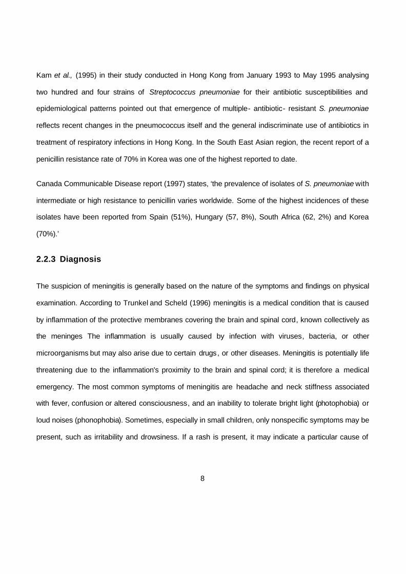
TABLE OF CONTENTS
ACKNOWLEDGEMENTS ..................................................................................................................ii
SUMMARY ....................................................................................................................................iii
TABLE OF CONTENTS..................................................................................................................... v
LIST OF TABLES ..............................................................................................................................viii
LIST OF FIGURES ............................................................................................................................. ix
CHAPTER 1: INTRODUCTION................................................................................................... 1
CHAPTER 2: LITERATURE REVIEW....................................................................................... 5
2.1 MENINGITIS ....................................................................................................................... 5
2.1.1 EPIDEMIOLOGY ............................................................................................................ 5
2.1.2 PATHOGENESIS AND PATHOPHYSIOLOGY ........................................................ 6
2.2 BACTERIAL MENINGITIS ................................................................................................ 6
2.2.1 CLINICAL PRESENTATION ........................................................................................ 6
2.2.2 AETIOLOGY ................................................................................................................... 7
2.2.3 DIAGNOSIS .................................................................................................................... 8
2.2.4 TREATMENT ................................................................................................................ 10
2.3 VIRAL MENINGITIS......................................................................................................... 14
2.3.1 DEFINITION .................................................................................................................. 14
2.3.2 PATHOGENESIS AND PATHOPHYSIOLOGY ...................................................... 15
2.3.3 ANTIVIRAL THERAPY ................................................................................................ 16
2.4 CRYPTOCOCCAL MENINGITIS (FUNGAL) ............................................................... 17
2.4.1 BACKGROUND ............................................................................................................ 17
2.4.2 PATHOPHYSIOLOGY AND PATHOGENESIS ...................................................... 17

vi
2.4.3 ANTIFUNGAL THERAPY ........................................................................................... 18
2.5 TUBERCULOUS MENINGITIS ...................................................................................... 18
2.5.1 ANTI-TB THERAPY ..................................................................................................... 19
2.6 DRUG-INDUCED ASEPTIC MENINGITIS ................................................................... 20
2.6.1 PATHOPHYSIOLOGY................................................................................................. 21
2.7 PRESCRIBING PATTERNS........................................................................................... 21
2.7.1 RECOMMENDED MENINGITIS PRESCRIBING PATTERNS ............................. 21
2.8 CURRENT RECOMMENDED PRESCRIBING FOR MENINGITIS IN DR
GEORGE MUKHARI HOSPITAL................................................................................... 23
CHAPTER 3: METHODOLOGY................................................................................................ 26
3.1 AIM...................................................................................................................................... 26
3.2 STUDY OBJECTIVES ..................................................................................................... 26
3.3 METHOD AND MATERIALS .......................................................................................... 26
3.4 STUDY SITE ..................................................................................................................... 27
3.5 THE STUDY DESIGN ..................................................................................................... 27
3.6 DATA COLLECTION ....................................................................................................... 27
CHAPTER 4: RESULTS .............................................................................................................. 29
4.1 INTRODUCTION .............................................................................................................. 29
4.2 PATIENT DEMOGRAPHICS.......................................................................................... 29
4.2.1 AGE AND GENDER .................................................................................................... 29
4.3 DURATION OF STAY IN HOSPITAL ........................................................................... 30
4.4 OUTCOMES...................................................................................................................... 31

vii
4.5 TYPES OF MENINGITIS ................................................................................................ 33
4.6 DURATION OF TREATMENT ....................................................................................... 35
4.7 COST OF TREATMENT ................................................................................................. 37
CHAPTER 5: DISCUSSION ....................................................................................................... 41
5.1 AGE AND GENDER ........................................................................................................ 41
5.2 DURATION OF STAY IN HOSPITAL ........................................................................... 41
5.3 TREATMENT COST ........................................................................................................ 44
5.4 OUTCOMES...................................................................................................................... 45
5.5 LIMITATIONS ................................................................................................................... 47
CHAPTER 6: CONCLUSION AND RECOMMENDATIONS ............................................ 48
REFERENCES .................................................................................................................................. 50
APPENDICES .................................................................................................................................. 56
APPENDIX 1: ........................................................................................................................................ 56

viii
LIST OF TABLES
TABLE 4.1: DURATION OF STAY IN HOSPITAL ........................................................... 30
TABLE 4.2: OUTCOMES ......................................................................................................... 31
TABLE 4.3: DIAGNOSES ........................................................................................................ 32
TABLE 4.4: TREATMENT OUTCOMES ............................................................................. 33
TABLE 4.5: TYPES OF MENINGITIS .................................................................................. 34
TABLE 4.6: ALL ANTI-INFECTIVES GIVEN TO PATIENTS DURING
HOSPITALISATION ........................................................................................... 35
TABLE 4.7: DURATION OF TREATMENT ....................................................................... 36
TABLE 4.8: DURATION OF TREATMENT WITH ANTI-INFECTIVES FOR
MENINGITIS......................................................................................................... 37
TABLE 4.9: COST OF TREATMENT ................................................................................... 37
TABLE 4.10: COST OF ANTI-INFECTIVES......................................................................... 38
TABLE 4.11: EIGHT MOST COSTLY MEDICINES FOR MENINGIT IS TREATMENT
.................................................................................................................................. 39
TABLE 4.12: DURATION OF TREATMENT WITH ANTI-INFECTIVES FOR
MENINGITIS......................................................................................................... 40

ix
LIST OF FIGURES
FIGURE 4.1: PATIENT DEMOGRAPHICS........................................................................... 30
FIGURE 4.2: DISTRIBUTION OF THE DURATION OF HOSPITAL STAY BY
STUDY PATIENTS (N=66) .............................................................................. 31
FIGURE 4.3: PERCENTAGE OF TOTAL COST OF MENINGITIS MEDICINES ..... 40

1
CHAPTER 1: INTRODUCTION
Meningitis is one of the major causes of morbidity and mortality around the world. Hearing impairment,
obstructive hydrocephalus and damage to the brain parenchyma are common neurological sequelae
from meningitis (Leib and Täuber, 1999).
According to DiP iro et al., (1997) Different types of meningitis have been identified; “purulent” (bacterial
aetiology) and “aseptic” (acute meningeal irritation, usually benign and self-limiting, with complete
recovery and sterile pleocytic CSF and at least 70% are caused by viruses). Most cases of meningitis
are due to infection with viruses, followed by bacteria, fungi, and parasites (Helbok et al., 2009).
Viruses that can cause meningitis include various enterovirus subtypes, herpes simplex virus 2 (and
less commonly HSV-1), Varicella zoster virus (known for causing chickenpox and shingles), mumps
virus and HIV. The types of bacteria that cause bacterial meningitis vary by age group. In premature
and newborn babies up to three months, common bacteria are group B streptococcus (subtype III)–
especially in the first week of life–and bacteria that normally inhabit the digestive tract such as
Escherichia coli (carrying K1 antigen). Listeria monocytogenes (serotype IVb) may affect the newborn
and occurs in epidemics. Older children are more commonly affected by Neisseria meningitidis
(meningococcus), Streptococcus pneumoniae (serotypes 6, 9, 14, 18 and 23) and those under five by
Haemophilus influenzae type B. In adults, N. meningitidis and S. pneumoniae together cause 80% of all
cases of meningitis, with increased risk of L. monocytogenes in those over 50. In trauma, neurosurgery,
or other contact between the skin and the meninges, staphylococci are more likely, as well as infections
with pseudomonas and related Gram-negative bacilli. The same pathogens are also more common in
those with an impaired immune system. Tuberculous meningitis due to infection with Mycobacterium
tuberculosis, is more common in those from countries where tuberculosis is common, but is also
encountered in those with immune deficiencies , such as AIDS (Katti, 2004).

2
According to (Steele, 2002) Streptococcus pneumoniae is the most frequent cause of meningitis in the
United States, accounting for 47% of all cases, followed by Neisseria meningitides, the meningococcus
(25%), group B streptococcus (12%), Listeria monocytogenes (8%), and Haemophilus influenzae (7%).
The pneumococcus continues to exhibit the highest mortality and morbidity rates among these more
common bacterial causes of meningitis, with a 4% to 16% mortality rate in children and a substantially
higher rate in adults. Death is the result in more than half of the elderly adults (older than 70 years of
age) infected with pneumococcal meningitis. S pneumoniae strains that are non-susceptible to penicillin
and third-generation cephalosporins (i.e., ceftriaxone and cefotaxime) have been identified worldwide,
with as many as 8% of pneumococci demonstrating this multiresistant pattern in some regions
Meningitis and intracranial tuberculoma are types of tuberculosis (TB) infection of the central nervous
system (CNS). These types of infections are seen in regions where the incidence of tuberculosis is high
and there is a prevalence of dissemination after primary TB infection. Treatment of CNS tuberculosis
should begin as soon as possible when clinically suspected because patient outcome is improved when
therapy is begun in the early stage of disease. For all types of CNS tuberculosis, treatment should
begin with a combination of bactericidal drugs that penetrate the CSF (Katti, 2004; Be et al ., 2009).
The first line of therapy should include isoniazid (INH), rifampicin, and pyrazinamide, all of which are
bactericidal and achieve effective levels in the CSF. Pyrazinamide is given with INH and rifampicin
during the first 2 months of therapy, and then is stopped. A fourth drug should be used during the first 2
months of therapy when INH drug resistance is a concern. Drug resistance is a concern if the region
has more than a 4% INH resistance rate, if the patient has received prior antituberculosis therapy, or if
the patient has been exposed to a resistant case. Ethambutol or streptomycin are the medications
recommended as the fourth agent. Ethambutol has moderate CSF penetration and streptomycin has

3
poor penetration, so it has been given as a combination of intramuscular and intrathecal delivery
(Steele, 2002).
Pharmacotherapy in infectious diseases is constantly changing. The change is affected by the constant
change in pathogen resistance to therapy and emerging infections (Drew et al., 2004). The principles of
antimicrobial therapy for acute meningitis include use of agents that penetrate well into cerebrospinal
fluid and attain appropriate cerebrospinal fluid concentrations, are active in purulent cerebrospinal fluid,
and are susceptible against the infecting pathogen.
In the hospital where the study was conducted certain antimicrobials were used for the treatment of
various causes of meningitis. The guidelines of neurology department (2004) state that acute bacterial
meningitis be treated with cefotaxime 3g 6hourly IV or ceftriaxone 2g 12 hourly IV. Meningococcal
meningitis is treated with benzyl penicillin 6 million units 6 hourly IV for 10 days. Cefotaxime 3g 6 hourly
IV or cefriaxone 2g 12 hourly IV would also be effective. Pneumococcal meningitis, if it is penicillin
sensitive is treated with benzyl penicillin 6 million units 6 hourly IV for 14 days or ceftriaxone 2g 12
hourly IV. If penicillin resistant, it is treated with cefotaxime 3g 6 hourly IV for 14 days or ceftriaxone 2g
12 hourly IV for 14 days. In practice meningococcal meningitis was treated with ceftriaxone 2g 12
hourly IV for 13 days in one case while pneumococcal meningitis with culture resistant to penicillin was
treated with ceftriaxone 2g 12 hourly IV for 15 days in one case and for 16 days in another case. The
current practice is therefore showing deviation from the hospital treatment guidelines. The hospital
antibiotics committee has had concerns about the usage of antibiotics and their prescribing patterns in
the hospital, hence the study. This study was therefore designed to determine the prescribing patterns
for adult patients with meningitis in the medical wards of Dr. George Mukhari Hospital. The specific
objectives were to record the prescribing patterns in patients diagnosed with meningitis, to record the

4
causative organisms and sensitivity patterns, to record the outcome of treatment, to determine the
treatment cost, and to determine the length of treatment.
The following chapter will deal with the literature review.

5
CHAPTER 2: LITERATURE REVIEW
2.1 Meningitis
2.1.1 Epidemiology
Victor and Ropper (2001) state that pneumococcal, influenzal (Haemophilus influenza), and
meningococcal forms of meningitis have a worldwide distribution, occurring mainly during the fall,
winter, and spring and predominating in males. Each has a relatively constant yearly incidence,
although epidemics of meningococcal meningitis seem to occur roughly in 10-year cycles. Drug-
resistant strains occur with varying frequency. H. influenzae meningitis, formerly encountered mainly in
infants and young children, has been nearly eliminated in this age group as a result of vaccination
programmes in developed countries. It continues to be common in less developed nations and is now
occurring with increasing frequency in adults. Meningococcal meningitis occurs mostly in children and
adolescents but is also encountered throughout much of adult life, with a sharp decline in incidence
after the age of 50. Pneumococcal meningitis predominates in the very young and in older adults.
Perhaps the greatest change in the epidemiology of bacterial meningitis, aside from the one related to
H. influenza vaccination, has been the increasing incidence of nosocomial infections. Gram-negative
bacilli and Staphylococcus spp. account for a large proportion of these. The yearly incidence rate (per
100,000) of the responsible pathogens is as follows: Streptococcus pneumoniae, 1.1; Nesseria
meningitidis , 0.6; group B streptococcus, 0.3; Listeria monocytogenes, 0.2; and H. influenzae, 0.2.
According to Miller (2008) meningitis is an inflammation of the leptomeninges and underlying
subarachnoid cerebrospinal fluid (CSF). It can be useful to divide symptom onset into acute, sub-acute,
and chronic categories. Unlike sub-acute (1-7 days) or chronic (>7 days) meningitis, which have myriad

6
infectious and non-infectious aetiologies, acute meningitis (<24 hours) is almost always a bacterial
infection caused by one of several organisms. Depending on age and general condition, these gravely
ill patients present acutely with signs and symptoms of meningeal inflammation and systemic infection
of less than 1 day's duration. Patients may deteriorate quickly and require emergency care, including
antimicrobial therapy, within 30 minutes of emergency department (ED) presentation.
2.1.2 Pathogenesis and pathophysiology
To cause disease, the pathogen must, in the absence of a neurosurgical procedure or CSF leak,
colonise the nasopharynx, traverse the nasopharynx into the blood stream, survive host defence
mechanisms in the intravascular space, invade the blood-brain barrier (BBB), and survive and replicate
in the subarachnoid space, producing disease. Various cell wall products of meningeal pathogens are
well-known inducers of the inflammatory host responses. The inflammatory response in the
subarachnoid space characteristic of acute purulent meningitis can be reproduced by the intracisternal
challenge with whole heat- killed unencapsulated pneumococi, their isolated cell walls, lipoteichoic acid,
or peptidoglycan, but not by the injection of heat- killed encapsulated strains or isolated capsular
polysaccharide. For meningeal pathogens, the major inflammatory stimuli are lipopolysaccharide (LPS)
and peptidoglycan for Gram-negative and Gram- positive organisms, respectively. Multiple cytokines
play an important regulatory role in the control of inflammation (Trunkel and Scheld, 1996).
2.2 Bacterial meningitis
2.2.1 Clinical presentation
According to Razonable et al., (2007) depending on the duration of symptoms, meningitis may be
classified as acute or chronic. Mehlhorn and Sucher (2005) have pointed out that adults with meningitis

7
typically present with the classic triad of fever, neck stiffness and altered mental status. Other
symptoms are headache, photophobia, nausea and vomiting, rash, and seizures. It was found that only
44% with meningitis presented with the triad of symptoms (indicating a low sensitivity), while 95% had
at least two of four symptoms: fever, neck stiffness, altered mental status, and/ or headache. Infants
often have nonspecific symptoms, e.g., irritability, crying, vomiting, seizures, or altered sleeping or
eating patterns.
2.2.2 Aetiology
According to Trunkel and Scheld, (1996), Victor and Ropper (2001) and Beers and Berkow (2005)
almost any bacterium gaining entrance to the body may produce meningitis, but, as already noted, by
far the most common are H. influenzae, N. meningitidis, S. pneumoniae, which account for about 75%
of sporadic cases. Infection with L. monocytogenes is now the fourth common type of nontraumatic or
nonsurgical bacterial meningitis in adults. The following are less frequent causes: Staphylococcus.
aureus and group A and group D streptococci, usually in association with brain abscess, head trauma,
neurosurgical procedures or cranial thrombophlebitis; E. coli and group B streptococci in newborns; and
the other Enterobacteriaceae such as Klebsiella, Proteus, and Pseudomonas, which are usually a
consequence of lumbar puncture, spinal anaesthesia, or shunting procedures to relieve hydrocephalus.
Rarer meningeal pathogens include Salmonella, Shigella, Clostridium, Nesseria gonorrhoeae, and
Acinetobacter calcoaceticus , which may be difficult to distinguish from Haemophilus and Neisseria. In
endemic areas, mycobacterial infections are as frequent as those due to other bacterial organisms.
They have also assumed greater importance in developed countries as the number of
immunosuppressed persons increases.
8
Kam et al., (1995) in their study conducted in Hong Kong from January 1993 to May 1995 analysing
two hundred and four strains of Streptococcus pneumoniae for their antibiotic susceptibilities and
epidemiological patterns pointed out that emergence of multiple- antibiotic- resistant S. pneumoniae
reflects recent changes in the pneumococcus itself and the general indiscriminate use of antibiotics in
treatment of respiratory infections in Hong Kong. In the South East Asian region, the recent report of a
penicillin resistance rate of 70% in Korea was one of the highest reported to date.
Canada Communicable Disease report (1997) states, ‘the prevalence of isolates of S. pneumoniae with
intermediate or high resistance to penicillin varies worldwide. Some of the highest incidences of these
isolates have been reported from Spain (51%), Hungary (57, 8%), South Africa (62, 2%) and Korea
(70%).’
2.2.3 Diagnosis
The suspicion of meningitis is generally based on the nature of the symptoms and findings on physical
examination. According to Trunkel and Scheld (1996) meningitis is a medical condition that is caused
by inflammation of the protective membranes covering the brain and spinal cord, known collectively as
the meninges The inflammation is usually caused by infection with viruses, bacteria, or other
microorganisms but may also arise due to certain drugs, or other diseases. Meningitis is potentially life
threatening due to the inflammation's proximity to the brain and spinal cord; it is therefore a medical
emergency. The most common symptoms of meningitis are headache and neck stiffness associated
with fever, confusion or altered consciousness, and an inability to tolerate bright light (photophobia) or
loud noises (phonophobia). Sometimes, especially in small children, only nonspecific symptoms may be
present, such as irritability and drowsiness. If a rash is present, it may indicate a particular cause of

9
meningitis; for instance, meningitis caused by meningococcus bacteria may be accompanied by a
characteristic rash.
Meningitis is a medical emergency, and referral to hospital is indicated. Investigations include blood
tests and usually X-ray examination of the chest. The most important test in identifying or ruling out
meningitis is analysis of the cerebrospinal fluid. However, if the patient is at risk for a cerebral mass
lesion or elevated intracranial herniation, in such cases CT or MRI scan is generally performed prior to
the lumbar puncture to exclude this possibility (Lazoff, 2007).
According to Roos (2005) the opening pressure is elevated and the CSF appears cloudy in patients
with bacterial meningitis. In patients with extremely low CSF glucose concentrations (< 5mg/dl), usually
a high number of microorganisms can be found on microscopy of the CSF. Diagnosis of acute bacterial
meningitis is established by identification of the bacterial pathogen by microscopy of a Gram-stained
smear or methylene blue- stained smear and by a positive bacterial CSF culture. Bacterial
microorganisms are detectable in the CSF in 70 to 90 percent of patients by at least one of the above-
mentioned methods. Blood cultures are positive in 50 percent of patients with bacterial meningitis.
Blood and CSF cultures should be obtained prior to the start of antibiotic therapy. Antigen detection in
the CSF using latex particle agglutination ( e.g. antigen detection of N. meningitides, S. pneumoniae, H.
influenzae, and S. agalactiae) can provide diagnostic confirmation or give additional information if there
are suspected bacterial pathogens on a microscopically stained smear. The use of antigen detection
tests to detect classic meningeal pathogens is indicated (1) to confirm unclear microscopic CSF results,
(2) if the CSF shows marked pleocytosis but a negative microscopic bacterial result, and (3) if there is a
history of antibiotic pre-treatment.

10
If there is clinical suspicion of meningococcal disease but negative microscopic and culture results,
then the polymerase chain reaction (PCR) assay to detect meningococcal DNA in the CSF should be
done. A broad- range PCR can detect small numbers of viable and nonviable organisms in CSF. When
the broad- range PCR is positive, a PCR that uses specific bacterial primers to detect the nucleic acid
of S. pneumoniae, N. meningitides, E. coli, L. monocytogenes, H. influenzae, and S. agalactiae should
be done.
2.2.4 Treatment
Antimicrobial agents are among the most dramatic examples of the advances of modern medicine.
Many infectious diseases once considered incurable and lethal are now amenable to treatment (Trunkel
and Scheld, 1996; Katzung, 2001). Of various classes of drugs used in the world, antibiotics receive
special attention because more money is spent on them than any other drugs (Hasan et al., 1997).
Infectious diseases are said to account for approximately 30% of hospital admissions in the United
States and an estimated 26- 53% of hospitalized patients receive at least one antibiotic (Gums et al.,
1999). Prevalence rates of meningitis are not known, despite the fact that in many countries public
health bodies need to be notified of every episode; studies have shown that bacterial meningitis occurs
in about 3 people per 100,000 annually in Western countries.
Beers and Berkow (2005) remarked that if the patient appears acutely ill, empiric therapy with multiple
antibiotics should be started immediately after an IV line has been placed and blood for culture has
been drawn. Lumbar puncture can be performed afterward. If the patient does not appear acutely ill, a
lumbar puncture should be promptly performed before starting therapy, but after CT scan has excluded
a mass lesion. Gram stain of CSF sediment can usually discriminate between meningococcus, H.
influenza, pneumococcus, staphylococcus, and gram-negative organisms. Antibiotics should be started

11
immediately after CSF, blood, nasopharyngeal secretions, and other body fluids have been sent for
culture (Mark et al., 2005). Fitch and van de Beek (2007) state that when initial choice of antibiotics is
considered, practice guidelines and expert opinions recommend broad-spectrum coverage until
bacterial identification can be obtained. According to Beers and Berkow (2005) and Prasad et al.,
(2009) a 3rd- generation cephalosporin (e.g., ceftriaxone or cefotaxime) should be included because it is
highly active against common meningeal pathogens in patients of all ages. Ceftriaxone is widely
distributed after parenteral administration. It penetrates well into CSF, especially when the meninges
are inflamed; CSF clearance is low (Gibbon, 2005). However, because pneumococcal strains resistant
to ceftriaxone and cefotaxime are becoming increasingly prevalent, vancomycin, with or without
rifampicin, is usually added. Ampicillin is added to cover Listeria sp. Herfindal and Gourley (2000), note
that if a steroid is used, the preferred regimen is ceftriaxone IV plus rifampicin PO or IV.Short courses
(i.e., first 2 days of therapy) of corticosteroid may be optimal. According to Fitch and van de Beek
(2007) in newborns, gentamicin may be added to expand Gram-negative coverage. These
recommendations are likely to change as new resistance patterns evolve and new antibiotics are
developed. When lumbar puncture results become available, antibiotics are adjusted.
Neisseria meningitidis meningitis: When the CSF Gram stain reveals Gram-negative cocci,
meningococcal meningitis is assumed and empiric therapy is directed at that organism. The drug of
choice for the treatment of meningococcal meningitis is penicillin G administered 4 million units every 4
hours for 7 days for adults with normal renal function. Penicillin dose adjustment should be considered
in patients with an estimated creatinine clearance of less than 30mL/min. Cefotaxime (2g every 4 to 6
hours) or ceftriaxone (2g every 12 to 24 hours) are used as second-line agents for patients with
penicillin allergy, although cross-sensitivity is sometimes seen. Chloramphenicol may be used in
patients with allergy to both penicillin and cephalosporins. Other alternatives include sulfonamides and
12
fluoroquinolones. Antimicrobial prophylaxis is ind icated for intimate contacts of sporadic cases of
meningococcal disease. Hospital staff do not require treatment unless they have given the patient
mouth-to-mouth resuscitation. Options include rifampicin or ceftriaxone (adult, children), or ciprofloxacin
(preferred option in adults). Ceftriaxone: adults, 250mg as a single dose. Children under 12 years, 125
mg. Ciprofloxacin: adults, 500mg as a single dose. Rifampicin: adults, 600mg 12 hourly for 4 doses.
Children, 1- 12 years, 10mg/kg/dose; under 1 year, 5 mg/kg/dose. Patients who have had meningitis
and recovered fully should also receive treatment as above for elimination of the nasopharyngeal
carrier state, unless they received a third generation cephalosporin as treatment (Gibbon, 2005).
Haemophilus influenzae type b meningitis: Given the tremendous success that conjugated Hib
vaccines have had on reducing the incidence of Hib meningitis in the United States, all children > 2
months of age should receive the vaccination series (Koda- Kimble et al., 2005).
According to Standard Treatment Guidelines and Essential Drugs List (1998) chloramphenicol , IV,
100mg/kg/day in 4- 6 divided doses is recommended, if there is resistance, a third generation
cephalosporin, eg. cefotaxime,IV, 8- 12g in 4 divided doses, 6 hourly or ceftriaxone, IV, 4g/day divided
in 12 hourly doses for at least 7 days.
Listeria monocytogenes meningitis: When listeric meningitis occurs, the overall mortality may reach
70%; from septicemia 50%, from perinatal/neonatal infections greater than 80%. In infections during
pregnancy, the mother usually survives. Reports of successful treatment with parenteral penicillin or
ampicillin exist. Trimethoprim-sulfmethoxazole has been shown effective in patients allergic to penicillin
(Wikipedia, 2008).

13
Enterobacteriaceae: A third generation cephalosporin, e.g. cefotaxime IV, 8- 12g/day in 4 divided
doses, 6 hourly or ceftriaxone, IV, 4 g/day divided in 12 hourly doses for 3 weeks (Standard Treatment
Guidelines and Essential Drugs List,1998).
Pseudomonas aeruginosa: A third generation cephalosporin, eg. cefotaxime, IV, 8- 12g/day in 4
divided doses, 6 hourly or ceftriaxone, IV, 4g divided in 12 hourly doses plus gentamicin, IV, 3-5 mg/
kg/day in 3 divided doses, 8 hourly for 3 weeks (Standard treatment Guidelines and Essential Drugs
list,1998).
Staphylococcus aureus:
S. aureus has become resistant to many commonly used antibiotics (Naesens et al., 2009). In the UK,
only 2% of all S. aureus isolates are sensitive to penicillin with a similar picture in the rest of the world,
due to a penicillinase (a form of ß-lactamase). The ß-lactamase-resistant penicillins (methicillin,
oxacillin, cloxacillin and flucloxacillin) were developed to treat penicillin-resistant S. aureus and are still
used as first-line treatment. Methicillin was the first antibiotic in this class to be used (it was introduced
in 1959), but only two years later, the first case of methicillin-resistant S. aureus (MRSA) was reported
in England. Despite this, MRSA generally remained an uncommon finding even in hospital settings until
the 1990s when there was an explosion in MRSA prevalence in hospitals where it is now endemic.
First-line treatment for serious invasive infections due to MRSA is currently glycopeptide antibiotics
(vancomycin and teicoplanin). There are a number of problems with these antibiotics, mainly centred
around the need for intravenous administration, toxicity and the need to monitor drug levels regularly by
means of blood tests. There are also concerns that glycopeptide antibiotics do not penetrate very well
into infected tissues (this is a particular concern with infections of the brain and meninges and in
endocarditis). Glycopeptides must not be used to treat methicillin-sensitive S. aureus as outcomes are
14
inferior. When the infection is confirmed to be due to a methicillin-susceptible strain of S. aureus, then
treatment can be changed to flucloxacillin.
Hospital acquired meningitis, organism unknown: A third generation cephalosporin, e.g.
cefotaxime, IV, 8- 12g/day in 4 divided doses, 6 hourly or ceftriaxone,IV, 4g/day divided in 12 hourly
doses plus tobramycin, IV, 5mg/kg/day in 3 divided doses for 3 weeks (Standard Treatment Guidelines
and Essential Drugs List,1998).
2.3 Viral Meningitis
2.3.1 Definition
According to Trunkel and Scheld, (1996) and Ramachadran (2007) aseptic meningitis syndrome is not
caused by pyogenic bacteria, but can be caused by multiple conditions including infectious viral and
non-viral causes and many non-infectious aetiologies. Acute viral infections of the nervous system may
manifest clinically in three forms: viral (aseptic) meningitis, encephalitis, or myelitis, the latter of which is
infrequent. Viral meningitis is usually a self-limiting illness characterized by signs of meningeal irritation,
such as headache, photophopia, and neck stiffness. Encephalitis entails involvement of parenchymal
brain tissue, as indicated by convulsive seizures, alterations in the state of consciousness, and focal
neurologic abnormalities. When both meningeal and encephalitis are present, the term
meningoencephalitis may be used. Viral infection may also localize to the parenchyma of the spinal
cord, resulting in myelitis. Myelitis may occur from infection of spinal motor neurons, sensory neurons,
autonomic neurons, or demyelination of white matter. When both encephalitis and myelitis occur, the
term encephalomyelitis is used. The CSF findings in these three acute viral syndromes are usually

15
similar, consisting of an increase in pressure, pleocytosis of varying degree, a moderate protein content
elevation, and normal sugar content (Rowland, 2005).
According to Parasuram et al., (2000) concerns about the costs of viral meningitis (VM) are growing,
and accurate estimates of the economic impact of VM are becoming increasingly relevant. Based on a
conservatively estimated 300,000 annual cases in the United States, the economic impact in direct
costs to payers exceeds $1.5 billion per year.
2.3.2 Pathogenesis and pathophysiology
After the colonization of mucosal surfaces throughout the body by various viruses, the host possesses
numerous barriers to prevent viral entry. For example, the respiratory tract contains a thin film of mucus
and mucociliary elevator that moves viral particles away from the lower respiratory tract; even if this
barrier is crossed, alveolar macrophages are actively phagocytic for viral particles. Gastric acidity
inactivates most swallowed viruses and gastrointestinal enzymes and bile also disrupts viral envelopes,
capsid proteins, and lipoprotein membranes; however, some nonenveloped, acid- resistant viruses
(e.g., enteroviruses, adenoviruses, reoviruses, and parvoviruses) are adapted for replication in the
gastrointestinal tract. If certain viruses are able to escape initial host defence mechanisms, they may
replicate and disseminate with the potential for CNS invasion. CNS invasion by viruses may occur via
several mechanisms. Most viruses invade directly across cerebral capillary endothelial cells, the major
site of the blood-brain-barrier (BBB). Some viruses directly infect cerebral microvascular endothelial
cells before infection of adjacent glia and neurons, whereas others initially infect glia without evidence
of endothelial cell infection. Still other viruses may be carried between cerebral endothelial cells in
infected leukocytes after BBB disruption. Another site of virus entry is the choroids plexus epithelium
(Trunkel and Scheld, 1996; Mandell, et al., 2000).

16
According to Lo Re III (2004) enteroviruses remain the most common cause of aseptic meningitis in
those cases in which diagnosis has been made. Approximately two thirds of all culture-negative CSF
from patients with aseptic meningitis will be positive for enteroviruses by polymerase chain reaction
(PCR). The most common enteroviruses causing meningitis include coxsackieviruses A and B,
echovirus, and enterovirus 68 to 71. Poliovirus is also classified as an enterovirus, but its incidence has
declined greatly since vaccination.
2.3.3 Antiviral therapy
Walker and Edwards (2003) pointed out that it is fortunate that most cases of viral meningitis are self-
limiting since none of the currently available antiviral agents have useful activity against the viruses that
commonly cause this condition. CNS infections with herpes simplex viruses tend to present as
encephalitis rather than meningitis. Herpes simplex meningoencephaliltis is treated with high doses
acyclovir injection, 10mg/kg three times daily.
Symptomatic control with antipyretics, analgesics, and antiemetics is usually all that is needed in the
management of uncomplicated viral meningitis. Anti-HIV therapy is initiated when the history is strongly
suggestive and/or confirmatory tests have proven an infection. Ganciclovir for cytomegalovirus , CMV-
related infections is reserved for severe cases with positive CMV culture, congenital infection, or
immunocompromised patients. Pleconaril is an antipicornavirus drug that held potential for treatment
of enteroviral meningitis. However, to date, no study has demonstrated clear efficacy, and pleconaril
trials have now shifted focus to treatment of rhinovirus upper respiratory infections (Vokshoor and Wan,
2007)
17
2.4 Cryptococcal meningitis (Fungal)
2.4.1 Background
The most common cause of fungal meningitis is cryptococcal meningitis. Cryptococcus neoformans is
an encapsulated yeast. In 1984, a pathologist named Busse first described the yeast in a paper he
presented to the Greifswald Medical Society (King, 2007). Cryptococcal meningitis is the third most
neurological condition that people with acquired immune deficiency syndrome (AIDS) present with
(Harrison et al., 1998).
According to Wilks et al., (2003), fungal meningitis is usually associated with immunodeficiency such as
HIV infection. Diagnosis is made by India ink staining of CSF and antigen detection in CSF and serum.
2.4.2 Pathophysiology and Pathogenesis
Meningitis, space-occupying intracranial lesions, and invasion into arterial or venous structures are the
main manifestations of CNS fungal infection. The production of toxins and antigens may also be
harmful to humans. The most important mechanism of disease production is the invasion of tissue.
Invasive diseases caused by fungi are termed mycoses. Entrance into the body occurs from inhalation
of fungal spores, and by penetration of the skin, the mouth, the gastrointestinal tract or nasal sinuses,
as well as via indwelling arterial or venous catheters. Fungal infections of the central nervous system
can produce meningitis, cerebritis, ventriculitis, brain abscess, granuloma, meningeal vasculitis with
brain infarctions and, in rare instances, septic (mycotic) sinus venous thrombosis or intracranial
haemorrhage in case of rupture of a mycotic aneurysm (Trunkel and Scheld, 1996; Noseworthy, 2003).

18
2.4.3 Antifungal therapy
According to Standard Treatment Guidelines and Essential Drugs List (2006) in human
immunodeficiency virus negative patients the aim is to cure the infection, whilst in HIV infection the aim
is to suppress the infection until the immune restoration occurs with antiretroviral therapy.
Katzung (2004) states that amphotericin B remains the antifungal agent with the broadest spectrum of
action.
The recommended treatment of cryptococcal meningitis is divided into three stages. They are: induction
phase, consolidation phase and maintenance phase. Amphotericin B given at 0.7mg/kg/day is the drug
of choice in the induction phase. It can be used alone or in combination with 5-flucytosine given at
100mg/kg/day. An alternative to this regimen is fluconazole at 400- 800mg/day. Either alone or with 5-
flucytosine. Treatment is to be continued for two weeks (Harrison,1998). In the consolidation phase of
therapy, fluconazole is given at 400mg/day for 8 weeks or until the CSF cultures are sterile.
Maintenance therapy is given to patients who were successfully treated to prevent relapse (Saag et al.,
2000). In AIDS-related extrapulmonary cryptococcosis, amphotericin B is administered for the initial 2
weeks, followed by fluconazole. In non- AIDS- related extrapulmonary cryptococcosis, amphotericin is
continued for longer periods (4- 8 weeks), as the goal is to cure rather than long- term suppression.
(Gibbon, 2005; Sloan, 2009).
2.5 Tuberculous Meningitis
Tuberculosis is the most prevalent infection in the world, infecting roughly one third of the world. The
World health estimates that there are approximately 8 million new cases every year (Iseman, 2000).
This increase is due mainly though not exclusively to the HIV epidemic. In developing countries,
19
particularly in sub-Saharan Africa, the incidence of tuberculosis is estimated to be more than 25 times
that in the United States, again largely because of the prevalence of HIV infection (Victor and Ropper,
2001).
Ramachadran (2007) states that in a healthy host, CNS tuberculosis usually takes the form of a
meningitis that causes an acute-to-subacute illness characterized by fever, headache, drowsiness,
meningism, and confusion over a period of approximately 2-3 weeks. Less frequent presentations
include atypical febrile seizures in children, isolated cranial nerve palsies, bilateral papilledema, and
acute confusional states. Tuberculous meningitis (TBM) is difficult to diagnose, and a high index of
suspicion is needed for making an early diagnosis. Establish the patient's recent contact with
tuberculosis. The prodrome is usually nonspecific, including headache, vomiting, photophobia, and
fever. In one study, only 2% of subjects reported meningitic symptoms. The duration of presenting
symptoms may vary from 1 day to 9 months, although 55% of individuals presented with symptoms of
fewer than 2 weeks' duration.
2.5.1 Anti-TB therapy
The treatment of tuberculous meningitis consists of the administration of a combination of drugs:
isoniazid (INH), rifampicin (RMP), and a third and sometimes a fourth drug, which may be ethambutol
(EMB), ethionamide (ETA), or preferably pyrazinamide (PZA). All of these drugs have a capacity to
penetrate blood-brain-barrier, with INH, ETA, and PZA ranked higher than the others in this respect.
Resistant strains are emerging, which require the use of PZA and ETA in addition to the two main drugs
(INH, RMP). Antibiotics must be given for a prolonged period, 18 to 24 months as a general rule
(although it may not be necessary to give all three or four drugs for the entire period). Isoniazid is the
single most effective drug. It can be given in a single daily dose of 5mg/kg in adults and 10mg/kg in

20
children. The most important adverse effects of INH are neuropathy and hepatitis, particularly in
alcoholics. Neuropathy can be prevented by the administration of 50mg pyridoxine daily. In patients
who develop the symptoms of hepatitis or have abnormal liver function tests, INH should be
discontinued. The usual dose of RMP is 600mg daily for adults, 15mg/kg in children. Ethambutol is
given in a single daily dose of 15mg/kg. The dose of ETA is 15 to 25mg/kg daily for adults; because of
its tendency to produce gastric irritation, it is given in divided doses, after meals. The latter two drugs
(EMB and ETA) may cause optic neuropathy, so that patients taking them should have regular
examinations of visual acuity and red-green colour discrimination. Pyrazinamide is given once daily in
doses of 20 to 35 mg/kg. Rash, gastrointestinal disturbances and hepatitis are the main adverse
effects. Except for INH, all these drugs can only be given orally, or by stomach tube. Isoniazid can be
given parenterally, in the same dose as with oral use. Corticosteroids should be used only in patients
whose lives are threatened by the effects of subarachnoid block or raised intracranial pressure, and
only in conjunction with antituberculous drugs (Victor and Ropper, 2001).
2.6 Drug-induced aseptic meningitis
Kepa et al., (2005) state that drug-induced aseptic meningitis (DIAM) is a relatively uncommon
impairment of the CNS. It may be associated with autoimmune disorders- diseases of connective tissue
and the use of some drugs and chemicals. Causative therapeutic agents potentially inducing DIAM are
the following: nonsteroidal anti-inflammatory drugs (NSAIDs) - ibuprofen, naproxen, tolmetin, sulindac,
diclofenac, ketoprofen; antimicrobials- co-trimoxazole, isoniazid, pyrazinamide, penicillin, ciprofloxacin,
metronidazole, cephalosporins; intravenous immunoglobulins (IV IG); intrathecal agents; monoclonal
antibodies; vaccines.
21
2.6.1 Pathophysiology
Aseptic meningitis caused by nonsteroidal anti-inflammatory drugs occurs primarily in patients with
connective tissue disorders, especially those with systemic lupus erythematosus (SLE) and mixed
connective tissue disease. Clinically, patients develop the classic signs of meningitis, usually including
fever. In most cases, this reaction occurs within hours to 1 day of exposure and is reproducible upon re-
exposure to the offending agent. CSF studies in general show lymphocyte- predominant pleocytosis
unless the fluid was collected early in the course, at which time polymorphonuclear cells may
predominate. Eosinophils may also be present. The CSF protein level is mildly elevated, and glucose
concentration is normal in most patients. Generally, the symptoms resolve without sequelae, and only
supportive therapy is needed (Samuels and Feske, 2003).
To avoid inducing drug- related meningitis, avoid antimicrobial treatment of respiratory tract infections
(e.g., sinusitis and pneumonia) with oral antibiotics that do not have optimal activity against
pneumococci. A substantial number of patients have acquired fatal pneumoccal sepsis and meningitis
while taking such antibiotics (Wilson and Sande, 2001).
2.7 Prescribing patterns
2.7.1 Recommended meningitis prescribing patterns
Members of Antimicrobial Committee Johannesburg Hospital (2005) state the following about
guidelines for treatment of acute bacterial meningitis:
Negative CSF Gram stain

22
Aetiological agents: Group B streptococcus, E. coli, Listeria monocytogenes, and other Gram negative
bacilli.
Suggested regimen: ampicillin 50mg/kg q8h IV + cefotaxime 50mg/kg q8h IV ×21 days .
Aetiological agents: S. pneumoniae, N. meningitidis, H. influenzae, E. coli, S. agalactiae
Suggested regimen: cefotaxime 50mg/kg q6- 8h IV + dexamethasone (0.15mg/kg q6h × 2- 4 days).
Aetiological agents: S. pneumoniae, N. meningitidis, H. influenzae
Suggested regimens: ceftriaxone 2g IV q 12h or cefotaxime 2g q4-6h IV + dexamethasone 0.15mg/kg
IV q6 h × 2- 4 days, give with or just before 1st dose of antibiotics × 10 – 14 days.
Aetiological agents: S. pneumoniae, Listeria monocytogenes, Gram- negative bacilli
Suggested regimen: ampicillin 2g q4h IV+ ceftriaxone 2g q 12h or cefotaxime 2g q 6h+ vancomycin
500- 750mg q 6h IV + dexamethasone 0.15mg/kg q6 IV ×2days.
Positive CSF Gram stain
Gram positive diplococci- S. pneumoniae- ceftriaxone 2g IV q 12h or cefotaxime 2g IV q6h × 10-14
days + dexamethasone 0.15mg/ kg q 6h IV × 2 days.
Gram negative diplococci- N. meningitidis- Pen G 4MU q4h IV × 7 days + dexamethasone 0.15mg/kg
q6h IV × 2 days.
Gram positive bacilli or coccobacilli- L. monocytogenes - ampicillin 2g IV q4h × 21 days + gentamicin
1.7mg/kg q8h initially.

23
Gram negative bacilli- H. influenzae, P. aeroginosa, coliforms- ceftazidime 2g IV q8h × 21 days +
gentamicin 1.7mg/kkg q8h × initially.
Antifungal regimen
Aetiological agents: C. neoforms
Suggested regimen: amphotericin B 0.7- 1mg/ kg/day IV × 14 days followed by fluconazole 400mg q
24h PO × 10 wks followed by secondary prophylaxis: fluconazole 200mg q24h PO
Candida spp. - fluconazole 400mg PO q24h × 6 months
Antiviral regimen
Aetiological agents: HSV-
Suggested regimens: acyclovir 10mg/kg IV q8h × 14- 21 days.
Aetiological agents: Cytomegalovirus
Suggested regimens: ganciclovir 5mg/kg IV q12h × 3- 6wks.
2.8 Current recommended prescribing for meningitis in Dr George
Mukhari Hospital
According to Neurology department Dr George Mukhari Hospital (2004) the following guide serves as
an empiric therapy for treatment of suspected diagnosis of acute bacterial meningitis: cefotaxime 3g 6
hourly IV or ceftriaxone 2g IV 12 hourly.
24
Pneumococcal meningitis: If penicillin sensitive (MIC= 0.06 µg/ ml) benzyl penicillin 6MU 6 hourly for 14
days or ceftriaxone 2g IV 12 hourly. If penicillin intermediate resistant (MIC 0.12- 1 µg/ ml) cefotaxime
3g 6hourly IVI for 14 days or ceftriaxone 2g 12 hourly IVI for 14 days.
If penicillin resistant (MIC>- 2µg/ ml) cefotaxime 3g 6 hourly IVI for 14 days or ceftriaxone 2g 12 hourly
IVI for 14 days. If cefotaxime resistant (MIC = 4mcg/ml), as recommended by the microbiological
pathologist.
Meningococcal meningitis: benzyl penicillin 6 MU 6 hourly IV for a period of 10 days. Cefotaxime 3g 6
hourly IV or ceftriaxone 2g IV 12 hourly would also be effective.
Prophylaxis – ciprofloxacin 500mg orally single dose. NB: Prophylaxis should be given only to those
who had close contact with patient (household contacts, children at day care centre and health care
workers who had examined the patient and those who had close contact with the patient). It is a
notifiable disease.
Haemophilus infuenzae meningitis: cefotaxime 3g 6 hourly or ceftriaxone 2g IV 12 hourly for 14 days
Gram negative bacterial meningitis: cefotaxime 3g 6 hourly IV or ceftriaxone 2g IV 12 hourly for 21 days
or depending on the antibiotic susceptibility results.
TB meningitis: rifampicin, isoniazid, pyrazinamide, ethambutol or streptomycin plus 25 – 50 mg
pyridoxine orally daily for 2 months. If there is satisfactory clinical improvement it may be followed by
rifampicin plus isoniazid orally 5 times a week for 10 months. Tubercoloma may need 2 years of
treatment.

25
Cryptococcal meningitis: amphotericin B 0.7 mg/kg IVI in 5%dextrose for 14 days to be given slowly as
follows: amphotericin B 1mg in 5% dextrose solution infused slowly through a central venous line over
4- 6 hours on day 1. The rate of infusion may be varied according to the severity of the side effects.
Increase the dose at an incremental rate at 5-15mg per day (according to the severity of the infection)
and one aims usually at a treatment dose of approximately 50 mg per day. Switch over to fluconazole
400mg orally daily for 8 weeks followed by maintenance dose of 200mg orally per day for life. Or
fluconazole 800mg orally stat followed by 400mg orally daily for 10 weeks, if there is associated HIV
infection; one could consider the maintenance dose of 200mg daily thereafter.
In the following chapter the methodology of the study will be addressed.

26
CHAPTER 3: METHODOLOGY
3.1 Aim
To determine prescribing patterns for adult patients with meningitis in the medical wards of Dr. George
Mukhari Hospital
3.2 Study Objectives
• To record prescribing patterns in patients diagnosed with meningitis
• To record causative organisms and sensitivity patterns
• To record the outcome of treatment
• To determine the length of treatment
• To determine the treatment cost
3.3 Method and Materials
Permission was obtained from the Research and Ethics Committee of Medunsa Campus, University of
Limpopo, Department of Internal Medicine in University of Limpopo and the Superintendent of Dr
George Mukhari Hospital before the study commenced (see Appendix 1).

27
3.4 Study site
The study was conducted in the following wards of internal medicine (36, 34, 37, 38 and 39) at Dr
George Mukhari Hospital.
3.5 The study design
This was a prospective study. Adults patients 18 years and older admitted to the admission ward (ward
36) between May and August 2008 were included in the study. Patients with suspected and confirmed
meningitis were included in the study and followed through until they were discharged from the hospital.
Patients who were lost in the process due to wrong patients ̀ hospital number capturing in the ward
register or electronic patients` database were eliminated from the study.
3.6 Data collection
• Patients admitted to internal medicine admission ward (ward 36) with suspected or
confirmed meningitis were included in the study. Both male and female patients were
included.
• Patients` demographics, diagnosis and treatment the patients were put on were
recorded on data collection sheets (see Appendix 2).
• Patients transferred from the admission ward to other wards were followed until they
were discharged.
• Prescribed anti-infectives for different types of meningitis were recorded.

28
• All anti-infectives used in the study were assigned to an ATC codes (Anatomical
Therapeutic Chemical Classification System code (WHO, 2009).
• Culture and sensitivity tests done on patients` blood or cerebrospinal fluid specimens
were documented.
• Any change in any of the anti-infectives prescribed during patients` hospitalisation was
noted.
• Dose, frequency and duration of treatment for each patient were recorded.
• The treatment outcome of each patient hospitalised was recorded.
• The Gauteng provisional code list was used to calculate daily cost of anti-infectives and
other medicines used by patients during their hospital stay.
The following chapter will show results obtained during the study.

29
CHAPTER 4: RESULTS
4.1 Introduction
This chapter presents the findings of the study. For easy reference, the specific objectives are listed
again below.
• To record the prescribing patterns in patients diagnosed with meningitis
• To record the causative organisms and sensitivity patterns
• To record the outcome of treatment
• To determine the treatment cost
• To determine the length of treatment
4.2 Patient demographics
4.2.1 Age and gender
There were 33 male and 33 female patients enrolled in the study. The distribution of patients per age
group (years) is depicted in Figure 1. The age group of 31 to 40 years had the highest number of
patients enrolled in the study with male patients showing the highest number in this age group as
compared with other age groups. In the age group of 61 to 70 years there were no female patients
represented.

30
0
5
10
15
20
25
30
1120 2130 3140 4150 5160 6170
Age group (Years)
Nu
mb
er o
f p
atie
nts
Male
Female
Total
Figure 4.1: Patient demographics
4.3 Duration of stay in hospital
As shown in Table 1, the average duration of stay in hospital by female patients (n = 33) was 13.5 and
that of male patients was 13.0 days. The average number of days in the hospital was 13.3 days, with a
very wide range of 3 to 47 days.
Table 4.1: Duration of stay in hospital
Female Male Total N= 33 33 66 Average (days) 13.5 13.0 13.3 Range 3-47 4-29 3-47 Median 9 13 11.5

31
0
2
4
6
8
10
12
14
16
1-3 4-6 7-9 10-12 13-15 16-18 19-21 22-24 25-27 28-30 31-33 37-39 46-48
Duration of hospital stay (days)
Num
ber
of p
atie
nts
Figure 4.2: Distribution of the duration of hospital stay by study patients (n=66)
4.4 Outcomes
From a total number of 66 patients who were enrolled in the study, one female patient absconded, 13
female patients and nine male patients died, 17 female patients and 24 male patients recovered with
two female patients refusing hospital treatment.
Table 4.2: Outcomes
Outcome Female Male Total Absconded 1 1 Death 13 9 22 Recovered 17 24 41 Refusal of hospital treatment 2 2 Total 33 33 66
The diagnoses are shown in Table 3.

32
Table 4.3: Diagnoses
Diagnosis Female Male Total Meningitis 31 94% 32 97% 63 95% RVD 16 48% 22 67% 38 58% TB 10 30% 7 21% 17 26% Pneumonia or bronchopneumonia 2 6% 2 6% 4 6% Diarrhoea 2 6% 1 3% 3 5% Headache 3 9% 0 0% 3 5% Hypertension 1 3% 1 3% 2 3% Otitis media 2 6% 0 0% 2 3% Pleural effusion 1 3% 1 3% 2 3% Other (one patient each)* 5 15% 6 18% 11 17% Total 33 100% 33 100% 66 100%
* Patients could have more than one diagnoses.
* Other conditions included genital ulcer, nephrotic syndrome, Herpes zoster, systemic lupus
erythematosus (SLE), hyperosmolar nonketoacidosis (HONK), Herpes labialis, upper respiratory
tract infection (URTI), carvenous sinus thrombosis (CST), nodular rash, chronic obstructive
pulmonary disease (COPD), oropharyngeal thrush, and lung cancer.

33
Table 4.4: Treatment outcomes
Type of meningitis RVD Died Recovered Total Tuberculosis No RVD 2 4 6 RVD 1 6 7 Total 3 (23%) 10 (77%) 13 Fungal RVD 2 8 10 Not known 1 1 2 Total 3 (25%) 9 (75%) 12 Bacterial No RVD 1 1 2 RVD 0 1 1 Total 1 (33%) 2 (67%) 3 Not specified No RVD 6 10 16 RVD 8 9 17 Not known 0 2 2 Total 14 (40%) 21 (60%) 35 Grand total 21 (33%) 42 (67%) 63
Two patients refused hospital treatment (one with fungal and one with unspecified meningitis). One
patient with unspecified meningitis absconded. Three patients did not have meningitis. These patients
were excluded from further analysis
4.5 Types of meningitis
From the 63 patients with the diagnoses of meningitis, five patients were tested for bacterial meningitis
(four patients were tested positive and one patient tested negative). From the four patients who were
tested positive, three of them had S. pneumoniae and one had N. meningitides. Specimens were sent
from 16 patients for fungal meningitis. Ten were positive for fungal meningitis and six were negative.
One patient tested positive for TB meningitis. No tests were done for non- specific meningitis.

34
Table 4.5: Types of meningitis
Suspected cause of meningitis
No. of patients with lab
test Lab results
Total no. of
patients % of patients
Bacterial
5
Positive 4
3 (S.pneumoniae) 1 (N. meningitides)
Negative 1
Not tested 2
7
11.1%
Fungal 16 10 6 2 18 28.6% Tuberculosis 1 1 0 8 9 14.3% Non-specific 0 0 0 29 29 46.0% Total 22 15 7 41 63 100%
• Viral and drug-induced meningitis were not included as differential diagnosis.
• From 66 subjects, three cultures were identified to have Streptococcus pneumoniae, one was found to be resistant to cloxacillin, penicillin and ampicillin respectively with high level sensitivity to gentamicin and sensitive to ceftriaxone and cefotaxime, two cultures were not tested for sensitivity.
• Neisseria meningitidis culture was found to be sensitive to both cefotaxime and ceftriaxone

35
Table 4.6: All anti-infectives given to patients during hospitalisation
Prescriptions for anti-infectives
ATC Code Lookup
Tuber-culous
meningitis (n=13
patients)
Other bacterial
meningitis (n=3
patients)
Fungal meningitis
(n=12 patients)
Meningitis not further specified
(n=35 patients)
Total (n=63
patients) J01AA02 Doxycycline 1 1 J01DA06 Cefuroxime 3 2 5 J01DA13 Ceftriaxone 11 3 8 33 55 J01DH02 Meropenem 1 1 J01EE01 Co-trimoxazole 4 8 8 20 J01FA01 Erythromycin 1 1 2 J01GA01 Streptomycin 3 1 4 J01HB02 Cloxacillin 2 3 J01MA02 Ciprofloxacin 1 1 J01XE01 Nitrofurantoin 1 1 J02AA01 Amphotericin B 10 10 J02AC01 Fluconazole 8 2 9 19 38 J04AK02 Ethambutol 1 1 J04AM02 Rifinah® 2 2 J04AM06 Rifafour® 12 2 3 24 41 J05AB01 Aciclovir 1 1 J05AB11 Valaciclovir 1 1 J05AF01 Zidovudine 1 1 J05AF04 Stavudine 2 1 3 J05AF05 Lamivudine 3 2 5 J05AG03 Efavirenz 2 2 4 Total 50 7 43 99 199
4.6 Duration of treatment
This section presents results on the duration with the different types of medicines (Tables 4.7 and 4.8).
36
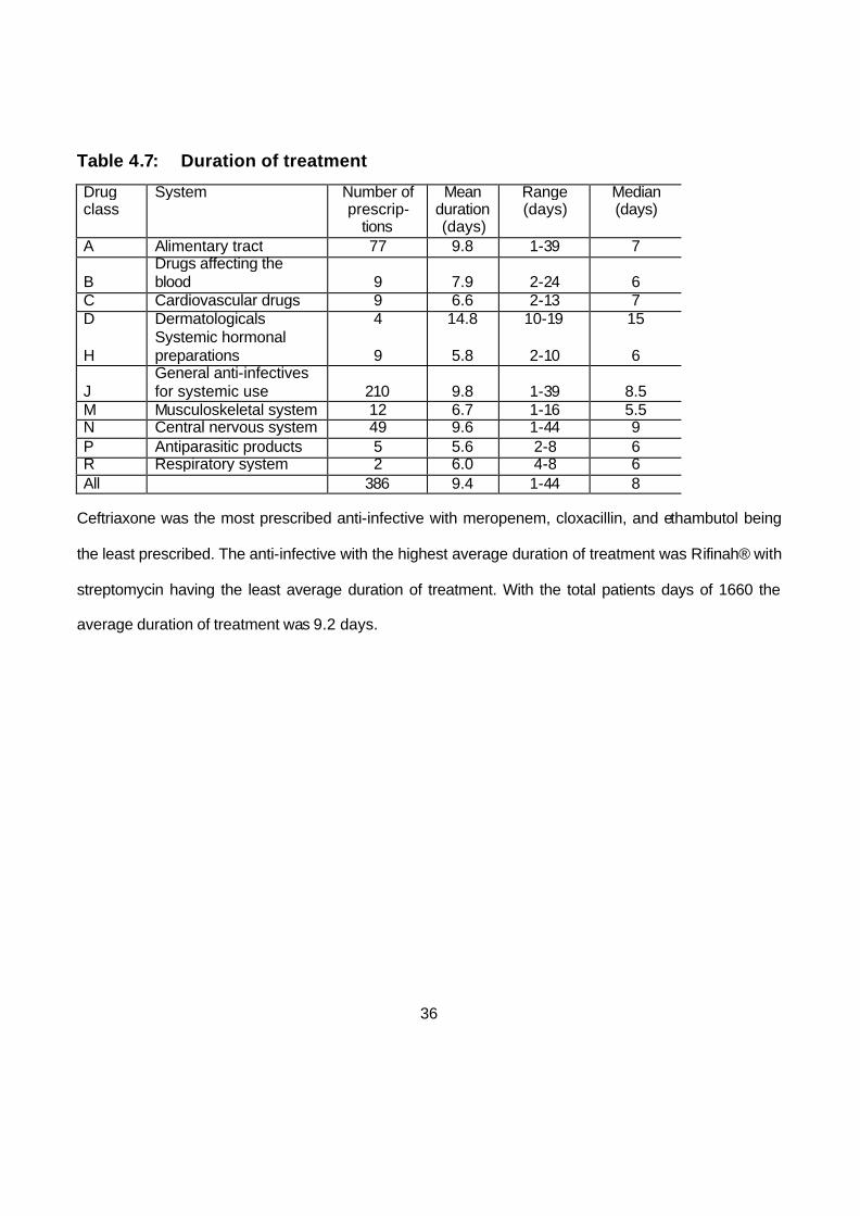
Table 4.7: Duration of treatment
Drug class
System Number of prescrip-
tions
Mean duration (days)
Range (days)
Median (days)
A Alimentary tract 77 9.8 1-39 7
B Drugs affecting the blood 9 7.9 2-24 6
C Cardiovascular drugs 9 6.6 2-13 7 D Dermatologicals 4 14.8 10-19 15
H Systemic hormonal preparations 9 5.8 2-10 6
J General anti-infectives for systemic use 210 9.8 1-39 8.5
M Musculoskeletal system 12 6.7 1-16 5.5 N Central nervous system 49 9.6 1-44 9 P Antiparasitic products 5 5.6 2-8 6 R Respiratory system 2 6.0 4-8 6 All 386 9.4 1-44 8
Ceftriaxone was the most prescribed anti-infective with meropenem, cloxacillin, and ethambutol being
the least prescribed. The anti-infective with the highest average duration of treatment was Rifinah® with
streptomycin having the least average duration of treatment. With the total patients days of 1660 the
average duration of treatment was 9.2 days.

37
Table 4.8: Duration of treatment with anti-infectives for meningitis
ATC code Anti-infective Total patient-
days
Number of treatment courses
Average duration of treatment (days)
J01DA06 Cefuroxime 41 5 8.2 J01DA13 Ceftriaxone 539 58 9.3 J01DH02 Meropenem 11 1 11.0 J01GA01 Streptomycin 20 4 5.0 J01HB02 Cloxacillin 11 1 5.5 J01MA02 Ciprofloxacin 14 2 7.0 J02AA01 Amphotericin B 126 10 12.6 J02AC01 Fluconazole 425 41 10.4 J04AK02 Ethambutol 8 1 8.0 J04AM02 Rifinah® 28 2 14.0 J04AM06 Rifafour® 437 43 10.2 Total 1660 168 9.2
4.7 Cost of treatment
Table 4.9: Cost of treatment
Drug class (ATC code)
System Number of prescriptions
Number of patients (n=66)
Total cost (ZAR)
A Alimentary tract 77 49 245.33 B Drugs affecting the blood 9 9 1,812.04 C Cardiovascular drugs 9 4 48.00 D Dermatologicals 4 3 203.90
H Systemic hormonal preparations 9 8 346.76
J General anti-infectives for systemic use 210 66 113,174.95
M Musculoskeletal system 12 10 30.97 N Central nervous system 49 34 368.65 P Antiparasitic products 5 5 253.65 R Respiratory system 2 2 6.19 All drug classes 386 66 116,490.43
38
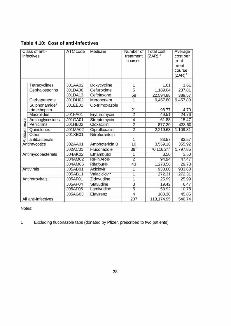
Table 4.10: Cost of anti-infectives
Class of anti-infectives
ATC code Medicine Number of treatment courses
Total cost (ZAR) 2
Average cost per treat-ment course (ZAR)4
Ant
ibac
teria
ls
Tetracyclines J01AA02 Doxycycline 1 1.61 1.61 Cephalosporins J01DA06 Cefuroxime 5 1,189.04 237.81 J01DA13 Ceftriaxone 58 22,594.88 389.57 Carbapenems J01DH02 Meropenem 1 9,457.80 9,457.80 Sulphonamide/ trimethoprim
J01EE01 Co-trimoxazole 21 98.77 4.70
Macrolides J01FA01 Erythromycin 2 49.51 24.76 Aminoglycosides J01GA01 Streptomycin 4 61.88 15.47 Penicillins J01HB02 Cloxacillin 2 877.20 438.60 Quinolones J01MA02 Ciprofloxacin 2 2,219.63 1,109.81 Other antibacterials
J01XE01 Nitrofurantoin 1 83.57 83.57
Antimycotics J02AA01 Amphotericin B 10 3,559.18 355.92 J02AC01 Fluconazole 391 70,116.241 1,797.85 Antimycobacterials J04AK02 Ethambutol 1 3.50 3.50 J04AM02 RIFINAR® 2 94.94 47.47 J04AM06 Rifafour® 43 1,278.56 29.73 Antivirals J05AB01 Aciclovir 1 933.60 933.60 J05AB11 Valaciclovir 1 272.31 272.31 Antiretrovirals J05AF01 Zidovudine 1 25.99 25.99 J05AF04 Stavudine 3 19.42 6.47 J05AF05 Lamivudine 5 53.92 10.78 J05AG03 Efavirenz 4 183.38 45.85 All anti-infectives 207 113,174.95 546.74
Notes:
1 Excluding fluconazole tabs (donated by Pfizer, prescribed to two patients)

39
Fluconazole was the most costly anti-infective that was used. It was used on patients with fungal
meningitis. Cloxacillin was the least costly anti-infective that was used. It was used as combination
therapy together with streptomycin and ethambutol on patients with TB re-infection.
Table 4.11: Eight most costly medicines for meningitis treatment
Medicine Number of prescriptions Cost (ZAR)
Percentage of Total cost
1 Fluconazole 41 70,116.24 63.00%
2 Ceftriaxone 58 22,594.88 20.30%
3 Meropenem 1 9,457.80 8.50%
4 Amphotericin B 10 3,559.18 3.20%
5 Ciprofloxacin 2 2,219.63 1.99%
6 Rifafour® 43 1,278.56 1.15%
7 Cefuroxime 5 1,189.04 1.07%
8 Cloxacillin 3 877.20 0.79%
All medicines 163 111,292.53 100%

40
Percentage of Total cost of Meningitis medicines
1 Fluconazole
2 Ceftriaxone
3 Meropenem
4 Amphotericin B
5 Ciprofloxacin
6 Rifafour
7 Cefuroxime
8 Cloxacillin
Figure 4.3: Percentage of total cost of meningitis medicines
Table 4.12: Duration of treatment with anti-infectives for meningitis
ATC code Anti-infective Total
patient-days
Number of treatment courses
Average duration of treatment
(days) J01DA06 Cefuroxime 41 5 8.2 J01DA13 Ceftriaxone 539 58 9.3 J01DH02 Meropenem 11 1 11.0 J01GA01 Streptomycin 20 4 5.0 J01HB02 Cloxacillin 11 1 5.5 J01MA02 Ciprofloxacin 14 2 7.0 J02AA01 Amphotericin B 126 10 12.6 J02AC01 Fluconazole 425 41 10.4 J04AK02 Ethambutol 8 1 8.0 J04AM02 Rifinah® 28 2 14.0 J04AM06 Rifafour® 437 43 10.2 Total 1660 168 9.2
The following chapter will deal with the discussion of the results obtained during the study.

41
CHAPTER 5: DISCUSSION
5.1 Age and gender
During the course of the study that was conducted from February to August 2008; 66 patients were
enrolled and 33 patients were male and 33 of them female. The largest number of patients was
between ages 31 and 40. After provisional diagnosis; 63 patients were later confirmed as meningitis
cases and only three were confirmed otherwise. In all the subjects of the study no differential diagnosis
of viral meningitis was made. This is different from findings according to Morbidity and Weekly Report
(2003) which states that aseptic or viral meningitis is the most common type of meningitis and is
associated with an estimated 26,000-42,000 hospitalizations each year in the United States
Enteroviruses are the most common cause of aseptic meningitis. Echovirus 9 (E9) and echovirus 30
(E30) have been associated frequently with outbreaks of aseptic meningitis. During March 2003,
several state public health departments noted increased reports of aseptic meningitis and, as of August
7, seven states (Arizona, California, Georgia, Idaho, Oregon, South Carolina, and Texas) had reported
outbreaks associated with either E9 or E30.
5.2 Duration of stay in hospital
The average stay in hospital for patients was 13.3 days. Recommendations regarding length of therapy
depend on the offending organism. Patients with meningococcal meningitis or meningitis caused by H.
influenzae should be treated for 7 days. Patients with pneumococcal meningitis are treated for 10 to 14
days, and those with meningitis caused by L. monocytogenes or S. agalactiae are treated for 14 to 21
days. Therapy for patients with Listeria meningitis may need to be individualized, since some patients

42
require longer courses. Meningitis caused by Enterobacteriaceae or P. aeruginosa requires 21 days of
treatment (Aronin and Qualiarello, 2003).
According to the treatment guideline for treatment of different bacterial meningitis the number of
treatment days in the hospital is as follows; for community acquired with unknown bacterial aetiology;
ceftriaxone, IV, 2g 12 hourly is given for 10 days. For confirmed meningococcal disease only: benzyl
penicillin (Penicillin G), IV, 20- 24 million units daily in 4-6 divided doses for one week. For
pneumococcal meningitis, if sensitive, the following is given; benzyl penicillin (Penicillin G), IV, 20-24
million units daily in 4-6 divided doses for 10 days. If any degree of resistance is present or cannot be
excluded: the following is given; ceftriaxone, IV, 2g 12 hourly for at least 10 days (Standard Treatment
Guidelines and Essential Drug List, 2006).
Tuberculosis is the most prevalent infection in the world, infecting roughly one third of the world. The
World Health Organization estimates that there are approximately eight million new cases every year.
Exposure to HIV significantly increases susceptibility to tuberculosis (Iseman, 2000). Treatment of CNS
tuberculosis should begin as soon as possible when clinically suspected because patient outcome is
improved when therapy is begun in the early stage of disease. For all types of CNS tuberculosis,
treatment should begin with a combination of bactericidal drugs that penetrate into the CSF. The first
line of therapy should include isoniazid (INH), rifampicin, and pyrazinamide, all of which are bactericidal
and achieve effective levels in the CSF. Pyrazinamide is given with INH and rifampicin during the first
two months of therapy, and then is stopped. A fourth drug should be used during the first two months of
therapy when INH drug resistance is a concern (Bajaj, 2002). In the ward a patient with confirmed TB
meningitis was treated with Rifafour®. For the first two months patients are treated with Rifafour® and
then switched to Rifinah®; the total duration of treatment is 12 months. However, after TB meningitis
patients have been discharged, they are referred to their local clinic for further management. Treatment

43
for tuberculous meningitis, intracranial tuberculoma, or tuberculoma with meningitis should be given for
12 months when the tuberculosis strain is sensitive to the antibiotics. Treatment should be lengthened
to 18 months if the patient does not receive pyrazinamide during the first two months of therapy. If a
patient has a multidrug-resistant strain of CNS tuberculosis, therapy is often extended to 24 months. It
is also recommended that patients with active tuberculosis have directly observed therapy to insure
compliance, reduce the development of a resistant organism, and monitor clinical response (Bajaj,
2002).
In the study most patients who were on antituberculosis therapy were started on Rifafour®, two were
started on Rifinah® (combination of rifampicin and isoniazid), later during the course of treatment
Rifafour® was added to the therapy of one patient but there was an overlap of six days between the
two drugs and the 2nd one only continued with Rifinah®. Another patient was started on Rifafour®
which was stopped after four days of therapy and streptomycin injection was added to the therapy one
day later but was subsequently stopped after one day of therapy. The reason for stopping
antituberculosis treatment was because of the fact that the patient was confirmed TB negative. One
patient was started on both Rifafour® and streptomycin injection for 8 days. Another patient was put on
Rifafour® for 15 days and was later given ciprofloxacin oral, ethambutol oral, and streptomycin
injection, until the patient was discharged. Patients with confirmed TB meningitis and other forms of TB
were given Rifafour® on discharge and referred to local clinic for further management. It must be noted
that there was not consistency in the treatment of TB as is shown in the above-mentioned remarks.
Causative organisms and sensitivity patterns
According to the specimens that were cultured the following micro-organisms were identified;
Streptococcus pneumonia (3) and Neisseria meningitides (1) (Table 4.5). This is in line with the

44
findings of Razonable, Keating, (2007) which state the following in terms of the causative micro-
organisms in adult patients: age 18-50 years;(1) Streptococcus pneumonia, Neisseria meningitides and
H. influenzae, age older than 50 years;(2) Streptococcus pneumonia, Neisseria meningitides, Listeria
monocytogenes and aerobic Gram- negative bacilli.
Out of 18 patients with suspected fungal meningitis, 10 were positive with cryptococcus and six were
negative. Out of nine patients treated for TB meningitis only one patient had a positive result for TB.
The rest were not tested. There were 29 non-specified meningitis cases that were not tested for micro-
organisms and sensitivity. Because there was no routine testing of specimens from patients, it was
difficult to record causative organisms and sensitivity patterns. This, therefore, made it difficult to
establish any level of resistance to ceftriaxone that is being used as the antibiotic of choice in the
treatment of bacterial meningitis in the internal medicine wards of Dr George Mukhari Hospital. This
also had a negative effect in containing the cost of anti-infectives used as there was no consistent
tailoring of anti-infectives used even when the final diagnosis was pointing to other than bacterial
meningitis.
5.3 Treatment cost
The total cost of anti-infectives used for treatment of meningitis amounted to R111, 292.53; and the
average cost per patient was R1 686.25. All anti-infectives prescribed for the patient cost R113, 174.95
with an average cost of R546.74 per patient. Cost of all medicines prescribed for the 66 patients
amounted to R116, 490.43. Cefriaxone was the most prescribed anti-infective; 58 patients out of 66
were started on ceftriaxone. This is due to the fact that ceftriaxone is an anti-infective of choice to
patients suspected with meningitis in the internal medicine wards of Dr George Mukhari Hospital.
According to (Steele 2002) S pneumoniae strains that are non-susceptible to penicillin and third-

45
generation cephalosporins (i.e., ceftriaxone and cefotaxime) have been identified worldwide, with as
many as 8% of pneumococci demonstrating this multi-resistant pattern in some regions. Fluconazole
was the second most prescribed anti-infective, and though it was prescribed to only 41 patients it was
the most expensive anti-infective used.
5.4 Outcomes
One female patient absconded treatment. The patient was on the ninth day of treatment when she
absconded. She was still on ceftriaxone, IV, 2g q12h that was started on the day she was admitted and
Rifafour®, four tablets daily from the second day of her admission. According to her blood specimen,
meningitis was confirmed though no mention of the type of meningitis was made. This poses a question
to the addition of Rifafour® to her treatment. But it has been established in the study that patients who
were admitted with provisional meningitis diagnosis and who were very sick with concomitant diseases
like retroviral disease were put on different anti-infectives ranging from antibacterials, anti-TB drugs and
antifungals. It was only when the differential diagnosis has been made that the medication would be
tailored. There was, however, no routine blood or cerebrospinal fluid specimens taken to establish
whether the current treatment given to a patient was relevant. Sputums for TB would also be sent for
testing but were not part of the sample types that were identified to be recorded for the purpose of the
study.
In two patients therapy was initiated but they refused hospital treatment. One patient had unspecified
meningitis and was put on ceftriaxone, IV, 2g q12h and fluconazole, IV, 400mg q12h, and the second
patient had a confirmed cryptococcal meningitis and was put on amphotericin B, IV, 50mg daily. These
two patients were then discharged from the hospital.
46
A total number of 41 (62%) patients recovered. All the patients were started on ceftriaxone, IV, 2g q12h
except for one patient who had a final diagnosis of severe headaches. It is advised that treatment of
bacterial meningitis should be initiated as soon as possible as it is a medical emergency, and patients
should be hospitalized. They can deteriorate as rapidly as in less than 24 hours. Empiric treatment is
advised according to the likely organisms, until the organism has been identified (Spiro et al.,2004)
According to Mark et al., (2005) if the patient appears acutely ill, empiric therapy with multiple
antibiotics should be started immediately after an IV line has been placed and blood for culture has
been drawn. Lumbar puncture can be performed afterwards. A third generation cephalosporin (e.g.,
ceftriaxone or cefotaxime) should be included because it is highly active against common meningeal
pathogens in patients of all ages. During the study severely ill patients were put on multiple anti-
infectives with the addition of Rifafour® and amphotericin B to ceftriaxone. It was observed that there
was no routine drawing of blood for culture before initiating anti-infectives that was observed. Lumbar
puncture was performed on some patients suspected of meningitis and some of the CSF results were
not available in the patients` files. There was no evidence of routine de-escalation of anti-infective
therapy noted as culture sensitivity was not prescribed for most of the patients` specimens. It was only
in one case where there was a tailoring of antibiotics, with ceftriaxone stopped after one day of
commencement and switched over to fluconazole, IV, 400mg bd and amphotericin, IV; 50mg daily after
the patient was confirmed with cryptococcal meningitis, which was not the case with other differential
meningitis diagnoses. From the 24 males that recovered, two were confirmed with cryptococcal
meningitis and the first one was started on amphotericin B,IV, 50mg and the second one was started
on both amphotericin B, IV, 50mg daily and fluconazole, IV, 400mg bd respectively after one day of
ceftriaxone, IV, 2g bd. In two other cases where cryptococcal meningitis was confirmed on admission,
one patient was started on amphotericin B, IV, 50mg daily for 13 days and the second patient was
started on amphotericin B, IV, 50mg daily for 14 days and later during the course of treatment

47
fluconazole, IV, 400mg bd for six days but overlapped with amphotericin B treatment for six days. one
patient was started on anti-TB drugs. All other male patients were started on ceftriaxone, IV, 2g bd. The
concomitant conditions in male patients who recovered were the following; hypertension, nephritic
syndrome, LRTI, retroviral disease, pulmonary TB, herpes labialis, pneumonia, pleural effusion,
oropharangeal thrush, diffused nodular rash, multi- drug resistant tuberculosis (MDR), chronic
diarrhoea.
5.5 Limitations
Several factors contributed to the limitations of the study. The following were the limitations observed:
Following-up of some patients was not easy as there was no consistency in updating the list of patients
in some of the wards. It must be explained that some patients would be moved from the admission
ward (ward 36) to one of the other internal medicine wards and if the patient is diagnosed to have TB
he will be moved to a TB ward (ward 39).
Some of the patients who were discharged or demised their files would be taken to the filing room
before final information is obtained from the files. This was due to the fact that in the beginning of the
study only one clerk was involved in keeping the files for final recording of information for all the internal
medicine wards. It was also very difficult to get files that were taken to the filing room as some of them
were taken to finance department for billing which took a long time for them to be returned to the filing
room. The files of the patients who were demised were not filed in any particular order and it made it
very difficult to access them.
The following chapter will deal with conclusion and recommendations.

48
CHAPTER 6: CONCLUSION AND RECOMMENDATIONS
Most patients in the study were put on ceftriaxone which is the anti-infective of choice for empiric
treatment in patients with meningitis. There was no consistent tapering down of empiric therapy as the
culture and sensitivity tests were not done for most of the patients.
The lack of culture and sensitivity results made it difficult to establish which type of meningitis is
prevalent and also to understand resistance patterns in the internal medicine wards.
From a total number of 66 patients who were enrolled in the study, one female patient absconded, 13
female patients and nine male patients died, 17 female patients and 24 male patients recovered with
two female patients refusing hospital treatment.
The total cost of anti-infectives used for treatment of meningitis amounted to R111, 292.53; and the
average cost per patient was R1 686.25. All anti-infectives prescribed for the patient cost R113, 174.95
with an average cost of R546.74 per patient. Cost of all medicines prescribed for the 66 patients
amounted to R116, 490.43.
The anti-infective with the highest average duration of treatment was Rifinah® with streptomycin having
the least average duration of treatment. With the total patients days of 1660 the average duration of
treatment was 9.2 days.
To be able to address issues of culture and sensitivity results in future a researcher will need to involve
clinicians at the beginning of the study in making sure that for every suspected meningitis case a
request is made to get the culture and sensitivity tests done. The researcher can also play a proactive
role in accessing the results from the microbiology laboratory. This will also make it easy for him to

49
inform the clinicians about any resistance patterns picked up in the process and to suggest an
appropriate alternative. This will also help to draft a new treatment protocol should it be necessary to do
so.

50
REFERENCES
Aronin, S.I., 2003. Bacterial Meningitis. [online]. 1-4, [Accessed 6th July 2009]. Available from World
Wide Web:http://www.medscape.com/viewarticle/451596.
Bajaj, B.K. 2002. Duration of Therapy for Tubercular Meningitis. [ Online]. Pages ?. [ accessed 22nd
December 2008]. Available from World Wide Web: http://www.medscape.com/criticalcare
Be N.A., Kim K.S., Bishai W.R., Jain S.K. 2009. Pathogenesis of central nervous system tuberculosis.
Curr Mol Med. 9:94-9.
Beers, M.H., Berkow, R.2005. Merck Manual of diagnosis and therapy. Seventh Edition. 1431, 1435.
Canada Communicable Disease Report (1997). [Online]. 65-72, [Accessed 3rd June 2006]. Available
from: http://www.aspc.gc.ca
Di Piro J. T., Talbert R.L., Yee G.C., Matzke G.R., Wells B.G., Posey L.M.1997. A Pathophysiologic
Approach. Third Edition.Appleton & Lange.1986,1987.
Drew R.H., American College of Clinical Pharmacy Infectious Diseases Practice and Research Network
Bibliography Group. 2004. Key references in infectious diseases pharmacotherapy. Pharmacotherapy
24: 1586-1593.
Fitch, M.T., van de Beek, D. 2007. Emergency diagnosis and treatment of adult meningitis. 191-200.
Gums, J.G., Yancey, R.W., A.C., Kubilis, P.S 1999. A randomised, prospective study measuring
outcomes after antibiotic therapy intervention by a multidisciplinary consult team. Pharmacotherapy.
19 (12): 1369- 1377. Available from:http://www.medscape.com/viewarticle/418076 [ Accessed 02 April
2003].
Gibbon, C.J. 2005. South African Medicine Formulary. Seventh Edition. Health and Medical Publishing
Group. 255.

51
Harrison M.J.G., McArthur, J.C. 1998. Cerebral infections in AIDS:cryptococcal meningitis. Infect Med.
15 (6): 396- 409.
Hasan, Y.M., Das, M., Mourad, F. 1997. Drug utilisation and antibiotic use in the Primary Health Care
Centres in Sharjah. Eastern Mediterranean health journal. 3 (3): 444-451.
Helbok R., Broessner G., Pfausler B., Schmutzhard E. 2009. Chronic meningitis. J Neurol. 256:168-
175.
Herfindal, E.T., Gourley D.R. 2000. Textbook of therapeutics, drugs and diseases management.
Seventh edition. Lippincolt Williams & Wilkins. 1508- 1510.
Iseman, M.D. 2000. Antibiotic resistance: A serious public health threat- The global threat. Page 18-
19. National Health Council, Inc.
Kam, K.M., Luey K.T., Fung, S.M., Yie, P.P., Harden, T.J., Cheung, M.M. 1995. Emergence of Multiple-
Antibiotic- Resistant Streptococcus pneumoniae in Hong Kong, Journal of American Society for
Microbiology. [Online]. 2667-2670, [Accessed 3rd June 2006]. Available from
http://www.pubmedcentral.nih.gov.
Katti M.K. 2004. Pathogenesis, diagnosis, treatment, and outcome aspects of cerebral tuberculosis.
Med Sci Monit. 10:215-29.
Katzung, B.G.2001. Basic and Clinical Pharmacology, 8th Edition. Lange Medical Books/McGraw- Hill
Medical Publishing Division. 753.
Katzung, B.G. 2004. Basic & Clinical Pharmacology. Ninth Edition. Appleton & Lange.794.
Kepa, L., Oczko-Grzesik B., Stolarz, W., Sobala- Szczgel, B. 2005. Drug- induced aseptic meningitis in
Suspected Central nervous system infections. Journal of Clinical Neuroscience. 562-564.

52
King J.W. 2007. Cryptococcosis. [Online].1-33, [Accessed 7th June 2008]. Available from: http//
www.emedicine.com/med/TOPIC 482.HTM.
Koda- Kimble, M.A., Young, L.Y., Kradjan, W.A., Guglielmo, B.J. 2005. Applied Therapeutics: THE
CLINICAL USE OF DRUGS. Eigth Edition. Lippincott Williams & Wilkins. 58-13.
Lazoff M. 2007. Meningitis. [Online]. 1-41, [Accessed 7th June 2008]. Available from: http://
www.emedicine.com/emerg/TOPIC 309.HTM.
Leib S.L. ,Täuber M.G. 1999. Pathogenesis of bacterial meningitis. Infectious disease clinics of North
America. 13;3:527-548.
Lo Re III, V. 2004. Infectious Diseases. Hanley and Belfus. 13.
Mandell,G.L., Bennet, J.E., Dolin, R. 2000. Principles and Practice of Infectious Diseases. Fifth edition.
Churchill Livingstone.964, 980.
Mehlhorn, A.; Sucher, B. (2005). An overview of Bacterial Meningitis, Journal of U.S. Pharmacist.
[online]. 8-12, [accessed 19th May 2006]. Available from World Wide Web: http:www.uspharmacist.com.
Members of Antimicrobial Committee Johannesburg Hospital. 2005. Antimicrobial Therapy Guide. 1st
Edition. 6,7,10.
Miller, M.L., Gaur, A.H., Kumar, A. 2008. Meningitis, Bacterial. [Online].1-31, [Accessed 6th June 2008].
Available from: http:www.emedicine.com/ped/TOPIC 198.HTM.
Morbidity and Mortality Weekly Report. 2003. Outbreaks of aseptic meningitis associated with
Echoviruses 9 and 30 and Preliminary Surveillance Reports on Enterovirus Activity- United States.
[online].1-2, [Accessed 11 July 2009]. Available from World Wide Web:
http://www.medscape.com.viewarticle/1460226.

53
Naesens R, Ronsyn M, Druwé P, Denis O, Ieven M, Jeurissen A. 2009. Central nervous system
invasion by community-acquired meticillin-resistant Staphylococcus aureus. J Med Microbiol. 58:1247-
51.
Parasuraman, T.V., Deverka, P.A., Toscani M.R. 2000. Estimating the economic impact of viral
meningitis in the United States.[online]. 1-2, [Accessed 11 July 2009]. Available
from:http://www.medscape.com/viewarticle/410064.
Prasad, K., Karlupia, N., Kumar, A. 2009. Treatment of bacterial meningitis: An overview of Cochrane
systematic reviews. Respiratory Medicine 103:945-950.
Ramachadran, T.S. 2007. Tuberculous Meningitis. [online]. 1-54. [accessed 6/06/2008]. Available
from:http://www.emedicine.com.
Razonable, R.R; Keating M.R. 2007. [Online]. 1-70, [Accessed 11th February 2008]. Available from:
http://www.emedicine.com/med/TOPIC 2613.HTM.
Razonable, R.R; Keating M.R. 2007. Meningitis eMedicine Infectious Diseases.[Online].1-18.
[Accessed 12 July 2009]. Available from:http://emedicine.medscape.com/article/232915-overview.
Roos, K.L. 2005. Principles of Neurologic Infectious Diseases. McGraw- Hill Companies. 18-20.
Rowland, L. P.2005. Merritt’s Neurology. Eleventh Edition. Lippincott Williams & Wilkins. 175-176.
Saag, M.S., Graybill, R.J., Larson, A.R., Pappas P.G., Perfect, J.R., Powderly, W.G., Sobel, J.D.,
Dismukes E.W. 2000. Practical guidelines for the management of cryptococcal disease. Clinical
Infectious Diseases. 30: 710-8.
Samuels, M.A., Feske, S. K.2003. Office Practice of Neurology. Second edition. Churchill Livingstone.
511.

54
Sloan, D.J., Dedicoat, M.J., Lalloo, D.G. 2009. Treatment of cryptococcal meningitis in resource limited
settings. Curr Opin Infect Dis. 22:455-63.
Spiro, E.S., Spiro, D.M. 2004. Acute meningitis: Focus on bacterial infection. Clinical Reviews. 14 (3):
53- 60.
Standard Treatment Guidelines and Essential Drugs list. 1998 edition. Adult hospital level. Essential
drugs programme South Africa. 222- 223.
Standard Treatment Guidelines and Essential Drugs List. 2006 Edition. Adult hospital level. Essential
Drugs Programme South Africa. 208.
Steele, R.W. 2002. Recognizing, Managing, and Preventing Pneumococcal Meningitis: An Expert
Interview With Russell W. Steele, MD. [ Online]. Pages. [Accessed 29th December 2008]. Available
from: http:www.medscape.com/viewpublication/206
Trunkel, A.R., Scheld, W.M. 1996. Central nervous system infections. In A practical approach to
infectious diseases. Edited by Reese, R.E., Betts, R.F. Fourth edition. Little Brown and Company, New
York. 133-183
Van der Meyden, C.H. 2004.Neurology Department Dr George Mukhari Hospital.2004. Antibiot ics
Policy of Neurology Department. 1-3.
Victor, M., Ropper, A.H.2001. Principles of Neurology. Seventh edition. McGraw- Hill.
Pages:735,737,757,760.
Vokshoor, A., Wan, C. 2007. Viral meningitis. . [Online].1-11, [Accessed 19th August 2008. Available
from World Wide Web: http://www.emedicine.com./neuro/TOPIC 607. HTM.

55
Walker, R., Edwards, C.2003. Clinical Pharmacy and Therapeutics. Third Edition.Churchill Livingstone.
565.
WHO.2009. The Anatomical Therapeutic Chemical Classification System With Defined Daily Doses
(ATC/DDD). [Online]. 1-2, [Accessed 31st August 2009]. Available from World Wide Web:
http://www.who.int/classifications/atcddd/en/
Wikimedia Foundation (2008). [Online]. 1-12, [Accessed 8th February 2008]. Available from:
http://en.wikipedia.org/wiki/Meningitis.
Wilks, D., Farrington, M., Rubenstein, D. 2003. The Infectious Diseases Manual. Second Edition.
Blackwell Publishing. 101.
Wilson, W.R., Sande, M. A.2001. Current Diagnosis & Treatment in Infectious Diseases. LANGE
Medical Books/ McGrw- Hill. 75.

56
APPENDICES
Appendix 1: