Embed Size (px)
Citation preview
1

2
The authors declare no conflicts of interest
No financial support was taken for this
cross-sectional study

ASNR 2015 Annual Meeting Abstract No: EP-04
Submission Number: 1813
Hormone replacement therapy-related changes in the early postmenopausal period
(critical window): an in vivo brain proton magnetic resonance spectroscopy study
Kamran Mahmutyazıcıoglu1, Fahri Halit Besir2, Mustafa Bardakci3, Hamit Alper Tanrıverdi4,
Handan Ankarali5
1Department of Radiology, Faculty of Medicine, Fatih University Sema Training Hospital, İstanbul, Turkey 2Department of Radiology, Duzce University Faculty of Medicine, Duzce, Turkey
3Department of Radiology, Faculty of Medicine, Bülent Ecevit University, Zonguldak, Turkey 4Department of Obstetrics and Gynecology, Faculty of Medicine, Bülent Ecevit University,Zonguldak, Turkey
5Department of Statistics, Duzce University Faculty of Medicine, Duzce, Turkey
3
Purpose
Hormonal replacement therapy (HRT) is a medical
treatment to relieve the symptoms of menopause in
surgically or naturally postmenopausal women
Magnetic resonance spectroscopy (MRS) is an in vivo
method used to study brain metabolism; in particular, the
changes that occur during aging and cognitive diseases
have attracted interest.

Purpose
However, brain MRS data on the effect of HRT in the
postmenopausal period are lacking.
We sought todetermine whether there are metabolic
changes related with HRT usage in the brains of healthy
postmenopausal women with no cognitive complaints who
started HRT in the early postmenopausal period (critical
window).
Materials and Methods
The cross-sectional study was enrolling postmenopausal
women. Healthy, literate postmenopausal women, between 45
and 65 years old, were included in the study.
Postmenopausal women in the present study were
assembled into 2 groups, as HRT users and non-HRT users.
The 2 groups were matched for age, education, and
postmenopausal period.

Materials and Methods

Materials and Methods
MR examinations were conducted with a 1.5 T scanner
(Gyroscan Intera, Philips, Best, the Netherlands). A
standard head coil was used for conventional MR imaging
and MRS.
Sixty-eight postmenopausal women aged between 45 and 55 years
old were included in the study. Of these, 21 subjects were excluded
from the study due to anxiety disorder (2 subjects), depression (5
subjects), vertebrobasilar insufficiency (1 subject), and hypertension
or heart failure (13 subjects). Of the 47 subjects included in the study,
21 (45%) used HRT and 26 (55%) did not.
Results

Results The results of the comparison of HRT users and
nonusers in terms of age and duration of menopause are
given in Table.
The differences between the 2 groups regarding age
and duration of menopause were not statistically
significant.
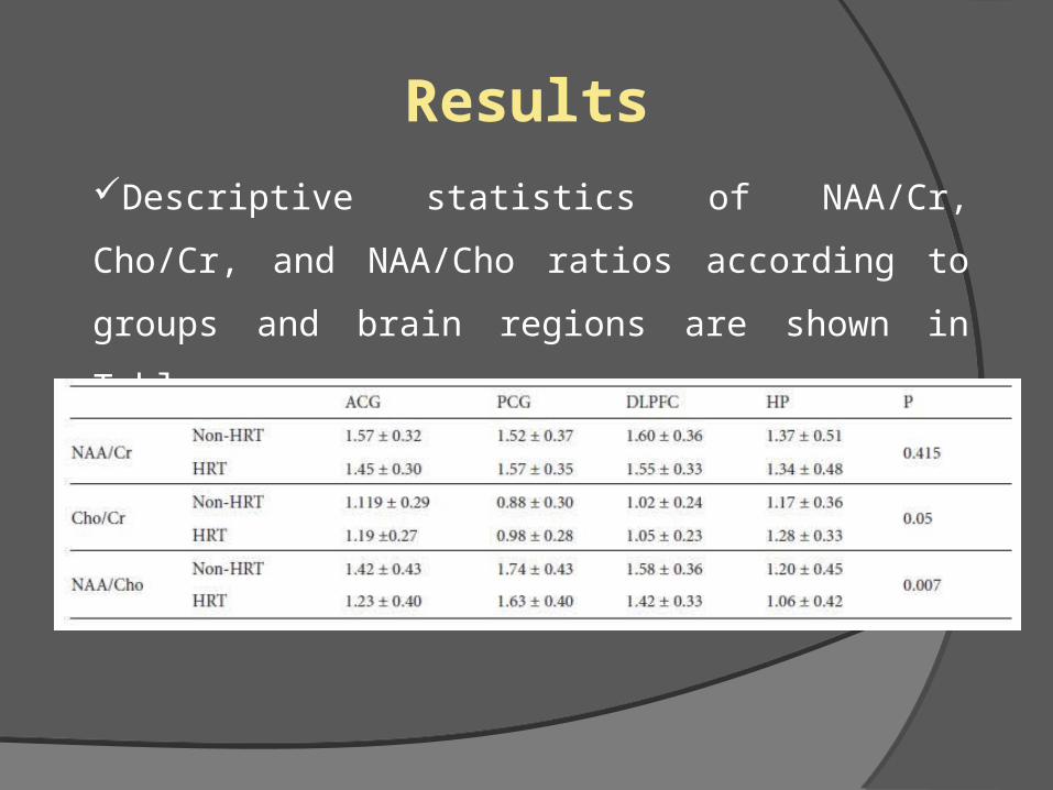
Descriptive statistics of NAA/Cr, Cho/Cr, and
NAA/Cho ratios according to groups and brain regions
are shown in Table.
Results
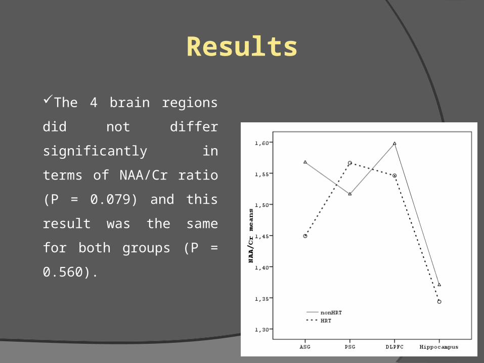
The 4 brain regions did not
differ significantly in terms of
NAA/Cr ratio (P = 0.079) and
this result was the same for
both groups (P = 0.560).
Results

Results
Cho/Cr ratio of the HRT user
group was significantly higher in all
4 regions when controlling for the
differences between brain region
and the group-by-region interaction
(P = 0.05)

Results
NAA/Cho ratios correlated
negatively with duration of
menopause when controlling for the
effect of age (r=–0.350, p=0.05

Results NAA/Cho ratio of the HRT-
user group was significantly
lower in all 4 brain regions when
controlling for the differences
between brain region and the
group-by region interaction
(p=0.007)
The critical period or window theory suggests a beneficial effect of estrogen
therapy in the early postmenopausal stage. The present findings of decreased
NAA/Cho and elevated Cho/Cr do not support this theory and argue against
the neuroprotective effect of HRT in early postmenopausal women during the
critical period. It is unclear whether our results were affected by the addition of
progestin to estrogen. Because all hormone treated women in our study
received tibolone, which has combined estrogenic and progestogenic
properties, our study provides no evidence regarding the effect of unopposed
estrogen therapy on neuronal integrity. Further comparative studies are
necessary to resolve this issue.
Conclusions

The current data suggest, but do not prove, that
postmenopausal HRT with tibolone, a synthetic
steroid with estrogenic, progestogenic, and
androgenic properties, do not have a protective
effect on the neurochemical structure of the brain in
selected regions. Since this study is of a cross-
sectional design, further longitudinal studies are
needed to validate this finding.
Conclusions

1. Taylor HS, Manson JE. Update in hormone therapy use in menopause. J Clin Endocrinol Metab 2011; 96: 255–64.2. Tiidus PM. Benefits of estrogen replacement for skeletal muscle mass and function in post-menopausal females: evidence from human and animal studies. EAJM 2011; 43: 109–14.3. Malatyalıoğlu E, Kökçü A, Yanık FF, Alper T. Effects of four different hormone replacement therapy regimens on certain cardiovascular risk factors. Turk J Med Sci 2000; 30: 469–73.4. Loucks TL, Berga SL. Does postmenopausal estrogen use confer neuroprotection? Semin Reprod Med 2009; 27: 260–74.5. Resnick SM, Henderson VW. Hormone therapy and risk of Alzheimer disease: a critical time. JAMA; 2002: 288: 2170–2.6. Maki PM. Hormone therapy and cognitive function: is there a critical period for benefit? Neuroscience 2006; 138: 1027–30.7. Maki PM, Dumas J. Mechanisms of action of estrogen in the brain: insights from human neuroimaging and psychopharmacologic studies. Semin Reprod Med 2009; 27:250–9.
References