Embed Size (px)
Citation preview
1
Chapter 19Chapter 19The Cardiovascular System: The Cardiovascular System:
The Blood The Blood

2
Cells of the body are serviced by 2 fluidsCells of the body are serviced by 2 fluids bloodblood
composed of plasma and a variety of cellscomposed of plasma and a variety of cells transports nutrients and wastestransports nutrients and wastes
interstitial fluidinterstitial fluid bathes the cells of the bodybathes the cells of the body
Nutrients and oxygen diffuse from the blood Nutrients and oxygen diffuse from the blood into the interstitial fluid & then into the into the interstitial fluid & then into the cellscells
Wastes move in the reverse directionWastes move in the reverse direction Hematology is study of blood and blood Hematology is study of blood and blood
disordersdisorders
Fluids of the Body Fluids of the Body

3
Functions of BloodFunctions of Blood TransportationTransportation
O2, CO2, metabolic wastes, nutrients, heat O2, CO2, metabolic wastes, nutrients, heat & hormones& hormones
RegulationRegulation helps regulate pH through buffershelps regulate pH through buffers helps regulate body temperaturehelps regulate body temperature
coolant properties of water coolant properties of water vasodilatation of surface vessels dump heatvasodilatation of surface vessels dump heat
helps regulate water content of cells by helps regulate water content of cells by interactions with dissolved ions and interactions with dissolved ions and proteinsproteins
Protection from disease & loss of bloodProtection from disease & loss of blood

4
Physical Characteristics Physical Characteristics of Bloodof Blood
Thicker (more viscous) than water and Thicker (more viscous) than water and flows more slowly than waterflows more slowly than water
Temperature of 100.4 degrees FTemperature of 100.4 degrees F pH 7.4 (7.35-7.45)pH 7.4 (7.35-7.45) 8 % of total body weight8 % of total body weight Blood volumeBlood volume
5 to 6 liters in average male5 to 6 liters in average male 4 to 5 liters in average female4 to 5 liters in average female hormonal negative feedback systems hormonal negative feedback systems
maintain constant blood volume and maintain constant blood volume and osmotic pressureosmotic pressure
5
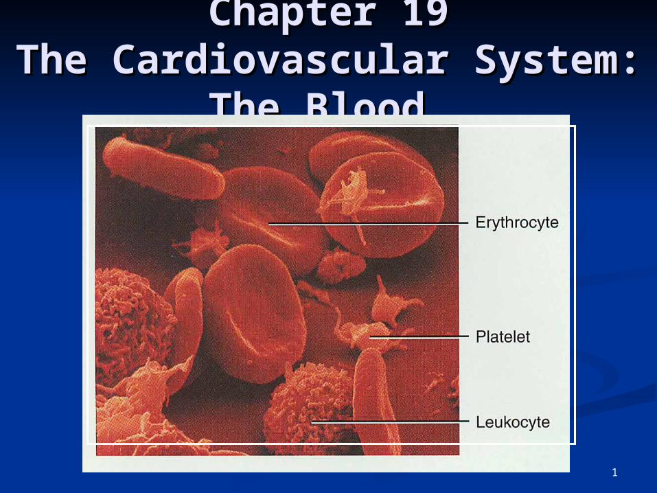
Techniques of Blood Techniques of Blood SamplingSampling
VenipunctureVenipuncture sample taken from vein with sample taken from vein with
hypodermic needle & syringehypodermic needle & syringe median cubital vein (see page 717)median cubital vein (see page 717) why not stick an artery?why not stick an artery?
less pressureless pressure closer to the surfacecloser to the surface
Finger or heel stickFinger or heel stick common technique for diabetics to common technique for diabetics to
monitor daily blood sugarmonitor daily blood sugar method used for infantsmethod used for infants

6
Components of BloodComponents of Blood HematocritHematocrit
55% plasma55% plasma 45% cells 45% cells
99% RBCs99% RBCs < 1% WBCs and platelets< 1% WBCs and platelets

7
Blood PlasmaBlood Plasma 0ver 90% water0ver 90% water 7% plasma proteins7% plasma proteins
created in livercreated in liver confined to bloodstreamconfined to bloodstream
albuminalbumin maintain blood osmotic pressuremaintain blood osmotic pressure
globulins (immunoglobulins)globulins (immunoglobulins) antibodies bind to foreignantibodies bind to foreign
substances called antigenssubstances called antigens form antigen-antibody complexesform antigen-antibody complexes
fibrinogenfibrinogen for clotting for clotting
2% other substances 2% other substances electrolytes, nutrients, hormones, gases, waste electrolytes, nutrients, hormones, gases, waste
productsproducts

8
Formed Elements of BloodFormed Elements of Blood Red blood cells ( erythrocytes )Red blood cells ( erythrocytes ) White blood cells ( leukocytes )White blood cells ( leukocytes )
granular leukocytesgranular leukocytes neutrophilsneutrophils eosinophilseosinophils basophilsbasophils
agranular leukocytesagranular leukocytes lymphocytes = T cells, B cells, and natural killer lymphocytes = T cells, B cells, and natural killer
cellscells monocytesmonocytes
Platelets (special cell fragments)Platelets (special cell fragments)

9
HematocritHematocrit Percentage of blood occupied by cellsPercentage of blood occupied by cells
female normal rangefemale normal range 38 - 46% (average of 42%)38 - 46% (average of 42%)
male normal rangemale normal range 40 - 54% (average of 46%)40 - 54% (average of 46%) testosteronetestosterone
Anemia Anemia not enough RBCs or not enough hemoglobinnot enough RBCs or not enough hemoglobin
PolycythemiaPolycythemia too many RBCs (over 65%)too many RBCs (over 65%) dehydration, tissue hypoxia, blood doping in athletesdehydration, tissue hypoxia, blood doping in athletes
10
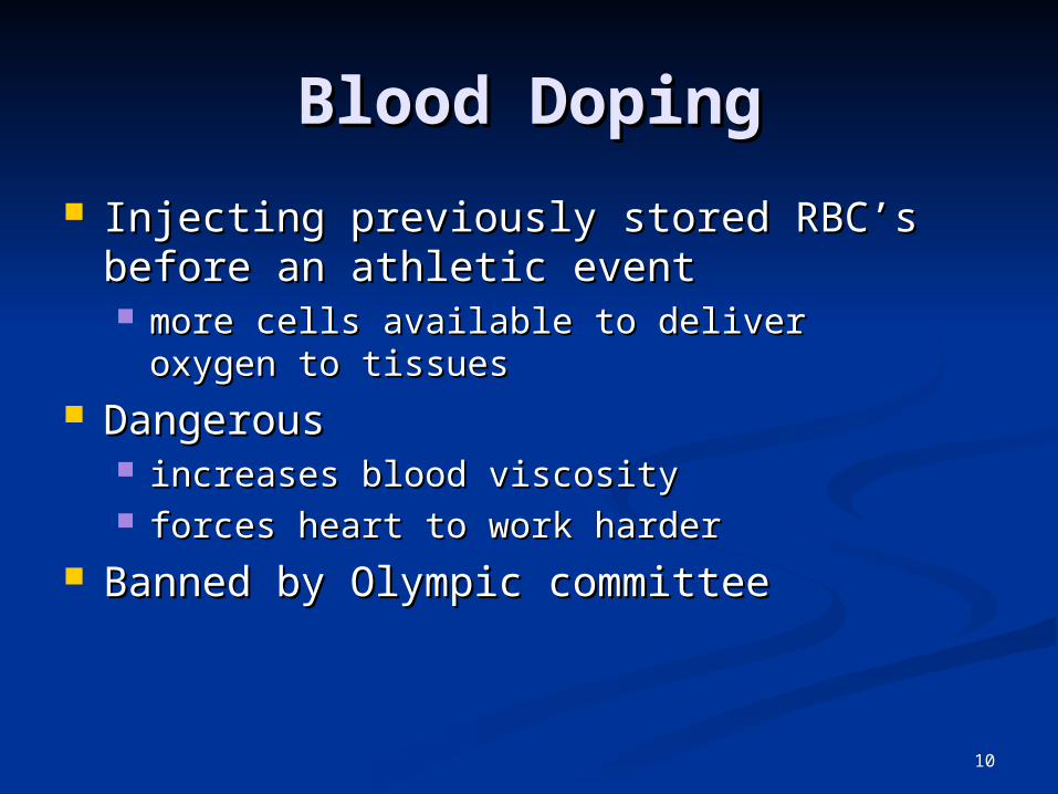
Blood DopingBlood Doping
Injecting previously stored RBC’s Injecting previously stored RBC’s before an athletic eventbefore an athletic event more cells available to deliver oxygen to more cells available to deliver oxygen to
tissuestissues Dangerous Dangerous
increases blood viscosityincreases blood viscosity forces heart to work harderforces heart to work harder
Banned by Olympic committeeBanned by Olympic committee

11
Formation of Blood Formation of Blood CellsCells
Most blood cells types need to be Most blood cells types need to be continually replacedcontinually replaced die within hours, days or weeksdie within hours, days or weeks process of blood cells formation is process of blood cells formation is
hematopoiesis or hemopoiesishematopoiesis or hemopoiesis In the embryoIn the embryo
occurs in yolk sac, liver, spleen, thymus, occurs in yolk sac, liver, spleen, thymus, lymph nodes & red bone marrowlymph nodes & red bone marrow
In adultIn adult occurs only in red marrow of flat bones like occurs only in red marrow of flat bones like
sternum, ribs, skull & pelvis and ends of sternum, ribs, skull & pelvis and ends of long boneslong bones

12
HematopoiesisHematopoiesis

13
Medical Uses of Growth Medical Uses of Growth FactorsFactors
Available through recombinant DNA Available through recombinant DNA technologytechnology recombinant erythropoietin (EPO) very recombinant erythropoietin (EPO) very
effective in treating decreased RBC effective in treating decreased RBC production of end-stage kidney diseaseproduction of end-stage kidney disease
other products given to stimulate WBC other products given to stimulate WBC formation in cancer patients receiving formation in cancer patients receiving chemotherapy which kills bone marrowchemotherapy which kills bone marrow granulocyte-macrophage colony-stimulating factorgranulocyte-macrophage colony-stimulating factor granulocyte colony stimulating factorgranulocyte colony stimulating factor
thrombopoietin helps prevent platelet thrombopoietin helps prevent platelet depletion during chemotherapydepletion during chemotherapy

14
Contain oxygen-carrying protein Contain oxygen-carrying protein hemoglobin that gives blood its red colorhemoglobin that gives blood its red color 1/3 of cell’s weight is hemoglobin1/3 of cell’s weight is hemoglobin
Biconcave disk 8 microns in diameterBiconcave disk 8 microns in diameter increased surface area/volume ratio increased surface area/volume ratio flexible shape for narrow passagesflexible shape for narrow passages no nucleus or other organellesno nucleus or other organelles
no cell division or mitochondrial ATP formationno cell division or mitochondrial ATP formation Normal RBC countNormal RBC count
male 5.4 million/drop ---- female 4.8 male 5.4 million/drop ---- female 4.8 million/dropmillion/drop
new RBCs enter circulation at 2 million/secondnew RBCs enter circulation at 2 million/second
Red Blood Cells or Red Blood Cells or ErythrocytesErythrocytes

15
HemoglobinHemoglobin
Globin protein consisting of 4 polypeptide chainsGlobin protein consisting of 4 polypeptide chains One heme pigment attached to each polypeptide One heme pigment attached to each polypeptide
chainchain each heme contains an iron ion (Fe+2) that can combine each heme contains an iron ion (Fe+2) that can combine
reversibly with one oxygen moleculereversibly with one oxygen molecule
16
RBC Life CycleRBC Life Cycle
RBCs live only 120 daysRBCs live only 120 days wear out from bending to fit through wear out from bending to fit through
capillariescapillaries no repair possible due to lack of organellesno repair possible due to lack of organelles
Worn out cells removed by fixed Worn out cells removed by fixed macrophages in spleen & livermacrophages in spleen & liver
Breakdown products are recycledBreakdown products are recycled

17
Feedback Control of RBC Feedback Control of RBC ProductionProduction
Tissue hypoxia (cells not Tissue hypoxia (cells not getting enough O2)getting enough O2) high altitude since air has less O2high altitude since air has less O2 anemiaanemia
RBC production falls below RBC RBC production falls below RBC destructiondestruction
circulatory problemscirculatory problems Kidney response to hypoxiaKidney response to hypoxia
release erythropoietinrelease erythropoietin speeds up development of speeds up development of
proerythroblasts into proerythroblasts into reticulocytesreticulocytes
18
WBC Anatomy and TypesWBC Anatomy and Types
All WBCs (leukocytes) have a nucleus All WBCs (leukocytes) have a nucleus and no hemoglobinand no hemoglobin
Granular or agranular classification Granular or agranular classification based on presence of cytoplasmic based on presence of cytoplasmic granules made visible by staininggranules made visible by staining granulocytes are neutrophils, eosinophils or granulocytes are neutrophils, eosinophils or
basophilsbasophils agranulocytes are monocyes or lymphocytesagranulocytes are monocyes or lymphocytes
19
Neutrophils (Granulocyte)Neutrophils (Granulocyte)
Polymorphonuclear Leukocytes or PolysPolymorphonuclear Leukocytes or Polys Nuclei = 2 to 5 lobes connected by thin Nuclei = 2 to 5 lobes connected by thin
strandsstrands older cells have more lobesolder cells have more lobes young cells called band cells because of young cells called band cells because of
horseshoe shaped nucleus (band)horseshoe shaped nucleus (band) Fine, pale lilac practically invisible granules Fine, pale lilac practically invisible granules Diameter is 10-12 microns Diameter is 10-12 microns 60 to 70% of circulating WBCs60 to 70% of circulating WBCs

20
Eosinophils Eosinophils (Granulocyte)(Granulocyte)
Nucleus with 2 or 3 lobes Nucleus with 2 or 3 lobes connected by a thin strandconnected by a thin strand
Large, uniform-sized granules Large, uniform-sized granules stain orange-red with acidic dyesstain orange-red with acidic dyes do not obscure the nucleusdo not obscure the nucleus
Diameter is 10 to 12 micronsDiameter is 10 to 12 microns 2 to 4% of circulating WBCs2 to 4% of circulating WBCs

21
Basophils (Granulocyte)Basophils (Granulocyte)
Large, dark purple, variable-sized Large, dark purple, variable-sized granules stain with basic dyesgranules stain with basic dyes obscure the nucleusobscure the nucleus
Irregular, s-shaped, bilobed nuclei Irregular, s-shaped, bilobed nuclei Diameter is 8 to 10 micronsDiameter is 8 to 10 microns Less than 1% of circulating WBCsLess than 1% of circulating WBCs

22
Lymphocyte Lymphocyte (Agranulocyte)(Agranulocyte)
Dark, oval to round nucleusDark, oval to round nucleus Cytoplasm sky blue in colorCytoplasm sky blue in color
amount varies from rim of blue to normal amount varies from rim of blue to normal amountamount
Small cells 6 - 9 microns in diameterSmall cells 6 - 9 microns in diameter Large cells 10 - 14 microns in diameterLarge cells 10 - 14 microns in diameter
increase in number during viral infectionsincrease in number during viral infections 20 to 25% of circulating WBCs20 to 25% of circulating WBCs

23
Monocyte (Agranulocyte)Monocyte (Agranulocyte) Nucleus is kidney or horse-shoe shapedNucleus is kidney or horse-shoe shaped Largest WBC in circulating bloodLargest WBC in circulating blood
does not remain in blood long before migrating to the tissuesdoes not remain in blood long before migrating to the tissues differentiate into macrophagesdifferentiate into macrophages
fixed group found in specific tissuesfixed group found in specific tissues alveolar macrophages in lungsalveolar macrophages in lungs kupffer cells in liverkupffer cells in liver
wandering group gathers at sites of infectionwandering group gathers at sites of infection
Diameter is 12 - 20 micronsDiameter is 12 - 20 microns Cytoplasm is a foamy blue-gray Cytoplasm is a foamy blue-gray 3 to 8% o circulating WBCs3 to 8% o circulating WBCs

24
WBC PhysiologyWBC Physiology Less numerous than RBCsLess numerous than RBCs
5000 to 10,000 cells per drop of blood5000 to 10,000 cells per drop of blood 1 WBC for every 700 RBC1 WBC for every 700 RBC
Leukocytosis is a high white blood cell countLeukocytosis is a high white blood cell count microbes, strenuous exercise, anesthesia or microbes, strenuous exercise, anesthesia or
surgerysurgery Leukopenia is low white blood cell countLeukopenia is low white blood cell count
radiation, shock or chemotherapyradiation, shock or chemotherapy Only 2% of total WBC population is in Only 2% of total WBC population is in
circulating blood at any given timecirculating blood at any given time rest is in lymphatic fluid, skin, lungs, lymph rest is in lymphatic fluid, skin, lungs, lymph
nodes & spleennodes & spleen

25
Emigration & Phagocytosis Emigration & Phagocytosis in WBCsin WBCs
WBCs roll along WBCs roll along endothelium, stick to it & endothelium, stick to it & squeeze between cells.squeeze between cells. adhesion molecules (selectins) adhesion molecules (selectins)
help WBCs stick to endotheliumhelp WBCs stick to endothelium displayed near site of injurydisplayed near site of injury
molecules (integrins) found on molecules (integrins) found on neutrophils assist in movement neutrophils assist in movement through wallthrough wall
Neutrophils & macrophages Neutrophils & macrophages phagocytize bacteria & phagocytize bacteria & debrisdebris chemotaxis of bothchemotaxis of both
kinins from injury site & toxinskinins from injury site & toxins

26
Neutrophil FunctionNeutrophil Function Fastest response of all WBC to bacteriaFastest response of all WBC to bacteria Direct actions against bacteriaDirect actions against bacteria
release lysozymes which destroy/digest release lysozymes which destroy/digest bacteriabacteria
release defensin proteins that act like release defensin proteins that act like antibiotics & poke holes in bacterial cell antibiotics & poke holes in bacterial cell walls destroying themwalls destroying them
release strong oxidants (bleach-like, strong release strong oxidants (bleach-like, strong chemicals ) that destroy bacteriachemicals ) that destroy bacteria

27
Monocyte FunctionMonocyte Function
Take longer to get to site of infection, but Take longer to get to site of infection, but arrive in larger numbersarrive in larger numbers
Become wandering macrophages, once they Become wandering macrophages, once they leave the capillariesleave the capillaries
Destroy microbes and clean up dead tissue Destroy microbes and clean up dead tissue following an infectionfollowing an infection
28
Basophil FunctionBasophil Function Involved in inflammatory and allergy Involved in inflammatory and allergy
reactionsreactions Leave capillaries & enter connective Leave capillaries & enter connective
tissue as mast cellstissue as mast cells Release heparin, histamine & serotoninRelease heparin, histamine & serotonin
heighten the inflammatory response and heighten the inflammatory response and account for hypersensitivity (allergic) account for hypersensitivity (allergic) reactionreaction
29
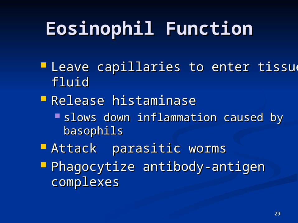
Eosinophil FunctionEosinophil Function
Leave capillaries to enter tissue fluidLeave capillaries to enter tissue fluid Release histaminase Release histaminase
slows down inflammation caused by slows down inflammation caused by basophilsbasophils
Attack parasitic wormsAttack parasitic worms Phagocytize antibody-antigen Phagocytize antibody-antigen
complexescomplexes

30
Lymphocyte FunctionsLymphocyte Functions B cellsB cells
destroy bacteria and their toxinsdestroy bacteria and their toxins turn into plasma cells that produces turn into plasma cells that produces
antibodiesantibodies T cellsT cells
attack viruses, fungi, transplanted organs, attack viruses, fungi, transplanted organs, cancer cells & some bacteriacancer cells & some bacteria
Natural killer cellsNatural killer cells attack many different microbes & some tumor attack many different microbes & some tumor
cellscells destroy foreign invaders by direct attackdestroy foreign invaders by direct attack

31
Differential WBC Count Differential WBC Count Detection of changes in numbers of circulating Detection of changes in numbers of circulating
WBCs (percentages of each type)WBCs (percentages of each type) indicates infection, poisoning, leukemia, indicates infection, poisoning, leukemia,
chemotherapy, parasites or allergy reactionchemotherapy, parasites or allergy reaction Normal WBC countsNormal WBC counts
neutrophils 60-70% (up if bacterial infection)neutrophils 60-70% (up if bacterial infection) lymphocyte 20-25% (up if viral infection)lymphocyte 20-25% (up if viral infection) monocytes 3 -- 8 % (up if fungal/viral infection)monocytes 3 -- 8 % (up if fungal/viral infection) eosinophil 2 -- 4 % (up if parasite or allergy eosinophil 2 -- 4 % (up if parasite or allergy
reaction)reaction) basophil <1% (up if allergy reaction or basophil <1% (up if allergy reaction or
hypothyroid)hypothyroid)

32
Bone Marrow Bone Marrow TransplantTransplant
Intravenous transfer of healthy bone Intravenous transfer of healthy bone marrow marrow
ProcedureProcedure destroy sick bone marrow with radiation & destroy sick bone marrow with radiation &
chemotherapychemotherapy donor matches surface antigens on WBCdonor matches surface antigens on WBC put sample of donor marrow into patient's vein put sample of donor marrow into patient's vein
for reseeding of bone marrowfor reseeding of bone marrow success depends on histocompatibility of donor success depends on histocompatibility of donor
& recipient& recipient Treatment for leukemia, sickle-cell, breast, Treatment for leukemia, sickle-cell, breast,
ovarian or testicular cancer, lymphoma or ovarian or testicular cancer, lymphoma or aplastic anemiaaplastic anemia

33
Platelet (Thrombocyte) Platelet (Thrombocyte) AnatomyAnatomy
Disc-shaped, 2 - 4 micron cell Disc-shaped, 2 - 4 micron cell fragment with no nucleusfragment with no nucleus
Normal platelet count is 150,000-Normal platelet count is 150,000-400,000/drop of blood400,000/drop of blood
Other blood cell countsOther blood cell counts 5 million red & 5-10,000 white blood 5 million red & 5-10,000 white blood
cells cells
34
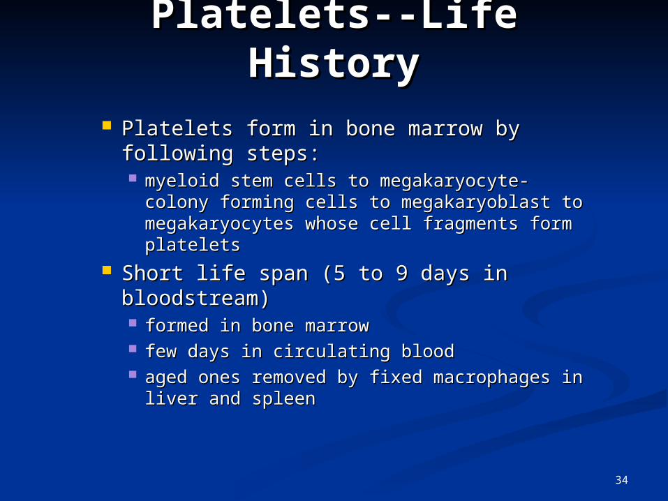
Platelets--Life HistoryPlatelets--Life History
Platelets form in bone marrow by Platelets form in bone marrow by following steps: following steps: myeloid stem cells to megakaryocyte-colony myeloid stem cells to megakaryocyte-colony
forming cells to megakaryoblast to forming cells to megakaryoblast to megakaryocytes whose cell fragments form megakaryocytes whose cell fragments form plateletsplatelets
Short life span (5 to 9 days in Short life span (5 to 9 days in bloodstream)bloodstream) formed in bone marrowformed in bone marrow few days in circulating bloodfew days in circulating blood aged ones removed by fixed macrophages in aged ones removed by fixed macrophages in
liver and spleenliver and spleen

35
Complete Blood CountComplete Blood Count
Screens for anemia and infectionScreens for anemia and infection Total RBC, WBC & platelet counts; Total RBC, WBC & platelet counts;
differential WBC; hematocrit and differential WBC; hematocrit and hemoglobin measurementshemoglobin measurements
Normal hemoglobin rangeNormal hemoglobin range infants have 14 to 20 g/100mL of bloodinfants have 14 to 20 g/100mL of blood adult females have 12 to 16 g/100mL of adult females have 12 to 16 g/100mL of
bloodblood adult males have 13.5 to 18g/100mL of bloodadult males have 13.5 to 18g/100mL of blood
36
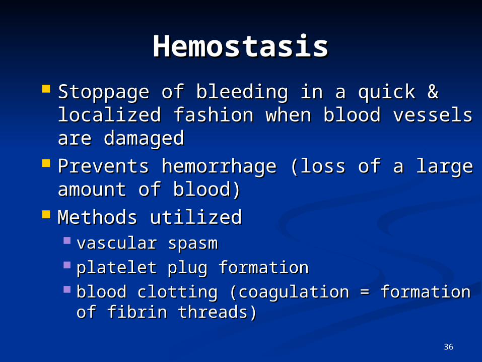
HemostasisHemostasis Stoppage of bleeding in a quick & Stoppage of bleeding in a quick &
localized fashion when blood vessels are localized fashion when blood vessels are damageddamaged
Prevents hemorrhage (loss of a large Prevents hemorrhage (loss of a large amount of blood)amount of blood)
Methods utilizedMethods utilized vascular spasmvascular spasm platelet plug formationplatelet plug formation blood clotting (coagulation = formation of blood clotting (coagulation = formation of
fibrin threads)fibrin threads)
37
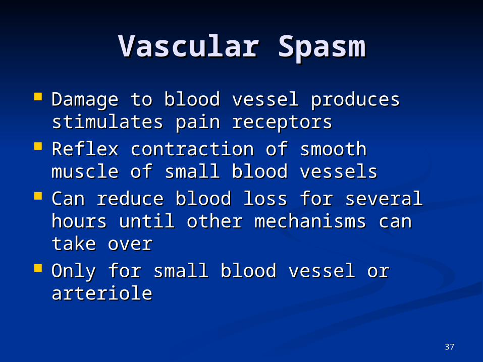
Vascular SpasmVascular Spasm
Damage to blood vessel produces Damage to blood vessel produces stimulates pain receptorsstimulates pain receptors
Reflex contraction of smooth muscle of Reflex contraction of smooth muscle of small blood vesselssmall blood vessels
Can reduce blood loss for several hours Can reduce blood loss for several hours until other mechanisms can take overuntil other mechanisms can take over
Only for small blood vessel or arterioleOnly for small blood vessel or arteriole

38
Platelet Plug FormationPlatelet Plug Formation Platelets store a lot of chemicals in Platelets store a lot of chemicals in
granules needed for platelet plug granules needed for platelet plug formationformation alpha granulesalpha granules
clotting factors clotting factors platelet-derived growth factorplatelet-derived growth factor
cause proliferation of vascular endothelial cells, cause proliferation of vascular endothelial cells, smooth muscle & fibroblasts to repair damaged smooth muscle & fibroblasts to repair damaged vesselsvessels
dense granulesdense granules ADP, ATP, Ca+2, serotonin, fibrin-stabilizing ADP, ATP, Ca+2, serotonin, fibrin-stabilizing
factor, & enzymes that produce thromboxane A2factor, & enzymes that produce thromboxane A2 Steps in the processSteps in the process
(1) platelet adhesion (2) platelet release (1) platelet adhesion (2) platelet release reaction (3) platelet aggregationreaction (3) platelet aggregation

39
Platelet AdhesionPlatelet Adhesion
Platelets stick to exposed collagen underlying Platelets stick to exposed collagen underlying damaged endothelial cells in vessel wall damaged endothelial cells in vessel wall

40
Platelet Release Platelet Release ReactionReaction
Platelets activated by adhesionPlatelets activated by adhesion Extend projections to make contact with each other Extend projections to make contact with each other Release thromboxane A2 & ADP activating other plateletsRelease thromboxane A2 & ADP activating other platelets Serotonin & thromboxane A2 are vasoconstrictors Serotonin & thromboxane A2 are vasoconstrictors
decreasing blood flow through the injured vesseldecreasing blood flow through the injured vessel

41
Platelet AggregationPlatelet Aggregation Activated platelets stick together and activate new Activated platelets stick together and activate new
platelets to form a mass called a platelet plugplatelets to form a mass called a platelet plug Plug reinforced by fibrin threads formed during Plug reinforced by fibrin threads formed during
clotting processclotting process
42
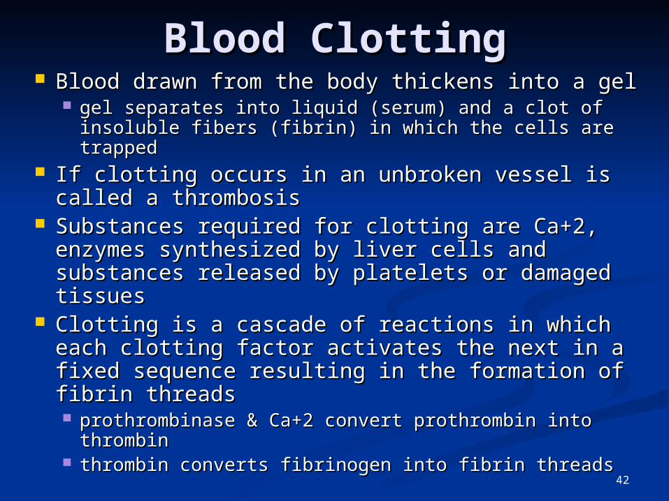
Blood ClottingBlood Clotting Blood drawn from the body thickens into a gelBlood drawn from the body thickens into a gel
gel separates into liquid (serum) and a clot of gel separates into liquid (serum) and a clot of insoluble fibers (fibrin) in which the cells are trappedinsoluble fibers (fibrin) in which the cells are trapped
If clotting occurs in an unbroken vessel is called If clotting occurs in an unbroken vessel is called a thrombosisa thrombosis
Substances required for clotting are Ca+2, Substances required for clotting are Ca+2, enzymes synthesized by liver cells and enzymes synthesized by liver cells and substances released by platelets or damaged substances released by platelets or damaged tissuestissues
Clotting is a cascade of reactions in which each Clotting is a cascade of reactions in which each clotting factor activates the next in a fixed clotting factor activates the next in a fixed sequence resulting in the formation of fibrin sequence resulting in the formation of fibrin threadsthreads prothrombinase & Ca+2 convert prothrombin into prothrombinase & Ca+2 convert prothrombin into
thrombinthrombin thrombin converts fibrinogen into fibrin threadsthrombin converts fibrinogen into fibrin threads
43
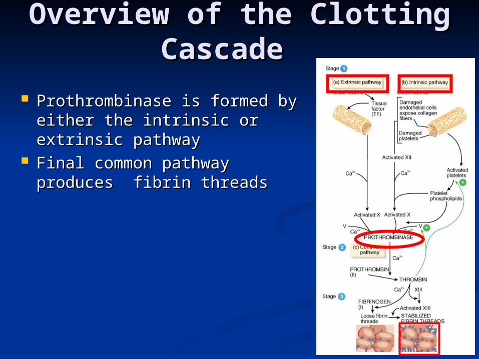
Overview of the Clotting Overview of the Clotting Cascade Cascade
Prothrombinase is formed by Prothrombinase is formed by either the intrinsic or extrinsic either the intrinsic or extrinsic pathwaypathway
Final common pathway Final common pathway produces fibrin threadsproduces fibrin threads

44
Extrinsic PathwayExtrinsic Pathway
Damaged tissues leak Damaged tissues leak tissue factor tissue factor (thromboplastin) into (thromboplastin) into bloodstreambloodstream
Prothrombinase forms in Prothrombinase forms in secondsseconds
In the presence of Ca+2, In the presence of Ca+2, clotting factor X combines clotting factor X combines with V to form with V to form prothrombinaseprothrombinase
45
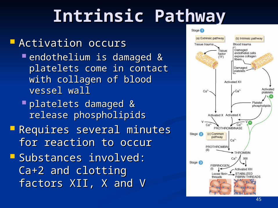
Intrinsic PathwayIntrinsic Pathway Activation occursActivation occurs
endothelium is damaged & endothelium is damaged & platelets come in contact platelets come in contact with collagen of blood with collagen of blood vessel wallvessel wall
platelets damaged & platelets damaged & release phospholipidsrelease phospholipids
Requires several minutes Requires several minutes for reaction to occurfor reaction to occur
Substances involved: Substances involved: Ca+2 and clotting factors Ca+2 and clotting factors XII, X and VXII, X and V
46
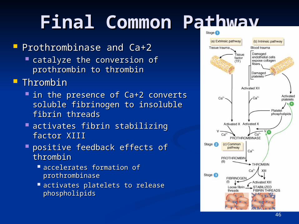
Final Common PathwayFinal Common Pathway Prothrombinase and Ca+2 Prothrombinase and Ca+2
catalyze the conversion of catalyze the conversion of prothrombin to thrombinprothrombin to thrombin
ThrombinThrombin in the presence of Ca+2 converts in the presence of Ca+2 converts
soluble fibrinogen to insoluble fibrin soluble fibrinogen to insoluble fibrin threadsthreads
activates fibrin stabilizing factor XIII activates fibrin stabilizing factor XIII positive feedback effects of thrombinpositive feedback effects of thrombin
accelerates formation of prothrombinaseaccelerates formation of prothrombinase activates platelets to release activates platelets to release
phospholipidsphospholipids
47
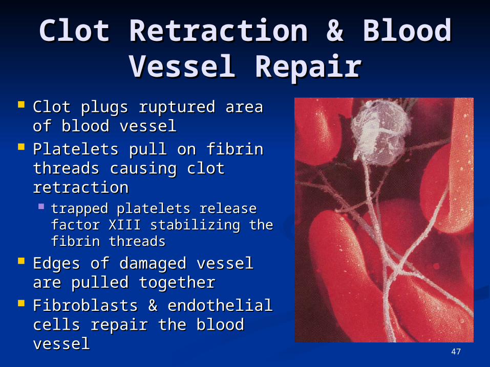
Clot Retraction & Blood Clot Retraction & Blood Vessel RepairVessel Repair
Clot plugs ruptured area of Clot plugs ruptured area of blood vesselblood vessel
Platelets pull on fibrin Platelets pull on fibrin threads causing clot threads causing clot retraction retraction trapped platelets release trapped platelets release
factor XIII stabilizing the factor XIII stabilizing the fibrin threadsfibrin threads
Edges of damaged vessel Edges of damaged vessel are pulled togetherare pulled together
Fibroblasts & endothelial Fibroblasts & endothelial cells repair the blood vesselcells repair the blood vessel

48
Role of Vitamin K in Role of Vitamin K in ClottingClotting
Normal clotting requires adequate Normal clotting requires adequate vitamin Kvitamin K fat soluble vitamin absorbed if lipids are fat soluble vitamin absorbed if lipids are
presentpresent absorption slowed if bile release is absorption slowed if bile release is
insufficientinsufficient Required for synthesis of 4 clotting Required for synthesis of 4 clotting
factors by hepatocytesfactors by hepatocytes factors II (prothrombin), VII, IX and Xfactors II (prothrombin), VII, IX and X
Produced by bacteria in large Produced by bacteria in large intestineintestine

49
Hemostatic Control Hemostatic Control MechanismsMechanisms Fibrinolytic system dissolves small, inappropriate Fibrinolytic system dissolves small, inappropriate
clots & clots at a site of a completed repairclots & clots at a site of a completed repair fibrinolysis is dissolution of a clotfibrinolysis is dissolution of a clot
Inactive plasminogen is incorporated into the clotInactive plasminogen is incorporated into the clot activation occurs because of factor XII and thrombinactivation occurs because of factor XII and thrombin plasminogen becomes plasmin (fibrinolysin) which plasminogen becomes plasmin (fibrinolysin) which
digests fibrin threadsdigests fibrin threads Clot formation remains localizedClot formation remains localized
fibrin absorbs thrombinfibrin absorbs thrombin blood disperses clotting factorsblood disperses clotting factors endothelial cells & WBC produce prostacyclin that endothelial cells & WBC produce prostacyclin that
opposes thromboxane A2 (platelet adhesion & release)opposes thromboxane A2 (platelet adhesion & release) Anticoagulants present in blood & produced by Anticoagulants present in blood & produced by
mast cellsmast cells
50
Intravascular ClottingIntravascular Clotting ThrombosisThrombosis
clot (thrombus) forming in an unbroken blood clot (thrombus) forming in an unbroken blood vesselvessel forms on rough inner lining of BVforms on rough inner lining of BV if blood flows too slowly (stasis) allowing clotting if blood flows too slowly (stasis) allowing clotting
factors to build up locally & cause coagulationfactors to build up locally & cause coagulation may dissolve spontaneously or dislodge & travelmay dissolve spontaneously or dislodge & travel
Embolus Embolus clot, air bubble or fat from broken bone in the clot, air bubble or fat from broken bone in the
bloodblood pulmonary embolus is found in lungspulmonary embolus is found in lungs
Low dose aspirin blocks synthesis of Low dose aspirin blocks synthesis of thromboxane A2 & reduces inappropriate thromboxane A2 & reduces inappropriate clot formationclot formation strokes, TIAs and myocardial infarctionsstrokes, TIAs and myocardial infarctions

51
Anticoagulants and Anticoagulants and Thrombolytic AgentsThrombolytic Agents
Anticoagulants suppress or prevent blood clottingAnticoagulants suppress or prevent blood clotting heparinheparin
administered during hemodialysis and surgeryadministered during hemodialysis and surgery warfarin (Coumadin)warfarin (Coumadin)
antagonist to vitamin K so blocks synthesis of clotting factorsantagonist to vitamin K so blocks synthesis of clotting factors slower than heparinslower than heparin
stored blood in blood banks treated with citrate stored blood in blood banks treated with citrate phosphate dextrose (CPD) that removes Ca+2phosphate dextrose (CPD) that removes Ca+2
Thrombolytic agents are injected to dissolve clotsThrombolytic agents are injected to dissolve clots directly or indirectly activate plasminogendirectly or indirectly activate plasminogen streptokinase or tissue plasminogen activator (t-PA)streptokinase or tissue plasminogen activator (t-PA)
52
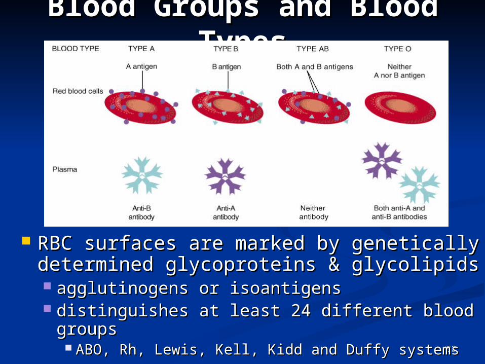
Blood Groups and Blood Groups and Blood TypesBlood Types
RBC surfaces are marked by genetically RBC surfaces are marked by genetically determined glycoproteins & glycolipids determined glycoproteins & glycolipids agglutinogens or isoantigensagglutinogens or isoantigens distinguishes at least 24 different blood distinguishes at least 24 different blood
groups groups ABO, Rh, Lewis, Kell, Kidd and Duffy systemsABO, Rh, Lewis, Kell, Kidd and Duffy systems

53
ABO Blood GroupsABO Blood Groups Based on 2 glycolipid isoantigens called A and B Based on 2 glycolipid isoantigens called A and B
found on the surface of RBCsfound on the surface of RBCs display only antigen A -- blood type Adisplay only antigen A -- blood type A display only antigen B -- blood type Bdisplay only antigen B -- blood type B display both antigens A & B -- blood type ABdisplay both antigens A & B -- blood type AB display neither antigen -- blood type Odisplay neither antigen -- blood type O
Plasma contains isoantibodies or agglutinins to Plasma contains isoantibodies or agglutinins to the A or B antigens not found in your bloodthe A or B antigens not found in your blood anti-A antibody reacts with antigen Aanti-A antibody reacts with antigen A anti-B antibody reacts with antigen Banti-B antibody reacts with antigen B

54
RH blood groupsRH blood groups Antigen was discovered in blood of Antigen was discovered in blood of RhesusRhesus
monkeymonkey People with Rh agglutinogens on RBC surface People with Rh agglutinogens on RBC surface
are Rh+. Normal plasma contains no anti-Rh are Rh+. Normal plasma contains no anti-Rh antibodiesantibodies
Antibodies develop only in Rh- blood type & only Antibodies develop only in Rh- blood type & only with exposure to the antigenwith exposure to the antigen transfusion of positive bloodtransfusion of positive blood during a pregnancy with a positive blood type fetusduring a pregnancy with a positive blood type fetus
Transfusion reaction upon 2nd exposure to the Transfusion reaction upon 2nd exposure to the antigen results in hemolysis of the RBCs in the antigen results in hemolysis of the RBCs in the donated blooddonated blood

55
Hemolytic Disease of Hemolytic Disease of NewbornNewborn
Rh negative mom and Rh+ fetus will have mixing of blood at Rh negative mom and Rh+ fetus will have mixing of blood at birthbirth
Mom's body creates Rh antibodies unless she receives a Mom's body creates Rh antibodies unless she receives a RhoGam shot soon after first delivery, miscarriage or RhoGam shot soon after first delivery, miscarriage or abortionabortion RhoGam binds to loose fetal blood and removes it from body before RhoGam binds to loose fetal blood and removes it from body before
she reactsshe reacts In 2nd child, hemolytic disease of the newborn may develop In 2nd child, hemolytic disease of the newborn may develop
causing hemolysis of the fetal RBCscausing hemolysis of the fetal RBCs

56
Transfusion and Transfusion and Transfusion ReactionsTransfusion Reactions
Transfer of whole blood, cells or plasma into Transfer of whole blood, cells or plasma into the bloodstream of recipientthe bloodstream of recipient used to treat anemia or severe blood lossused to treat anemia or severe blood loss
IIncompatible blood transfusionsncompatible blood transfusions antigen-antibody complexes form between plasma antigen-antibody complexes form between plasma
antibodies & “foreign proteins” on donated RBC's antibodies & “foreign proteins” on donated RBC's (agglutination)(agglutination)
donated RBCs become leaky (complement proteins) & donated RBCs become leaky (complement proteins) & burstburst
loose hemoglobin causes kidney damageloose hemoglobin causes kidney damage Problems caused by incompatibility between Problems caused by incompatibility between
donor’s cells and recipient’s plasmadonor’s cells and recipient’s plasma Donor plasma is too diluted to cause problemsDonor plasma is too diluted to cause problems

57
Universal Donors and Universal Donors and RecipientsRecipients
People with type AB blood called People with type AB blood called “universal recipients” since have “universal recipients” since have no antibodies in plasmano antibodies in plasma only true if cross match the blood only true if cross match the blood
for other antigensfor other antigens People with type O blood cell People with type O blood cell
called “universal donors” since called “universal donors” since have no antigens on their cellshave no antigens on their cells theoretically can be given to anyone theoretically can be given to anyone

58
Typing and Cross-Matching Typing and Cross-Matching BloodBlood
Mixing of incompatible blood causes Mixing of incompatible blood causes agglutination (visible clumping)agglutination (visible clumping) formation of antigen-antibody complex that formation of antigen-antibody complex that
sticks cells togethersticks cells together not the same as blood clottingnot the same as blood clotting
Typing involves testing blood with known Typing involves testing blood with known antisera that contain antibodies A, B or Rh+antisera that contain antibodies A, B or Rh+
Cross-matching is to test by mixing donor Cross-matching is to test by mixing donor cells with recipient’s serumcells with recipient’s serum
Screening is to test recipient’s serum Screening is to test recipient’s serum against known RBC’s having known against known RBC’s having known antigensantigens

59
Anemia = Not Enough Anemia = Not Enough RBCsRBCs
SymptomsSymptoms oxygen-carrying capacity of blood is reducedoxygen-carrying capacity of blood is reduced fatigue, cold intolerance & palenessfatigue, cold intolerance & paleness
lack of O2 for ATP & heat productionlack of O2 for ATP & heat production
Types of anemiaTypes of anemia iron-deficiency =lack of absorption or loss of ironiron-deficiency =lack of absorption or loss of iron pernicious = lack of intrinsic factor for B12 absorptionpernicious = lack of intrinsic factor for B12 absorption hemorrhagic = loss of RBCs due to bleeding (ulcer)hemorrhagic = loss of RBCs due to bleeding (ulcer) hemolytic = defects in cell membranes cause rupturehemolytic = defects in cell membranes cause rupture thalassemia = hereditary deficiency of hemoglobinthalassemia = hereditary deficiency of hemoglobin aplastic = destruction of bone marrow aplastic = destruction of bone marrow
(radiation/toxins)(radiation/toxins)
60
Sickle-cell Anemia (SCA)Sickle-cell Anemia (SCA) Genetic defect in hemoglobin molecule (Hb-Genetic defect in hemoglobin molecule (Hb-
S) that changes 2 amino acids S) that changes 2 amino acids at low very O2 levels, RBC is deformed by at low very O2 levels, RBC is deformed by
changes in hemoglobin molecule within the RBCchanges in hemoglobin molecule within the RBC sickle-shaped cells rupture easily = causing anemia & sickle-shaped cells rupture easily = causing anemia &
clotsclots
Found among populations in malaria beltFound among populations in malaria belt Mediterranean Europe, sub-Saharan Africa & Mediterranean Europe, sub-Saharan Africa &
AsiaAsia Person with only one sickle cell genePerson with only one sickle cell gene
increased resistance to malaria because RBC increased resistance to malaria because RBC membranes leak K+ & lowered levels of K+ kill membranes leak K+ & lowered levels of K+ kill the parasite infecting the red blood cellsthe parasite infecting the red blood cells

61
HemophiliaHemophilia Inherited deficiency of clotting factors Inherited deficiency of clotting factors
bleeding spontaneously or after minor traumableeding spontaneously or after minor trauma subcutaneous & intramuscular hemorrhagingsubcutaneous & intramuscular hemorrhaging nosebleeds, blood in urine, articular bleeding & nosebleeds, blood in urine, articular bleeding &
painpain Hemophilia A lacks factor VIII (males only)Hemophilia A lacks factor VIII (males only)
most commonmost common Hemophilia B lacks factor IX (males only)Hemophilia B lacks factor IX (males only) Hemophilia C (males & females)Hemophilia C (males & females)
less severe because alternate clotting activator less severe because alternate clotting activator existsexists
Treatment is transfusions of fresh plasma or Treatment is transfusions of fresh plasma or concentrates of the missing clotting factorconcentrates of the missing clotting factor

62
Disseminated Intravascular Disseminated Intravascular ClottingClotting
Life threatening paradoxical presence of Life threatening paradoxical presence of blood clotting and bleeding at the same blood clotting and bleeding at the same time throughout the whole bodytime throughout the whole body so many clotting factors are removed by so many clotting factors are removed by
widespread clotting that too few remain to widespread clotting that too few remain to permit normal clottingpermit normal clotting
Associated with infections, hypoxia, low Associated with infections, hypoxia, low blood flow rates, trauma, hypotension & blood flow rates, trauma, hypotension & hemolysishemolysis
Clots cause ischemia and necrosis Clots cause ischemia and necrosis leading to multisystem organ failureleading to multisystem organ failure

63
LeukemiaLeukemia Acute leukemiaAcute leukemia
uncontrolled production of immature uncontrolled production of immature leukocytesleukocytes
crowding out of normal red bone marrow crowding out of normal red bone marrow cells by production of immature WBCcells by production of immature WBC
prevents production of RBC & plateletsprevents production of RBC & platelets Chronic leukemiaChronic leukemia
accumulation of mature WBC in accumulation of mature WBC in bloodstream because they do not diebloodstream because they do not die
classified by type of WBC that is classified by type of WBC that is predominant---monocytic, lymphocytic.predominant---monocytic, lymphocytic.