Embed Size (px)
Citation preview
1
Chapter 2a CNS Gross Anatomy
Chris RordenUniversity of South CarolinaNorman J. Arnold School of Public HealthDepartment of Communication Sciences and DisordersUniversity of South Carolina
2
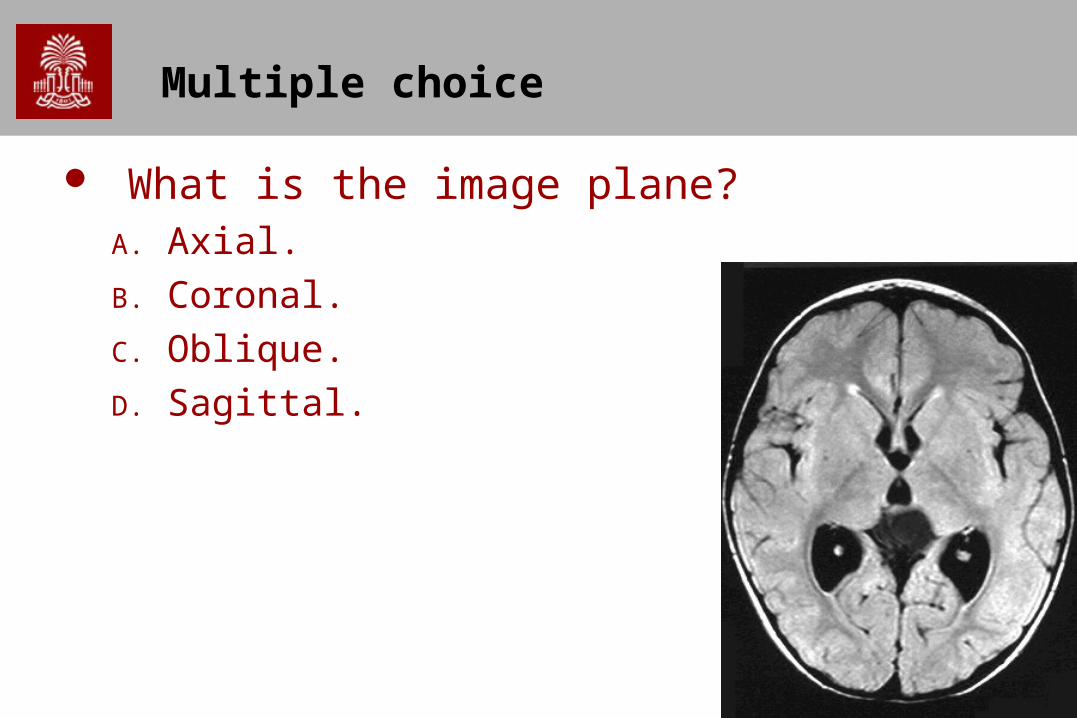
Multiple choice
What is the image plane?A. Axial.
B. Coronal.
C. Oblique.
D. Sagittal.
3
Multiple choice
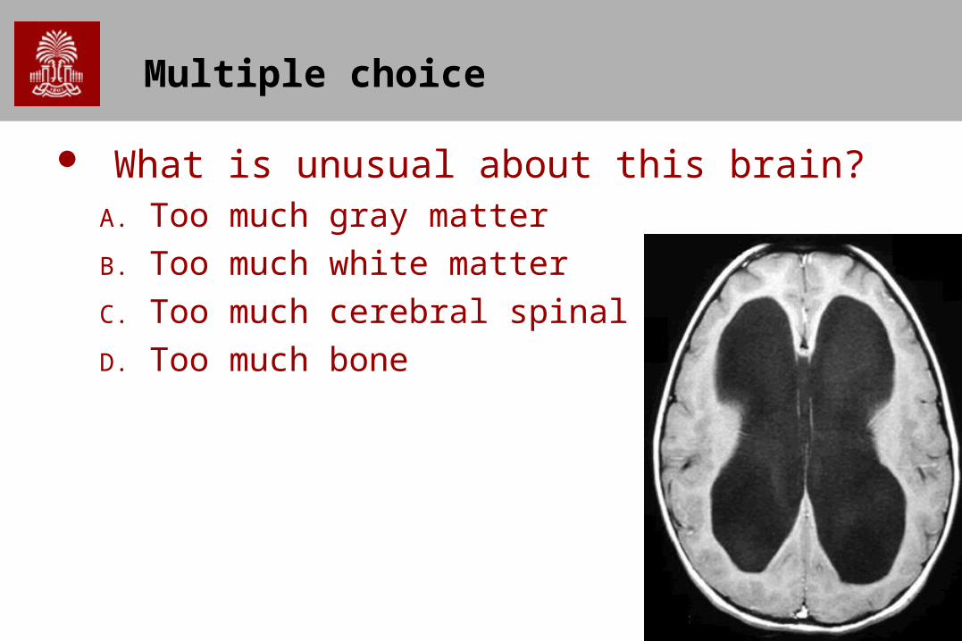
What is unusual about this brain?A. Too much gray matter
B. Too much white matter
C. Too much cerebral spinal fluid
D. Too much bone
4
Roles of the CNS
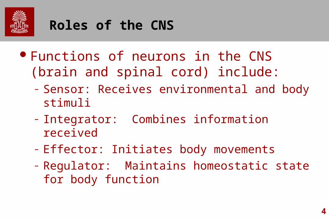
Functions of neurons in the CNS (brain and spinal cord) include:– Sensor: Receives environmental and body stimuli– Integrator: Combines information received– Effector: Initiates body movements– Regulator: Maintains homeostatic state for body
function
5
Nervous System
The CNS is protected and isolated.– Bone offers protection from injury
Skull covers brainVertebral Column covers spinal cord
– The is encased in soft-tissue membranes – The brain’s blood vessels stop many subastances
from entering the brain (blood-brain barrier)Protects from contamination/infection
– The brain floats in cerebral spinal fluidOffers protection from impact
6
The Meninges
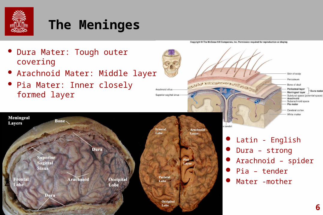
Dura Mater: Tough outer covering Arachnoid Mater: Middle layer Pia Mater: Inner closely formed
layer
Latin - English Dura – strong Arachnoid – spider Pia – tender Mater -mother
7
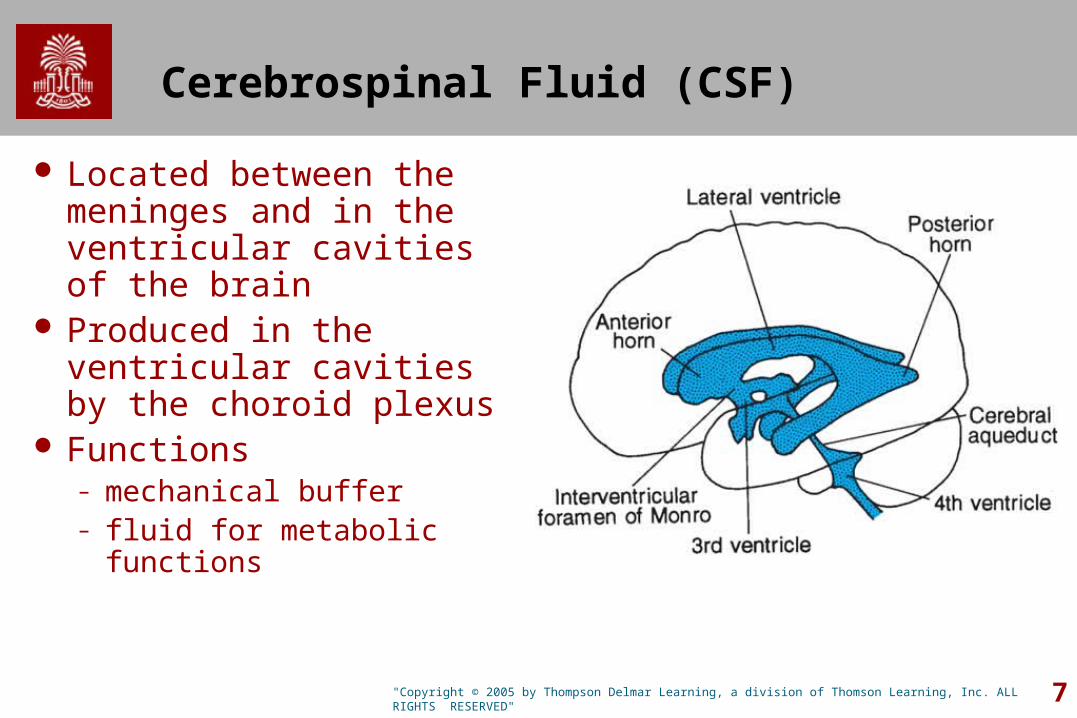
Cerebrospinal Fluid (CSF)
Located between the meninges and in the ventricular cavities of the brain
Produced in the ventricular cavities by the choroid plexus
Functions– mechanical buffer – fluid for metabolic functions
"Copyright © 2005 by Thompson Delmar Learning, a division of Thomson Learning, Inc. ALL RIGHTS RESERVED"
8
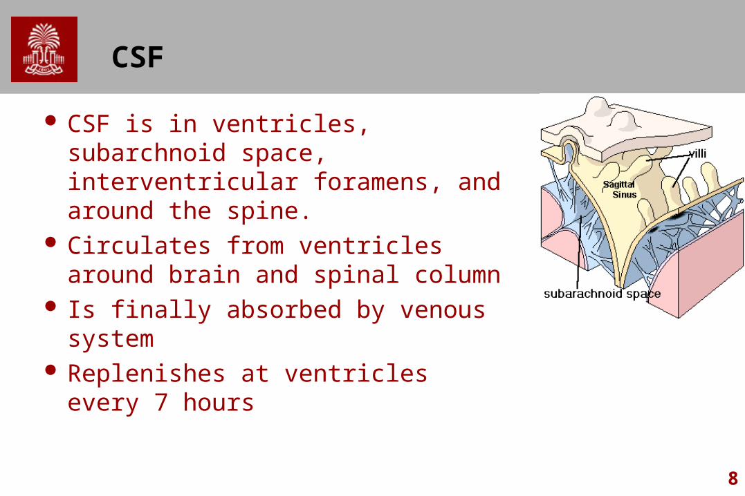
CSF
CSF is in ventricles, subarchnoid space, interventricular foramens, and around the spine.
Circulates from ventricles around brain and spinal column
Is finally absorbed by venous system Replenishes at ventricles every 7
hours
9
Divisions of the PNS
Somatic Nervous System (under voluntary control)– Sensory and Motor – Skin and Muscles
Autonomic Nervous System (can not be voluntarily controlled).– Sensory and Motor– Visceral organs and glands– Two main subdivisions:– Sympathetic: Fight, Flight, Fear
Prepare to expend energy– Parasympathetic: Regulates normal function
Prepare to conserve energy
10
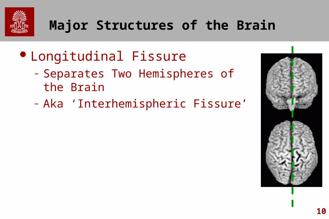
Major Structures of the Brain
Longitudinal Fissure– Separates Two Hemispheres of the Brain– Aka ‘Interhemispheric Fissure’
11
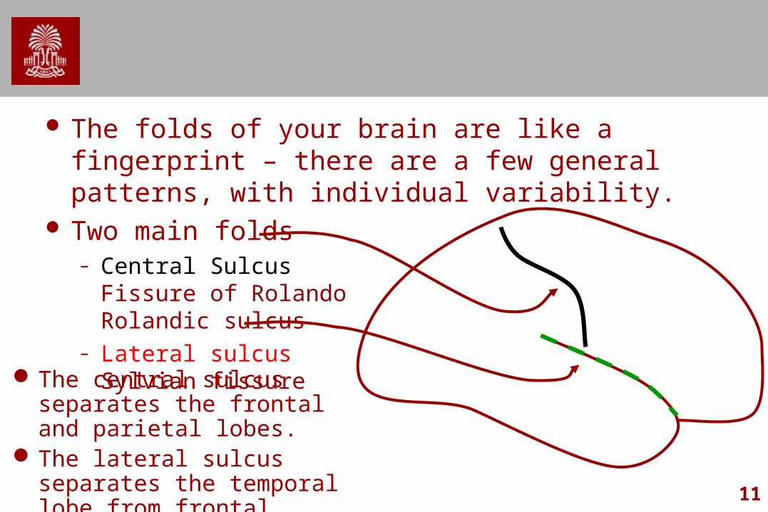
The folds of your brain are like a fingerprint – there are a few general patterns, with individual variability.
Two main folds– Central Sulcus
Fissure of RolandoRolandic sulcus
– Lateral sulcusSylvian fissure
The central sulcus separates the frontal and parietal lobes.
The lateral sulcus separates the temporal lobe from frontal, parietal, insula
12
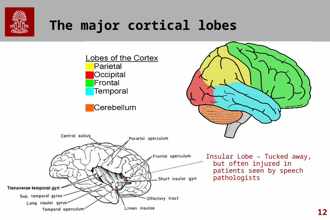
The major cortical lobes
Insular Lobe – Tucked away, but often injured in patients seen by speech pathologists
13
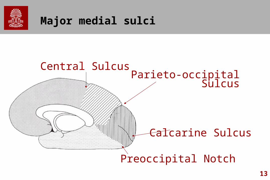
Major medial sulci
Central Sulcus
Preoccipital Notch
Calcarine Sulcus
Parieto-occipital Sulcus
14
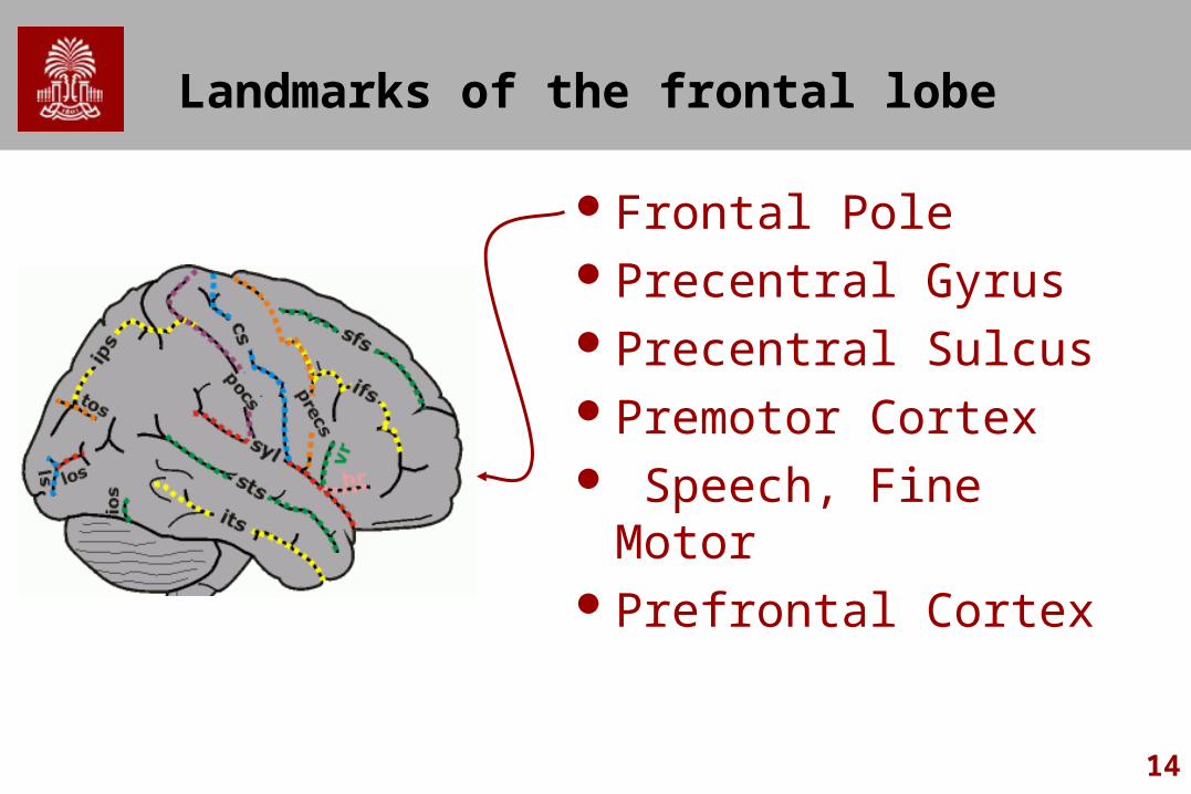
Landmarks of the frontal lobe
Frontal PolePrecentral GyrusPrecentral SulcusPremotor Cortex Speech, Fine MotorPrefrontal Cortex
15
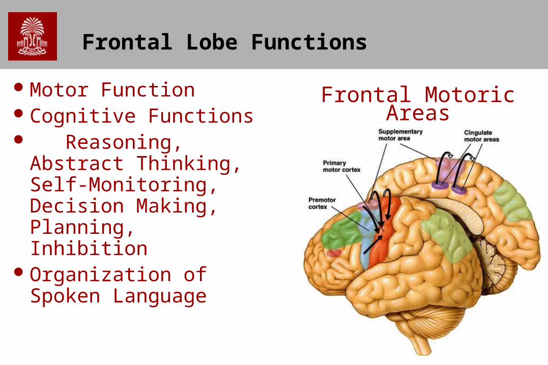
Frontal Lobe Functions
Motor FunctionCognitive Functions Reasoning, Abstract
Thinking, Self-Monitoring, Decision Making, Planning, Inhibition
Organization of Spoken Language
Frontal Motoric Areas
16
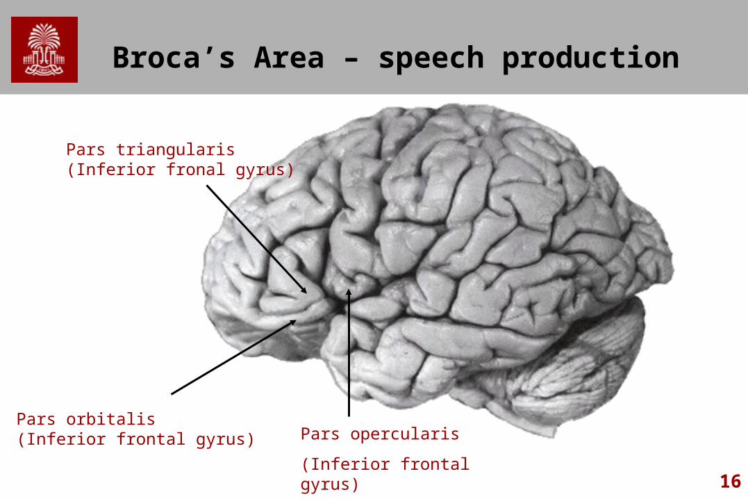
Broca’s Area – speech production
Pars opercularis
(Inferior frontal gyrus)
Pars triangularis(Inferior fronal gyrus)
Pars orbitalis(Inferior frontal gyrus)
17
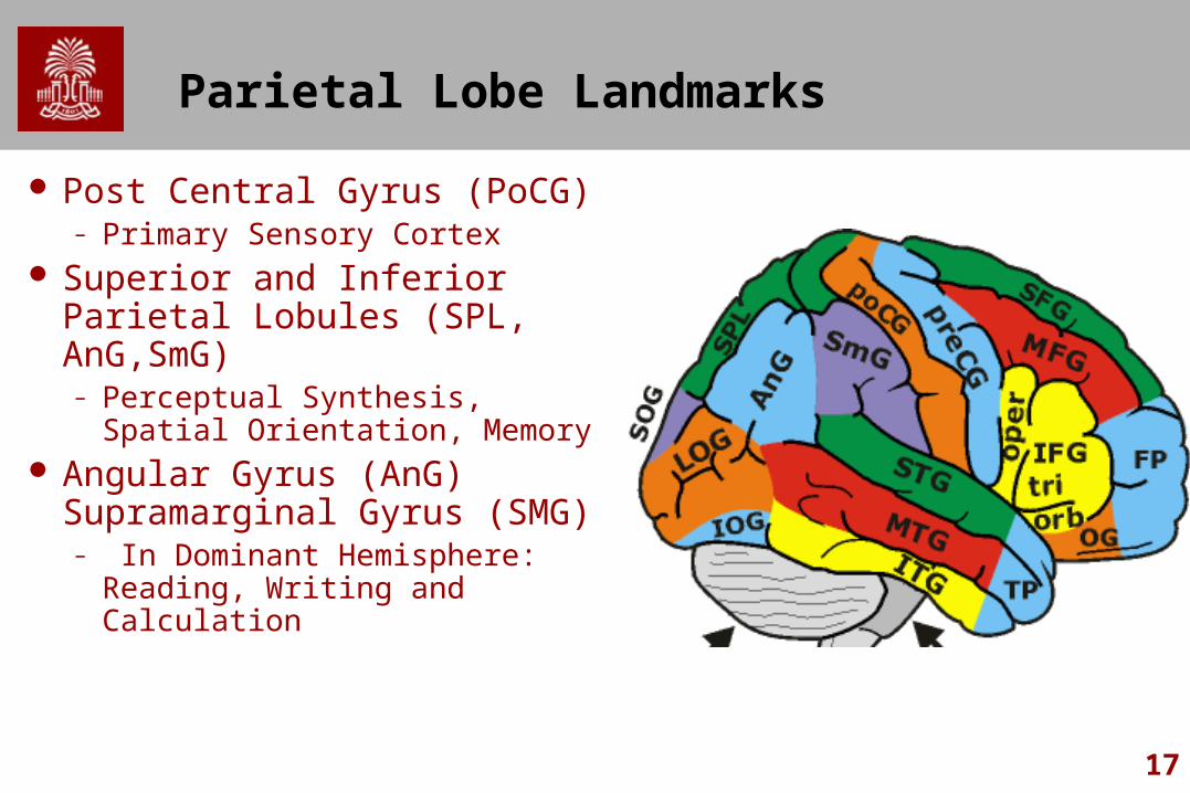
Parietal Lobe Landmarks
Post Central Gyrus (PoCG)– Primary Sensory Cortex
Superior and Inferior Parietal Lobules (SPL, AnG,SmG)– Perceptual Synthesis, Spatial
Orientation, Memory Angular Gyrus (AnG)
Supramarginal Gyrus (SMG)– In Dominant Hemisphere:
Reading, Writing and Calculation
18
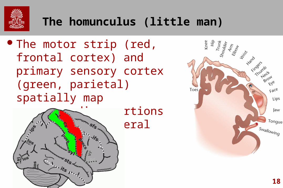
The homunculus (little man)
The motor strip (red, frontal cortex) and primary sensory cortex (green, parietal) spatially map corresponding portions of the contralateral hemisphere.
19
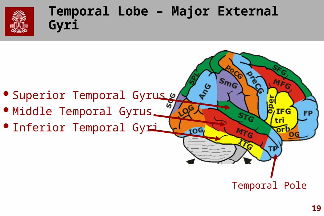
Temporal Lobe – Major External Gyri
Superior Temporal GyrusMiddle Temporal Gyrus Inferior Temporal Gyri
Temporal Pole
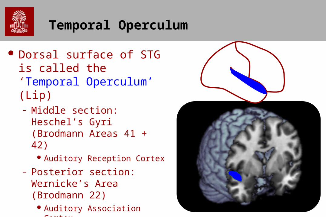
Temporal Operculum
Dorsal surface of STG is called the ‘Temporal Operculum’ (Lip) – Middle section: Heschel’s Gyri
(Brodmann Areas 41 + 42) Auditory Reception Cortex
– Posterior section: Wernicke’s Area (Brodmann 22)
Auditory Association Cortex
21
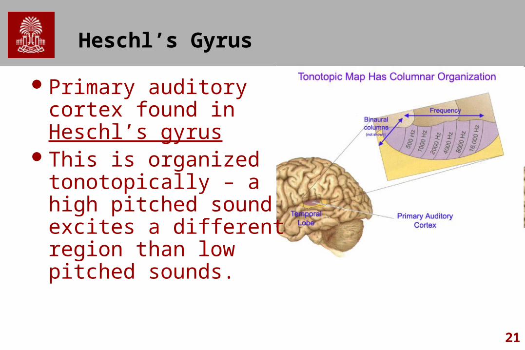
Heschl’s Gyrus
Primary auditory cortex found in Heschl’s gyrus
This is organized tonotopically – a high pitched sound excites a different region than low pitched sounds.
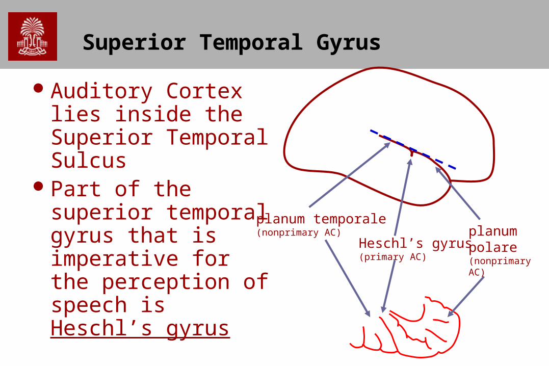
Superior Temporal Gyrus
Auditory Cortex lies inside the Superior Temporal Sulcus
Part of the superior temporal gyrus that is imperative for the perception of speech is Heschl’s gyrus
Heschl’s gyrus (primary AC)
planum temporale (nonprimary AC) planum
polare (nonprimary AC)
23
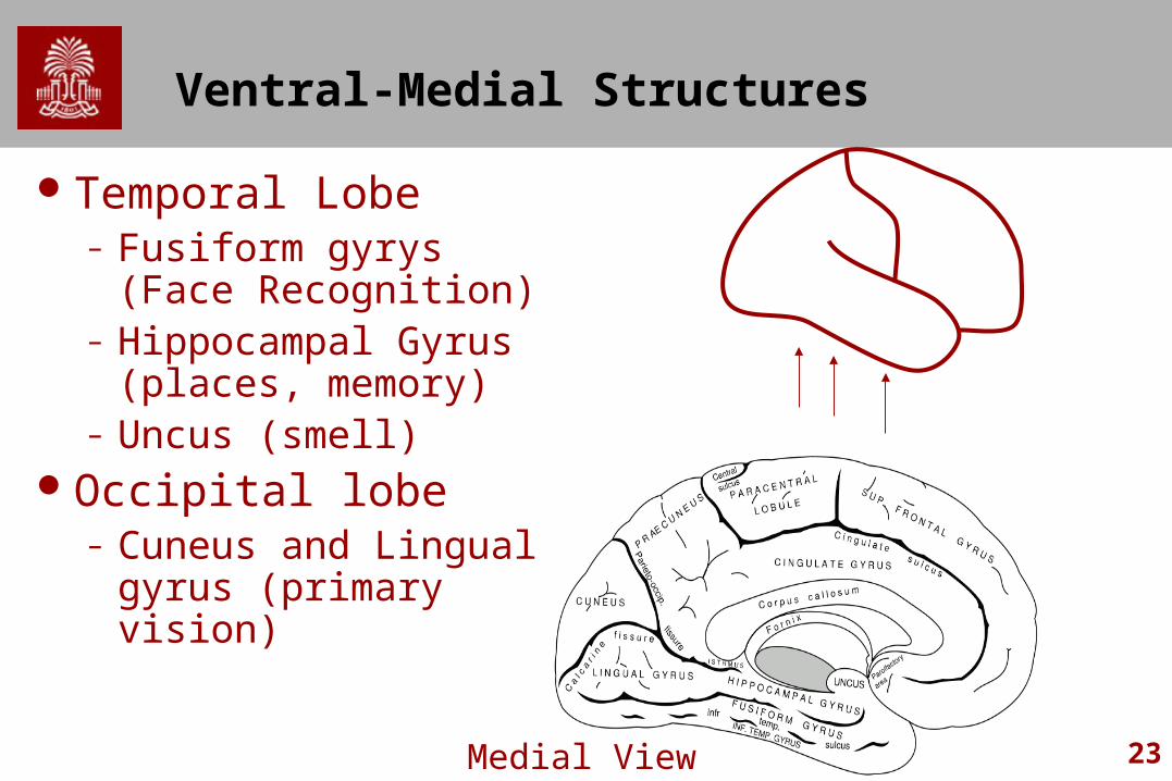
Ventral-Medial Structures
Temporal Lobe– Fusiform gyrys (Face
Recognition)– Hippocampal Gyrus
(places, memory)– Uncus (smell)
Occipital lobe– Cuneus and Lingual
gyrus (primary vision)
Medial View
24
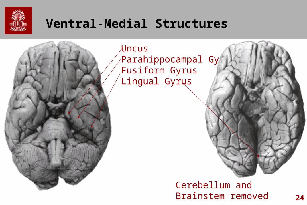
Ventral-Medial Structures
UncusParahippocampal GyrusFusiform GyrusLingual Gyrus
Cerebellum and Brainstem removed
25
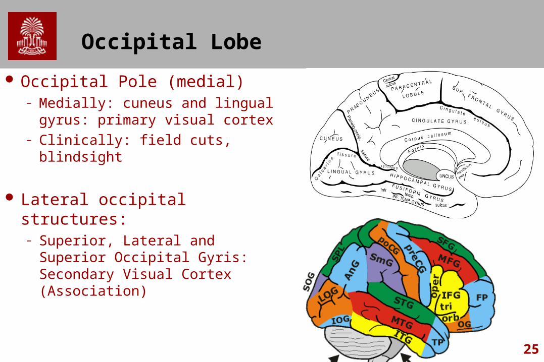
Occipital Lobe
Occipital Pole (medial)– Medially: cuneus and lingual
gyrus: primary visual cortex– Clinically: field cuts, blindsight
Lateral occipital structures:– Superior, Lateral and Superior
Occipital Gyris: Secondary Visual Cortex (Association)

26
Language Areas
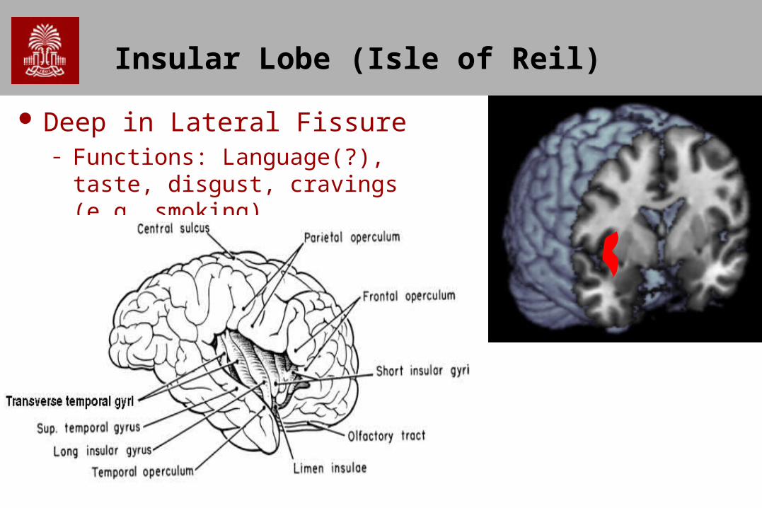
Insular Lobe (Isle of Reil)
Deep in Lateral Fissure– Functions: Language(?), taste, disgust,
cravings (e.g. smoking)
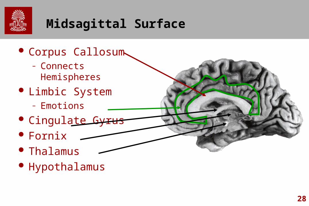
28
Midsagittal Surface
Corpus Callosum – Connects Hemispheres
Limbic System– Emotions
Cingulate Gyrus Fornix Thalamus Hypothalamus
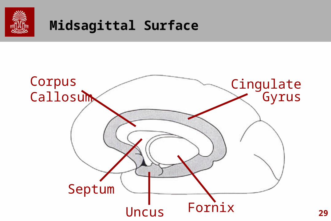
29
Midsagittal Surface
Uncus Fornix
Septum
Corpus Callosum
CingulateGyrus
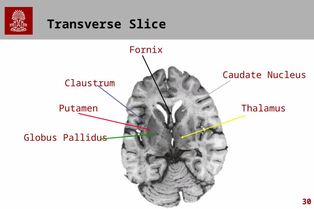
30
Transverse Slice
Fornix
ThalamusPutamen
Caudate Nucleus
Globus Pallidus
Claustrum
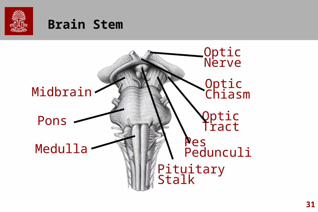
31
Brain Stem
Midbrain
Pons
Medulla
OpticNerve
OpticChiasm
OpticTract
PituitaryStalk
Pes Pedunculi
32
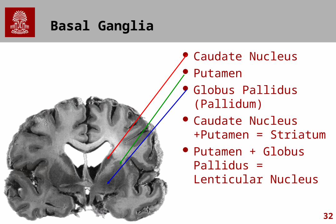
Basal Ganglia
Caudate Nucleus Putamen Globus Pallidus (Pallidum) Caudate Nucleus
+Putamen = Striatum Putamen + Globus
Pallidus = Lenticular Nucleus
33
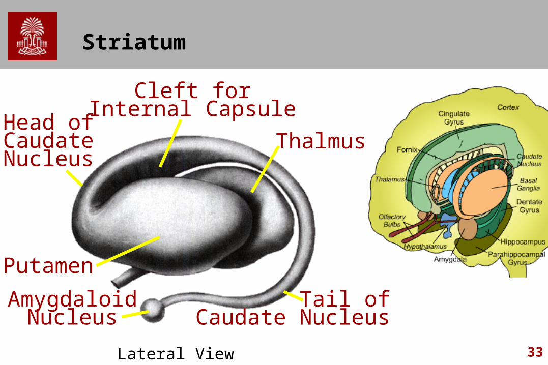
Striatum
Cleft forInternal Capsule
Head ofCaudateNucleus
Thalmus
Putamen
AmygdaloidNucleus
Tail ofCaudate Nucleus
Lateral View