Embed Size (px)
Citation preview
1
Chapter 6
Bones and Skeletal Tissue
Lecture 13
Figure: © 1998 A.D.A.M. Software, Inc.
Marieb’s HumanAnatomy and
Physiology
Marieb Hoehn
2
Lecture Overview
• Introduction to the skeletal system
• Function and classification of bones
• Structure of bones
• Bone development and growth
• Bone homeostasis
• Factors affecting bone growth, development and repair
• Functions of bone
3
Overview of the Skeletal System
• The human skeleton contains a total of 206 bones– 80 Axial– 126 Appendicular
• Bone is also called osseous tissue and is a hard (supporting) form of connective tissue composed of two (2) types of bone– Compact bone (osteons are the main units)– Spongy (cancellous) bone
4
Skeletal System Functions
• Support and Protection• gives shape to head, etc.• supports body’s weight• protects thoracic organs and brain
• Body Movement• interacts with muscles• bones act as rigid bar of a lever
• Blood Cell Formation• hematopoiesis• red marrow
• Inorganic Salt Storage• calcium • phosphate• magnesium• sodium• potassium• electrolyte and acid/base balance
7
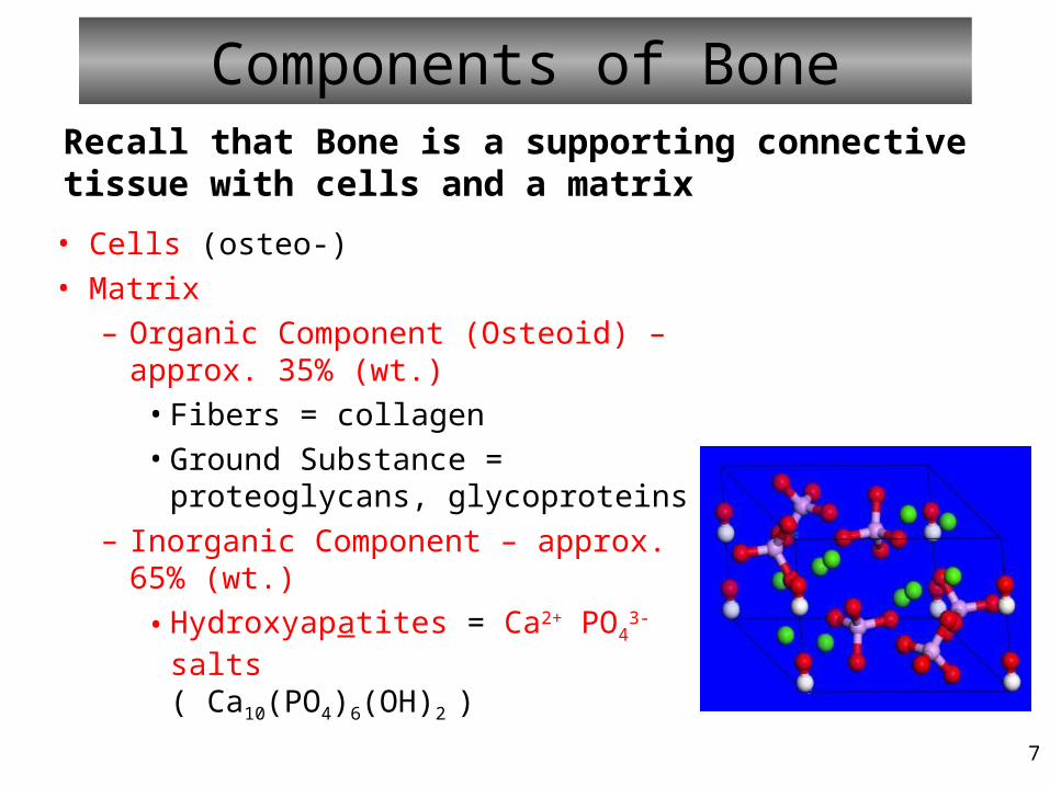
Components of Bone
• Cells (osteo-)
• Matrix
– Organic Component (Osteoid) – approx. 35% (wt.)
• Fibers = collagen
• Ground Substance = proteoglycans, glycoproteins
– Inorganic Component – approx. 65% (wt.)
• Hydroxyapatites = Ca2+ PO43- salts
( Ca10(PO4)6(OH)2 )
Recall that Bone is a supporting connective tissue with cells and a matrix
8
Cells of Bone Tissue• Osteoprogenitor (osteogenic) – Mesenchymal
precursors of osteoblasts
• Osteoblasts – mesenchymal-derived; secrete matrix of bone (osteogenesis, i.e., creation of new bone)
• Osteocytes - osteoblasts trapped in lacunae
(mature cells that maintain bone)
• Osteoclasts – monocyte-derived; break down (‘eat’) bone (this is called osteolysis)
9
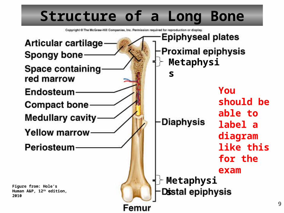
Structure of a Long Bone
Metaphysis
Metaphysis
You should be able to label a diagram like this for the exam
Figure from: Hole’s Human A&P, 12th edition, 2010
10
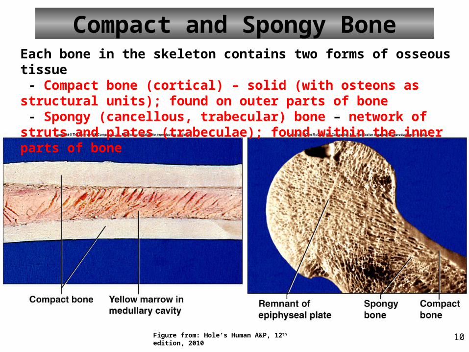
Compact and Spongy BoneEach bone in the skeleton contains two forms of osseous tissue - Compact bone (cortical) – solid (with osteons as structural units); found on outer parts of bone - Spongy (cancellous, trabecular) bone – network of struts and plates (trabeculae); found within the inner parts of bone
Figure from: Hole’s Human A&P, 12th edition, 2010
11
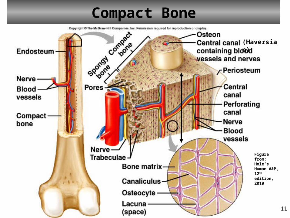
Compact Bone
(Haversian)
Figure from: Hole’s Human A&P, 12th edition, 2010
12
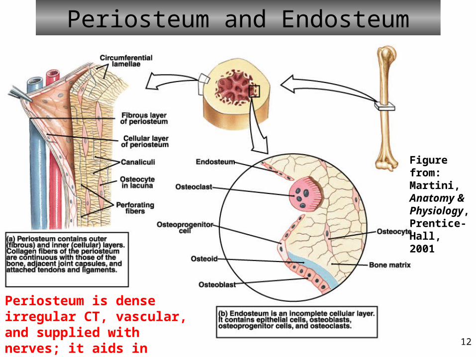
Periosteum and Endosteum
Figure from: Martini, Anatomy & Physiology, Prentice-Hall, 2001
Periosteum is dense irregular CT, vascular, and supplied with nerves; it aids in growth/repair
13
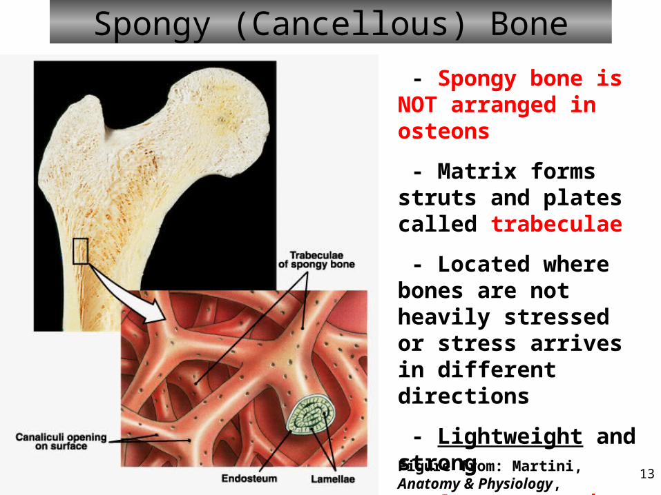
Spongy (Cancellous) Bone
- Spongy bone is NOT arranged in osteons
- Matrix forms struts and plates called trabeculae
- Located where bones are not heavily stressed or stress arrives in different directions
- Lightweight and strong
- Supports and protects cells of the red marrow
Figure from: Martini, Anatomy & Physiology, Prentice-Hall, 2001
14
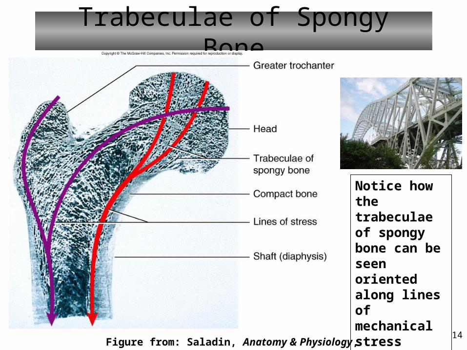
Trabeculae of Spongy Bone
Notice how the trabeculae of spongy bone can be seen oriented along lines of mechanical stress applied by the weight of the body
Figure from: Saladin, Anatomy & Physiology, McGraw Hill, 2007
15
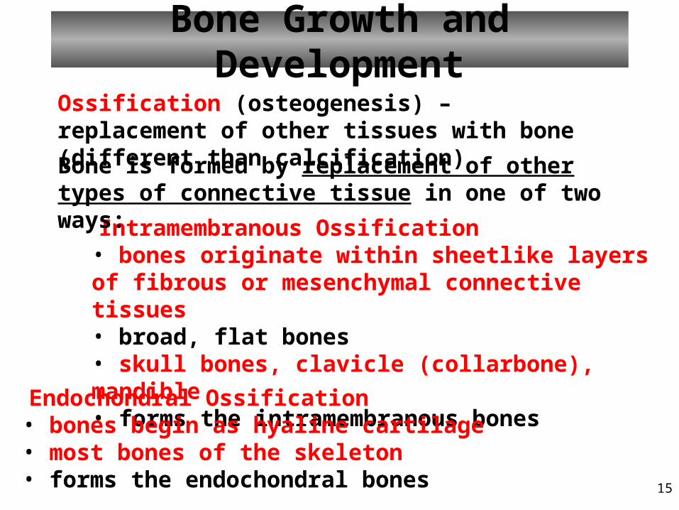
Bone Growth and Development
Intramembranous Ossification• bones originate within sheetlike layers of fibrous or mesenchymal connective tissues• broad, flat bones• skull bones, clavicle (collarbone), mandible• forms the intramembranous bones
Endochondral Ossification• bones begin as hyaline cartilage• most bones of the skeleton• forms the endochondral bones
Bone is formed by replacement of other types of connective tissue in one of two ways:
Ossification (osteogenesis) – replacement of other tissues with bone (different than calcification)
16
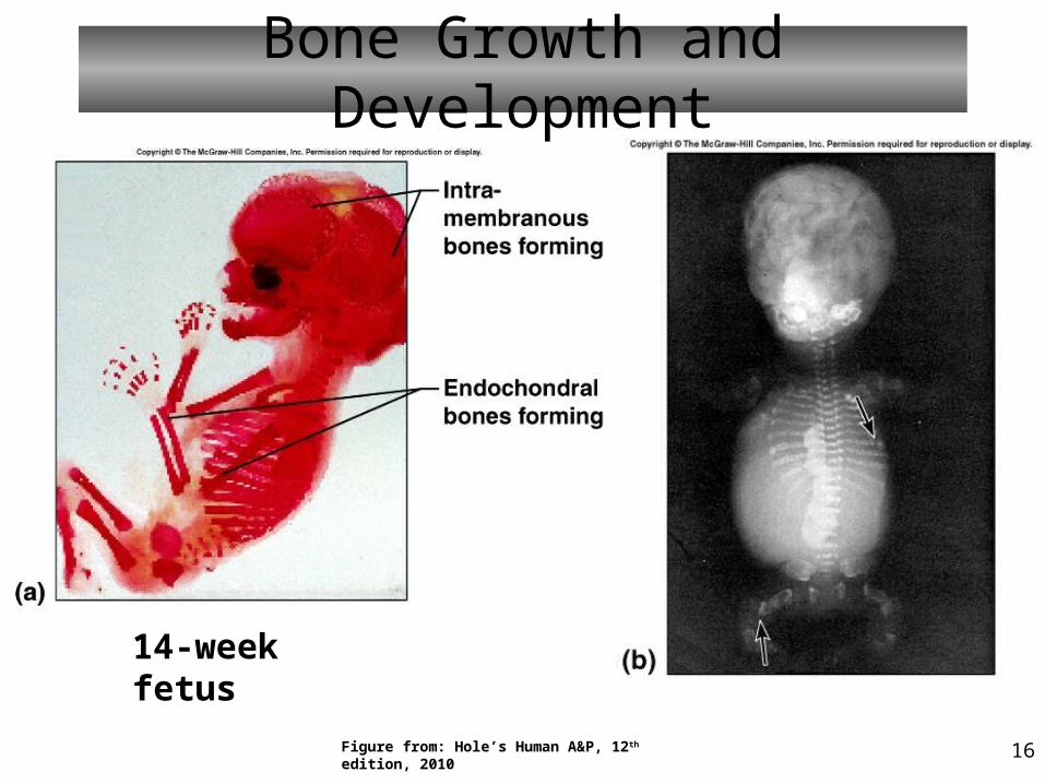
Bone Growth and Development
14-week fetus
Figure from: Hole’s Human A&P, 12th edition, 2010
17
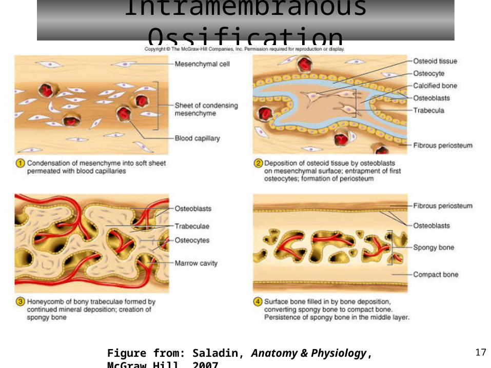
Intramembranous Ossification
Figure from: Saladin, Anatomy & Physiology, McGraw Hill, 2007
18
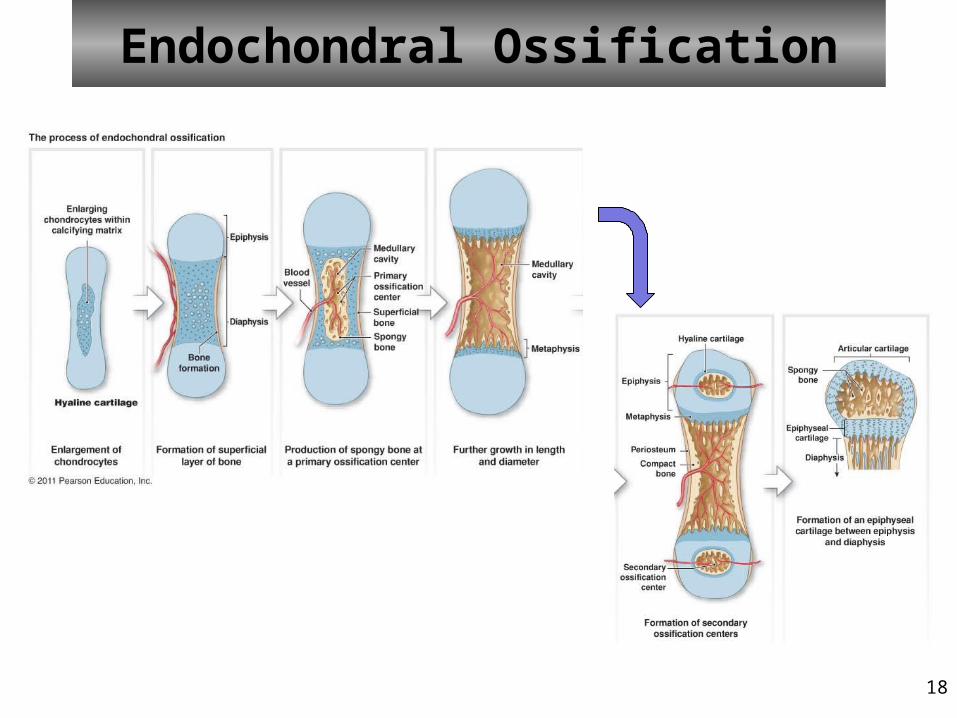
Endochondral Ossification
19
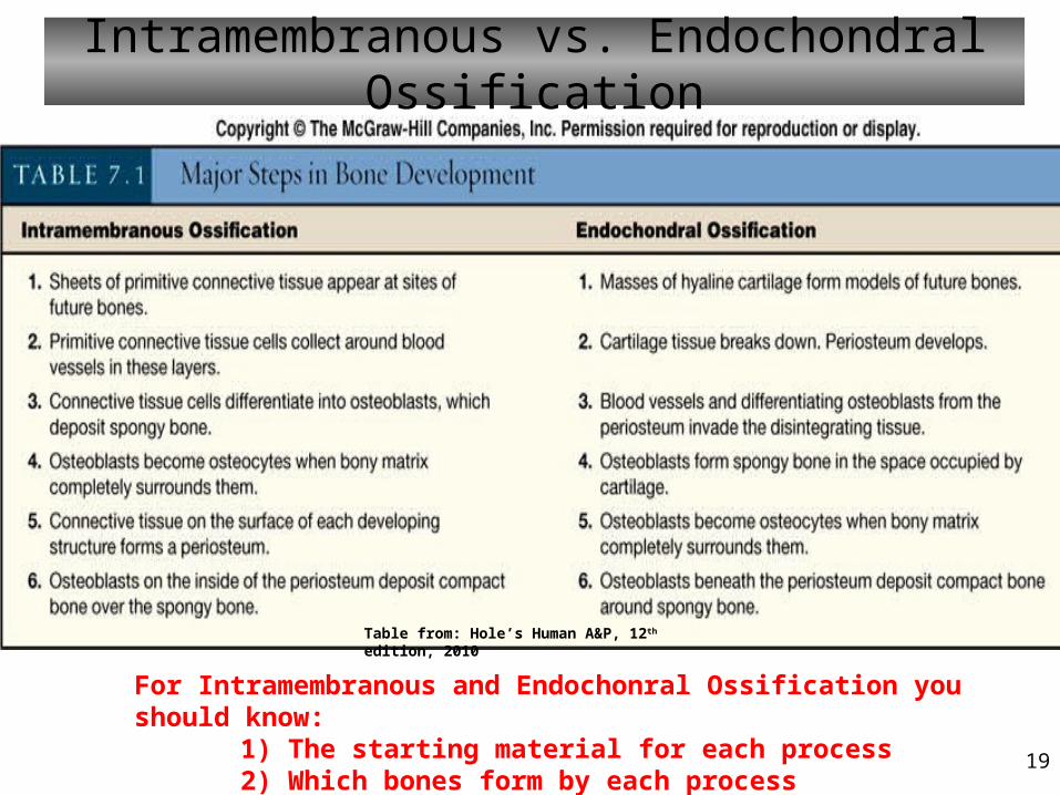
Intramembranous vs. Endochondral Ossification
For Intramembranous and Endochonral Ossification you should know:1) The starting material for each process2) Which bones form by each process
Table from: Hole’s Human A&P, 12th edition, 2010
20
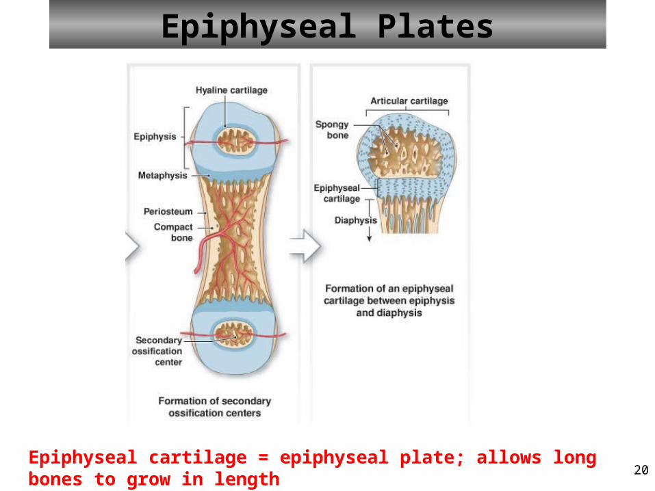
Epiphyseal Plates
Epiphyseal cartilage = epiphyseal plate; allows long bones to grow in length
21
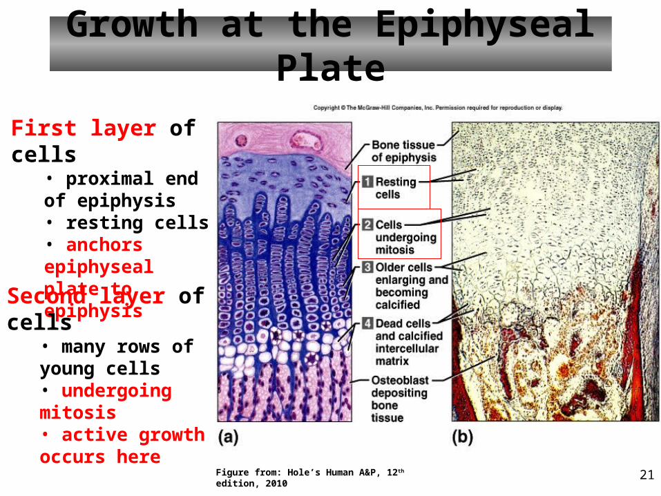
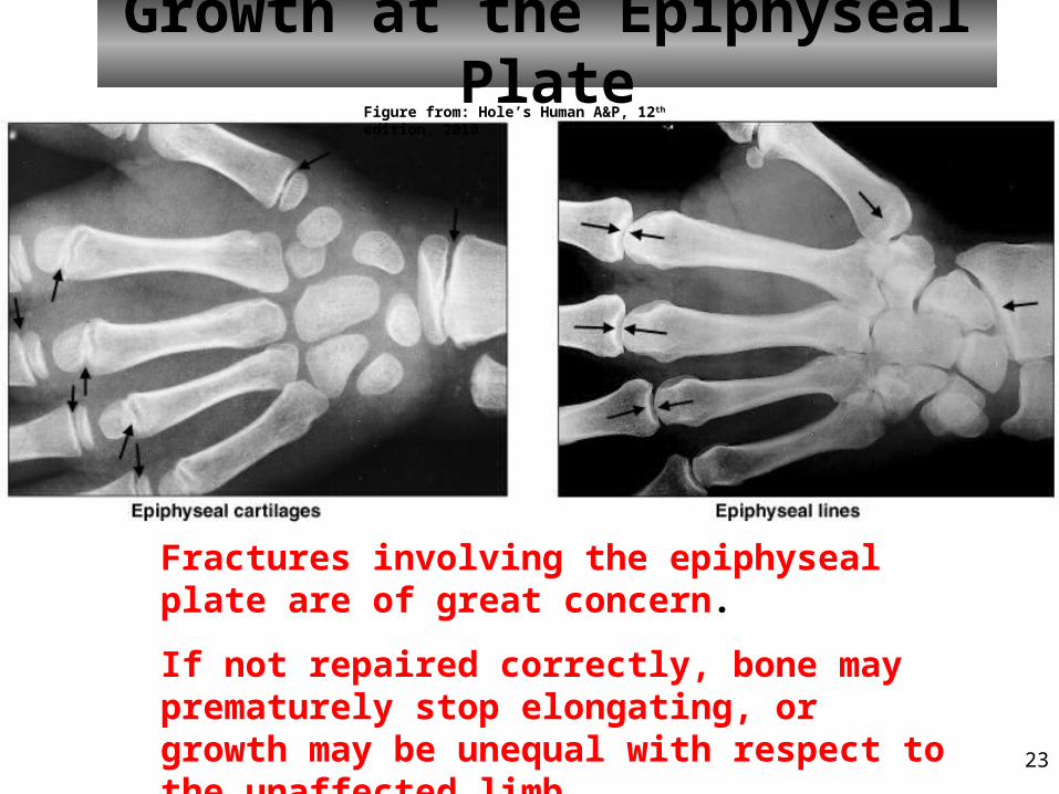
Growth at the Epiphyseal Plate
First layer of cells• proximal end of epiphysis• resting cells• anchors epiphyseal plate to epiphysis
Second layer of cells• many rows of young cells• undergoing mitosis• active growth occurs here
Figure from: Hole’s Human A&P, 12th edition, 2010
22
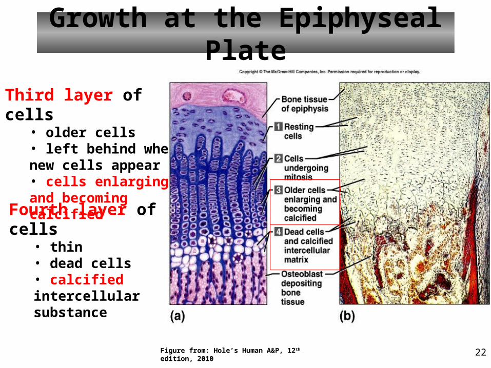
Growth at the Epiphyseal Plate
Third layer of cells• older cells• left behind when new cells appear• cells enlarging and becoming calcified
Fourth layer of cells• thin• dead cells• calcified intercellular substance
Figure from: Hole’s Human A&P, 12th edition, 2010
23
Growth at the Epiphyseal Plate
Fractures involving the epiphyseal plate are of great concern.
If not repaired correctly, bone may prematurely stop elongating, or growth may be unequal with respect to the unaffected limb.
Figure from: Hole’s Human A&P, 12th edition, 2010
24
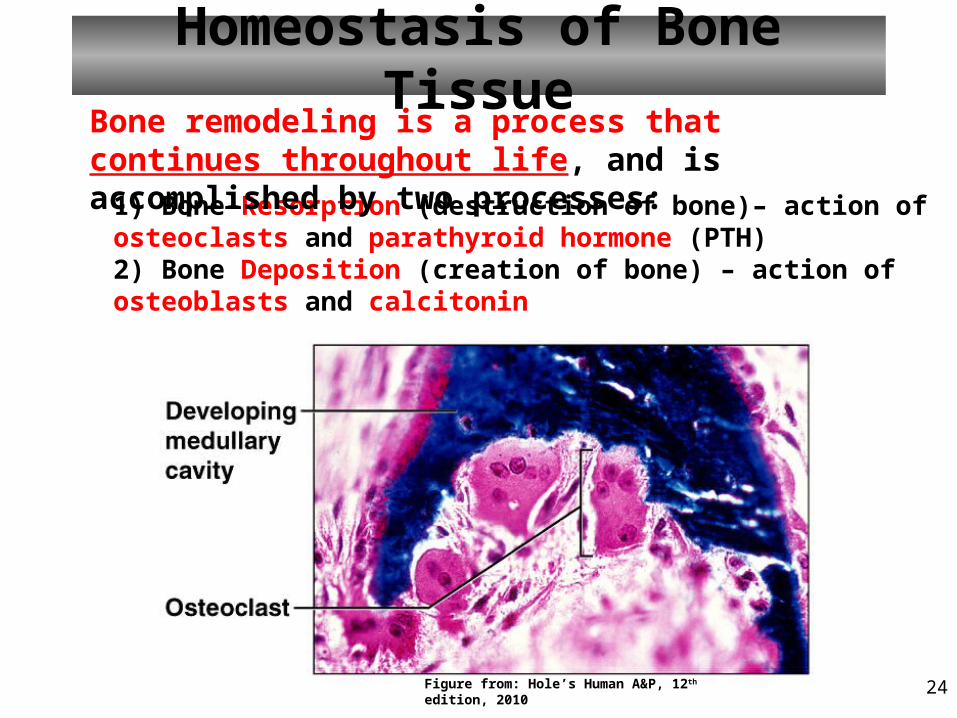
Homeostasis of Bone Tissue
1) Bone Resorption (destruction of bone)– action of osteoclasts and parathyroid hormone (PTH)2) Bone Deposition (creation of bone) – action of osteoblasts and calcitonin
Bone remodeling is a process that continues throughout life, and is accomplished by two processes:
Figure from: Hole’s Human A&P, 12th edition, 2010
25
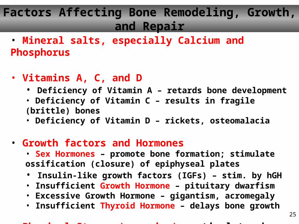
Factors Affecting Bone Remodeling, Growth, and Repair
• Mineral salts, especially Calcium and Phosphorus
• Vitamins A, C, and D• Deficiency of Vitamin A – retards bone development• Deficiency of Vitamin C – results in fragile (brittle) bones • Deficiency of Vitamin D – rickets, osteomalacia
• Growth factors and Hormones• Sex Hormones – promote bone formation; stimulate ossification (closure) of epiphyseal plates• Insulin-like growth factors (IGFs) – stim. by hGH• Insufficient Growth Hormone – pituitary dwarfism• Excessive Growth Hormone – gigantism, acromegaly • Insufficient Thyroid Hormone – delays bone growth
• Physical Stress (exercise) – stimulates bone growth
27
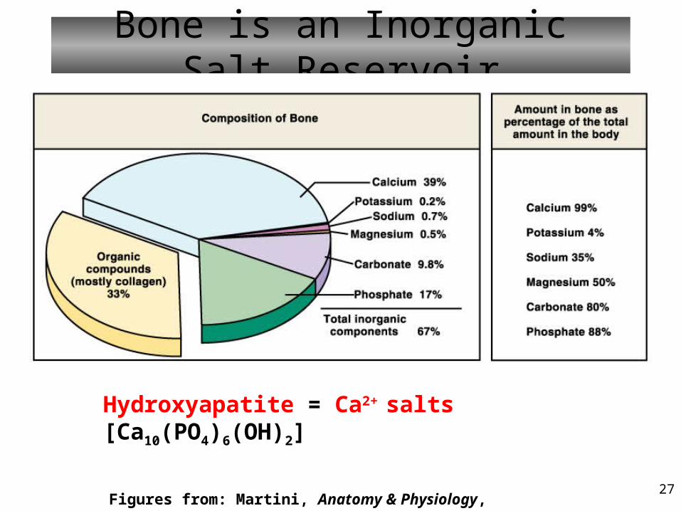
Bone is an Inorganic Salt Reservoir
Figures from: Martini, Anatomy & Physiology, Prentice-Hall, 2001
Hydroxyapatite = Ca2+ salts [Ca10(PO4)6(OH)2]
28
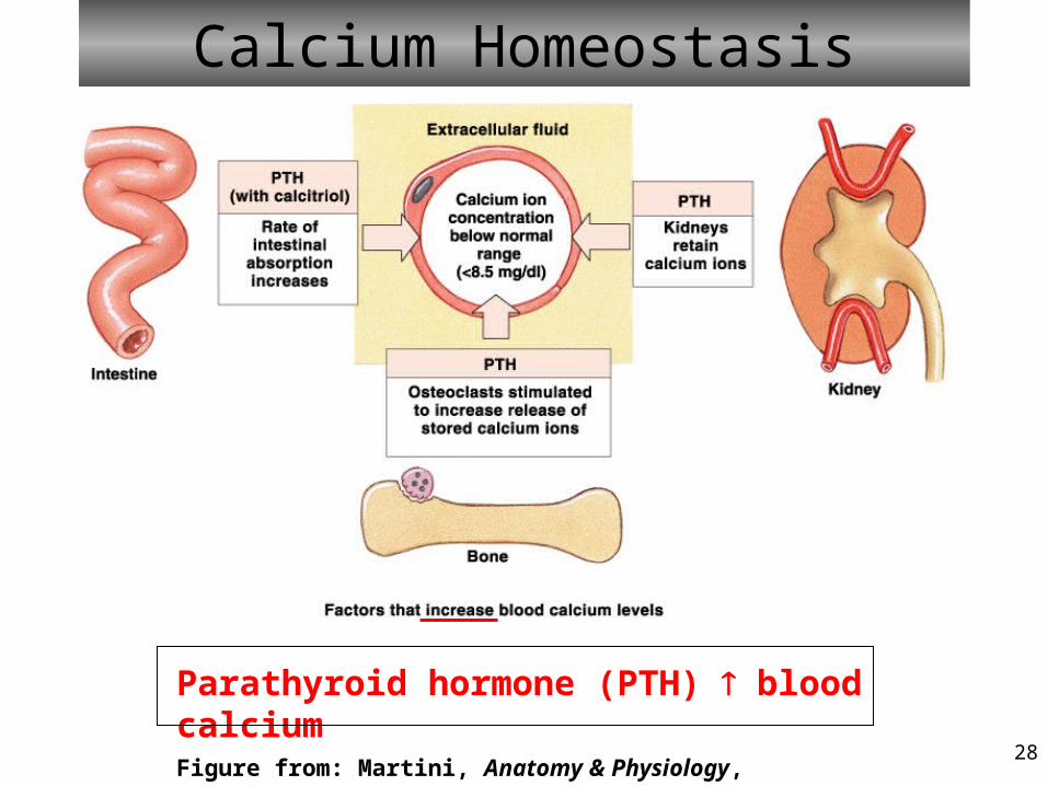
Calcium Homeostasis
Figure from: Martini, Anatomy & Physiology, Prentice-Hall, 2001
Parathyroid hormone (PTH) blood calcium
29
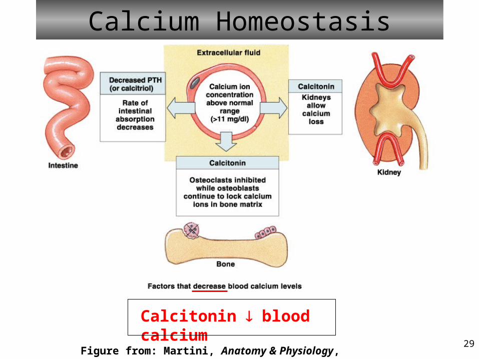
Calcium Homeostasis
Figure from: Martini, Anatomy & Physiology, Prentice-Hall, 2001
Calcitonin blood calcium
30
Blood Cell Formation
• Hematopoiesis is the process of blood cell formation (also called ‘hemopoiesis’)
• At different points in development, hematopoiesis takes place in the:– Yolk sac (embryo)– Liver and spleen (fetus)– Red bone marrow (fetus, newborn, and adult)
31
Blood Cell Formation Takes Place in the Red Bone Marrow
• Red marrow functions in development of – Myeloid cells - red blood cells, platelets,
eosinophils, basophils, neutrophils, and monocytes– Lymphocytic cells – T lymphocytes and B
lymphocytes
• Adult red marrow is primarily found in the spongy bone of the skull, ribs, sternum, clavicles, vertebrae, pelvis, and epiphyses of long bones
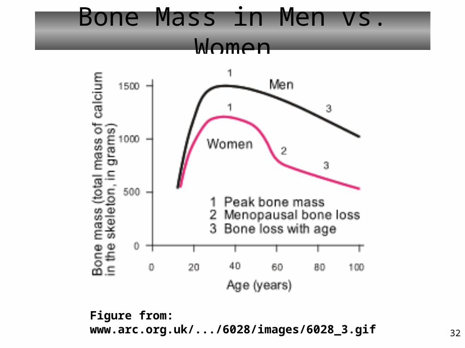
32
Bone Mass in Men vs. Women
Figure from: www.arc.org.uk/.../6028/images/6028_3.gif
33
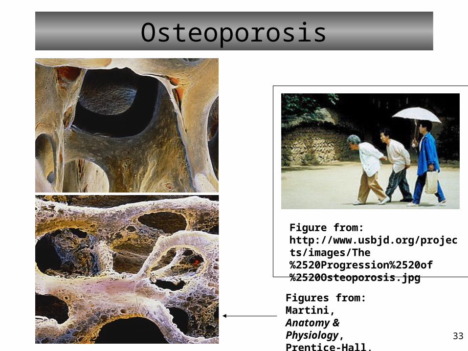
Osteoporosis
Figure from: http://www.usbjd.org/projects/images/The%2520Progression%2520of%2520Osteoporosis.jpg
Figures from: Martini, Anatomy & Physiology, Prentice-Hall, 2001
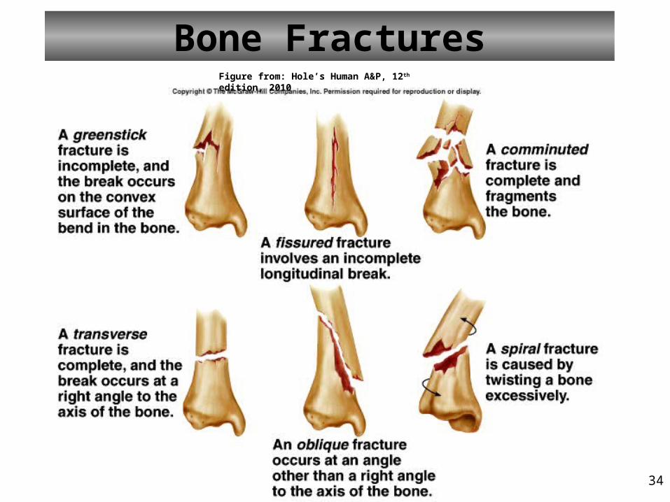
34
Bone FracturesFigure from: Hole’s Human A&P, 12th edition, 2010
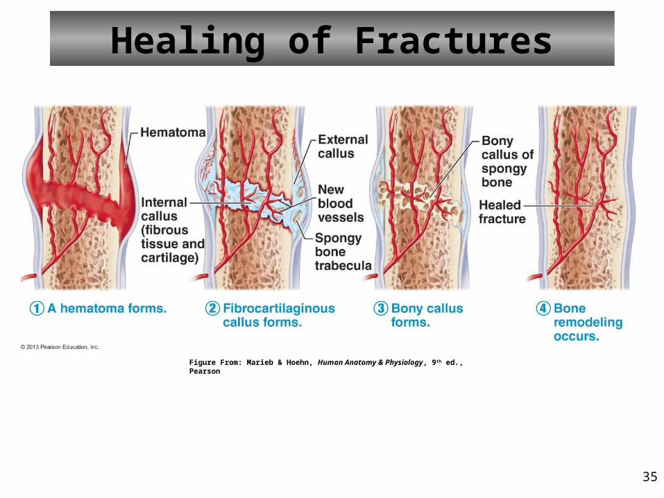
35
Healing of Fractures
Figure From: Marieb & Hoehn, Human Anatomy & Physiology, 9th ed., Pearson