Embed Size (px)
Citation preview

1
Graded potential
Could be
depolarisation or
hyperpolarisation
Magnitude various;
can be added;
reduces while
propagated

2
Action potential
Changes in membrane potential reach a
threshold; becomes a brief reversal of
membrane potential
e.g. -70mV +30mV
Depolarisation only
Magnitude is the same; does not reduce
while propagated (all-or-none)

Mem
bra
ne
po
ten
tial
(m
V)
-55
+30
-70
Phase 1 Phase 2
Phase 3
Threshold
Resting potential
Ion permeability
PNa
PK
Action potential
Mem
bra
ne
po
ten
tial
(m
V)
-55
+30
-70
Phase 1 Phase 2
Phase 3
Threshold
Resting potential
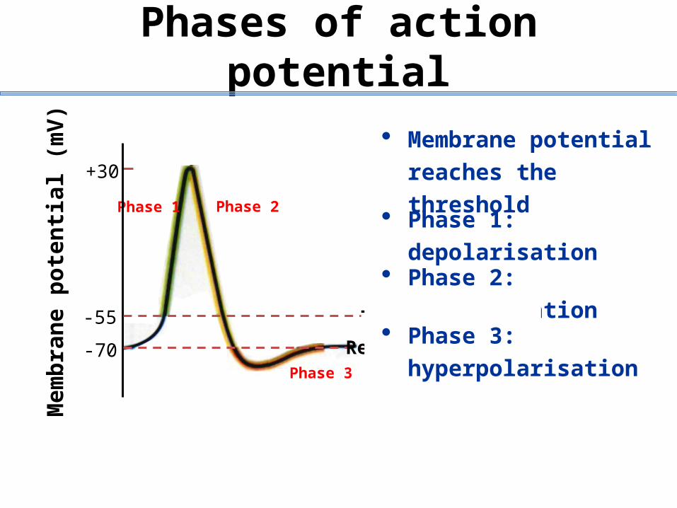
Phases of action potential
Membrane potential
reaches the threshold
Phase 1: depolarisation
Phase 2: repolarisation
Phase 3:
hyperpolarisation
5
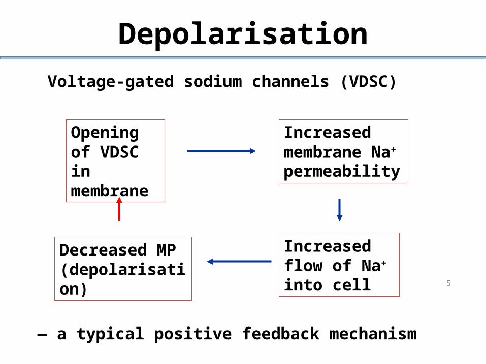
Depolarisation
Opening of VDSC in membrane
Increased membrane Na+ permeability
Decreased MP (depolarisation)
Increased flow of Na+ into cell
— a typical positive feedback mechanism
Voltage-gated sodium channels (VDSC)

6
Repolarisation
After Na+ channels open, they become
deactivated & Na+ flux stops
Voltage-gated K+ channels slowly open
K+ ions rush outwards
Membrane potential returns to the resting one (-70mV)

7
Hyperpolarisation
Voltage-gated K channels close slowly
K ions continue to flow outwards
Membrane becomes more polarised (-80mV)

8
Refractory period
Period of reduced excitability
Absolute refractory period
Relative refractory period
Significance: single direction of propagation

9
Refractory period
Stanfield & Germann Fig 7.17

10
Propagation of action potential
myelinated
not myelinated

11
Propagation of action potential - myelinated axon
Myelin sheath acting as insulator to prevent
leakage of charges from the axon
Current can only pass through the membrane at
the Node of Ranvier
Propagation is much faster than unmyelinated
axon

Dr. Fang Lou
12
Overview of the musculoskeletal system
including bone and soft tissue

Learning Outcomes:
Describe the types of connective tissue found in the body and indicate their characteristic functions
Describe the functional properties of the three types of cartilage tissue
Name the major regions of the skeleton and describe their relative functions
List and describe five important functions of bones
13
Following this session and appropriate independent study, you should be able to:

Connective tissue proper (inc. fat and fibrous tissue of ligament)
Cartilage
Bone tissue
Blood
Connective Tissues
Four main classes

Functions of connective tissue
Binding and support
Protection
Insulation
Transportation

Major classes of connective tissue
Mesenchyme
Connective tissue proper
Cartilage Osseous Blood
1. Loose connective tissue- Areolar- Adipose- Reticular2. Dense connective tissue- Regular- Irregular
Subclasses1. Hyaline cartilage2.Fibrocartilage3. Elastic cartilage
1. Compact bone2. Spongy (cancellous) bone
Class of connective tissue
Common embryonic form

Has qualities between dense connective
tissue and bone
Tough but flexible
Stands up to both tension & compression
Lack of nerve fibres and is avascular
Up to 80% water
Calcify/ossify in later life
Cartilage

Varieties of cartilageHyaline Elastic
Fibrocartilage
www.kumc.edu/instruction/medicine/anatomy/histoweb/cart/cart.htm

Location: embryonic skeleton
covers end of long bones in joint cavity
costal cartilage of the ribs
respiratory tract (nose, larynx, trachea, etc.)
Provide firm support with some flexiability
Hyaline Cartilage

20
The bones and cartilage of the human skeleton
Fig. 6.1 Marieb
Bones of axial skeleton
Bones of appendicular skeleton
Hyaline cartilage
Elastic cartilage
Fibrocartilage

Location: External ear
epiglottis
More elastic fibres
Provide strength and exceptional stretchability
Elastic Cartilage

Location: intervertebral disc
disc of knee joint
Between hyaline cartilage and dense regular connective tissue
Absorb shock
Fibrocartilage
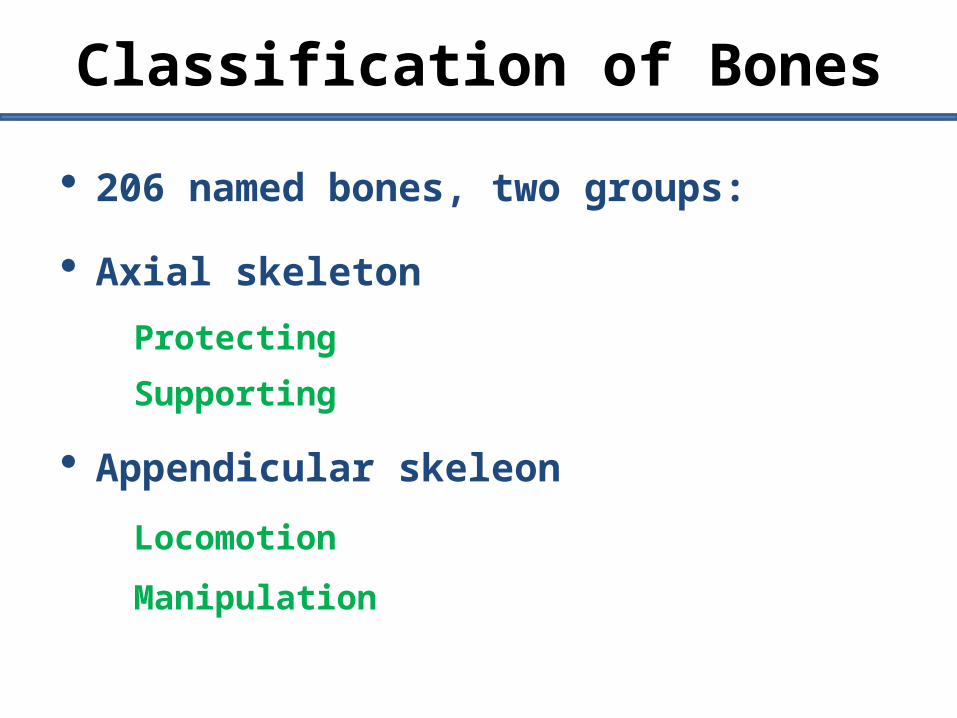
206 named bones, two groups:
Axial skeleton
Protecting
Supporting
Appendicular skeleon
Locomotion
Manipulation
Classification of Bones

The axial skeleton The skull
The vertebral column
The bony thorax
The appendicular skeleton The pectoral (shoulder) girdle
The upper limb
The pelvic (hip) girdle
The lower limb
Major regions of the skeleton

25
skull
vertebraethoracic
Axial SkeletonSupport & protect
Modified from Fig. 7.1 Marieb 7th ed.

26
pectoral (shoulder) girdle
the lower limb
the upper limb
Appendicular SkeletonSupportMovementMineralBlood cells
Modified from Fig. 7.21 Marieb 7th ed.
pelvic (hip) girdle

Support (framework)
Protection (skull, vertebrae & ribs)
Movement (lever for muscles)
Store minerals (calcium & phosphate)
Blood cell formation (marrow)
Functions of Bones