Embed Size (px)
Citation preview

1
Haemophilus
2
Introduction• During the influenza pandemic of 1918 a “pleomorphic” Gram
negative bacterium was isolated from the respiratory tract of most of the patients who were dying of influenza
• The bacterium grew only on media containing blood from rabbits, horses, and cows. It did not on media containing unheated sheep’s blood unless it was first heated to 80oC, a temp that releases X & V factors without destroying them. This was the 1st use of what we now call chocolate blood agar (CBA).
• The bacterium was named Haemophilus, or “blood loving” because it grew only on media containing blood. The species name “influenzae” was used because it was mistakenly presumed to cause influenza.
• In the 1930s, after cell cultures and the electron microscope were invented, it was demonstrated that influenza is caused by a virus, the influenza virus (orthomyxovirus).

3
• Blood of all animal species contains 2 growth factors, both of which are stable at 80oC.
1) The “labile factor” is extracellular but is destroyed by a heat labile enzyme found in sheep’s blood, but not in rabbit, horse, or cow blood. This is NAD and is known now as V factor.
2) 2) The “stable factor” is stable in the presence of the enzyme found in sheep’s blood, but it is intracellular, and is released from RBCs heated at 80oC. This is now known to be heme, and is referred to as X factor.
• H. ducreyi requires the addition of X factor but not V factor. H. aphrophilus is unable to synthesize X factor on primary cultures but gains the ability upon subculture. Species names with the prefix “para” cannot synthesize V factor but can synthesize X factor. All other Haemophilus species and a few other species require both growth factors because they lack the enzymes to synthesize them.

4
Introduction• The inactivating enzyme in sheep's blood but not the blood of
rabbits, horses, or cows is NADase
• H. influenzae will grow on unheated SBA if NAD is added in sufficient quantities to overcome NADase. Alternatives to adding NAD are to add the B vitamin (not really) niacin, or by growing an NAD-producing microbe along with H. influenzae. Various yeast species are the best microbes to use for this purpose.
• To test an organism for the necessity of added X &/or V factors, conduct a “cross streak” of the organism with S. aureus on agar containing blood previously heated to 100oC. The isolate will grow as tiny “satellite” colonies next to the S. aureus
• Even though the “staph streak” method will allow H. influenzae and other species to grow, the colonies remain tiny.
• H. influenzae grows much larger colonies on chocolate agar

5
“Cross streak” technique for growth factors
StaphylococcusGrowth
“Satellite” coloniesIncubation
for 24 hours

6
Introduction• The modern version of chocolate agar, which allows H. influenzae
to grow up to 3mm diameter size colonies in 48 hours, involves adding hemoglobin powder to a basal agar medium such as TSA. Autoclave it, cool to 50oC, and add filter-sterilized yeast extract as an NAD source to the molten 50oC agar base.
• As said earlier, H. influenza does not cause influenza, but can cause secondary bacterial infections including pneumonia and septicemia.
• If the microbiologists who misnamed the bacterium had followed Koch’s postulates, another species name almost certainly would have been chosen.
• As we will discuss later, the primary pathology associated with the “b” strain of this organism is meningitis. This strain is now referred to as HIb.
7
General characteristics• The genus Haemophilus consists of Gram negative, pleomorphic
rods. In clinical specimens they range from small coccobacilli to long filamentous rods, and are usually encapsulated.
• They are nonmotile and aerobic to facultatively anaerobic • Most species are catalase and oxidase positive (may be weak)• There are 10 species of Haemophilus associated with humans • Most, including H. influenzae, are part of the normal microbiota
of the URT and other mucous membranes. • The two major pathogens are H. influenzae and H. ducreyi. The
others are occasionally associated with opportunistic infections• Several Haemophilus species are hemolytic on rabbit or horse
blood

8
Clinical specimens and culture• Haemophilus species can be isolated from various clinical specimens
including blood, CSF, middle-ear exudate, joint fluids, URT and LRT, conjunctivae, vagina, and wounds and abscesses (although not routinely plated unless specifically requested)
• H. ducreyi is more fastidious than other Haemophilus species. If the physician suspects H. ducreyi, the media should be incubated for at least one week
• Media for isolating Haemophilus should be incubated in a humid environment rich in carbon dioxide
• Colonies of Haemophilus are 1-3 mm in diameter in 24-48 hours of incubation. On CBA they are slightly convex moist, circular smooth and translucent, and have a distinct “mousy” or a very faint “bleach-like” odor
• Highly virulent encapsulated strains of H. influenzae produce a slightly grayish and very moist appearing colonies
9
Identification• The first clue that Haemophilus may be present in “pure” culture
is good growth on CBA and no growth on SBA. If the culture is from a non-sterile site (mixed culture), tiny satellite colonies growing around colonies of other species is another clue
• The most virulent H. influenzae strains or “biotypes” can be detected using the spot indole test and oxidase test. Most virulent strains of H. influenzae are “positive” for both tests
• Most virulent strains of H. influenzae also produce a very strong urease. Actual growth is not necessary if a heavy inoculum is used - the transferred urease will give a rapid positive reaction
• The next step is to confirm X, V, or X&V requirements. The traditionally test involves placing paper discs (or strips) containing X and V on media devoid of both factors. Discs of both factors are arranged on the agar close together and distant from each other.
10
Identification• Organisms that grow only between the closely placed disks
require both factors.
• Those that grow only around the V disks cannot synthesize V (“require V”)
• Those that grow only around the X disks cannot synthesize X (“require X”)
• When identifying Haemophilus species using X and V discs a very light inoculum should be used (less than 0.5 MacFarland)
• When picking the colony be careful not to touch the medium because “carry-over” of factors from the media can result in erroneous results

11
X and V Requirements
X V
X V
Requires X and V
Growth Between X and V Disk Only

12
X and V Requirements
V
X V
Requires X only
Growth Around X Disks only
X

13
X and V Requirements
X V
X V
Requires V only
Growth Around V Disks only
14
Identification• Most clinical microbiology labs now use some commercial
system to identify Haemophilus species such as the “quad” plate
• One quadrant of a divided plate contains only X, one contains only V, one contains X and V, and one contains rabbit or horse blood for detection hemolysis. The plate is read very much like the paper strip method previously described.
• Many microbiologists abandoned the use of strips and media containing X and V because they claim that it is impossible to avoid carry-over, especially for the X factor
• Instead, they determine if an isolate can synthesize the X factor by using the “porphyrin” test (recall that heme is derived form a protoporphyrin)

16
Identification• The porphyrin tube test is performed by making a heavy
suspension in buffered aminolevulinic acid (ALA) solution
• If the isolate can synthesize heme, it will enzymatically convert ALA to porphobilinogen (PBG) - the lack of the enzymes to do this limits all Haemophilus that cannot synthesize heme. From PBG an organism can produce protoporphyrin and then heme
• If PBG is produced, a red color will develop in the presence of Ehrlich’s reagent (pDAB) – recall from urinalysis
• Protoporphyrin can also be detected rather easily because it fluoresces red when exposed to ultraviolet light

17
“Porphyrin” Test
ALA PBG ProtoporphyrinBacterialenzymes
Bacterialenzymes
Red Color(pDAB)
Red Fluorescence(UV light)
Interpretation: Organisms that produce a red color with either pDAB or UV light can synthesize heme; heme does not have to be add to a medium to grow this organism; it does not “require” X. Organisms failing to produce the red color(s) do “require” X to be added to a growth medium

18
Porphyrin Tube Test
After addition of pDAB:If red appears, it does not “require” X; If no red appears it does “require”X
UV Light: If red appears, it does not “require” X; If no red appears it does “require” X
ALA medium exposed to pDAB or UV light

19
Identification• The UV test can be performed on an agar plate containing ALA
• The unknown is “band” streaked on the plate & after overnight incubation the surface of the plate is exposed to UV light
• A red fluorescence indicates the organism can synthesize porphyrin (does not “require” X)
• No red fluorescence indicates the organism cannot synthesize porphyrin (does “require” X)
• Several commercial ID products use miniaturized tubes with dehydrated substrates. RapID NH is one such product
• It can identify Haemophilus and other fastidious coccobacilli as well as Neisseria and related organisms

20
Plate Porphyrin Test
A: Red Fluorescence with UV light(Does not “require” X)
B: No red fluorescence with UV light (does “require” X)
Bacteria “band” streakedacross a plate containing
- ALA
Unknown A
Unknown B
21
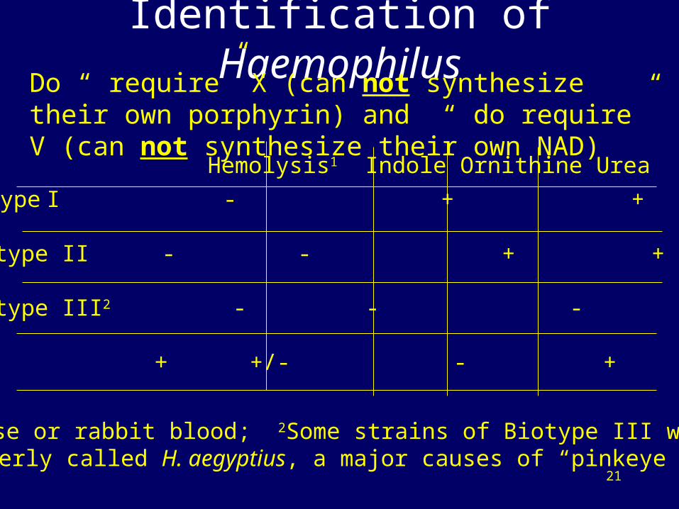
Identification of HaemophilusDo “ require” X (can not synthesize their own porphyrin) and “ do require” V (can not synthesize their own NAD)
H. influenzae Biotype I - + + +
H. influenzae Biotype II - - + +
H. influenzae Biotype III2 - - - +
H. hemolyticus + +/- - +
Hemolysis1 Indole Ornithine Urea
1Horse or rabbit blood; 2Some strains of Biotype III were formerly called H. aegyptius, a major causes of “pinkeye”

22
Identification of HaemophilusDo not “require” X (can synthesize their own porphyrin); Do “require” V (can not synthesize their own NAD)
H. parainfluenzae - - + +
H. parahaemolyticus + - +/- +
H. segnis - - - -
H. paraphrophilus - - - +
Hemolysis1 Indole Ornithine Urea
1Horse or rabbit blood

23
Identification of HaemophilusDoes “require X (can not synthesize its own porphyrin);Does not “require” V (can synthesize its own NAD)
H. ducreyi + /- - - -
Hemolysis1 Indole Ornithine Urea
1 May require heme on primary isolation 2Horse or rabbit blood
Does not “require” X1 (can synthesize its own porphyrin; does not “require” V ( can synthesize its own NAD)
Hemolysis2 Indole Ornithine Urea
H. aphrophilus - - - -
24
Virulence factors of H. influenzae
• The following virulence factors play a role in the invasiveness of this organism and the initiation of infection
– “Type b” polysaccharide capsule: attachment & anti-phagocytosis – appears critical for virulence
– Endotoxin (LPS) plays role but not understood
– IgA protease & neuraminidase (like orthomyxovirus) produced by all virulent strains but role in virulence unknown
– Adherence factors: fimbriae and protein “adhesins” required for initial colonization
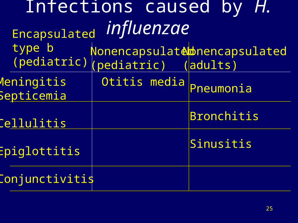
25
Infections caused by H. influenzae Encapsulated type b (pediatric)
Nonencapsulated(pediatric)
Nonencapsulated (adults)
MeningitisSepticemia
Cellulitis
Epiglottitis
Conjunctivitis
Otitis mediaPneumonia
Bronchitis
Sinusitis
26
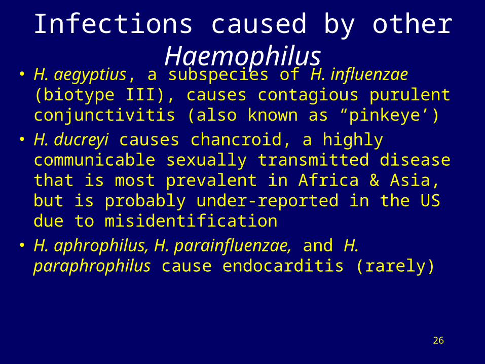
Infections caused by other Haemophilus• H. aegyptius, a subspecies of H. influenzae (biotype III),
causes contagious purulent conjunctivitis (also known as “pinkeye’)
• H. ducreyi causes chancroid, a highly communicable sexually transmitted disease that is most prevalent in Africa & Asia, but is probably under-reported in the US due to misidentification
• H. aphrophilus, H. parainfluenzae, and H. paraphrophilus cause endocarditis (rarely)
27
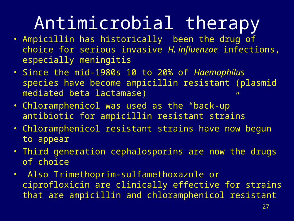
Antimicrobial therapy• Ampicillin has historically been the drug of choice for serious
invasive H. influenzae infections, especially meningitis
• Since the mid-1980s 10 to 20% of Haemophilus species have become ampicillin resistant (plasmid mediated beta lactamase)
• Chloramphenicol was used as the “back-up” antibiotic for ampicillin resistant strains
• Chloramphenicol resistant strains have now begun to appear
• Third generation cephalosporins are now the drugs of choice
• Also Trimethoprim-sulfamethoxazole or ciprofloxicin are clinically effective for strains that are ampicillin and chloramphenicol resistant
28
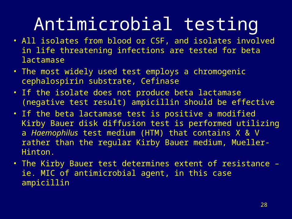
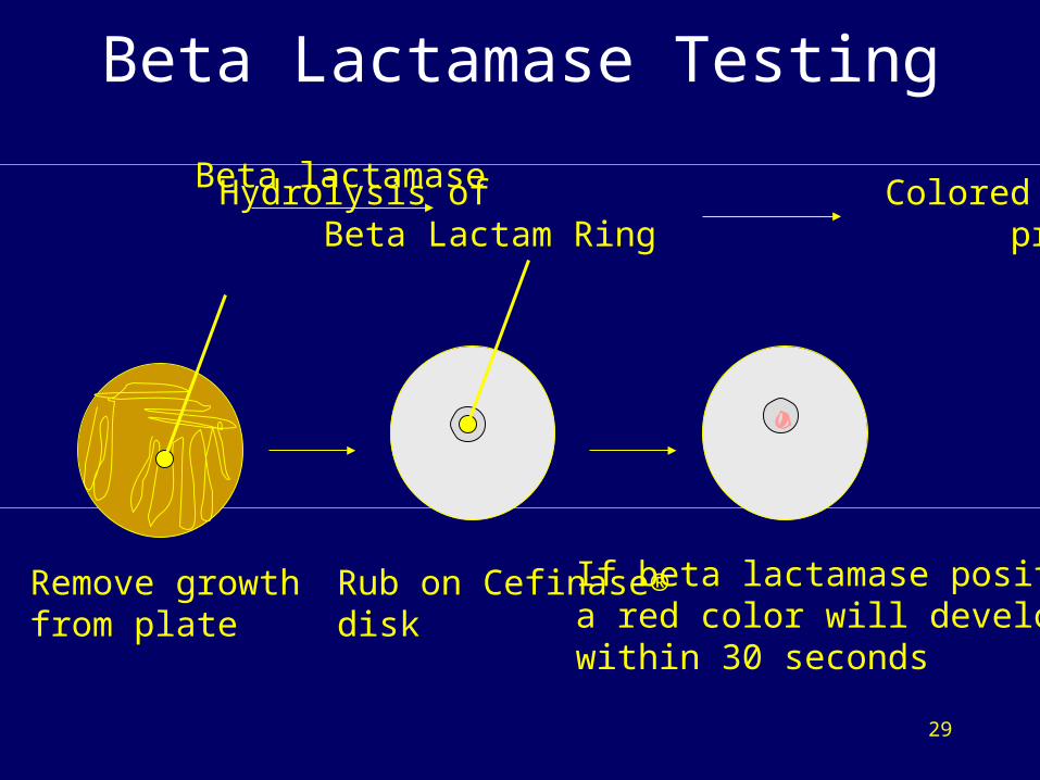
Antimicrobial testing• All isolates from blood or CSF, and isolates involved in life
threatening infections are tested for beta lactamase
• The most widely used test employs a chromogenic cephalospirin substrate, Cefinase
• If the isolate does not produce beta lactamase (negative test result) ampicillin should be effective
• If the beta lactamase test is positive a modified Kirby Bauer disk diffusion test is performed utilizing a Haemophilus test medium (HTM) that contains X & V rather than the regular Kirby Bauer medium, Mueller-Hinton.
• The Kirby Bauer test determines extent of resistance – ie. MIC of antimicrobial agent, in this case ampicillin
29
Beta Lactamase Testing
Chromogenic Hydrolysis of Colored Cephalosporin Beta Lactam Ring products Reagent Cefinase ® released
Beta lactamase
Remove growthfrom plate
Rub on Cefinase®disk
If beta lactamase positivea red color will developwithin 30 seconds
30
Antimicrobial testing• The E-test on HTM agar can also be used to test for
antimicrobial susceptibility of Haemophilus
• The E-test uses a plastic strip that is impregnated with a concentration gradient of antimicrobial agents
• The strip is placed on a plate of HTM agar that has been spread-plate inoculated with a 0.5 MacFarland broth culture
• The antimicrobial gradient diffuses into the agar
• Susceptible strains forms an elliptical pattern of inhibition
• The point near the low concentration end of the strip that is intersected by the zone of inhibition represents the minimal inhibitory concentration (MIC) of the antimicrobial agent

31
E-Test
E
O.50ug/mlline
High concentrationend of the strip
Zone of Inhibition
BacterialGrowth
Low concentrationend of the strip


![Haemophilus [NavIn]](https://img.pdfslide.net/doc/110x75/577d27191a28ab4e1ea30e7f/haemophilus-navin.jpg)


![[PAPER] Pleomorphic Adenoma Print.docx](https://img.pdfslide.net/doc/110x75/56d6bd9b1a28ab30168ea546/paper-pleomorphic-adenoma-printdocx.jpg)