Embed Size (px)
Citation preview
Antigen specific CD4 cells assist CD8 T effector cells in eliminating
keratinocytes.
Jennifer K Broom1, Andrew M Lew 2, Hiroaki Azukizawa3 Tony J Kenna1,
Graham R Leggatt1, Ian H Frazer1
1 The University of Queensland Diamantina Institute for Cancer, Immunology
and Metabolic Medicine,
2 The Walter and Eliza Hall Institute of Medical Research, Victoria, Australia
3 Department of Dermatology, Course of Molecular Integrated Medicine,
Osaka University Graduate School of Medicine, Japan
Correspondence : Ian Frazer, The University of Queensland Diamantina
Institute for Cancer, Immunology and Metabolic Medicine Level 4, R Wing,
Princess Alexandra Hospital, Ipswich Road, Woolloongabba, Queensland
4102, Australia
Phone :61-7-3240 5954 , Fax: 61-7-3240 5946, Email : [email protected]
Short title: CD4 help for skin effector CD8 cells
Key words: memory T cell, helper T cell, epithelial antigens, skin grafts
Abbreviations: OVA ovalbumin, HPV human papillomavirus, CTL cytotoxic T
lymphocyte
Abstract
Keratinocytes expressing tumour or viral antigens can be eliminated by
antigen primed CD8 cytotoxic T cells. CD4 T helper cells help induction of
CD8 cytotoxic T cells from naïve precursors and generation of CD8 T cell
memory. Here we show, unexpectedly,that CD4 cells are also required to
assist primed CD8 effector T cells in rejection of skin expressing hGH, a neo-
self antigen, in keratinocytes. The requirement for CD4 cell help can be
substituted by CD40 co-stimulation. Rejection of skin expressing OVA, a non-
self antigen, by primed CD8 cytotoxic T cells can in contrast occur without
help from antigen specific CD4 T cells. However, rejection of OVA expressing
keratinocytes is helped by antigen specific CD4 T cells if only low numbers of
primed or naïve OVA specific CD8 T cells are available. Effective
immunotherapy directed at antigens expressed in squamous cancer may
therefore be facilitated by induction of tumour antigen specific CD4 helper T
cells as well as cytotoxic CD8 T cells.

Introduction
Development of epithelial malignancy is regulated by the immune system, as
chronic immunosuppression after organ transplantation is associated with a
greatly increased risk of squamous cancer. Many cancers of epithelial origin
express tumour specific modified self proteins as neo-self antigens
(Buckwalter and Srivastava, 2008). Epithelial cancer can also arise from skin
persistently infected with virus, including human papillomavirus (Al-Daraji and
Smith, 2009) and Merkel tumour associated retrovirus (Feng et al., 2008), and
these tumours continue to express virus encoded antigen. Evidently, neo-
self or viral antigens expressed in squamous epithelial cancer fail to induce an
immune response adequate to eliminate the cancer. Better understanding of
the requirement for effective immune mediated elimination of keratinocytes
expressing viral or neo self antigen would therefore assist in the design of
appropriate immunotherapeutic interventions for squamous epithelial cancer.
We have developed a murine model to study the requirements for effective
immunotherapy directed at antigen expressed in keratinocytes. Skin
expressing a non-self or neo-antigen as a transgene in keratinocytes from a
keratin promoter is grafted to a syngeneic non transgenic recipient(Zhong et
al., 2004). This allows us to study the requirements for inducing and
delivering an effective immune response to antigen expressed only in
keratinocytes, and not directly presented to the immune system by
professional antigen presenting cells(APC).
The fate of a graft of skin expressing non self antigen in keratinocytes
depends on the antigen. Some antigens including papillomavirus non-

structural proteins fail to invoke spontaneous rejection of grafts (Dunn et al.,
1997), allowing study of the requirements for effective induction of immunity
after antigen cross presentation from keratinocytes. Other antigens, including
ovalbumin(Holcmann et al., 2009) and human growth hormone(Zhong et al.,
2004), which is closely related in sequence to mouse growth hormone and
therefore a neo-self antigen in the mouse, induce spontaneous graft rejection,
allowing study of elimination of keratinocytes by primed T cells in a second
graft following immune mediated elimination of a priming graft.
To prime a cytotoxic CD8 effector T cell response from naïve antigen specific
CD8 T cell precursors, antigens expressed only in epithelial cells must be
cross-presented by professional APC, particularly dermal dendritic cells in the
local lymph node (Bedoui et al., 2009). CD4 helper T cells are required to
enable priming of memory and effector CD8 T cells (Schuurhuis et al.,
2000;Janssen et al., 2003;Smith et al., 2004). CD4 T cells also assist in the
maintenance of a pool of memory CD8 T cells(Sun et al., 2004). However,
once they are primed by antigen, CD8 effector T cells are held capable of
elimination of their cognate target cells without the need for CD4 T cell help.
Recent in vitro evidence suggests however that CD4 T cells can assist in
secondary responses to cross-presented but not directly presented
antigen(Blachere et al., 2006). We have also previously shown a requirement
for CD4 cells in a secondary immune response to human growth hormone in
FVB mice of H-2q x H-2b genetic background (Zhong et al., 2008). To extend
this observation to mice of a genetic background more commonly used for
studies of immune physiology and therefore enable further dissection of the
mechanisms by which CD4 cells contribute to elimination of keratinocytes
following priming or immunisation, we created K14.hGH transgenic mice on a
C57/Bl6 genetic background. We evaluated the role of CD4 T cells in
rejection of epithelial cells expressing various non-self antigens using these
mice. Here we present data showing that CD4 T cell help improves the
efficacy of rejection by already primed CD8 effector T cells of skin expressing
either non self antigen or neo-self antigen in keratinocytes, and demonstrate
that the requirement for CD4 cell responses can be substituted by CD40 co-
stimulation.

Results
Second-set syngeneic skin grafts expressing hGH as a neo-self antigen are
more rapidly rejected by C57Bl/6 recipients than the primary graft
We grafted skin expressing antigen as a transgene in keratinocytes to naïve
C57Bl/6 (C57) recipient animals, and measured time to graft rejection to
assess the immune response induced to the expressed antigen. We first
studied recall responses to human growth hormone (hGH), a near homologue
of mouse growth hormone representing modified self (neo-self). Animals that
rejected a hGH transgenic graft were considered primed to hGH, and
received a further hGH graft. Primary hGH grafts on C57 mice were rejected
in a median of 31.5 days (Fig. 1A), as was observed for C57 x FVB mice and
in contrast to the findings for FVB mice, which rarely reject primary hGH
grafts (Zhong et al., 2004). Second hGH grafts placed on graft primed C57
recipients were rejected with a median survival time of 21 days, significantly
earlier than the initial graft (p=0.018). The recall response to hGH by C57
mice is consistent with observations for FVB x C57 mice (Zhong et al., 2004).
Rejection of second-set hGH skin grafts requires CD4 cells
Rejection by FVB x C57 mice of skin expressing hGH antigen in
keratinocytes requires both CD4 and CD8 T cells (Zhong et al., 2004;Zhong et
al., 2008). To establish, for C57 mice, whether CD4 T cells are required for
rejection of hGH transgenic skin following priming, hGH graft primed C57
animals were depleted of CD4 T cells, and given a second hGH graft (Fig
1B). Rejection of second hGH grafts occurred in 8% (1/13) of CD4 depleted
recipients, compared with 87% (7/8) of CD4 replete recipients (p<0.0001).
Thus, CD4 T cells are important not only for priming but also for optimal
rejection by CD8 T cells of hGH grafts from an hGH graft primed C57animal.
CD40 stimulation abrogates the requirement for CD4 cells in rejection of
second-set K14hGH.H-2b skin grafts
CD4 T helper cells provide co-stimulatory signals to dendritic cells to assist in
priming antigen-specific primary cytotoxic T cell responses (Schoenberger et
al., 1998). Co-stimulatory signals delivered by dendritic cells to activated T
cells have recently been shown to play a significant role in secondary immune
responses (Wang et al., 2007;Salek-Ardakani et al., 2008) . We therefore
investigated whether CD40 stimulation could replace the requirement for CD4
T cells and enable effector T cell function and hence graft rejection in an hGH
primed animal. Mice that had rejected an hGH graft were depleted of CD4 T
cells, re-grafted with a further hGH graft, and treated with agonist anti-CD40
antibody to provide a costimulatory signal, or with control rat serum, at 0, 5,
10, 15 and 20 days post-grafting. Rejection of second hGH grafts occurred in
53% (8/15) of mice receiving the CD40 stimulatory antibody FGK45 (Fig. 2B),
and in 0% (0/6) mice receiving rat IgG, demonstrating that CD40 stimulation
could substitute for primed antigen specific CD4 cells in promoting graft
rejection by CD8 T cells from a primed animal, and suggesting that re-
exposure of primed CD8 T cells to antigen presented by professional APC is
required for optimal function.
Primed, OVA-specific CD8 T cells can reject second-set OVA-expressing
skin grafts without the need for help

To examine further how help from CD4 T cells enables CD8 T cell mediated
rejection of skin grafts in antigen primed recipients, we used an antigen,
ovalbumin, for which CD8 and CD4 MHC Class I and II restricted epitopes are
known, and for which mice with T cells transgenic for T cell receptors
recognising CD4 and CD8 restricted OVA epitopes are available. We first
grafted skin from mice expressing a membrane associated form of OVA from
a keratin 5 promoter (K5-mOVA). K5-mOVA graft recipients successfully
rejected their grafts, with a median survival time of 19 days. OVA graft primed
recipients were then depleted of CD4 cells, and received a second K5-mOVA
graft on the opposite flank. CD4 T cell depleted and OVA graft primed
recipients of a second K5-mOVA graft rejected the second graft with a median
survival time of 12 days, significantly shorter than the time to primary graft
rejection (p=0.01; Fig. 3A) and no different to the rejection time (12 days) for
OVA graft primed and otherwise unmanipulated animals
To confirm that there was no requirement for CD4 cells for rejection of OVA
grafts by OVA specific T cells, we examined the response to a graft
expressing a minimal CTL epitope of OVA (SIINFEKL) expressed from a
keratin 14 promoter (K14.SIIN), and hence unable to invoke antigen specific
CD4 cells. K14SIIN skin grafts were rejected by 12 of 15 unprimed recipients,
in a median of 21 days. Recipients that had rejected a first K14.SIIN graft
were depleted of CD4 cells, and regrafted. Ten of 10 recipients rejected a
repeat graft, in a median of 11 days (p<0.0001; Fig. 3B) similar to that for
OVA grafts. To further investigate whether MHC Class 1 restricted CD8 T
cells are sufficient for rejection of K5.mOVA grafts, 1x10⁶ Rag1-/- OT-I cells,
specific for the MHC-1 restricted SIINFEKL peptide, were transferred to
immune deficient Rag1-/- recipients(n=4), that were then grafted with K5-
mOVA grafts. Four of 4 grafts were rejected, with a median survival time of 10
days (Fig. 5A). Thus, CD8 T cells are sufficient to reject skin grafts
expressing either OVA or the major MHC Class 1 restricted CD8 epitope of
OVA. in keratinocytes.
CD4 T cells alone are not sufficient for graft rejection
To establish whether CD4 T cells alone could effect rejection of a K5.mOVA
graft, 2.5x10⁷ OT-II spleen cells, specific for a CD4 restricted epitope of OVA,
were transferred into Rag1-/- recipients(n=16), which 3 days later received a
K5.mOVA skin graft. FACS staining of peripheral blood on the day of grafting
confirmed successful transfer of CD4 T cells(Fig 4). K5.mOVA grafts (13/16)
were generally accepted by immunodeficient Rag 1 -/- animals whether or not
they received OVA specific CD4 OT-II T cells (p=0.37; Fig. 4), in contrast to
the findings for OVA specific CD8 T cells described above. Thus OVA specific
CD4 T cells are unable, in the absence of OVA specific CD8 cytotoxic T cells,
to reject OVA expressing skin grafts.
The number of transferred Rag-OT-I spleen cells determines the rate of
K5mOVA graft rejection
As CD4 T cells are necessary for rejection of skin expressing hGH but not
OVA from naïve and antigen primed animals, and are insufficient to achieve
rejection of skin grafts without CD8 T cells, we investigated whether CD4 T
cells might accelerate CD8 T cell-mediated K5.mOVA skin graft rejection.
OT-I OVA specific CD8 cytotoxic T cells (106) were transferred with or without
OT- II OVA specific CD4 helper T cells (106) to immunodeficient recipient

animals, 3 days prior to grafting with K5m.OVA skin. Grafts were rejected in a
median of 10 days whether or not OT-II cells were transferred (Fig 5B). To
establish whether CD4 cells might contribute to rejection when antigen
specific CD8 T cells were present in limiting numbers, graded number of
Rag-OT-1 OVA specific CD8 spleen cells, combined with sufficient irrelevant
TCR transgenic T cells to equalise the total number of transferred T cells and
minimise T cell activation through homeostatic proliferation which would
otherwise occur in the T cell deficient Rag 1 -/- recipient. Rejection of OVA
grafts from these immune reconstituted animals was fastest in animals
recipient of larger numbers of transferred OT-1 cells (p=0.0082; Fig. 5A). To
determine whether there was a role of OVA specific helper CD4 T cells in
graft rejection when OVA specific CD8 cell numbers were limiting, 102 OVA
specific CD8 T cells were transferred to 2C TCR transgenic recipients, which
have normal numbers of T cells without any OVA specific T cell repertoire.
OVA specific OT-1 CD8 T cells of “naïve” phenotype as shown (Figure 6 A,B)
were transferred with or without 106 OVA specific OT-II CD4 T cells(Fig. 5C).
K5.mOVA skin grafts were rejected from recipients of OVA specific CD8
spleen cells alone with a median survival time of 40 days. When both OVA
specific CD8 and OVA specific CD4 T cells were transferred, median graft
survival time was significantly shorter at 16 days (p=0.022). Thus in animals
with physiological precursor frequencies of antigen specific CD8 T cells of
100-1000 (Lammermann and Sixt, 2008) antigen specific CD4 T cells
enhance skin graft rejection.
CD4 T cell help for memory CD8 T cells.
OVA specific CD4 T cells might enhance OVA graft rejection by OVA specific
CD8 cells in the previously described experiments by assisting with the
priming of naïve OVA specific CD8 T cells. To investigate whether memory
CD8 T cell function could also be enhanced by CD4 T cells, we created CD8
memory T cells specific for OVA in vitro by a protocol shown to produce cells
predominantly of a central memory phenotype (CD44CD62Lhi) , and 98% of
the cells demonstrated this phenotype (Fig 6). Memory phenotype OVA
specific CD8 OT-1 cells (100/animal) were then transferred to Rag1-/- C57
mice bearing a K5.mOVA skin graft , with or without 106 CD4 enriched OVA
specific cells from OT-II mice (Fig 6C). Grafts were rejected by mice
receiving OT-1 CD8 central memory T cells alone with a median graft survival
time of 24 days. Mice receiving both OT-I CD8 central memory T cells and
OT-II OVA specific CD4 cells (n=7) rejected grafts more quickly, with a
median survival time of 12 days (p=0.008; Fig 6D). Thus, OVA specific CD4 T
cells enhanced graft rejection by activated OVA specific CD8 T cells,
confirming that antigen specific CD4 T cells can assist primed antigen
effector and memory CD8 T cell function in eliminating skin grafts expressing
antigen in keratinocytes.
Discussion
The data presented here show that antigen specific CD4 T cells can enhance
the function of antigen primed effector and memory CD8 T cells, and hence
enable, or increase the rapidity of, rejection of keratinocytes expressing non
or neo self antigen. Further, the function of CD4 T cells can be substituted by
CD40 stimulation, suggesting that the likely mechanism of CD4 mediated
enhancement is activation of APCs cross presenting antigen to CD8 T cells.
These findings provide in vivo evidence to complement previous in vitro
generated data (Blachere et al., 2006) that CD8 effector function is enhanced
by antigen specific CD4 T cells, and extend this finding specifically to
keratinocyte elimination. Our previous observations for hGH antigen on an
F1 (H-2 b x H-2q) genetic background demonstrated that CD8 T cells were
necessary for elimination of keratinocytes, that CD4 T cells assisted the
process, and that antibody, although induced, was not sufficient to enable
rejection (Zhong et al., 2004;Zhong et al., 2008). Extension of this observation
from H-2q to H-2b mice eliminates the possibility that the previous findings
represented a genetically determined limitation of CD8 T cell function in F1
(H-2q x H-2b) animals, as H-2q mice did not present hGH as a neo-self
antigen for rejection.
As the mechanism by which epithelial cells expressing antigen are eliminated
by CD8 T cells is uncertain, it was important to establish that CD4 T cells
themselves were not effecting graft rejection where antigen was expressed in
keratinocytes, as it is well recognised that allograft rejection can occur via an
CD4 dependent and MHC class II restricted mechanism(Rosenberg et al.,
1989).Large numbers of antigen specific CD4 T cells were unable to induce
graft rejection in our model, indicating that direct cellular cytotoxicity by CD4 T
cells is unlikely to account for help provided in enhancement of skin graft
rejection in this system.
CD4 T cells assist priming of naïve CD8 T cells (Mintern et al., 2002c),
activating DCs to express co-stimulatory molecules, and providing key
cytokines for CD8 T cell expansion including IL-2. High precursor frequencies
of antigen-specific CD8 T cells can overcome the need for CD4 T cells, as
evidenced by ovalbumin-specific cytotoxicity ex vivo of OT-I T cells generated
in MHC II-deficient hosts(Mintern et al., 2002b;Mintern et al., 2002a;Wang et
al., 2007;Salek-Ardakani et al., 2008). The precursor frequency of antigen-
specific CD8 T cells determines the fate of skin grafts, and previous
experiments have established that for priming of responses to OVA,
expressed constitutively in all cells including APC, higher precursor frequency
abrogates the need for CD28- and CD154-mediated co-stimulation to expand
the precursor pool(Ford et al., 2007). However, while priming can occur in the
absence of help from antigen specific CD4 T cells, particularly if CD8 cells
generate their own IL-2, availability of antigen specific CD4 cells at priming
appears critical for generation of central memory CD8 T cells(Fernando et al.,
2002;Fuse et al., 2009;Castellino and Germain, 2007a).
More recently, CD4 T cells have also been shown in vitro to play a role in the
reactivation of memory cells through an mechanism in which Il-2 plays a part,
though it cannot entirely substitute for CD 4 cells(Blachere et al., 2006). In
our experiments, OT-I OVA specific CD8 T cells were sufficient to reject a
K5mOVA graft from a Rag -/- mouse, and the rate of rejection was related to
the number of transferred Rag OT-I cells. Thus, at least for OVA, the
requirement for CD4 T cells is not absolute, and likely assists in amplification
of effector numbers. IL-2 dependent proliferation of CD8 memory cells
(Mintern et al., 2002c) is important in increasing cell numbers, and production
of IL-2 by OT-1 cells in response to antigen is well described. Proliferation of
CD8 cells can occur locally in the target tissue (Wakim et al., 2008b;Kim et al.,
2009) and CD4 T cells may help activate local dendritic cells (Wakim et al.,
2008a) as these have also been shown to play a role in enabling memory T
cell function, or upregulate MHC Class 1 presentation on KC, as these cells
can also promote proliferation of antigen specific T cells in vivo, even in the
absence of professional APC (Kim et al., 2009). Our data however show that
as OT-1 cells are sufficient to enable rejection of well healed skin grafts from
CD4 deficient animals, local dendritic cell activation and CD4 T cell induction
are not mandatory for CD8 effector function for a non self antigen, as they
appear to be for a neo-self antigen, but rather assist in amplifying the CD8
effector response. We have previously established in a graft model where the
target antigen is the E7 protein of HPV 16 that the inflammation associated
with grafting promotes local CD8 effector T cell function in an antigen primed
host, as well healed grafts are relatively resistant to CD8 T cell
function(Matsumoto et al., 2004). In the hGH graft rejection model, where the
antigen is a neo self antigen, and partial tolerance would be expected(Zhong
et al., 2004), inflammation and CD4 cells were required for CD8 effector
function, and we now show that CD40 co-stimulation was able to substitute for
CD4 T cells, as has been shown in other systems (Hernandez et al., 2008).
Our system differs from previous data in demonstrating the enhancement of a
secondary immune response by CD40 co-stimulation. This suggests a role for

CD40 signalling in antigen presentation to memory CD8 T cells. Together,
these data demonstrate that the likely in vivo mechanism of enhancement of
skin graft rejection by CD4 cells and local inflammation involves effective
cross presentation of antigen by CD4 activated APC in skin to enable
amplification of CD8 T effector cells. Alternatively, albeit less likely, direct
CD40 stimulation of CD8 T cells may enhance CTL function and could
account for rejection mediated by CD40 stimulation in this system(Bourgeois
et al., 2002). A further mechanism by which inflammation may promote graft
rejection is through chemokine mediated attraction of T cells(Castellino and
Germain, 2007b), both antigen specific and non specific(Wakim et al., 2008b),
to the site of grafting, enabling selective proliferation of antigen specific
effector cells.
Elucidation of the cellular requirements for an effective immune response to
antigen cross-presented at epithelium has significant clinical relevance.
Failure to clear chronic HPV infection leads in some cases to cervical cancer,
and many epithelial cancers express tumour specific antigens. Both non self
antigen and neo-self antigen presentation in these clinical scenarios is
chronic, invoking regulatory rather than effector CD4 responses, Our data
demonstrate that absence of antigen specific helper CD4 T cells may
significantly impair the effectiveness of effector or memory CD8 T cell
responses to neo-self antigen in keratinocytes, whether the immune response
is induced by specific immunotherapy or in response to antigen cross
presented from an epithelial tumour. They therefore support the need for
induction of appropriate CD4 T cell responses as part of any
immunotherapeutic intervention targeted at epithelium.

Materials and Methods
Mice
All experimental protocols were approved by the animal ethics committee of
the University of Queensland. Mice were maintained under conventional
conditions in specific pathogen-free holding rooms in the Princess Alexandra
Hospital Biological Resources Facility, University of Queensland.
Transgenic mice used in these studies are detailed in Table 1. OT-II and
K5m.OVA mice were obtained from Dr W.R. Heath, Walter and Eliza Hall
Institute, Parkville, Australia. OT-I and Rag1-/- mice were purchased from the
Animal Resource Centre, Perth Australia. 2C mice were obtained from Dr B.
Fazekas, Centenary Institute, Sydney. K14hGH.H-2b mice were produced by
back-crossing male K14hGH. H-2q mice obtained from Dr Jie Zhong,
Diamantina Institute with C57Bl/6 mice for 10 generations. To assess whether
backcrossing had successfully generated a C57Bl/6 background, offspring not
expressing hGH were used as skin graft donors. Ear skin grafts applied to the
flanks of C57Bl/6 recipients were uniformly accepted, indicating
histocompatibility.
Screening of transgenic mice
To screen for the K14.hGH transgene, serum hGH was assayed using the
Bioclone Elegance Growth Hormone ELISA Kit according to the
manufacturer’s instructions. To screen for the K5.mOVA transgene, DNA from
tail tips was detected by PCR with forward primer 5’-CTG TGC AGA TGA
TGT AC-3’ and reverse primer 5’-TGG TTG CGA TGT GCT TG-3’.

Skin grafting
Whole-thickness ear skin grafting was performed as previously
described(Frazer et al., 1998). Grafts were examined every second day and
were classified as rejected if there was >80% loss of epithelium, and as
accepted if there was no rejection by day 50. Rejection or acceptance of
transgenic skin was confirmed genetically and histologically in a random
sample of mice. Where repeat grafting was undertaken it was performed on
the opposite flank.
CD4 cell depletion
CD4 cells were depleted by intraperitoneal injection with anti-CD4 mAb
(GK1.5) at 1mg/mouse, 3 days prior to grafting. An equal amount of rat IgG
was used as a control. Efficacy of depletion was evaluated by FACS staining
of 100-200μL peripheral blood. Briefly, peripheral blood mononuclear cells
were prepared and washed with FACS buffer (1% FCS in PBS). Cells were
stained with FITC-conjugated anti-CD4 (RM4-4, BD Biosciences, San Jose,
CA; RM4-4 does not cross-compete with GK1.5) and phycoerythrin-
conjugated anti-CD3 (BD Biosciences) mAbs and visualized with flow
cytometry.
Monoclonal antibody production
Anti mouse-CD40 agonist Rat IgG2a antibody was isolated from FGK45
hybridoma cells used with permission of Professor Antionius Rolink (Basel
Institute, Switzerland). Briefly, FGK45 cells were cultured, and protein
produced was isolated with a protein G column and quantified by the
NanoDrop 1000 spectrophotometer. Rat IgG was prepared by separating an
immunoglobulin enriched fraction of rat serum by ammonium sulphate
precipitation. The product was dialysed against PBS, filtered (0.2µm filter) and
the protein concentration measured by BCA Protein Assay kit (Pierce).
Antibody was administered according to a published schedule (Fischbein et
al., 2000) shown effective in our laboratory .
Preparation and transfer of spleen cells
A single cell suspension of spleen cells in DMEM (Invitrogen) was prepared.
Red cells were lysed by addition of 2mL ACK lysing buffer (Invitrogen,
Carlsbad, CA), and cells washed in sterile PBS. Cell viability was determined
by trypan blue (Invitrogen) dye exclusion.
In vitro activation of OT-1 T cells
OT-I T cells were in vitro activated as described by Kenna et al(Kenna et al.,
2008). Briefly, OT-I lymph node cells were harvested and cultured in complete
RPMI (RPMI 1640 supplemented with 1mM sodium pyruvate, 0.1mM
nonessential amino acids, 50μM 2-ME) with 1% normal mouse serum,
0.1μg/mL OVA257-264 and 10ng/mL interleukin-2. After 3 days, cells were
harvested and washed (3x) with RPMI 1640 and re-cultured in 6-well plates at
2x10⁶/mL in the absence of antigen but with 10ng/mL IL-15 for an additional 5
days. Cells were harvested, wash and resuspended in sterile PBS prior to
transfer into recipients.
Purification of OT-II cells by positive selection

A single cell suspension of OT-II spleen cells was resuspended at 90μL 4%
foetal bovine serum in sterile PBS per 107 cells and incubated with MACS
anti-CD4 beads [Miltenyi Biotech, Bergisch Gladbach, Germany] (10μL beads
per 107 total cells) for 15 min at 4oC. Magnetic separation was performed with
the autoMACS Separator using the Possel positive selection programme as
per manufacturer’s instructions [Miltenyi Biotec]. Purity was assessed by
FACS staining with anti-CD4 (FITC conjugated, BD Pharmingen), and anti-
CD3 (PE conjugated, eBioscience). Cells were washed twice in sterile PBS
prior to transfer.
Statistics
The statistical comparisons were determined by Mantel-Cox analysis of
Kaplan-Meier survival curves. Differences with p <0.05 were considered
significant.

Acknowledgements
IHF is funded by a Queensland Government Premiers Fellowship. JB was
funded by a NHMRC postdoctoral studentship. Work was funded by an
NHMRC Program Grant.
Declaration of conflict of interest.
The Authors declare no conflicts of interest
Reference List
1 Al-Daraji WI, Smith JH: Infection and cervical neoplasia: facts and fiction. Int J Clin Exp
Pathol 2:48-64 (2009).
2 Azukizawa H, Kosaka H, Sano S, Heath WR, Takahashi I, Gao XH, Sumikawa Y, Okabe M,
Yoshikawa K, Itami S: Induction of T-cell-mediated skin disease specific for antigen
transgenically expressed in keratinocytes. Eur J Immunol 33:1879-1888 (2003).
3 Bedoui S, Whitney PG, Waithman J, Eidsmo L, Wakim L, Caminschi I, Allan RS,
Wojtasiak M, Shortman K, Carbone FR, Brooks AG, Heath WR: Cross-presentation of
viral and self antigens by skin-derived CD103(+) dendritic cells. Nat Immunol (2009).
4 Blachere NE, Morris HK, Braun D, Saklani H, Di Santo JP, Darnell RB, Albert ML: IL-2 Is
Required for the Activation of Memory CD8+ T Cells via Antigen Cross-Presentation. J
Immunol 176:7288-7300 (2006).
5 Bourgeois C, Rocha B, Tanchot C: A role for CD40 expression on CD8+ T cells in the
generation of CD8+ T cell memory. Science 297:2060-2063 (2002).
6 Buckwalter MR, Srivastava PK: "It is the antigen(s), stupid" and other lessons from over
a decade of vaccitherapy of human cancer. Semin Immunol 20:296-300 (2008).
7 Castellino F, Germain RN: Chemokine-guided CD4+ T cell help enhances generation of
IL-6RalphahighIL-7Ralpha high prememory CD8+ T cells. J Immunol 178:778-787
(2007a).
8 Castellino F, Germain RN: Chemokine-guided CD4+ T cell help enhances generation of
IL-6RalphahighIL-7Ralpha high prememory CD8+ T cells. J Immunol 178:778-787
(2007b).
9 Dunn LA, Evander M, Tindle RW, Bulloch AL, De Kluyver RL, Fernando GJ, Lambert PF,
Frazer IH: Presentation of the HPV16E7 protein by skin grafts is insufficient to allow
graft rejection in an E7-primed animal. Virology 235:94-103 (1997).
10 Feng H, Shuda M, Chang Y, Moore PS: Clonal integration of a polyomavirus in human
Merkel cell carcinoma. Science 319:1096-1100 (2008).
11 Fernando GJP, Khammanivong V, Leggatt GR, Liu WJ, Frazer IH: The number of long-
lasting functional memory CD8+ T cellls generated depends on the nature of the initial
non-specific stimulation. Eur J Immunol 32:1541-1549 (2002).
12 Fischbein MP, Ardehali A, Yun J, Schoenberger S, Laks H, Irie Y, Dempsey P, Cheng G,
Fishbein MC, Bonavida B: CD40 signaling replaces CD4+ lymphocytes and its blocking
prevents chronic rejection of heart transplants. J Immunol 165:7316-7322 (2000).
13 Ford ML, Koehn BH, Wagener ME, Jiang W, Gangappa S, Pearson TC, Larsen CP:
Antigen-specific precursor frequency impacts T cell proliferation, differentiation, and
requirement for costimulation. J Exp Med 204:299-309 (2007).
14 Frazer IH, Fernando GJP, Fowler N, Leggatt GR, Lambert PF, Liem A, Malcolm K, Tindle
RW: Split tolerance to a viral antigen expressed in thymic epithelium and
keratinocytes. Eur J Immunol 28:2791-2800 (1998).
15 Fuse S, Tsai CY, Molloy MJ, Allie SR, Zhang W, Yagita H, Usherwood EJ: Recall
responses by helpless memory CD8+ T cells are restricted by the up-regulation of PD-1.
J Immunol 182:4244-4254 (2009).
16 Hernandez MG, Shen L, Rock KL: CD40 on APCs is needed for optimal programming,
maintenance, and recall of CD8+ T cell memory even in the absence of CD4+ T cell
help. J Immunol 180:4382-4390 (2008).
17 Hogquist KA, Jameson SC, Heath WR, Howard JL, Bevan MJ, Carbone FR: T cell receptor
antagonist peptides induce positive selection. Cell 76:17-27 (1994).
18 Holcmann M, Stoitzner P, Drobits B, Luehrs P, Stingl G, Romani N, Maurer D, Sibilia M:
Skin Inflammation Is Not Sufficient to Break Tolerance Induced against a Novel
Antigen. J Immunol 183:1133-1143 (2009).
19 Janssen EM, Lemmens EE, Wolfe T, Christen U, Von Herrath MG, Schoenberger SP:
CD4+ T cells are required for secondary expansion and memory in CD8+ T
lymphocytes. Nature 421:852-856 (2003).
20 Kenna TJ, Thomas R, Steptoe RJ: Steady-state dendritic cells expressing cognate
antigen terminate memory CD8+ T-cell responses. Blood 111:2091-2100 (2008).
21 Kim BS, Miyagawa F, Cho YH, Bennett CL, Clausen BE, Katz SI: Keratinocytes Function
as Accessory Cells for Presentation of Endogenous Antigen Expressed in the Epidermis.
J Invest Dermatol (2009).

22 Lammermann T, Sixt M: The microanatomy of T-cell responses. Immunol Rev 221:26-
43 (2008).
23 Matsumoto K, Leggatt GR, Zhong J, Liu XS, De Kluyver RL, Peters T, Fernando GJP, Liem
A, Lambert PF, Frazer IH: Impaired antigen presentation and effectiveness of
combined active/passive immunotherapy for epithelial tumors. JNCI 96:1611-1619
(2004).
24 McGargill MA, Mayerova D, Stefanski HE, Koehn B, Parke EA, Jameson SC,
Panoskaltsis-Mortari A, Hogquist KA: A spontaneous CD8 T cell-dependent
autoimmune disease to an antigen expressed under the human keratin 14 promoter. J
Immunol 169:2141-2147 (2002).
25 Mintern JD, Belz G, Gerondakis S, Carbone FR, Heath WR: The cross-priming APC
requires a Rel-dependent signal to induce CTL. J Immunol 168:3283-3287 (2002a).
26 Mintern JD, Davey GM, Belz GT, Carbone FR, Heath WR: Cutting edge: Precursor
frequency affects the helper dependence of cytotoxic T cells. J Immunol 168:977-980
(2002b).
27 Mintern JD, Davey GM, Belz GT, Carbone FR, Heath WR: Cutting Edge: Precursor
Frequency Affects the Helper Dependence of Cytotoxic T Cells. J Immunol 168:977-980
(2002c).
28 Rosenberg AS, Katz SI, Singer A: Rejection of skin allografts by CD4+ T cells is antigen-
specific and requires expression of target alloantigen on Ia- epidermal cells. J Immunol
143:2452-2456 (1989).
29 Salek-Ardakani S, Moutaftsi M, Crotty S, Sette A, Croft M: OX40 drives protective
vaccinia virus-specific CD8 T cells. J Immunol 181:7969-7976 (2008).
30 Schoenberger SP, Toes REM, Van der Voort EIH, Offringa R, Melief CJM: T-cell help for
cytotoxic T lymphocytes is mediated by CD40-CD40L interactions. Nature 393:480-483
(1998).
31 Schuurhuis DH, Laban S, Toes REM, Ricciardi-Castagnoli P, Kleijmeer MJ, Van der Voort
EIH, Rea D, Offringa R, Geuze HJ, Melief CJM, Ossendorp F: Immature dendritic cells
acquire CD8+ cytotoxic T lymphocyte priming capacity upon activation by T helper cell-
independent or -dependent stimuli. J Exp Med 192:145-150 (2000).
32 Sha WC, Nelson CA, Newberry RD, Pullen JK, Pease LR, Russell JH, Loh DY: Positive
selection of transgenic receptor-bearing thymocytes by Kb antigen is altered by Kb
mutations that involve peptide binding. Proc Natl Acad Sci U S A 87:6186-6190 (1990).
33 Smith CM, Wilson NS, Waithman J, Villadangos JA, Carbone FR, Heath WR, Belz GT:
Cognate CD4(+) T cell licensing of dendritic cells in CD8(+) T cell immunity. Nat
Immunol 5:1143-1148 (2004).
34 Sun JC, Williams MA, Bevan MJ: CD4(+) T cells are required for the maintenance, not
programming, of memory CD8(+) T cells after acute infection. Nat Immunol (2004).
35 Wakim LM, Waithman J, van RN, Heath WR, Carbone FR: Dendritic cell-induced
memory T cell activation in nonlymphoid tissues. Science 319:198-202 (2008a).
36 Wakim LM, Gebhardt T, Heath WR, Carbone FR: Cutting Edge: Local Recall Responses
by Memory T Cells Newly Recruited to Peripheral Nonlymphoid Tissues. J Immunol
181:5837-5841 (2008b).
37 Wang C, Wen T, Routy JP, Bernard NF, Sekaly RP, Watts TH: 4-1BBL induces TNF
receptor-associated factor 1-dependent Bim modulation in human T cells and is a
critical component in the costimulation-dependent rescue of functionally impaired
HIV-specific CD8 T cells. J Immunol 179:8252-8263 (2007).
38 Zhong J, Hadis U, De Kluyver R, Leggatt GR, Fernando GJP, Frazer IH: TLR7 stimulation
augments T effector-mediated rejection, of skin expressing neo-self antigen in
keratinocytes. Eur J Immunol 38:73-81 (2008).
39 Zhong J, Matsumoto K, De Kluyver R, Fernando GJP, Leggatt GR, Frazer IH: Human
growth hormone presented by K14hGH-transgenic skin grafts induces a strong
immune response but no graft rejection. Immunol Cell Biol 82:577-586 (2004).

Figure Legends
FIGURE 1 hGH graft primed animals require CD4 T cells for optimal second
graft rejection. A,B: Graft survival time is shown for (A) naïve or K14hGH.C57
graft primed C57Bl/6 animals (n=12) that received a K14hGH.C57 or a
C57Bl/6 graft, and (B) K14hGH.C57 graft primed animals that received a
K14hGH.C57 graft with or without simultaneous depletion of CD4 T cells.
Graft survival was compared using log-rank (Mantel- Cox) test. Multiple mice
rejecting grafts on the same day are shown as a single graph point on that
day. C,D. For control antibody treated (C) and GK1.5 anti CD4 treated (D)
animals, efficacy of CD4 cell depletion was assessed by flow cytometry
analysis of peripheral blood, using anti-CD4 and anti-CD3 mAb. Percentage
of CD4 cells at 7 days are shown for each treatment. Results are combined
from two experiments.
FIGURE 2 CD40 stimulation abrogates the requirement for CD4 cells to
enable rejection of hGH grafts from hGH primed animals. (A) Graft survival
over time for K14hGH.C57 grafts (n=15) and for C57Bl/6 grafts (n=6) placed
on hGH graft primed and CD4 depleted C57Bl/6 recipients treated with
agonist CD40 antibody FKG 45. (B) Graft survival over time for
K14hGH.C57 grafts placed on graft primed and CD4 depleted C57Bl/6
recipients treated either with agonist CD40 antibody (n=15) or with control
antibody (n=6). Graft survival differences were assessed for significance
using log-rank (Mantel- Cox) test. Results are from three separate
experiments.

FIGURE 3 Ovalbumin and SIINFEKL-expressing skin grafts do not require
CD4 cells for rejection. (A) Graft survival over time is shown for K5.mOVA
grafts placed on naïve, or OVA graft primed and CD4 depleted, C57Bl/6
recipient animals (n=12) , (B) Graft survival over time is shown for K14
SIINFEKL grafts placed on naïve or SIINFEKL graft primed and CD4
depleted C57Bl/6 recipient animals (n=12). Survival differences were
assessed for significance using log-rank (Mantel- Cox) test.
FIGURE 4 Transfer of CD4 T cells alone does not significantly increase graft
rejection. (A) Survival over time of OVA grafts placed on immunodeficient
Rag1-/- recipients (n=16) reconstituted with 106 OVA specific helper T cells
from OT-II transgenic spleen or on control Rag 1 -/- animals receiving no
cells (n=8). (B,C) Effective transfer of OVA specific helper T cells from OTII
mice was confirmed by staining of peripheral blood from mice receiving OT-II
cells (B) and control mice(C) with anti-CD3 and anti-Vα2 antibodies specific
for OT-II cells. Results are from two separate experiments.
FIGURE 5 The rate of K5mOVA graft rejection is dependent on CD8
precursor frequency and CD4 T cells A, Immunodeficient Rag1-/- recipients
of K5.mOVA grafts received increasing numbers of Rag-/- OTI OVA specific
CD8 T cells from spleen as shown together with sufficient irrelevant TCR
transgenic T cells to equalise the number of transferred cells, and graft
survival was observed (n=>4 per group). B, Rag-OT-I OVA specific CD8 T
cells from spleen (106) were administered with (n=4) or without ( n=4) OVA
specific T helper (OT-II) spleen cells (106) to immunodeficient Rag1-/-
recipients. After 3 days, K5mOVA skin grafts were placed and followed to
rejection. C, Rag -/- 2C immunodeficient mice received 102 OVA specific CD8
OT-1 cells , or 102 OT-1 cells and 106 OVA specific CD4 OT-2 cells.
K5.mOVA skin grafts were placed and followed to rejection. Graft survival
over time is shown. Survival differences were assessed for significance using
log-rank (Mantel- Cox) test. Results are from two separate experiments.
FIGURE 6 CD4 T cells provide help to activated CD8 T cells. (A, B) OT-I
CD8 Ova specific T cells, either activated in vitro (Left panels) or not so
activated (“ Naïve”; right panels) were assessed for CD8, CD44(A) or
CD62L(B) expression by flow cytometry. (C) OT-II spleen cells were enriched
for OVA specific T helper cells using anti-CD4 magnetic beads and
enrichment assessed with anti-CD4 and CD3 antibodies by flow cytometry.
D. Mice received 102 activated OVA specific CD8 OT-I cells (n=7) or 102
activated OT-I and 106 CD4-enriched OVA specific helper OT-II cells (n=7).
K5.mOVa skin grafts were placed and followed to rejection. Graft survival over
time is shown. Survival differences were assessed for significance using log-
rank (Mantel- Cox) test.

Figure1

Figure 2
0 10 20 30 40 500
25
50
75
100
K14hGH.C57 graftswith FGK45
C57Bl/6 graftswith FGK45
time (days)
gra
ft s
urv
iva
l (%
)
0 10 20 30 40 500
25
50
75
100
K14hGH.C57 with FGK45
K14hGH.C57 with Rat Ig
time (days)
gra
ft s
urv
iva
l (%
)
A
B
p=0.007
p=0.03

Figure 3
0 10 20 30 40 500
20
40
60
80
100
ova priming
ova repeat (CD4 deplete)
time (days)
gra
ft s
urv
ival
(%
)
0 10 20 30 40 500
25
50
75
100
SIINFEKL priming
SIINFEKL repeat
time (days)
gra
ft s
urvi
val (
%)
A
B
p=<0.0001
p=0.016

Figure4
Figure 5
no cel
ls
cel
ls
2
1x10
cel
ls
3
1x10
cel
ls
4
1x10
cel
ls
5
1x10
cel
ls
6
1x10
0
10
20
30
40
50
RagOTI cells transferred (total cells)
Gra
ft s
urv
iva
l (d
ay
s)
0 10 20 30 40 500
20
40
60
80
100
OTI transfer
OTI/OTII transfer
p=0.92
time (days)
gra
ft s
urv
ival
(%)
A
B
C
0 10 20 30 40 500
20
40
60
80
100OTI transfer (n=6)
OTI/OTII transfer (n=8)
p=0.022
time (days)
gra
ft s
urv
ival
(%)
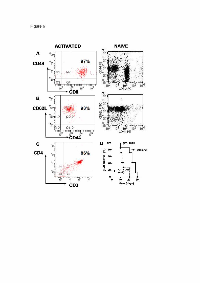
Figure 6
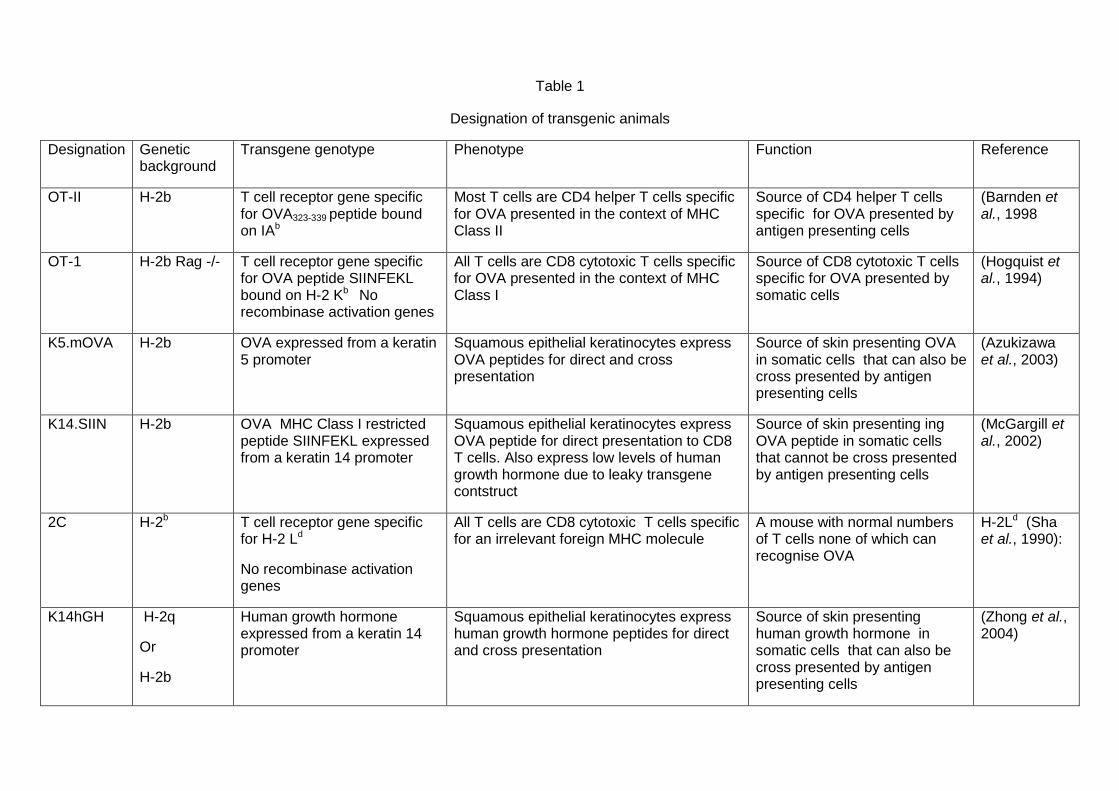
Table 1
Designation of transgenic animals
Designation Genetic background
Transgene genotype Phenotype Function Reference
OT-II H-2b T cell receptor gene specific for OVA323-339 peptide bound on IAb
Most T cells are CD4 helper T cells specific for OVA presented in the context of MHC Class II
Source of CD4 helper T cells specific for OVA presented by antigen presenting cells
(Barnden et al., 1998
OT-1
H-2b Rag -/- T cell receptor gene specific for OVA peptide SIINFEKL bound on H-2 Kb No recombinase activation genes
All T cells are CD8 cytotoxic T cells specific for OVA presented in the context of MHC Class I
Source of CD8 cytotoxic T cells specific for OVA presented by somatic cells
(Hogquist et al., 1994)
K5.mOVA H-2b OVA expressed from a keratin 5 promoter
Squamous epithelial keratinocytes express OVA peptides for direct and cross presentation
Source of skin presenting OVA in somatic cells that can also be cross presented by antigen presenting cells
(Azukizawa et al., 2003)
K14.SIIN H-2b OVA MHC Class I restricted peptide SIINFEKL expressed from a keratin 14 promoter
Squamous epithelial keratinocytes express OVA peptide for direct presentation to CD8 T cells. Also express low levels of human growth hormone due to leaky transgene contstruct
Source of skin presenting ing OVA peptide in somatic cells that cannot be cross presented by antigen presenting cells
(McGargill et al., 2002)
2C H-2b T cell receptor gene specific for H-2 Ld
No recombinase activation genes
All T cells are CD8 cytotoxic T cells specific for an irrelevant foreign MHC molecule
A mouse with normal numbers of T cells none of which can recognise OVA
H-2Ld (Sha et al., 1990):
K14hGH H-2q
Or
H-2b
Human growth hormone expressed from a keratin 14 promoter
Squamous epithelial keratinocytes express human growth hormone peptides for direct and cross presentation
Source of skin presenting human growth hormone in somatic cells that can also be cross presented by antigen presenting cells
(Zhong et al., 2004)