Embed Size (px)
DESCRIPTION
TJPRC JOURNALS
Citation preview
www.tjprc.org [email protected]
COMPARATIVE STUDIES ON THE MORPHOLOGY, ANATOMY OF CROTON
BONPLADIANUMBAILL AND MICROCOCCAMERCURIALIS(L)
BENTH(EUPHORBIACEAE.)
SAI PRASANNA C. G1 & KARPAGAM .S 2 1Research Scholars, Department of Botany, Queen Mary’s College, Chennai, Tamil Nadu, India
2Associate Professor of Botany, Queen Mary’s College, Chennai, Tamil Nadu, India
ABSTRACT
Croton bonplandianumBaill and Micrococcamercurialis (L).Benthbelonging to Euphorbiaceae were
distinguished apart from their ethanobotanicalvalues,for which its morphological and characters are included. The
present study was undertaken in complete morphology and anatomical characters of leaf, stem, root of both the plants.
Morphological features was observed in leaf, stem,root , fruit with seed. Anatomical features were noted by rotary
microtome sections and free hand safranin stained sections, and examined under bright field microscope. Both the
plants posses the common features like calcium oxalatecrystals distributed in leaf,well representedvascular bundles in
petiole, leaf, stem and root. Their distinct features like vasculature in petiole, sclerenchyma, lactifers are observed only
in Croton bonplandianum.,andMicrococcamercurialishas advance organization of phloem elements concentric
,amphicribralvascularisation in leaf and stem, no sclerenchyma , prominent bicollateral vasculature in wing-bundlesof
petiole.Leaf lamina showed loosely arranged spongy parenchyma with wide air-space. The root shows oxidative
hermidin blue layer with crushed cortex. These anatomical results are helpful to learn more about Claoxylonfeaturesin
family Euphorbiaceae .
KEYWORDS :Amphicribral ,Ethanobotinal ,Hermidin, Lactifers , Mesophyll, Petiole , Sclerenchyma,
Received: Oct 30, 2015; Accepted: Nov 04, 2015 ; Published: Nov 14, 2015 ; Paper Id.: IJBRDEC20151
INTRODUCTION
Each plant have some unique characters to make its identity clear from others Croton bonpladianumBaill
and Micrococcamercurialis (L) Benth are common weedsbelongs to family Euphorbiaceae.
Complexity in habitat range and variability in morphology and genetics has made Euphorbiaceae
classification difficult. The leaves are mostly alternate but may be opposite or whorled and they may be simple,
compound and sometimes highly reduced. Stipules are present but may be reduced to glands or spines. Flowers
are unisexual and usually actinomorphic.They may be highly reduced by suppression of parts, in extream form
consisting of naked stamen as a staminate flower and a naked pistil as a pistillate flower. A specialized type of
miniature inflorescence occurs in about 1,500 species comprising the genera Euphorbia.It consisitsof a single
stamen. These flowers are all enclosed in cup –like involucre that is provided with peripheral nectarines and
petalloid appendages such that the whole aggregations closely resemble a single flower( R.Elumalai et
al.,2014).Extra floral nectaries are also common.The hairs are diverse and include glandular and non-
glandular(C.R. Metcalfe and L.Chalk,1957) .The enumeration of several anatomic features like wood structure,
laticifer type, trichomes and nature of stomata as being important for family classification, while other like pollen
Original A
rticle International Journal of Botany and Research (IJBR) ISSN(P): 2277-4815; ISSN(E): 2319-4456 Vol. 5, Issue 6, Dec 2015, 1-16 © TJPRC Pvt. Ltd.
2 Sai Prasanna C. G & Karpagam .S
Impact Factor (JCC): 2.1259 Index Copernicus Value (ICV): 3.0
nuclear, exine structures, type of pollination and inflorescence types are important for classifying genera, tribes and
subfamilies(Yuvraj D. Adsul, Raghunath T. Mahajan and Shamkant B. Badgujar 2013).
Croton is a genus comprising around 1,300 species, widespread in tropicalregions of the Old and New
Worlds.Several species have a long role in the traditional use of medicinalplants in Africa, Asia and South America.
Popular uses include treatment of cancer, constipation,diabetes, digestive problems, dysentery, external wounds, fever,
hypercholesterolemia, hypertension,inflammation, intestinal worms, malaria, pain, ulcers and weight-loss. Several species
of Croton have a red sap, in some species containing proanthocyanidinsand/or alkaloids. The latter may betaspine or some
of several benzylisoquinoline-like compounds. Diterpenes are very common in Croton, corresponding to clerodanes,
cembranoid, halimanes, kauranes, labdanes, phorbol esters,trachylobanes and sarcopetalanes.(Antonio Salatino,* Maria L.
FariaSalatino and Giuseppina Negri. 2007).
Micrococcais a genuscomporising 18 species, a native to tropical Africa.Most are herbs,but some are succulent
and resemble cacti. Itis a wild food plant contributing to local household food and livelihood(Jeyachandradan .R ,Bastin
2013). The whole plant is used to treat children with fever and the plant- sap is instilled into the nose, eyes, or ears to treat
headache, filariarisof the eye or otitis, respectively(Yuvraj D. Adsul, Raghunath T. Mahajan and Shamkant B. Badgujar
2013).The phytochemical screening analysis showed the presence of alkaloids, phenols, amino acids, diterpenesterpenoids,
proteins, oxalate, cardiac glycosides, xanthoproteins, anthocyanin and saponin (Sai Prasanna C. G,., Poongani .M and
Karpagam .S Phytochemical content of the leaf, stem and root of Micrococcamercurialis (L.)Benth. A promising
herb.IOSR Journal of Pharmacy and Biological Sciences Volume 10, Issue 3 Ver. II (May - Jun. 2015), PP 24-27
MATERIALS AND METHODS
Plant Material
Healthy plants for studies have collected in Queen Mary’S College for Micrococcamercurialis (L) Benth (Reg
.no. PARC/2015/3092) and Croton bonplandianum Baill was collected from agricultural fields of Nellikuppam ,
Kancheepuram District. The plant materials was authenticated by Prof. Jayaraman, Director , Plant Anatomy Research
Centre (PARC) Chennai 45.
Macroscopic Evaluation
Macroscopic characters like habitat, habit, shape, size, morphology of the two plants including their vegetative,
reproductive and embryonic structures were observed.
Microscopic Evaluation
Different parts of the plant, such as leaf (midrib and lamina), petiole, stem, rhizome and roots were fixed in FAA
(Formalin: Acetic acid: 70% Ethanol; 5:5:90) for 24 hrs. After fixation the specimens were washed, dehydrated by passing
through Tertiary butyl alcohol (TBA) series following the procedure of Sass, 1940. After dehydration, the specimens were
infiltrated with paraffin wax (melting point 56-58°C) ad embedded in the paraffin blocks. Sections were cut using Rotary
microtome at a thickness of 10µm. Sections were stained with Toluidine blue (0.01%) aqueous solution. Photomicrographs
were prepared with Nikon trinologular microscope and Nikon digital camera.
The saffri n stained free hand sections are taken for studying the cortex layers and vascular bundle organization
only in Micrococcamercurialis. It was observed under various magnifications (10x, 20x, 40x and 100x) of trinocular
Comparative Studies on the Morphology, Anatomy of croton 3
Bonpladianum Baill and Micrococcamercurialis(L) Benth(Euphorbiaceae)
www.tjprc.org [email protected]
microscope and pictures are captured accordingly.
RESULT AND DISCUSSIONS
Macroscopic Evaluation
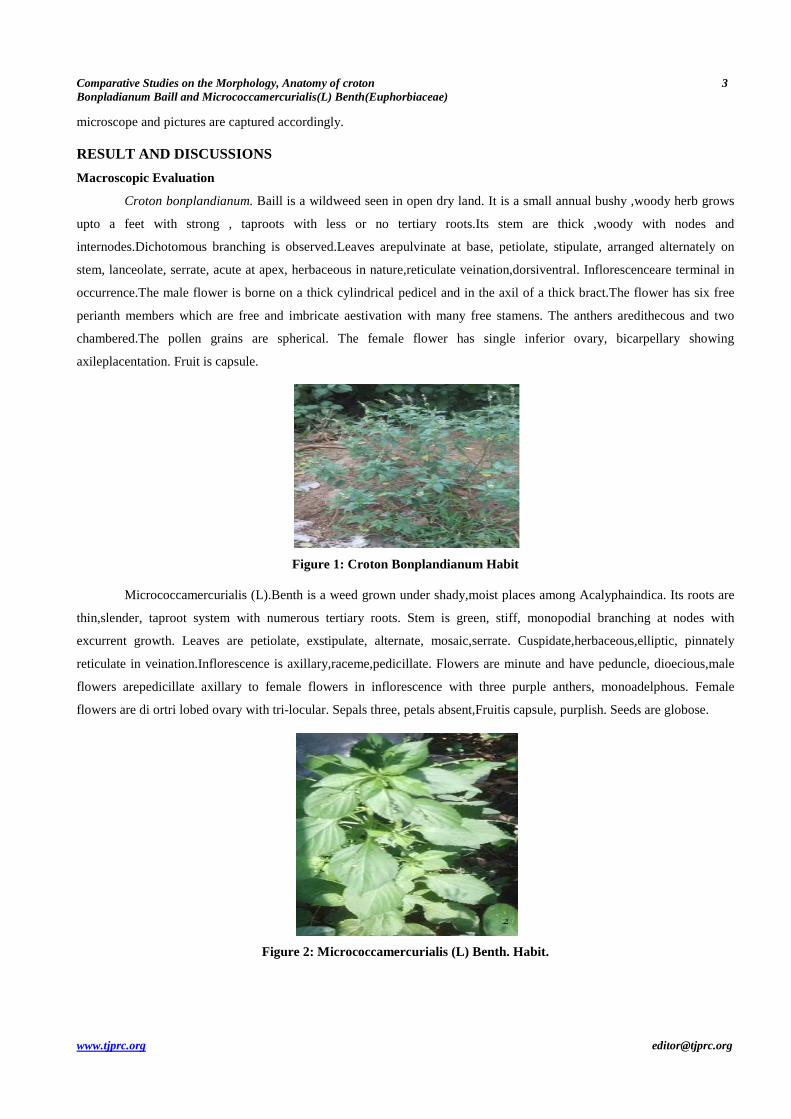
Croton bonplandianum. Baill is a wildweed seen in open dry land. It is a small annual bushy ,woody herb grows
upto a feet with strong , taproots with less or no tertiary roots.Its stem are thick ,woody with nodes and
internodes.Dichotomous branching is observed.Leaves arepulvinate at base, petiolate, stipulate, arranged alternately on
stem, lanceolate, serrate, acute at apex, herbaceous in nature,reticulate veination,dorsiventral. Inflorescenceare terminal in
occurrence.The male flower is borne on a thick cylindrical pedicel and in the axil of a thick bract.The flower has six free
perianth members which are free and imbricate aestivation with many free stamens. The anthers aredithecous and two
chambered.The pollen grains are spherical. The female flower has single inferior ovary, bicarpellary showing
axileplacentation. Fruit is capsule.
Figure 1: Croton Bonplandianum Habit
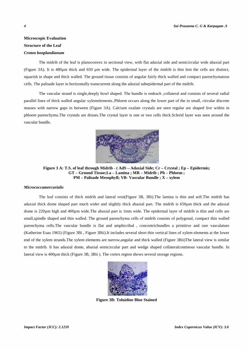
Micrococcamercurialis (L).Benth is a weed grown under shady,moist places among Acalyphaindica. Its roots are
thin,slender, taproot system with numerous tertiary roots. Stem is green, stiff, monopodial branching at nodes with
excurrent growth. Leaves are petiolate, exstipulate, alternate, mosaic,serrate. Cuspidate,herbaceous,elliptic, pinnately
reticulate in veination.Inflorescence is axillary,raceme,pedicillate. Flowers are minute and have peduncle, dioecious,male
flowers arepedicillate axillary to female flowers in inflorescence with three purple anthers, monoadelphous. Female
flowers are di ortri lobed ovary with tri-locular. Sepals three, petals absent,Fruitis capsule, purplish. Seeds are globose.
Figure 2: Micrococcamercurialis (L) Benth. Habit.
4
Impact Factor (JCC): 2.1259
Microscopic Evaluation
Structure of the Leaf
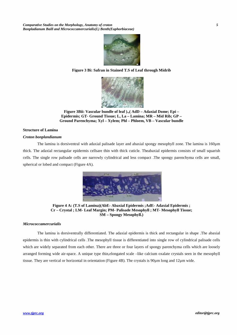
Croton bonplandianum
The midrib of the leaf is planoconvex in sectional view, with
(Figure 3A). It is 480µm thick and 650 µm wide. The
squarish in shape and thick walled. The ground tissue consists of angular
cells. The palisade layer in horizontally transcurrent along the adaxial subepidermal part of the midrib.
The vascular strand is single,deeply bowl shaped. The bundle
parallel lines of thick walled angular xylem
masses with narrow gaps in between (
phloem parenchyma.The crystals are druses.
vascular bundle.
Figure 3 A: T.S. of leaf through GT – Ground Tissue;La
PM – Palisade Mesophyll; V
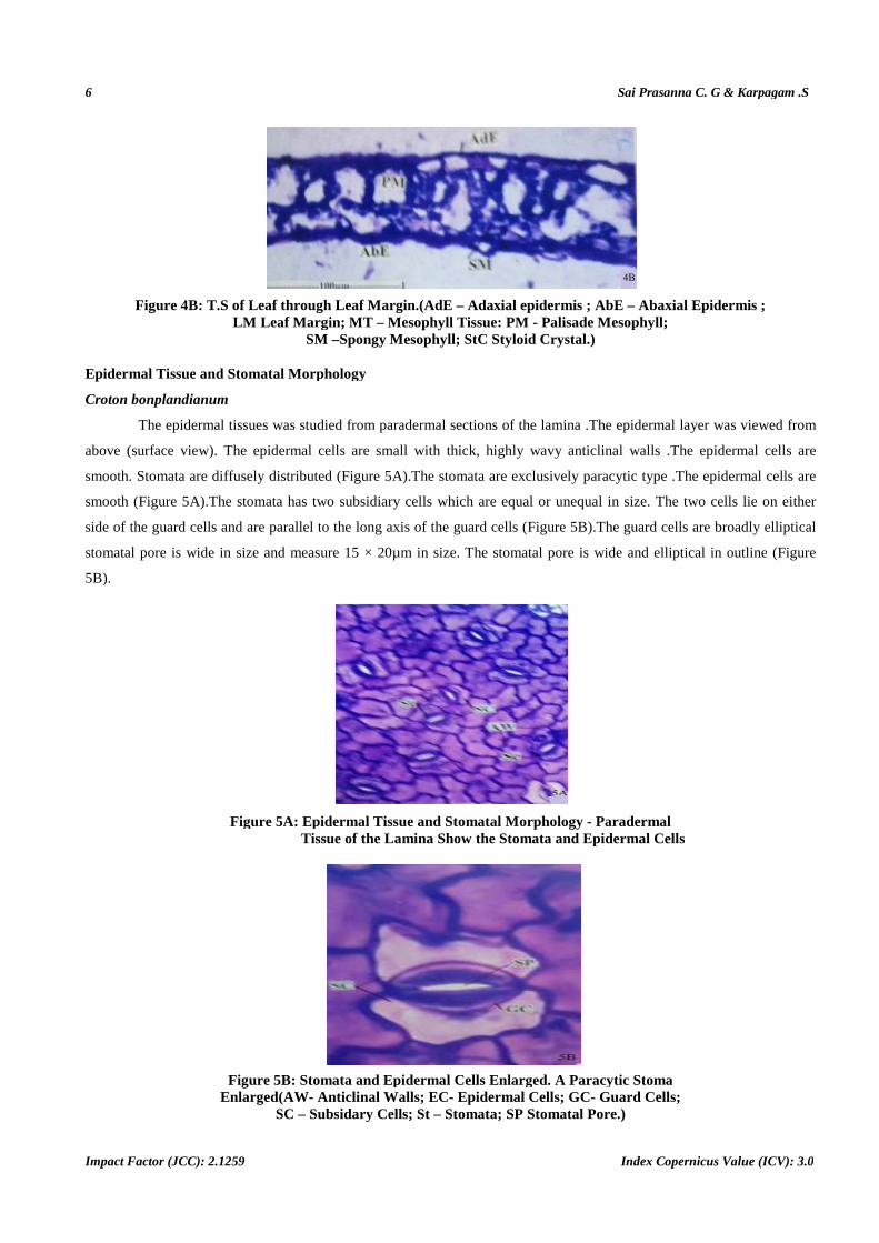
Micrococcamercurialis
The leaf consists of thick midrib and lateral vein(
adaxial thick dome shaped part much wider and slightly thick abaxial part. The midrib is 650µm thick and the adaxial
dome is 220µm high and 400µm wide.The abaxial part is 1mm wide. The epidermal layer of mid
small,spindle shaped and thin walled. The ground parenchyma cells
parenchyma cells.The vascular bundle is flat and
(Katherine Esau 1965) (Figure 3Bi , Fig
end of the xylem strands.The xylem elements are narrow,
to the midrib. It has adaxial dome, abaxial semicir
lateral view is 400µm thick (Figure 3B, 3Bii
Sai Prasanna C. G & Karpagam .S
Index Copernicus Value (ICV): 3.0
oconvex in sectional view, with flat adaxial side and semicircular wide
. It is 480µm thick and 650 µm wide. The epidermal layer of the midrib is thin lent the cells are distinct
. The ground tissue consists of angular fairly thick walled and compact parenchymatous
cells. The palisade layer in horizontally transcurrent along the adaxial subepidermal part of the midrib.
,deeply bowl shaped. The bundle is endrach ,collateral and consists of se
parallel lines of thick walled angular xylemelements..Phloem occurs along the lower part of the in smal
masses with narrow gaps in between (Figure 3A). Calcium oxalate crystals are seen regular are shaped live withi
oem parenchyma.The crystals are druses.The crystal layer is one or two cells thick.Sclerid layer was seen around the
T.S. of leaf through Midrib - ( AdS – Adaxial Side; Cr – Crystal ; Ep Ground Tissue;La – Lamina ; MR – Midrib ; Ph – Phloem ;
Palisade Mesophyll; VB- Vascular Bundle ; X – xylem
The leaf consists of thick midrib and lateral vein(Figure 3B, 3Bi).The lamina is thin and soft.
adaxial thick dome shaped part much wider and slightly thick abaxial part. The midrib is 650µm thick and the adaxial
dome is 220µm high and 400µm wide.The abaxial part is 1mm wide. The epidermal layer of mid
d thin walled. The ground parenchyma cells of midrib consists of polygonal
bundle is flat and amphicribal , concentricbundles a primitive and rare vasculature
, Figure 3Bii).It includes several short thin vertical lines of xylem elements at the
end of the xylem strands.The xylem elements are narrow,angular and thick walled (Figure 3B
, abaxial semicircular part and wedge shaped collateralcontinous
, 3Bii ). The cortex region shows several storage regions.
Figure 3B: Toluidine Blue Stained
Sai Prasanna C. G & Karpagam .S
Index Copernicus Value (ICV): 3.0
t adaxial side and semicircular wide abaxial part
hin lent the cells are distinct,
fairly thick walled and compact parenchymatous
cells. The palisade layer in horizontally transcurrent along the adaxial subepidermal part of the midrib.
collateral and consists of several radial
g the lower part of the in small, circular discrete
). Calcium oxalate crystals are seen regular are shaped live within in
Sclerid layer was seen around the
Crystal ; Ep – Epidermis; Phloem ; xylem
).The lamina is thin and soft.The midrib has
adaxial thick dome shaped part much wider and slightly thick abaxial part. The midrib is 650µm thick and the adaxial
dome is 220µm high and 400µm wide.The abaxial part is 1mm wide. The epidermal layer of midrib is thin and cells are
of midrib consists of polygonal, compact thin walled
bundles a primitive and rare vasculature
It includes several short thin vertical lines of xylem elements at the lower
3Bii)The lateral view is similar
continous vascular bundle. In
The cortex region shows several storage regions.
Comparative Studies on the Morphology, Anatomy ofBonpladianum Baill and Micrococcamercurialis(L) Benth(Euphorbiaceae
www.tjprc.org
Figure 3 Bi:
Figure 3Bii: Vascular bundle of leaf ).,( AdD Epidermis; GT
Ground Parenchyma;
Structure of Lamina
Croton bonplandianum
The lamina is dorsiventral with ada
thick. The adaxial rectangular epidermis
cells. The single row palisade cells are narrowly cylindrical and less c
spherical or lobed and compact (Figure
Figure 4 A: (T.S of LaminaCr – Crystal ; LM- Leaf Margin; PM
Micrococcamercurialis
The lamina is dorsiventrally differentiated. The adaxial epidermis is t
epidermis is thin with cylindrical cells
which are widely separated from each other.
arranged forming wide air-space. A unique type thin,elongated
tissue. They are vertical or horizontal in orientation (
Comparative Studies on the Morphology, Anatomy of croton curialis(L) Benth(Euphorbiaceae)
Figure 3 Bi: Safran in Stained T.S of Leaf through Midrib
Figure 3Bii: Vascular bundle of leaf ).,( AdD – Adaxial Dome; Epi Epidermis; GT- Ground Tissue; L, La – Lamina; MR – Mid Rib; GP
Ground Parenchyma; Xyl – Xylem; Phl – Phloem, VB – Vascular bundle
lamina is dorsiventral with adaxial palisade layer and abaxial spongy mesophyll zone.
epidermis cellsare thin with thick cuticle. Theabaxial epidermis consists of small squarish
are narrowly cylindrical and less compact .The spongy parenchyma cells
4A).
T.S of Lamina)(AbE- Abaxial Epidermis ;AdE- Adaxial Epidermis ;Leaf Margin; PM - Palisade Mesophyll ; MT- Mesophyll Tissue;
SM – Spongy Mesophyll.)
lamina is dorsiventrally differentiated. The adaxial epidermis is thick and rectangular in shape .
.The mesophyll tissue is differentiated into single row of cylindrical palisade cells
dely separated from each other. There are three or four layers of spongy parenchyma cells which are loosely
A unique type thin,elongated scale –like calcium oxalate crystals seen in the mesop
or horizontal in orientation (Figure 4B). The crystals is 90µm long and 12µm wide.
5
Adaxial Dome; Epi – Mid Rib; GP –
Vascular bundle
ngy mesophyll zone. The lamina is 160µm
al epidermis consists of small squarish
ompact .The spongy parenchyma cells are small,
Adaxial Epidermis ; Mesophyll Tissue;
hick and rectangular in shape .The abaxial
ll tissue is differentiated into single row of cylindrical palisade cells
three or four layers of spongy parenchyma cells which are loosely
like calcium oxalate crystals seen in the mesophyll
). The crystals is 90µm long and 12µm wide.
6
Impact Factor (JCC): 2.1259
Figure 4B: T.S of Leaf through Leaf Margin.(AdE LM Lea f Margin; MT
SM
Epidermal Tissue and Stomatal Morphology
Croton bonplandianum
The epidermal tissues was studied from
above (surface view). The epidermal cells are small
smooth. Stomata are diffusely distributed (
smooth (Figure 5A).The stomata has two subsidiary cells which
side of the guard cells and are parallel t
stomatal pore is wide in size and measure 15 × 20µm in size.
5B).
Figure 5A: Epidermal Tissue of the Lamina Show the Stomata and Epidermal Cells
Figure 5B: Stomata and Epidermal Cells Enlarged.Enlarged(AW- Anticlinal Walls;
SC – Subsidary
Sai Prasanna C. G & Karpagam .S
Index Copernicus Value (ICV): 3.0
T.S of Leaf through Leaf Margin.(AdE – Adaxial epidermis ; AbE – f Margin; MT – Mesophyll Tissue: PM - Palisade Mesophyll
SM –Spongy Mesophyll; StC Styloid Crystal.)
Tissue and Stomatal Morphology
idermal tissues was studied from paradermal sections of the lamina .The epidermal layer was viewed from
above (surface view). The epidermal cells are small with thick, highly wavy anticlinal walls .The epidermal cells are
y distributed (Figure 5A).The stomata are exclusively paracytic type .
The stomata has two subsidiary cells which are equal or unequal in size.
side of the guard cells and are parallel to the long axis of the guard cells (Figure 5B).The guard cells are broadly elliptical
and measure 15 × 20µm in size. The stomatal pore is wide and elliptical in outline
Figure 5A: Epidermal Tissue and Stomatal Morphology - Paradermal Tissue of the Lamina Show the Stomata and Epidermal Cells
Stomata and Epidermal Cells Enlarged. A Paracytic Stoma Anticlinal Walls; EC- Epidermal Cells; GC- Guard Cells;
Subsidary Cells; St – Stomata; SP Stomatal Pore.)
Sai Prasanna C. G & Karpagam .S
Index Copernicus Value (ICV): 3.0
Abaxial Epidermis ; Palisade Mesophyll;
The epidermal layer was viewed from
highly wavy anticlinal walls .The epidermal cells are
re exclusively paracytic type .The epidermal cells are
equal or unequal in size. The two cells lie on either
The guard cells are broadly elliptical
and elliptical in outline (Figure
Paradermal Tissue of the Lamina Show the Stomata and Epidermal Cells
A Paracytic Stoma Guard Cells;
Comparative Studies on the Morphology, Anatomy of croton 7
Bonpladianum Baill and Micrococcamercurialis(L) Benth(Euphorbiaceae)
www.tjprc.org [email protected]
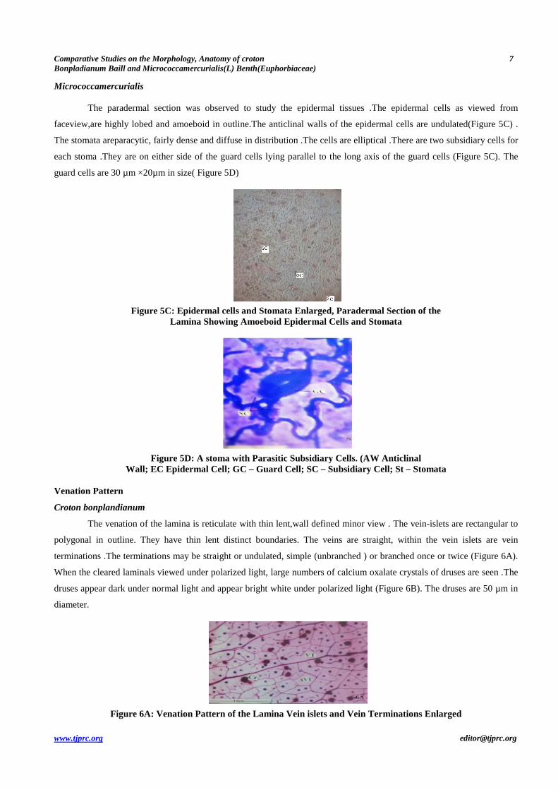
Micrococcamercurialis
The paradermal section was observed to study the epidermal tissues .The epidermal cells as viewed from
faceview,are highly lobed and amoeboid in outline.The anticlinal walls of the epidermal cells are undulated(Figure 5C) .
The stomata areparacytic, fairly dense and diffuse in distribution .The cells are elliptical .There are two subsidiary cells for
each stoma .They are on either side of the guard cells lying parallel to the long axis of the guard cells (Figure 5C). The
guard cells are 30 µm ×20µm in size( Figure 5D)
Figure 5C: Epidermal cells and Stomata Enlarged, Paradermal Section of the Lamina Showing Amoeboid Epidermal Cells and Stomata
Figure 5D: A stoma with Parasitic Subsidiary Cells. (AW Anticlinal Wall; EC Epidermal Cell; GC – Guard Cell; SC – Subsidiary Cell; St – Stomata
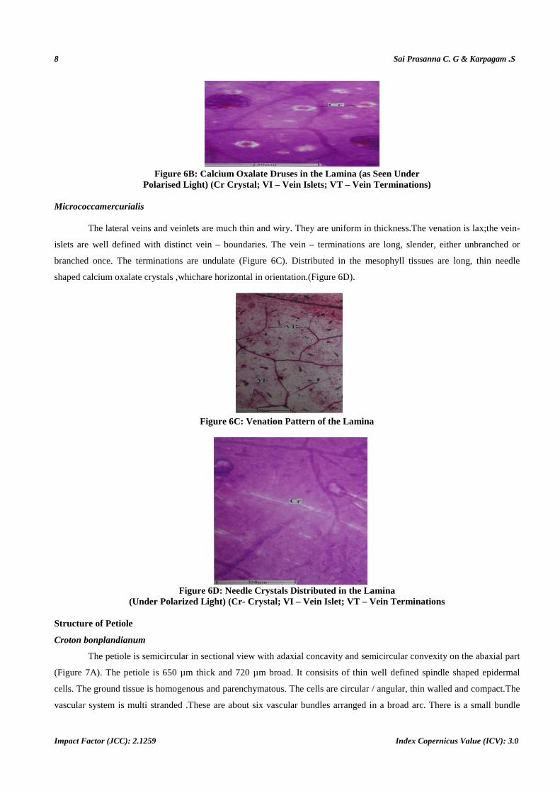
Venation Pattern
Croton bonplandianum
The venation of the lamina is reticulate with thin lent,wall defined minor view . The vein-islets are rectangular to
polygonal in outline. They have thin lent distinct boundaries. The veins are straight, within the vein islets are vein
terminations .The terminations may be straight or undulated, simple (unbranched ) or branched once or twice (Figure 6A).
When the cleared laminals viewed under polarized light, large numbers of calcium oxalate crystals of druses are seen .The
druses appear dark under normal light and appear bright white under polarized light (Figure 6B). The druses are 50 µm in
diameter.
Figure 6A: Venation Pattern of the Lamina Vein islets and Vein Terminations Enlarged
8 Sai Prasanna C. G & Karpagam .S
Impact Factor (JCC): 2.1259 Index Copernicus Value (ICV): 3.0
Figure 6B: Calcium Oxalate Druses in the Lamina (as Seen Under Polarised Light) (Cr Crystal; VI – Vein Islets; VT – Vein Terminations)
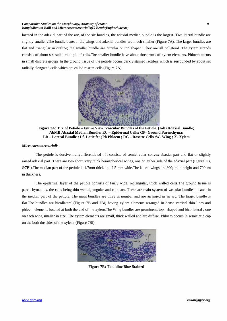
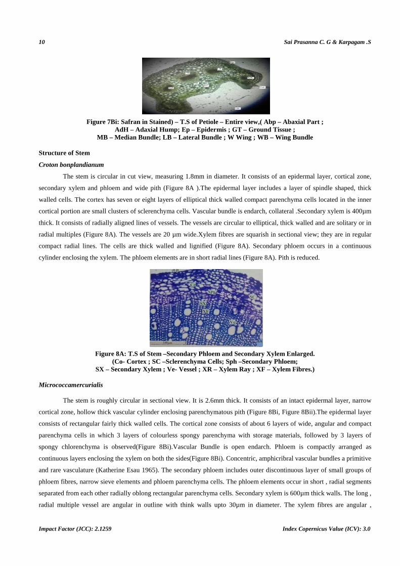
Micrococcamercurialis
The lateral veins and veinlets are much thin and wiry. They are uniform in thickness.The venation is lax;the vein-
islets are well defined with distinct vein – boundaries. The vein – terminations are long, slender, either unbranched or
branched once. The terminations are undulate (Figure 6C). Distributed in the mesophyll tissues are long, thin needle
shaped calcium oxalate crystals ,whichare horizontal in orientation.(Figure 6D).
Figure 6C: Venation Pattern of the Lamina
Figure 6D: Needle Crystals Distributed in the Lamina (Under Polarized Light) (Cr- Crystal; VI – Vein Isl et; VT – Vein Terminations
Structure of Petiole
Croton bonplandianum
The petiole is semicircular in sectional view with adaxial concavity and semicircular convexity on the abaxial part
(Figure 7A). The petiole is 650 µm thick and 720 µm broad. It consisits of thin well defined spindle shaped epidermal
cells. The ground tissue is homogenous and parenchymatous. The cells are circular / angular, thin walled and compact.The
vascular system is multi stranded .These are about six vascular bundles arranged in a broad arc. There is a small bundle
Comparative Studies on the Morphology, Anatomy ofBonpladianum Baill and Micrococcamercurialis(L) Benth(Euphorbiaceae
www.tjprc.org
located in the adaxial part of the arc, of the six bundles
slightly smaller .The bundle beneath the wings and adaxial bundles are much smaller (
flat and triangular in outline; the smaller bundle are circular or top s
consists of about six radial multiple of cells.
in small discrete groups In the ground tissue of the petiole occurs darkly stained lactifers which is surrounded by about six
radially elongated cells which are called rosette cells (
Figure 7A: T.S. of Petiole –AbMB Abaxial Median Bundle
LB – Lateral Bundle ; Lf
Micrococcamercurialis
The petiole is dorsiventrallydifferentiated . It consists of semicircular convex abaxial part and flat or slightly
raised adaxial part. There are two short
&7Bi) .The median part of the petiole is 1.7mm thick and
in thickness.
The epidermal layer of the petiole consists of fairly wide,
parenchymatous, the cells being thin walled
the median part of the petiole. The ma
flat.The bundles are bicollateral,(Figure
phloem elements located at both the end of t
on each wing smaller in size. The xylem elements are smal
on the both the sides of the xylem. (Figure
Comparative Studies on the Morphology, Anatomy of croton curialis(L) Benth(Euphorbiaceae)
of the arc, of the six bundles, the adaxial median bundle is the largest. Two
The bundle beneath the wings and adaxial bundles are much smaller (Figure
flat and triangular in outline; the smaller bundle are circular or top shaped. They are all collateral
ut six radial multiple of cells.The smaller bundle have about three rows of xylem elements
In the ground tissue of the petiole occurs darkly stained lactifers which is surrounded by about six
ially elongated cells which are called rosette cells (Figure 7A).
– Entire View. Vascular Bundles of the Petiole. (AdB Adaxial Bundle; AbMB Abaxial Median Bundle; EC – Epidermal Cells; GP- Ground Parenchyma
Lateral Bundle ; Lf - Laticifer ;Ph Phloem ; RC – Rosette Cells ;W- Wing ; X
The petiole is dorsiventrallydifferentiated . It consists of semicircular convex abaxial part and flat or slightly
two short, very thick hemispherical wings, one on either side of the adaxial part (
.The median part of the petiole is 1.7mm thick and 2.5 mm wide.The lateral wings are 800µm in height and 700µm
The epidermal layer of the petiole consists of fairly wide, rectangular, thick walled cells.
walled, angular and compact. These are main system of vascular bundles located in
etiole. The main bundles are three in number and are arranged in an
ure 7B and 7Bi) having xylem elements arranged in dense vertical thin lines and
end of the xylem.The Wing bundles are prominent, top
The xylem elements are small, thick walled and are diffuse. Ph
ure 7Bi).
Figure 7B: Toluidine Blue Stained
9
, the adaxial median bundle is the largest. Two lateral bundle are
ure 7A). The larger bundles are
haped. They are all collateral. The xylem strands
ut three rows of xylem elements. Phloem occurs
In the ground tissue of the petiole occurs darkly stained lactifers which is surrounded by about six
Bundles of the Petiole. (AdB Adaxial Bundle; Ground Parenchyma;
Wing ; X- Xylem
The petiole is dorsiventrallydifferentiated . It consists of semicircular convex abaxial part and flat or slightly
, one on either side of the adaxial part (Figure 7B,
2.5 mm wide.The lateral wings are 800µm in height and 700µm
rectangular, thick walled cells.The ground tissue is
, angular and compact. These are main system of vascular bundles located in
and are arranged in an arc. The larger bundle is
having xylem elements arranged in dense vertical thin lines and
prominent, top –shaped and bicollateral , one
hloem occurs in semicircle cap
10 Sai Prasanna C. G & Karpagam .S
Impact Factor (JCC): 2.1259 Index Copernicus Value (ICV): 3.0
Figure 7Bi: Safran in Stained) – T.S of Petiole – Entire view,( Abp – Abaxial Part ; AdH – Adaxial Hump; Ep – Epidermis ; GT – Ground Tissue ;
MB – Median Bundle; LB – Lateral Bundle ; W Wing ; WB – Wing Bundle
Structure of Stem
Croton bonplandianum
The stem is circular in cut view, measuring 1.8mm in diameter. It consists of an epidermal layer, cortical zone,
secondary xylem and phloem and wide pith (Figure 8A ).The epidermal layer includes a layer of spindle shaped, thick
walled cells. The cortex has seven or eight layers of elliptical thick walled compact parenchyma cells located in the inner
cortical portion are small clusters of sclerenchyma cells. Vascular bundle is endarch, collateral .Secondary xylem is 400µm
thick. It consists of radially aligned lines of vessels. The vessels are circular to elliptical, thick walled and are solitary or in
radial multiples (Figure 8A). The vessels are 20 µm wide.Xylem fibres are squarish in sectional view; they are in regular
compact radial lines. The cells are thick walled and lignified (Figure 8A). Secondary phloem occurs in a continuous
cylinder enclosing the xylem. The phloem elements are in short radial lines (Figure 8A). Pith is reduced.
Figure 8A: T.S of Stem –Secondary Phloem and Secondary Xylem Enlarged. (Co- Cortex ; SC –Sclerenchyma Cells; Sph –Secondary Phloem;
SX – Secondary Xylem ; Ve- Vessel ; XR – Xylem Ray ; XF – Xylem Fibres.)
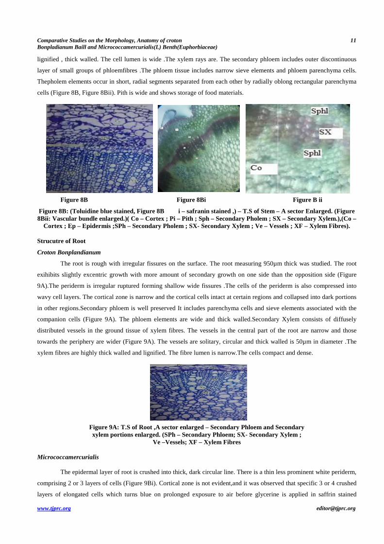
Micrococcamercurialis
The stem is roughly circular in sectional view. It is 2.6mm thick. It consists of an intact epidermal layer, narrow
cortical zone, hollow thick vascular cylinder enclosing parenchymatous pith (Figure 8Bi, Figure 8Bii).The epidermal layer
consists of rectangular fairly thick walled cells. The cortical zone consists of about 6 layers of wide, angular and compact
parenchyma cells in which 3 layers of colourless spongy parenchyma with storage materials, followed by 3 layers of
spongy chlorenchyma is observed(Figure 8Bi).Vascular Bundle is open endarch. Phloem is compactly arranged as
continuous layers enclosing the xylem on both the sides(Figure 8Bi). Concentric, amphicribral vascular bundles a primitive
and rare vasculature (Katherine Esau 1965). The secondary phloem includes outer discontinuous layer of small groups of
phloem fibres, narrow sieve elements and phloem parenchyma cells. The phloem elements occur in short , radial segments
separated from each other radially oblong rectangular parenchyma cells. Secondary xylem is 600µm thick walls. The long ,
radial multiple vessel are angular in outline with think walls upto 30µm in diameter. The xylem fibres are angular ,
Comparative Studies on the Morphology, Anatomy ofBonpladianum Baill and Micrococcamercurialis(L) Benth(Euphorbiaceae
www.tjprc.org
lignified , thick walled. The cell lumen is wide .
layer of small groups of phloemfibres .
Thepholem elements occur in short, radial segments separated from each other by radially oblong rectangular parenchyma
cells (Figure 8B, Figure 8Bii). Pith is wide
Figure 8B
Figure 8B: (Toluidine blue stained, Fig8Bii: Vascular bundle enlarged.)( Co
Cortex ; Ep – Epidermis ;SPh – Secondary Pholem ; SX
Strucutre of Root
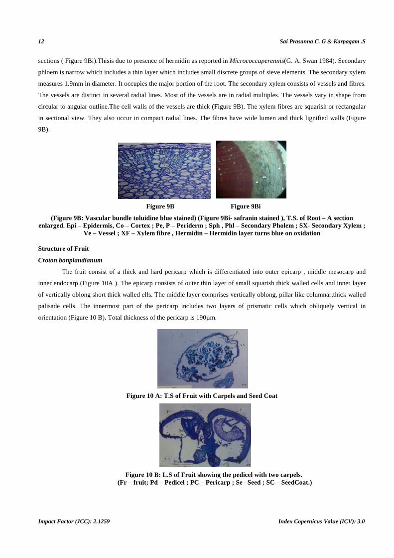
Croton Bonplandianum
The root is rough with irregular fissures on the surface
exihibits slightly excentric growth with more amount of secondary growth on one side than the opposition side (
9A).The periderm is irregular ruptured forming shall
wavy cell layers. The cortical zone is narrow and the cortical cells intact at certai
in other regions.Secondary phloem is well preserved
companion cells (Figure 9A). The phloem elem
distributed vessels in the ground tissue of xylem fibres.
towards the periphery are wider (Figure
xylem fibres are highly thick walled and lignifi
Figure 9A: T.S of Root ,A sector enlarged xylem portions enlarged.
Micrococcamercurialis
The epidermal layer of root is crushed into thick, dark
comprising 2 or 3 layers of cells (Figure
layers of elongated cells which turns blue on prolonged exposure to air before glycerine is applied in saffrin stained
Comparative Studies on the Morphology, Anatomy of croton curialis(L) Benth(Euphorbiaceae)
alled. The cell lumen is wide .The xylem rays are. The secondary phloem includes outer discontinuous
fibres .The phloem tissue includes narrow sieve elements and phloem parenchyma cells.
, radial segments separated from each other by radially oblong rectangular parenchyma
Pith is wide and shows storage of food materials.
Figure 8Bi
Figure 8B i – safranin stained ,) – T.S of Stem –( Co – Cortex ; Pi – Pith ; Sph – Secondary Pholem ; SX
Secondary Pholem ; SX- Secondary Xylem ; Ve – Vessels ; XF
regular fissures on the surface. The root measuring 950µm thick was studied. The root
exihibits slightly excentric growth with more amount of secondary growth on one side than the opposition side (
).The periderm is irregular ruptured forming shallow wide fissures .The cells of the periderm is also compressed into
wavy cell layers. The cortical zone is narrow and the cortical cells intact at certain regions and collapsed into dark portions
ndary phloem is well preserved It includes parenchyma cells and sieve elements associ
). The phloem elements are wide and thick walled.Secondary Xylem consists of diffusely
ground tissue of xylem fibres. The vessels in the central part of the root are
ure 9A). The vessels are solitary, circular and thick walled is 50µm in diameter .
xylem fibres are highly thick walled and lignified. The fibre lumen is narrow.The cells compact and dense.
T.S of Root ,A sector enlarged – Secondary Phloem and Secondary xylem portions enlarged. (SPh – Secondary Phloem; SX- Secondary Xylem ;
Ve –Vessels; XF – Xylem Fibres
ot is crushed into thick, dark circular line. There is a thin less prominent
ure 9Bi). Cortical zone is not evident,and it was observed that
turns blue on prolonged exposure to air before glycerine is applied in saffrin stained
11
The secondary phloem includes outer discontinuous
The phloem tissue includes narrow sieve elements and phloem parenchyma cells.
, radial segments separated from each other by radially oblong rectangular parenchyma
Figure B ii
– A sector Enlarged. (Figure Pholem ; SX – Secondary Xylem.),(Co –
Vessels ; XF – Xylem Fibres).
. The root measuring 950µm thick was studied. The root
exihibits slightly excentric growth with more amount of secondary growth on one side than the opposition side (Figure
The cells of the periderm is also compressed into
regions and collapsed into dark portions
des parenchyma cells and sieve elements associated with the
Secondary Xylem consists of diffusely
The vessels in the central part of the root are narrow and those
ck walled is 50µm in diameter .The
The cells compact and dense.
Secondary Phloem and Secondary Secondary Xylem ;
circular line. There is a thin less prominent white periderm,
,and it was observed that specific 3 or 4 crushed
turns blue on prolonged exposure to air before glycerine is applied in saffrin stained
12
Impact Factor (JCC): 2.1259
sections ( Figure 9Bi).Thisis due to presence of
phloem is narrow which includes a thin layer which includes small
measures 1.9mm in diameter. It occupies the major portion of the root. The secondary xylem consists of vessels and fibres.
The vessels are distinct in several radial lines. Most of the vessels are in
circular to angular outline.The cell walls of the vessels are thick (
in sectional view. They also occur in compact radial lines. The fibres have wide
9B).
(Figure 9B: Vascular bundle toluidine blue stained) enlarged. Epi – Epidermis, Co – Cortex ; Pe
Ve – Vessel ; XF – Xylem fibre , Hermidin
Structure of Fruit
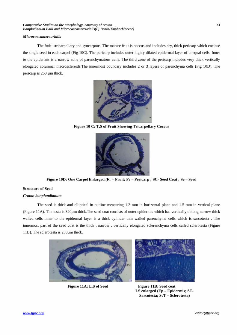
Croton bonplandianum
The fruit consist of a thick and hard pericarp which is differentiated into outer epicarp , middle mesocarp and
inner endocarp (Figure 10A ). The epicarp consists of outer thin layer of small squari
of vertically oblong short thick walled ell
palisade cells. The innermost part of the pericarp includes two layers of prismatic cells which obliquely vertical in
orientation (Figure 10 B). Total thickness of the peric
Figure 10 A:
Figure (Fr – fruit
Sai Prasanna C. G & Karpagam .S
Index Copernicus Value (ICV): 3.0
presence of hermidin as reported in Micrococcaperennis(G. A. Swan
hin layer which includes small discrete groups of sieve elements. The secondary xylem
measures 1.9mm in diameter. It occupies the major portion of the root. The secondary xylem consists of vessels and fibres.
The vessels are distinct in several radial lines. Most of the vessels are in radial multiples. The vessels vary in shape f
The cell walls of the vessels are thick (Figure 9B). The xylem fibres are squarish or rectangular
in sectional view. They also occur in compact radial lines. The fibres have wide lumen and thick lignified walls (
Figure 9B Figure 9Bi
Vascular bundle toluidine blue stained) (Figure 9Bi- safranin stained ), Cortex ; Pe, P – Periderm ; Sph , Phl – Secondary Pholem ; SX
Xylem fibre , Hermidin – Hermidin layer turns blue on
thick and hard pericarp which is differentiated into outer epicarp , middle mesocarp and
). The epicarp consists of outer thin layer of small squarish thick walled cells and inner
of vertically oblong short thick walled ells. The middle layer comprises vertically oblong, pillar like columnar,
palisade cells. The innermost part of the pericarp includes two layers of prismatic cells which obliquely vertical in
). Total thickness of the pericarp is 190µm.
ure 10 A: T.S of Fruit with Carpels and Seed Coat
ure 10 B: L.S of Fruit showing the pedicel with two carpels.fruit ; Pd – Pedicel ; PC – Pericarp ; Se –Seed ; SC – SeedCoat.)
Sai Prasanna C. G & Karpagam .S
Index Copernicus Value (ICV): 3.0
(G. A. Swan 1984). Secondary
discrete groups of sieve elements. The secondary xylem
measures 1.9mm in diameter. It occupies the major portion of the root. The secondary xylem consists of vessels and fibres.
radial multiples. The vessels vary in shape from
). The xylem fibres are squarish or rectangular
lumen and thick lignified walls (Figure
, T.S. of Root – A section Secondary Pholem ; SX- Secondary Xylem ;
turns blue on oxidation
thick and hard pericarp which is differentiated into outer epicarp , middle mesocarp and
sh thick walled cells and inner layer
yer comprises vertically oblong, pillar like columnar,thick walled
palisade cells. The innermost part of the pericarp includes two layers of prismatic cells which obliquely vertical in
carpels. SeedCoat.)
Comparative Studies on the Morphology, Anatomy of croton 13
Bonpladianum Baill and Micrococcamercurialis(L) Benth(Euphorbiaceae)
www.tjprc.org [email protected]
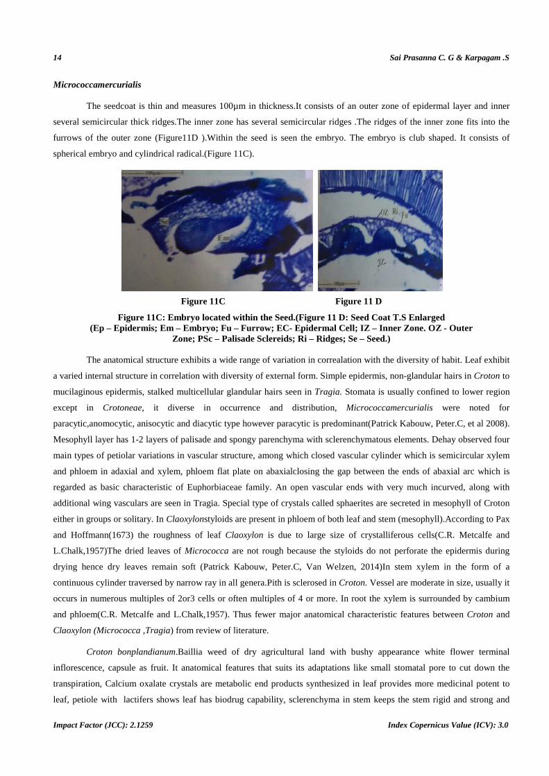
Micrococcamercurialis
The fruit istricarpellary and syncarpous .The mature fruit is coccus and includes dry, thick pericarp which enclose
the single seed in each carpel (Fig 10C). The pericarp includes outer highly dilated epidermal layer of unequal cells. Inner
to the epidermis is a narrow zone of parenchymatous cells. The third zone of the pericarp includes very thick vertically
elongated columnar macrosclereids.The innermost boundary includes 2 or 3 layers of parenchyma cells (Fig 10D). The
pericarp is 250 µm thick.
Figure 10 C: T.S of Fruit Showing Tricarpellary Coccus
Figure 10D: One Carpel Enlarged.(Fr – Fruit; Pe – Pericarp ; SC- Seed Coat ; Se – Seed
Structure of Seed
Croton bonplandianum
The seed is thick and elliptical in outline measuring 1.2 mm in horizontal plane and 1.5 mm in vertical plane
(Figure 11A). The testa is 320µm thick.The seed coat consists of outer epidermis which has vertically oblong narrow thick
walled cells inner to the epidermal layer is a thick cylinder thin walled parenchyma cells which is sarcotesta . The
innermost part of the seed coat is the thick , narrow , vertically elongated sclerenchyma cells called sclerotesta (Figure
11B). The sclerotesta is 230µm thick.
Figure 11A: L.S of Seed Figure 11B: Seed coat LS enlarged (Ep – Epidermis; ST-
Sarcotesta; ScT – Sclerotesta)
14 Sai Prasanna C. G & Karpagam .S
Impact Factor (JCC): 2.1259 Index Copernicus Value (ICV): 3.0
Micrococcamercurialis
The seedcoat is thin and measures 100µm in thickness.It consists of an outer zone of epidermal layer and inner
several semicircular thick ridges.The inner zone has several semicircular ridges .The ridges of the inner zone fits into the
furrows of the outer zone (Figure11D ).Within the seed is seen the embryo. The embryo is club shaped. It consists of
spherical embryo and cylindrical radical.(Figure 11C).
Figure 11C Figure 11 D
Figure 11C: Embryo located within the Seed.(Figure 11 D: Seed Coat T.S Enlarged (Ep – Epidermis; Em – Embryo; Fu – Furrow; EC- Epidermal Cell; IZ – Inner Zone. OZ - Outer
Zone; PSc – Palisade Sclereids; Ri – Ridges; Se – Seed.)
The anatomical structure exhibits a wide range of variation in correalation with the diversity of habit. Leaf exhibit
a varied internal structure in correlation with diversity of external form. Simple epidermis, non-glandular hairs in Croton to
mucilaginous epidermis, stalked multicellular glandular hairs seen in Tragia. Stomata is usually confined to lower region
except in Crotoneae, it diverse in occurrence and distribution, Micrococcamercurialis were noted for
paracytic,anomocytic, anisocytic and diacytic type however paracytic is predominant(Patrick Kabouw, Peter.C, et al 2008).
Mesophyll layer has 1-2 layers of palisade and spongy parenchyma with sclerenchymatous elements. Dehay observed four
main types of petiolar variations in vascular structure, among which closed vascular cylinder which is semicircular xylem
and phloem in adaxial and xylem, phloem flat plate on abaxialclosing the gap between the ends of abaxial arc which is
regarded as basic characteristic of Euphorbiaceae family. An open vascular ends with very much incurved, along with
additional wing vasculars are seen in Tragia. Special type of crystals called sphaerites are secreted in mesophyll of Croton
either in groups or solitary. In Claoxylonstyloids are present in phloem of both leaf and stem (mesophyll).According to Pax
and Hoffmann(1673) the roughness of leaf Claoxylon is due to large size of crystalliferous cells(C.R. Metcalfe and
L.Chalk,1957)The dried leaves of Micrococca are not rough because the styloids do not perforate the epidermis during
drying hence dry leaves remain soft (Patrick Kabouw, Peter.C, Van Welzen, 2014)In stem xylem in the form of a
continuous cylinder traversed by narrow ray in all genera.Pith is sclerosed in Croton. Vessel are moderate in size, usually it
occurs in numerous multiples of 2or3 cells or often multiples of 4 or more. In root the xylem is surrounded by cambium
and phloem(C.R. Metcalfe and L.Chalk,1957). Thus fewer major anatomical characteristic features between Croton and
Claoxylon (Micrococca ,Tragia) from review of literature.
Croton bonplandianum.Baillia weed of dry agricultural land with bushy appearance white flower terminal
inflorescence, capsule as fruit. It anatomical features that suits its adaptations like small stomatal pore to cut down the
transpiration, Calcium oxalate crystals are metabolic end products synthesized in leaf provides more medicinal potent to
leaf, petiole with lactifers shows leaf has biodrug capability, sclerenchyma in stem keeps the stem rigid and strong and
Comparative Studies on the Morphology, Anatomy of croton 15
Bonpladianum Baill and Micrococcamercurialis(L) Benth(Euphorbiaceae)
www.tjprc.org [email protected]
stores food , lignified xylem fibres with smaller lumen in root helps in effective water transportation in dry fields . Fruit
has three layers of pericarp so it is thick and rigid.
Micrococcamercurialis.(L)Benth is a slender herb , grown in shady moist place, with axillary green inflorescence
with inconspicuous male flower, green coccus capsule as fruit . The leaf and stem shares same type of concentric,
amphicribral vasculature which is observed in this species. Leaf has wide air space inmesophyll and also big stomatal
guard cells that helps in respiration. No sclerenchymatous layer is observed which serves the plant to be slender.Petiole has
advance vasculature pattern three vascular bundles in in midrib region and two in wings. The vascular bundles are
bicollateral. Leaf and veins has calcium oxalate layers which provides bio-drug potent to this herb. Root shows a peculiar
character of changing into blue on long exposure to air due hermidin. Xylem fibres of root has wide lumen shows less rigid
in nature. Fruit has only three layers of pericarp so it is tender
CONCLUSIONS
The above anatomical studies are useful for easy identification, taxonomic position and for authentication of these
plants for their drug potency.
ACKNOWELDGEMENT
Authors acknowledge the FIST – DST instrumentation facility of Queen Mary’s College, Dr.Prof. Jayaraman,
Director , Plant Anatomy Research Centre (PARC) Chennai – 45 , Head of the Department Mrs.R .Banumathy , Queen
Mary’s College , for providing needful help.
REFERENCES
1. Salatino,* Maria L. FariaSalatino and Giuseppina Negri. Traditional uses, Chemistry and Pharmacology of Croton species
(Euphorbiaceae). J. Braz. Chem. Soc., Vol. 18, No. 1, 11-33, 2007.Printed in Brazil - ©2007 SociedadeBrasileira de Antonio
Química0103 – 5053.
2. R. Elumalai et al., Foliar anatomical studies of some taxa of Euphobiaceae. African journal of Plant science vol 8(6) pp 271-
277, june 2014.
3. Jeyachandradan .R ,Bastin. M. An efficient protocol for in vitro flowering and fruiting in Micrococcamercurialis (L) Benth.
Internatiinal journal of Natural and Applied Science 2013;2(1); 18-22.
4. Katherine Esau 1965. PLANT ANATOMY. Second Edition, Wiley Eastern Limited, pp 368-369.
5. C.R. Metcalfe and L.Chalk,1957. ANATOMY OF DICOTYLEDONS. LEAVES ,STEM ,AND WOOD IN RELATION TO
TAXONOMY WITH NOTES ON ECONOMIC USES. Vol .II. Oxford University Press, Amen House, London E.C.4. p.1207-
1235.
6. Yuvraj D. Adsul, Raghunath T. Mahajan and Shamkant B. Badgujar. Ethnobotanical Euphorbian plants of North Maharashtra
Region . IOSR Journal of Pharmacy and Biological Sciences (IOSR-JPBS) e-ISSN: 2278-3008, p-ISSN:2319-7676. Volume 7,
Issue 1 (Jul. – Aug. 2013), PP 29-35 .
7. Patrick Kabouw, Peter.C, Van Welzen, Pieter Baas and Bertie.J, Van Heuven. Styloid crystals in Claoxylon(Euphorbiaceae)
and allies (Cloxylinae) with notes on leaf anatomy. Botanical journal of the Linnean Society, 2008,156,445-457.
8. Patrick Kabouw, Peter.C, Van Welzen,. Foliar Epidermal Studies of Plants in Euphorbiaceae.Taiwania,59(1): p:59-70,2014..
16 Sai Prasanna C. G & Karpagam .S
Impact Factor (JCC): 2.1259 Index Copernicus Value (ICV): 3.0
9. Ricardo de et al., An overview of recent taxonomic studies on Euphorbiaceaes.I. in Brazil. Rodriguesia 63(1); 227 – 247.
2012.
10. Sai Prasanna C. G,.,Poongani .M and Karpagam .S Phytochemical content of the leaf, stem and root of
Micrococcamercurialis (L.)Benth. A promising herb. IOSR Journal of Pharmacy and Biological Sciences Volume 10, Issue 3
Ver. II (May - Jun. 2015), PP 24-27
11. G. A. Swan. Hermidin, a chromogen from Mercurialisperennis L.Experientia15. 7.1984, Volume 40, Issue 7, pp 687-688