Embed Size (px)
Citation preview

1
Interactions Between CdsD, CdsQ and CdsL, Three Putative Chlamydophila pneumoniae 1
Type III Secretion Proteins 2
Dustin L. Johnson, Chris B. Stone and James B. Mahony* 3
4
M.G. DeGroote Institute for Infectious Disease Research, Faculty of Health Sciences and the Department of 5
Pathology and Molecular Medicine, McMaster University, and the Father Sean O’Sullivan Research Centre, St. 6
Joseph’s Healthcare, Hamilton, Ontario, CANADA 7
8
9
10
11
12
13
14
15
16
* Address correspondence to: 17
Dr. J.B. Mahony 18
Regional Virology and Chlamydiology Laboratory, St. Joseph’s Hospital 19
50 Charlton Avenue East, Hamilton, Ontario, CANADA 20
L8N 4A6 21
Phone: 905-521-6021 22
Fax: 905-521-6083 23
Email: [email protected] 24
25
Running title: Interaction of Cds proteins in C. pneumoniae 26
27
ACCEPTED
Copyright © 2008, American Society for Microbiology and/or the Listed Authors/Institutions. All Rights Reserved.J. Bacteriol. doi:10.1128/JB.01997-07 JB Accepts, published online ahead of print on 15 February 2008
on Septem
ber 16, 2018 by guesthttp://jb.asm
.org/D
ownloaded from

2
Abstract 1
Chlamydophila pneumoniae is a gram negative obligate intracellular bacterial pathogen that 2
causes pneumonia, bronchitis and may contribute to atherosclerosis. The developmental cycle of 3
C. pneumoniae includes a morphological transition from an infectious extracellular elementary 4
body (EB) to a non-infectious intracellular reticulate body (RB) that divides by binary fission. 5
The C. pneumoniae genome encodes a type III secretion (T3S) apparatus that may be used to 6
infect eukaryotic cells and to evade the host immune response. In the present study Cpn0712 7
(CdsD), Cpn0704 (CdsQ) and Cpn0826 (CdsL), three C. pneumoniae genes encoding Yersiniae 8
type III secretion YscD, YscQ and YscL homologs, respectively, were cloned and expressed as 9
histidine- and GST-tagged proteins in E. coli. Purified recombinant proteins were used to raise 10
hyper-immune polyclonal antiserum, and were used in GST pull-down and co-purification assays 11
to identify protein-protein interactions. CdsD was detected in both EB and RB lysates by 12
Western blot, and immunofluorescent staining demonstrated the presence of CdsD within 13
inclusions. Triton X-114 solubilization and phase separation of chlamydial EB proteins 14
indicated that CdsD partitions with cytoplasmic proteins, suggesting it is not an integral 15
membrane protein. GST pull-down assays indicated that recombinant CdsD interacts with CdsQ 16
and CdsL, and co-purification assays with chlamydial lysates confirmed that native CdsD 17
interacts with CdsQ and CdsL. To the best of our knowledge this is the first report demonstrating 18
interactions between YscD, YscQ and YscL homologs of bacterial T3S systems. These novel 19
protein interactions may play important roles in the assembly or function of the chlamydial T3S 20
apparatus. 21
22
23
ACCEPTED
on Septem
ber 16, 2018 by guesthttp://jb.asm
.org/D
ownloaded from

3
Introduction 1
Gram negative bacterial pathogens such as Salmonella, Shigella, Yersinia and 2
Escherichia coli use T3S to deliver toxins and effector proteins directly into the cytoplasm of 3
host cells, resulting in bacterial uptake, survival and virulence. The T3S apparatus is composed 4
of approximately 20-25 functionally conserved proteins that assemble to form a unified structure 5
containing oligomeric protein complexes in the bacterial cytoplasm (C-ring), inner membrane 6
(inner ring), periplasm (inner rod), outer membrane (outer ring; secretin), extracellular space 7
(needle, needle extension, ruler) and host cell membrane (translocons). This elongated needle 8
complex is also known as the injectisome, and upon cellular contact the translocons insert into 9
the host cell plasma membrane, forming a conduit for delivery of effector proteins into the host 10
cytoplasm (10). 11
Chlamydophila pneumoniae is an obligate intracellular gram negative pathogen with a 12
unique biphasic developmental cycle. C. pneumoniae EB attach to host cells and are internalized 13
via an unknown mechanism involving activation of Rho family GTPases, actin polymerization, 14
and formation of microvilli (7,9,43). Once within cells, metabolically inert EB transform into RB 15
and divide by binary fission within membrane-bound intracellular inclusions. The developmental 16
cycle culminates with transformation of RB back into EB and release of infectious EB from the 17
host cell (22). Type III secretion likely plays a key role in this infection process (16) and it has 18
recently been suggested that chlamydial T3S may be involved in several virulence mechanisms 19
including progression of the developmental cycle (45), inhibition of host-cell apoptosis (37) and 20
preventing immune system recognition of Chlamydia-infected cells (12,25,48). 21
Interestingly, phylogenetic analyses of four conserved proteins of the injectisome indicate 22
the Chlamydiae encode a distinct family of the T3S apparatus (18), and many T3S proteins of 23
ACCEPTED
on Septem
ber 16, 2018 by guesthttp://jb.asm
.org/D
ownloaded from

4
Chlamydia are homologous with ‘Yersinia secretion complex’ and flagellar T3S system proteins 1
(10). Conclusive evidence of the use and presence of type III secretion structures on the surface 2
of Chlamydia has yet to be reported, however recent biochemical and genetic evidence suggests 3
a functional T3S system in Chlamydia (16,19). For instance, heterologous bacterial T3S systems 4
are able to translocate chlamydial effectors including the inclusion membrane proteins 5
(14,21,41). Additionally, TARP (translocated actin recruiting phosphoprotein), CopN (the 6
putative T3S plug protein), CopB/CopB2 (putative translocons) and CPAF (Chlamydia protease-7
like activity factor) are thought to be secreted into the host cytoplasm by Chlamydia 8
(8,14,15,28,42,48). In the absence of a genetic transformation system, biochemical 9
characterization of the putative chlamydial T3S proteins provides important information into the 10
roles of chlamydial T3S proteins. 11
The inner membrane ring of the T3S system is composed of approximately 6 proteins 12
including YscJ, YscR, YscS, YscT, YscU, and YscV (17,46). Cytoplasmic and peripheral 13
membrane proteins include YscQ, YscL, YscN, LcrE, YscK and chaperones (10). YscD, a 14
protein shown to be essential in T3S (30,38), localizes to the inner membrane of Yersinia (38), 15
although Pas, the YscD homolog of enterohemorrhagic E. coli, was located in both the 16
cytoplasm and inner membrane (27). YscQ and YscL form cytoplasmic complexes in Yersina 17
(23) and crystallization of the YscQ homologs HrcQB-C and FliN (5,13) revealed YscQ 18
tetramerizes in order to form building blocks of the C-ring of the T3S apparatus. YscL inhibits 19
and tethers YscN, an ATPase, to the inner membrane ring of the Yersinia type III secretion 20
apparatus (3) as well as interacts with YscQ, YscK and YscN in cytoplasmic multiprotein T3S 21
complexes (23). The precise role of these molecular interactions is not known. Three genes of C. 22
pneumoniae, Cpn0704 (CdsQ), Cpn0826 (CdsL), Cpn0712 (CdsD) encode homologs of Yersinia 23
ACCEPTED
on Septem
ber 16, 2018 by guesthttp://jb.asm
.org/D
ownloaded from

5
YscQ, YscL and YscD, respectively. Transcripts for CdsQ, CdsD and CdsL were not detected in 1
EB (29), although regulated transcription of these genes throughout the chlamydial 2
developmental cycle has been shown (1,2,34,40). CdsD has also been detected by mass 3
spectrometry and immunoblot, indicating that the protein is made in Chlamydia (20,44). CdsD, 4
CdsQ and CdsL may therefore play important roles in the assembly and functioning of the 5
putative chlamydial T3S apparatus. 6
Given the abundance of data supporting a functional type III secretion system in 7
Chlamydia, and the importance of interactions between protein subunits in the assembly and 8
function of the injectisome in other bacteria, surprisingly little is known regarding the 9
associations between proteins of the putative chlamydial T3S apparatus. To date only a few 10
studies have elucidated interactions between proteins predicted to be part of the chlamydial T3S 11
system, and these have focused on identifying interactions between chaperones and effectors 12
(14,39). We report for the first time interactions between CdsD, CdsQ and CdsL, proteins 13
predicted to be related to the structure and function of the chlamydial T3S apparatus. Importantly 14
we identify CdsQ and CdsL as interacting partners of CdsD, two interactions never before 15
demonstrated in bacterial type III secretion systems that may be important in the function of this 16
apparatus in Chlamydia. 17
18
Methods 19
Construction of Expression Plasmids 20
Genomic DNA was isolated from C. pneumoniae CWL029 (VR1310; ATCC) (GenBank 21
accession # AE001363) using the Sigma GenElute kit. AttB-containing primers (Gateway; 22
Invitrogen) specific to the full length Cpn0826 ORF (CdsL: amino acids 1-233), full length 23
ACCEPTED
on Septem
ber 16, 2018 by guesthttp://jb.asm
.org/D
ownloaded from

6
Cpn0704 ORF (CdsQ: amino acids 1-371), full length Cpn0324 ORF (CopN: amino acids 1-1
399), full length Cpn0707 ORF (CdsN: amino acids 1-442) and full length Cpn0478 ORF (HflX: 2
amino acids 1-472) were used to amplify the genes with flanking attB sites. Full length Cpn0712 3
ORF (CdsD: amino acids 1-845), the FHA-2 domain of CdsD (FHA-2; amino acids 398-547), 4
full length Cpn0095 (PknD: amino acids 1-932) and the PknD kinase domain (KD; amino acids 5
1-293) were cloned previously (24). The amplified products were cloned into pDONR201 6
(Gateway; Invitrogen) to generate pENT vectors. The pENT vectors were subcloned into 7
pDEST15 or pDEST17 to generate the expression vectors pEX15CdsQ, pEX15CdsL, pEX17CdsQ, 8
pEX17CdsL, pEX15CopN, pEX15CdsN, and pEX17HflX (pEX17CdsD, pEX17FHA-2, pEX17PknD 9
and pEX15KD were from the previous study). All constructs were verified by sequencing. 10
11
Separation of EB into Soluble and Integral Membrane Protein Fractions 12
Phase separation of EB proteins was carried out essentially as described (4,16). Briefly, 13
chlamydial EB were treated with 5 mM DTT and pelleted for 20 min. at 4 oC. EB were 14
resuspended in 50 mM Tris-HCl pH 7.4, 100 mM KCl, 1% Triton X-114 and incubated on ice 15
for 10 min. followed by incubation at 37 oC for 10 min. The sample was pelleted at 16, 000 x g 16
in a microcentrifuge, and the top phase (soluble proteins) removed to a separate tube and treated 17
with Triton X-114 while the bottom phase (containing integral membrane proteins) was treated 18
with buffer without Triton X-114. This cycle was repeated 4 x and proteins from each phase 19
were precipitated over-night with 10% TCA. 20
21
Production of Recombinant Protein 22
ACCEPTED
on Septem
ber 16, 2018 by guesthttp://jb.asm
.org/D
ownloaded from

7
E. coli Rosetta pLysS or BL21(DE3) were transformed with protein expression vectors (see 1
above) and plated on Luria Bertani plates containing 100 µg/mL ampicillin with or without 34 2
µg/mL chloramphenicol. Three medium-sized colonies from each plate were pooled into 5 mL 3
LB broth containing the appropriate antibiotics and grown over-night at 37 oC. Over-night 4
cultures were inoculated into 750 mL LB broth containing antibiotics, incubated at 37 oC at 270 5
rpm until OD600 = 0.4 - 0.6 then cooled on ice to 20 oC. Production of recombinant protein was 6
initiated with addition of 0.2 mM IPTG, and cultures were incubated at room temperature (23 7
oC) at 270 rpm for 2 hours. Cultures were then centrifuged at 6,000 x g for 20 minutes and 8
pellets washed with 200 mL ice-cold mtPBS buffer consisting of 140 mM NaCl, 16 mM 9
Na2HPO4, and 4 mM NaH2PO4. Pellets were resuspended in 10 mL ice-cold mtPBS (containing 10
1 x complete EDTA-free protease inhibitors) for purification on glutathione agarose and frozen 11
over-night at –20 oC. 12
13
Purification of GST Proteins 14
Culture suspensions were thawed on ice and sonicated (as above) and insoluble material 15
removed by centrifugation at 20,000 x g for 20 minutes at 4 oC. Supernatants were filtered 16
through 0.2 µm acrodisc filters (Pall Corporation) onto 250 µL glutathione-agarose (Sigma) and 17
rotated on a rocking platform for 16 hours at 4 oC. Beads were collected by centrifugation at 500 18
x g for 10 min. at 4 oC and washed 4 x with mtPBS. GST fusion proteins on glutathione-agarose 19
beads were stored at 4 oC for use in the pull-down assays. 20
21
Production and Affinity Purification of Rabbit Polyclonal Antibody to CdsD 22
ACCEPTED
on Septem
ber 16, 2018 by guesthttp://jb.asm
.org/D
ownloaded from

8
Hyper-immune guinea pig and rabbit antisera were raised against CdsD and the FHA-2 domain 1
of CdsD, respectively. Briefly, His-CdsD and His-FHA-2 were expressed in Rosetta pLysS and 2
purified on Ni NTA-agarose (Qiagen) according to established procedures (24) and 100 µg or 3
500 µg delivered to Cocalicobiologicals Inc. (Reamstown, PA) as a coomassie-stained SDS-4
PAGE gel slice. Three rounds of immunizations with the adjuvant Titermax were employed. IgG 5
molecules were precipitated from the hyper-immune antisera with 50% saturated ammonium 6
sulfate (SAS) solution, resuspended in cold PBS and dialyzed into PBS. Activated CH-sepharose 7
beads (Sigma) were coupled with 10 mg His-FHA-2 and used to affinity-purify the rabbit anti-8
FHA-2 IgG. Briefly, antibody was incubated with the His-FHA-2-conjugated beads O/N at 4 oC 9
with rocking and washed with ice-cold PBS until the A280 was less than 0.02. Glycine (100 mM) 10
pH = 3.0 was used to elute the rabbit anti-FHA-2 IgG molecules in 1 mL fractions into eppendorf 11
tubes containing 10 µL 1.5 M Tris pH 8.8. Fractions were analyzed by probing Western blots 12
containing chlamydial EB lysate, E. coli lysate containing over-expressed His-CdsD and E. coli 13
lysate containing His-PknD. 14
15
Indirect Immunofluorescence 16
HeLa cells at 60-70% confluency on 1.1 cm2 coverslips were infected with C. pneumoniae 17
CWL029 at a multiplicity of infection of 1 and grown for 48 hours at 37 oC in the presence of 1 18
µg/mL cycloheximide in minimal essential medium (with L-glutamine and Earle’s salts) and a 5 19
% CO2 atmosphere. Infected HeLa cells were washed 3 x with cold PBS and fixed for 10 20
minutes in 3.7 % formaldehyde in PBS. Coverslips were then washed 3 x cold PBS and stored at 21
4 oC for up to 2 weeks. Cells were permeabilized with 0.05% Tween-20 in PBS for 5 minutes, 22
washed 3 x with PBS and blocked with 1 % BSA in PBS for 1 hour at room temperature. 23
ACCEPTED
on Septem
ber 16, 2018 by guesthttp://jb.asm
.org/D
ownloaded from

9
Primary antibody solutions were prepared by diluting either rabbit preimmune serum to 1:1250 1
(32 µg/mL total serum protein) or affinity purified rabbit anti-FHA-2 IgG to 1:25 (16 µg/mL 2
final concentration) in normal mouse gamma globulin DFA reagent (Diagnostics Hybrids) 3
containing Evan’s blue counterstain. Antibody was incubated on the coverslips over-night at 4 oC 4
with shaking. Coverslips were then washed 3 x with PBS, and FITC conjugated sheep anti-rabbit 5
IgG (Sigma) was added at 1:200 in 1% BSA in PBS for 1 hour at room temperature. Coverslips 6
were washed 3 x PBS and mounted on a 1:1 PBS/glycerol solution and visualized using an 7
Olympus BX51 fluorescent microscope. Images were captured at 400 x magnification with a 8
mounted camera (Q color 5; Olympus) using the software Q capture. 9
10
GST Pull-down Assays 11
In order to investigate protein-protein interactions between predicted components of the 12
chlamydial T3S system, pull-down assays were carried out essentially as described (35). Briefly, 13
10 µL glutathione agarose beads bound to twenty micrograms of prey protein (GST or CdsQ, 14
CdsL, CopN, CdsN, or PknD KD fused with GST) (see purification of GST proteins above) were 15
blocked O/N in 4% BSA TBS-T. The agarose beads were then briefly centrifuged at 4 oC and the 16
supernatant was discarded. The agarose beads were then resuspended and incubated with E. coli 17
lysate containing bait protein (His-CdsD, His-FHA-2, His-CdsQ or His-CdsL) in binding buffer 18
(50 mM potassium phosphate pH 7.2, 150 mM KCl, 1 mM MgCl2) containing 2 % (v/v) glycerol 19
and 0.2 % (v/v) Triton X-100 for 3 hours at 4 oC. Beads were collected by brief centrifugation 20
and washed 4 x with 50 mM Tris-HCl pH 7.45 containing either 0, 200 or 500 mM NaCl. Each 21
wash step was completed within 5 minutes; essentially the beads were resuspended in wash 22
buffer, the tubes inverted 3 x, then allowed to settle on ice prior to brief centrifugation and 23
ACCEPTED
on Septem
ber 16, 2018 by guesthttp://jb.asm
.org/D
ownloaded from

10
removal of the supernatants. Washed beads were resuspended in 20 µL 2x SDS-PAGE loading 1
buffer and boiled for 5 minutes prior to SDS-PAGE, Western blotting and ECL. 2
3
Co-purification Assays 4
Co-purification assays were carried out essentially as described (23) in order to investigate the 5
interaction of CdsD from RB with His-CdsQ and His-CdsL. Briefly, 42 hr post-infection RB 6
were purified on a discontinuous gradient (6) and lysed with a freeze-thaw cycle and incubated 7
on ice for 30 min. in PBS containing 1% Triton X-100. RB lysates were precleared by 8
microcentrifugation at 16, 000 x g for 30 minutes and then incubated with E. coli lysate 9
containing equal amounts of His-CdsQ, His-CdsL or His-HflX GTPase (negative control) in 10
binding buffer for 6 hours. Twenty microlitres of Ni-NTA agarose beads were then added to each 11
tube over-night. Beads were collected by brief centrifugation, washed 4 x with binding buffer 12
containing 20 mM imidazole and boiled in 2x SDS-PAGE loading buffer. Co-purified CdsD was 13
detected by Western blotting and ECL using hyper-immune guinea pig anti-CdsD antiserum. 14
15
Results 16
Sequence Analysis of CdsD, CdsQ and CdsL 17
CdsD (gene nomenclature: Cpn0712) consists of 845 amino acids with a predicted molecular 18
weight of approximately 93.2 kDa. CdsD contains two FHA domains, one located near the N-19
terminus of the molecule and the other within the C-terminal 444 amino acids that are 20
homologous with YscD (1.2e-43
; see www.tigr.org) (fig. 1A). Putative transmembrane and 21
phospholipid binding domains are also located within the region of YscD homology. CdsQ (gene 22
nomenclature: Cpn0704) consists of 371 amino acids with a predicted molecular weight of 23
ACCEPTED
on Septem
ber 16, 2018 by guesthttp://jb.asm
.org/D
ownloaded from

11
approximately 41.2 kDa. Amino acids 69-371 are homologous with YscQ (4.3e-38
) (fig 1B), 1
amino acids 303-371 are homologous with the ‘surface presentation of antigens’ (SpoA) domain 2
(e = 0.0019) and amino acids 309-371 are homologous to FliN (7.9e-06
), a flagellar motor switch 3
of the flagellar T3S apparatus. CdsL (gene nomenclature: Cpn0826) consists of 233 amino acids 4
with a predicted molecular weight of approximately 25.8 kDa. Amino acids 17-188 are 5
homologous with YscL (2.6e-35
) (fig 1C), and amino acids 13-201 are homologous with FliH (e 6
= 0.00084), a flagellar assembly protein. 7
8
Detection of full-length CdsD in chlamydial elementary bodies 9
The presence of proteins in metabolically inert but infectious EB may indicate a role for these 10
proteins early in the chlamydial developmental cycle. In order to determine if C. pneumoniae EB 11
contain CdsD, we separated EB and RB by differential centrifugation on a discontinuous 12
gastrografin gradient (6), separated the EB proteins by SDS-PAGE, and carried out Western 13
blotting and ECL detection with hyper-immune rabbit anti-CdsD antiserum. Pre-immune rabbit 14
serum did not detect CdsD in EB or His-tagged CdsD in E. coli (fig 2A, lanes 3 and 2), but rabbit 15
antiserum raised against the FHA-2 domain of CdsD detected a protein of ~100 kDa in an EB 16
lysate (fig 2B, lane 3), which correlates well with the predicted size of CdsD (93 kDa). Antibody 17
reactivity towards lower molecular weight proteins was seen in both the pre-immune and 18
immune sera (figs 2A and B), indicating the presence of non-specific cross-reactive antibodies. 19
In order to remove these antibodies we affinity purified the rabbit anti-FHA-2 antiserum on CH-20
sepharose beads cross-linked with His-FHA-2. The affinity purified antibody retained the ability 21
to detect CdsD in both chlamydial EB lysate and E. coli lysate containing His-CdsD and 22
exhibited minimal cross-reactivity with lower molecular weight proteins (fig 2C). The affinity 23
ACCEPTED
on Septem
ber 16, 2018 by guesthttp://jb.asm
.org/D
ownloaded from
12
purified antibody did not cross-react with His-PknD, and did not yield a signal in the molecular 1
weight range of CdsD in an E. coli lysate containing His-PknD, demonstrating the specificity of 2
the affinity purified antiserum for CdsD (fig 2C, compare lanes 1 and 2). These results 3
demonstrate that the affinity purified antibody specifically reacts with CdsD in Chlamydia. 4
5
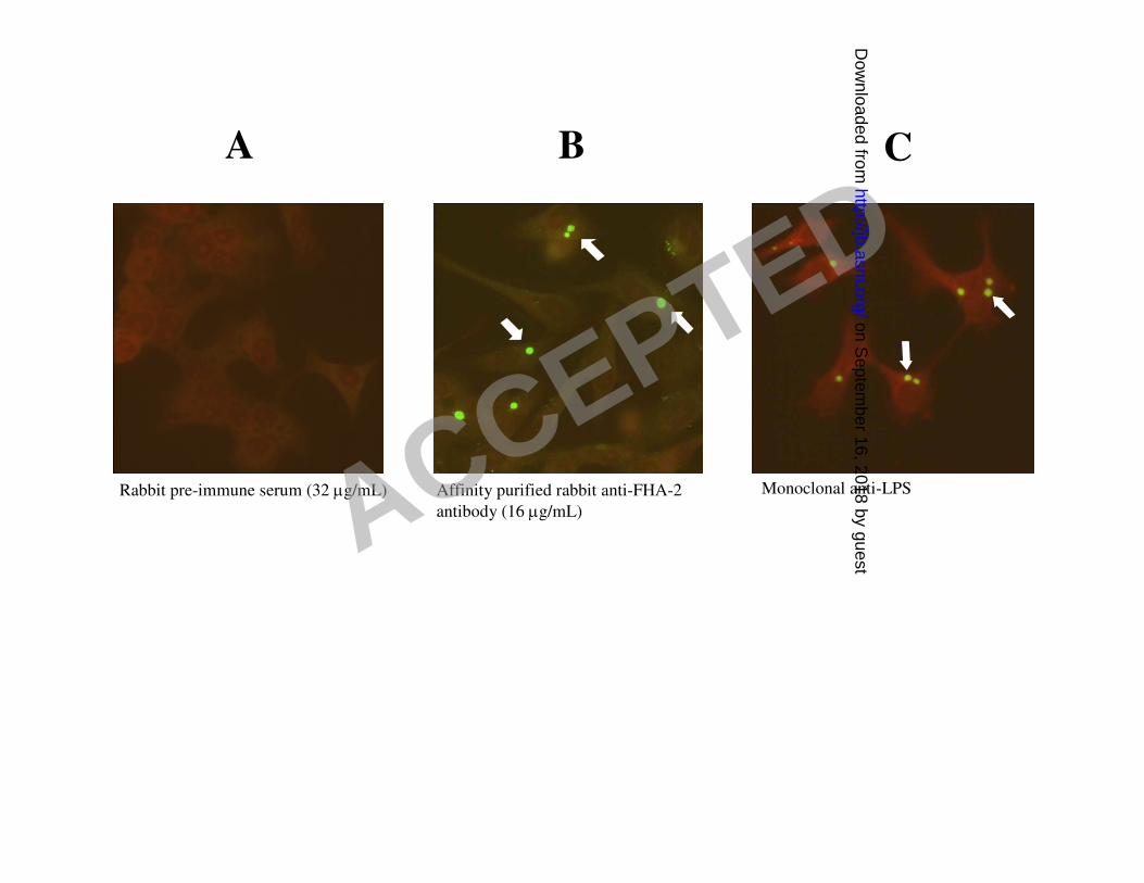
Detection of CdsD in chlamydial reticulate bodies 6
Indirect immunofluorescence of Chlamydia-infected HeLa cells at 48 hr post-infection was used 7
in order to visualize CdsD in RB in infected cells. Affinity purified rabbit anti-FHA-2 IgG 8
uniformly stained whole chlamydial inclusions 48 hr post-infection (fig 3B), suggesting the 9
presence of CdsD in chlamydial RB. Rabbit pre-immune serum did not react with chlamydial 10
inclusions (fig 3A). Monoclonal antibody to chlamydial lipopolysaccharide produced a similar 11
staining pattern as the anti-CdsD antibody (fig 3C). These results suggest that CdsD is present in 12
chlamydial RB which predominate in inclusions 48 hours post-infection. 13
14
Localization of CdsD with cytoplasmic proteins in Chlamydia 15
In order to determine the localization of CdsD in Chlamydia, soluble and integral membrane 16
protein fractions of EB were prepared based on differential solubility in Triton X-114 (4,16); the 17
protein fractions were then separated by SDS-PAGE and stained with Coomassie blue (fig 4A). 18
Western blotting followed by ECL detection of CdsD with anti-CdsD antibody demonstrated that 19
CdsD is predominantly in the soluble fraction of Triton X-114-solubilized EB (fig 4B, top 20
panel). A fraction of CdsD molecules were also detected in the integral membrane protein 21
fraction (fig 4B, top panel), although this could be due to minor contamination with soluble 22
proteins. Two other chlamydial proteins, Hsp60 and CopN, not thought to be membrane proteins, 23
ACCEPTED
on Septem
ber 16, 2018 by guesthttp://jb.asm
.org/D
ownloaded from

13
were not detected in the integral membrane protein fraction even after extended exposure of the 1
Western blot to film (fig 4B, bottom panels). Together the results suggest CdsD is predominantly 2
soluble in EB but may localize to the inner membrane, potentially as a peripheral membrane 3
protein. CdsD is also present in RB (fig 4B). 4
5
Interaction of CdsD, CdsQ and CdsL in GST pull-down assays 6
GST pull-down assays were used to determine if putative chlamydial type III secretion proteins 7
interact. When incubated with an E. coli lysate containing His-CdsD as ‘bait’, GST fusion ‘prey’ 8
proteins CdsQ and CdsL but not GST alone bound His-CdsD (fig 5A). In order to test the avidity 9
of the interactions, three tubes containing each bait-prey combination were set up and washed 10
with increasing amounts of salt. Each tube was washed with 50 mM Tris buffer containing 0, 200 11
or 500 mM NaCl (fig 5A, lanes 1, 2 and 3, respectively). CopN, CdsN, and the PknD kinase 12
domain exhibited weak and presumably non-specific interactions with CdsD under low salt 13
concentrations, and these interactions were readily prevented with quick washes containing 500 14
mM salt. Both CdsQ and CdsL remained associated with CdsD after washing with 500 mM 15
NaCl. C-terminally truncated His-CdsD present in the E. coli lysate and visible by ECL with 16
anti-His antibody (the His-tag is located on the N-terminus), did not bind to CdsQ or CdsL (fig 17
5B) suggesting that the C-terminal region of CdsD is essential for binding to CdsQ and CdsL. 18
Pull-down assays with GST-CdsQ as prey and E. coli lysates containing the bait proteins His-19
CdsQ, His-CdsL and His-FHA-2 (fig 5C, left panel) revealed CdsQ-CdsQ and CdsQ-CdsL 20
interactions (fig 5C, right panel). Interactions between CdsQ and the FHA-2 domain of CdsD 21
were not observed and served as a negative control. These results suggest that the FHA-2 region 22
ACCEPTED
on Septem
ber 16, 2018 by guesthttp://jb.asm
.org/D
ownloaded from

14
of CdsD alone does not interact with CdsQ, but that full-length CdsQ, CdsL and CdsD form 1
stable intermolecular interactions. 2
3
Co-purification of CdsD with CdsQ and CdsL 4
Co-purification assays were used in order to determine if native CdsD binds with His-tagged 5
CdsQ or CdsL. Briefly, E. coli lysates containing equivalent amounts of His-tagged CdsQ, CdsL 6
or HflX were incubated with a chlamydial RB lysate prepared from 42 hr post-infection RB. Ni-7
NTA agarose beads were added over-night, and the beads were collected and washed with 8
binding buffer containing 20 mM imidazole. CdsD co-purified with His-CdsQ and His-CdsL but 9
not with His-HflX (a chlamydial GTPase presumably not related to type III secretion) as detected 10
by Western blot using anti-CdsD antibody (fig 5D, top panel). The blot was reprobed with anti-11
His antibody to visualize the purified recombinant proteins (fig 5D, bottom panel). The results 12
corroborate the GST pull-down assays and demonstrate that CdsD interacts with CdsQ and 13
CdsL. 14
15
Discussion 16
In the present study we investigated the expression and localization of CdsD in C. 17
pneumoniae and identified novel interactions between components of the putative chlamydial 18
type III secretion apparatus. CdsD was expressed in chlamydial EB and RB as a ~105 kDa 19
protein, localized with cytoplasmic proteins in EB after Triton X-114 phase partitioning and was 20
evenly distributed throughout the inclusion body at 48 hr post-infection based on a consolidated 21
staining pattern. We demonstrated interactions between CdsQ molecules and between CdsQ and 22
CdsL, and for the first time identify CdsQ and CdsL as molecular binding partners of CdsD. 23
ACCEPTED
on Septem
ber 16, 2018 by guesthttp://jb.asm
.org/D
ownloaded from

15
These results suggest CdsD, CdsQ and CdsL may interact in Chlamydia to form part of the 1
chlamydial type III secretion apparatus. 2
Virulence and flagella-associated type III secretion systems have a base located in the 3
bacterial cytoplasm that contains over 100 molecules of FliN (26,47), a YscQ homolog that 4
forms part of a concentric structure known as the C-ring (5). Structural analysis of HrcQB-C, the 5
conserved C-terminal region of a YscQ homolog in Pseudomonas syringae, revealed a tetrameric 6
‘dimer of dimers’ complex and two clusters of conserved residues that may mediate interactions 7
with other proteins (13). Spa33, a Shigella YscQ homolog, interacts with multiple T3S proteins 8
including structural components of the basal body, MxiN (a YscL homolog), Spa47 (a YscN 9
homolog), Spa32 (molecular ruler determining the needle length) and several effectors (32). 10
Spa33 has been localized to the C-ring of the T3S apparatus using electron microscopy, and was 11
shown to be required for injectisome formation and protein secretion, and may act as a recruiting 12
platform or scaffold for concentration of effector molecules prior to secretion (32). Yeast three 13
hybrid assays demonstrated that YscQ simultaneously interacts with YscK and YscL, suggesting 14
separate regions on YscQ may mediate binding to different proteins in Yersinia (23). Similarly, 15
YscL, a negative regulator of the ATPase YscN (3), has been shown to bring together YscN and 16
YscQ (23). Based on the data it was suggested that interaction between YscQ, YscL, YscN and 17
YscK may be important in the assembly and/or function of the Y. pestis type III secretion 18
apparatus (23). We have shown using a GST pull-down assay interactions between CdsQ 19
molecules, consistent with the requirement for YscQ dimerization in creating building blocks of 20
the T3S C-ring (5,13). Furthermore, intermolecular associations were identified between CdsQ 21
and CdsL, mirroring the interactions between their counterpart proteins in Yersinia and Shigella 22
and suggesting these two proteins may be important in chlamydial T3S. Using both pull-down 23
ACCEPTED
on Septem
ber 16, 2018 by guesthttp://jb.asm
.org/D
ownloaded from

16
and co-purification assays with chlamydial lysates we identified interactions between CdsQ and 1
CdsD and between CdsL and CdsD, indicating that CdsD interacts with both CdsQ and CdsL and 2
is likely a component of the T3S system. Given that recombinant CdsQ and CdsL were used in 3
the pull-down and co-purification assays, the interaction of CdsD with CdsQ and CdsL remains 4
to be demonstrated in Chlamydia. Weaker interactions were observed between CdsD and CopN, 5
PknD KD and CdsN, but these interactions were eliminated in the presence of 500 mM NaCl, 6
and therefore further study is required to determine their specificity. Interestingly, CdsQ did not 7
pull-down a 150 amino acid fragment encompassing the FHA-2 domain of CdsD, and both CdsQ 8
and CdsL were unable to pull-down C-terminal truncations of CdsD, collectively suggesting that 9
the C-terminus of CdsD plays a critical role in mediating stable protein-protein interactions with 10
CdsQ and CdsL. Studies are currently underway to assess these interactions in C. pneumoniae 11
and to determine the domains responsible for mediating interactions between CdsD, CdsQ and 12
CdsL. 13
Determining the localization of T3S proteins is important in order to elucidate and 14
understand their roles in secretion. To date, strong evidence for the localization of various YscD 15
homologs has not been forthcoming. EscD, the enteropathogenic E. coli YscD homolog, was 16
shown to interact with EscC in a Yeast two hybrid assay (11); mass spectrometry was used to 17
identify EscC as a major component of the T3S outer membrane ring, and the interaction 18
between EscD and EscC was corroborated using a GST pull-down assay (35). Together with the 19
presence of a putative transmembrane domain in EscD it was suggested EscD localizes as an 20
integral membrane protein in order to interact with EscC in the outer membrane. Biochemical 21
evidence supporting membrane localization of YscD comes from a study in Yersinia where it 22
was shown that YscD localizes to the inner membrane in cell fractionation experiments (38). 23
ACCEPTED
on Septem
ber 16, 2018 by guesthttp://jb.asm
.org/D
ownloaded from

17
Conversely, CdsD has been detected in the sarkosyl-insoluble ‘outer membrane’ fraction by 1
Mass Spectrometry (44), shown to transiently exist in the inclusion membrane by 2
immunofluorescence (20) and to be in the soluble protein fraction of Chlamydia (this report). 3
Tanzer and Hatch recognized that the localization of CdsD to the outer membrane of C. 4
trachomatis is likely an artifact of the sarkosyl extraction method (44). CdsD was not labeled 5
when intact bacteria were treated with the lipophilic, photoactivatable chemical [125
I]TID, in 6
contrast with labeling of canonical outer membrane proteins such as the major outer membrane 7
protein (44). Additionally, treatment of Chlamydia with trypsin prior to cell fractionation with 8
sarkosyl did not result in reduction in the size of the band representing CdsD in the sarkosyl 9
insoluble ‘outer membrane’ fraction. Together the data indicate CdsD is not surface exposed. 10
Recently a single immunofluorescent image was provided as evidence that CdsD is secreted into 11
the inclusion membrane at 20 hr post-infection (20). Closer inspection of the image, however, 12
reveals that one may interpret the image as CdsD-laden chlamydial RB directly associated with 13
the inclusion membrane. The authors also present Western blot data contradicting the secretion 14
of CdsD into the inclusion membrane by showing accumulation of CdsD in Chlamydia 15
throughout the developmental cycle, and report that immunofluorescent images from other time 16
points (0.5, 6, 48, 72 hr post-infection) do not reveal inclusion membrane staining for CdsD but 17
do reveal intra-luminal staining (suggesting CdsD is found within the bacteria). In addition, the 18
84 kDa CdsD protein detected by Western blot brings into question antibody specificity as 19
Tanzer and Hatch detected CdsD as a 120 kDa protein using mass spectrometry (44) and we 20
detected CdsD migrating slower than a 100 kDa reference protein. Given the discrepancy of 21
localization of YscD homologs in the literature, we prepared an affinity-purified antibody to the 22
FHA-2 domain of CdsD and detected full length CdsD in both EB and RB lysates by Western 23
ACCEPTED
on Septem
ber 16, 2018 by guesthttp://jb.asm
.org/D
ownloaded from

18
blot analysis. We detected CopN, a putative T3S plug protein, in EB but absent from RB lysates, 1
consistent with CopN secretion by Chlamydia early in the replication cycle (15). By analogy the 2
presence of CdsD in both RB and EB suggests that it is not secreted, consistent with a structural 3
T3S system component. Phase separation of chlamydial EB proteins into cytoplasmic and 4
integral membrane protein fractions based on differential solubility in Triton X-114 revealed that 5
CdsD is predominantly soluble in EB. It could be that chlamydial CdsD is a peripheral 6
membrane protein with a high hydrophilic character. Cytoplasmic localization of CdsD would be 7
consistent with the interactions of CdsD with the putative chlamydial C-ring protein CdsQ and 8
with the tethering protein CdsL, both presumably cytoplasmic proteins. 9
A consistent and essential role for YscD homologs in type III secretion and pathogenicity 10
has been well documented. E. coli ∆escD mutants were unable to produce the T3S apparatus 11
(35), Yersina yscD was shown to be necessary for secretion of the effector Yop proteins (30,38) 12
and Pseudomonas aeruginosa ∆pscD mutants were attenuated in a caterpillar model of infection 13
(31). CdsD, however, has an additional 400 amino acids relative to YscD homologs and contains 14
two FHA domains that may mediate binding to phosphorylated proteins, suggesting CdsD may 15
have an additional role in T3S. The presence of FHA domains on CdsD has led to the recent 16
suggestion that it may bind phosphorylated chaperone-substrate complexes of the T3S system 17
(36). A report in Pseudomonas aeruginosa, however, identified a type VI secretion apparatus 18
protein with an FHA domain that was phosphorylated by the protein kinase PpkA, triggering 19
protein secretion; it was proposed that after kinase autophosphorylation, the FHA domain of this 20
scaffolding protein is recruited to and binds the kinase, resulting in FHA domain phosphorylation 21
and induction of protein secretion (33). We have recently shown that the membrane-localized C. 22
pneumoniae protein kinase PknD autophosphorylates and phosphorylates CdsD on both its FHA-23
ACCEPTED
on Septem
ber 16, 2018 by guesthttp://jb.asm
.org/D
ownloaded from

19
1 and FHA-2 domains (24). In addition to potentially recognizing phosphorylated chaperone-1
substrate complexes of the T3S system as proposed (36), the FHA domains of CdsD may 2
mediate transient interactions with PknD as an important step in chlamydial T3S. It is tempting 3
to speculate that phosphorylation of CdsD FHA domains by PknD may be an activating or 4
triggering event in chlamydial T3S. 5
6
7
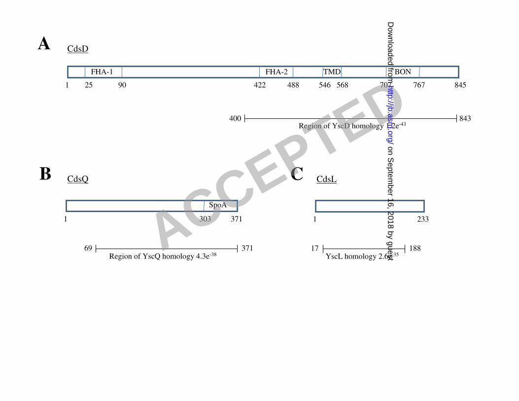
Figure 1. A: Domain organization of CdsD, CdsQ and CdsL. A: CdsD contains a predicted 8
transmembrane domain (TMD), phospholipid binding domain (BON) and two forkhead 9
associated (FHA) domains. The region of YscD homology (E-value 1.2e-43
) is shown. B: CdsQ 10
contains a ‘surface presentation of antigens’ (SpoA) domain. The region of YscQ homology (E-11
value 4.3e-38
) is shown. C: The region of YscL homology (E-value 2.6e-35
) is shown. Amino acid 12
numbers are given. 13
14
Figure 2. Detection of full-length CdsD in C. pneumoniae elementary bodies. A: Pre-immune 15
rabbit serum does not detect CdsD in EB by Western blot. B: Hyper-immune rabbit anti-FHA-2 16
antiserum detects His-CdsD in E. coli lysate and CdsD in chlamydial EB lysate. C: Affinity 17
purified rabbit anti-FHA-2 IgG detects His-CdsD in E. coli and CdsD in chlamydial EB and 18
exhibits minimal cross-reactivity with lower molecular weight species. Lane 1: E. coli lysate 19
containing His-PknD (migrates at approximately 108 kDa, location marked with an arrow head); 20
lane 2: E. coli lysate containing His-CdsD; lane 3: chlamydial EB lysate. An asterisks in A) and 21
B) indicates the location of non-specific antibody cross-reactivity. A double asterisks in C) 22
indicates a probable CdsD degradation product. 23
ACCEPTED
on Septem
ber 16, 2018 by guesthttp://jb.asm
.org/D
ownloaded from

20
1
Figure 3. Detection of CdsD within C. pneumoniae inclusions at 48 hr post-infection using 2
indirect immunofluorescence. A: Unstained inclusions; rabbit preimmune serum used at 32 3
µg/mL as the primary antibody. B: Stained inclusions; affinity purified rabbit anti-FHA-2 4
antibody used at 16 µg/mL as the primary antibody. C: Stained inclusions; FITC-conjugated 5
monoclonal anti-chlamydial LPS antibody was used to directly stain C. pneumoniae inside 6
inclusions. Evan’s blue was used as a counterstain to visualize HeLa cells and images were 7
captured at x400 magnification. Arrows in B and C indicate stained chlamydial inclusions. 8
9
Figure 4. CdsD localizes with cytoplasmic EB proteins. A: Chlamydial EB proteins were 10
separated into cytoplasmic (soluble) and integral membrane (membrane) protein fractions using 11
Triton X-114 and stained with coomassie blue. B: Fractions in A) were tested for the presence of 12
CdsD by Western blot using guinea pig anti-CdsD antiserum. CdsD was found in both RB and 13
EB lysates, and predominantly localized with cytoplasmic EB proteins although a small 14
proportion was found in the membrane fraction. The Western blot was reprobed for chlamydial 15
Hsp60 (shown directly below) which was not present in the integral membrane fraction (note: the 16
signal representing Hsp60 in the ‘EB soluble’ lane is seen encroaching into the ‘EB membrane’ 17
lane). An identical Western blot was probed for CopN (a Chlamydia protein predicted to be 18
cytoplasmic) using guinea pig anti-CopN antibody (shown below the Hsp60 Western blot). 19
CopN was detected in EB lysate and in the soluble fraction, but was not detected in RB lysates or 20
the EB integral membrane fraction even after over-exposing the membrane to the film (bottom). 21
Absence of CopN in RB is consistent with secretion of CopN by Chlamydia. 22
23
ACCEPTED
on Septem
ber 16, 2018 by guesthttp://jb.asm
.org/D
ownloaded from

21
Figure 5. Interaction between CdsD, CdsQ and CdsL in GST pull-down and co-purification 1
assays. A: CdsD binds with CdsQ and CdsL but not CopN, PknD KD, CdsN or GST in a GST 2
pull-down assay. E. coli lysate containing His-CdsD as bait was incubated with 20 µg of GST or 3
20 µg of the indicated GST chlamydial fusion proteins (CopN, PknD KD, CdsN, CdsQ or CdsL) 4
on glutathione agarose beads. Beads were collected by centrifugation, washed three times with 5
either 50 mM Tris-HCl pH 7.45 (lane 1), 50 mM Tris-HCl pH 7.45 containing 200 mM NaCl 6
(lane 2) or 50 mM Tris-HCl pH 7.45 containing 500 mM NaCl (lane 3), and proteins eluted in 2x 7
SDS loading buffer. Binding of CdsD with the prey proteins was revealed by anti-His Western 8
blotting. B: GST-CdsL and GST-CdsQ interact with full length His-CdsD but not with C-9
terminally truncated His-CdsD in a GST pull-down assay. C: CdsQ-CdsQ and CdsQ-CdsL 10
interactions identified by GST pull-down assay. E. coli lysates containing the bait proteins His-11
tagged CdsQ, CdsL and FHA-2 are shown in the left panel, and the results from the pull-down is 12
shown in the right panel. CdsQ did not bind with the FHA-2 domain of CdsD. D: CdsD from a 13
chlamydial RB lysate co-purifies with His-CdsQ and His-CdsL but not with His-HflX on Ni-14
NTA agarose as revealed by Western blot using anti-CdsD antibody (top). The nitrocellulose 15
membrane was reprobed with anti-His antibody and demonstrates equivalent amounts of His-16
tagged proteins purified and loaded in each lane (bottom). 17
18
19
20
21
22
ACCEPTED
on Septem
ber 16, 2018 by guesthttp://jb.asm
.org/D
ownloaded from

22
Acknowledgements 1
We thank Dr. Ken Fields for providing the phase separation protocol and for helpful 2
technical advice and discussions. We thank Dr. Brian Coombes for editing the manuscript. We 3
wish to acknowledge Rami El-Sebai for his contribution in constructing pENT-CdsQ and pENT-4
CdsL, and the members of the Mahony lab for helpful discussions on this project. 5
D.L.J. and C.B.S. are supported by a grant to J.B.M. from the Canadian Institutes of 6
Health Research. This work was funded in part by a grant from the Canadian Institutes of Health 7
Research. 8
ACCEPTED
on Septem
ber 16, 2018 by guesthttp://jb.asm
.org/D
ownloaded from

1
Reference List 1
2
1. Belland, R. J., D. E. Nelson, D. Virok, D. D. Crane, D. Hogan, D. Sturdevant, W. L. Beatty, 3
and H. D. Caldwell. 2003. Transcriptome analysis of chlamydial growth during IFN-gamma-4
mediated persistence and reactivation. Proc. Natl. Acad. Sci. U. S. A 100:15971-15976. 5
2. Belland, R. J., G. Zhong, D. D. Crane, D. Hogan, D. Sturdevant, J. Sharma, W. L. Beatty, 6
and H. D. Caldwell. 2003. Genomic transcriptional profiling of the developmental cycle of 7
Chlamydia trachomatis. Proc. Natl. Acad. Sci. U. S. A 100:8478-8483. 8
3. Blaylock, B., K. E. Riordan, D. M. Missiakas, and O. Schneewind. 2006. Characterization of 9
the Yersinia enterocolitica type III secretion ATPase YscN and its regulator, YscL. J. Bacteriol. 10
188:3525-3534. 11
4. Bordier, C. 1981. Phase separation of integral membrane proteins in Triton X-114 solution. J. 12
Biol. Chem. 256:1604-1607. 13
5. Brown, P. N., M. A. Mathews, L. A. Joss, C. P. Hill, and D. F. Blair. 2005. Crystal structure of 14
the flagellar rotor protein FliN from Thermotoga maritima. J. Bacteriol. 187:2890-2902. 15
6. Caldwell, H. D., J. Kromhout, and J. Schachter. 1981. Purification and partial characterization 16
of the major outer membrane protein of Chlamydia trachomatis. Infect. Immun. 31:1161-1176. 17
7. Carabeo, R. A., S. S. Grieshaber, A. Hasenkrug, C. Dooley, and T. Hackstadt. 2004. 18
Requirement for the Rac GTPase in Chlamydia trachomatis invasion of non-phagocytic cells. 19
Traffic. 5:418-425. 20
8. Clifton, D. R., K. A. Fields, S. S. Grieshaber, C. A. Dooley, E. R. Fischer, D. J. Mead, R. A. 21
Carabeo, and T. Hackstadt. 2004. A chlamydial type III translocated protein is tyrosine-22
phosphorylated at the site of entry and associated with recruitment of actin. Proc. Natl. Acad. Sci. 23
U. S. A 101:10166-10171. 24
9. Coombes, B. K. and J. B. Mahony. 2002. Identification of MEK- and phosphoinositide 3-kinase-25
dependent signalling as essential events during Chlamydia pneumoniae invasion of HEp2 cells. 26
Cell Microbiol. 4:447-460. 27
10. Cornelis, G. R. 2006. The type III secretion injectisome. Nat. Rev. Microbiol. 4:811-825. 28
11. Creasey, E. A., R. M. Delahay, S. J. Daniell, and G. Frankel. 2003. Yeast two-hybrid system 29
survey of interactions between LEE-encoded proteins of enteropathogenic Escherichia coli. 30
Microbiology 149:2093-2106. 31
12. Dong, F., J. Sharma, Y. Xiao, Y. Zhong, and G. Zhong. 2004. Intramolecular dimerization is 32
required for the chlamydia-secreted protease CPAF to degrade host transcriptional factors. Infect. 33
Immun. 72:3869-3875. 34
13. Fadouloglou, V. E., A. P. Tampakaki, N. M. Glykos, M. N. Bastaki, J. M. Hadden, S. E. 35
Phillips, N. J. Panopoulos, and M. Kokkinidis. 2004. Structure of HrcQB-C, a conserved 36
component of the bacterial type III secretion systems. Proc. Natl. Acad. Sci. U. S. A 101:70-75. 37
ACCEPTED
on Septem
ber 16, 2018 by guesthttp://jb.asm
.org/D
ownloaded from

2
14. Fields, K. A., E. R. Fischer, D. J. Mead, and T. Hackstadt. 2005. Analysis of putative 1
Chlamydia trachomatis chaperones Scc2 and Scc3 and their use in the identification of type III 2
secretion substrates. J. Bacteriol. 187:6466-6478. 3
15. Fields, K. A. and T. Hackstadt. 2000. Evidence for the secretion of Chlamydia trachomatis 4
CopN by a type III secretion mechanism. Mol. Microbiol. 38:1048-1060. 5
16. Fields, K. A., D. J. Mead, C. A. Dooley, and T. Hackstadt. 2003. Chlamydia trachomatis type 6
III secretion: evidence for a functional apparatus during early-cycle development. Mol. Microbiol. 7
48:671-683. 8
17. Ghosh, P. 2004. Process of protein transport by the type III secretion system. Microbiol. Mol. 9
Biol. Rev. 68:771-795. 10
18. Gophna, U., E. Z. Ron, and D. Graur. 2003. Bacterial type III secretion systems are ancient and 11
evolved by multiple horizontal-transfer events. Gene 312:151-163. 12
19. Hefty, P. S. and R. S. Stephens. 2007. Chlamydial type III secretion system is encoded on ten 13
operons preceded by sigma 70-like promoter elements. J. Bacteriol. 189:198-206. 14
20. Herrmann, M., A. Schuhmacher, I. Muhldorfer, K. Melchers, C. Prothmann, and S. 15
Dammeier. 2006. Identification and characterization of secreted effector proteins of 16
Chlamydophila pneumoniae TW183. Res. Microbiol. 157:513-524. 17
21. Ho, T. D. and M. N. Starnbach. 2005. The Salmonella enterica serovar typhimurium-encoded 18
type III secretion systems can translocate Chlamydia trachomatis proteins into the cytosol of host 19
cells. Infect. Immun. 73:905-911. 20
22. Hybiske, K. and R. S. Stephens. 2007. Mechanisms of host cell exit by the intracellular 21
bacterium Chlamydia. Proc. Natl. Acad. Sci. U. S. A 104:11430-11435. 22
23. Jackson, M. W. and G. V. Plano. 2000. Interactions between type III secretion apparatus 23
components from Yersinia pestis detected using the yeast two-hybrid system. FEMS Microbiol. 24
Lett. 186:85-90. 25
24. Johnson, D. L. and J. B. Mahony. 2007. Chlamydophila pneumoniae PknD exhibits dual amino 26
acid specificity and phosphorylates Cpn0712, a putative type III secretion YscD homolog. J. 27
Bacteriol. 189:7549-7555. 28
25. Kawana, K., A. J. Quayle, M. Ficarra, J. A. Ibana, L. Shen, Y. Kawana, H. Yang, L. 29
Marrero, S. Yavagal, S. J. Greene, Y. X. Zhang, R. B. Pyles, R. S. Blumberg, and D. J. 30 Schust. 2007. CD1d degradation in Chlamydia trachomatis-infected epithelial cells is the result of 31
both cellular and chlamydial proteasomal activity. J. Biol. Chem. 282:7368-7375. 32
26. Khan, I. H., T. S. Reese, and S. Khan. 1992. The cytoplasmic component of the bacterial 33
flagellar motor. Proc. Natl. Acad. Sci. U. S. A 89:5956-5960. 34
27. Kresse, A. U., K. Schulze, C. Deibel, F. Ebel, M. Rohde, T. Chakraborty, and C. A. Guzman. 35
1998. Pas, a novel protein required for protein secretion and attaching and effacing activities of 36
enterohemorrhagic Escherichia coli. J. Bacteriol. 180:4370-4379. 37
28. Lugert, R., M. Kuhns, T. Polch, and U. Gross. 2004. Expression and localization of type III 38
secretion-related proteins of Chlamydia pneumoniae. Med. Microbiol. Immunol. 193:163-171. 39
ACCEPTED
on Septem
ber 16, 2018 by guesthttp://jb.asm
.org/D
ownloaded from

3
29. Maurer, A. P., A. Mehlitz, H. J. Mollenkopf, and T. F. Meyer. 2007. Gene expression profiles 1
of Chlamydophila pneumoniae during the developmental cycle and iron depletion-mediated 2
persistence. PLoS. Pathog. 3:e83. 3
30. Michiels, T., J. C. Vanooteghem, R. C. Lambert de, B. China, A. Gustin, P. Boudry, and G. 4
R. Cornelis. 1991. Analysis of virC, an operon involved in the secretion of Yop proteins by 5
Yersinia enterocolitica. J. Bacteriol. 173:4994-5009. 6
31. Miyata, S., M. Casey, D. W. Frank, F. M. Ausubel, and E. Drenkard. 2003. Use of the 7
Galleria mellonella caterpillar as a model host to study the role of the type III secretion system in 8
Pseudomonas aeruginosa pathogenesis. Infect. Immun. 71:2404-2413. 9
32. Morita-Ishihara, T., M. Ogawa, H. Sagara, M. Yoshida, E. Katayama, and C. Sasakawa. 10
2006. Shigella Spa33 is an essential C-ring component of type III secretion machinery. J. Biol. 11
Chem. 281:599-607. 12
33. Mougous, J. D., C. A. Gifford, T. L. Ramsdell, and J. J. Mekalanos. 2007. Threonine 13
phosphorylation post-translationally regulates protein secretion in Pseudomonas aeruginosa. Nat. 14
Cell Biol. 9:797-803. 15
34. Nicholson, T. L., L. Olinger, K. Chong, G. Schoolnik, and R. S. Stephens. 2003. Global stage-16
specific gene regulation during the developmental cycle of Chlamydia trachomatis. J. Bacteriol. 17
185:3179-3189. 18
35. Ogino, T., R. Ohno, K. Sekiya, A. Kuwae, T. Matsuzawa, T. Nonaka, H. Fukuda, S. Imajoh-19
Ohmi, and A. Abe. 2006. Assembly of the type III secretion apparatus of enteropathogenic 20
Escherichia coli. J. Bacteriol. 188:2801-2811. 21
36. Peters, J., D. P. Wilson, G. Myers, P. Timms, and P. M. Bavoil. 2007. Type III secretion a la 22
Chlamydia. Trends Microbiol. 15:241-251. 23
37. Pirbhai, M., F. Dong, Y. Zhong, K. Z. Pan, and G. Zhong. 2006. The secreted protease factor 24
CPAF is responsible for degrading pro-apoptotic BH3-only proteins in Chlamydia trachomatis-25
infected cells. J. Biol. Chem. 281:31495-31501. 26
38. Plano, G. V. and S. C. Straley. 1995. Mutations in yscC, yscD, and yscG prevent high-level 27
expression and secretion of V antigen and Yops in Yersinia pestis. J. Bacteriol. 177:3843-3854. 28
39. Slepenkin, A., L. M. de la Maza, and E. M. Peterson. 2005. Interaction between components of 29
the type III secretion system of Chlamydiaceae. J. Bacteriol. 187:473-479. 30
40. Slepenkin, A., V. Motin, L. M. de la Maza, and E. M. Peterson. 2003. Temporal expression of 31
type III secretion genes of Chlamydia pneumoniae. Infect. Immun. 71:2555-2562. 32
41. Subtil, A., C. Delevoye, M. E. Balana, L. Tastevin, S. Perrinet, and A. utry-Varsat. 2005. A 33
directed screen for chlamydial proteins secreted by a type III mechanism identifies a translocated 34
protein and numerous other new candidates. Mol. Microbiol. 56:1636-1647. 35
42. Subtil, A., C. Parsot, and A. utry-Varsat. 2001. Secretion of predicted Inc proteins of 36
Chlamydia pneumoniae by a heterologous type III machinery. Mol. Microbiol. 39:792-800. 37
43. Subtil, A., B. Wyplosz, M. E. Balana, and A. utry-Varsat. 2004. Analysis of Chlamydia caviae 38
entry sites and involvement of Cdc42 and Rac activity. J. Cell Sci. 117:3923-3933. 39
ACCEPTED
on Septem
ber 16, 2018 by guesthttp://jb.asm
.org/D
ownloaded from
4
44. Tanzer, R. J. and T. P. Hatch. 2001. Characterization of outer membrane proteins in Chlamydia 1
trachomatis LGV serovar L2. J. Bacteriol. 183:2686-2690. 2
45. Wilson, D. P., P. Timms, D. L. McElwain, and P. M. Bavoil. 2006. Type III secretion, contact-3
dependent model for the intracellular development of chlamydia. Bull. Math. Biol. 68:161-178. 4
46. Yip, C. K. and N. C. Strynadka. 2006. New structural insights into the bacterial type III 5
secretion system. Trends Biochem. Sci. 31:223-230. 6
47. Zhao, R., N. Pathak, H. Jaffe, T. S. Reese, and S. Khan. 1996. FliN is a major structural protein 7
of the C-ring in the Salmonella typhimurium flagellar basal body. J. Mol. Biol. 261:195-208. 8
48. Zhong, G., P. Fan, H. Ji, F. Dong, and Y. Huang. 2001. Identification of a chlamydial protease-9
like activity factor responsible for the degradation of host transcription factors. J. Exp. Med. 10
193:935-942. 11
12
13
14
ACCEPTED
on Septem
ber 16, 2018 by guesthttp://jb.asm
.org/D
ownloaded from
A
400 843Region of YscD homology 1.2e-43
CdsD
1 25 90 422 488 546 568 707 767 845
FHA-1 FHA-2 TMD BON
C
1 233
CdsL
17 188YscL homology 2.6e-35
69 371
B
Region of YscQ homology 4.3e-38
1 303 371
SpoA
CdsQ
ACCEPTED on S
eptember 16, 2018 by guest
http://jb.asm.org/
Dow
nloaded from

affinity purified R α
FHA-2 (ECL 8 min.)
CdsD
pre-immune serum
(ECL 2 min.)
crude R α FHA-2
(ECL 1 sec.)
CdsD
A B C
1 2 3 1 2 3 1 2 3
180
110
80
60
48
MW
(kDa)
His-CdsD His-CdsD
*
*
**
**ACCEPTED on S
eptember 16, 2018 by guest
http://jb.asm.org/
Dow
nloaded from
Rabbit pre-immune serum (32 µg/mL) Affinity purified rabbit anti-FHA-2
antibody (16 µg/mL)
A B C
Monoclonal anti-LPSACCEPTED on S
eptember 16, 2018 by guest
http://jb.asm.org/
Dow
nloaded from

ARB EB EB EB
B
lysate lysate soluble membrane
CdsD
Hsp60
CopN
100
kDa
60
kDa
45
kDa
CopN
over-exposed
WB
WBs and ECL
EB EB EB
lysate soluble membrane
coomassie blue
45
kDa
66
97
116
200
55
36
MW
(kDa)
ACCEPTED on S
eptember 16, 2018 by guest
http://jb.asm.org/
Dow
nloaded from

180
110
80
His-CdsD
CopN PknD KD CdsN GST CdsQ CdsL
1 2 3 1 2 3 1 2 3 1 2 3 1 2 3 1 2 3
ECL: anti-His WB
A
C D
ECL: anti-CdsD WB
CdsD
His
-Hfl
X
RB
lys
ate
His
-Cds
L
His
-Cds
Q
ECL: anti-His WB
His-HflX
His-CdsQ
His-CdsL
Bla
nk
MW
(kDa)
Bait lysates GST-CdsQ Pull-down
ECL: anti-His WB
His-CdsQ
His-CdsL
His-FHA-2
CdsQ CdsL FHA-2 CdsQ CdsL FHA-2
His-CdsD
N-terminal
His-CdsD
fragments
Cds
L
Cds
Q
Bla
nkB
ail l
ysat
e
B
ACCEPTED on S
eptember 16, 2018 by guest
http://jb.asm.org/
Dow
nloaded from