Embed Size (px)
Citation preview
International Journal of Scientific & Engineering Research, Volume 7, Issue 2, February-2016 857 ISSN 2229-5518
IJSER © 2016 http://www.ijser.org
Semi-Automatic Seeded Region Growing for Object Extracted in MRI
Ziad M. Abood, Iman Hussein AL-Qinani, Kawther Thabt Saleh, Mushtaq Talib Ajjah
Abstract— this research characterizes a semi-automatic way to segemt objects found in medical images by using seeded region growing method, which increasingly became a popular method because of its ability to involve high-level knowledge of anatomical structures in seed selection process. Region based segmentation of the medical images is widely used in various clinical applications such as bone and tumor detection, visualization, and unsupervised image retrieval in clinical databases. Because of fuzziness of medical images in nature; segmenting regions depending on intensity is a very challenging task. In this paper, the popular seeded region grow methodology, which is used to segment anatomical structures in computed topography angiography images, is discussed. Homogeneity criteria used to control the region grow process during segmenting images is proposed.
Index Terms— Segmentation, Seeded Region Grow, Medical Imaging, Homogeneity, Thresholding, Region of Interest (ROI), Computed Topography.
—————————— ——————————
1 INTRODUCTION mage processing is a method used to convert an image
into digital form by performing some operations on it, in order to get an enhanced image or to extract some useful
information from it [1]. Color information is gaining an ever-greater meaning in digital image processing [2].
Region segmentation can be considered the essential and first step to verify images related to visualization, and applica-tions tasks. Besides, segmenting the medical images is consid-ered important due to the fact that it help physicians to find out the diseases that internally infect the body without carry-ing out a surgical procedure; this helps to decide the lesion location, which would reduce time used to read an image, and to estimate the disease probability [3].
Segmentation algorithms depend on one of two basic prop-erties of intensity values; discontinuity and similarity. The first category is done by partitioning an image depending on ab-rupt changes in intensity, such as edges in an image. The se-cond category is done by partitioning an image into similar regions according to predefined criteria [4].
Region growing algorithms depend on the growth of a re-gion whenever its interior is homogeneous according to cer-tain characteristics like intensity, color or texture. The imple-mented algorithm follows the strategy of a typical Region Growing; it depends on the growth of a region by adding sim-ilar neighbors [5].
Seeded Region Growing (SRG) is considered one of the simplest and most popular algorithms for region based seg-mentation. The most traditional implementation of this algo-rithm starts by selecting a starting point called “seed pixel”. Then, the region grows by adding similar adjacent pixels ac-cording to certain homogeneity criteria, which step by step increase the size of the region [5]. ———————————————— • Ziad M. Abood, Al -Mustansiriyah University, College of Education, Depart-ment of ComputerScience, [email protected]
Iman Hussein AL-Qinani, Al -Mustansiriyah University, College of Educa-tion, Department of ComputerScience, [email protected]
Kawther Thabt Saleh, Al -Mustansiriyah University, College of Education, Department of ComputerScience, [email protected]
Mushtaq Talib Ajjah, Al -Mustansiriyah University, College of Education, Department of ComputerScience, [email protected] 1.1 Related Work
Qiyao Yu and et al. suggested an image segmentation ap-proach named “iterative region growing using semantics”, which can be distinguished by comprising two aspects; the first aspect uses the functions of the gradually increased edge penalty inside the traditional Markov random field context model to formulate the objective functions. Whreas, the se-cond aspect (IRGS) uses the techniques of region growing to search for a solution to those objective functions. The suggest-ed (IRGS) is considered the improved approach of the tradi-tional ones which are baced on MRF in which the information of edge strength is applied and the achieved model parame-ters will be more stable extimated. Moreover, the (IRGS) pro-cedure enables representing image content hierarchically; it allows various features of region and even it incorporates the domain knowledge to be used in the segmentation process. This algorithm was successfully examined on many artificial images and on images of synthetic aperture radar (SAR) [6].
Shilpa Kamdi and R. K. Krishna provided a survey of achievements, problems being encountered, the open issues in the image segmentation research area and usage of the tech-niques in different areas. They considered the techniques un-der the following three groups: Threshold-based, Edge-based and Region-based [7].
In his thesis, Luis Garriza provided effective method to au-tomatically segment simple to complex images. The algorithm depends on color edge-detection and dynamic region grow-ing, completed by a multi resolution region merging. The segmentation procedure was tested on Berkeley database which is publicly available, and its results quality was meas-ured. This algorithm robustness was shown in the results compared with those obtained for the same image when seg-mented using other methods [8].
Valliappan Raman and et al. provided the methodology of segmentation with partial results, and explained theoretically how a mammogram tumor classification is performed through case base reasoning method. The first stage of mammogram is
I
IJSER
International Journal of Scientific & Engineering Research, Volume 7, Issue 2, February-2016 858 ISSN 2229-5518
IJSER © 2016 http://www.ijser.org
mass segmentation result. The basic idea of the algorithm is to find a set of seed pixels in the image, and then to grow itera-tively and aggregate the pixels that have similar properties. The second stage is under implementation, so the conceptual framework of classification method is described on the paper. The info structure that is presented in the paper when success-fully implemented would have an immense impact on com-puter-aided diagnosis systems field [9].
2 IMAGE SEGMENTATION:
2.1 Image Segmentation: Image segmentation is useful in many applications. It can
identify the regions of interest in a scene or annotate the data. The existing segmentation algorithm is categorized into re-gion-based segmentation, data clustering, and edge-base seg-mentation [10].
Region-based segmentation includes the algorithms of growing seeded and unseeded regions, the JSEG, and the fast scanning algorithm. All of them expand each region pixel wise based on their pixel value or quantized¬-value so that each cluster has high positional relation [11]. For data clustering, the concept of them depends on the whole image and consid-ers the distance between each data. The characteristic of data clustering is that each pixel of a cluster does not certainly con-nective. The basis method of data clustering can be divided into hierarchical and partitioned clustering. Furthermore, the extension of data clustering that is called “mean shift algo-rithm” was shown, although this algorithm belongs to density estimation [11]. The last classification of segmentation is edge-based segmentation. This type of segmentations generally ap-plies edge detection or the concept of edge. The typical one is the watershed algorithm, but it always has the over-segmentation problem; so that the use of markers was pro-posed to improve the watershed algorithm by smoothing and selecting markers [11].
2.2 Segmentation of Color Image Image segmentation is the process by which any image is
divided into connected image areas but non-overlapping those areas are called regions, by using the criteria basis that govern-ing similarity and homogeneity. Similarly, color image seg-mentation is the process of extracting from the image domain one or more connected regions that satisfying uniformity (ho-mogeneity) criteria which depend on features derived from spectral image components. Those components are defined in a chosen color space. The segmentation process can be sup-ported by some additional knowledge about the objects in the scene such as geometric and optical properties [2].
Perhaps one of the most important features of a segmenta-tion process is the region definition. Almost four types of re-gion definitions can be differentiated [2], as follows: 1. The region is a connected component of a specific pixel
group that can be specified using a class membership func-tion that is determined in the color space. The color signals grouping can be carried out in the color space. One condi-tion for grouping should be fulfuled that is the pixel color must lie within certain plane or polyhedra in the color space [2].
2. Region is the maximal connected group of pixels found in the image plane for which the uniformity condition is satis-fied. In contrast to type 1, grouping the color signals takes place in the image plane instead of the color space. A uni-form region is obtained, for example, when larger, non-uniform regions are split or when a region is determined by merging other pixels (or blocks of pixels) in the sur-rounding area of a starting pixel [2].
3. The region is a connected set of pixels bounded by edge pixels that create a color contour. The color contour is de-termined by applying an operator for edge detection on the color image and possibly by an ensuing filling of the gaps in the contour. In a certain sense, the regions are also uni-form, because they represent the complementary set of a non-uniform set that is created by edge pixels [2].
4. The region is a connected component of a pixel set whose grouping results from a physical modeling of the color sig-nal in the color space. The objective of the segmentation is to extract regions in the color image that correspond to the surfaces of objects in the scene; each one consists of one homogeneous material. Shading, shadow, and highlight should not have any influence on the result of this image segmentation, although the color values in the image are changing [2].
3 REGION-BASED SEGMENTATION METHOD-OLOGY
3.1 Seeded Region Growing (SRG) The algorithm of seeded region growing (SRG) is consid-
ered one of the simplest segmentation methods, which are region-based. It implements any image segmentation by ex-amining the surrounding pixels of a group of points, which known as seed points; and deciding if those pixels can be clas-sified into the seed point cluster or not. The algorithm steps are as follows [12]:
Step (1): Starting with a set of seed points, which are clustered into (n) clusters, and then called C1, C2, …, Cn. The locations of the initial seed points are p1, p2,…, p3.
Step (2): Computing the pixel value difference between the initial seed point (pi) and its surrounding points, if the differ-ence was smaller than the previously defined threshold (crite-rion), the surrounding point will be classified into Ci, where i = 1, 2, …,n.
Step (3): Recomputing the boundary of (Ci) and refer to those surrounding points as the new seed points pi (s). Besides, recomputing the (Ci) mean pixel values, respectively [12].
Step (4): Repeating Steps (2 and 3) until all pixels found in the image have been assigned to a appropriate cluster [12].
The user makes the threshold, which usually depends on gray level, color values or intensity. The regions are chosen to be as regular as possible [12].
There is no suspicion that all SRG segmentation regions have a high color similarity and they haven’t fragmentary problem. Nevertheless, it still has two blemishes, which are time-consuming, and the initial seed-points. The later problem means that different groups of initial seed points lead to dif-
IJSER

International Journal of Scientific & Engineering Research, Volume 7, Issue 2, February-2016 859 ISSN 2229-5518
IJSER © 2016 http://www.ijser.org
ferent segmentation results, which reduces the same image segmentation results stability. Furthermore, the seed points number should be initially decided, it is an important issue because various images have individually suitable segmenta-tion number. The other problem is time-consuming, because SRG requires too much computation time, and it is the most serious problem of SRG [12].
We have been used the following algorithm:
3.2 Growing of Unseeded Region: The algorithm os unseeded region growing (URG) is con-
sidered derivision of the seeded region growing that is pro-posed by Lin et al. [13]. Its distinction is that no explicit seed selection is necessary. In the segmentation procedure, the seeds could be automatically generated. So, this method can fully perform automatic segmentation with the added benefit of robustness from being a region-based segmentation. The algorithm steps of URG are as follows [13].
Step1. The process initializes with cluster C1 containing a sin-gle image pixel, and the running state of the process compose of a set of identified clusters, C1, C2, …, Cn.
Step2. The set of all unsigned pixels which borders at least one of those clusters is defined as in equation (1) [13]:
Where N(x) are current neighboring pixels of point x. Moreover, let δ be a difference measure, is defined as in equation (2) [13]:
Where g(x) denotes the pixel value of point x, and i is an index of the cluster such that N(x) intersect Ci. Step3. To choose a point z Є S and cluster Cj where j Є [1,n] such that, is defined as in equation (3) [13]:
If δ(z,Cj) is less than the predefined threshold t, the pixel is clustered to Cj. Otherwise, the most considerable similar clus-
ter C must be selected, such that in equation (4) [13]:
If δ(z, C)<t, then the pixel to C can be allocated, if neither of two conditions conform, it is obvious that the pixel is substan-tially from all the clusters found so far, so that a new cluster Cn+1 would be generated and initialized with point z [13].
Step4. After the pixel has been allocated to the cluster, the mean pixel value of the cluster must be updated [13].
Step5. Iterate Step2 to 4 until all pixels have been assigned
to a cluster [13].
4. EXPERIMENTAL AND RESULTS
4.1 Experimental Our experimental segmentation results obtained of a medi-
cal image is presented. A set of experimental results to show the effectiveness of the proposed algorithm is reported. Figure (1) shows the general diagram of the present method.
(ROI: Region of Interest).
Fig. 1. The General Diagram of the Present Method
4.1.1 Steps of Design and Implementation Seeded Region Growing Algorithm (SRG)
IJSER
International Journal of Scientific & Engineering Research, Volume 7, Issue 2, February-2016 860 ISSN 2229-5518
IJSER © 2016 http://www.ijser.org
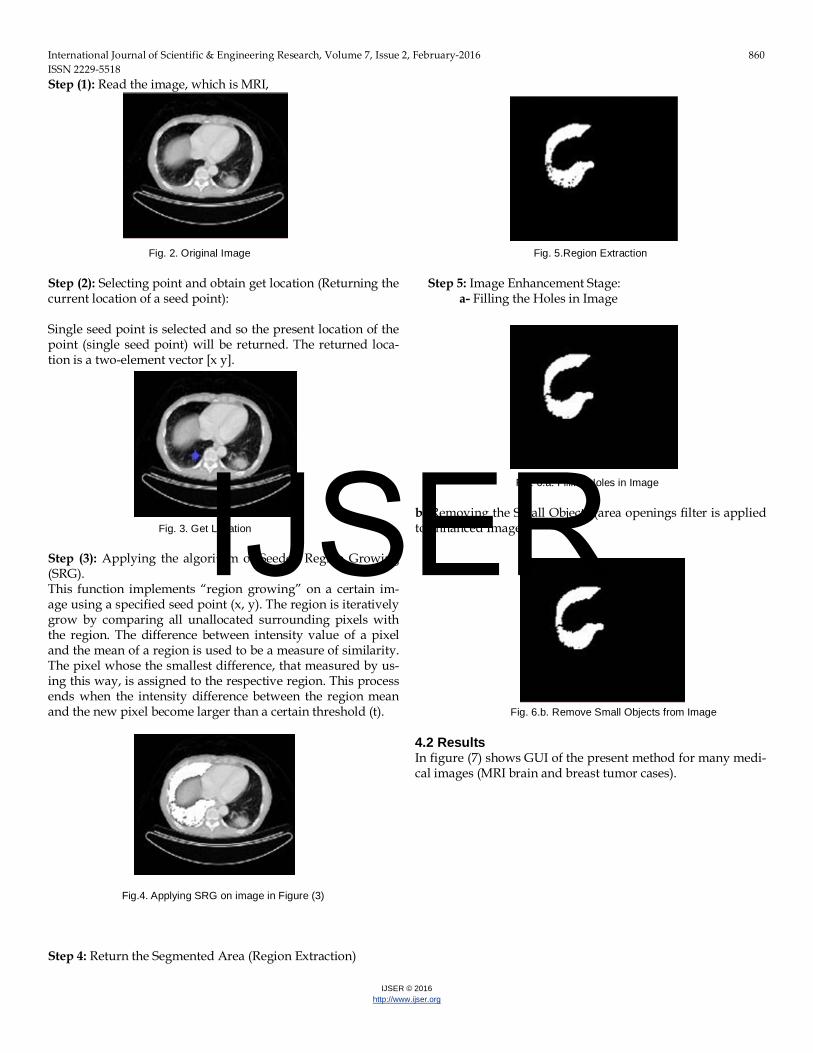
Step (1): Read the image, which is MRI,
Fig. 2. Original Image Step (2): Selecting point and obtain get location (Returning the current location of a seed point): Single seed point is selected and so the present location of the point (single seed point) will be returned. The returned loca-tion is a two-element vector [x y].
Fig. 3. Get Location Step (3): Applying the algorithm of Seeded Region Growing (SRG). This function implements “region growing” on a certain im-age using a specified seed point (x, y). The region is iteratively grow by comparing all unallocated surrounding pixels with the region. The difference between intensity value of a pixel and the mean of a region is used to be a measure of similarity. The pixel whose the smallest difference, that measured by us-ing this way, is assigned to the respective region. This process ends when the intensity difference between the region mean and the new pixel become larger than a certain threshold (t).
Fig.4. Applying SRG on image in Figure (3)
Step 4: Return the Segmented Area (Region Extraction)
Fig. 5.Region Extraction
Step 5: Image Enhancement Stage: a- Filling the Holes in Image
Fig. 6.a. Filling Holes in Image
b- Removing the Small Objects (area openings filter is applied to enhanced Images)
Fig. 6.b. Remove Small Objects from Image
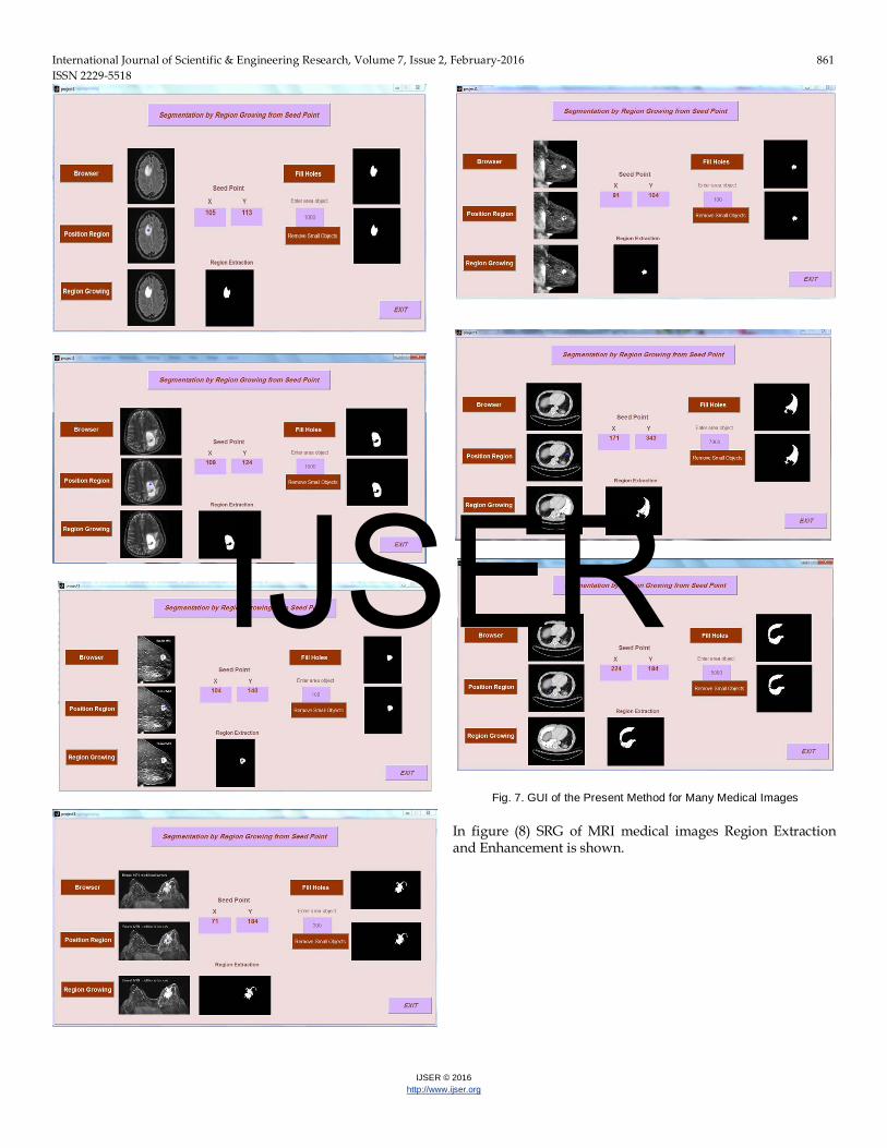
4.2 Results In figure (7) shows GUI of the present method for many medi-cal images (MRI brain and breast tumor cases).
IJSER
International Journal of Scientific & Engineering Research, Volume 7, Issue 2, February-2016 861 ISSN 2229-5518
IJSER © 2016 http://www.ijser.org
Fig. 7. GUI of the Present Method for Many Medical Images
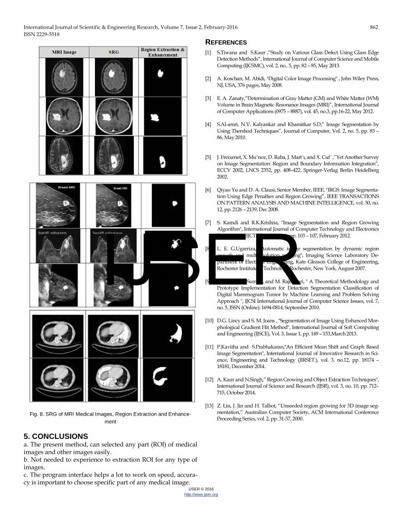
In figure (8) SRG of MRI medical images Region Extraction and Enhancement is shown.
IJSER
International Journal of Scientific & Engineering Research, Volume 7, Issue 2, February-2016 862 ISSN 2229-5518
IJSER © 2016 http://www.ijser.org
Fig. 8. SRG of MRI Medical Images, Region Extraction and Enhance-
ment
5. CONCLUSIONS a. The present method, can selected any part (ROI) of medical images and other images easily. b. Not needed to experience to extraction ROI for any type of images. c. The program interface helps a lot to work on speed, accura-cy is important to choose specific part of any medical image.
REFERENCES [1] S.Tiwana and S.Kaur ,“Study on Various Glass Defect Using Glass Edge
Detection Methods”, International Journal of Computer Science and Mobile Computing (IJCSMC), vol. 2, no.. 5, pp. 82 – 85, May 2013.
[2] A. Koschan, M. Abidi, "Digital Color Image Processing" , John Wiley Press, NJ, USA, 376 pages, May 2008 .
[3] E. A. Zanaty,”Determination of Gray Matter (GM) and White Matter (WM) Volume in Brain Magnetic Resonance Images (MRI)”, International Journal of Computer Applications (0975 – 8887), vol. 45, no.3, pp.16-22, May 2012.
[4] S.Al-amri, N.V. Kalyankar and Khamitkar S.D,” Image Segmentation by Using Thershod Techniques”, Journal of Computer, Vol. 2, no. 5, pp. 83 – 86, May 2010.
[5] J. Freixenet, X. Mu˜noz, D. Raba, J. Mart´ı, and X. Cuf´ ,”Yet Another Survey
on Image Segmentation: Region and Boundary Information Integration”, ECCV 2002, LNCS 2352, pp. 408–422. Springer-Verlag Berlin Heidelberg 2002.
[6] Qiyao Yu and D. A. Clausi, Senior Member, IEEE, "IRGS: Image Segmenta-tion Using Edge Penalties and Region Growing”, IEEE TRANSACTIONS ON PATTERN ANALYSIS AND MACHINE INTELLIGENCE, vol. 30, no. 12, pp. 2126 – 2139, Dec 2008.
[7] S. Kamdi and R.K.Krishna, "Image Segmentation and Region Growing Algorithm", International Journal of Computer Technology and Electronics Engineering (IJCTEE). vol.2, no 1, pp. 103 – 107, February 2012.
[8] L. E. G.Ugarriza, "Automatic image segmentation by dynamic region growth and multiresolution merging", Imaging Science Laboratory De-partment of Electrical Engineering, Kate Gleason College of Engineering, Rochester Institute of Technology Rochester, New York, August 2007.
[9] V. Raman, P. Sumari, and M. Rajeswari, " A Theoretical Methodology and Prototype Implementation for Detection Segmentation Classification of Digital Mammogram Tumor by Machine Learning and Problem Solving Approach ", IJCSI International Journal of Computer Science Issues, vol. 7, no. 5, ISSN (Online): 1694-0814, September 2010.
[10] D.G. Lincy and S. M. Joans , "Segmentation of Image Using Enhanced Mor-phological Gradient Hit Method", International Journal of Soft Computing and Engineering (IJSCE), Vol. 3, Issue 1, pp. 149 – 153,March 2013.
[11] P.Kavitha and S.Prabhakaran,"An Efficient Mean Shift and Graph Based Image Segmentation", International Journal of Innovative Research in Sci-ence, Engineering and Technology (JIRSET.), vol. 3, no.12, pp. 18174 – 18181, December 2014.
[12] A. Kaur and N.Singh,” Region Growing and Object Extraction Techniques", International Journal of Science and Research (IJSR), vol. 3, no. 10, pp. 712– 715, October 2014.
[13] Z. Lin, J. Jin and H. Talbot, “Unseeded region growing for 3D image seg-mentation,” Australian Computer Society, ACM International Conference Proceeding Series, vol. 2, pp. 31-37, 2000.
IJSER