Embed Size (px)
Citation preview
1Life and Cells
• What is Life?– Can grow, i.e. increase in size.– Can reproduce.– Responsive to environment.– Metabolism: can acquire and utilize energy.
• Schwann and Schleiden: cells basic unit of life– Prokaryotes and eukaryotes from microscopy.– Our focus: prokaryotic cells.
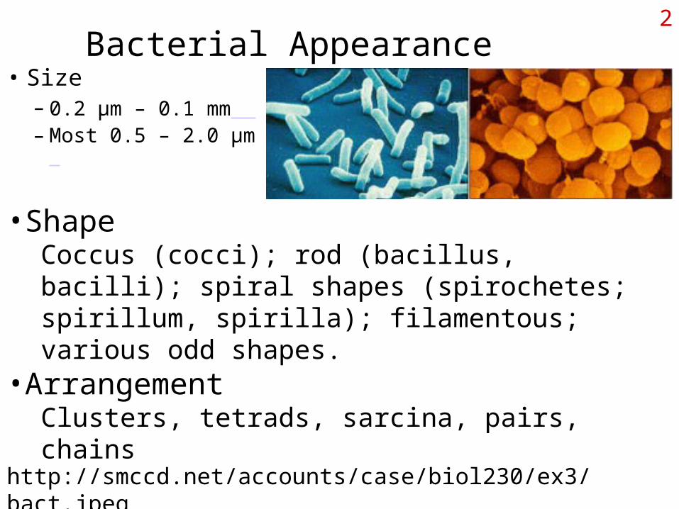
2Bacterial Appearance
• Size– 0.2 µm – 0.1 mm – Most 0.5 – 2.0 µm
•ShapeCoccus (cocci); rod (bacillus, bacilli); spiral shapes (spirochetes; spirillum, spirilla); filamentous; various odd shapes.
•ArrangementClusters, tetrads, sarcina, pairs, chains
http://smccd.net/accounts/case/biol230/ex3/bact.jpeg
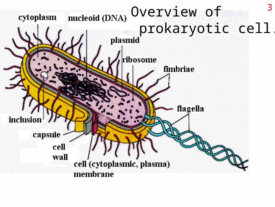
3Overview of prokaryotic cell.
4From Membrane Out:lecture order
• Examination of layers of bacterial cell
– Starting at cell membrane, working to outside
• A look at how cells move
• Examination of inside of bacterial cell
• A look at how things get into cells
• Review eukaryotic cell structure on your own.
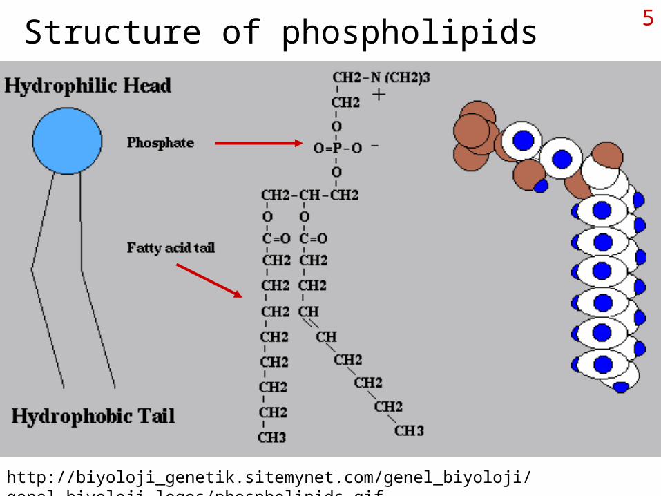
5Structure of phospholipids
http://biyoloji_genetik.sitemynet.com/genel_biyoloji/genel_biyoloji_logos/phospholipids.gif
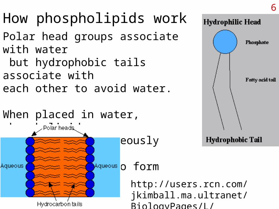
6How phospholipids workPolar head groups associate with water but hydrophobic tails associate with each other to avoid water.
When placed in water, phospholipids associate spontaneously side by side and tail to tail to form membranes.
http://users.rcn.com/jkimball.ma.ultranet/BiologyPages/L/LipidBilayer.gif
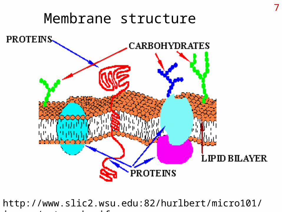
7Membrane structure
http://www.slic2.wsu.edu:82/hurlbert/micro101/images/cytomemb.gif
8Cell Membranes
• 50/50 lipids and proteins• Fluid mosaic model• Effective barrier to large and hydrophilic molecules
– O2, CO2, H2O, lipid substances can pass through– Salts, sugars, amino acids, polymers, cannot.
• Proteins can be on inner, outer surfaces or transmembrane– Involved primarily with transport– Degradation and biosynthesis– Site of ATP synthesis

9Outside the cell membrane:the Cell Wall
Animal cells do not have a cell wall outside the cell membrane.
Plant cells and fungal cells do.
So do most prokaryotic cells, provided structural support and determining the shape of the cell.
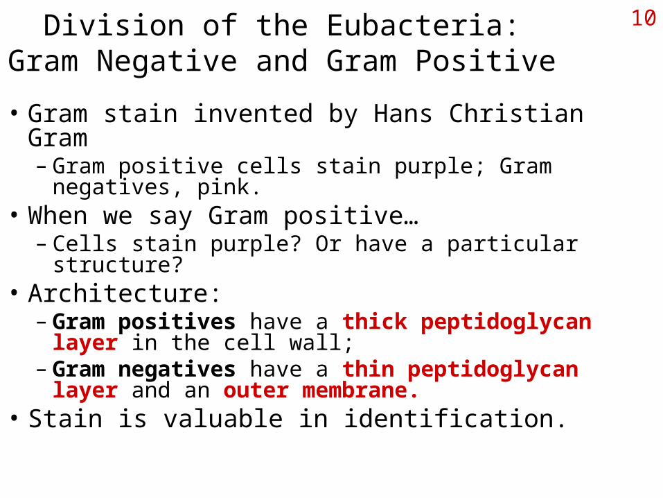
10Division of the Eubacteria:Gram Negative and Gram Positive
• Gram stain invented by Hans Christian Gram– Gram positive cells stain purple; Gram negatives, pink.
• When we say Gram positive…– Cells stain purple? Or have a particular structure?
• Architecture: – Gram positives have a thick peptidoglycan layer in the
cell wall;– Gram negatives have a thin peptidoglycan layer and an
outer membrane.• Stain is valuable in identification.
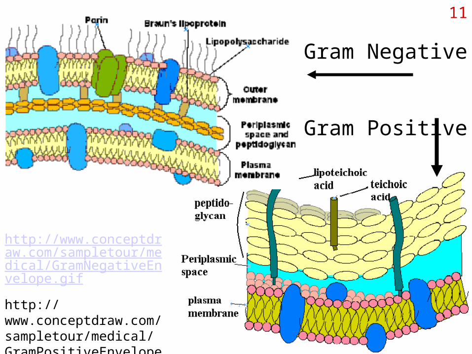
11
http://www.conceptdraw.com/sampletour/medical/GramNegativeEnvelope.gif
http://www.conceptdraw.com/sampletour/medical/GramPositiveEnvelope.gif
Gram Negative
Gram Positive
12Function and Structure of peptidoglycan
• Provides shape and structural support to cell• Resists damage due to osmotic pressure• Provides some degree of resistance to diffusion of
molecules• Single bag-like, seamless molecule• Composed of polysaccharide chains cross linked
with short chains of amino acids: “peptido” and “glycan”.
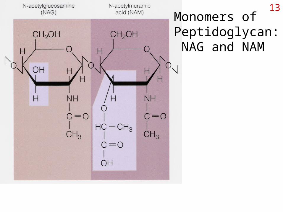
13Monomers of Peptidoglycan: NAG and NAM
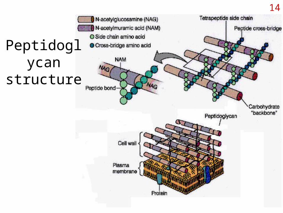
14
Peptidoglycan structure
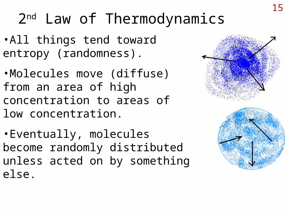
152nd Law of Thermodynamics
•All things tend toward entropy (randomness).
•Molecules move (diffuse) from an area of high concentration to areas of low concentration.
•Eventually, molecules become randomly distributed unless acted on by something else.
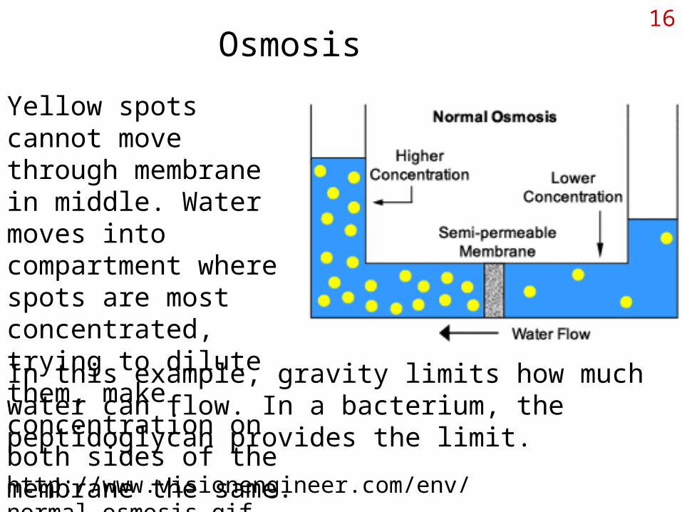
16Osmosis
Yellow spots cannot move through membrane in middle. Water moves into compartment where spots are most concentrated, trying to dilute them, make concentration on both sides of the membrane the same.
In this example, gravity limits how much water can flow. In a bacterium, the peptidoglycan provides the limit.
http://www.visionengineer.com/env/normal_osmosis.gif
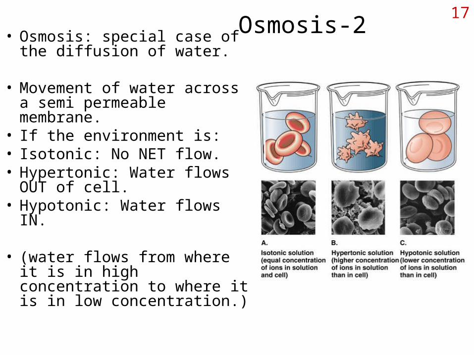
17Osmosis-2• Osmosis: special case of the
diffusion of water.
• Movement of water across a semi permeable membrane.
• If the environment is:• Isotonic: No NET flow.• Hypertonic: Water flows OUT
of cell.• Hypotonic: Water flows IN.
• (water flows from where it is in high concentration to where it is in low concentration.)
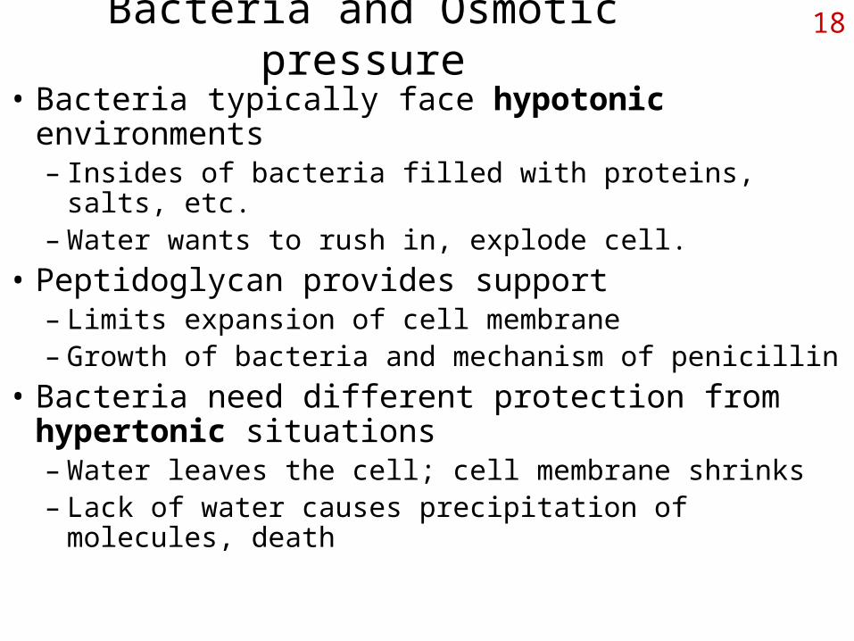
18Bacteria and Osmotic pressure
• Bacteria typically face hypotonic environments– Insides of bacteria filled with proteins, salts, etc.– Water wants to rush in, explode cell.
• Peptidoglycan provides support– Limits expansion of cell membrane– Growth of bacteria and mechanism of penicillin
• Bacteria need different protection from hypertonic situations– Water leaves the cell; cell membrane shrinks– Lack of water causes precipitation of molecules, death

19Effect of osmotic pressure on cells
• Hypotonic: water rushes in; PG prevents cell rupture.
• Hypertonic:
water leaves cell, membrane pulls away from cell wall.
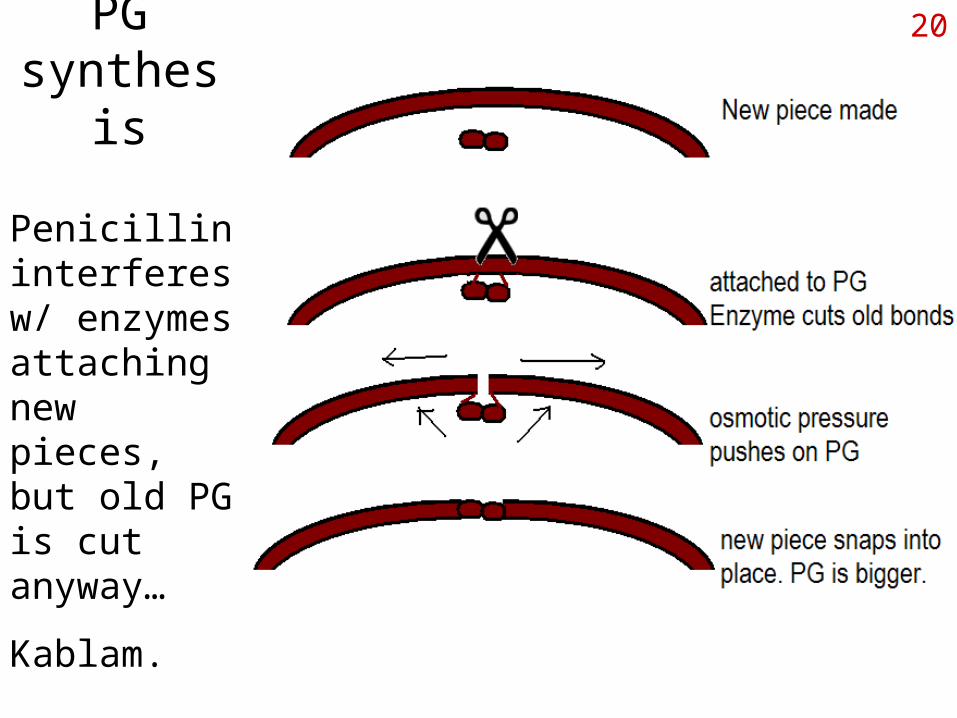
20PG synthesis
Penicillin interferes w/ enzymes attaching new pieces, but old PG is cut anyway…
Kablam.
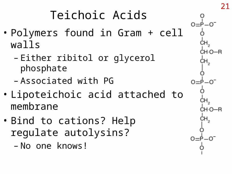
21Teichoic Acids
• Polymers found in Gram + cell walls– Either ribitol or glycerol phosphate– Associated with PG
• Lipoteichoic acid attached to membrane
• Bind to cations? Help regulate autolysins?– No one knows!
22Cell Wall Exceptions
• Mycobacterium and relatives– Wall contains lots of waxy mycolic acids– Attached covalently to PG
• Mycoplasma: no cell wall– Parasites of animals, little osmotic stress
• Archaea, the 3rd domain– Pseudomurein and other chemically different wall
materials (murein another name for PG)