Embed Size (px)
Citation preview

1
Neuroscience and Behavior
Chapter 2

2
“The brain is wider than the sky.”
Emily Dickinson, 1830-1886
3
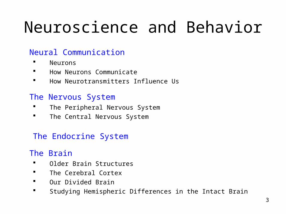
Neuroscience and BehaviorNeural Communication Neurons How Neurons Communicate How Neurotransmitters Influence Us
The Nervous System The Peripheral Nervous System The Central Nervous System
The Endocrine System
The Brain Older Brain Structures The Cerebral Cortex Our Divided Brain Studying Hemispheric Differences in the Intact Brain

4
Neural Communication
The body’s information system is built from billions of interconnected cells called neurons.

5
Neuron
A nerve cell, or a neuron, consists of many different parts.

6
Parts of a Neuron
Cell Body: Life support center of the neuron.
Dendrites: Branching extensions at the cell body. Receive messages from other neurons.
Axon: Long single extension of a neuron, covered with myelin [MY-uh-lin] sheath to insulate and speed up messages through neurons.
Terminal Branches of axon: Branched endings of an axon that transmit messages to other neurons.

7
Neurons: Communication
• Membrane: a skin that separates the inside from the outside of the neuron
• Ions: Molecules that are positively or negatively charged

8
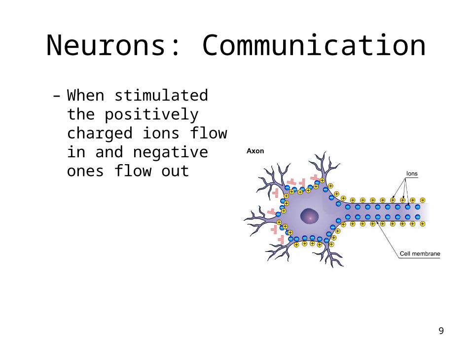
Neurons: Communication
• Resting Neuron:– When a neuron is
polarized, more negative ions are inside.
9
Neurons: Communication
– When stimulated the positively charged ions flow in and negative ones flow out

10
Neuron: Communication
• Action Potential– When neurons depolarized– When soma receives
enough stimulation, the gates open and lets some negative ions out and some positive ones in.
– Then the whole cell fires, more gates open and more positive ions rush in
– The electric charge of the neuron suddenly changes from negative to positive

11
Action Potential
12
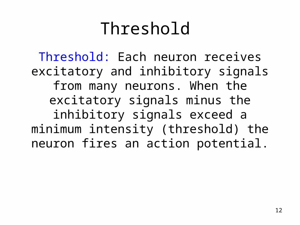
Threshold
Threshold: Each neuron receives excitatory and inhibitory signals from many neurons. When the excitatory signals minus the inhibitory signals
exceed a minimum intensity (threshold) the neuron fires an action
potential.

13
Action Potential Properties
All-or-None Response: A strong stimulus can trigger more neurons to fire, and to fire more often, but it does not affect the
action potentials strength or speed.
Intensity of an action potential remains the same throughout the length of the
axon.

14
Synapse
Synapse [SIN-aps] a junction between the axon tip of the sending neuron and the
dendrite or cell body of the receiving neuron. This tiny gap is called the synaptic gap or
cleft.

15
Neurotransmitters
Neurotransmitters (chemicals) released
from the sending neuron travel across the synapse and bind to receptor sites on
the receiving neuron, thereby influencing it to generate an action
potential.

16
Reuptake
Neurotransmitters in the synapse are
reabsorbed into the sending neurons
through the process of reuptake. This
process applies the brakes on
neurotransmitter action.

17
• Excitatory Neurotransmitters
• Inhibitory Neurotransmitters

18

19
• Location
Brain, spinal cord, peripheral nervous system, especially some organs of the parasympathetic nervous system
• Effect
Excitatory in brain and autonomic nervous system; inhibitory elsewhere
• Function
Muscle movement; cognitive functioning
• Related to Drug Curare
poisonous darts that paralyze skeletal muscles
• Deficiency in ACh production in patients suffering from Alzheimer’s disease
Acetylcholine (ACh)
Neurotransmitters

20
How Neurotransmitters Influence Us
Serotonin
From Mapping the Mind, Rita Carter, © 1989 University of California Press
LocationBrain, spinal cord
EffectInhibitory
FunctionSleeping, eating,
mood, pain, depression, alcoholism, suicide, impulsivity, aggression, and coping with stress

21
Dopamine Pathways
LocationBrain
•EffectInhibitory or excitatory
•FunctionMuscle disorders, mental disorders, Parkinson’s From Mapping the Mind, Rita Carter, © 1989
University of California Press

22Endorphins
• LocationBrain, spinal cord
• EffectPrimarily inhibitory, except in hippocampus
• FunctionPain suppression, pleasurable feelings, appetites, placebos
– “runners high”
Neurotransmitters

23

24
Lock & Key Mechanism
Neurotransmitters bind to the receptors of the receiving neuron in a key-lock mechanism.

25
Agonists

26
Antagonists
27
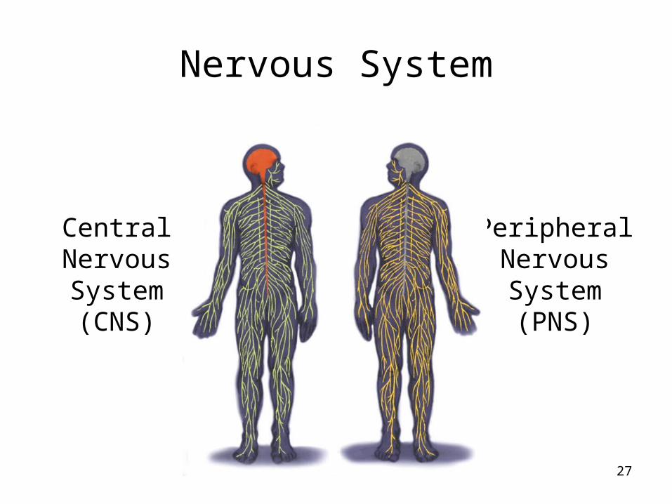
Nervous System
CentralNervousSystem(CNS)
PeripheralNervousSystem(PNS)

28
The Nervous System

29
Peripheral Nervous System
Somatic Nervous System: The division of the peripheral nervous system that controls the body’s skeletal muscles.
Autonomic Nervous System: Part of the PNS that controls the glands and other muscles.

30
Autonomic Nervous System (ANS)
Sympathetic NS “Arouses”
(fight-or-flight)
Parasympathetic NS “Calms”
(rest and digest)

31
Central Nervous System
• Receives, processes, interprets, and stores incoming sensory information– The brain is the control center– The spinal cord is an extension of the brain
• Has some autonomy• Produces spinal reflexes requiring no conscious
efforts

32
Spinal Reflex

33
Read the list
• Blue• Green• Red• Yellow• Red• Orange• Green• Brown• Yellow• Purple• Red

34
Name the color

35
Name the color
BrownBlueRedPurpleOrangeBlackBlueGreenBrown

36
The Brain
Brain Stem
Cerebellum (little brain)
Limbic System
Cerebral Cortex

37
The Brain: Older Brain Structures
The Brainstem is the oldest part of the brain, beginning where the spinal cord swells and enters the skull. It is responsible for automatic survival
functions.

38
Brainstem
The Medulla [muh-DUL-uh] is the base
of the brainstem that controls heartbeat
and breathing.

39
Brainstem
The Thalamus [THAL-uh-muss] is the brain’s sensory switchboard, located on top of the brainstem. It directs
messages to the sensory areas in the cortex and transmits
replies to the cerebellum and
medulla.
40
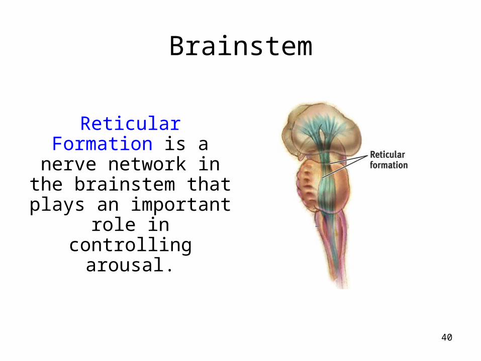
Brainstem
Reticular Formation is a nerve network in the brainstem that plays an important role in controlling
arousal.

41
The “little brain” attached to the rear of the brainstem. It
helps coordinate voluntary movements
and balance.
Cerebellum

42
The Brain
Techniques to Study the Brain
A brain lesion experimentally
destroys brain tissue to study animal behaviors after such destruction.
Hubel (1990)

43
Clinical ObservationClinical observations have shed light on a
number of brain disorders. Alterations in brain morphology due to neurological and
psychiatric diseases are now being catalogued.
Tom
Landers/ B
oston Globe

44
Electroencephalogram (EEG)An amplified recording of the electrical waves
sweeping across the brain’s surface, measured by electrodes placed on the scalp.
AJ P
hoto/ Photo R
esearchers, Inc.

45
PET Scan
PET (positron emission tomography)
Scan is a visual display of brain
activity that detects a radioactive form of glucose while the
brain performs a given task.
Courtesy of N
ational Brookhaven N
ational Laboratories

46
MRI ScanMRI (magnetic
resonance imaging) uses magnetic fields and radio waves to produce computer-
generated images that distinguish among
different types of brain tissue. Top images show ventricular enlargement in a
schizophrenic patient. Bottom image shows brain regions when a
participants lies.
Both photos from Daniel Weinberger, M.D., CBDB, NIMH
James Salzano/ Salzano Photo Lucy Reading/ Lucy Illustrations

47
The Limbic System is a doughnut-shaped system of neural
structures at the border of the brainstem and cerebrum, associated with emotions such as fear, aggression and
drives for food and sex. It includes the hippocampus, amygdala, and hypothalamus.
The Limbic System
48
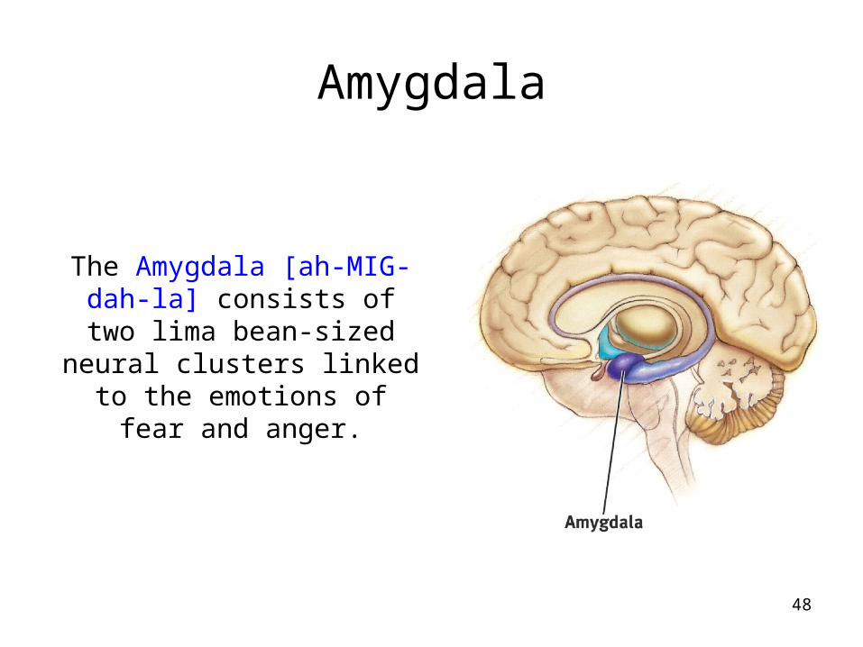
Amygdala
The Amygdala [ah-MIG-dah-la] consists of two lima bean-sized neural clusters linked to the emotions of fear and
anger.
49
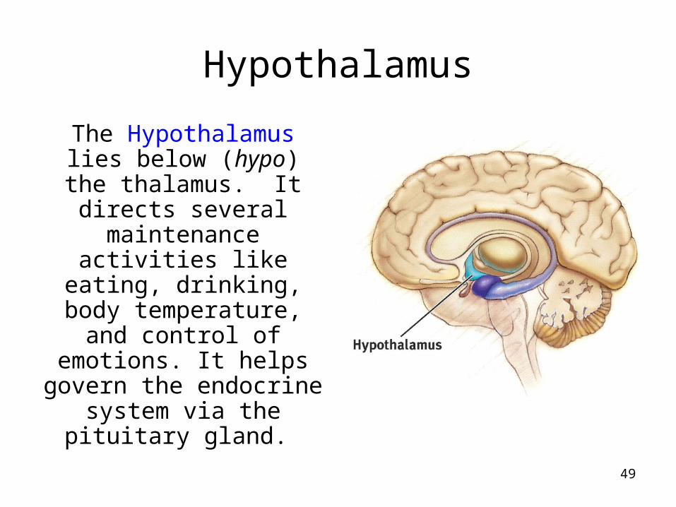
Hypothalamus
The Hypothalamus lies below (hypo) the thalamus. It directs several maintenance activities like eating,
drinking, body temperature, and
control of emotions. It helps govern the
endocrine system via the pituitary gland.

50
Rats cross an electrified grid for self-
stimulation when electrodes are placed
in the reward (hypothalamus) center (top picture). When the
limbic system is manipulated, a rat will navigate fields or climb
up a tree (bottom picture).
Reward CenterS
anjiv Talw
ar, SU
NY
Dow
nstate

51
Brain: Structures

52
Structure of the Cortex
Each brain hemisphere is divided into four
lobes that are separated by
prominent fissures. These lobes are the
frontal lobe (forehead), parietal lobe (top to rear head), occipital lobe (back head) and temporal lobe (side of
head).

53
BrainFrontal LobeEmotion, planning, creative thinking and motor cortex.
Left lobe, Broca’s area.
Parietal lobe receives information from pressure, pain, touch and temperature, from overall body.
Temporal lobe regulates hearing, balance, and equilibrium. It is important in face recognition, and contains auditory cortex.

54
Visual Function
The functional MRI scan shows the visual cortex is active as the subject looks at faces.
Courtesy of V
.P. Clark, K
. Keill, J. M
a. M
aisog, S. Courtney, L
.G.
Ungerleider, and J.V
. Haxby,
National Institute of M
ental Health

55
Auditory Function
The functional MRI scan shows the
auditory cortex is active in patients who
hallucinate.

56
More intelligent animals have increased “uncommitted” or association areas of the
cortex.
Association Areas
57
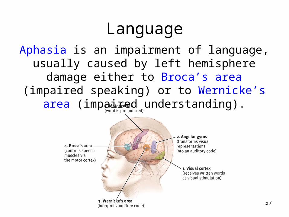
LanguageAphasia is an impairment of language,
usually caused by left hemisphere damage either to Broca’s area (impaired speaking)
or to Wernicke’s area (impaired understanding).

58
Specialization & Integration
Brain activity when hearing, seeing, and speaking words

59
Our Divided Brain
Our brain is divided into two hemispheres. The left hemisphere processes reading,
writing, speaking, mathematics, and comprehension skills. In the 1960s, it was
termed as the dominant brain.

60
Non-Split Brains
People with intact brains also show left-right hemispheric differences in mental
abilities.
A number of brain scan studies show normal individuals engage their right
brain when completing a perceptual task and their left brain when carrying out a
linguistic task.

61
Splitting the BrainA procedure in which the two hemispheres of the brain are isolated by cutting the connecting fibers
(mainly those of the corpus callosum) between them.
Corpus Callosum
Ma
rtin M
. Ro
the
r
Courtesy of T
erence William
s, University of Iow
a

62
Split Brain Patients
With the corpus callosum severed, objects (apple) presented in the right visual field can be named.
Objects (pencil) in the left visual field cannot.

63
Divided Consciousness

64
Try This!
Try drawing one shape with your left hand and one with your right hand,
simultaneously.B
BC


![chapter 1. biography and main workschapter 1. biography ... V – EMILY DICKINSON 115 EMILY DICKINSON, 1830-1886 chapter 1. biography and main works George MCMICHAEL [1025] One day](https://img.pdfslide.net/doc/110x75/5aa6ebd67f8b9ac5648b681c/chapter-1-biography-and-main-workschapter-1-biography-v-emily-dickinson.jpg)


![Emily Dickinson 1886 [ Complete Poems]](https://img.pdfslide.net/doc/110x75/549e317ab4795988208b4684/emily-dickinson-1886-complete-poems.jpg)