Embed Size (px)
Citation preview

1
Part 3Coenzyme-Dependent Enzyme
MechanismsProfessor A. S. Alhomida
Part 3Coenzyme-Dependent Enzyme
MechanismsProfessor A. S. Alhomida
Disclaimer• The texts, tables and images contained in this course presentation
are not my own, they can be found on: – References supplied– Atlases or– The web
King Saud University
College of Science
Department of Biochemistry

2

3
Vitamins and Coenzymes

4

5

6

7

8

9
Glycolysis
TCAcycle
Glycogenolysis
aKGDHvit B1,B2,B3
PP avit B6
Glc
GlycogenG1P
R5PTK
vit B1
PDHvit B1,B2,B3
aKGSCoA
Acetyl-CoA
G6P
Pyr
G3PALT
vit B6
Ala
ASTvit B6
OAAsp
vit B6 Glu
PPP
Vitamins in Metabolic Pathways

10
Coenzymes and Vitamins
Apoenzyme + Cofactor Holoenzyme
(protein only) (active)
(inactive)
• Some enzymes require cofactors for activity
(1) Essential ions (mostly metal ions)
(2) Coenzymes (organic compounds)

11
Coenzymes
• Coenzymes act as group-transfer reagents
• Hydrogen, electrons, or other groups can be transferred
• Larger mobile metabolic groups can be attached at the reactive center of the coenzyme
• Coenzyme reactions can be organized by their types of substrates and mechanisms

12
Types of cofactors

13
Inorganic Cations
• Enzymes requiring metal ions for full activity:
(1) Metal-activated enzymes have an absolute requirement or are stimulated by metal ions (examples: K+, Ca2+, Mg2+)
(2) Metalloenzymes contain firmly bound metal ions at the enzyme active sites (examples:
iron, zinc, copper, cobalt )

14
Carbonic Anhydrase

15
• Carbon dioxide (CO2) is a major end product of aerobic metabolism
• In mammals, this CO2 is released into the blood and transported to the lungs for exhalation
• While in the blood, CO2 reacts with water• The product of this reaction is a moderately
strong acid, carbonic anhydride (pKa = 3.5), which becomes bicarbonate ion on the loss of a H+
Carbonic Anhydrase

16
• Almost all organisms contain enzyme, carbonic ahydrase, that catalyzes the below reaction
• Cabonic anhydrase accelerates CO2 hydration dramatically at rate as high as kcat =
106 s-1
Carbonic Anhydrase, Cont’d

17
Types of Carbonic Anhydrases
• -Carbonic anydrase:– Found in human, animals, some bacteria and algae– Trimer
• -Carbonic anydrase– Higher plants and many bacteria and E. coli– Has only one conserved His whereas in a has three
His• -Carbonic anhydrase
– Found in bacteria Methanoscarcina thermophila– Has three zn sites similar to carbonic anhydrase

18
Structure of -Carbonic Anydrase
• Zn2+ is coordinated by the imidazole rings of three His residues, His-94, His-96 and His-119
• The primary function of the enzyme in animals is to interconvert CO2 and bicarbonate to maintain acid-base balance in blood and other tissues and to help transport CO2 out of tissues

19
Structure of β-Carbonic Anhydrase
• Found in plans which is an evolutionarily distinct enzyme but participates in the same reaction and also uses a Zn2+ in its active site
• It helps raise the concentration of CO2 within the chloroplast to increase the carboxylation rate of the enzyme Rubisco
• It integrates CO2 into organic carbon sugars during photosynthesis, and can only use the CO2 form of carbon, not carbonic acid nor bicarbonate

20
Structure of -Carbonic Anydrase
Zn bound to three HisThree subunits

21
Structure of -Carbonic Anhydrase
• (Left) the Zn site, (Middle) the trimeric structure (A, B, and C) and (Right) the enzyme is rotated to show a top-down view position of the Zn sites

22
Human carbonic anhydrase

23
• How does this Zn2+ complex facilitates CO2 hydration?
• A major clue comes from the pH profile of the enzymatic ally catalyzed CO2 hydration:
Carbonic Anhydrase, Cont’d

24
Carbonic Anhydrase, Cont’d
• At pH 8, the reaction proceeds near its maximal rate
• As the pH decreases, the rate of the reaction drops
• The midpoint of this transition is near pH 7, suggesting that a group with pKa = 7 plays an important role in the activity of this enzyme

25
Carbonic Anhydrase, Cont’d
• The deprotonated (high pH) form of this group participates more effectively in the catalysis
• Although His have pKa value near 7, a variety of evidence suggest that the group responsible for this transition is not His but it is the Zn2+-bound water molecule
• The binding of water to the positively charged Zn2+ center reduces the pKa of the water from 15.7 to 7

26
Carbonic Anhydrase, Cont’d

27
• The lowered pKa generates Zn2+-OH-
complex that is sufficiently nucleophilic to attack CO2 more readily than water does
Carbonic Anhydrase, Cont’d

28
Mechanism of Carbonic Anhydrase

29
Mechanism of Carbonic Anhydrase
Zn2+
His
HisHis
OH
Zn2+
His
HisHis
OH
B:
C OO
B:
CO2

30
• Zn2+ ion promotes the ionization of bound H2O. Resulting nucleophilic OH- attacks carbon of CO2
• The pKa of water drops from 15.7-7 when it is coordinate to Zn2+
• HO- is 4 orders of magnitude more nucleophlic than is water
Mechanism of Carbonic Anhydrase, Cont’d

31
Zn2+
His
HisHis
OH
C
OO
B:
H2O
Zn2+
His
HisHis
OH
C
OO
O
HH
B :

32
Bicarbonate
Zn2+
His
HisHis
OH
C
OO
O
H
BH
O
C OHO
Zn2+
His
HisHis
OH
BH
Tetrahedral intermediate

33
1. The important of Zn2+-OH- comlpex suggests a simple mechanism of CO2 hydration:
2. Zn2+ facilitates the release of a H+ from water, which generates a OH-
3. The CO2 binds to the enzyme’s active site and is positioned to react with the OH-
Mechanism of Carbonic Anhydrase, Cont’d

34
3. The OH- attacks nucleophilically CO2, converting it into bicarbonate ion
4. The catalytic site is regenerated with release of the bicarbonate ion and the binding of another molecule of water
Mechanism of Carbonic Anhydrase, Cont’d

35
Iron in Metalloenzymes• Iron undergoes reversible oxidation and
reduction:Fe3+ + e- (reduced substrate) Fe2+ + (oxidized substrate)
• Enzyme heme groups and cytochromes contain iron

36
Iron in Metalloenzymes, Cont’d
• Nonheme iron exists in iron-sulfur clusters (iron is bound by sulfide ions and S- groups from cysteines)
• Iron-sulfur clusters can accept only one e- in a reaction

37
Iron-sulfur clusters
• Iron atoms are complexed with an equal number of sulfide ions (S2-) and with thiolate groups of Cys side chains

38
Coenzyme Classification
• There are two classes of coenzymes
(1) Cosubstrates are altered during the reaction and regenerated by another enzyme
(2) Prosthetic groups remain bound to the enzyme during the reaction, and may be
covalently or tightly bound to enzyme

39
Classification of Coenzymes in Mammals
(1) Metabolite coenzymes- synthesized from common metabolites
(2) Vitamin-derived coenzymes- derivatives of vitamins (vitamins cannot be synthesized by mammals, but must be obtained as nutrients)

40
Metabolite Coenzymes

41
Metabolite Coenzymes
• Nucleoside triphosphates are examples
ATP 5`-C

42
Reactions of ATP
• ATP is a versatile reactant that can donate its:
(1) Phosphoryl group (-phosphate)
(2) Pyrophosphoryl group (, phosphates)
(3) Adenylyl group (AMP)
(4) Adenosyl group

43
• Nucleotide-sugar coenzymes are involved in carbohydrate metabolism
• UDP-Glucose is a sugar coenzyme. It is formed from UTP and glucose 1-phosphate
(UDP-glucose product next slide)

44

45
Carbon-Carbon Bond Formation

46
Alkylation Reactions
• Methylation is an important transformation in the biosynthesis of many secondary metabolites
• Organic chemists use methyl iodide or methyl sulphonates for methylations
• The biological equivalent is S-adenosyl methionine (SAM)
• The driving force for methyl group transfer is the conversion of a sulphonium ion into a neutral sulphide

47
Alkylation Reactions, Cont’d

48
Aldol and Claisen Reactions
• Reactions between enolates (and their equivalents) with aldehydes or ketones are referred to as aldol reactions
• Reaction of enolates with esters are referred to as Claisen reactions
• They are the most common method to form carbon-carbon bonds
• The biological equivalent of enolates are enamines and coenzyme A
• These are co-factors of aldolase enzymes
49
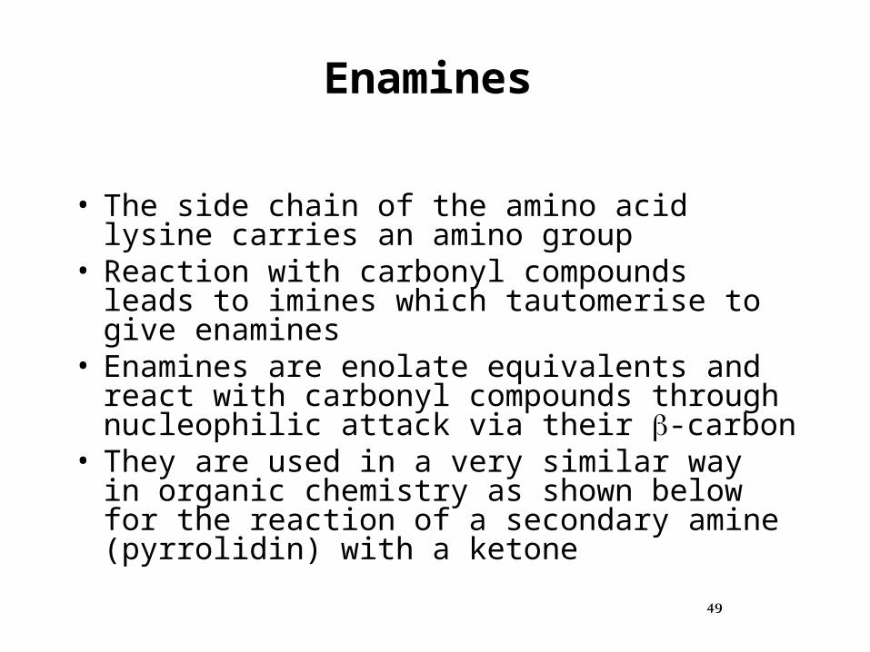
Enamines
• The side chain of the amino acid lysine carries an amino group
• Reaction with carbonyl compounds leads to imines which tautomerise to give enamines
• Enamines are enolate equivalents and react with carbonyl compounds through nucleophilic attack via their -carbon
• They are used in a very similar way in organic chemistry as shown below for the reaction of a secondary amine (pyrrolidin) with a ketone

50
Enamines, Cont’d

51
Aldol Reactions
• Aldol Reactions Require Several Levels of Control:
• Enol versus carbonyl component: • carbonyl compounds with acidic -protons
can either be deprotonated and react as nucleophiles, or react as electrophiles through their carbonyl group
• If this is not carefully controlled an intractable mixture of products (cross-aldol products) is obtained

52
• Formation of an enamine avoids this problem• The enamine is only nucleophilic• Regioselectivity: enamines are ambident
nucleophiles• They can in principle react through the
carbon or the nitrogen atom• For aldol-type processes, only reactions
through the carbon atom lead to the desired product
Aldol Reactions, Cont’d

53
• In biological systems the regioselectivity is controled by the steric environment of the enzyme active site
Aldol Reactions, Cont’d

54
• Stereoselectivity: The stereochemistry of aldol reactions is highly complex-syn, anti, matched case, mismatched case-are just a few keywords highlighting how difficult it is to control the relative and absolute stereochemistry of aldol products
• In biological systems this is again taken care of by the stereochemistry of the active site of an enzyme
Aldol Reactions, Cont’d

55
S-Adenosylmethionine (SAM)

56
• ATP is a source of other metabolite coenzymes such as S-adenosylmethionine (SAM)
• SAM donates methyl groups in many biosynthesis reactions
Methionine + ATP SAM + Pi + PPi
SAM Biosynthesis

57
Structure of SAM
• Activated methyl group in red

58
Functions of SAM
1. SAM donates the methyl group for many methylation reactions: Methylation of norepinephrins

59
Functions of SAM, Cont’d
2. SAM involves in redox radical-dependent enzymes: Pyruvate formate lyase; Anerobic ribonucleotide reductase
3. Until this point, the only know role for SAM was for methyl group transfer, thus it was surprising to find SAM involved in redox biochemistry

60
Functions of SAM, Cont’d
4. The involvement of SAM in redical biochemistry was first established for Lys 2,3-aminomustase (from C. subterminale) which converts Lys with -Lys
5. Lys 2,3-aminomustase catalyzes the reaction by 1,2 rearrangement mechanism similar to Vit B12-dependent mutase, but didn’t use Vit B12
instead required PLP and SAM for activity and a reduced [4Fe4S] cluster

61
SAM as Methyl Group Donor
– Methylation of bases in tRNA– Methylation of cytosine residues in DNA– Methylation of norepinephrine

62
SAM Cycle
1. SAM synthase (Met adenosyl transferase)
2. Methyltransferase3. S-adenosyl
homocysteinase 4. Homocysteine
methyltransferase

63
Mechanism of SAM Synthase
(Met Adenosyl Transferase)

64
Mechanism of SAM Synthase
C COO
H
S
O
N
NN
N
OH OH
HHH H
NH2
OO O
O OP
O
OP
O
OP
O
CH2CH2 2
CH3
H3N
MethionineATP
Unusual displacement of triphosphate reaction
:
Nucleophilic attack (SN2 mechanism)

65
• Met is not a sufficient reactive to be a good methyl donor because of the homosysteine mercaptide anion is a poor leaving group
• SAM synthase catalyzes an unusual displacement reaction because of Met sulfur atom attacks nucleophilically on the 5` carbon of ATP to produced the sulfonium compound and and inorganic triphosphate (PPPi ) is formed
Mechanism of SAM Synthase, Cont’d

66
SAM synthase
SAM
PPiPPPiPi
Mechanism of SAM Synthase
Very good leaving group because of positively charged of S atom
C COO
H
S
CH2 2
CH3
H3N
O
N
NN
N
OH OH
HHH H
NH2
CH2
OO O
O P
O
OP
O
OP
O
O
O O
O P
O
OP
O
O
O
O OP
O
2
O
O OP
O
H2O
Supernucleophile

67
• PPPi is then hydrolyzed by the same enzyme into PPi and Pi making the reaction thermodynamically more favorable
• This is one of two reactions in which a displacement of this kind is known to occur in biological system
Mechanism of SAM Synthase, Cont’d

68
• The other being the formation of adenosylcobalamin
• The hydrolysis of PPPi drives the reaction to right highly exergoic in the synthetic direction
Mechanism of SAM Synthase, Cont’d

69
SAM-Dependent Methyltransferase

70
SAM-Dependent Methyltransferase
• The functional roles of methylation are wide ranging and include biosynthesis, metabolism, detoxification, signal transduction, protein sorting and repairing, nucleic acid processing, gene silencing and imprinting
• The majority of methylation reactions are carried out by the SAM-dependent methyltransferases

71
• Human thiopurine S-methyltransferase (TPMT) in complex with SAH
• TPMT is a cytosolic drug-metabolizing enzyme that catalyzes the S-methylation of thiopurine drugs such as 6-mercaptopurine, azathioprine, 6-thioguanine
SAM-Dependent Methyltransferase, Cont’d

72
• All methylation reactions requiring SAM are simple SN2 (Substitution of nucleophilic bimolecular) displacements
• SAH is a potent inhibitor of all reactions in which a methyl group is transferred from SAM to an acceptor
• It is important to prevent the accummulation of SAH in cells
SAM-Dependent Methyltransferase, Cont’d

73
• This is accomplished through the action of S-adenosylhomocysteinase that converts SAH into adenosine and homocysteine
• Homocysteine is converted into Met and adenosine (Ado) into inosine (via SAM cycle)
SAM-Dependent Methyltransferase, Cont’d

74
Mechanism of SAM-Dependent Methyltransferase

75
Norepinephrine
SAM
Mechanism of SAM-Dependent Methyltransferase
HO
HO
CH2CH2NH2
OH
..
C COO
H
S
CH2 2
CH3
H3N
O
N
NN
N
OH OH
HHH H
NH2
CH2
Nucleophilic attack (SN2 Mechanism)

76
Mechanism of SAM-Dependent Methyltransferase, Cont’d
HO
HO
CH2CH2NH2
OH
C COO
H
CH2 2
CH3H3N
O
N
NN
N
OH OH
HHH H
NH2
CH2S
+
Epinephrine
S-adenosylhomocysteine (SAH)

77
SAM-Dependent Radical Enzymes

78
SAM-Dependent Radical Enzymes
• Organic radicals are used by a number of enzymes to catalyze biochemical transformations with high-energy barriers that would be difficult to accomplish through non-radical heterolytic chemistry
• Well known examples include:– Reduction of an alcohol to an alkane catalyzed by
ribonucleotide reductase– Carbon chain rearrangements catalyzed by
methylmalonyl CoA mutase or glutamate mutase

79
SAM-Dependent Radical Enzymes
• Organic radicals can be generated in enzymes through only three general mechanisms: – Metal-activated oxygen biochemistry– Adenosylcobalamin (Vit B12) biochemistry,
or – Reduction of the sulfonium of SAM

80
Pyruvate Formate Lyase(Formate C-Acetyltransferase)

81
Pyruvate Formate Lyase
• It is an important enzyme (found in Escherichia coli and other organisms) that helps regulate anaerobic glucose metabolism
• Using radical biochemistry, it catalyzes the reversible conversion of pyruvate and CoA into formate and acetyl-CoA

82
Structure of Pyruvate Formate Lyase
• It is a homodimer made of 85 kD, 759-residue subunits
• It has a 10-stranded / barrel motif into which is inserted a finger that contains major catalytic residues
• The active site of the enzyme, elucidated by X-ray crystallography, holds three essential amino acids that perform catalysis:– Gly-734– Cys-418– Cys-419

83
Structure of Pyruvate Formate Lyase, Cont’d
• It is a homodimeric protein (2 x 85 kD) and catalytically inactive when isolated
• Activated enzyme contains one protein radical per dimer at Gly-734 and has a half of the sites reactivity

84
Structure of Pyruvate Formate Lyase, Cont’d
• Three major residues that hold the substrate pyruvate close by Arg-435, Arg-176, and Ala-272), and two flanking hydrophobic residuesTrp-333 and Phe-432
• The active site of enzyme is a similar to that of class I and class III ribonucleotide reductase

85
• The interaction of SAM with the [4Fe–4S]1+ of activated en\yme
• -N and -carboxyl O of Met anchors the SAM to the cluster with the sulfonium interacting with a sulfide from the cluster
SAM-[4Fe4S] ClusterSAM
[4Fe4S] cluster

86
(AE) Activase (DE) Deactivase
Regulationn of Pyruvate Formate Lyase
Radical Radical Gly-734

87
Reaction of Pyruvate Formate Lyase
N
H
N
O
HHH
734
[4Fe4S]+
SAM
N
H
N
O
H
734
Gly-734 Gly-734 radical
Pyruvate formate lyase
red

88
Reaction of Pyruvate Formate Lyase, Cont’d
Gly-734 radical
H3C
O
O
OCoA
H3C S
O
CoA
H
O
O
N
H
N
O734
H
Pyruvate
Formate
Acetyl-CoA
Pyruvate formate lyase

89
Mechanism of Pyruvate Formate Lyase

90
Role of Catalytic Residues
Gly-734 (glycyl radical)– Transfers the radical on and off Cys-418, via Cys-
419
• Cys-418 (thiyl radical)– Does acylation reaction on the carbon atom of the
pyruvate carbonyl
• Cys-419 (thiyl radical) – Performs hydrogen-atom transfers

91
Generation of 5`-deoxyadenosyl Radical from SAM by [4Fe4S] Cluster
S Fe
SFe
Fe S
FeS
O
OH
O O
OH
Ad
SH3CHN
H2
S Fe
SFe
Fe S
FeS
O
OH
O O
OH
Ad
SH3CHN
H2
H2C
2
SAM
5`-deoxyadenosyl radical
Enz Enz

92
Mechanism of Pyruvate Formate Lyase

93
Mechanism of Pyruvate Formate Lyase
Gly-734
Cys-419
Cys-418
SH
SHRadical transfer from Gly-734 to Cys-419

94
Gly-734
Cys-419
Cys-418 S
SH
HH
Pyruvate
Gly-734
Cys-419
Cys-418
S
SH
H3C
O
O
O
HH
Radical transfer from Cys-419 to Cys-418
Mechanism of Pyruvate Formate Lyase, Cont’d

95
Gly-734
Cys-419
Cys-418
S
HH
H3C
O
O
O
SH
Gly-734
Cys-419
Cys-418
S
H3C O
HH
O
O
SH
Tetrahedral radical intermediate
formate radical intermediate
Thioester (acyl-enzyme)
Mechanism of Pyruvate Formate Lyase, Cont’d

96
Formate
Radical transfer from Cys-419 to CoA
Gly-734
Cys-419
Cys-418
S
H3C O
HH S
CoA-SO
O
H H
CoA-S H
Mechanism of Pyruvate Formate Lyase, Cont’d

97
Gly-734
Cys-419
Cys-418
S
H3C O
HH
CoA-S
SH
Gly-734
Cys-419
Cys-418
S
H3C O
HH
CoA-S
SH
Radical transfer from CoA to acetate
Tetrahedral radical intermediate
Mechanism of Pyruvate Formate Lyase, Cont’d

98
Radical Cys-418
Acetyl-CoA
Mechanism of Pyruvate Formate Lyase, Cont’d
Gly-734
Cys-419
Cys-418
S
HH SH
C
O
CoA-SH3

99
Mechanism of Pyruvate Formate Lyase, Cont’d
Gly-734
Cys-419
Cys-418
S
HH SH
e
Gly-734
Cys-419
Cys-418
HH SH
SH
Cys-418 radical enzyme Cys-418 radical inactivated enzyme

100
1. The proposed mechanism begins with radical transfer from Gly-734 to Cys-418, via Cys-419
2. The Cys-418 thiyl radical adds covalently to C-2 of pyruvate, generating an acetyl-enzyme intermediate (which now contains the radical)
3. The acetyl-enzyme intermediate releases a formyl radical that undergoes hydrogen-atom transfer with Cys-419
Mechanism of Pyruvate Formate Lyase, Cont’d

101
4. CoA comes in and undergoes hydrogen-atom transfer with the Cys-419 radical to generate a CoA radical
5. The CoA radical then picks up the acetyl group from Cys-418 to generate acetyl-CoA, leaving behind a Cys-418 radical
6. Enzyme can then undergo radical transfer to put the radical back onto Gly-734
7. Note that each step is reversible
Mechanism of Pyruvate Formate Lyase, Cont’d

102
Mechanism for Generating Radical Gly-734
Trasfer radical to inactivated Gly-724 enzyme
From favorodoxin

103
1. Activated enzyme has a novel radical mechanism that utilizes an Fe–S cluster and SAM to facilitate generation of a putative adenosyl radical
2. The Fe–S cluster has a unique iron site in the [4Fe–4S] cluster which is used to coordinate an amino -nitrogen and -carboxyl oxygen to anchor SAM in the active site
Mechanism for Generating Radical Gly-734

104
3. Inner-sphere electron transfer from a bridging sulfide of the [4Fe–4S]1+ cluster to the sulfonium of SAM (AdoMet) causes C–S bond homolysis, which produces a 5′-deoxyadenosyl radical and Met
4. This anchoring allows for the potential inner-sphere electron transfer from the bridging sulfide to the sulfonium of SAM, and facilitates homolytic bond cleavage and creation of the adenosyl radical
Mechanism for Generating Radical Gly-734, Cont’d

105
5. The adenosyl radical abstracts a hydrogen from Gly-734 of enzyme and 5′-deoxyadenosine and Met are replaced with another SAM
6. The active cluster of enzyme has to be in reduced form ([4Fe–4S]1+), which is oxidized to [4Fe–4S]2+ during turnover catalysis
7. The source of the electron is proposed to be a reduced flavodoxin
Mechanism for Generating Radical Gly-734, Cont’d

106
Vitamin-Derived Coenzymes

107
Vitamin-Derived Coenzymes
• Vitamins are required for coenzyme synthesis and must be obtained from nutrients
• Animals rely on plants and microorganisms for vitamin sources (meat supplies vitamins also)
• Most vitamins must be enzymatically transformed to the coenzyme

108
Vitamin C

109
Vitamin C: a Vitamin but not a Coenzyme
• A reducing reagent for hydroxylation of collagen
• Deficiency leads to the disease scurvy
• Most animals (not primates) can synthesize Vit C

110
Vitamin C (ascorbic acid) in Foods

111
Nicotinamide Adenine Dinucleotide

112
Niacin in Foods

113
Niacin in Foods

114
Reduction Reactions
• The biological equivalent of hydride transfer reagents, such as NaBH4, is nicotinamide adenine dinucleotide (NADH) and its phosphorylated analog NADPH
• These are coenzymes of reductase enzymes• The stick model of NAD is taken from an
actual X-ray crystallographic analysis of human alcohol dehydrogenase enzyme

115
Reduction Reactions, Cont’d

116
• The pyridinium ring acts as hydride acceptor in the oxidation step, whilst 1,4-dihydropyridine system acts as hydride donor in the reduction step:
Reduction Reactions, Cont’d

117
• The stereoselectivity of the reduction step relies on the "chiral environment" provided by the active side of the enzyme
• NADH is a coenzyme which is held in the acitve site of the enzyme (alcohol dehydrogenase in this case) by non-covalent interactions
• The image below shows NADH and amino acids in a distance of 5 Å from NADH
Reduction Reactions, Cont’d

118
Reduction Reactions, Cont’d

119
• The image on the left is a close-up view of the residues neighbouring NADH in the active site
• The image on the right shows the whole enzyme (the enzyme is actually a dimer and only one half is shown for clarity)
Reduction Reactions, Cont’d

120
• NAD-dependant Enzymes• Oxidation is the reverse of reduction and the
oxidized form of NADH can act as an oxidant• In oxidation-mode NAD/NADH-dependant
enzymes are referred to as oxidase enzymes• This form is called NAD
Oxidation Reactions

121
• In fact, NAD and NADH have to be reversible redox pairs to allow the coenzyme and the enzyme to act as true catalysts
Oxidation Reactions, Cont’d

122
Cytochrome-P450-dependant Enzymes
• The redox-active species in this class of enzymes is the Fe(III)-Fe(II) couple
• The iron centre is coordinated to a porphorine system
• Together they form the hem coenzyme of oxygenase enzymes (note the difference to oxidase enzymes which contain NAD as coenzyme)
• The name cytochrome P450 is due to the strong absorption at 450 nm of enzymes that contain a hem coenzyme when co-ordinated to carbon monoxide

123
Cytochrome-P450-dependant Enzymes, Cont’d

124
Non-Hem -Ketoglutarate-Dependant Oxygenases
• Enzymes belonging to this class contain an iron centre, but no hem coenzyme
• Isopenicillin-N-synthase, the crucial enzyme in the biosynthesis of penicillin belongs to this class

125
NAD+ and NADP+
• Nicotinic acid (niacin) is precursor of NAD and NADP
• Lack of niacin causes the disease pellagra
• Humans obtain niacin from cereals, meat, legumes

126
Oxidized, reduced forms of NAD (NADP)

128
NAD and NADP are cosubstrates for dehydrogenases
• Oxidation by pyridine nucleotides always occurs two electrons at a time
• Dehydrogenases transfer a hydride ion (H:-) from a substrate to pyridine ring C-4 of NAD+ or NADP+
• The net reaction is:
NAD(P)+ + 2e- + 2H+ NAD(P)H + H+

129
Biosynthesis of NAD(P)

130
Oxidoreductase and Dehydrogenase

131
Oxidoreductase and Dehydrogenase
• Oxidoreductases that transfer electron from one molecule to another
• These enzymes catalyze the oxidation reaction: A(red) + B(oxid) A(oxid) + B(red)
• In reality, free electrons do not exists as these reactions involve atoms transfer

132
Oxidoreductase and Dehydrogenase
• Dehydrogenases: that involve removing hydrogen from the electron donor during metabolic oxidation reactions
• Oxidases are used only for the enzymes in which the oxidation reaction with molecular oxygen (O2) participating as the electron acceptor

133
Dehydrogenase Nomenclature
• The common scheme for making names for oxidoreductases is adding donor name to the dehydrogenase, i.e. donor dehydrogenase.
• For example: alcohol dehydrogenase, lactate dehydrogenase, etc
• The proper name consists from the donor name, acceptor name together with oxidoreductase, i.e. donor: acceptor oxidoreductase

134
Dehydrogenase Nomenclature
• Sometimes the construction acceptor reductase is used: – Example: Enzyme EC 1.1.1.1
Systematic name: alcohol:NAD+
oxidoreductaseAccepted name: alcohol dehydrogenase

135
Enzymatic Classification of Dehydrogenases
• According to the Enzyme Nomenclature from NC-IUBMB the nomenclature and classification of enzymes is based on the reaction they catalyze
• Each reaction, catalyzed by enzyme is specified by the Enzyme Commission number or EC number

136
Enzymatic Classification of Dehydrogenases
• Each EC number consists of the EC and for digits separated by periods
• Each digit represents the progressively higher level of enzyme classification
• Dehydrogenases are belongs to the EC 1 Oxidoreductases group
• Oxidoreductases classification according to the substrate they utilize:

137
• EC 1.1 - Acting on the CH-OH group of donors • EC 1.2 - Acting on the aldehyde or oxo group of donors • EC 1.3 - Acting on the CH-CH group of donors • EC 1.4 - Acting on the CH-NH2 group of donors • EC 1.5 - Acting on the CH-NH group of donors • EC 1.6 - Acting on NADH or NADPH • EC 1.7 - Acting on other nitrogenous compounds as
donors • EC 1.8 - Acting on a sulfur group of donors • EC 1.9 - Acting on a heme group of donors • EC 1.10 - Acting on diphenols and related substances
as donors • EC 1.11 - Acting on a peroxide as acceptor • EC 1.12 - Acting on hydrogen as donor

138
• EC 1.13 - Acting on single donors with incorporation of molecular oxygen (oxygenases)
• EC 1.14 - Acting on paired donors, with incorporation or reduction of molecular oxygen
• EC 1.15 - Acting on superoxide as acceptor • EC 1.16 - Oxidizing metal ions • EC 1.17 - Acting on CH or CH2 groups • EC 1.18 - Acting on iron-sulfur proteins as donors• EC 1.19 - Acting on reduced flavodoxin as donor • EC 1.20 - Acting on phosphorus or arsenic in donors • EC 1.21 - Acting on X-H and Y-H to form an X-Y bond • EC 1.97 - Other oxidoreductases • EC 1.98 - Enzymes using H2 as reductant • EC 1.99 - Other enzymes using O2 as oxidant

139
Structural Classification of Dehydrogenases
• Currently, two different classifications of dehydrogenases are exists:– One is historical for polyol dehydrogenases and – Another is modern UniProt protein classification
for dehydrogenases and oxydoreductases• You still can use ancient classification, but it
is necessary to remember, that these classification are slightly different
• Please also remember, that alcohol dehydrogenase classification is slightly inconsistent

140
Dehydrogenase Catalytic Mechanism
• Dehydrogenases transfer protons to an acceptor or coenzymes such as NAD+/NADH or NADP+/NADPH, FAD/FMN
• The wide diversity of dehydrogenases does not allow to develop a uniform catalytic mechanism for all cases
• All NAD+/NADH reactions in the body involve 2 electron hydride transfers

141
• NAD+/NADH can undergo two electron redox steps, in which a hydride is transferred from a substrate to the NAD+, with the electrons flowing to the positively charged nitrogen of NAD+ which serves as an electron sink
Dehydrogenase Catalytic Mechanism

142

143
• NADH does not react well with dioxgyen (O2)• Since single electron transfers to/from
NAD+/NADH produce free radical species which can not be stabilized effectively
Dehydrogenase Catalytic Mechanism

144
Dehydrogenase Catalytic Mechanism

145
Hydrogenases
• The enzymes that catalyze hydrogen production are hydrogenases (not dehydrogenses)
• Crystal structures of hydrogenases show them to be unique among metal-containing enzymes
• They contain two metals bonded to each other. The metal centers can either be both iron or one each of iron and nickel

146
Experimental Evidences for Hydride Ion Transfer

147
Alcohol Dehydrogenase
N
CONH2
R
CH3CH2OHN
CONH2
R
HH
O
C HH3C+
NAD NADH
• if run in T2O or D2O, no T or D incorporation in NADH• if run with H3CCD2OH, complete D incorporation in NADH• Results consistent with a hydride-transfer (H-) mechanism and not
a proton-transfer (H+)
N
CONH2
R
CH3CH2OHN
CONH2
R
HH
NAD NADH
C O
H
H
H3CH
Enzyme
:BC O
H3C
Enzyme
B
H
H

148
N
CONH2
R
CH3CH2OHN
CONH2
R
HH
O
C HH3C+
NAD NADH
ADH
EthanolAcetaldehyde
1. If run in T2O or D2O, no T or D incorporation in NADH
2. If run with H3CCD2OH, complete D incorporation in NADH

149
3. Results consistent with a hydride-transfer (H-) mechanism and not a proton-transfer (H+)
N
CONH2
R
CH3CH2OHN
CONH2
R
HH
NAD NADH
C O
H
H
H3CH
Enzyme
:BC O
H3C
Enzyme
B
H
H

150
Experimental Evidence for a Hydride-transfer vs an Electron-
transfer mechanism
• Cyclopropyl carbinyl radical ring opening as a probe for radical intermediates
k ~ 108 s-1
cyclopropyl carbinylradical (radical clock)
4-butenyl radical

151
Experimental Evidence for a Hydride-transfer vs an
Electron-transfer mechanism

152
CO2H
O
CO2H
O lactatedehydrogenase
lactatedehydrogenase
CO2H
OH
CO2H
OH
NADH
NADH
Product consistent with a hydride-transfer mechanism
2˚ alcohol
2˚ alcohol

153
• If an electron-transfer mechanism:
CO2H
O
CO2H
O+ e-
CO2H
O + e-
CO2H
O2 H+
CO2H
O
- keto acid

154

155
Lactate Dehydrogenase

156
Lactate Dehydrogenase
• It is a tetramer of MW 14000• It provides a good example of the occurrence
of isoenzymes• There are five forms of the enzymes can be
separated by electrophoresis• The different forms arise from five possible
way of assembling a tetramer from two types of subunits (4, 3, 22, 3 and 4)

157
Lacte Dehydrogenase Isoenzymes
LD 1 LD 2 LD 3 LD 4 LD 5

158
Lactate Dehydrogenase Isoenzymes, Cont’d
0
10
20
30
40
50
60
% Distribution
LD-1LD-2LD-3LD-4LD-5
Heart

15905
1015202530354045
% Distribution
LD-1LD-2LD-3LD-4LD-5
Skeletal Muscle
Lactate Dehydrogenase Isoenzymes, Cont’d

160
Molecular Structure of LDH
H H
H
H H
M H
H H
M MH
H M
M M
M M
M M
LD 1 LD 2 LD 3
LD4LD 5

161
LDH Isoenzymes in Liver
0
10
20
30
40
50
60
70
80
% Distribution
LD 1
LD 2
LD 3
LD 4
LD 5

162
LDH Isoenzymes in Serum
0
5
10
15
20
25
30
35
40
% Total Activity
LD-1
LD-2
LD-3
LD-4 & LD-5

163

164
LDH Assays
CH3 C
O
COOH
Pyruvate Lactate
NADH NAD
CH3 CH
OH
COOH
H+ +

165
• The NAD (colored) is bound in a bent conformation: – Only part of the LDH
enzyme is shown– The -helices are
displayed as bands, the-pleated sheets as arrows
– Amino acid side chains that are in direct contact with NAD are outlined

166
NAD Binding Domain
• (a) It consists of a 6-stranded parallel -sheet and a 4 -helix
• (b) NAD binds in an extended conformation through H bonds and salt bridges
(a)
(b)

167
The tetramer of the M4 isoenzyme

168
Active Site of LDH
• The active site of LDH showing the relative arrangement of reacting groups
• The substrate pyruvate is shown; the ‑CH3 group is replaced by ‑NH2 to form oxamate
• The hydride transfer is indicated by the bold arrow, hydrogen transfer by light arrow

169
Mechanism of Lactate Dehydrogease

170
Mechanism of Lactate Dehydrogease
Arg
N
NH
OH
C
H
CH3
B:
N
R
NH2
O
His
NAD+
+
Arg-171
-109
-195
C
O
O
Lactate
Hydride ion (H:-) is transferred from C-2 of lactate to the C-4 of
NAD+
Electron sink (Stored 2 electrons and one H+). Source & Where?

171
CH3C
Lacate Dehydrogeanse
H
N
N
His
O
NADH
+N
NH2
R
HH
..
O
BH+
COO
Pyruvate

172
Ordered mechanism for lactate dehydrogenase
• Reaction of lactate dehydrogenase
• NAD+ is bound first and NADH released last

173
Alcohol Dehydrogenase

174
• ADH is a homodimer• Each monomer has 374 residues with molecular
weight of 74000 dalton• There are two domains:
– The NAD+-binding domain (residues 176-318) consists of a central -sheet of 6 strands flanked by helices. NAD+ binds to the C-terminus of the -sheet
– The catalytic domain (residues 1-175, 319-374) also has a / structure
Alcohol Dehydrogenase

175
• ADH binds two zinc ions:– One structural role– One catalytic role
• There are two Zn2+ cations per monomer, one at the catalytic site being mandatory for catalysis
• The catalytic zinc coordinates with two sulfur atoms from (3) Cys 46, Cys 174, and a nitrogen atom from His 67
• An ionizable water molecule occupies the fourth position on the zinc
Alcohol Dehydrogenase

176
• The fifth and final zinc coordinate is the oxygen from the alcohol
• In the active site there are three amino acids, Phe-93, Leu-57 and Leu-116, that work in concert to provide the three point binding of the alcohol substrate
• This binding accounts for the stereo-specific removal of the pro-R hydrogen
Alcohol Dehydrogenase

177
Alcohol Dehydrogenase

178
Alcohol Dehydrogenase

179
Alcohol Dehydrogenase
ADH is a homodimer

180
Reaction of ADH

181
Dehydrogenase Stereospecificity

182
STEREOCENTERSSTEREOCENTERS
One of the ways a molecule can be chiral is to have astereocenter
A stereocenter is an atom, or a group of atoms, that can potentially cause a molecule to be chiral
stereocenters cangive rise to chirality

183
BrFH
Cl
STEREOGENIC CARBONSSTEREOGENIC CARBONS
A stereogenic carbon is tetrahedral and has four different groups attached
stereocenter
(called “chiral carbons” in older literature)
HFClBr

184
Cl
ClBrBr
Cl Cl
plane ofsymmetry
side view edge view
Cl Cl

185
CONFIGURATIONCONFIGURATION
ABSOLUTE CONFIGURATION (R /S)

186
CONFIGURATIONCONFIGURATION
The three dimensional arrangement of the groups attached to an atom
Stereoisomers differ in the configuration at one ormore of their atoms

187
2
CONFIGURATIONCONFIGURATION: relates to the three dimensionalsense of attachment for groups attached to a chiralatom or group of atoms (i.e., attached to a stereocenter)
clockwise counterclockwise
(rectus) (sinister)
view with substituentof lowestpriority inback
1 2
4
3
C C
1
4
3
R S

188
DETERMINATION OF DETERMINATION OF R/S CONFIGURATIONR/S CONFIGURATIONIN FISCHER PROJECTIONSIN FISCHER PROJECTIONS

189
H
CH2OH
CHO
OH
PLACE THE PRIORITY = 4 GROUP IN ONE OF THE VERTICALPOSITIONS, THEN LOOK AT THE OTHER THREE
H
OH
OHC CH2OH1
2
3
4
1
2 3
4
R
alternatively:
H
CH2OH
CHO
OH1
2
3
4 HOCH2 CHO
OH2
1
4
3
H
R
#4 at top position
#4 at bottom position
BOTH IN BACKSAME RESULT

190
H
CH2OH
CHO
OH 1
2
3
4
FOR THE MENTALLY AGILEFOR THE MENTALLY AGILEWHY BOTHER INTERCHANGING? JUST REVERSE YOUR RESULT!
H comingtoward you
Same moleculeas on previousslide. S reverse R
Same resultas before.

191
THE SIMPLEST WAY OF ASSIGNING R/S CONFIGURATION WAS GIVEN BY EPLING (1982)
1. FIX THE PRIORITY
2. TRACE A SEMICIRCLE JOINING a b c IGNORING d
3. CLOCKWISE IS ‘R’ AND ANTICLOCKWISE ‘S’ IF ‘d’ IS VERTICAL (TOP OR BOTTOM)
4. IF ‘d’ IS ON THE HORIZONTAL LINE REVERSE THE NOTATION

192
Prochiral Center
Ethanol Acetaldehyde

193
Prochiral Center
NAD+ NADH

194
Alcohol Dehydrogenase: Pro-chirality
OH3C H
R-
R-
Pro-S face
Pro-R face
H3C
R
OHH
S 1
2
3
4
H3C
R
HOH
R 4
2
3
1
enantiomers
H3C
OH
HH
ethanol
pro-Shydrogen
pro-Rhydrogen
H3C
OH
DH
H3C
OH
HD
1
1
2
2
4
4
3
3S
R
H’s are enantiotopic,chemically equivalent

195