Embed Size (px)
Citation preview

1. The Respiratory Mechanism
Overview and objectives of this dissection
The principal aim of this dissection is to observe the muscles that control the flow of air required for speech. Air is drawn into and pushed out of the lungs by the action of the respiratory muscles shown in figure 1.1. Speech involves first filling the lungs with air and then pushing this air out so as to supply sufficient power for the production of the various sounds
The volume of air in the lungs depends on the size of the thoracic (chest) cavity, which is bounded by the ribs and the diaphragm, a large dome shaped muscle separating the thoracic cavity from the abdominal cavity. The thoracic cavity can be enlarged by raising the rib-cage with the external intercostal muscles, or by contracting (and hence lowering) the diaphragm. The external intercostals are attached between the ribs and are most prominent laterally and posteriorly (at the side and the back). Anteriorly (in the front) at the costochondral junction (near where the ribs attach to the sternum, the breast bone), they are replaced by a membrane which attaches to the sternum. The direction of the muscle fibers is the same as that of the fingers insertednto the front pockets of the pants i.e. posterosuperiorly to anteroinferiorly (from abovand behind to below and in front).
1
Figure 1.1. The respiratory mechanism. (In this, as in many of the figures in this manual, sometimes only one part of a paired muscle or structure is shown.)
ribs
internalintercostals
externalintercostals
rectus abdominis
externalobliques
sternum
costochondraljunction
diaphragm

Without the action of inspiratory forces such as the external intercostals and the diaphragm, the lungs collapse back to their resting state. Normal expiration is entirely passive. The more the lungs are inflated, the greater their tendency to return to the resting position. This restoring force is known as the elastic recoil of the lung. It produces the relaxation pressure, the air pressure in the lungs that occurs when there are no inspiratory forces and the flow of air out of the lungs is impeded.
In speech, the task is to produce a relatively constant positive air pressure in the lungs, despite the varying relaxation pressure. During normal conversation, this is achieved by the expiratory action of the internal intercostal muscles. Usually these are the only respiratory muscles active in quiet speech. The internal intercostal muscles are situated deep to (inside) the external intercostal muscles and their muscle fibers run posteroinferiorly to anterosuperiorly (from behind and below to above and in front). This is the direction of your fingers if you cross your arms on your chest and let your fingers point up towards your shoulders. Contracting the internal intercostals pulls the ribs together, thus forcing air out.
The size of the thoracic cavity can also be reduced by pushing up on the diaphragm. The rectus abdominis,(the “abs” in body builder’s terms) and the external obliques (the muscles on your sides on top of your ribs) are mainly used for controlling the posture of the body, but when they contract they decrease the size of the abdominal cavity, and, as a result push the diaphragm upwards, and assist in pushing air out of the lungs. Major activity of these muscles usually occurs only at the end of an utterance when action of the internal intercostals alone is not sufficient to produce the required pressure.
This dissection also allows you to observe that the lungs are inside the pleura, which will be illustrated later. The pleura form the lining of the thoracic cavity. The part of the pleura attached to the thoracic wall is the parietal pleura, and the part surrounding the lungs (and other organs) is the visceral pleura. The visceral pleura surrounds the cavities containing the lungs. As the rib cage is raised, or the diaphragm is lowered, these cavities are enlarged. The enlargement of the thoracic cavity during inspiration results in a decrease in the pressure in the pleural cavity To prevent a vacuum (or, at least, too great a lowering of the intrapleural pressure), the visceral pleura expands, and this in turn forces the lungs to expand. Schematically the lungs are like a pair of balloons being filled by suction from the surrounding cavity. When a balloon is inside a jar, removing the air from the jar will make the balloon expand.
Bearing all this in mind, at least the following anatomical structures should be observed during the first dissection: the external and internal intercostal muscles, the ribs, the diaphragm, the external oblique muscles, the rectus abdominis muscle, the lungs within the pleura, and the trachea. As many measurements of subglottal pressure during speech are inferred from pressures recorded in the esophagus, its anatomical relationship with the trachea should also be noted. You will also have to remove other muscles that will be described in the course of the dissection, in order to see these structures.
Students should refer to Kahane and Folkins (1984), figures 2-1 to 2-23.
2
Before you begin
Before beginning the dissection, you should take a few minutes to familiarize yourselves with the cadaver. In particular, you should review the anatomical planes as presented in the Introduction. Also, observe the cadaver from the outside, noting tubes and incisions from surgery and/or the embalming process. Before taking the scalpel in hand, open the anatomy atlas to the appropriate pages and put on gloves. Remember that every cadaver has two halves, so two people can be performing the same dissection simultaneously, as long as the structures of interest are symmetrical.
Exposing the rib cage
1. Place the cadaver on its back. (see Figure 1.2) In the midline (a fixed line that could be drawn along the center of the cadaver, from the head downwards) feel the jugular notch (also called the sternal notch) in the breast bone below the neck. Make a skin incision from the jugular notch along the clavicle (the collar bone) out towards each arm.
Figure 1.2 Skin incisions for exposing the rib-cage.
2. Make a midline incision from the jugular notch down to the cut edge of the torso. If a complete cadaver is used, continue the incision to 5 cm below the umbilicus (the belly button); a further incision will have to be made from this point laterally (out to the side) to the edge of the abdomen.
Peel away the skin beginning at the jugular notch. Do so by reflecting (pulling back) a corner of the skin and making a small incision about a centimeter away from and parallel to the corner in order and insert a finger into this incision. By pulling on this flap with the flexed inserted finger, the skin can be more easily separated from the underlying fascia (the lining between muscles or between muscles and the skin) with a sharp scalpel. The scalpel blade
3
jugular notch
clavicle(underneath)

should be aimed upward towards the skin to avoid damage to the underlying musculature (Figure 1.2). Reflect laterally the complete rectangle of skin from the clavicle down to the inferior (lower) edge of the torso (or the inferior incision line). Try to keep the skin all in one piece so that it can be used to cover the body at the end of the day’s dissection. Scrape or pick away underlying fatty tissue with a scalpel and forceps, until all muscles are revealed.
3 Locate the pectoralis major muscleThe pectoralis major’s medial (midline) attachments are along the clavicle, thsternum and the ribs (Figure 1.3), its lateral attachments are on the upper arm.
4. Remove the pectoralis major by inserting a probe underneath it, starting along the upper arm, and lifting it away from all underlying structures. Cut the muscle along its medial and inferior attachments and reflect it towards the arm. This will expose the pectoralis minor.
5. The pectoralis minor is attached anteriorly (in front) to ribs 3, 4 and 5. (See figure 1.4.) Detach this muscle from its insertions into the ribs and reflect it towards the shoulder.
6. Locate the external intercostal muscles between each rib (Figure 1.5). From the front, they can be seen best in the area to the side of and under the pectoralis minor.
Note that the external intercostal muscle fibers run superolaterally to inferomedially (from top at the side to the bottom middle) and the muscle does not extend all the way to the sternum. The direction of the fibers is the same as that of your forearms when putting your hands into your pockets. The external intercostal muscle fibers change to membrane as you move from the bony ribs to the cartilaginous ribs to the sternum. This membrane is
4
sternum
clavicle
Figure 1.3 Pectoralis major. Figure 1.4 Pectoralis minor
ribs
upper arm
sternum

easiest to see on the superior (upper) ribs where you can even observe the internal intercostal muscle fibers through the external intercostal membrane.
Figure 1.5. The external and internal intercostal muscles. External intercostals, which are involved in lifting the rib cage and thus causing inspiration, are shown on the left of the figure. Internal intercostals (on the right) cause controlled expiration (as in egressive pulmonic airflow) by lowering and contracting the rib cage.
7. Peel the external intercostal membrane upwards to reveal the internal intercostal muscles as shown in Figure 1.6. Insert a probe in the plane between the external intercostals and the internal intercostals, then cut the external intercostals along one rib and reflect them upward.
The internal intercostal muscles are most prominent near the midline at the inferior part of the thoracic cage (around ribs 9 and 10). Note that the fibers of the internal intercostal muscle run superomedially to inferolaterally (from the top middle to the bottom side). This is similar to the direction of your forearms when crossing your arms on your chest.
5
Externalintercostals
Internalintercostals

6
Figure 1.7. Left rectus abdominis and left external oblique
sternum internal intercostals
pectoralismajor
pectoralisminor
externalintercostals
8. Locate the rectus abdominis muscles near the midline (Figure 1.7). They are attached to the costal margin (the lower edge of the rib cage) and can extend superiorly as high as the posterior aspect of the fifth costal cartilage (the back of the cartilage of the fifth rib).
9. Locate the external and internal oblique muscles.
The external obliques lie lateral to the rectus abdominis muscles, (Figure 1.7) and sweep downward and medially from the bony part of the lower ribs to the pelvic bone. Their fibers run in the same direction as the fibers of the external intercostal muscles.
Reflect the external oblique muscle to expose the internal oblique muscle.
The fibers of the internal oblique muscle sweep upward and medially from the pelvic bone to the cartilaginous part of the lower ribs, in the same direction as the fibers of the internal intercostal muscles. If only a torso is available for dissection, these abdominal muscles will be sectioned close to their attachments to the costal margin.
ternalobliques
sternum
rectusabd.
Figure 1.6. Reflecting the external intercostals. (Viewed from over the right shoulder. The headwould be to the left).

Depending on the time available, the following may be left to a second dissection session, With this in mind, the numbering of the instructions has been started again at 1.
Opening the thoracic cavity.
1. Cut along the side of the sternum and reflect the ribs laterally, using a saw or a strong pair of scissors as shown in figure 1.8. Be aware that reflecting the ribs may cause them to break. This procedure may be done on either one or both sides of the rib cage.
Figure 1.8. Opening the thoracic cavity.
2. Locate the pleura, the membrane surrounding the lungs, inside the rib cage. There are two parts to the pleura, the outer or parietal part is attached to the interior of the rib cage, and the inner or visceral part, is attached to the lungs. The two layers are actually part of a single sheet, folded in on itself. The external part of the pleura may be seen on the interior of the reflected part of the rib cage.
7

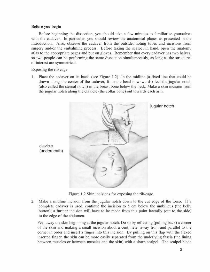
3. Locate the left and right lungs, remembering that left and right refer to the cadaver’s perspective, not your own (figure 1.10)
The right lung has three lobes and the left lung has two lobes. Additionally, the left lung is smaller than the right lung because the heart, within the pericardial sac, lies slightly to the left of the midline. The lungs may appear to be quite small because they are usually partially deflated. Some cadavers’ lungs may have black streaks or spots from smoking or exposure to air pollution. Note the spongy consistency of the lungs.
The slightly larger right lung is divided into a superior, a middle and an inferior lobe by the oblique and horizontal fissures. The left lung is divided only by the oblique fissure into a superior and an inferior lobe. Observe that the visceral pleura extends into these interlobar clefts.
Study the root of the lungs where the bronchial tubes, pulmonary arteries and veins are grouped together. This close relationship between the air ducts and the blood vessels is maintained deep into the substance of the lungs, to facilitate gas exchange. If time permits, dissect a lung by following a bronchus down its branching structure.
8
Figure 1.9. A schematized coronal section of the pleural
layers, showing them as a single continuous membrane.
parietalpleura
visceralpleura
Figure 1.10. Structures of the thoracic cavity. The heart is within the pericardial sac.
4. Reflect back the oblique muscles and the rectus abdominis muscle to reveal the abdominal cavity.
Locate the diaphragm and its attachments inside the rib cage and the thoracic cavity (Figure 1.11).
The diaphragm muscle is like a large inverted bowl dividing the abdominal and thoracic cavities. The muscle fibers of the diaphragm radiate out from the central tendon and form sternal, costal and vertebral attachments.
Contraction of the diaphragm pulls the top of the “inverted bowl” in and down expanding the thoracic cavity.
Observe the domes of the diaphragm from underneath by removing the contents of the abdominal cavity, particularly the liver. Note that the diaphragm is pierced by three structures: the aorta at the level of thoracic vertebra 12 (T12), the esophagus at T10 and the inferior vena cava at T8. Shine a light into the area beneath the diaphragm to see these structures.
9
cut ribs
rightlung lobes pericardial sac
diaphragm (cut)

Figure 1.11. The diaphragm and its attachments.
5. Return to the upper torso to observe the relationship between the trachea (windpipe) and esophagus. Identify the individual tracheal rings, noting that they are actually C-shaped cartilages bound posteriorly by a sheet of smooth muscle called the trachealis (figure 1.12). This shape allows for the expansion of the esophagus when food is being swallowed. The expansion is necessary because the esophagus is bounded posteriorly by the bony vertebrae of the spinal column. Because only a thin sheet of muscle separates the trachea from the esophagus, the subglottal (tracheal) air pressure can be approximated by measurement of the esophageal air pressure.
The trachea divides above the lungs into two bronchi which are composed of cartilaginous rings bound together by fibroelastic and smooth muscle tissue. Since all cartilage ossifies (becomes bony) with age, depending on the age of your cadaver, the cartilages of the bronchi and trachea may be more or less bony.
10
sternal
costal
vertebral
diaphragm

Figure 1.12. The tracheal rings, showing the C-shaped ‘open ring’ structure.
11
trachealcartilage
trachealis


![Respiratory 2[1]](https://img.pdfslide.net/doc/110x75/577d1d3d1a28ab4e1e8be43a/respiratory-21.jpg)