Embed Size (px)
Citation preview
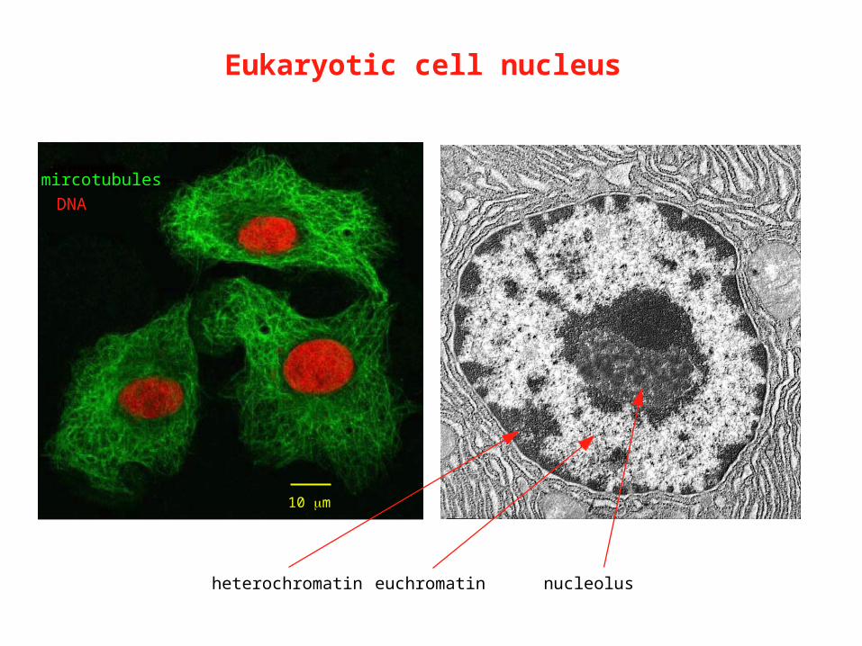
10 m
mircotubules
DNA
Eukaryotic cell nucleus
heterochromatin euchromatin nucleolus

DNA
Chromatin organization of higher eukaryotes

Chromatin in the nucleus
• In 1973, Olins et al and Woodcock et al observed that chromatin shows a “beads on a string” structure by EM
• treatment of chromatin with micrococcal nuclease preferentially cuts between the beads
• In 1884, Albrecht Kossel coined the term “histon” to describe the proteins he found by extracting avian erythrocyte nuclei using diluted acids

Nucleosome structure
Roger Kornberg• based on EM images, nuclease digestion patterns, X-ray diffraction data, and purification of nucleoprotein complexes, proposed that the nucleosome is the repeating unit of chromatin and that every ~200 bp of DNA forms a complex with four histone pairs (1974)
core histone octamer=2 copies each of
H2AH2BH3H4
DNA
H1 nucleosome

Core histones
• core histones (H2A, H2B, H3 and H4) are small (11 to 14 kD), highly basic proteins
• they are evolutionarily highly conserved (from yeast to humans)
• they all share similar structural motifs
N C
N-terminal tail C-term tailhelicies
histone fold
= “hand shake” motif

• histones can dimerize through their “hand shake motifs”
Assembly of a nucleosome
• H3 can only dimerize with H4 and H2A always dimerizes with H2B
• nucleosome assembly starts with two H3-H4 dimers forming a tetramer
• this is followed by addition of two H2A-H2B dimers to form the octamer
• DNA is wrapped around the histone octamer

H3
H4
Luger et al, Nature, 1997
Nucleosome crystal structure

H3
H4
H2A
H2B
Luger et al, Nature, 1997
Nucleosome crystal structure
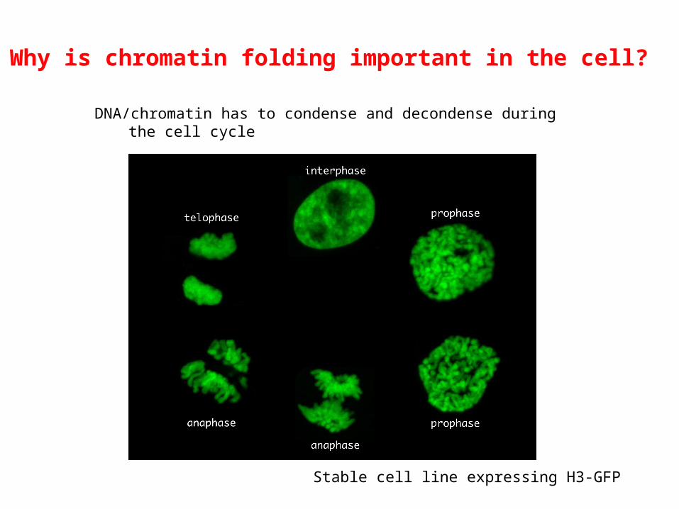
Why is chromatin folding important in the cell?
DNA/chromatin has to condense and decondense during the cell cycle
Stable cell line expressing H3-GFP

How does chromatin folding affect nuclear functions?
• nucleosomes inherently function as barrier to nuclear factors that need to access and bind to DNA elements
• e.g. chromatinized template inhibits transcription of underlying genes
• also affects other DNA-templated processes such as DNA replication, repair etc.
• in order to activate gene expression, the cell has developed ways to “open” up chromatin
a. ATP-dependent chromatin remodeling factorsb. histone modifying enzymesc. insert histone variants at strategic locations within genome

ubiquitination
Post translational modifications on histones
• different modifications occur on specific residues to perform specific regulatory functions

• Histone PTM has been a "hot” research topic in the last 15 yrs
Post translational modifications on histones
Frequently asked questions:
• What biological processes are associated with/regulated by site-specific histone modifications?
• What are the enzymes (acetylases, kinases, methyl-transferases) that directly modify histones at specific sites?
• What are the upstream pathways that regulated these enzymes?
• What are the downstream effects of histone PTMs -- i.e. mechanism?
• What are the enzymes that remove specific histone PTMs?
• What pathways that regulate these de-acetylases, phosphatases, de-methylases etc?

Histone acetylation regulates transcription activation
• It has long been known that histones in vivo are acetylated, and as early as in the 60’s, Vincent Allfrey has suggested that histone acetylation (and methylation) regulate RNA synthesis
• e.g. by the 70’s, Allfrey et al showed that drugs that increase histone acetylation in cells also increased DNase sensitivity of the cellular DNA
• by special labeling techniques, it was shown that more accessible chromatin are enriched for acetylated histones
• However, the direct link between histone acetylation and transcription regulation wasn’t discovered till 1996 when the first transcription-associated histone acetyltransferase (HAT) was identified

Identification of the first histone acetyltransferase
The first transcription-associated histone acetyltransferase (HAT) was identified by an “in gel” histone acetyltransferase assay
- + - +histone substrates
+ 3H Ac-CoA
Coomassie stain Autorad
cut out for peptide sequencing, protein ID
SDSPAGE
denature and renature proteins in the gel
Brownell et al, Cell, 1996

Transcription is regulated by the balance of HATs and HDACs
• The first HAT identified was Gcn5, which was a well-studied transcription co-activator identified by genetics studies in yeast
• Also in 1996, the first histone deacetylase (HDAC) was identified, and the enzyme Rpd3 was also a long studied transcription repressor identified by yeast genetic studies
• Many other transcription co-activators and repressors were found to be HATs and HDACs respectively, and these enzymes are recruited to promoters during transcription activation or repression
hyper Ac-histone hypo Ac-histone HATs
HDACstranscriptionactivation
transcriptionrepression

Technical advances that helped the study of histone modifications
1. Development and refinement of in vitro assays
enzyme source substrateradioactiveco-factor modified histones+ +
nuclear extracts
IP’d protein
recombinant protein
histones
nucleosomes
peptides
3H-Ac-CoA (acetylation)
3H-SAM (methylation)
32P-ATP (phosphorylation)
32P-NAD (ADP-ribosylation)

Wang et al, Mol Cell, 2001
Example: identification of a histone H3 methyltransferase
add histone H3 substrate and 3H SAM
separate proteins by SDS PAGEstain gel or do autoradiography
identify fractions that contain radio-actively labeled H3repeat fractionation if necessary
identify histone modifying enzyme
fractionate nuclear lysates bychromatography techniques
collect fractions

ubiquitination
How to identify site of histone modification?

How to identify site of histone modification?
histonemethyl-transferase
H3 peptide 3H-SAM radioactively-labeled peptide+ +
protein sequencing (Edman degradation)
Strahl et al, PNAS, 1999
detect radioactive amino acid

2. Development and usage of histone modification-specific antibodies
• antibodies are very useful reagents for research
• they can have exquisite specificities and sensitivities for detection of proteins
• can generate and purify antibodies that specifically detect site-specifically modified histones
Technical advances that helped the study of histone modifications

Development and usage of histone modification-specific antibodies
Technical advances that helped the study of histone modifications

Recent article in BMC Bioinformatics on epigenetics and histone modifications

2. Development and usage of histone modification-specific antibodies
• antibodies are very useful reagents for research
• they can have exquisite specificities and sensitivities for detection of proteins
• can generate and purify antibodies that specifically detect site-specifically modified histones
• these antibodies can be used for Western blot analyses, immunofluorescence (IF) studies, and chromatin immunoprecipitation (ChIP) assays
Technical advances that helped the study of histone modifications

2a. Western blot analyses
• modification-specific histone antibodies are useful for monitoring overall abundance and global changes of specific histone modifications
Briggs et al, Genes Dev, 2001
Uses of modification-specific histone antibodies

2b. Immunofluorescence assays
• modification-specific histone antibodies can be used to examine localization of the modified histones within the nucleus
Me(Lys9) H3
Chromosome enriched in Lys9-methylated H3
Uses of modification-specific histone antibodies

2c. Chromatin immunoprecipitation assay
Uses of modification-specific histone antibodies
• ChIP assay is useful for examining the enrichment of specific histone- modifications or binding of specific factors to the gene of interest in vivo

2c. Chromatin immunoprecipitation (ChIP) assay
• can be coupled to gene activation procedures to look at changes in histone- modifications or transcription factor binding to specific genes before and after transcription activation
• can also be used in combination with microarray analyses (ChIP on chip) or deep- DNA sequencing (ChIP-seq) to do genome-wide mapping of histone modifications and chromatin-binding proteins
• while ChIP-chip or ChIP-seq provide correlational information, detailed ChIP analyses of specific genes can help eludicate step-wise mechanisms
Uses of modification-specific histone antibodies

Transcription activation of the -interferon gene
adapted from Agalioti et al, Cell, 2000
mRNA levels
ChIP assays
• The -interferon gene is highly activated upon viral infections and has served as a model system to study gene activations

How does histone acetylation promote How does histone acetylation promote transcription?transcription?
Jacobson et al; Science 2000
• Acetylated histones recruit and stabilize binding of transcription or chromatin remodeling factors via interactions of the acetylated lysines with the Bromodomains of these nuclear factors
• Acetylation neutralizes the positively charged lysine residues on histones and thus reduces the interactions of the histones with the negatively charged DNA
AcBD
H45/8 12/16
TAFII250
AcBD

Histone acetylation precedes recruitment of transcription factors
adapted from Agalioti et al, Cell, 2000
mRNA levels
ChIPassays
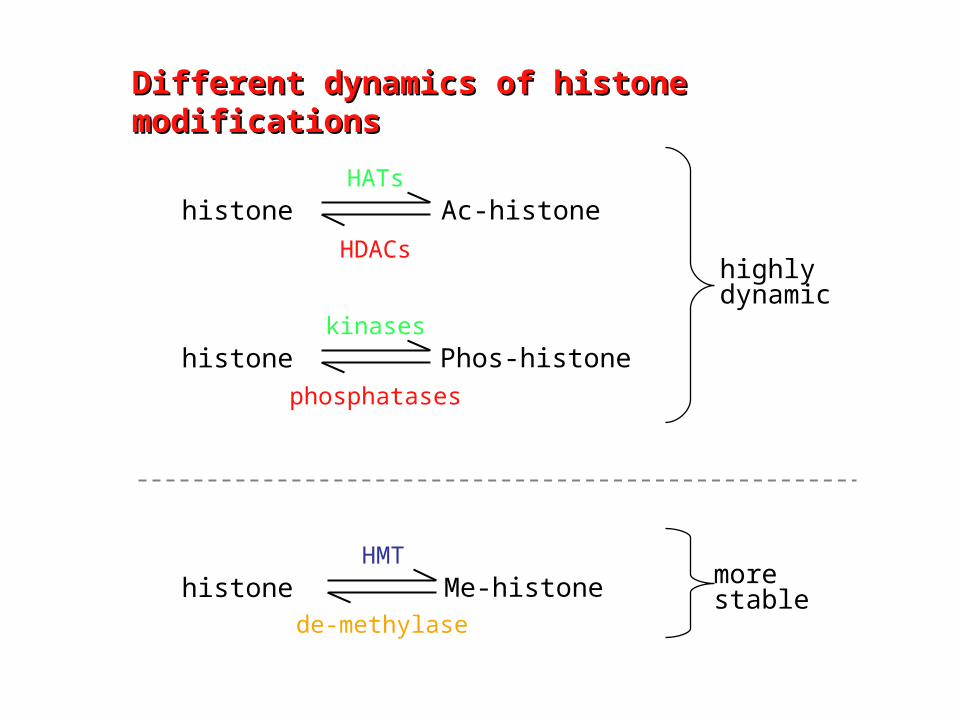
Different dynamics of histone Different dynamics of histone modificationsmodifications
histone Ac-histoneHATs
HDACs
histone Phos-histonekinases
phosphatases
histone Me-histoneHMT
de-methylase
highlydynamic
morestable