Embed Size (px)
Citation preview

Normal labour is characterised by:
• Regular uterine contractions• Dilatation of the cervix• Descent of the presenting part.
It encompasses the time from the onset of regular contractions tospontaneous vaginal delivery of the infant (within 24 hours).
UTERINE CONTRACTIONS
• Contractions begin in two ‘pace-makers’ near the uterotubaljunctions.
• Only one is operative in each contraction that spreads like a waveover the whole uterus.
• Relaxation begins simultaneously in all areas of the uterus.
Labour is characterised by:
• Strong and sustained action of the muscle of the uterine funduswhich increases as labour progresses
• Less strong contractions of the mid-zone• Relative inactivity of the lower segment
Normal uterine contractions are characterised by:
• A frequency of one every 2 to 3 minutes with at least 1 minutebetween contractions
• A duration of 40 to 70 seconds• An intensity (measured by intrauterine catheter) of around
50 mmHg with a resting tonus of <10 mmHg.
CERVICAL DILATATION
• This occurs from above downwards accompanied by effacement(thinning).
• It is caused by co-ordinated contraction and retraction of theupper segment.
OBSTETRICS10
136
10. Labour and intrapartumproblems
NORMAL LABOUR
F07223-10.qxd 5/12/03 11:07 AM Page 136

• The forewaters may act as a hydrostatic wedge, and dilatation isfacilitated by close apposition of the cervix and presenting part.
FIRST STAGE OF LABOUR
Latent and active phases—see Table 10.1 and Fig. 10.1.The latent phase starts from the onset of regular uterinecontractions and ends when the cervix is 2–3 cm dilated and fullyeffaced. It occurs because the thinning of the lower segment andcervix take a lot of uterine work before rapid dilatation can begin.In the active phase the cervix dilates at 1–3 cm per hour inprimigravidae and up to 6 cm per hour in multigravidae.
Control of uterine activity in labourProstaglandins are, and oxytocin may be, important for themaintenance of progressive labour.
The autonomic nervous system has little or no motor function.
Progress in labour is best assessed using a partogram on which canbe recorded:
LABOUR AND INTRAPARTUM PROBLEMS 10
137
Table 10.1 Length of first stage of labour
Mean length in hours (± 1 SD)Primigravidae Multigravidae
Latent phase 9 ± 6 5 ± 4
Active phase 5 ± 3.5 2 ± 1.5
Cer
vica
l dila
tatio
n
2 4 6 8 10 12 14 16
2
4
6
8
10
Latent phase Active phase
Time in hours
Fig 10.1 Cervical dilatation time curve
F07223-10.qxd 5/12/03 11:07 AM Page 137

• Cervical dilatation—marked in centimetres at zero time (the timeof admission to labour ward) and at every subsequentexamination.
• Descent of the head (in fifths palpable above the pelvic brim).• Contractions—frequency, duration and strength assessed for
10 minutes each half-hour.• Fetal heart rate (see p. 150).• Condition of the liquor and time and manner of membrane rupture.• Moulding of the fetal skull.• Dosage of oxytocin, if used.• Maternal status (BP, pulse, temperature, urinalysis) and medication
(including epidural block, if used).
MANAGEMENT OF NORMAL LABOUR
Each obstetric unit should set down (and review regularly) agreedguidelines for the management of labour to include a clearstatement about the diagnosis of the onset of labour.
Routine perineal shaving and an enema on admission are outmodedpractices.
Maternal informed choice is to be encouraged. (This includesrecognition of responsibility for that choice.)
Posture• Mobility should be encouraged during the latent phase.• In the active phase (in the absence of complications) allow the
mother to adopt the position she finds most comfortable.• Maternal posture should at all times be as upright because:
• the supine position tends to impair placental perfusion• the need for augmentation (see below) and analgesia is reduced
Normal progress• In primigravidae delivery should be expected within 8 hours of the
diagnosis of labour and achieved within 12 hours.Delay in primigravid labour may be due to:• Inefficient uterine action• Occipito-posterior position of the fetal head• True cephalo-pelvic disproportion (rarely)
• Labour is much more rapid in multiparous women, and inefficientuterine action is rare. If delay is occurring its cause should besought and corrected if possible.Obstruction must be considered as a possible cause for prolongedlabour in a multiparous woman.
Augmentation of labour in primigravidae• This practice is widespread but remains controversial. Its aim is to
achieve safe delivery within 8 hours of admission to the labour ward.
OBSTETRICS10
138
F07223-10.qxd 5/12/03 11:07 AM Page 138

• It is usually considered if the rate of cervical dilatation is less than1 cm per hour in the active phase of labour.
• The membranes are ruptured and oxytocin infusion is set up 1 hour later if labour does not accelerate.
• Among the contraindications are:• obstetric anomalies, e.g. breech presentation or multiple
pregnancy.• fetal compromise.
• It is relatively contraindicated in multigravidae because the risk ofuterine rupture is increased.
• Review of trials to date (see Enkin et al in Further reading)suggests that:• no beneficial effects have been demonstrated• women find ambulation more acceptable• further trials are necessary.
Oral intake in labour• The major risk to be avoided is aspiration of gastric contents. This
only occurs in the context of general anaesthesia. • In general, therefore, low-fat, low-residue food and drink can be
given to a mother during labour.• There is no evidence that the routine use of antacids or H2 receptor
antagonists is beneficial.• The significance of ketonuria is exaggerated:
• intravenous infusions of dextrose solutions (particularly dextrose10% are contra-indicated because of their deleterious effect onmother and baby (fetal hyponatraemia).
• if dehydration needs to be corrected, normal saline should beinfused but the volume should not exceed 3 litres/24 hours.
SECOND STAGE OF LABOUR
• It begins with full dilatation of the cervix and ends with delivery ofthe baby.
• Its average length in primigravidae is 40 minutes, and inmultiparae is 20 minutes.
• It has two phases: • the propulsive phase from full dilatation until the presenting
part has descended to the pelvic floor.• the expulsive phase which ends with delivery of the baby and is
recognised by the mother’s irresistible desire to bear downand/or distension of the perineum.
• A woman should not usually be encouraged to bear down untilshe has entered the expulsive phase.
• A prolonged propulsive phase in primigravidae due to inefficientuterine action (and not cephalo-pelvic disproportion) can betreated judiciously with oxytocin.
LABOUR AND INTRAPARTUM PROBLEMS 10
139
F07223-10.qxd 5/12/03 11:07 AM Page 139

• Induction of labour can be justified when:• the intrauterine risks to the fetus outweigh those from delivery• the risk to the mother’s health from the continuation of
pregnancy outweighs the risk to the fetus from delivery.• If the added risk of labour is unacceptable, delivery must be by
caesarean section.• Otherwise, labour can be induced and the maternal and fetal state
monitored throughout.• Fetal well-being should be confirmed immediately prior to
induction of labour.• A policy of offering routine induction of labour after 41 weeks
reduces perinatal mortality without increasing the caesareansection rate.
• An ultrasound scan to confirm gestation should be offered before20 weeks gestation as this reduces the need for induction forperceived post term pregnancy.
• Women who have pregnancies complicated by diabetes should beoffered induction of labour prior to their estimated date fordelivery (see p. 88).
• Women with pre-labour rupture of the membranes at term shouldbe offered a choice of immediate induction of labour or expectantmanagement. Expectant management at term should not exceed96 hours following membrane rupture.
• ‘Social’ indications rarely constitute an adequate reason forinduction but each situation must be considered on its merits. Ifoffered, the cervix should be favourable.
CONTRAINDICATIONS
Absolute• The fetal lie is not longitudinal.• Caesarean section has been carried out in a previous pregnancy for
a recurrent reason (e.g. pelvic contraction).• Two previous caesarean sections have been performed.• Placenta praevia.• A tumour occupies the pelvis that will obstruct labour.
Relative• The cervix has previously been repaired. Previous cone biopsy
merits caution.• Highly multiparous woman.
Other factors to be borne in mind• An unfavourable cervix• Uncertain gestational age.
OBSTETRICS10
140
INDUCTION OF LABOUR
F07223-10.qxd 5/12/03 11:07 AM Page 140

HAZARDS
• Iatrogenic prematurity—early pregnancy dating by ultrasoundreduces this risk.
• Infection—there is little appreciable risk in practice but amnionitiscan always be detected within 36 hours of amniotomy.
• Neonatal jaundice—there is a small risk if the total dose ofoxytocin exceeds 20 units.
• Failed induction—defined as failure to deliver vaginally a patientin whom safe vaginal delivery was expected. The incidence is about2% of all inductions.
CERVICAL RIPENESS
This is assessed by a modified Bishop’s score that gives marks of 0 to3 for five cervical features—see Table 10.2.
• If the cervix is ‘ripe’ (score >5) induction of labour is likely to besuccessful
• With a score of <5 induction is more likely to fail, the latent phasewill tend to be longer, and a higher total dose of medication islikely to be necessary to reach optimal uterine activity.
METHODS OF INDUCTION
• Membrane sweeping—should be offered to women prior to formalinduction. It is associated with discomfort during the procedureand light bleeding.
• Prostaglandins—should be used in preference to oxytocin in bothnulliparous and multiparous women with intact membranesregardless of their cervical favourability. Intravaginal PGE2 shouldbe used in preference to intracervical preparations, as vaginaladministration is as effective but less invasive.
LABOUR AND INTRAPARTUM PROBLEMS 10
141
Table 10.2 Modified Bishop’s score
Score0 1 2 3
Cervical dilatation (cm) <1 1–2 2–4 >4
Cervical length (cm) >4 2–4 1–2 <1
Station (cm above ischial –3 –2 –1/0 +1/+2spines)
Consistency Firm Average Soft –
Position Posterior Mid/ – –Anterior
F07223-10.qxd 5/12/03 11:07 AM Page 141

• Artificial rupture of membranes—following ruptured membranes,prostaglandins and oxytocin are equally effective.
Fetal well-being should be established once contractions areestablished or reported.
In women with intact membranes amniotomy should be performedwhere feasible prior to commencement of oxytocin infusion.
Oxytocin should not be started for 6 hours following theadministration of prostaglandins.
Oxytocin should be delivered through a syringe driver or an infusionpump with a non-return valve. A recommended regimen is:
• Starting dose of 1–2 milliunits/min.• Increased at intervals of 30 minutes or more.• Minimum dose should be used, titrated against uterine
contractions aiming for a maximum of 3–4 contractions every 10 minutes.
• Adequate contractions may be established at 12 milliunits/minalthough doses up to 32 milliunits/min are sometimes needed.
• As labour becomes established the dose of oxytocin may need tobe reduced to avoid hyper-stimulation, particularly inmultigravidae.
Water intoxication has occasionally followed the infusion of largevolumes of fluid containing dilute oxytocin. Confusion andconvulsions can proceed to coma and even death. This is totallyavoidable.
For more discussion see Further reading.
Fetal heart rate monitoring is necessary if induction is being carriedout for fetal reasons and during oxytocin infusion.
Pain is a normal part of labour and delivery although emotional,cultural and other influences alter individual responses.
CAUSES OF PAIN IN LABOUR:
• Dilatation of the cervix• Accumulation of pain-producing substances due to ischaemia
during uterine contractions • Pressure on other organs (e.g. bladder and rectum) or the lumbo-
sacral plexus; spasm in skeletal muscles • Distension of vagina and perineum.
Sensory pathways are T10 to L1 for both uterine body and cervix. T11 and 12 are stimulated during the latent phase when pain is not
OBSTETRICS10
142
PAIN IN LABOUR
F07223-10.qxd 5/12/03 11:07 AM Page 142

severe, T10–L1 are stimulated during the active phase. Referred painis experienced in the dermatomes of the above segments.
FACTORS AFFECTING PAIN IN CHILDBIRTH
Physical factors
• Intensity and duration of contractions• Speed of dilatation of cervix• Vaginal and perineal distension• Others, e.g. age, parity, size of infant, condition of patient.
Physiological factors• Pain blocking, e.g. customs, culture, preparation, distractive activity• Pain aggravating, e.g. customs, culture, fear, apprehension,
anxiety, ignorance, misinformation• Antenatal preparation of the mother and father is very important.
Antenatal education is a vital part of preparing women for the painof labour.
METHODS FOR PAIN RELIEF
Psychological methods• Counteract the ‘fear-tension’ sequence.• Pain-relieving drugs can be used to supplement the mother’s own
efforts.• With proper preparation and support, up to 30–40% of women
can go through labour without requiring analgesic drugs.
Inhalational agents• Nitrous oxide (50%) and oxygen (50%)—‘Entonox’ apparatus• Inhalational agents are often used too late and too hesitantly• They can be highly effective and appear safe for mother and baby.
Transcutaneous electrical nerve stimulation (TENS)• This aims to reduce pain by stimulating large myelinated nerve
fibres to reduce input from small myelinated and non-myelinatedfibres linked to peripheral pain receptors.
• Low-intensity continuous stimulation is applied to the dermatomesassociated with the pain.
• It can provide good to moderate pain relief but success dependson time spent teaching and supporting the mother before andduring use.
Narcotic drugs• e.g. intramuscular pethidine
LABOUR AND INTRAPARTUM PROBLEMS 10
143
ANALGESIA
F07223-10.qxd 5/12/03 11:07 AM Page 143
Note: combination with a phenothiazine (e.g. promazine orpromethazine) provides no additional benefit, may producematernal and fetal tachycardia and can rarely cause an oculogyriccrisis. It should, therefore, not be used.
Advantages• Ease of administration• Reasonably rapid analgesia• Low incidence of serious side-effects• Antagonists available.
Disadvantages• Inadequate analgesia in up to 40% of patients• Nausea and vomiting common• Psychic disturbances common (e.g. confusion, inability to
cooperate)• Delayed gastric emptying• Neonatal respiratory depression.
Contraindications• Previous idiosyncratic reactions• Current mono-amine oxidase inhibitors.
Epidural analgesiaDespite its widespread use, little is known about the short or longterm effects of epidural block on mother or baby.
Lumbar analgesia will provide total or adequate analgesia in over90% of patients.
IndicationsEpidural analgesia can be helpful for:
• prolonged labour • instrumental delivery• maternal distress • hypertension in labour• multiple pregnancy • breech presentation.
Contraindications• Lack of experienced personnel • Shock & hypovolaemia• Infection at the injection site • Bony abnormalities of the spinal
column• Coagulation defects or • Idiosyncratic reactions to local
bleeding diathesis anaesthetic agents.• Anticoagulant therapy
Pre-existing neurological disease is not necessarily a contraindicationas long as it is understood that coincidental relapses can occurunrelated to the epidural block.
OBSTETRICS10
144
F07223-10.qxd 5/12/03 11:07 AM Page 144

Immediate maternal problems• Dural tap—dural puncture by needle or catheter; it may lead to
‘spinal’ headache (see below).• Total spinal block—loss of all sensory and motor function; can include
unconsciousness, severe hypotension and apnoea; it results fromsubarachnoid injection of epidural dose of local anaesthetic agent.
• Hypotension—can be avoided by nursing the patient on her sideand by the intravenous infusion of Hartmann’s solution before theblock is established (also used for treatment of hypotension).
• Motor paralysis—reduces maternal expulsive effort, tends toprevent rotation of the fetal head and makes instrumental deliverymore likely. Motor block may be reduced by modification ofanaesthetic agents used (see below).
• Prolongation of second stage of labour.• Toxic reactions to local anaesthetic agents.
Delayed maternal hazards• Severe spinal headache due to spinal tap.
• Ensure adequate hydration and analgesia. • Infuse 1 litre of normal saline through the epidural catheter over
24 hours.• If no improvement within 48 hours consider a ‘blood patch’ (i.e.
injection of up to 20 ml autologous blood into epidural space).• Urinary retention ?—more usually due to method and
circumstances of delivery.• Sepsis—extremely unlikely if bacterial filter is used.• Temporary diminished sensation of dermatomes affected.• Local backache is an occasional temporary problem. Chronic long-
term backache is not.
Fetal effectsThere are no direct adverse effects on the fetus. Temporary changesin the fetal heart rate, sometimes related to hypotension, are notuncommon.
Guidelines for use• The regional block may be continuous during labour or as a single
injection for operative delivery.• Bupivacaine (0.5, 0.375 or 0.25%) is the preferred anaesthetic—a
test dose should be injected initially. Lower concentrations ofBupivicaine (0.1–0.25%) combined with an opiate (e.g. fentanyl)reduce the motor block and may allow a ‘walking epidural’.
• Anaesthetic agents are often given by continuous infusion.• Constant monitoring of maternal and fetal condition is mandatory.• Top-up dose must be individually chosen when the patient begins
to experience discomfort.
Epidural analgesia and previous caesarean sectionEpidural block is permissible in any woman who is being allowed tolabour having previously been delivered by caesarean section. FHR
LABOUR AND INTRAPARTUM PROBLEMS 10
145
F07223-10.qxd 5/12/03 11:07 AM Page 145

should be monitored throughout. Significant FHR abnormalities maybe a sign of scar dehiscence (and see p. 171).
DefinitionRegular, painful uterine contractions accompanied by effacementand dilatation of the cervix after 20 and before 37 completed weeksof pregnancy. It accounts for 5–10% of all deliveries but 85% ofneonatal deaths.
FACTORS ASSOCIATED WITH PRE-TERM DELIVERY
• Spontaneous labour—cause unknown: 40%• Spontaneous labour due to maternal or fetal conditions other than
multiple pregnancy: 25%• Multiple pregnancy: 10%• Elective delivery: 25%
CAUSES OF SPONTANEOUS PRE-TERM LABOUR
These are given in the information box (roughly in order of importance).
PREDICTION OF RISK
• No scoring system yet devised has proven itself superior to clinicaljudgement.
• The strongest association is with previous pre-term delivery. Amongthe measures suggested for prediction of high risk and possibleprevention and for which no evidence of benefit exists are:• routine cervical examination• home monitoring of uterine activity• prophylactic beta-sympathomimetics• routine screening for bacterial vaginosis• prophylactic antibiotics
• It has, however, been suggested that the risk tends to increase theshorter the cervix measured by vaginal ultrasound from 20–28 weeks. See information box over:
OBSTETRICS10
146
PRE-TERM LABOUR AND DELIVERY
• Multiple pregnancy • Congenital uterine anomaly• Antepartum haemorrhage • Diabetes• Intrauterine growth restriction • Polyhydramnios• Cervical incompetence • Pyelonephritis• Amnionitis • Other infections
Spontaneous pre-term labour: causes
F07223-10.qxd 5/12/03 11:07 AM Page 146

MANAGEMENT
Management varies according to five main factors:
• The state of the membranes—it is generally inadvisable to inhibitpre-term labour when the membranes are ruptured.
• Dilatation of the cervix—labour is likely to progress if the cervix is>4 cm dilated
• Gestational age—the earlier the gestation, the more strenuousattempts to inhibit labour must be. Labour should be allowed toprogress if the estimated fetal weight is >2000 g or gestation is>34 weeks.
• The cause of pre-term labour—delivery is indicated if fetal welfareis prejudiced.• Carry out an infection screen on the mother and consider
amniocentesis for bacteriological culture if suspicion of chorio-amnionitis.
• Assess fetal well-being (see p. 54).• The availability of neonatal intensive care facilities—if all cots are
full or facilities are inadequate consider transfer of the patient (ingood time) to a unit with better facilities.
Glucocorticoid therapy and the prevention of respiratorydistress syndrome• Corticosteroids given to the mother between 24 and 34 weeks can
induce pulmonary surfactant in the lungs of the immature fetusand reduce the severity of respiratory depression syndrome.
• One regimen is dexamethasone or betamethasone 12 mg i.m. on 2 successive days. Where the risk of pre-term labour is very high(e.g. triplet pregnancy) this may be repeated two-weekly to 34 weeks.
INHIBITION OF PRE-TERM LABOUR
In up to 50% of patients contractions will stop spontaneously andthe pregnancy will continue to term without any treatmentwhatsoever.
The clinical problem is to discern correctly those in whom drugtherapy is indicated.
LABOUR AND INTRAPARTUM PROBLEMS 10
147
75th 40 210th 26 65th 22 9.51st 13 14
Length of cervix Length of cervix Relative risk of(at or below centile) (mm) preterm delivery
F07223-10.qxd 5/12/03 11:07 AM Page 147

�-sympathomimetic drugs• These drugs (e.g. salbutamol or ritodrine hydrochloride) suppress
uterine activity.• Prolongation of pregnancy is, however, not necessarily beneficial
to the fetus.• The only true indication for their use is to delay delivery for long
enough to allow:• glucocorticoids to stimulate fetal lung surfactant (see above)• transfer of the mother to a centre with adequate facilities for
preterm delivery.
Potential side-effects• Maternal tachycardia • Hyperkalaemia• Hypotension • Hyperglycaemia• Palpitations, headache, visual • Rarely right heart failure may
disturbances, skin flushing, develop (usually when nausea and vomiting glucocorticoids have also been
• Fetal tachycardia given).
Contra-indications:• Antepartum haemorrhage• Severe pre-eclampsia• Maternal anti-hypertensive therapy (risk of myocardial infarction)• Maternal cardiac disease or thyrotoxicosis• Any other situation in which the prolongation of pregnancy could
be hazardous• Extreme caution must be exercised if the woman has diabetes, or is
being treated with corticosteroids.
Other tocolytics• Atosiban—oxytocin receptor antagonist. Licensed for the inhibition
of uncomplicated pre-term labour between 24 and 33 weeks ofgestation. Reported to have fewer side effects than β-sympathomimetics but more expensive.
• Indomethacin and nifedipine have also been used, but their use isunlicensed.
METHOD OF DELIVERY IN PRE-TERM LABOUR
• If the fetus is viable it must be delivered by the route least likely tocause trauma or hypoxia.
• Aim to have an experienced neonatal paediatrician present fordelivery.
• In general, aim for vaginal delivery if the presentation is cephalic.• Caesarean sections at very early gestation <26 weeks, and with
infants under 1000 g, can be hazardous for the mother, and arenot necessarily safer for the baby.
• The indications for caesarean section are stronger but not absolutein multiple pregnancy and breech presentation.
OBSTETRICS10
148
F07223-10.qxd 5/12/03 11:07 AM Page 148

• Ventouse delivery should be avoided below 34 weeks.• Pre-term labour is unpredictable and the woman may become fully
dilated quickly and silently.
This is defined as rupture of the membranes before the onset oflabour without reference to gestational age.
• It can be managed conservatively before 34 to 36 weeks’ gestationunless intrauterine infection is present or likely to develop. In theabsence of infection prophylactic erythromycin should be given for10 days.
• A high vaginal swab should be taken on admission. If it grows anysignificant organisms (particularly beta-haemolytic streptococci),delivery should be expedited and the neonatal paediatricians alerted.
• Any intrauterine infection must be treated vigorously andexpeditiously (see ‘Further reading’).
• In term pregnancies 86% of women with PROM go into labourwithin 24 hours and deliver satisfactorily. The rate of spontaneouslabour after this is about 5% per day. In the absence of anyevidence of infection or cord presentation/prolapse, the onset oflabour can be awaited for 24 hours. Expectant management ofwomen with pre-labour rupture of the membranes at term shouldnot exceed 96 hours following membrane rupture.
Prior to any form of fetal monitoring, the maternal pulse should bepalpated simultaneously with fetal heart rate auscultation in orderto differentiate between maternal and fetal heart rates.
The aim of monitoring is to detect fetal hypoxia. The effects ofhypoxia depend on the fetal glycogen reserves. A growth-restrictedfetus will, therefore, be affected earlier and more severely than awell-nourished fetus.
• Anaerobic glycolysis results in an accumulation of lactate. Thiscauses a fetal metabolic acidosis.
• The fetal pC02 rises, causing a respiratory acidosis.• The blood pH falls.• Fetal heart rate (FHR) patterns change (see below) the most serious
being late decelerations associated with a fetal tachycardia.
Fetal distressThe traditional diagnosis of ‘fetal distress’ depended predominantlyon the crude observation of heart changes.
‘Fetal distress’ is an imprecise and rather unhelpful term. Half of allbabies delivered by forceps or caesarean section because of ‘fetal
LABOUR AND INTRAPARTUM PROBLEMS 10
149
PRE-LABOUR RUPTURE OF THE MEMBRANES (PROM)
INTRAPARTUM FETAL MONITORING
F07223-10.qxd 5/12/03 11:07 AM Page 149

distress’ are not hypoxic; and half of the most hypoxic babies do notexhibit classical signs of ‘fetal distress’.
METHODS OF INTRAPARTUM MONITORING
Intermittent auscultation of FHR using a fetal stethoscope• In the active stage of labour this should occur after a contraction
for a minimum of 60 seconds, and at least:• Every 15 minutes in the first stage.• Every 5 minutes in the second stage.
• It is applicable to low-risk patients with no significant obstetricabnormalities.
• More intensive monitoring should be used if any risk factors arepresent (see below).
Continuous monitoring of FHR and uterine activity(cardiotocography; CTG)• The FHR is obtained by an external Doppler ultrasound monitor or
an electrode attached to fetal scalp.• The monitor measures the interval between paired beats, converts
it into ‘beats per minute’ (bpm) and registers it.• Uterine activity can be assessed by an external strain gauge
transducer or measured by intrauterine catheter.• This is a screening technique that facilitates the detection of fetal
hypoxic stress. It is not diagnostic.• Even when the most ominous pattern is present (see below) only
50% of the babies have a low Apgar score (see p. 188) at birth.• The use of continuous FHR recordings must therefore be backed up
by measurement of fetal scalp pH (see below).
Guide to indications for continuous FHR monitoring in labour
Antepartum risk factors• Previous caesarean section • Diabetes• High multiparity • Multiple pregnancy• Suspected FGR • Rhesus iso-immunisation• Hypertension/pre-eclampsia • Oligohydramnios• History of APH in this • Reduced fetal movements
pregnancy • Abnormal antenatal FHR tracing.• Poor obstetric history
Intrapartum risk factors• FHR >160 or <110 bpm • Augmented or induced labour• Meconium-stained liquor • Pre-term labour• Prolonged labour • Breech presentation• Epidural anaesthesia • Prolonged rupture of membranes.
InterpretationThe whole clinical situation must be considered particularlygestational age, stage and progress of labour.
OBSTETRICS10
150
F07223-10.qxd 5/12/03 11:07 AM Page 150
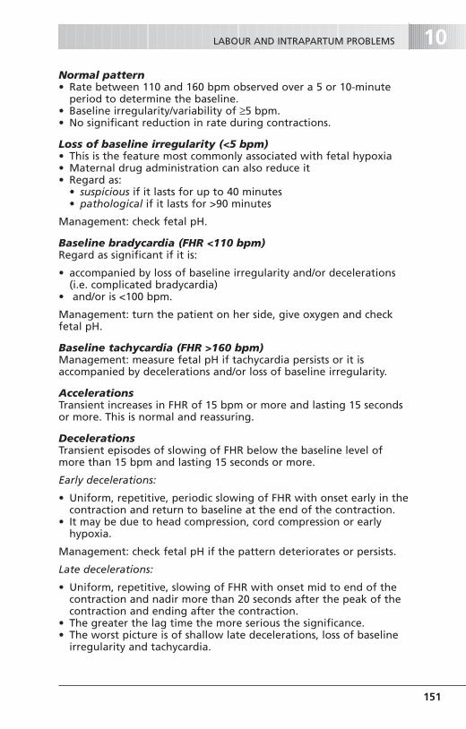
Normal pattern• Rate between 110 and 160 bpm observed over a 5 or 10-minute
period to determine the baseline.• Baseline irregularity/variability of ≥5 bpm.• No significant reduction in rate during contractions.
Loss of baseline irregularity (<5 bpm)• This is the feature most commonly associated with fetal hypoxia• Maternal drug administration can also reduce it • Regard as:
• suspicious if it lasts for up to 40 minutes• pathological if it lasts for >90 minutes
Management: check fetal pH.
Baseline bradycardia (FHR <110 bpm)Regard as significant if it is:
• accompanied by loss of baseline irregularity and/or decelerations(i.e. complicated bradycardia)
• and/or is <100 bpm.
Management: turn the patient on her side, give oxygen and checkfetal pH.
Baseline tachycardia (FHR >160 bpm)Management: measure fetal pH if tachycardia persists or it isaccompanied by decelerations and/or loss of baseline irregularity.
AccelerationsTransient increases in FHR of 15 bpm or more and lasting 15 secondsor more. This is normal and reassuring.
DecelerationsTransient episodes of slowing of FHR below the baseline level ofmore than 15 bpm and lasting 15 seconds or more.
Early decelerations:
• Uniform, repetitive, periodic slowing of FHR with onset early in thecontraction and return to baseline at the end of the contraction.
• It may be due to head compression, cord compression or earlyhypoxia.
Management: check fetal pH if the pattern deteriorates or persists.
Late decelerations:
• Uniform, repetitive, slowing of FHR with onset mid to end of thecontraction and nadir more than 20 seconds after the peak of thecontraction and ending after the contraction.
• The greater the lag time the more serious the significance.• The worst picture is of shallow late decelerations, loss of baseline
irregularity and tachycardia.
LABOUR AND INTRAPARTUM PROBLEMS 10
151
F07223-10.qxd 5/12/03 11:07 AM Page 151

Management: a fetal pH measurement is mandatory.
Variable decelerations:
• Variable, repetitive, periodic slowing of FHR with rapid onset andrecovery. Time relationships with contraction cycles are variableand they may occur in isolation.
• Sometimes they resemble other types of deceleration patterns intiming and shape.
• If they appear consistently, fetal hypoxia is likely.
Management: check fetal pH if the pattern persists after turning thepatient on her side (or if other adverse features are present).
FHR in the second stage of labour• The fetal heart patterns are complex and may be difficult to
interpret in the second stage of labour. • The CTG trace should be interpreted in conjunction with the
pattern in the first stage of labour.• Brief profound early decelerations are not uncommon but
persistent late decelerations or prolonged bradycardia must not beignored.
Fetal ECG• Uses the same scalp clip as for the FHR• It depends on analysis of the ST waveform• The following adverse features have been suggested:
• T/QRS ratio >0.25• a negative T wave• ST depression with T elevation
• Its main benefit is to provide reassurance in the presence of anabnormal CTG and, perhaps, reduce the rate of unnecessarycaesarean sections.
Fetal blood sampling (FBS)• FHR and scalp pH measurement are complementary.• The former without the latter increases the caesarean section rate
unnecessarily because of false positive diagnoses.• The indications for FBS are outlined above.
• A fetal scalp pH of 7.0 or less is strongly associated with a pooroutcome (particularly if the Apgar score is 3 or under at 5 minutes)
• A pH of 7.20 or less suggests the need to deliver• A base deficit of >12 mmol/l is also abnormal
Significance of meconium staining of the liquor• Meconium is present in the liquor of about 15% of all deliveries at
term and up to 40% post-term.• It’s significance as a diagnostic sign of ‘fetal distress’ has been over-
emphasised although gross staining is more likely to be significant.
OBSTETRICS10
152
F07223-10.qxd 5/12/03 11:07 AM Page 152

• Aspiration by the baby of the liquor heavily stained withmeconium causes a severe and sometimes fatal pneumonitis.
ConclusionThe sensitivity and specificity of all methods for intrapartummonitoring of the fetus are still poor. New initiatives are still badlyneeded. For further discussion see NICE guidelines and Enkin et al inFurther reading.
AVERAGE DIAMETERS
PELVIC SHAPE—IN THE NORMAL PELVIS
• The brim is round, and the sacral promontory is not prominent.• The angle of inclination is about 55° to the horizontal.• The cavity is shallow with straight, non-converging walls.• The sacrum is smoothly curved.• In the outlet the sacro-sciatic notches are wide and shallow.• The sacrum does not project forwards.• The ischial spines are not prominent.• The pubic arch is wide and domed.• The sub-pubic angle is about 90°.• The inter-tuberous diameter is wide
PELVIMETRY
• Clinical assessment of pelvic size and shape is only likely to be ofbenefit if the pelvis is severely contracted.
LABOUR AND INTRAPARTUM PROBLEMS 10
153
Diameters (cm)
Anteroposterior
Brim
Cavity
Outlet
11–11.5
12
12.5
12 12
TransverseOblique
12
12 11–11.5
12.5
Fig 10.2 Normal Pelvis
THE NORMAL PELVIS
F07223-10.qxd 5/12/03 11:07 AM Page 153

• X-ray pelvimetry has been superseded in many places by CTscanning. This reduces the radiation dosage and is likely to bemore accurate.
• However, the criteria for normality have not yet been set. • Pelvimetry is of no clinical value if the presentation is cephalic.• There is also no reliable evidence of benefit for the traditional
indications of primigravid breech presentation or after caesareansection for suspected disproportion. Pelvimetry after previouscaesarean section has been shown to increase caesarean sectionrates in subsequent pregnancies.
Definition: The failure of the head to pass through the pelvis safelybecause the pelvis is too small and/or the head too large
• CPD is more likely if maternal height is 1.5 metres or under.• The diagnosis is made in labour if the fetal head fails to descend
and the cervix to dilate. With increasing oedema of the scalp,caput forms and excessive moulding of the skull bones occurs.
MANAGEMENT
• Elective section is rarely necessary in primigravidae unless there areother indications e.g. a malpresentation or the true conjugate is<7.5 cm.
• Otherwise an attempt at vaginal delivery is justifiable. This is thenregarded as a trial of labour.• A trial of labour should be allowed to continue for as long as
progress is occurring in labour with regular forceful contractions. • If the woman has not delivered within 12 hours of the onset of
regular contractions the situation must be reviewed critically.• Such a trial of labour has no place in multigravid women or in
the presence of a breech presentation.
BREECH PRESENTATION
Incidence3–4% of all labours. Up to one-third are undiagnosed.
Definitions• Frank breech (65%)—both legs extended at the knee.• Complete breech (10%)—both legs flexed at hip and knee.• Footling breech (25%)—one or both feet tucked underneath the
buttocks; more common in multiparous women due to laxity ofabdomen.
OBSTETRICS10
154
CEPHALO-PELVIC DISPROPORTION (CPD)
ABNORMALITIES OF LIE, PRESENTATION AND POSITION
F07223-10.qxd 5/12/03 11:07 AM Page 154

Causes• Extended legs preventing spontaneous version• Those conditions preventing the presenting part entering the
pelvic cavity• Uterine anomaly• Chance.
Associations• Fetal anomaly• Pre-term delivery• Multiple pregnancy.
Antenatal management• Spontaneous version is likely up to 34 weeks but may occur later.• External cephalic version (ECV) is safe for mother and baby in
carefully selected patients and reduces the need for electivecaesarean section. It should be actively offered and discussed.
• ECV is not advised before 36 weeks’. Tocolytic agents may increasesuccess rates. Do not use in women with heart disease, diabetes orthyroid disease.
• ECV at term will reduce non-cephalic births by up to 60%. Over95% remain cephalic.
Hazards of ECV. See the information box.
Contraindications to ECV. See the information box.
Prerequisites for ECV• Gestation at least 36 weeks’
LABOUR AND INTRAPARTUM PROBLEMS 10
155
• Pre-term labour • Cord accident• Placental abruption • Uterine rupture (if previous section)
Hazards of ECV
Absolute Relative• Multiple pregnancy • Previous caesarean section• APH • FGR• Ruptured membranes • Hypertension• Oligohydramnios • Rhesus iso-immunisation• Significant fetal anomaly • High multiparity• Caesarean section indicated for • Anterior placenta
other reasons• Placenta praevia • Obesity
Contraindications to ECV
F07223-10.qxd 5/12/03 11:07 AM Page 155

• Recent ultrasound to confirm presentation, normal fetus andadequate liquor volume
• Reactive FHR• Informed consent of mother• Facilities for rapid progression to caesarean section, if necessary• Rh-D-negative women must be given anti-D immunoglobulin
(50 µg or more as Kleihauer test dictates).
Management of deliveryCurrent evidence suggests that:
• Vaginal breech delivery is safe for 97% of babies in whom thereare no other risk markers (see below).
• However, the overall risk of perinatal death for the term singletonbreech delivered by planned caesarean section is reduced by 75%[Relative risk (RR) 0.23; Confidence interval (CI) 0.07–0.8].
Thus, women should be offered an elective caesarean section if ECVis declined or unsuccessful.
Although there is insufficient evidence to support routine caesareansection for the delivery of the pre-term breech and the first orsecond breech twin, most obstetricians would now recommendcaesarean section for the pre-term breech or if twin one is breech.
Some mothers will still opt for an attempt at a vaginal breechdelivery and should be further assessed (see below).
Pre-delivery assessment• There is no evidence that routine pelvimetry is beneficial. It has
not been shown to improve outcome. If thought to be necessaryuse CT pelvimetry (see p. 154).
• Carry out ultrasound assessment of BPD, fetal mass, fetal attitudeand flexion/extension of fetal head.
• Major fetal anomalies should have been excluded.
Vaginal delivery• An attempt at vaginal delivery can be considered with:
• Term pregnancy and fetal weight estimated at 2500–3800 g• Frank or complete breech• Presumed or demonstrated normal pelvic dimensions• No other complications of pregnancy (e.g. pre-eclampsia)• Normal fetal assessment
• Epidural anaesthesia can be useful during a breech labour but isnot essential.
• Augmentation of labour with oxytocin should only be used withextreme caution and close monitoring. It is contraindicated if thereis any evidence of disproportion.
• The baby should be born by the patient’s own efforts with littleassistance from the obstetrician (assisted breech delivery).
OBSTETRICS10
156
F07223-10.qxd 5/12/03 11:07 AM Page 156

• Any more active intervention involving breech extraction iscontraindicated because the perinatal consequences are so severe.
Caesarean sectionAmong the definite indications are any medical or obstetriccomplications that are likely to be associated with mechanicaldifficulties at delivery or a compromised fetus:
• Any abnormality of bony • Diabetespelvis
• Fetal weight estimated at • Severe pre-eclampsia>3.8 kg
• Hyperextension of fetal head • Failure to progress in first stage• Previous difficult labour • Failure of descent of breech in
second stage• FGR • Any condition which would • Bad obstetric history apply whatever the presentation,
e.g. fetal hypoxia.
OCCIPITO-POSTERIOR POSITION
Incidence: approximately 20% in early labour.
If the baby’s head is partially extended it does not fit into the loweruterine pole well, with the following consequences in labour:
• The membranes rupture early and the cervix is not well apposed tothe cervix.
• The sinciput reaches the pelvic floor first and, therefore, rotates tothe front, i.e. the occiput is posterior.
• The larger occipito-frontal diameter (10 cm) of the head presents,making its passage through the pelvis more difficult.
• The first stage of labour is prolonged.• The moment of the forces pushes the head posteriorly causing
backache and inducing bearing-down efforts before full dilatation.• The second stage of labour may be prolonged.
The occiput may:
• Rotate anteriorly and deliver relatively easily (75%)• Persist posteriorly (POP) and delivery spontaneously if the pelvis is
capacious (i.e. face to pubes) or require assisted delivery (5%)• Begin to rotate anteriorly but undergo deep transverse arrest at
the level of the ischial spines. Instrumental delivery will berequired (20%).
Predisposing factors• Slight reduction in pelvic inlet• Large baby
DiagnosisAntenatally—this is inaccurate:
LABOUR AND INTRAPARTUM PROBLEMS 10
157
F07223-10.qxd 5/12/03 11:07 AM Page 157

• the maternal abdomen may be flattened or fetal parts palpableeasily on both sides of the midline.
• the head is unengaged and feels larger than usual.
Intrapartum—by vaginal examination:
• both fontanelles can be felt more easily.• moulding and caput may make recognition difficult and palpation
of an ear may be necessary for correct positioning.
Management• Provide adequate analgesia: an epidural anaesthetic is ideal.• Prevent ‘maternal distress’, ketosis and dehydration.• Observe progress in labour carefully.• Monitor fetal welfare.• Syntocinon may be used with care only in primigravidae to
encourage spontaneous rotation to occipito-anterior.
Relative cephalo-pelvic disproportion may occur and there is anincreased risk of instrumental delivery and caesarean section.
The criteria for assisted delivery are discussed on p. 164.
BROW PRESENTATION
Incidence: approximately 1/1000
A brow presentation discovered antenatally may be due to:
• chance—and may correct itself spontaneously• a swelling in the neck causing extension of the head, e.g. goitre,
cystic hygroma• spasm of the sterno-mastoid muscles.
Suspect a brow presentation in a multiparous woman with delay inthe first stage of labour despite good contractions when she hasdelivered vaginally easily before.
Diagnosis• Supra-orbital ridges and anterior fontanelle palpable p.v.• Confirm by ultrasound.
Management• In early labour a brow presentation may flex to become a vertex or
extend further to a face presentation. Both are potentiallydeliverable vaginally.
• If the brow presentation persists into, or is discovered in,established labour delivery should be by caesarean section.
FACE PRESENTATION
Incidence: approximately 1/500. (75% mento-anterior).
OBSTETRICS10
158
F07223-10.qxd 5/12/03 11:07 AM Page 158

A face presentation has the same causes as a brow presentation butcausing full extension of the head on the neck.
In labour, anterior rotation of the chin is essential: a mento-posteriorposition cannot deliver vaginally.
DiagnosisPalpation of supra-orbital ridges and the alveolar margins (confusionmay arise between a face and the breech).
ManagementAn attempt at vaginal delivery should be allowed unless:
• Something is obstructing the entry into the pelvis.• The pelvis is too small.• The chin is posterior.
TRANSVERSE AND OBLIQUE LIE
Incidence: approximately 1/300
Among the causes are:
• High multiparity • Hydramnios• Pre-term labour • Obstructing tumour or placenta praevia• Multiple pregnancy • Severe pelvic contraction.• Uterine anomaly
Antenatal management See p. 132.
lntrapartum management (singleton pregnancy)In a neglected shoulder presentation an arm may well be prolapsedand the baby already dead. In these circumstances vaginaldecapitation may be possible but only by an experienced operator.This is very seldom performed in the UK.
Otherwise caesarean section with decapitation in utero is lesshazardous for the mother.
SHOULDER DYSTOCIA
Incidence: about 0.2–1%.
This is one of the most frightening obstetric emergencies
It occurs when the fetal shoulders fail to negotiate the pelvic inlet
Prompt (but not forcible) action is required to prevent fetalmorbidity or mortality (see Stirrat and Taylor in ‘Further reading’)
Antenatal risk factors• Mother’s birthweight >90th centile• Maternal obesity or massive weight gain
LABOUR AND INTRAPARTUM PROBLEMS 10
159
F07223-10.qxd 5/12/03 11:07 AM Page 159

• Diabetes mellitus—can be despite seemingly good blood sugarcontrol
• Prolonged pregnancy (beyond 42 completed weeks)• Previous shoulder dystocia (10% risk of recurrence) or large baby • Recognised macrosomia this pregnancy.
Intrapartum risk factors First stage:
• ‘dysfunctional labour’• secondary arrest after 8 cms.
Second stage:
• midcavity arrest• need for midcavity instrumental delivery in multiparous woman.
Prediction• Consideration of above risk factors predicts fewer than 20% of
cases!• Where practiced it has not reduced fetal asphyxia or trauma.• When interpreted too rigidly many women will have unnecessary
interventions.• Clinical prediction of excessive birthweight is unreliable.• Ultrasound estimates are inaccurate at upper centiles.
Risks to the baby• Neurological injury—occurs in 1–2/1000 births. It can involve:
• cervical cord • brachial plexus—Erb’s palsy (C5,6,7): Klumpke’s palsy (C7,8,T1)• phrenic nerve.
• Hypoxic ischaemic encephalopathy (HIE)—0.5–1/1000 births• Fractures: clavicle (2–3/1000); humerus (0.2–0.3/1000).
Management • Recognition and recording of possible risk factors
(keep good records!)• Clear plan of action in guidelines• Rapid reaction—the midwife has a vital role• Immediate response:
• call for help—summon experienced obstetrician, anaesthetistand paediatrician
• place woman in ‘McRoberts position’, i.e. hip joints fullyabducted, rotated outwards and flexed with thighs touchingmaternal abdomen. This encourages release of the anteriorshoulder.
• make good-sized episiotomy.• Next steps:
• Apply suprapubic pressure to try to encourage the anteriorshoulder to flex and rotate transversely.
OBSTETRICS10
160
F07223-10.qxd 5/12/03 11:07 AM Page 160

• Attempt to deliver posterior shoulder if unable to deliveranterior (adequate analgesia needed for this and manoeuvresbelow).
• Try to dislodge and rotate fetal shoulders vaginally (Wood’sscrew manoeuvre).
• Cephalic replacement following tocolysis and delivery bycaesarean section (Zavanelli manoeuvre) has been described butonly as a ‘desperate solution’.
• Symphysiotomy may allow delivery as a last resort, but is seldomused in the UK.
TWINS
• Pre-term labour is common.• Placenta praevia may be present.• Prolapse of the cord must be watched for.• Malpresentations are more likely—the presentations in order of
frequency being:• Vertex: vertex • Breech: breech• Vertex: breech • Vertex: transverse• Breech: vertex • Breech: transverse.
POSSIBLE INDICATIONS FOR ELECTIVE CAESAREAN SECTION
• First twin presenting as a breech• Triplets and higher multiples• Proteinuric pre-eclampsia• Any indication which would also apply in singleton pregnancies,
e.g. FGR, APH.
MANAGEMENT OF LABOUR AND DELIVERY
• An intravenous line should be set up; a paediatrician and ananaesthetist should be present for delivery lest rapid generalanaesthesia becomes necessary.
• An epidural is often useful, particularly to assist in delivery of thesecond twin.
• After vaginal delivery of the first twin, check the lie is longitudinal.External cephalic version is usually possible. If necessary,presentation can be checked by portable ultrasound in the deliveryroom. The second sac should be ruptured once uterine activitybegins.
• If contractions do not begin within 15 minutes, commence anoxytocin infusion.
• If the cord of the second twin prolapses, proceed to ventouseextraction (if the presentation is cephalic) or breech extraction (if the presentation is breech).
LABOUR AND INTRAPARTUM PROBLEMS 10
161
MULTIPLE PREGNANCY AND LABOUR
F07223-10.qxd 5/12/03 11:07 AM Page 161

OPERATIVE OBSTETRICS
• Anaesthesia for the latter should be epidural if alreadyestablished, or general.
• The interval between delivery of the first and second twins shouldbe no more than 20 minutes.
• Beware of postpartum haemorrhage. The third stage should beactively managed and a syntocinon infusion given if the uterus ispoorly contracted.
• After delivery check placentae and membranes for zygosity.Histological confirmation is necessary.
EPISIOTOMY
The need for an episiotomy is a matter for experienced clinicaljudgement. ‘Routine’ episiotomy is no longer practiced.
IndicationsAmong these will be:
• When a major perineal tear appears inevitable• In cases of fetal distress late in the second stage• Most forceps deliveries (except low cavity forceps)• Pre-term delivery• Breech delivery• Failure to advance because of perineal rigidity.
TechniqueAn episiotomy must be:
• Performed at the correct time—incise too early and unnecessaryblood loss will result.
• Carried out with adequate local or regional anaesthesia. Failure touse anaesthesia is to be deprecated.
• Made with sharp scissors in the correct place. The medio-lateralepisiotomy is more common in the UK. Midline incisions increasethe risk of third degree tears. The episiotomy must always start inthe midline.
• Repaired properly within as short a time of delivery as possible.
Side-effects• Pain. This can be severe and is the main reason for avoiding
episiotomy. It can be reduced by prompt, careful and expert repair.• Bleeding. The average blood loss is about 100 ml and much larger
losses are all too common.• Breakdown. Inversely related to the expertise of the person
repairing the episiotomy. • potential causes are delay in suturing, inappropriate suture
materials and bad technique.
OBSTETRICS10
162
F07223-10.qxd 5/12/03 11:07 AM Page 162
• primary repair with antibiotic cover should be carried out wherepossible.
• Dyspareunia. This can be so severe that it becomes a factor inmarital breakdown.
THIRD- AND FOURTH-DEGREE PERINEAL TEARS
All women having a vaginal delivery should have a systematicexamination of the perineum, vagina and rectum to assess theseverity of damage prior to suturing.
Third-degree tears involve the external anal sphincter muscle.Fourth-degree tears also involve the rectal mucosa.
• They must be repaired in theatre under epidural or generalanaesthesia by an experienced obstetrician.
• Give broad spectrum intra-operative and post-operative antibioticsto reduce infection risk and wound dehiscence.
• Use of stool softeners such as lactulose and a bulking agent such asFybrogel for 10 days is recommended.
• If the repair breaks down eradicate local sepsis before attemptinganother repair.
• The prevalence of anal symptoms is reported to be 25–57%.• Women should be followed up at 6 weeks and 6 months.
Subsequent deliveryDiscuss the possibility of recurrence and deterioration of faecalsymptoms. If symptomatic, offer elective caesarean section, as thereis risk of deterioration in symptoms.
There is no evidence regarding the role of prophylactic episiotomy.
INSTRUMENTAL DELIVERY
Potential indications• Failure to advance in the second stage, frequently due to failure of
maternal effort, epidural analgesia and/or malposition of the fetalhead
• Maternal conditions in which (prolonged) expulsive efforts may bedetrimental e.g. cardiac and respiratory disease, severe pre-eclampsia or eclampsia
• ‘Fetal distress’ in the second stage• Prolapse of the cord in the second stage
Delivery can be by the vacuum extractor (ventouse) or obstetricforceps.
The ventouse must not be considered as an easy way out whenadverse features are present or the position of the fetal head isunknown.
LABOUR AND INTRAPARTUM PROBLEMS 10
163
F07223-10.qxd 5/12/03 11:07 AM Page 163
Prior conditions for instrumental delivery • A legitimate indication must be present.• The presentation must be suitable, i.e. vertex, face (mento-anterior
—not ventouse!) or after-coming head in a breech delivery.• There must be no cephalo-pelvic disproportion. Moulding of the
fetal skull must not be excessive.• The head must be engaged. Ideally, no part of the fetal head
should be palpable per abdomen, and if more than 1/5 can bepalpated vaginal delivery must not be contemplated.
• The position of the head must be known.• For forceps, the cervix must be fully dilated. • The ventouse can, in some circumstances, be used before the cervix
has reached full dilatation (see below).• Analgesia must be adequate.• The bladder must be empty.• The uterus must be contracting.
Forceps to the after-coming head (ACH) in a breech delivery This is the method of choice for delivery of the ACH because of thedegree of control the operator can exercise.
Forceps in the delivery of low birthweight infants (<2500 g)Lift-out forceps neither protects against nor induces birth trauma inLBW infants. Rotational forceps are best avoided for LBW infants.Ventouse should be avoided <34 weeks.
‘Trial of forceps’• This is justifiable when it is likely, but not entirely certain, that
vaginal delivery by forceps will be successful. Otherwise the patientshould be delivered by caesarean section.
• It should be carried out in a theatre by an experiencedobstetrician, with the mother fully prepared for caesarean sectionto reduce delay in delivery if unable to deliver vaginally.
The use of the ventouseVaginal delivery is technically possible before full dilatation butshould not be attempted if there is any suspicion of cephalo-pelvicdisproportion.
The following points are a guide to its proper use:
• The patient’s expulsive efforts are used to assist delivery.• The fetal head must be at least at the level of the spines.• The largest possible of the four cups should be used.• If delivery is not imminent after pulling on the ventouse during
three contractions the attempt must cease and the patient must bedelivered by caesarean section.
Analgesia for instrumental delivery• Perineal infiltration alone is suitable for episiotomy, and low outlet
deliveries using the ventouse or ‘outlet’ forceps.
OBSTETRICS10
164
F07223-10.qxd 5/12/03 11:07 AM Page 164

• Pudendal nerve block is useful for mid-cavity forceps and someventouse deliveries. It does not provide adequate analgesia forrotational forceps. The transvaginal route is recommended forinsertion of the block.
• Epidural anaesthesia is ideal particularly for rotational forceps; it isalso suitable for emergency caesarean sections in which an existingepidural block is providing good analgesia.
CAESAREAN SECTION
Definition of classes of caesarean section adopted by the RCOG:
• Emergency—immediate threat to life of woman or fetus.• Urgent—maternal or fetal compromise that is not immediately life
threatening.• Scheduled—needing early delivery but no maternal or fetal
compromise.• Elective—at a time to suit the patient and the maternity team.• Perimortem—carried out in extremis while the mother is
undergoing active resuscitation to save the fetus or the mother.• Postmortem—carried out after the mother has died in order to try
to save the fetus.
The overall caesarean rate in the UK is about 20%, but there arewide variations between regions and individual hospitals.
• It must not be carried out without good reasons. • It is indicated when delivery must be effected rapidly for fetal and/
or maternal reasons and when it is not thought to be safe vaginally.• The transperitoneal lower segment caesarean section accounts for
virtually all of the operations in modern obstetrics.• Classical caesarean section is very occasionally indicated, e.g. for
transverse lie with PROM, or for caesarean section at 26 to 28 weeks. In this latter situation, the vertical incision starts in thelower segment but extends into the upper segment.
Epidural or spinal anaesthesia and caesarean section
Advantages• It is safer for the mother.• She is awake and sees the child at delivery.• The father can usually be present.• Consciousness is not impaired immediately post-operatively.• Post-operative problems and pain are less than after general
anaesthesia.• Breast-feeding and mobilisation can start early.
Disadvantages• The procedure takes longer.• Occasionally anaesthesia is not complete. Therefore the patient
should be fully prepared for general anaesthesia (GA).
LABOUR AND INTRAPARTUM PROBLEMS 10
165
F07223-10.qxd 5/12/03 11:07 AM Page 165

• Contraindications to epidural/spinal anaesthesia are discussed on p. 144.
SOME IMPORTANT TECHNICAL POINTS ABOUT LOWER SEGMENTCAESAREAN SECTION (LSCS)
• H2-receptor antagonists should be given pre-operatively to reducethe risk from aspiration of acid gastric contents.
• This also applies to procedures using epidural/spinal block lestconversion to GA becomes necessary.
• Induction of GA should take place at the last possible moment toreduce fetal exposure to the anaesthetic agents.
• The operation is carried out with a 10–15° left lateral tilt toprevent supine hypotension.
• A cuffed endotracheal tube must be used.• Special care must be taken in Rh-negative women to remove
residual blood from the peritoneal cavity because some of it maybe Rh-D-positive fetal blood.
• Thrombo-prophylaxis must be considered (see also p. 75):• Low risk—early mobilisation and hydration.• Moderate risk—subcutaneous heparin and/or mechanical
methods.• High risk—heparin prophylaxis and leg stockings.
DELIVERY IN SUBSEQUENT PREGNANCIES
• Elective caesarean section is advised if the cause is recurrent (e.g. CPD)
• If vaginal delivery is attempted, oxytocin must be used withextreme care and only with the strongest of indications. (It may behelpful to monitor intrauterine pressure).
• Epidural anaesthesia can be used; the pain of ruptured uterus willbreak through the epidural block.
• Significant increased risk of placenta accreta if placenta praevia.
MATERNAL MORTALITY AND CAESAREAN SECTION
See Further reading and Chapter 12.
• There were 40 direct and 51 indirect deaths in women who had acaesarean section in the UK in 1997–1999.
• The main associated causes of direct deaths (in order of frequency)were:• Hypertensive disease 12• Thrombosis 11• Sepsis 6• Haemorrhage 4• Amniotic fluid embolism 2• Trauma/other 1
OBSTETRICS10
166
F07223-10.qxd 5/12/03 11:07 AM Page 166

• Surgical case fatality rates are a combination of the risk associatedwith the disorder for which the surgery is performed and that ofthe procedure itself (including anaesthesia and peri-operativecare).
• The direct death case fatality rate for following caesarean sectionwas:• About ×5 greater than for vaginal delivery.• ×12 greater for emergency caesarean section• ×2 greater for elective caesarean section.
• Substandard care was deemed to be a factor in a significantnumber of cases, the main criticisms being:• lack of facilities and staff for ‘high risk’ cases• failure to understand the severity of the woman’s condition• misjudgement of fluid balance and transfusion requirements• inappropriate delegation or assumption of responsibility
Clear guidelines should be set for perimortem and postmortemcaesarean sections and be made known in all Obstetric and Accident& Emergency Units.
FURTHER READING
Chamberlain G, Steer P (ed) 2001 Turnbull’s Obstetrics, 3rd edn. ChurchillLivingstone, Edinburgh
Creasy RK, Resnik R 1998 Maternal Fetal Medicine, 4th edn. Saunders,Philadelphia
Enkin M, Keirse MJNC, Neilson J, et al 2000 A guide to effective care inpregnancy and childbirth. Oxford University Press, Oxford
Hankins GDV, Clark SL, Gilstrap L, Cunningham G 1995 Operative Obstetrics.Appleton Lange
Iams JD, et al 1996 The length of the cervix and the risk of spontaneouspremature delivery. New England Journal of Medicine 334: 567–72
James DK, Steer PJ, Weiner CP, Gonik B (eds) 1999 High Risk Pregnancy-Management Options, 2nd edn. Saunders, London
National Institute for Clinical Excellence. Clinical Guideline C 2001 The use ofelectronic fetal monitoring. NICE, London
National Institute for Clinical Excellence. Clinical Guideline D 2001 Inductionof Labour. NICE, London
O’Driscoll K, Meagher D 1986 Active management of labour, 2nd edn.Saunders, London
RCOG, Greentop Guideline No.29. 2001. Management of third- and fourth-degree perineal tears following vaginal delivery. RCOG, London
RCOG, Greentop Guideline No.20. 2001. Breech presentation. RCOG, LondonRCOG 2001 Why Mothers Die 1997–1999. The fifth report of the Confidential
Enquiries into Maternal Deaths in the United Kingdom. RCOG Press, LondonRCOG Clinical Guideline No. 1 (B) 2002. Tocolytic drugs for women in preterm
labour. RCOG, LondonRCOG Scientific Advisory Committee Opinion Paper 3, 2002 Intrauterine
infection and perinatal brain injury RCOG, LondonStirrat GM, Taylor RW 2002. Mechanism of obstetric medial plexus palsy: a
critical analysis. Clinical Risk 8: 218–222.
LABOUR AND INTRAPARTUM PROBLEMS 10
167
F07223-10.qxd 5/12/03 11:07 AM Page 167