Embed Size (px)
Citation preview
1142 SECTION 14: Neurology
Stroke Syndromes Steven GoDaniel J. Worman
INTRODUCTION AND EPIDEMIOLOGY
In the United States, 795,000 people experience strokes annually (one stroke every 40 seconds).1 Of these events, 77% are primary strokes, whereas 23% represent recurrent strokes.1 In addition to the human costs, the financial implications of stroke are enormous—strokes accounted for an estimated $36.5 billion of total expenditures in the United States in 2010. Despite these grim statistics, from 2000 to 2010, the annual stroke death rate fell 35.8%.2 With the growing use of stroke units, thrombolysis, and other new therapies, there is increased hope for patients with acute stroke who present to the ED.
PATHOPHYSIOLOGY
Stroke is generally defined as any disease process that interrupts blood flow to the brain. Injury is related to the loss of oxygen and glucose substrates necessary for high-energy phosphate production and the presence of mediators of secondary cellular injury. Subsequent factors, such as edema and mass effect, may exacerbate the initial insult.
� ANATOMYAn understanding of the diagnosis and treatment of stroke begins with a working knowledge of the relevant vascular supply and neuroanatomy of the brain.
The arterial supply to the brain is illustrated in Figures 167-1 and 167-2.The vascular supply is divided into anterior and posterior circulations.
Clinical findings in stroke are determined by the location of the lesion(s)
(Table 167-1), but the degree of collateral circulation may cause varia-tions in the specific clinical symptoms and their severity.
� STROKE TYPEStroke results from two major mechanisms: ischemia and hemorrhage. Ischemic strokes account for 87% of all strokes and are categorized by cause: thrombotic, embolic, or hypoperfusion related. Hemorrhagic strokes are subdivided into intracerebral (accounting for 10% of all strokes) and nontraumatic subarachnoid hemorrhage (accounting for 3% of all strokes)1 (Table 167-2). The final common pathway for all of these mechanisms is altered neuronal perfusion. Neurons are exquisitely sensitive to changes in cerebral blood flow and die within minutes of complete cessation of perfusion—hence, the current treatment emphasis on rapid reperfusion strategies.
PREHOSPITAL CARE
The early detection of stroke must begin with the general public. In general, stroke knowledge among laypersons remains suboptimal and has led to several educational initiatives to raise stroke awareness.3
Three of the more commonly used prehospital tools are the Cincin-nati Prehospital Stroke Scale,4 the Los Angeles Prehospital Stroke Screen,5 and the Melbourne Ambulance Stroke Screen (Table 167-3).6
Time is a critical component in the care of stroke patients. EMS person-nel should quickly ascertain the time of onset of the patient’s symptoms, giving particular attention to bystander accounts to clarify details, because stroke patients may be poor historians. Family members and/or witnesses to the event should be urged to come to the ED as soon as possible to provide further medical information to the treating physician. Time at the scene should be limited: EMS personnel should rapidly stabilize the patient’s condition and transport the patient to a facility capable of opti-mally managing acute stroke, being sure to notify the receiving facility of the patient’s condition and estimated time of arrival. In some cases, it may be necessary to bypass closer, but less capable, facilities in order to increase the chances of the patient receiving the best possible care.7
Broca’s areaSensory cortexAuditory areaMotor cortexContraversive eye centerWernicke’s aphasia areaVisual cortex
Post. parietal a.
Ant. parietal a.
Rolandic a.
Prerolandic a.
Lateraloribitofrontal a.
Sup. divisionmiddle cerebral a.
Temporopolar a.
Middle cerebral a.
Inf. divisionmiddle cerebral a.
Angular a.
Post. temporal a.
Visual radiation
Ant. temporal a.
FIGURE 167-1. Cerebral hemisphere, lateral aspect. Note the branches and distribution of the middle cerebral artery and the principal regions of cerebral localization. The middle cerebral artery bifurcates into a superior and inferior division. a. = artery; ant. = anterior; inf. = inferior; post. = posterior; sup. = superior. [Modified with permission from Fauci AS, Braunwald E, Kasper DL, et al: Harrison’s Principles of Internal Medicine, 17th ed. New York, McGraw-Hill Professional, 2008.]
167C H A P T E R
Tintinalli_Sec14_p1125-1206.indd 1142 8/6/15 4:00 PM
CHAPTER 167: Stroke Syndromes 1143
Medial rolandic a.
Sensorycortex
Post.parietal a.
Splenial a.
Lateral posteriorchoroidal a.
Post. thalamic a.
Parietooccipital a.
Visual cortex
Striate areaalong calcarinesulcus
Calcarine a.
Post. temporal a.
Medial posteriorchoroidal a.
Hippocampal As.
Ant. temporal a.Post. cerebral a.
Penetratingthalamosubthalamic
paramedian As.
Post. communicating a.
Medial orbitofrontal a.
Ant. cerebral a.
Frontopolar a.
Callosomarginal a.
Medialprerolandic a.
Secondarymotor area
Pericallosal a.
Motor cortex
FIGURE 167-2. Cerebral hemisphere, medial aspect. Note the branches and distribution of the anterior cerebral artery, posterior cerebral artery, and the principal regions of cerebral localization. a. = artery; ant. = anterior; post. = posterior. [Reproduced with permission from Fauci AS, Braunwald E, Kasper DL, et al: Harrison’s Principles of Internal Medicine, 17th ed. New York, McGraw-Hill Professional, 2008.]
TABLE 167-1 Anterior and Posterior Circulation of the Brain
Circulation Major Arteries Major Regions of Brain Supplied
Anterior (internal carotid system)
Ophthalmic Optic nerve and retinaAnterior cerebral Frontal pole
Anteromedial cerebral cortexAnterior corpus callosum
Middle cerebral Frontoparietal lobeAnterotemporal lobe
Posterior (vertebral system)
Vertebral BrainstemPosteroinferior cerebellar CerebellumBasilar ThalamusPosterior cerebral Auditory/vestibular structuresMedial temporal lobe Visual occipital cortex
CLINICAL FEATURES
The diagnosis of stroke in the ED rests on the bedrock of a focused, accurate history and physical examination.8 The clinical presentation of stroke can range from the obvious (facial droop, arm drift, abnor-mal speech8) to the subtle (generalized weakness, lightheadedness, vague sensory changes, altered mental status). Women account for slightly more than half of new or recurrent strokes in the United States,9 and modest gender differences exist in terms of presenting signs and symptoms.10,11 In general, women tend to report diffuse, nontraditional symptoms and are less likely to report traditional symptoms when compared to men, but evidence of these distinctions is limited (Table 167-4).10-12
� HISTORY The timing of symptom onset, the presence of associated symptoms, and the medical history may point toward a particular mechanism of stroke. For example, sudden onset of symptoms suggests an embolic or hemorrhagic stroke, whereas a stuttering or waxing and waning deficit suggests a thrombotic or hypoperfusion-related stroke. A history of Valsalva maneuver immediately preceding a thunderclap headache or sudden onset of symptoms suggests a ruptured cerebral aneurysm, whereas a recent history of neck trauma or manipulation suggests cervical artery dissection. Risk factors for vessel thrombus include hypertension, diabetes mellitus, and coronary atherosclerotic disease. In contrast, atrial fibrillation, valvular replacement, or recent myocar-dial infarction suggests embolism. Transient neurologic deficits occur-ring in the same vascular distribution suggest underlying vascular disease consistent with a thrombotic stroke, whereas transient deficits involving different vascular distributions suggest embolism. Although adjunctive history can be helpful in determining the type of stroke, exhaustive or unduly prolonged attempts to elicit nonessential history should not delay therapy.
Accurately determine the time of symptom onset. Time of onset is frequently misreported as the time a patient was discovered with symp-toms or the time of awakening (if symptoms were noted upon awaken-ing). For the purposes of thrombolysis, if a patient awakens with the symptoms of stroke, the time of onset is the last known time when the patient’s condition was at baseline.7
Exclude as many stroke mimics as possible (Table 167-5).7,8,13 After stroke mimics are excluded, if acute stroke is still the most likely diagno-sis and if the symptom onset is within the recommended time limits for thrombolytic therapy, elicit information pertaining to inclusion and exclusion criteria for thrombolytic therapy (see Table 167-117,14 and Table 167-127,15 for the criteria for administration of recombinant tissue plasminogen activator [rtPA] in acute ischemic stroke).
Tintinalli_Sec14_p1125-1206.indd 1143 8/6/15 4:00 PM

1144 SECTION 14: Neurology
TABLE 167-2 Stroke Classification
Stroke Type Mechanism Major Causes Clinical Notes
IschemicThrombotic Narrowing of a damaged
vascular lumen by an in situ process—usually clot formation
AtherosclerosisVasculitisArterial dissectionPolycythemiaHypercoagulable stateInfection (human immunodeficiency virus infection, syphilis, trichinosis, tuberculosis, aspergillosis)
Symptoms often have gradual onset and may wax and wane.Common cause of transient ischemic attack.
Embolic Obstruction of a normal vascular lumen by intravascular material from a remote source
Valvular vegetationsMural thrombiParadoxical emboliCardiac tumors (myxomas)Arterial-arterial emboli from proximal sourceFat emboliParticulate emboli (IV drug use)Septic emboli
Typically sudden in onset.Account for 20% of ischemic strokes.
Hypoperfusion Low–blood flow state leading to hypoperfusion of the brain
Cardiac failure resulting in systemic hypotension Diffuse injury pattern in watershed regions.Symptoms may wax and wane with hemodynamic factors.
HemorrhagicIntracerebral Intraparenchymal hemorrhage
from previously weakened arterioles
HypertensionAmyloidosisIatrogenic anticoagulationVascular malformationsCocaine use
Intracranial pressure rise causes local neuronal damage.Secondary vasoconstriction mediated by blood breakdown products or neuronal mechanisms (diaschisis) can cause remote perfusion changes.Risks include advanced age, history of stroke, and tobacco or alcohol use.More common in those of Asian or African descent.
Nontraumatic subarachnoid
Hemorrhage into subarachnoid space
Berry aneurysm ruptureVascular malformation rupture
May be preceded by a sentinel headache (“warning leak”).
TABLE 167-3 Prehospital Stroke Scales
Cincinnati Prehospital Stroke Scale (If any of the three items is abnormal, sensitivity = 66%, specificity = 87% for acute stroke.)
1. Facial droop (abnormal: one side of face does not move as well as other side)2. Arm drift (abnormal: one arm does not move or one arm drifts down compared with the
other)3. Speech (abnormal: slurred, inappropriate words or mute)
Los Angeles Prehospital Stroke Screen (If answers to all items 1–6 are “Yes” or “Unknown,” sensitivity = 91% [95% confidence interval (CI) 76%–98%], specificity = 97% [95% CI 93%–99%] for acute stroke.)
1. Age >45 y2. No history of seizure disorder3. New onset of neurologic symptoms in last 24 h4. Patient ambulatory at baseline (prior to event)5. Blood glucose level of 60–400 milligrams/dL6. Obvious asymmetry in any of the following examinations: facial smile/grimace, grip, arm
strengthMelbourne Ambulance Stroke Screen (If answers to all items 1-4 are “Yes” PLUS at least one of 5-8 is present, sensitivity = 90% [95% CI: 81%-96%), specificity = 74% [95% CI: 53%-88%] for acute stroke.)
1. Age > 45 y2. No history of seizure/epilepsy3. Not wheelchair-bound/bedridden at baseline4. Blood Glucose 50-400 mg/dL5. Unilateral facial droop6. Unilateral hand grip weakness7. Unilateral arm drift8. Abnormal speech
� PHYSICAL EXAMINATIONAirway, breathing, and circulation are the top priorities. Next, the goals of examination are to confirm the diagnosis of stroke, exclude stroke mimics, and identify comorbidities. Fever should prompt an investigation for poten-tial infection. CNS infections (meningitis, encephalitis) may mimic a stroke,
or an infection such as aspiration pneumonia or a urinary tract infection may be a complication of the stroke. Look for meningismus, signs of emboli (Janeway lesions and Osler nodes), and bleeding diatheses (ecchymoses or petechiae). A funduscopic examination may identify signs of papilledema (suggesting a mass lesion, cerebral vein thrombosis, or hypertensive crisis) or preretinal hemorrhage (consistent with subarachnoid hemorrhage).
Tintinalli_Sec14_p1125-1206.indd 1144 8/6/15 4:00 PM
CHAPTER 167: Stroke Syndromes 1145
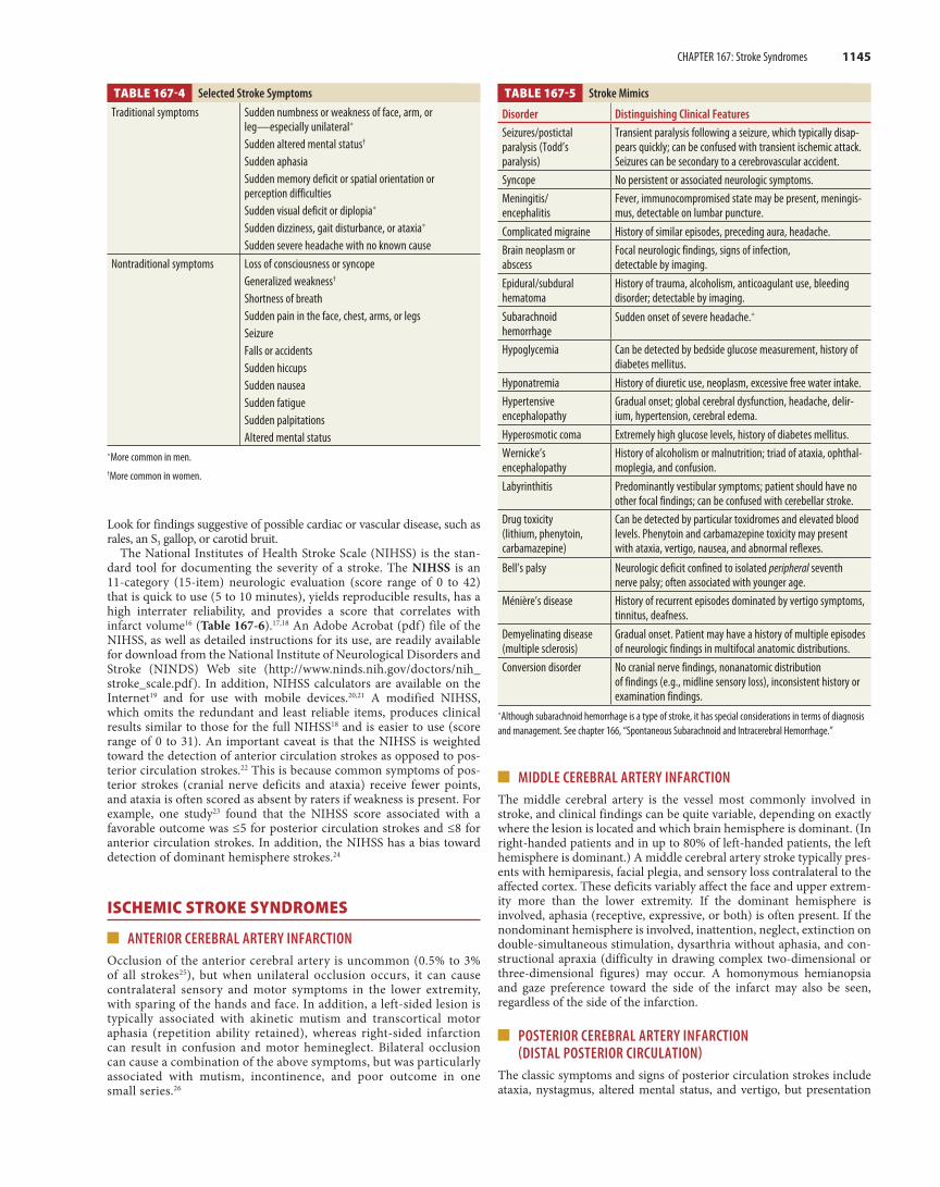
TABLE 167-5 Stroke Mimics
Disorder Distinguishing Clinical Features
Seizures/postictal paralysis (Todd’s paralysis)
Transient paralysis following a seizure, which typically disap-pears quickly; can be confused with transient ischemic attack. Seizures can be secondary to a cerebrovascular accident.
Syncope No persistent or associated neurologic symptoms.Meningitis/ encephalitis
Fever, immunocompromised state may be present, meningis-mus, detectable on lumbar puncture.
Complicated migraine History of similar episodes, preceding aura, headache.Brain neoplasm or abscess
Focal neurologic findings, signs of infection, detectable by imaging.
Epidural/subdural hematoma
History of trauma, alcoholism, anticoagulant use, bleeding disorder; detectable by imaging.
Subarachnoid hemorrhage
Sudden onset of severe headache.*
Hypoglycemia Can be detected by bedside glucose measurement, history of diabetes mellitus.
Hyponatremia History of diuretic use, neoplasm, excessive free water intake.Hypertensive encephalopathy
Gradual onset; global cerebral dysfunction, headache, delir-ium, hypertension, cerebral edema.
Hyperosmotic coma Extremely high glucose levels, history of diabetes mellitus.Wernicke’s encephalopathy
History of alcoholism or malnutrition; triad of ataxia, ophthal-moplegia, and confusion.
Labyrinthitis Predominantly vestibular symptoms; patient should have no other focal findings; can be confused with cerebellar stroke.
Drug toxicity (lithium, phenytoin, carbamazepine)
Can be detected by particular toxidromes and elevated blood levels. Phenytoin and carbamazepine toxicity may present with ataxia, vertigo, nausea, and abnormal reflexes.
Bell’s palsy Neurologic deficit confined to isolated peripheral seventh nerve palsy; often associated with younger age.
Ménière’s disease History of recurrent episodes dominated by vertigo symptoms, tinnitus, deafness.
Demyelinating disease (multiple sclerosis)
Gradual onset. Patient may have a history of multiple episodes of neurologic findings in multifocal anatomic distributions.
Conversion disorder No cranial nerve findings, nonanatomic distribution of findings (e.g., midline sensory loss), inconsistent history or examination findings.
*Although subarachnoid hemorrhage is a type of stroke, it has special considerations in terms of diagnosis and management. See chapter 166, “Spontaneous Subarachnoid and Intracerebral Hemorrhage.”
Look for findings suggestive of possible cardiac or vascular disease, such as rales, an S3 gallop, or carotid bruit.
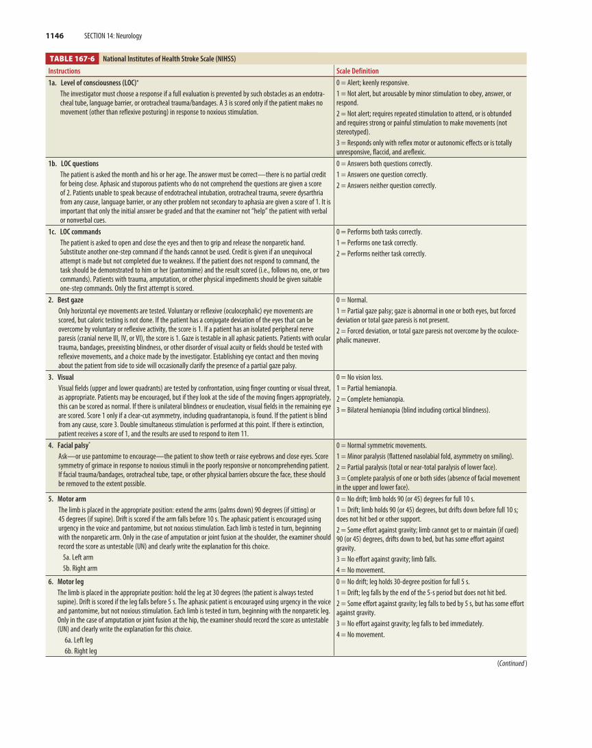
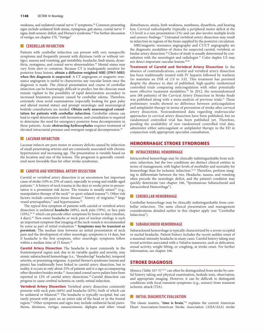
The National Institutes of Health Stroke Scale (NIHSS) is the stan-dard tool for documenting the severity of a stroke. The NIHSS is an 11-category (15-item) neurologic evaluation (score range of 0 to 42) that is quick to use (5 to 10 minutes), yields reproducible results, has a high interrater reliability, and provides a score that correlates with infarct volume16 (Table 167-6).17,18 An Adobe Acrobat (pdf) file of the NIHSS, as well as detailed instructions for its use, are readily available for download from the National Institute of Neurological Disorders and Stroke (NINDS) Web site (http://www.ninds.nih.gov/doctors/nih_stroke_scale.pdf). In addition, NIHSS calculators are available on the Internet19 and for use with mobile devices.20,21 A modified NIHSS, which omits the redundant and least reliable items, produces clinical results similar to those for the full NIHSS18 and is easier to use (score range of 0 to 31). An important caveat is that the NIHSS is weighted toward the detection of anterior circulation strokes as opposed to pos-terior circulation strokes.22 This is because common symptoms of pos-terior strokes (cranial nerve deficits and ataxia) receive fewer points, and ataxia is often scored as absent by raters if weakness is present. For example, one study23 found that the NIHSS score associated with a favorable outcome was ≤5 for posterior circulation strokes and ≤8 for anterior circulation strokes. In addition, the NIHSS has a bias toward detection of dominant hemisphere strokes.24
ISCHEMIC STROKE SYNDROMES
� ANTERIOR CEREBRAL ARTERY INFARCTIONOcclusion of the anterior cerebral artery is uncommon (0.5% to 3% of all strokes25), but when unilateral occlusion occurs, it can cause contralateral sensory and motor symptoms in the lower extremity, with sparing of the hands and face. In addition, a left-sided lesion is typically associated with akinetic mutism and transcortical motor aphasia (repetition ability retained), whereas right-sided infarction can result in confusion and motor hemineglect. Bilateral occlusion can cause a combination of the above symptoms, but was particularly associated with mutism, incontinence, and poor outcome in one small series.26
� MIDDLE CEREBRAL ARTERY INFARCTION The middle cerebral artery is the vessel most commonly involved in stroke, and clinical findings can be quite variable, depending on exactly where the lesion is located and which brain hemisphere is dominant. (In right-handed patients and in up to 80% of left-handed patients, the left hemisphere is dominant.) A middle cerebral artery stroke typically pres-ents with hemiparesis, facial plegia, and sensory loss contralateral to the affected cortex. These deficits variably affect the face and upper extrem-ity more than the lower extremity. If the dominant hemisphere is involved, aphasia (receptive, expressive, or both) is often present. If the nondominant hemisphere is involved, inattention, neglect, extinction on double-simultaneous stimulation, dysarthria without aphasia, and con-structional apraxia (difficulty in drawing complex two-dimensional or three-dimensional figures) may occur. A homonymous hemianopsia and gaze preference toward the side of the infarct may also be seen, regardless of the side of the infarction.
� POSTERIOR CEREBRAL ARTERY INFARCTION (DISTAL POSTERIOR CIRCULATION)
The classic symptoms and signs of posterior circulation strokes include ataxia, nystagmus, altered mental status, and vertigo, but presentation
TABLE 167-4 Selected Stroke Symptoms
Traditional symptoms Sudden numbness or weakness of face, arm, or leg—especially unilateral*
Sudden altered mental status†
Sudden aphasiaSudden memory deficit or spatial orientation or perception difficultiesSudden visual deficit or diplopia*
Sudden dizziness, gait disturbance, or ataxia*
Sudden severe headache with no known causeNontraditional symptoms Loss of consciousness or syncope
Generalized weakness†
Shortness of breathSudden pain in the face, chest, arms, or legsSeizureFalls or accidentsSudden hiccupsSudden nauseaSudden fatigueSudden palpitationsAltered mental status
*More common in men.†More common in women.
Tintinalli_Sec14_p1125-1206.indd 1145 8/6/15 4:00 PM
1146 SECTION 14: Neurology
(Continued )
TABLE 167-6 National Institutes of Health Stroke Scale (NIHSS) (Continued )
Instructions Scale Definition
1a. Level of consciousness (LOC)*
The investigator must choose a response if a full evaluation is prevented by such obstacles as an endotra-cheal tube, language barrier, or orotracheal trauma/bandages. A 3 is scored only if the patient makes no movement (other than reflexive posturing) in response to noxious stimulation.
0 = Alert; keenly responsive.1 = Not alert, but arousable by minor stimulation to obey, answer, or respond.2 = Not alert; requires repeated stimulation to attend, or is obtunded and requires strong or painful stimulation to make movements (not stereotyped).3 = Responds only with reflex motor or autonomic effects or is totally unresponsive, flaccid, and areflexic.
1b. LOC questionsThe patient is asked the month and his or her age. The answer must be correct—there is no partial credit for being close. Aphasic and stuporous patients who do not comprehend the questions are given a score of 2. Patients unable to speak because of endotracheal intubation, orotracheal trauma, severe dysarthria from any cause, language barrier, or any other problem not secondary to aphasia are given a score of 1. It is important that only the initial answer be graded and that the examiner not “help” the patient with verbal or nonverbal cues.
0 = Answers both questions correctly.1 = Answers one question correctly.2 = Answers neither question correctly.
1c. LOC commandsThe patient is asked to open and close the eyes and then to grip and release the nonparetic hand. Substitute another one-step command if the hands cannot be used. Credit is given if an unequivocal attempt is made but not completed due to weakness. If the patient does not respond to command, the task should be demonstrated to him or her (pantomime) and the result scored (i.e., follows no, one, or two commands). Patients with trauma, amputation, or other physical impediments should be given suitable one-step commands. Only the first attempt is scored.
0 = Performs both tasks correctly.1 = Performs one task correctly.2 = Performs neither task correctly.
2. Best gazeOnly horizontal eye movements are tested. Voluntary or reflexive (oculocephalic) eye movements are scored, but caloric testing is not done. If the patient has a conjugate deviation of the eyes that can be overcome by voluntary or reflexive activity, the score is 1. If a patient has an isolated peripheral nerve paresis (cranial nerve III, IV, or VI), the score is 1. Gaze is testable in all aphasic patients. Patients with ocular trauma, bandages, preexisting blindness, or other disorder of visual acuity or fields should be tested with reflexive movements, and a choice made by the investigator. Establishing eye contact and then moving about the patient from side to side will occasionally clarify the presence of a partial gaze palsy.
0 = Normal.1 = Partial gaze palsy; gaze is abnormal in one or both eyes, but forced deviation or total gaze paresis is not present.2 = Forced deviation, or total gaze paresis not overcome by the oculoce-phalic maneuver.
3. VisualVisual fields (upper and lower quadrants) are tested by confrontation, using finger counting or visual threat, as appropriate. Patients may be encouraged, but if they look at the side of the moving fingers appropriately, this can be scored as normal. If there is unilateral blindness or enucleation, visual fields in the remaining eye are scored. Score 1 only if a clear-cut asymmetry, including quadrantanopia, is found. If the patient is blind from any cause, score 3. Double simultaneous stimulation is performed at this point. If there is extinction, patient receives a score of 1, and the results are used to respond to item 11.
0 = No vision loss.1 = Partial hemianopia.2 = Complete hemianopia.3 = Bilateral hemianopia (blind including cortical blindness).
4. Facial palsy*
Ask—or use pantomime to encourage—the patient to show teeth or raise eyebrows and close eyes. Score symmetry of grimace in response to noxious stimuli in the poorly responsive or noncomprehending patient. If facial trauma/bandages, orotracheal tube, tape, or other physical barriers obscure the face, these should be removed to the extent possible.
0 = Normal symmetric movements.1 = Minor paralysis (flattened nasolabial fold, asymmetry on smiling).2 = Partial paralysis (total or near-total paralysis of lower face).3 = Complete paralysis of one or both sides (absence of facial movement in the upper and lower face).
5. Motor armThe limb is placed in the appropriate position: extend the arms (palms down) 90 degrees (if sitting) or 45 degrees (if supine). Drift is scored if the arm falls before 10 s. The aphasic patient is encouraged using urgency in the voice and pantomime, but not noxious stimulation. Each limb is tested in turn, beginning with the nonparetic arm. Only in the case of amputation or joint fusion at the shoulder, the examiner should record the score as untestable (UN) and clearly write the explanation for this choice.
5a. Left arm5b. Right arm
0 = No drift; limb holds 90 (or 45) degrees for full 10 s.1 = Drift; limb holds 90 (or 45) degrees, but drifts down before full 10 s; does not hit bed or other support.2 = Some effort against gravity; limb cannot get to or maintain (if cued) 90 (or 45) degrees, drifts down to bed, but has some effort against gravity.3 = No effort against gravity; limb falls.4 = No movement.
6. Motor legThe limb is placed in the appropriate position: hold the leg at 30 degrees (the patient is always tested supine). Drift is scored if the leg falls before 5 s. The aphasic patient is encouraged using urgency in the voice and pantomime, but not noxious stimulation. Each limb is tested in turn, beginning with the nonparetic leg. Only in the case of amputation or joint fusion at the hip, the examiner should record the score as untestable (UN) and clearly write the explanation for this choice.
6a. Left leg6b. Right leg
0 = No drift; leg holds 30-degree position for full 5 s.1 = Drift; leg falls by the end of the 5-s period but does not hit bed.2 = Some effort against gravity; leg falls to bed by 5 s, but has some effort against gravity.3 = No effort against gravity; leg falls to bed immediately.4 = No movement.
Tintinalli_Sec14_p1125-1206.indd 1146 8/6/15 4:00 PM
CHAPTER 167: Stroke Syndromes 1147
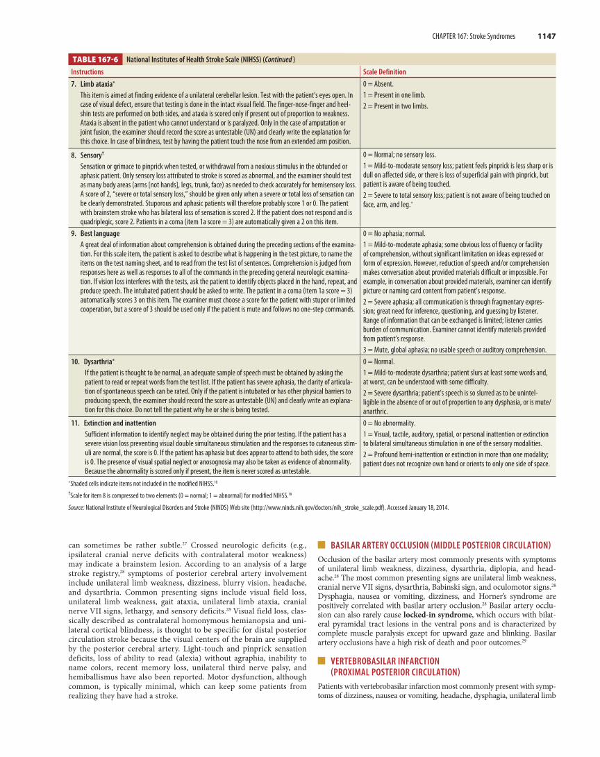
TABLE 167-6 National Institutes of Health Stroke Scale (NIHSS) (Continued )
Instructions Scale Definition
7. Limb ataxia*
This item is aimed at finding evidence of a unilateral cerebellar lesion. Test with the patient’s eyes open. In case of visual defect, ensure that testing is done in the intact visual field. The finger-nose-finger and heel-shin tests are performed on both sides, and ataxia is scored only if present out of proportion to weakness. Ataxia is absent in the patient who cannot understand or is paralyzed. Only in the case of amputation or joint fusion, the examiner should record the score as untestable (UN) and clearly write the explanation for this choice. In case of blindness, test by having the patient touch the nose from an extended arm position.
0 = Absent.1 = Present in one limb.2 = Present in two limbs.
8. Sensory†
Sensation or grimace to pinprick when tested, or withdrawal from a noxious stimulus in the obtunded or aphasic patient. Only sensory loss attributed to stroke is scored as abnormal, and the examiner should test as many body areas (arms [not hands], legs, trunk, face) as needed to check accurately for hemisensory loss. A score of 2, “severe or total sensory loss,” should be given only when a severe or total loss of sensation can be clearly demonstrated. Stuporous and aphasic patients will therefore probably score 1 or 0. The patient with brainstem stroke who has bilateral loss of sensation is scored 2. If the patient does not respond and is quadriplegic, score 2. Patients in a coma (item 1a score = 3) are automatically given a 2 on this item.
0 = Normal; no sensory loss.1 = Mild-to-moderate sensory loss; patient feels pinprick is less sharp or is dull on affected side, or there is loss of superficial pain with pinprick, but patient is aware of being touched.2 = Severe to total sensory loss; patient is not aware of being touched on face, arm, and leg.*
9. Best languageA great deal of information about comprehension is obtained during the preceding sections of the examina-tion. For this scale item, the patient is asked to describe what is happening in the test picture, to name the items on the test naming sheet, and to read from the test list of sentences. Comprehension is judged from responses here as well as responses to all of the commands in the preceding general neurologic examina-tion. If vision loss interferes with the tests, ask the patient to identify objects placed in the hand, repeat, and produce speech. The intubated patient should be asked to write. The patient in a coma (item 1a score = 3) automatically scores 3 on this item. The examiner must choose a score for the patient with stupor or limited cooperation, but a score of 3 should be used only if the patient is mute and follows no one-step commands.
0 = No aphasia; normal.1 = Mild-to-moderate aphasia; some obvious loss of fluency or facility of comprehension, without significant limitation on ideas expressed or form of expression. However, reduction of speech and/or comprehension makes conversation about provided materials difficult or impossible. For example, in conversation about provided materials, examiner can identify picture or naming card content from patient’s response.2 = Severe aphasia; all communication is through fragmentary expres-sion; great need for inference, questioning, and guessing by listener. Range of information that can be exchanged is limited; listener carries burden of communication. Examiner cannot identify materials provided from patient’s response.3 = Mute, global aphasia; no usable speech or auditory comprehension.
10. Dysarthria*
If the patient is thought to be normal, an adequate sample of speech must be obtained by asking the patient to read or repeat words from the test list. If the patient has severe aphasia, the clarity of articula-tion of spontaneous speech can be rated. Only if the patient is intubated or has other physical barriers to producing speech, the examiner should record the score as untestable (UN) and clearly write an explana-tion for this choice. Do not tell the patient why he or she is being tested.
0 = Normal.1 = Mild-to-moderate dysarthria; patient slurs at least some words and, at worst, can be understood with some difficulty.2 = Severe dysarthria; patient’s speech is so slurred as to be unintel-ligible in the absence of or out of proportion to any dysphasia, or is mute/anarthric.
11. Extinction and inattentionSufficient information to identify neglect may be obtained during the prior testing. If the patient has a severe vision loss preventing visual double simultaneous stimulation and the responses to cutaneous stim-uli are normal, the score is 0. If the patient has aphasia but does appear to attend to both sides, the score is 0. The presence of visual spatial neglect or anosognosia may also be taken as evidence of abnormality. Because the abnormality is scored only if present, the item is never scored as untestable.
0 = No abnormality.1 = Visual, tactile, auditory, spatial, or personal inattention or extinction to bilateral simultaneous stimulation in one of the sensory modalities.2 = Profound hemi-inattention or extinction in more than one modality; patient does not recognize own hand or orients to only one side of space.
*Shaded cells indicate items not included in the modified NIHSS.18
†Scale for item 8 is compressed to two elements (0 = normal; 1 = abnormal) for modified NIHSS.18
Source: National Institute of Neurological Disorders and Stroke (NINDS) Web site (http://www.ninds.nih.gov/doctors/nih_stroke_scale.pdf). Accessed January 18, 2014.
can sometimes be rather subtle.27 Crossed neurologic deficits (e.g., ipsilateral cranial nerve deficits with contralateral motor weakness) may indicate a brainstem lesion. According to an analysis of a large stroke registry,28 symptoms of posterior cerebral artery involvement include unilateral limb weakness, dizziness, blurry vision, headache, and dysarthria. Common presenting signs include visual field loss, unilateral limb weakness, gait ataxia, unilateral limb ataxia, cranial nerve VII signs, lethargy, and sensory deficits.28 Visual field loss, clas-sically described as contralateral homonymous hemianopsia and uni-lateral cortical blindness, is thought to be specific for distal posterior circulation stroke because the visual centers of the brain are supplied by the posterior cerebral artery. Light-touch and pinprick sensation deficits, loss of ability to read (alexia) without agraphia, inability to name colors, recent memory loss, unilateral third nerve palsy, and hemiballismus have also been reported. Motor dysfunction, although common, is typically minimal, which can keep some patients from realizing they have had a stroke.
� BASILAR ARTERY OCCLUSION (MIDDLE POSTERIOR CIRCULATION)Occlusion of the basilar artery most commonly presents with symptoms of unilateral limb weakness, dizziness, dysarthria, diplopia, and head-ache.28 The most common presenting signs are unilateral limb weakness, cranial nerve VII signs, dysarthria, Babinski sign, and oculomotor signs.28 Dysphagia, nausea or vomiting, dizziness, and Horner’s syndrome are positively correlated with basilar artery occlusion.28 Basilar artery occlu-sion can also rarely cause locked-in syndrome, which occurs with bilat-eral pyramidal tract lesions in the ventral pons and is characterized by complete muscle paralysis except for upward gaze and blinking. Basilar artery occlusions have a high risk of death and poor outcomes.29
� VERTEBROBASILAR INFARCTION (PROXIMAL POSTERIOR CIRCULATION)
Patients with vertebrobasilar infarction most commonly present with symp-toms of dizziness, nausea or vomiting, headache, dysphagia, unilateral limb
Tintinalli_Sec14_p1125-1206.indd 1147 8/6/15 4:00 PM
1148 SECTION 14: Neurology
weakness, and unilateral cranial nerve V symptoms.28 Common presenting signs include unilateral limb ataxia, nystagmus, gait ataxia, cranial nerve V signs, limb sensory deficit, and Horner’s syndrome.28 For further discussion of vertigo, see chapter 170, “Vertigo.”
� CEREBELLAR INFARCTIONPatients with cerebellar infarction can present with very nonspecific symptoms and frequently present with dizziness (with or without ver-tigo), nausea and vomiting, gait instability, headache, limb ataxia, dysar-thria, nystagmus, and cranial nerve abnormalities.30 Mental status may vary from alert to comatose. Because CT is inadequately sensitive for posterior fossa lesions, obtain a diffusion-weighted MRI (DWI-MRI) when this diagnosis is suspected. A CT angiogram or magnetic reso-nance angiogram is useful to characterize any vascular lesion once the diagnosis is made. The clinical presentation and course of cerebellar infarction can be frustratingly difficult to predict, but the clinician must remain vigilant to the possibility of rapid deterioration secondary to increased brainstem pressure caused by cerebellar edema. Therefore, extremely close serial examinations (especially looking for gaze palsy and altered mental status) and prompt neurologic and neurosurgical bedside consultations are needed. Obtain early neurosurgical consul-tation for patients with cerebellar infarction. Cerebellar edema can lead to rapid deterioration with herniation, and consultation is required to determine the need for emergency posterior fossa decompression in these patients. Acute obstructing hydrocephalus requires treatment of elevated intracranial pressure and emergent surgical decompression.30
� LACUNAR INFARCTIONLacunar infarcts are pure motor or sensory deficits caused by infarction of small penetrating arteries and are commonly associated with chronic hypertension and increasing age. The presentation is variable based on the location and size of the lesions. The prognosis is generally consid-ered more favorable than for other stroke syndromes.
� CAROTID AND VERTEBRAL ARTERY DISSECTION Carotid or vertebral artery dissection is an uncommon but important cause of stroke (10% to 25% of cases), especially in young and middle-aged patients.31 A history of neck trauma in the days to weeks prior to presen-tation is a prominent risk factor. The trauma is usually minor32 (e.g., manipulative therapy of the neck33 or sport-related trauma34). Other risk factors include connective tissue disease,35 history of migraine,36 large vessel arteriopathies,37 and hypertension.38
The typical first symptom of patients with carotid or vertebral artery dissection is unilateral headache (68%), neck pain (39%), or face pain (10%),39-41 which can precede other symptoms by hours to days (median, 4 days).42 New-onset headache or neck pain of unclear etiology is such an important symptom that imaging of the neck vessels is recommended by some as part of initial evaluation.41 Symptoms may be transient or persistent. The median time between an initial presentation of neck pain and the development of other neurologic symptoms is 14 days, but if headache is the first symptom, other neurologic symptoms follow within a median time of 15 hours.42 Carotid Artery Dissection The headache is most commonly in the frontotemporal region and, due to its variable quality and severity, may mimic subarachnoid hemorrhage (i.e., “thunderclap” headache), temporal arteritis, or preexisting migraine. A partial Horner’s syndrome (miosis and ptosis) has traditionally been linked to carotid artery dissection, but in reality, it occurs in only about 25% of patients and is a sign accompanying other disorders besides stroke.43 Associated cranial nerve palsies have been reported in 12% of carotid artery dissections.44 Carotid dissection can progress to cause cerebral ischemia or, rarely, retinal infarction.Vertebral Artery Dissection Vertebral artery dissection commonly presents with neck pain (66%) and headache (65%), both of which can be unilateral or bilateral.40 The headache is typically occipital, but can rarely present with pain on an entire side of the head or in the frontal region.39 Other symptoms and signs may include unilateral facial pares-thesia, dizziness, vertigo, nausea/emesis, diplopia and other visual
disturbances, ataxia, limb weakness, numbness, dysarthria, and hearing loss. Cervical radiculopathy (typically a peripheral motor deficit at the C5 level) is a rare presentation (1%) and can also involve multiple levels and sensory findings.45 Untreated vertebral artery dissection may result in infarction in regions of the brain supplied by the posterior circulation.
MRI/magnetic resonance angiography and CT/CT angiography are the diagnostic modalities of choice for suspected carotid, vertebral, or basilar artery dissection.46 Choice of study is usually determined by con-sultation with the neurologist and radiologist.47 Color duplex US may not detect important vascular lesions.48,49 Treatment of Carotid and Vertebral Artery Dissection In the absence of contraindications, carotid and vertebral artery dissection has been traditionally treated with IV heparin followed by warfarin (to maintain an INR of 2.0 to 3.0). This treatment has persisted despite the absence to date of published, high-quality randomized controlled trials comparing anticoagulation with other potentially more effective treatment modalities.50 In 2012, the nonrandomized arm (88 patients) of the Cervical Artery Dissection in Stroke Study was published along with a meta-analysis of previous trials.51 These preliminary results showed no difference between anticoagulation and antiplatelet therapy in terms of prevention of stroke after cervical artery dissection. Nonrandomized data regarding endovascular approaches to cervical artery dissection have been published, but no randomized controlled trial has been published yet. Therefore, pending the availability of new randomized controlled trial data, administer either anticoagulant or antiplatelet therapy in the ED in conjunction with appropriate specialist consultation.
HEMORRHAGIC STROKE SYNDROMES
� INTRACEREBRAL HEMORRHAGE Intracerebral hemorrhage may be clinically indistinguishable from isch-emic infarction, but the two conditions are distinct clinical entities in terms of management, with higher levels of morbidity and mortality for hemorrhage than for ischemic infarction.52,53 Therefore, perform imag-ing to differentiate between the two. Headache, nausea, and vomiting often precede the neurologic deficit, and the patient’s condition may quickly deteriorate (see chapter 166, “Spontaneous Subarachnoid and Intracerebral Hemorrhage”).
� CEREBELLAR HEMORRHAGECerebellar hemorrhage may be clinically indistinguishable from cere-bellar infarction. The same clinical presentation and management considerations detailed earlier in this chapter apply (see “Cerebellar Infarction”).
� SUBARACHNOID HEMORRHAGESubarachnoid hemorrhage is typically characterized by a severe occipital or nuchal headache. Patient history includes the recent sudden onset of a maximal-intensity headache in many cases. Careful history taking may reveal activities associated with a Valsalva maneuver, such as defecation, sexual activity, weight lifting, or coughing, at stroke onset. For further information, see chapter 166.
STROKE DIAGNOSIS
Mimics (Table 167-5)7,8,13 can often be distinguished from stroke by care-ful history taking and physical examination, bedside tests, observation, and appropriate imaging. However, it can be difficult to distinguish conditions with focal transient symptoms (e.g., seizure) from transient ischemic attack (TIA).
� INITIAL DIAGNOSTIC EVALUATIONThe classic mantra, “time is brain,”54 explains the current American Heart Association/American Stroke Association (AHA/ASA) stroke
Tintinalli_Sec14_p1125-1206.indd 1148 8/6/15 4:00 PM
CHAPTER 167: Stroke Syndromes 1149
guidelines Class IB recommendation to enact “an organized protocol for the emergency evaluation of patients with suspected stroke” in which “the goal is to evaluate and to decide treatment within 60 minutes of the patient’s arrival in an ED.”7 Creation of a multidisciplinary “stroke team” is encouraged, and the culture of the institution should be aligned to encourage rapid diagnosis and treatment of stroke. Train triage person-nel to suspect stroke and to quickly activate a stroke critical pathway that includes specific standing orders and procedures. Implement the critical pathway immediately upon that patient’s arrival, beginning in the triage area. Notify the emergency physician, the ED charge nurse, and the CT and laboratory technicians immediately when acute stroke is suspected. Do not delay the workup because ED beds are overcrowded or the emer-gency physician is currently otherwise engaged, and move the patient to a monitored bed as soon as feasible.
Concurrent with focused history taking and physical examination, perform a prioritized group of interventions and diagnostic studies rap-idly in the ED when acute stroke is suspected (Table 167-7).
Nonessential testing should NOT delay performing brain imaging within 25 minutes of the patient’s arrival (Table 167-8).7
� IMAGINGBrain Imaging Obtain emergency non–contrast-enhanced CT for sus-pected acute stroke. Most acute ischemic strokes are not visualized by a noncontrast brain CT in the early hours of a stroke.8 Therefore, the
utility of the first brain CT is primarily to exclude intracranial bleeding, abscess, tumor, and other stroke mimics, as well as to detect current contraindications to thrombolytics (e.g., evidence of more than one-third middle cerebral artery territory involvement).
The CT scan should be reviewed by the most expert interpreter available within 45 minutes of patient arrival, especially if thrombo-lytic therapy is being considered.7 The identification of subtle hemor-rhage, infarctions involving more than one third of the middle cerebral artery territory, and early cerebral infarction requires expertise. Tele-medicine consultation is an excellent option, because reports indicate
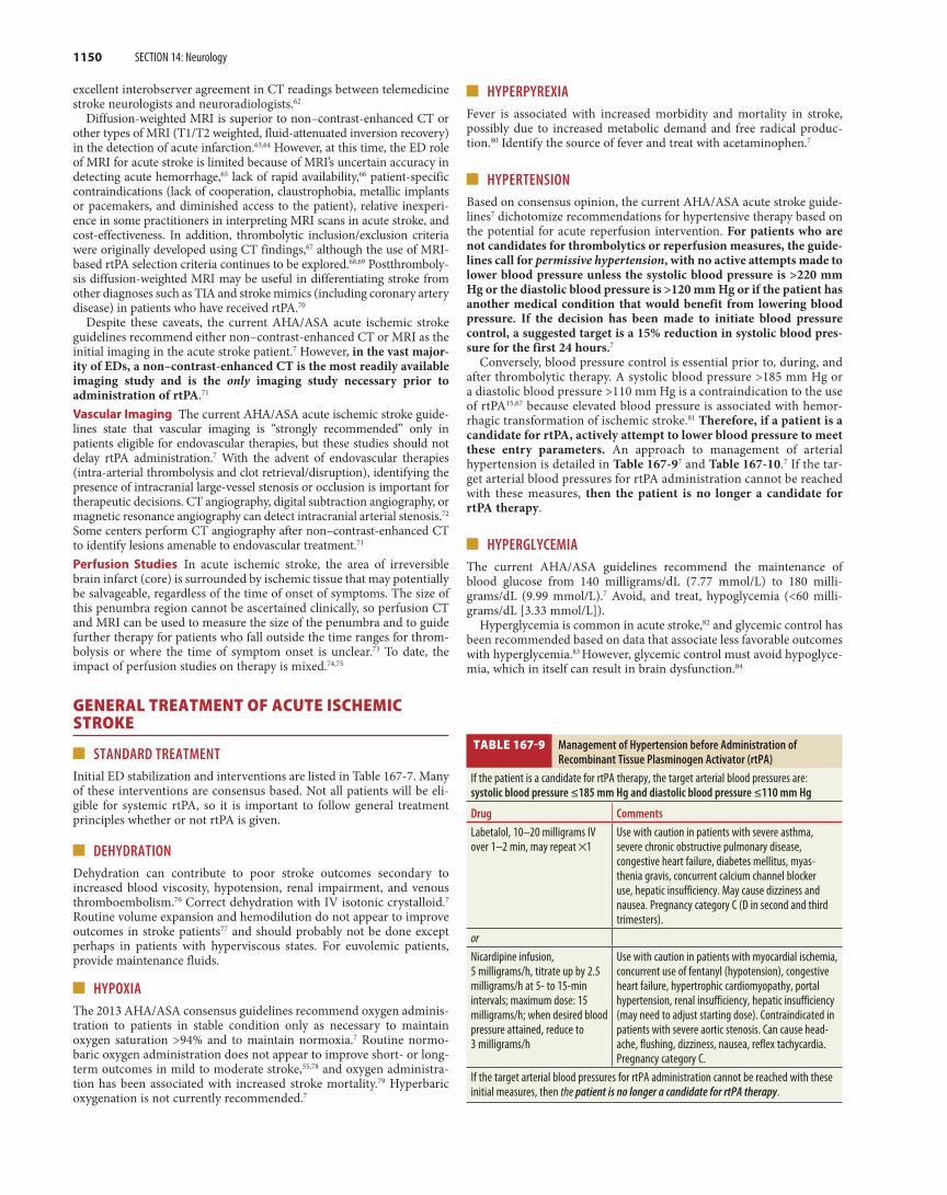
TABLE 167-7 Core ED Interventions for Suspected Acute Stroke
Intervention/Evaluation Rationale/Discussion
All patients
Assessment of airway, breathing, circulation
Immediate life threats must be addressed before other interventions are undertaken. Actively manage airway if necessary.
Establishment of IV access IV access is necessary for possible thrombolytic therapy.Oxygen administration (if hypoxia is present)
Routine oxygen supplementation is not indicated in mild to moderate stroke,55 and should only be given to keep oxygen saturation >94%.7
Cardiac monitoring Dysrhythmias, especially atrial fibrillation, are not infrequent in acute stroke and may predict 3-month mortality.56,57 Prophylactic administra-tion of antiarrhythmic agents is not indicated.
Bedside glucose determination To rapidly rule out hypoglycemia mimicking stroke. Treat hypoglycemia (<60 milligrams/dL) with IV dextrose.This is the only laboratory test result required prior to thrombolytic therapy7 unless the patient is taking/possibly taking oral anticoagulation therapy or heparin, or if there is a suspicion of thrombocytopenia or other bleeding diatheses (Table 167-11).
Pulse oximetry To detect hypoxia. ECG Acute coronary syndrome, dysrhythmias (atrial fibrillation, in particular), ECG changes, and elevated troponin T levels are frequently associ-
ated with acute stroke.58 ECG abnormalities may also predict 3-mo mortality.56 A large study (n = 9180) of consecutive stroke patients revealed that 2.3% suffered a subsequent myocardial infarction with 64.9% morbidity/mortality compared with 35.8% in the entire cohort.59
Noncontrast brain CT or MRI To exclude intracerebral hemorrhage, frank hypodensity (especially more than one third of the middle cerebral artery territory) (CT) or hyper-density of ischemia (MRI),7 abscess, and tumor. (See discussion in “Imaging“ section of this chapter.)
CBC including platelet count To detect polycythemia, thrombocytosis, or thrombocytopenia.Coagulation studies To detect preexisting coagulopathy in hemorrhagic stroke or when thrombolytics are being considered (Table 167-11).Electrolyte levels To detect electrolyte-imbalance stroke mimics (particularly Na+ and Ca2+).Cardiac enzyme levels See “ECG” earlier in this table.Nothing by mouth (NPO) order To protect against aspiration.Strict bedrest in the ED To protect against falls and seizures (in the period immediately after stroke). In patients who can maintain oxygenation, supine position
has been recommended to possibly improve cerebral blood flow7,60; however, this remains controversial.61 Head of bed elevation to 15 to 30 degrees may be used in patients at risk for hypoxia, airway compromise, aspiration, or suspected increased intracranial pressure.7
Selected patientsUrinalysis and/or chest radiograph (if infection suspected)
To detect infectious stroke mimics or stroke-associated infections. Routine chest radiography is not recommended.7
Pregnancy test (if female of childbearing age)
Pregnancy influences diagnosis and management considerations.
Toxicology screen and/or blood alcohol level (if ingestion suspected)
To detect stroke mimics as well as potential causes of stroke such as ingestion of a sympathomimetic (e.g., cocaine, methamphetamine, phencyclidine).
Lumbar puncture (if infection or subarach-noid hemorrhage suspected)
To detect stroke mimics. Thrombolytics should not be given before or after a lumbar puncture because of increased risk of post–lumbar puncture epidural hematoma.
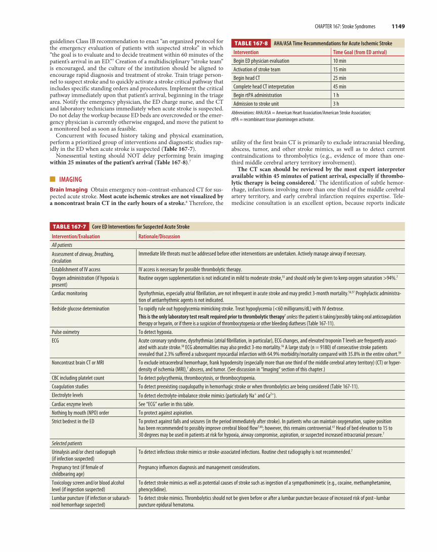
TABLE 167-8 AHA/ASA Time Recommendations for Acute Ischemic Stroke
Intervention Time Goal (from ED arrival)
Begin ED physician evaluation 10 minActivation of stroke team 15 minBegin head CT 25 min Complete head CT interpretation 45 minBegin rtPA administration 1 hAdmission to stroke unit 3 h
Abbreviations: AHA/ASA = American Heart Association/American Stroke Association; rtPA = recombinant tissue plasminogen activator.
Tintinalli_Sec14_p1125-1206.indd 1149 8/6/15 4:00 PM
1150 SECTION 14: Neurology
excellent interobserver agreement in CT readings between telemedicine stroke neurologists and neuroradiologists.62
Diffusion-weighted MRI is superior to non–contrast-enhanced CT or other types of MRI (T1/T2 weighted, fluid-attenuated inversion recovery) in the detection of acute infarction.63,64 However, at this time, the ED role of MRI for acute stroke is limited because of MRI’s uncertain accuracy in detecting acute hemorrhage,65 lack of rapid availability,66 patient-specific contraindications (lack of cooperation, claustrophobia, metallic implants or pacemakers, and diminished access to the patient), relative inexperi-ence in some practitioners in interpreting MRI scans in acute stroke, and cost-effectiveness. In addition, thrombolytic inclusion/exclusion criteria were originally developed using CT findings,67 although the use of MRI-based rtPA selection criteria continues to be explored.68,69 Postthromboly-sis diffusion-weighted MRI may be useful in differentiating stroke from other diagnoses such as TIA and stroke mimics (including coronary artery disease) in patients who have received rtPA.70
Despite these caveats, the current AHA/ASA acute ischemic stroke guidelines recommend either non–contrast-enhanced CT or MRI as the initial imaging in the acute stroke patient.7 However, in the vast major-ity of EDs, a non–contrast-enhanced CT is the most readily available imaging study and is the only imaging study necessary prior to administration of rtPA.71 Vascular Imaging The current AHA/ASA acute ischemic stroke guide-lines state that vascular imaging is “strongly recommended” only in patients eligible for endovascular therapies, but these studies should not delay rtPA administration.7 With the advent of endovascular therapies (intra-arterial thrombolysis and clot retrieval/disruption), identifying the presence of intracranial large-vessel stenosis or occlusion is important for therapeutic decisions. CT angiography, digital subtraction angiography, or magnetic resonance angiography can detect intracranial arterial stenosis.72 Some centers perform CT angiography after non–contrast-enhanced CT to identify lesions amenable to endovascular treatment.71 Perfusion Studies In acute ischemic stroke, the area of irreversible brain infarct (core) is surrounded by ischemic tissue that may potentially be salvageable, regardless of the time of onset of symptoms. The size of this penumbra region cannot be ascertained clinically, so perfusion CT and MRI can be used to measure the size of the penumbra and to guide further therapy for patients who fall outside the time ranges for throm-bolysis or where the time of symptom onset is unclear.73 To date, the impact of perfusion studies on therapy is mixed.74,75
GENERAL TREATMENT OF ACUTE ISCHEMIC STROKE
� STANDARD TREATMENTInitial ED stabilization and interventions are listed in Table 167-7. Many of these interventions are consensus based. Not all patients will be eli-gible for systemic rtPA, so it is important to follow general treatment principles whether or not rtPA is given.
� DEHYDRATIONDehydration can contribute to poor stroke outcomes secondary to increased blood viscosity, hypotension, renal impairment, and venous thromboembolism.76 Correct dehydration with IV isotonic crystalloid.7 Routine volume expansion and hemodilution do not appear to improve outcomes in stroke patients77 and should probably not be done except perhaps in patients with hyperviscous states. For euvolemic patients, provide maintenance fluids.
� HYPOXIAThe 2013 AHA/ASA consensus guidelines recommend oxygen adminis-tration to patients in stable condition only as necessary to maintain oxygen saturation >94% and to maintain normoxia.7 Routine normo-baric oxygen administration does not appear to improve short- or long-term outcomes in mild to moderate stroke,55,78 and oxygen administra-tion has been associated with increased stroke mortality.79 Hyperbaric oxygenation is not currently recommended.7
� HYPERPYREXIAFever is associated with increased morbidity and mortality in stroke, possibly due to increased metabolic demand and free radical produc-tion.80 Identify the source of fever and treat with acetaminophen.7
� HYPERTENSIONBased on consensus opinion, the current AHA/ASA acute stroke guide-lines7 dichotomize recommendations for hypertensive therapy based on the potential for acute reperfusion intervention. For patients who are not candidates for thrombolytics or reperfusion measures, the guide-lines call for permissive hypertension, with no active attempts made to lower blood pressure unless the systolic blood pressure is >220 mm Hg or the diastolic blood pressure is >120 mm Hg or if the patient has another medical condition that would benefit from lowering blood pressure. If the decision has been made to initiate blood pressure control, a suggested target is a 15% reduction in systolic blood pres-sure for the first 24 hours.7
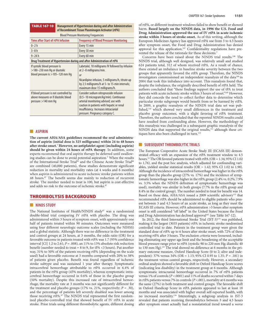
Conversely, blood pressure control is essential prior to, during, and after thrombolytic therapy. A systolic blood pressure >185 mm Hg or a diastolic blood pressure >110 mm Hg is a contraindication to the use of rtPA15,67 because elevated blood pressure is associated with hemor-rhagic transformation of ischemic stroke.81 Therefore, if a patient is a candidate for rtPA, actively attempt to lower blood pressure to meet these entry parameters. An approach to management of arterial hypertension is detailed in Table 167-97 and Table 167-10.7 If the tar-get arterial blood pressures for rtPA administration cannot be reached with these measures, then the patient is no longer a candidate for rtPA therapy.
� HYPERGLYCEMIAThe current AHA/ASA guidelines recommend the maintenance of blood glucose from 140 milligrams/dL (7.77 mmol/L) to 180 milli-grams/dL (9.99 mmol/L).7 Avoid, and treat, hypoglycemia (<60 milli-grams/dL [3.33 mmol/L]).
Hyperglycemia is common in acute stroke,82 and glycemic control has been recommended based on data that associate less favorable outcomes with hyperglycemia.83 However, glycemic control must avoid hypoglyce-mia, which in itself can result in brain dysfunction.84
TABLE 167-9 Management of Hypertension before Administration of Recombinant Tissue Plasminogen Activator (rtPA)
If the patient is a candidate for rtPA therapy, the target arterial blood pressures are: systolic blood pressure ≤185 mm Hg and diastolic blood pressure ≤110 mm Hg
Drug Comments
Labetalol, 10–20 milligrams IV over 1–2 min, may repeat ×1
Use with caution in patients with severe asthma, severe chronic obstructive pulmonary disease, congestive heart failure, diabetes mellitus, myas-thenia gravis, concurrent calcium channel blocker use, hepatic insufficiency. May cause dizziness and nausea. Pregnancy category C (D in second and third trimesters).
orNicardipine infusion, 5 milligrams/h, titrate up by 2.5 milligrams/h at 5- to 15-min intervals; maximum dose: 15 milligrams/h; when desired blood pressure attained, reduce to 3 milligrams/h
Use with caution in patients with myocardial ischemia, concurrent use of fentanyl (hypotension), congestive heart failure, hypertrophic cardiomyopathy, portal hypertension, renal insufficiency, hepatic insufficiency (may need to adjust starting dose). Contraindicated in patients with severe aortic stenosis. Can cause head-ache, flushing, dizziness, nausea, reflex tachycardia. Pregnancy category C.
If the target arterial blood pressures for rtPA administration cannot be reached with these initial measures, then the patient is no longer a candidate for rtPA therapy.
Tintinalli_Sec14_p1125-1206.indd 1150 8/6/15 4:00 PM
CHAPTER 167: Stroke Syndromes 1151
� ASPIRINThe current AHA/ASA guidelines recommend the oral administra-tion of aspirin (initial dose is 325 milligrams) within 24 to 48 hours after stroke onset.7 However, no antiplatelet agent (including aspirin) should be given within 24 hours of rtPA therapy. In addition, some experts recommend that oral aspirin therapy be deferred until swallow-ing studies can be done to avoid potential aspiration.7 When the results of the International Stroke Trial85 and the Chinese Acute Stroke Trial86 are combined (40,000 patients), these studies demonstrate significant reduction in mortality and morbidity rates (at 4 weeks and 6 months) when aspirin is administered to acute ischemic stroke patients within 48 hours.87 The benefit seems due mainly to reduction of recurrent stroke. The number needed to treat is 100, but aspirin is cost-effective and adds no risk to the outcome of ischemic stroke.87
THROMBOLYSIS BACKGROUND
� NINDS STUDY The National Institutes of Health/NINDS study67 was a randomized double-blind trial comparing IV rtPA with placebo. The drug was administered within 3 hours of symptom onset, with approximately one half of patients treated within 90 minutes. Outcomes were measured using four different neurologic outcome scales (including the NIHSS) and a global statistic. Although there was no difference in the treatment and control groups at 24 hours, at 3 months, the odds ratio (OR) for a favorable outcome in patients treated with rtPA was 1.7 (95% confidence interval [CI] 1.2 to 2.61; P = .008), an 11% to 13% absolute risk reduction benefit (number needed to treat = 8 to 9, for tPA <3 hours). Put another way, 31% to 50% of the patients receiving rtPA (depending on the scale used) had a favorable outcome at 3 months compared with 20% to 38% of patients given placebo. Benefit was found regardless of ischemic stroke subtype and was sustained 1 year after therapy. Symptomatic intracerebral hemorrhage attributable to rtPA occurred in 6.4% of patients in the rtPA group (45% mortality), whereas symptomatic intra-cerebral hemorrhage occurred in 0.6% of those in the placebo group (50% mortality). Despite this increased rate of intracerebral hemor-rhage, the mortality rate at 3 months was not significantly different for the treatment and placebo groups (17% vs. 21%, respectively; P = .30), and the percentage of patients left severely disabled was lower among those receiving rtPA.67 The NINDS trial represented the first random-ized placebo-controlled trial that showed benefit of IV rtPA in acute stroke. Prior trials using different thrombolytic agents, different dosing
of rtPA, or different treatment windows failed to show benefit or showed harm. Based largely on the NINDS data, in 1996 the U.S. Food and Drug Administration approved the use of IV rtPA in acute ischemic stroke within 3 hours of stroke onset. As of this writing, although the European Medicines Agency has approved rtPA use from 3 to 4.5 hours after symptom onset, the Food and Drug Administration has denied approval for this application.107 Confidentiality regulations have pre-vented the release of the rationale for these decisions.7
Concerns have been raised about the NINDS trial results.108 The NINDS trial, although well designed, was relatively small and studied 624 patients total, 312 of whom received rtPA. As a result of chance, there existed an imbalance in baseline stroke severity between the two groups that apparently favored the rtPA group. Therefore, the NINDS investigators commissioned an independent reanalysis of the data109 in 2004 that took this imbalance into account. This reanalysis found that, despite the imbalance, the originally described benefit of rtPA held. The authors concluded that “these findings support the use of tPA to treat patients with acute ischemic stroke within 3 hours of onset.”109 However, they did concede the need to collect further data to determine which particular stroke subgroups would benefit from or be harmed by rtPA. In 2009, a graphic reanalysis of the NINDS trial data set was pub-lished,110 which showed very small differences in the treatment and placebo group outcomes, with a slight favoring of rtPA treatment. Therefore, the authors concluded that the reported NINDS results could have resulted from confounding alone. However, the methodology of this reanalysis was challenged in a subsequent graphic reanalysis of the NINDS data that supported the original results,111 although these cri-tiques have also been challenged in turn.112
� SUBSEQUENT THROMBOLYTIC TRIALS The European Cooperative Acute Stroke Study III (ECASS III) demon-strated efficacy with an expansion of the rtPA treatment window to 4.5 hours.15 The OR favored patients treated with rtPA (OR = 1.34; 95% CI 1.02 to 1.76), and the post hoc analysis, which adjusted for confounding vari-ables, yielded similar statistical results (OR = 1.42; 95% CI 1.02 to 1.98). Although the incidence of intracerebral hemorrhage was higher in the rtPA group than the placebo group (27% vs. 17%) and the incidence of symp-tomatic hemorrhage was also higher in the rtPA group (2.4% vs. 0.2%; 7.9% vs. 3.5% when the NINDS definition of symptomatic hemorrhage was used), mortality was similar in both groups (7.7% in the rtPA group and 8.4% in the control group). The number needed to treat for benefit was 14. Based on these data, AHA/ASA issued a 2009 scientific advisory113 that recommended rtPA should be administered to eligible patients who pres-ent between 3 and 4.5 hours of an acute stroke, as long as they meet the ECASS III criteria. However, rtPA administration >3 hours after symptom onset is still considered “off label” in the United States because the Food and Drug Administration has declined approval107 (see Table 167-12).
In 2012, the third International Stroke Trial (IST-3)114 was published, making it the largest (3035 patients) rtPA in ischemic stroke randomized controlled trial to date. Patients in the treatment group were given the standard dose of rtPA up to 6 hours after stroke onset, with 72% of them receiving rtPA after 3 hours. The exclusion criteria were loosened, includ-ing eliminating any upper age limit and the broadening of the acceptable blood pressure range prior to rtPA (systolic 90 to 220 mm Hg; diastolic 40 to 130 mm Hg).115 The trial showed no difference at 6 months in the pri-mary outcome measure, Oxford Handicap Score (0 to 2; alive and inde-pendent): 37% versus 34% (OR = 1.13; 95% CI 0.95 to 1.35; P = .181) in the treatment versus control groups, respectively. However, a secondary ordinal analysis showed a favorable shift in Oxford Handicap Score scores (indicating less disability) in the treatment group at 6 months. Although symptomatic intracranial hemorrhage occurred in 7% of rtPA patients versus 1% of controls (P <.0001) and 11% of deaths occurred within 7 days in rtPA patients versus 7% in controls (P <.001), mortality at 6 months was the same (27%) in both treatment and control groups. The favorable shift in Oxford Handicap Score in rtPA patients appeared to last at least 18 months and was associated with higher overall self-reported health, with no increased mortality.116 Interestingly, a subgroup analysis in IST-3 revealed that patients receiving thrombolytics between 3 and 4.5 hours after symptom onset actually had a nonstatistical trend toward a worse
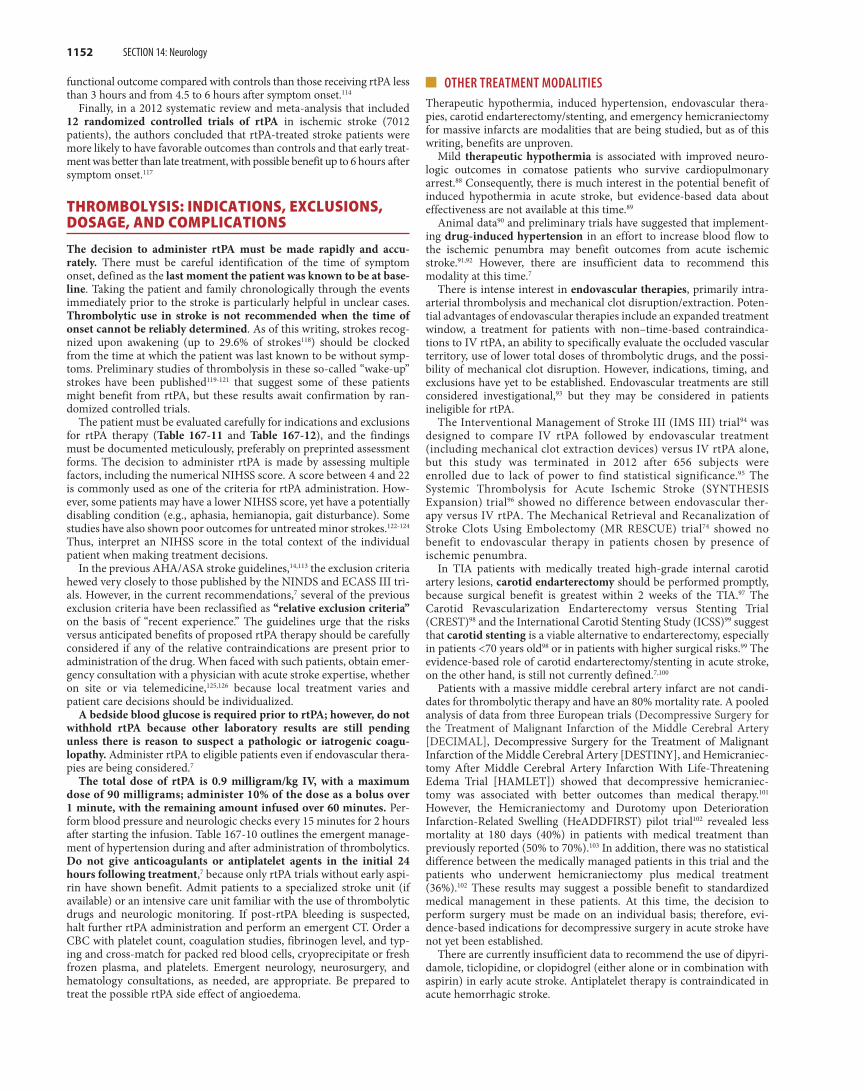
TABLE 167-10 Management of Hypertension during and after Administration of Recombinant Tissue Plasminogen Activator (rtPA)
Blood Pressure Monitoring FrequenciesTime after Start of rtPA Infusion Frequency of Blood Pressure Monitoring
0–2 h Every 15 min3–8 h Every 30 min9–24 h Every 60 minDrug Treatment of Hypertension during and after Administration of rtPA
If systolic blood pressure is >180–230 mm Hg or diastolic blood pressure is >105–120 mm Hg
Labetalol, 10 milligrams IV followed by infusion at 2–8 milligrams/min.orNicardipine infusion, 5 milligrams/h, titrate up by 2.5 milligrams/h at 5- to 15-min intervals; maximum dose 15 milligrams/h.
If blood pressure is not controlled by above measures or if diastolic blood pressure >140 mm Hg
Consider sodium nitroprusside infusion (0.5–10 microgram/kg/min). Continuous arterial monitoring advised; use with caution in patients with hepatic or renal insufficiency. Increases intracranial pressure. Pregnancy category C.
Tintinalli_Sec14_p1125-1206.indd 1151 8/6/15 4:00 PM
1152 SECTION 14: Neurology
functional outcome compared with controls than those receiving rtPA less than 3 hours and from 4.5 to 6 hours after symptom onset.114
Finally, in a 2012 systematic review and meta-analysis that included 12 randomized controlled trials of rtPA in ischemic stroke (7012 patients), the authors concluded that rtPA-treated stroke patients were more likely to have favorable outcomes than controls and that early treat-ment was better than late treatment, with possible benefit up to 6 hours after symptom onset.117
THROMBOLYSIS: INDICATIONS, EXCLUSIONS, DOSAGE, AND COMPLICATIONS
The decision to administer rtPA must be made rapidly and accu-rately. There must be careful identification of the time of symptom onset, defined as the last moment the patient was known to be at base-line. Taking the patient and family chronologically through the events immediately prior to the stroke is particularly helpful in unclear cases. Thrombolytic use in stroke is not recommended when the time of onset cannot be reliably determined. As of this writing, strokes recog-nized upon awakening (up to 29.6% of strokes118) should be clocked from the time at which the patient was last known to be without symp-toms. Preliminary studies of thrombolysis in these so-called “wake-up” strokes have been published119-121 that suggest some of these patients might benefit from rtPA, but these results await confirmation by ran-domized controlled trials.
The patient must be evaluated carefully for indications and exclusions for rtPA therapy (Table 167-11 and Table 167-12), and the findings must be documented meticulously, preferably on preprinted assessment forms. The decision to administer rtPA is made by assessing multiple factors, including the numerical NIHSS score. A score between 4 and 22 is commonly used as one of the criteria for rtPA administration. How-ever, some patients may have a lower NIHSS score, yet have a potentially disabling condition (e.g., aphasia, hemianopia, gait disturbance). Some studies have also shown poor outcomes for untreated minor strokes.122-124 Thus, interpret an NIHSS score in the total context of the individual patient when making treatment decisions.
In the previous AHA/ASA stroke guidelines,14,113 the exclusion criteria hewed very closely to those published by the NINDS and ECASS III tri-als. However, in the current recommendations,7 several of the previous exclusion criteria have been reclassified as “relative exclusion criteria” on the basis of “recent experience.” The guidelines urge that the risks versus anticipated benefits of proposed rtPA therapy should be carefully considered if any of the relative contraindications are present prior to administration of the drug. When faced with such patients, obtain emer-gency consultation with a physician with acute stroke expertise, whether on site or via telemedicine,125,126 because local treatment varies and patient care decisions should be individualized.
A bedside blood glucose is required prior to rtPA; however, do not withhold rtPA because other laboratory results are still pending unless there is reason to suspect a pathologic or iatrogenic coagu-lopathy. Administer rtPA to eligible patients even if endovascular thera-pies are being considered.7
The total dose of rtPA is 0.9 milligram/kg IV, with a maximum dose of 90 milligrams; administer 10% of the dose as a bolus over 1 minute, with the remaining amount infused over 60 minutes. Per-form blood pressure and neurologic checks every 15 minutes for 2 hours after starting the infusion. Table 167-10 outlines the emergent manage-ment of hypertension during and after administration of thrombolytics. Do not give anticoagulants or antiplatelet agents in the initial 24 hours following treatment,7 because only rtPA trials without early aspi-rin have shown benefit. Admit patients to a specialized stroke unit (if available) or an intensive care unit familiar with the use of thrombolytic drugs and neurologic monitoring. If post-rtPA bleeding is suspected, halt further rtPA administration and perform an emergent CT. Order a CBC with platelet count, coagulation studies, fibrinogen level, and typ-ing and cross-match for packed red blood cells, cryoprecipitate or fresh frozen plasma, and platelets. Emergent neurology, neurosurgery, and hematology consultations, as needed, are appropriate. Be prepared to treat the possible rtPA side effect of angioedema.
� OTHER TREATMENT MODALITIESTherapeutic hypothermia, induced hypertension, endovascular thera-pies, carotid endarterectomy/stenting, and emergency hemicraniectomy for massive infarcts are modalities that are being studied, but as of this writing, benefits are unproven.
Mild therapeutic hypothermia is associated with improved neuro-logic outcomes in comatose patients who survive cardiopulmonary arrest.88 Consequently, there is much interest in the potential benefit of induced hypothermia in acute stroke, but evidence-based data about effectiveness are not available at this time.89
Animal data90 and preliminary trials have suggested that implement-ing drug-induced hypertension in an effort to increase blood flow to the ischemic penumbra may benefit outcomes from acute ischemic stroke.91,92 However, there are insufficient data to recommend this modality at this time.7
There is intense interest in endovascular therapies, primarily intra-arterial thrombolysis and mechanical clot disruption/extraction. Poten-tial advantages of endovascular therapies include an expanded treatment window, a treatment for patients with non–time-based contraindica-tions to IV rtPA, an ability to specifically evaluate the occluded vascular territory, use of lower total doses of thrombolytic drugs, and the possi-bility of mechanical clot disruption. However, indications, timing, and exclusions have yet to be established. Endovascular treatments are still considered investigational,93 but they may be considered in patients ineligible for rtPA.
The Interventional Management of Stroke III (IMS III) trial94 was designed to compare IV rtPA followed by endovascular treatment (including mechanical clot extraction devices) versus IV rtPA alone, but this study was terminated in 2012 after 656 subjects were enrolled due to lack of power to find statistical significance.95 The Systemic Thrombolysis for Acute Ischemic Stroke (SYNTHESIS Expansion) trial96 showed no difference between endovascular ther-apy versus IV rtPA. The Mechanical Retrieval and Recanalization of Stroke Clots Using Embolectomy (MR RESCUE) trial74 showed no benefit to endovascular therapy in patients chosen by presence of ischemic penumbra.
In TIA patients with medically treated high-grade internal carotid artery lesions, carotid endarterectomy should be performed promptly, because surgical benefit is greatest within 2 weeks of the TIA.97 The Carotid Revascularization Endarterectomy versus Stenting Trial (CREST)98 and the International Carotid Stenting Study (ICSS)99 suggest that carotid stenting is a viable alternative to endarterectomy, especially in patients <70 years old98 or in patients with higher surgical risks.99 The evidence-based role of carotid endarterectomy/stenting in acute stroke, on the other hand, is still not currently defined.7,100
Patients with a massive middle cerebral artery infarct are not candi-dates for thrombolytic therapy and have an 80% mortality rate. A pooled analysis of data from three European trials (Decompressive Surgery for the Treatment of Malignant Infarction of the Middle Cerebral Artery [DECIMAL], Decompressive Surgery for the Treatment of Malignant Infarction of the Middle Cerebral Artery [DESTINY], and Hemicraniec-tomy After Middle Cerebral Artery Infarction With Life-Threatening Edema Trial [HAMLET]) showed that decompressive hemicraniec-tomy was associated with better outcomes than medical therapy.101 However, the Hemicraniectomy and Durotomy upon Deterioration Infarction-Related Swelling (HeADDFIRST) pilot trial102 revealed less mortality at 180 days (40%) in patients with medical treatment than previously reported (50% to 70%).103 In addition, there was no statistical difference between the medically managed patients in this trial and the patients who underwent hemicraniectomy plus medical treatment (36%).102 These results may suggest a possible benefit to standardized medical management in these patients. At this time, the decision to perform surgery must be made on an individual basis; therefore, evi-dence-based indications for decompressive surgery in acute stroke have not yet been established.
There are currently insufficient data to recommend the use of dipyri-damole, ticlopidine, or clopidogrel (either alone or in combination with aspirin) in early acute stroke. Antiplatelet therapy is contraindicated in acute hemorrhagic stroke.
Tintinalli_Sec14_p1125-1206.indd 1152 8/6/15 4:00 PM
CHAPTER 167: Stroke Syndromes 1153
DISPOSITION
Admit all acute stroke patients to monitored care units familiar with the care of stroke patients, preferably to specialized stroke units at desig-nated stroke centers. The use of stroke units has been associated with decreased complications and length of stay, improved daily function, decreased rate of discharge to long-term care facilities, and increased
likelihood of being able to live at home in the long term—with the ben-efit being independent of thrombolytic use.104-106 If a patient with acute stroke presents to a facility that lacks these resources, consider transfer-ring the patient to a higher level of care after the patient’s condition has stabilized and IV rtPA has been given, as indicated—the “drip and ship” model. Emergent, early consultation with an experienced stroke physi-cian at the accepting institution is desirable in these circumstances.
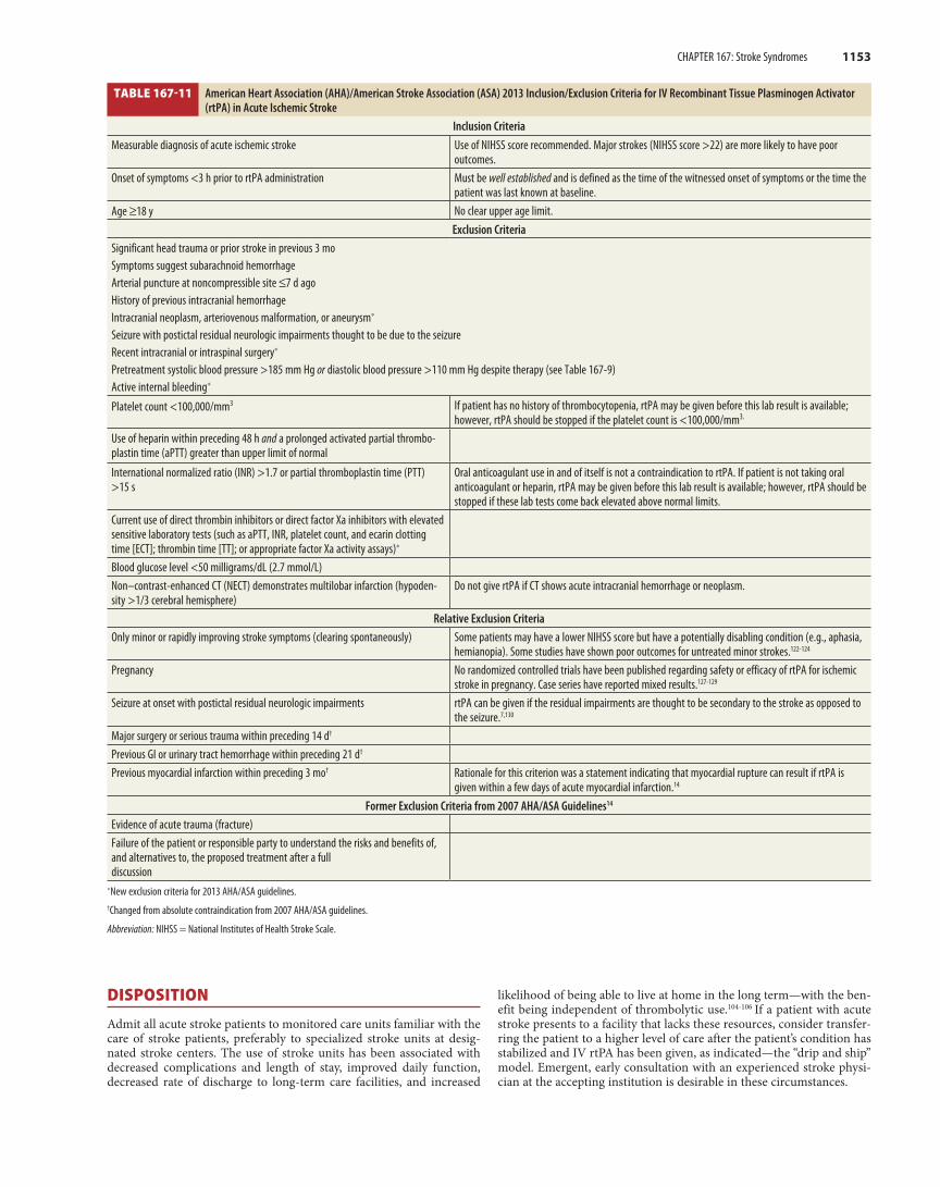
TABLE 167-11 American Heart Association (AHA)/American Stroke Association (ASA) 2013 Inclusion/Exclusion Criteria for IV Recombinant Tissue Plasminogen Activator (rtPA) in Acute Ischemic Stroke
Inclusion Criteria
Measurable diagnosis of acute ischemic stroke Use of NIHSS score recommended. Major strokes (NIHSS score >22) are more likely to have poor outcomes.
Onset of symptoms <3 h prior to rtPA administration Must be well established and is defined as the time of the witnessed onset of symptoms or the time the patient was last known at baseline.
Age ≥18 y No clear upper age limit.Exclusion Criteria
Significant head trauma or prior stroke in previous 3 moSymptoms suggest subarachnoid hemorrhageArterial puncture at noncompressible site ≤7 d agoHistory of previous intracranial hemorrhageIntracranial neoplasm, arteriovenous malformation, or aneurysm*
Seizure with postictal residual neurologic impairments thought to be due to the seizure Recent intracranial or intraspinal surgery*
Pretreatment systolic blood pressure >185 mm Hg or diastolic blood pressure >110 mm Hg despite therapy (see Table 167-9)Active internal bleeding*
Platelet count <100,000/mm3 If patient has no history of thrombocytopenia, rtPA may be given before this lab result is available; however, rtPA should be stopped if the platelet count is <100,000/mm3.
Use of heparin within preceding 48 h and a prolonged activated partial thrombo-plastin time (aPTT) greater than upper limit of normal
International normalized ratio (INR) >1.7 or partial thromboplastin time (PTT) >15 s
Oral anticoagulant use in and of itself is not a contraindication to rtPA. If patient is not taking oral anticoagulant or heparin, rtPA may be given before this lab result is available; however, rtPA should be stopped if these lab tests come back elevated above normal limits.
Current use of direct thrombin inhibitors or direct factor Xa inhibitors with elevated sensitive laboratory tests (such as aPTT, INR, platelet count, and ecarin clotting time [ECT]; thrombin time [TT]; or appropriate factor Xa activity assays)*
Blood glucose level <50 milligrams/dL (2.7 mmol/L)Non–contrast-enhanced CT (NECT) demonstrates multilobar infarction (hypoden-sity >1/3 cerebral hemisphere)
Do not give rtPA if CT shows acute intracranial hemorrhage or neoplasm.
Relative Exclusion Criteria
Only minor or rapidly improving stroke symptoms (clearing spontaneously) Some patients may have a lower NIHSS score but have a potentially disabling condition (e.g., aphasia, hemianopia). Some studies have shown poor outcomes for untreated minor strokes.122-124
Pregnancy No randomized controlled trials have been published regarding safety or efficacy of rtPA for ischemic stroke in pregnancy. Case series have reported mixed results.127-129
Seizure at onset with postictal residual neurologic impairments rtPA can be given if the residual impairments are thought to be secondary to the stroke as opposed to the seizure.7,130
Major surgery or serious trauma within preceding 14 d†
Previous GI or urinary tract hemorrhage within preceding 21 d†
Previous myocardial infarction within preceding 3 mo† Rationale for this criterion was a statement indicating that myocardial rupture can result if rtPA is given within a few days of acute myocardial infarction.14
Former Exclusion Criteria from 2007 AHA/ASA Guidelines14
Evidence of acute trauma (fracture)Failure of the patient or responsible party to understand the risks and benefits of, and alternatives to, the proposed treatment after a full discussion
*New exclusion criteria for 2013 AHA/ASA guidelines.†Changed from absolute contraindication from 2007 AHA/ASA guidelines.
Abbreviation: NIHSS = National Institutes of Health Stroke Scale.
Tintinalli_Sec14_p1125-1206.indd 1153 8/6/15 4:00 PM
1154 SECTION 14: Neurology
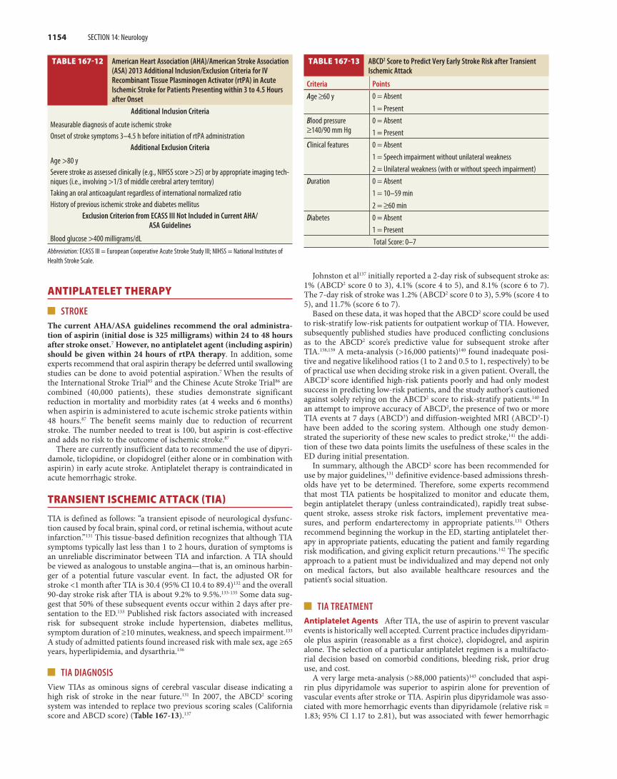
TABLE 167-12 American Heart Association (AHA)/American Stroke Association (ASA) 2013 Additional Inclusion/Exclusion Criteria for IV Recombinant Tissue Plasminogen Activator (rtPA) in Acute Ischemic Stroke for Patients Presenting within 3 to 4.5 Hours after Onset
Additional Inclusion Criteria
Measurable diagnosis of acute ischemic strokeOnset of stroke symptoms 3–4.5 h before initiation of rtPA administration
Additional Exclusion Criteria
Age >80 ySevere stroke as assessed clinically (e.g., NIHSS score >25) or by appropriate imaging tech-niques (i.e., involving >1/3 of middle cerebral artery territory)Taking an oral anticoagulant regardless of international normalized ratio History of previous ischemic stroke and diabetes mellitus
Exclusion Criterion from ECASS III Not Included in Current AHA/ ASA Guidelines
Blood glucose >400 milligrams/dLAbbreviation: ECASS III = European Cooperative Acute Stroke Study III; NIHSS = National Institutes of Health Stroke Scale.
ANTIPLATELET THERAPY
� STROKE The current AHA/ASA guidelines recommend the oral administra-tion of aspirin (initial dose is 325 milligrams) within 24 to 48 hours after stroke onset.7 However, no antiplatelet agent (including aspirin) should be given within 24 hours of rtPA therapy. In addition, some experts recommend that oral aspirin therapy be deferred until swallowing studies can be done to avoid potential aspiration.7 When the results of the International Stroke Trial85 and the Chinese Acute Stroke Trial86 are combined (40,000 patients), these studies demonstrate significant reduction in mortality and morbidity rates (at 4 weeks and 6 months) when aspirin is administered to acute ischemic stroke patients within 48 hours.87 The benefit seems mainly due to reduction of recurrent stroke. The number needed to treat is 100, but aspirin is cost-effective and adds no risk to the outcome of ischemic stroke.87
There are currently insufficient data to recommend the use of dipyri-damole, ticlopidine, or clopidogrel (either alone or in combination with aspirin) in early acute stroke. Antiplatelet therapy is contraindicated in acute hemorrhagic stroke.
TRANSIENT ISCHEMIC ATTACK (TIA)
TIA is defined as follows: “a transient episode of neurological dysfunc-tion caused by focal brain, spinal cord, or retinal ischemia, without acute infarction.”131 This tissue-based definition recognizes that although TIA symptoms typically last less than 1 to 2 hours, duration of symptoms is an unreliable discriminator between TIA and infarction. A TIA should be viewed as analogous to unstable angina—that is, an ominous harbin-ger of a potential future vascular event. In fact, the adjusted OR for stroke <1 month after TIA is 30.4 (95% CI 10.4 to 89.4)132 and the overall 90-day stroke risk after TIA is about 9.2% to 9.5%.133-135 Some data sug-gest that 50% of these subsequent events occur within 2 days after pre-sentation to the ED.133 Published risk factors associated with increased risk for subsequent stroke include hypertension, diabetes mellitus, symptom duration of ≥10 minutes, weakness, and speech impairment.133 A study of admitted patients found increased risk with male sex, age ≥65 years, hyperlipidemia, and dysarthria.136
� TIA DIAGNOSISView TIAs as ominous signs of cerebral vascular disease indicating a high risk of stroke in the near future.131 In 2007, the ABCD2 scoring system was intended to replace two previous scoring scales (California score and ABCD score) (Table 167-13).137
Johnston et al137 initially reported a 2-day risk of subsequent stroke as: 1% (ABCD2 score 0 to 3), 4.1% (score 4 to 5), and 8.1% (score 6 to 7). The 7-day risk of stroke was 1.2% (ABCD2 score 0 to 3), 5.9% (score 4 to 5), and 11.7% (score 6 to 7).
Based on these data, it was hoped that the ABCD2 score could be used to risk-stratify low-risk patients for outpatient workup of TIA. However, subsequently published studies have produced conflicting conclusions as to the ABCD2 score’s predictive value for subsequent stroke after TIA.138,139 A meta-analysis (>16,000 patients)140 found inadequate posi-tive and negative likelihood ratios (1 to 2 and 0.5 to 1, respectively) to be of practical use when deciding stroke risk in a given patient. Overall, the ABCD2 score identified high-risk patients poorly and had only modest success in predicting low-risk patients, and the study author’s cautioned against solely relying on the ABCD2 score to risk-stratify patients.140 In an attempt to improve accuracy of ABCD2, the presence of two or more TIA events at 7 days (ABCD3) and diffusion-weighted MRI (ABCD3-I) have been added to the scoring system. Although one study demon-strated the superiority of these new scales to predict stroke,141 the addi-tion of these two data points limits the usefulness of these scales in the ED during initial presentation.
In summary, although the ABCD2 score has been recommended for use by major guidelines,131 definitive evidence-based admissions thresh-olds have yet to be determined. Therefore, some experts recommend that most TIA patients be hospitalized to monitor and educate them, begin antiplatelet therapy (unless contraindicated), rapidly treat subse-quent stroke, assess stroke risk factors, implement preventative mea-sures, and perform endarterectomy in appropriate patients.131 Others recommend beginning the workup in the ED, starting antiplatelet ther-apy in appropriate patients, educating the patient and family regarding risk modification, and giving explicit return precautions.142 The specific approach to a patient must be individualized and may depend not only on medical factors, but also available healthcare resources and the patient’s social situation.
� TIA TREATMENT Antiplatelet Agents After TIA, the use of aspirin to prevent vascular events is historically well accepted. Current practice includes dipyridam-ole plus aspirin (reasonable as a first choice), clopidogrel, and aspirin alone. The selection of a particular antiplatelet regimen is a multifacto-rial decision based on comorbid conditions, bleeding risk, prior drug use, and cost.
A very large meta-analysis (>88,000 patients)143 concluded that aspi-rin plus dipyridamole was superior to aspirin alone for prevention of vascular events after stroke or TIA. Aspirin plus dipyridamole was asso-ciated with more hemorrhagic events than dipyridamole (relative risk = 1.83; 95% CI 1.17 to 2.81), but was associated with fewer hemorrhagic
TABLE 167-13 ABCD2 Score to Predict Very Early Stroke Risk after Transient Ischemic Attack
Criteria Points
Age ≥60 y 0 = Absent1 = Present
Blood pressure ≥140/90 mm Hg
0 = Absent1 = Present
C linical features 0 = Absent1 = Speech impairment without unilateral weakness2 = Unilateral weakness (with or without speech impairment)
Duration 0 = Absent1 = 10–59 min2 = ≥60 min
Diabetes 0 = Absent1 = PresentTotal Score: 0–7
Tintinalli_Sec14_p1125-1206.indd 1154 8/6/15 4:00 PM

CHAPTER 167: Stroke Syndromes 1155
events than aspirin and clopidogrel (relative risk = 0.38; 95% CI 0.25 to 0.56). However, a trial of 5170 patients found that the combination of clopidogrel and aspirin was superior to aspirin alone for reducing the risk of stroke in the first 90 days without increasing the risk of hemor-rhage.144 A trial (the Platelet-Oriented Inhibition in New TIA and Minor Ischemic Stroke [POINT] trial) is currently in progress to investigate this comparison as well.145 Anticoagulation Adjusted-dose oral anticoagulation with warfarin has been the historical therapy of choice for stroke prevention in patients with nonvalvular atrial fibrillation and TIA; however, an assessment of warfarin anticoagulation for stroke prevention in the United States dem-onstrated a dismal rate of optimal anticoagulation control.146 This has led to multiple randomized controlled trials of novel anticoagulants, which have been shown to have equivalent efficacy but with less risk of intracranial hemorrhage compared to warfarin.147 The risk of recurrent stroke in the presence of atrial fibrillation without anticoagulation is low, probably <5% over the next 48 hours; moreover, the risk of hemorrhagic transformation of an acute stroke is also greatest in the first 48 hours. Consequently, in the setting of acute atrial fibrillation, anticoagula-tion therapy typically should not be started in the ED but should be initiated in the inpatient setting.
Multiple studies have demonstrated that, although unfractionated heparin may help prevent recurrent stoke, its potential benefits are outweighed by the increased risk of intracranial hemorrhage. Multiple studies of low-molecular-weight heparin and heparinoids have found similarly disappointing results. A Cochrane systematic review of 24 randomized trials (23,748 patients) found no net benefit of antico-agulants in acute stroke.148 In addition, a meta-analysis of seven trials focused specifically on anticoagulant use in acute cardiothrombotic stroke and found no overall benefit.149 Therefore, the use of unfraction-ated heparin, low-molecular-weight heparin, or heparinoids for emergent treatment of a specific stroke subtype or TIA cannot be recommended based on available evidence, even in the presence of atrial fibrillation.150
SPECIAL POPULATIONS
� STROKE OR TIA WITH CONCURRENT ACUTE MYOCARDIAL INFARCTION
The co-occurrence of acute myocardial infarction and acute stroke has implications for acute treatment in the ED. There is little published infor-mation on co-occurrence of the two conditions, and treatment must be extrapolated. Troponin T elevation in acute stroke is likely to be related to acute myocardial infarction if renal failure and symptomatic heart failure are excluded and is associated with worse clinical outcomes.151 However, the newer highly sensitive troponin assays do not appear to add additional prognostic information if a fourth-generation troponin assay is normal.152 Although anticoagulation with heparin is not recommended for the treat-ment of acute stroke, heparin does reduce extension or reinfarction in the setting of acute myocardial infarction (relative risk = 0.40; number needed to treat = 33).153 However, the risk of mortality, the need for revasculariza-tion procedures, major or minor bleeding, and recurrent angina appear to be similar with and without heparin. Weigh the risks when requested to initiate heparin therapy by the cardiologist and when advised by the neu-rologist that heparin may induce hemorrhagic transformation. Consider the patient’s baseline cardiac and neurologic status. The balance often swings to the preservation of cardiac status.
� SICKLE CELL DISEASEStroke afflicts both adults and children with sickle cell disease, with an overall prevalence of 3.8%,154 compared to the overall prevalence of 2.8% (age ≥20 years) in the general population in the United States.1 Sickle cell disease is the most common cause of ischemic stroke in children. Patients homozygous for hemoglobin S have the highest incidence of stroke (0.61 in 100 patient-years), but all genotypes are at increased risk.154 The highest incidence of hemorrhagic stroke in these patients occurs from ages 20 to 29.154
Cerebral aneurysms and arterial abnormalities also occur with increased frequency in patients with sickle cell disease, and careful evaluation for subarachnoid hemorrhage is mandated for patients pre-senting with headache and neurologic findings. Initial management is similar to that for stroke patients without sickle cell disease, but care should also be taken to treat the underlying sickle cell disease with oxy-gen administration, hydration, and pain control, if necessary. Although evidence is limited, expert consensus recommends exchange transfusion in sickle cell patients with acute ischemic stroke, with the goal of reduc-ing hemoglobin S levels to <30%.155,156 The same therapy has been rec-ommended in hemorrhagic stroke in order to reduce vasospasm and secondary ischemic infarction.155 Sickle cell disease is not currently considered a contraindication to rtPA therapy in eligible adults. In any event, emergent consultation with a hematologist and a stroke neurolo-gist is in order, and admission to a stroke unit setting is indicated.
� YOUNG ADULTSExercise particular care when evaluating young adults (age 15 to 50 years) with acute stroke. In this group, cervical arterial dissection accounts for 20% of all ischemic strokes and may often be preceded by only minor trauma. The young adult with a cardioembolic event may have mitral valve prolapse, rheumatic heart disease, or paradoxical embolism as the originating cause. Migrainous stroke (infarction associ-ated with typical migraine attack, among those with established recur-rent migraines) is also a possibility in this age group. Some members of this population are at risk for ischemic stroke from substance abuse. Heroin, cocaine, amphetamines, and other sympathomimetic drugs are often implicated. Human immunodeficiency virus is also a risk factor for stroke in young adults.157
� PREGNANT WOMENWomen are at increased risk of ischemic and hemorrhagic stroke during pregnancy and the postpartum period (up to 6 weeks after birth). Poten-tial contributors to this increased risk include the presence of pre-eclampsia or eclampsia, hypertension, diabetes,158 sickle cell disease, and drug abuse, as well as the venous stasis, edema, and hypercoagulable state that naturally occur.9 Following birth, decreases in blood volume, alterations in hormonal status, and the potential for amniotic fluid embolism also contribute.159 The number of antenatal strokes is approx-imately 34 per 100,000 deliveries,160 with an adjusted relative risk in one series of 2.4 compared with nonpregnant females.161 Interestingly, the overall risk for all types of stroke is far greater during 6 weeks postpar-tum than during pregnancy itself, with adjusted relative risks versus nonpregnant females of 8.7 and 28.3 for ischemic and hemorrhagic stroke, respectively.161 Presentation of acute stroke is similar to that in nonpregnant patients, although pregnant patients are particularly sus-ceptible to cerebral venous thrombosis, especially during delivery or the postpartum period, and may present primarily with a headache. MRI is generally considered the safest brain imaging study,162 but the benefits of an emergent non–contrast-enhanced CT clearly outweigh the risks to the fetus in this setting if MRI is not immediately available. A head CT exposes the fetus to <1 rad. For further discussion, see chapter 99, “Comorbid Disorders in Pregnancy.” rtPA does not cross the placenta, and there are reports of successful IV rtPA use for stroke in pregnant patients.128,163-165 ED treatment of stroke in pregnant women should ide-ally involve early consultation with obstetricians, stroke neurologists, and neonatologists, as appropriate.
REFERENCES
The complete reference list is available online at www.TintinalliEM.com.
Tintinalli_Sec14_p1125-1206.indd 1155 8/6/15 4:00 PM