Embed Size (px)
Citation preview
Research reportFluoride 53(1 Pt 2):124-135
Fluoride toxicity in human fibroblastsKaszuba, Bentke, Krawczyk, Szlęzak, Wróbel
124124
Fluoride 2019 Dec 14. www.fluorideresearch.online/epub/files/065.pdf [Epub ahead of print]
January 2020
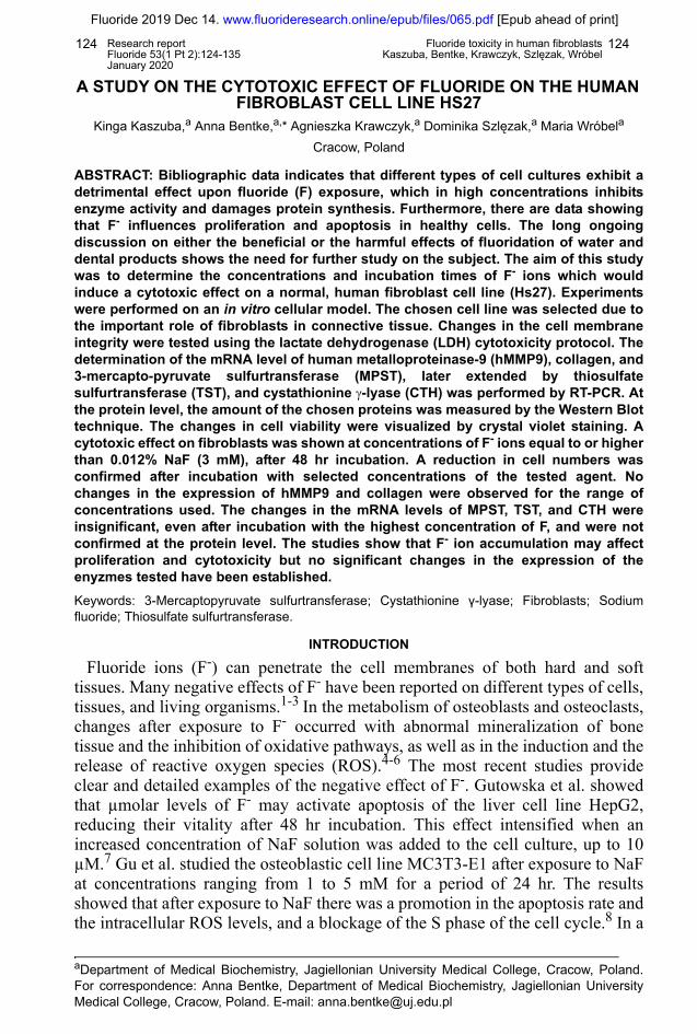
A STUDY ON THE CYTOTOXIC EFFECT OF FLUORIDE ON THE HUMAN FIBROBLAST CELL LINE HS27
Kinga Kaszuba,a Anna Bentke,a,* Agnieszka Krawczyk,a Dominika Szlęzak,a Maria Wróbela
Cracow, Poland
ABSTRACT: Bibliographic data indicates that different types of cell cultures exhibit adetrimental effect upon fluoride (F) exposure, which in high concentrations inhibitsenzyme activity and damages protein synthesis. Furthermore, there are data showingthat F- influences proliferation and apoptosis in healthy cells. The long ongoingdiscussion on either the beneficial or the harmful effects of fluoridation of water anddental products shows the need for further study on the subject. The aim of this studywas to determine the concentrations and incubation times of F- ions which wouldinduce a cytotoxic effect on a normal, human fibroblast cell line (Hs27). Experimentswere performed on an in vitro cellular model. The chosen cell line was selected due tothe important role of fibroblasts in connective tissue. Changes in the cell membraneintegrity were tested using the lactate dehydrogenase (LDH) cytotoxicity protocol. Thedetermination of the mRNA level of human metalloproteinase-9 (hMMP9), collagen, and3-mercapto-pyruvate sulfurtransferase (MPST), later extended by thiosulfatesulfurtransferase (TST), and cystathionine γ-lyase (CTH) was performed by RT-PCR. Atthe protein level, the amount of the chosen proteins was measured by the Western Blottechnique. The changes in cell viability were visualized by crystal violet staining. Acytotoxic effect on fibroblasts was shown at concentrations of F- ions equal to or higherthan 0.012% NaF (3 mM), after 48 hr incubation. A reduction in cell numbers wasconfirmed after incubation with selected concentrations of the tested agent. Nochanges in the expression of hMMP9 and collagen were observed for the range ofconcentrations used. The changes in the mRNA levels of MPST, TST, and CTH wereinsignificant, even after incubation with the highest concentration of F, and were notconfirmed at the protein level. The studies show that F- ion accumulation may affectproliferation and cytotoxicity but no significant changes in the expression of theenyzmes tested have been established. Keywords: 3-Mercaptopyruvate sulfurtransferase; Cystathionine γ-lyase; Fibroblasts; Sodiumfluoride; Thiosulfate sulfurtransferase.
INTRODUCTION
Fluoride ions (F-) can penetrate the cell membranes of both hard and softtissues. Many negative effects of F- have been reported on different types of cells,tissues, and living organisms.1-3 In the metabolism of osteoblasts and osteoclasts,changes after exposure to F- occurred with abnormal mineralization of bonetissue and the inhibition of oxidative pathways, as well as in the induction and therelease of reactive oxygen species (ROS).4-6 The most recent studies provideclear and detailed examples of the negative effect of F-. Gutowska et al. showedthat µmolar levels of F- may activate apoptosis of the liver cell line HepG2,reducing their vitality after 48 hr incubation. This effect intensified when anincreased concentration of NaF solution was added to the cell culture, up to 10µM.7 Gu et al. studied the osteoblastic cell line MC3T3-E1 after exposure to NaFat concentrations ranging from 1 to 5 mM for a period of 24 hr. The resultsshowed that after exposure to NaF there was a promotion in the apoptosis rate andthe intracellular ROS levels, and a blockage of the S phase of the cell cycle.8 In a
aDepartment of Medical Biochemistry, Jagiellonian University Medical College, Cracow, Poland.For correspondence: Anna Bentke, Department of Medical Biochemistry, Jagiellonian UniversityMedical College, Cracow, Poland. E-mail: [email protected]
Research reportFluoride 53(1 Pt 2):124-135
Fluoride toxicity in human fibroblastsKaszuba, Bentke, Krawczyk, Szlęzak, Wróbel
125125
Fluoride 2019 Dec 14. www.fluorideresearch.online/epub/files/065.pdf [Epub ahead of print]
January 2020
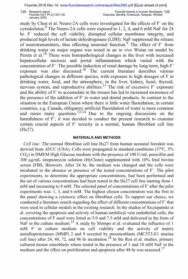
study by Chen et al. Neuro-2A cells were investigated for the effects of F- on thecytoskeleton.9 The Neuro-2A cells were exposed to 1, 2, 4, and 6 mM NaF for 24hr. F- reduced the cell viability, disrupted cellular membrane integrity, andproduced high levels of lactate dehydrogenase (LDH). NaF suppressed the releaseof neurotransmitters, thus effecting neuronal function.9 The effect of F- fromdrinking water on major organs was tested in an in vivo Wistar rat model byPereta et al.10 There were histopathological changes in the liver with increasedhepatocellular necrosis and portal inflammation which varied with theconcentration of F-. The possible induction of renal damage by long-term, high F-
exposure was also discussed.10 The current literature describes variouspathological changes in different species, with exposure to high dosages of F indrinking water, food, and the atmosphere, in the liver, kidney, heart, thyroid,nervous system, and reproductive abilities.11 The risk of excessive F- exposureand the ability of F- to accumulate in the tissues has led to increased awareness ofthe presence of the addition of F- to water and dental products. In contrast to thesituation in the European Union where there is little water fluoridation, in certaincountries, e.g. Canada, obligatory artificial fluoridation of water is more commonand raises many questions.12-14 Due to the ongoing discussions on theharmfulness of F-, it was decided to conduct the present research to examinecertain crucial aspects of F- toxicity in a normal, human fibroblast cell line(Hs27).
MATERIALS AND METHODS
Cell line: The normal fibroblast cell line Hs27 from human neonatal foreskin wasderived from ATCC (USA). Cells were propagated in standard conditions (37ºC, 5%CO2) in DMEM High Glucose medium with the presence of 100 U/mL penicillin and100 µg/mL streptomycin solution (HyClone) supplemented with 10% fetal bovineserum (FBS, Biowest). After 24 hr, the medium was changed and the cells wereincubated in the absence or presence of the tested concentrations of F-. The pilotexperiments, to determine the appropriate concentrations, had been performed andthe set of various concentrations had been tested in the Hs27 cell line starting from 1mM and increasing to 8 mM. The selected panel of concentrations of F- after the pilotexperiments was: 1, 3, and 6 mM. The highest chosen concentration was the first inthe panel showing a cytotoxic effect on fibroblast cells. To support our choice, weconducted a literature search regarding the effect of different concentrations of F- thatwere used in cellular models in the existing research. In the studies of Szczepański etal. covering the apoptosis and activity of human umbilical vein endothelial cells, theconcentrations of F used were listed as 5.0 and 7.5 mM and delivered in the form ofNaF in the culture medium.15 A study by Slompo et al. evaluated the influence of 1mM F- in culture medium on cell viability and the activity of matrixmetalloproteinases (MMP) 2 and 9 secreted by preosteoblasts (MC3T3-E1 murinecell line) after 24, 48, 72, and 96 hr incubation.16 In the Ren et al. studies, primarycultured mouse osteoblasts where tested in the presence of 1 and 10 mM NaF in themedium and the effect on proliferation and apoptosis after 48 hr was assessed.17

Research reportFluoride 53(1 Pt 2):124-135
Fluoride toxicity in human fibroblastsKaszuba, Bentke, Krawczyk, Szlęzak, Wróbel
126126
Fluoride 2019 Dec 14. www.fluorideresearch.online/epub/files/065.pdf [Epub ahead of print]
January 2020
Evaluation of cytotoxicity and quantitation of viable cells by crystal violet staining: The cells were seeded and grown on 96-well plates until they were 90% confluent.
The medium was changed, the respective concentrations of F- were added.Incubation was continued for 24 and 48 hr. The medium was collected and analyzedfor LDH activity using the Pierce LDH Cytotoxicity Assay Kit (Thermo Scientific),according to the producer’s manual. The remaining cells were treated with 0.2%crystal violet solution by modified Gillies et al. method.18
Expression of beta-actin, hMMP-9, collagen, MPST, CTH, and TST:a) ISOLATION OF RNA: Total RNA was extracted with Tri-Reagent (Lab Empire)
following the method described by Chomczyński and Sacchi.19 b) REVERSE TRANSCRIPTION: 3 µg of isolated RNA was reverse transcribed with
Reverse Transcriptase in 5 X Reaction buffer (GoScriptTM Promega) with MgCl2,RNAse Inhibitor, and dNTP mix (Thermo Scientific) in 10 µL of final volume ofreaction mixture following the Promega manufacturer’s protocol.
c) POLYMERASE CHAIN REACTION (PCR): PCR was performed using a mixture of:cDNA, adequate reverse (R) and forward (F) primers, DNA polymerase in Tris-HClpH 8.8 with MgCl2, KCl, Triton X-100, dNTP mix (Thermo Scientific), and H2O-DEPC in a total reaction volume of 12.5 µL. The PCR products were analyzed in2.0% agarose gels during electrophoresis and imaged with UVI-KS 4000i/ImagePC(Syngen Biotech).
d) DENSITOMETRIC EVALUATION OF PCR: Photos saved in the jpeg format (Bio-RadChemiDoc MP Imaging System) were used for the densitometry analysis. Thedensitometry data for band intensities in the different experiments were generated byanalyzing the gel images on the Gene Tools Software (Syngene).
Table 1. PCR conditions
Gene Initiation Denaturation Amplification Elongation Termination
MPST 5 min at 95°C
30 sec at 95°C
30 sec at 55°C
2 min at 72°C for 29 cycles
72°C for 8 min
CTH 5 min at 95°C
30 sec at 95°C
1 min at 51°C
8 min at 72°C for 30 cycles
72°C for 8 min
TST 5 min at 95°C
30 sec at 95°C
30 sec at 54.5°C
2 min at 72°C for 34 cycles
72°C for 8 min
β-actin
5 min at 95°C
30 sec at 95°C
30 sec at 55°C
2 min at 72°C for 30 cycles
72°C for 8 min

Research reportFluoride 53(1 Pt 2):124-135
Fluoride toxicity in human fibroblastsKaszuba, Bentke, Krawczyk, Szlęzak, Wróbel
127127
Fluoride 2019 Dec 14. www.fluorideresearch.online/epub/files/065.pdf [Epub ahead of print]
January 2020
Western blotting: The protein load for electrophoresis was 20 µg per well. SDS-PAGE electrophoresis and electrotransfer on PVDF membrane were conductedaccording to the Bio-Rad protocol. The relative amounts of CTH, MPST, and TSTwere determined using the appropriate antibody (Abnova, GeneTex, ProteinTech;1:1000). Anti-β-actin and anti-α-tubulin antibodies were used to check for loading(Sigma, ProteinTech; 1:5000). Proteins of interest were incubated with alkalinephosphatase-conjugated goat anti-rabbit or anti-mouse IgG antibody (ProteinTech,1:2000). Proteins were visualized by immunohistochemical detection with NBT/BCIP (Roche) staining solution.
Statistical analysis: Standard deviation was used for the error bars and for thestatistical analysis the Mann-Whitney U test was used.
RESULTS
The cytotoxic effect of a wide range of F- concentrations in the culture medium wasmeasured by the LDH cytotoxicity test. As shown in Table 2, the H27 fibroblast cellmembranes were treated with three concentrations of F-: 0.004% NaF (1 mM),0.012% NaF (3 mM), and 0.024% NaF (6 mM), and a control group was leftuntreated (non-treated cells, 0% NaF, 0 mM).
A cytotoxic effect was demonstrated for concentrations equal to or higher than0.012% after the 48 hr incubation time. Next, for the same F- concentrations, theproliferation protocol was introduced. The number of cells was determined withcrystal violet staining. A reduced number of cells was registered for the two testedconcentrations equal to and above 0.012% NaF. The amount of living fibroblast cells
Table 2. Changes in the integrity of the Hs27 fibroblast cell membranes after 24 and 48 hr incubation with F- measured by the LDH cytotoxicity kit
Fluoride concentration in cell culture medium % of cytotoxicity Incubation time
0.004% (1 mM)
0.012% (3 mM)
0.024% (6 mM)
0
0
2
24 hr
0.004% (1 mM)
0.012% (3 mM)
0.024% (6 mM)
0
10.5
16.4
48 hr
Cytotoxic effects of F- on fibroblast cells are shown for concentrations ³. 0.012% NaF (3mM) after 48 hr incubation. The tested concentrations of F as listed: 0.004%, 0.012%, and 0.024%. The amount of LDH present in the medium is expressed as a percentage of the total (0% in untreated cells; 100% in cells lysed with provided lysis buffer solution). The data refers to the values obtained for the control (non-treated) cells. The numbers listed in the table represent the results from at least 3 experiments done in 6 repetitions.

Research reportFluoride 53(1 Pt 2):124-135
Fluoride toxicity in human fibroblastsKaszuba, Bentke, Krawczyk, Szlęzak, Wróbel
128128
Fluoride 2019 Dec 14. www.fluorideresearch.online/epub/files/065.pdf [Epub ahead of print]
January 2020
decreased down to 70% in comparison to the control (listed as 100%), for bothincubation times (Figure 1).
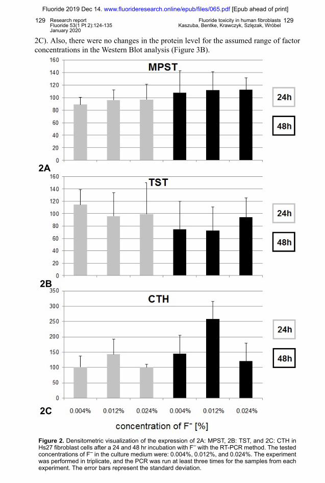
The following experiments included testing the expression of the chosen genesunder the same conditions. The tests were conducted using the RT-PCR and theWestern Blot. No changes in the mRNA levels for hMMP-9 and collagen wereobserved and therefore no confirmation was done at the protein level. Some changesin the expression in mRNA were observed in the MPST gene bars as shown with theRT-PCR method. The expression of this gene seemed to increase after incubationwith increased concentrations of F- with the most intense band appearing for 0.024%NaF after 24 hr (in comparison to the control). A range of experiments wereperformed to create the densitometric analysis for the MPST mRNA levels. Nostatistically significant changes were observed (Figure 2A). The results wereconfirmed at the protein level in Western Blot analysis for the human MPSTantibody, where no significant changes in the level of protein were observed after theincubation with F- (Figure 3C).
The MPST changed the expression in the RT-PCR and the anticipated role in theanti-oxidative response urged us to check the levels of other enzymes participating inthe metabolism of sulfur-containing compounds: CTH and TST. No statisticallysignificant changes in the mRNA levels for the TST gene in the RT-PCR methodwere observed, as shown in the graph representing densitometric analysis of the barsfrom multiple PCR tests (Figure 2B). Also, there were no changes in the proteinlevels for the assumed range of factor concentrations in the Western Blot analysis(Figure 3A). Furthermore, no statistically significant changes in the mRNA levels forthe CTH gene in the RT-PCR method were observed as shown on the graphrepresenting the densitometric analysis of the bars from multiple PCR tests (Figure
Figure 1. Cell viability in Hs27 cell line after the 24 and 48 hr incubation with F- measured bycrystal violet staining. The concentrations of F- tested in the medium were: 0.004%, 0.012%, and0.024%. The data refers to values obtained for the control (non-treated) cells normalized andlabeled as 100%. The bars represent the results from at least 3 experiments done in 6 repetitions.The error bars represent the standard deviation.
Research reportFluoride 53(1 Pt 2):124-135
Fluoride toxicity in human fibroblastsKaszuba, Bentke, Krawczyk, Szlęzak, Wróbel
129129
Fluoride 2019 Dec 14. www.fluorideresearch.online/epub/files/065.pdf [Epub ahead of print]
January 2020
2C). Also, there were no changes in the protein level for the assumed range of factorconcentrations in the Western Blot analysis (Figure 3B).
Figure 2. Densitometric visualization of the expression of 2A: MPST, 2B: TST, and 2C: CTH inHs27 fibroblast cells after a 24 and 48 hr incubation with F– with the RT-PCR method. The testedconcentrations of F– in the culture medium were: 0.004%, 0.012%, and 0.024%. The experimentwas performed in triplicate, and the PCR was run at least three times for the samples from eachexperiment. The error bars represent the standard deviation.
2A
2B
2C

Research reportFluoride 53(1 Pt 2):124-135
Fluoride toxicity in human fibroblastsKaszuba, Bentke, Krawczyk, Szlęzak, Wróbel
130130
Fluoride 2019 Dec 14. www.fluorideresearch.online/epub/files/065.pdf [Epub ahead of print]
January 2020
3A
3B
Figures 3A and 3B. Expression of 3A: TST and 3B: CTH in Hs27 fibroblast cells after a 24and 48 hr incubation with F– measured by the Western Blot. The tested concentrations of F–
in the culture medium were: 0.004%, 0.012%, and 0.024%. The experiment was performed atleast in duplicate and the Western Blot was run two times for the samples from eachexperiment with similar results. A representative result is shown.

Research reportFluoride 53(1 Pt 2):124-135
Fluoride toxicity in human fibroblastsKaszuba, Bentke, Krawczyk, Szlęzak, Wróbel
131131
Fluoride 2019 Dec 14. www.fluorideresearch.online/epub/files/065.pdf [Epub ahead of print]
January 2020
Figures 3C and 3D. Expression of 3C: MPST and 3D: β-actin in Hs27 fibroblast cells after a24 and 48 hr incubation with F– measured by the Western Blot. The tested concentrations ofF– in the culture medium were: 0.004%, 0.012%, and 0.024%. The experiment was performedat least in duplicate and the Western Blot was run two times for the samples from eachexperiment with similar results. A representative result is shown.
3D
3C

Research reportFluoride 53(1 Pt 2):124-135
Fluoride toxicity in human fibroblastsKaszuba, Bentke, Krawczyk, Szlęzak, Wróbel
132132
Fluoride 2019 Dec 14. www.fluorideresearch.online/epub/files/065.pdf [Epub ahead of print]
January 2020
DISCUSSION
Fibroblasts play a crucial role in the process that allows tissue reparation tooccur after tissue damage. Connective tissue cells are able to migrate to thedamaged part of the skin and proliferate to repair the injury.20,21 With there beinga considerable literature on the toxic and harmful effects of toxic substances ondifferent types of tissue and living organisms, the decision was made to examinefluoride toxicity using the normal fibroblast cell line Hs27. The effects of fluoride oncytotoxicity and the ability of cells to proliferate were examined by incubating cellcultures with fluoride in concentrations ranging from 1 mM to 6 mM NaF. The F-
concentrations were selected on the basis of a literature search on in vitro tests wherecytotoxic effects were induced with incubation times of between 24 and 48 hr.22-24
The proven ability of fluoride to inhibit certain groups of enzymes like phosphataseswas also taken into consideration.25,26 In the present study, we found a fluoride-induced cytotoxic effect on fibroblasts was shown at concentrations of F- equal to orhigher than 0.012% NaF (3 mM) but only after the longer 48 hr incubation time. Areduction of the percentage of viable cells was demonstrated after incubation with0.012% NaF for both incubation times of 24 and 48 hr. The results confirmed thecytotoxic effect of fluoride and the ability of fluoride to affect cell viability withexposure to F in millimolar concentrations. Similar results have been described byJeng et al. for another fibroblast cell line from human oral mucosa.27 To examine thecondition of fibroblasts and how NaF may affect this important connective tissueelement, two collagen genes and hMMP9 have been studied. Yan et al. foundevidence of a damaging effect of long-term F exposure on collagen 1A1 and 1A2expression in rat hard tissue.28 Brackett et al. reported reduced activity ofrccombinant human MMP9 protein (rhMMP9) in dentin matrices and, similarly, Katoet al. found that NaF exposure resulted in MMP9 inhibition in human saliva.29,30
Accordingly, in our study, we measured the mRNA levels for both of these genes.However, in our study, there was no visible change in the collagen and hMMP9expression after 24 and 48 hr incubation as measured by the RT-PCR method, evenfor the highest cytotoxic concentration of NaF. These results suggest that, for thistype of cell, the millimolar concentration range, up to 6 mM, has no effect on themRNA level of expression of collagen and hMMP9 and higher concentrations orlonger incubation times should be considered for future tests. These results confirmthe many reports in the literature that there is a non-specific response to the samelevels of F- in different types of cells.31
It is widely described in the literature that certain concentrations of F- in variouscell types are able to cause oxidative stress.32-34 Toxic amounts of F- take part in theprocesses of generating free radicals, such as OH· and ·O2
-, and have the ability tolower the enzymatic activity of antioxidant proteins that play a crucial role in copingwith free radicals in the main oxidative systems such as SOD.35,36 Recent literaturedata show that the group of sulfur-containing enzymes such as MPST, TST, and CTHmay play a role in the responses to oxidative stress. Recent studies have confirmedthe anti-oxidative effect of H2S metabolites produced by these enzymes. In addition,sulfurtransferases may also show a local antioxidant activity due to the presence offree SH groups.37-39 The long-term response to the cytotoxic effect of F may belinked to the level of sulfur enzyme expression. Thus, the enzymes involved in themetabolism of sulfur-containing compounds were chosen for testing to examine the
Research reportFluoride 53(1 Pt 2):124-135
Fluoride toxicity in human fibroblastsKaszuba, Bentke, Krawczyk, Szlęzak, Wróbel
133133
Fluoride 2019 Dec 14. www.fluorideresearch.online/epub/files/065.pdf [Epub ahead of print]
January 2020
relationship between the cytotoxicity of different concentrations of F- and the anti-oxidative response. Although there were some promising changes in the firstexperiments for the MPST enzyme after 48 hr incubation with the highest cytotoxicconcentration of F-, the further extended tests did not confirm the changes in theexpression of this enzyme. The other enzymes studied also did not show anystatistically significant changes in their expression after exposure to the tested rangeof NaF concentrations, as was confirmed by the Mann-Whitney U test. Some of thereports show a lack of any connection between the presence of F- and NaF-inducedoxidative stress. Moreover, in the research conducted by Lee et al., ROS productionwas shown in NaF-treated human gingival fibroblasts cells.40 It seems that, in thecase of the human fibroblast Hs27 cell line, the cytotoxic effect is not connected withthe changes in the sulfur-containing enzymes tested, elevated levels of which couldpossibly show an antioxidant response to the cytotoxic effect of F-.
In the present study, the cytotoxic effect of fluoride and the influence of fluoride onthe proliferation rate in the fibroblast cell line Hs27 were confirmed. Exposure to F-
did not result in any changes in the expression of collagen, hMMP9, MPST, CTH,and TST. There is no study in the literature on the effect of fluoride on the expressionof the aforementioned sulfur-containing enzymes, so the findings of the present studyare an addition to the knowledge on the effect of fluoride on the enzymatic activityprofile of this normal Hs27 cell line.
ACKNOWLEDGEMENTS
This work was financially supported by the Polish Ministry of Science and HigherEducation, and grant K/DSC/003573 of the Jagiellonian University Medical College.
CONFLICT OF INTEREST
The authors declare no conflict of interests in any relationships or with the financialsupport received. The authors alone are responsible for the content and writing of thispaper.
REFERENCES1 Wang ZH, Li XL, Yang ZQ, Xu M. Fluorine-induced apoptosis and lipid peroxidation in human
hair follicles in vitro. Biol Trace Elem Res 2010;137(3):280-8.2 He L-F, Chen J-G. DNA damage, apoptosis and cell cycle changes induced by fluoride in rat
oral mucosal cells and hepatocytes. World J Gastroenterol 2006;12(7):1144-8.3 Chouhan S, Flora SJS. Effects of fluoride on the tissue oxidative stress and apoptosis in rats:
Biochemical assays supported by IR spectroscopy data. Toxicology 2008;254(1-2):61-7.4 Bergandi L, Aina V, Malavasi G, Morterra C, Ghigo D. The toxic effect of fluoride on MG-63
osteoblast cells is also dependent on the production of nitric oxide. Chem Biol Interact2011;190(2-3):179-86.
5 Wang J, Guo Y, Liang Z, Hao J. Amino acid composition and histopathology of goat teeth in anindustrial fluoride polluted area. Fluoride 2003;36(3):177-84.
6 Zhang Y, Sun X, Sun G, Liu S, Wang L. DNA damage induced by fluoride in rat kidney cells.Fluoride 2006;39(3):191-4.
7 Gutowska I, Baranowska-Bosiacka I, Siwiec E, Szczuko M, Kolasa A, Kondarewicz A, et al.Lead enhances fluoride influence on apoptotic processes in the HepG2 liver cell line. ToxicolInd Health 2016;32(3):517-25.
Research reportFluoride 53(1 Pt 2):124-135
Fluoride toxicity in human fibroblastsKaszuba, Bentke, Krawczyk, Szlęzak, Wróbel
134134
Fluoride 2019 Dec 14. www.fluorideresearch.online/epub/files/065.pdf [Epub ahead of print]
January 2020
8 Gu X, Wang Z, Gao J, Han D, Zhang L, Chen P, et al. SIRT1 suppresses p53-dependentapoptosis by modulation of p21 in osteoblast-like MC3T3-E1 cells exposed to fluoride. ToxicolIn Vitro 2019;57:28-38.
9 Chen L, Ning H, Yin Z, Song X, Feng Y, Qin H, et al. The effects of fluoride on neuronalfunction occurs via cytoskeleton damage and decreased signal transmission. Chemosphere2017;185:589-94.
10 Perera T, Ranasinghe S, Alles N, Waduge R. Effect of fluoride on major organs with thedifferent time of exposure in rats. Environ Health Prev Med 2018:23(1):17. doi: 10.1186/s12199-018-0707-2.
11 Zuo H, Chen L, Kong M, Qiu L, Lu P, Wu P, et al. Toxic effects of fluoride on organisms. Life Sci2018;198:18-24.
12 Błaszczyk I, Ratajczak-Kubiak EB. Advantageous and harmful effects of fluorine. Farm Pol2009;18(9):623-6.
13 Public Health Capacity and Knowledge Management Unit, Quebec Region for the Office of theChief Dental Officer of Canada, Public Health Agency of Canada. The state of communitywater fluoridation across Canada. 2017 report. Ottawa and Winnipeg, Canada: Agence de lasanté publique du Canada, Public Health Agency of Canada; 2017. Available from: https://www.canada.ca/content/dam/hc-sc/documents/services/publications/healthy-living/community-water-fluoridation-across-canada-2017/community-water-fluoridation-across-canada-2017-eng.pdf
14 Wong E.The real cost of removing water fluoridation: a health equity impact assessment-report. Portland: Wellesley Institute; 2013.
15 Szczepański M, Kamianowska M, Kamianowski G. Effects of fluorides on apoptosis andactivation of human umbilical vein endothelial cells. Oral Dis 2012;18(3):280-4.
16 Slompo C, Buzalaf CP, Damante CA, Martins GM, Hannas AR, Buzalaf MA, et al. Fluoridemodulates preosteoblasts viability and matrix metalloproteinases-2 and -9 activities. Braz DentJ 2012;23(6):629-34.
17 Ren G, Ferreri M, Wang Z, Su Y, Han B, Su J. Sodium fluoride affects proliferation andapoptosis through insulin-like growth factor i receptor in primary cultured mouse osteoblasts.Biol Trace Elem Res 2011;144(1-3):914-23.
18 Gillies RJ, Didier N, Denton M. Determination of cell number in monolayer cultures. AnalBiochem 1986;159(1):109-13.
19 Chomczynski P, Sacchi N. Single-step method of RNA isolation by acid guanidiniumthiocyanate-phenol-chloroform extraction. Anal Biochem 1987;162(1):156-9.
20 Spyrou GE, Watt DAL, Naylor IL. The origin and mode of fibroblast migration and proliferationin granulation tissue. Br J Plast Surg 1998;51(6):455-61.
21 Zhang Q, Fong CC, Yu WK, Chen Y, Wei F, Koon CM, et al. Herbal formula Astragali Radix andRehmanniae Radix exerted wound healing effect on human skin fibroblast cell line Hs27 viathe activation of transformation growth factor (TGF-β) pathway and promoting extracellularmatrix (ECM) deposition. Phytomedicine 2012;20(1):9-16.
22 Chen L, Chen H, Yao C, Chang C, Xia H, Zhang C, et al. The toxicity of NaF on BmN cells anda comparative proteomics approach to identify protein expression changes in cells under NaF-stress. J Hazard Mater 2015;286:624-31.
23 Mendoza-Schulz A, Solano-Agama C, Arreola-Mendoza L, Reyez-Marquez B, Barbier O, DelRazo LM, et al. The effects of fluoride on cell migration, cell proliferation, and cell metabolismin GH4C1pituitary tumour cells. Toxicol Lett 2009;190(2):179-86.
Research reportFluoride 53(1 Pt 2):124-135
Fluoride toxicity in human fibroblastsKaszuba, Bentke, Krawczyk, Szlęzak, Wróbel
135135
Fluoride 2019 Dec 14. www.fluorideresearch.online/epub/files/065.pdf [Epub ahead of print]
January 2020
24 Tabuchi Y, Yunoki T, Hoshi N, Suzuki N, Kondo T. Genes and gene networks involved insodium fluoride-elicited cell death accompanying endoplasmic reticulum stress in oral epithelialcells. Int J Mol Sci 2014;15(5):8959-78.
25 Barbier O, Arreola-Mendoza L, Del Razo LM. Molecular mechanisms of fluoride toxicity. ChemBiol Interact 2010;188:319-33.
26 Adamek E, Pawłowska-Góral K BK. In vitro and in vivo effects of fluoride ions on enzymeactivity. Ann Acad Med Stetin 2005;12(51):69-85.
27 Jeng JH, Hsieh CC, Lan WH, Chang MC, Lin SK, Hahn LJ, et al. Cytotoxicity of sodium fluorideon human oral mucosal fibroblasts and its mechanisms. Cell Biol Toxicol 1998;14:383-9.
28 Yan X, Hao X, Nie Q, Feng C, Wang H, Sun Z, et al. Effects of fluoride on the ultrastructure andexpression of type I collagen in rat hard tissue. Chemosphere 2015;128:36-41.
29 Brackett MG, Agee KA, Brackett WW, Key WO, Sabatini C, Kato MT, et al. Effect of sodiumfluoride on the endogenous MMP activity of dentin matrices. J Nat Sci 2015;1(6):e118.
30 Kato MT, Bolanho A, Zarella BL, Salo T, Tjäderhane L, Buzalaf MAR. Sodium fluoride inhibitsMMP-2 and MMP-9. J Dent Res 2014;93(1):74-7.
31 Agalakova NI, Gusev GP. Molecular mechanisms of cytotoxicity and apoptosis induced byinorganic fluoride [review]. International Scholarly Research Network. ISRN Cell Biology2012;2012:Article ID 403835. doi:10.5402/2012/403835.
32 Chlubek D. Fluoride and oxidative stress [editorial review]. Fluoride 2003;36(4):217-28.33 Lu Y, Luo Q, Cui H, Deng H, Kuang P, Liu H, et al. Sodium fluoride causes oxidative stress and
apoptosis in the mouse liver. Aging (Albany NY) 2017;9(6):1623-39.34 Khatun S, Mandi M, Rajak P, Roy S. Interplay of ROS and behavioral pattern in fluoride
exposed Drosophila melanogaster. Chemosphere 2018;209:220-31.35 Gutiérrez-Salinas J, García-Ortíz L, Morales González JA, Hernandez-Rodriguez S, Ramirez-
Garcia S, Nunez-Ramos RR, et al. In vitro effect of sodium fluoride on malondialdehydeconcentration and on superoxide dismutase, catalase, and glutathione peroxidase in humanerythrocytes. ScientificWorldJournal 2013;2013:864718.
36 He X, Sun Z, Manthari RK, Wu P, Wang J. Fluoride altered rat’s blood testis barrier by affectingthe F-actin via IL-1α. Chemosphere 2018;211:826-33.
37 Nagahara N. Multiple role of 3-mercaptopyruvate sulfurtransferase: antioxidative function, H2Sand polysulfide production and possible SOx production. Br J Pharmacol 2018;175(4):577-89.
38 Wang R. Physiological implications of hydrogen sulfide: a whiff exploration that blossomed.Physiol Rev 2012;92(2):791-896.
39 Bronowicka-Adamska P, Bentke A, Wróbel M. Hydrogen sulfide generation from l-cysteine inthe human glioblastoma-astrocytoma U-87 MG and neuroblastoma SHSY5Y cell lines. ActaBiochim Pol 2017;64(1):171-6.
40 Lee HJ, Choi CH. Anti-inflammatory effects of bamboo salt and sodium fluoride in humangingival fibroblasts: An in vitro study. Kaohsiung J Med Sci 2015;31(6):303-8.
Copyright © 2019, ISFR, www.fluorideresearch.online, Dunedin, New Zealand


![Fluoride toothpastes for preventing dental caries in ...neuron.mefst.hr/docs/katedre/znanstvena_metodologija/Fluoride... · [Intervention Review] Fluoride toothpastes for preventing](https://img.pdfslide.net/doc/110x75/5ac7a33f7f8b9aa3298b67ff/fluoride-toothpastes-for-preventing-dental-caries-in-intervention-review-fluoride.jpg)