Embed Size (px)
Citation preview
125
Motor Neuron Disease
Lisa S. Krivickas, MD
DEFINITION
The term motor neuron disease refers to a progressive neuromuscular disorder in which upper
or lower motor neurons degenerate. The most common form of motor neuron disease is
amyotrophic lateral sclerosis (ALS), which is the primary focus of this chapter. Management
principles for other forms of motor neuron disease are similar. To meet diagnostic criteria for
ALS, an individual must have both upper and lower motor neuron dysfunction. If only lower
motor neuron dysfunction is present, the disease is called progressive muscular atrophy; if
only upper motor neuron dysfunction is present, it is called primary lateral sclerosis. If only
bulbar dysfunction is present, the disease is called progressive bulbar palsy. Most patients
initially diagnosed as having progressive muscular atrophy, primary lateral sclerosis, or
progressive bulbar palsy eventually have full-blown ALS. Those who do not convert to ALS
have a slower rate of disease progression.
Most cases of ALS are idiopathic. However, 5% to 10% of patients have a familial form,
usually transmitted in autosomal dominant fashion. In approximately 20% of these familial
cases, mutations in SOD1 (superoxide dismutase) can be identified. Other rare forms of
inherited adult motor neuron disease are Kennedy disease (X-linked recessive) and adult
spinal muscular atrophy (autosomal recessive), which both have only lower motor neuron
dysfunction.
ALS rapidly produces skeletal muscle weakness, eventually leading to the requirement for
ventilator support or death from respiratory failure. The onset of weakness may be in any
limb, the bulbar muscles, or in the respiratory muscles. The extraocular muscles and bowel
and bladder function are generally spared. Mean survival, without tracheostomy, is 3 years
from symptom onset, but the range may be less than 1 year to more than 20 years. One
explanation for the extreme variability in rate of disease progression is that ALS is probably a
heterogenous group of diseases rather than a single disease.1 The mean age at onset is in the
mid-50s, but ALS may develop in adults of any age. The cause of the disease is unknown, but
leading theories concerning pathogenesis implicate glutamate excitotoxicity, oxidative stress,
neuroinflammation, microglial cell activation, apoptosis, and mitochondrial dysfunction.
1
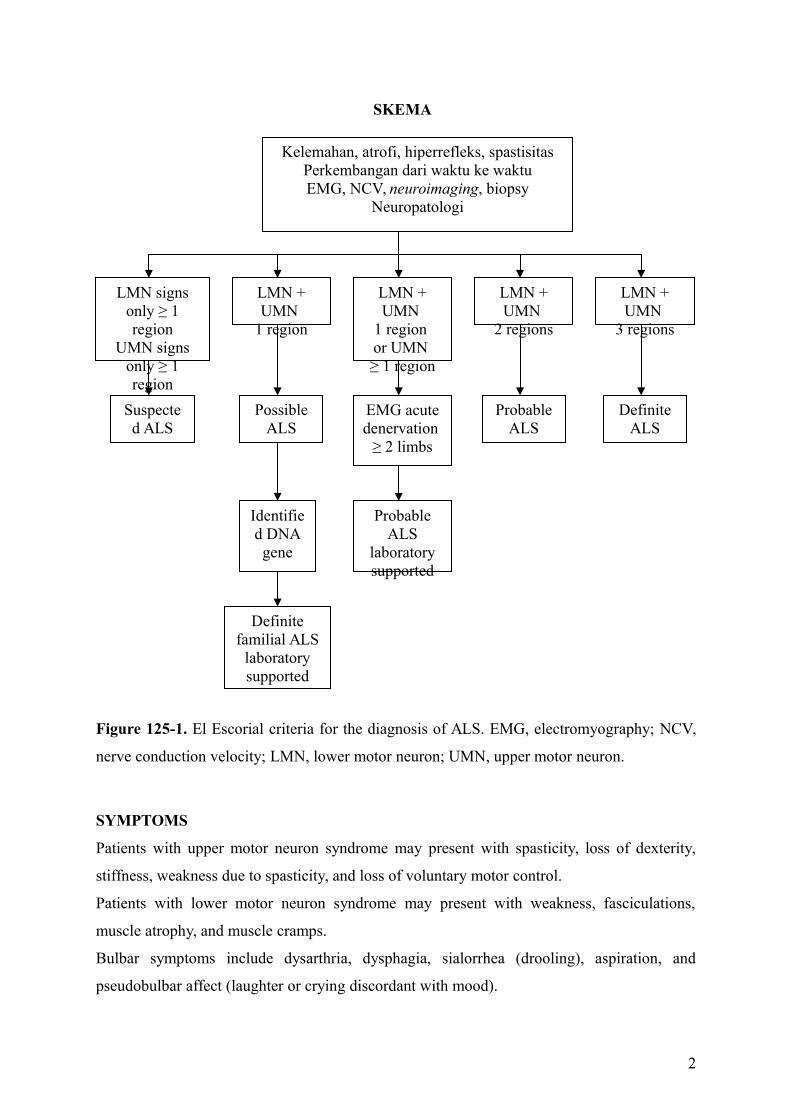
Figure 125-1. El Escorial criteria for the diagnosis of ALS. EMG, electromyography; NCV,
nerve conduction velocity; LMN, lower motor neuron; UMN, upper motor neuron.
SYMPTOMS
Patients with upper motor neuron syndrome may present with spasticity, loss of dexterity,
stiffness, weakness due to spasticity, and loss of voluntary motor control.
Patients with lower motor neuron syndrome may present with weakness, fasciculations,
muscle atrophy, and muscle cramps.
Bulbar symptoms include dysarthria, dysphagia, sialorrhea (drooling), aspiration, and
pseudobulbar affect (laughter or crying discordant with mood).
2
LMN signs only ≥ 1 region
UMN signs only ≥ 1 region
LMN + UMN
1 region
Suspected ALS
Possible ALS
Identified DNA gene
Definite familial ALS
laboratory supported
LMN + UMN
1 region or UMN ≥ 1 region
Probable ALS
laboratory supported
EMG acute denervation
≥ 2 limbs
LMN + UMN
2 regions
Probable ALS
Definite ALS
LMN + UMN
3 regions
SKEMA
Kelemahan, atrofi, hiperrefleks, spastisitasPerkembangan dari waktu ke waktuEMG, NCV, neuroimaging, biopsy
Neuropatologi
Respiratory failure and constitutional symptoms of weight loss and generalized fatigue may
be present. Cognitive symptoms include behavioral or executive dysfunction and
frontotemporal dementia in a minority of patients.
PHYSICAL EXAMINATION
The emphasis of the physical examination of a patient with suspected or diagnosed motor
neuron disease is on the neurologic, musculoskeletal, and cardiorespiratory systems. On
neurologic examination, one is looking for evidence of upper and lower motor neuron
dysfunction. The mental status, non-motor cranial nerve function, sensory examination, and
cerebellar examination findings are usually normal. The “gold standard” for the diagnosis of
upper motor neuron disease is the presence of pathologic reflexes-the Babinski sign,
Hoffmann sign, and brisk jaw jerk. Patients with ALS may be hyperreflexic or hyporeflexic,
depending on the stage at which they are in the disease process and whether they have a
predominance of upper or lower motor neuron disease. Evidence of lower motor neuron
disease includes muscle weakness, atrophy, hypotonia, hyporeflexia, and fasciculations.
Atrophy often appears first in hand intrinsic muscle. Although fasciculations are not a
necessary criterion for the diagnosis of ALS, one should question the diagnosis when none
are observed. The tongue is examined for fasciculations and atrophy, and tongue strength and
range of motion are assessed. The musculoskeletal examination focuses on assessment of
range of motion and evaluation of painful joints or soft tissue structures. Because progressive
respiratory failure develops, the cardiorespiratory system should be assessed at each visit.
Forced vital capacity (FVC) can be measured with a hand-held spirometer in the office
setting.
FUNCTIONAL LIMITATIONS
The majority of the functional limitations that develop in patients with ALS are the direct or
indirect of muscle weakness. As the disease progresses, patients have impaired mobility and
difficulties with performance of even the most basic activities of daily living, such as feeding
themselves. Bulbar muscle weakness produces dysarthria (difficulty speaking) and dysphagia
(difficulty swallowing); eventually, some patients become anarthric and unable to swallow
even their own saliva. Reactive depression, generalized fatigue, and musculoskeletal pain
may further limit function.
3
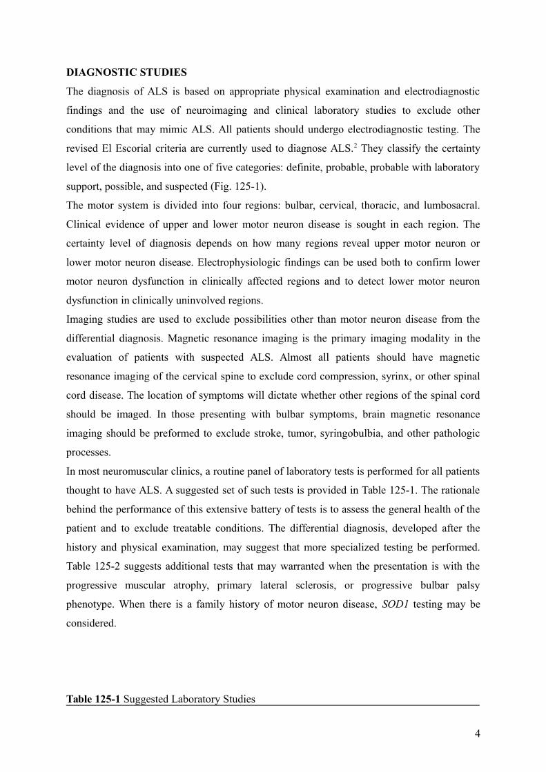
DIAGNOSTIC STUDIES
The diagnosis of ALS is based on appropriate physical examination and electrodiagnostic
findings and the use of neuroimaging and clinical laboratory studies to exclude other
conditions that may mimic ALS. All patients should undergo electrodiagnostic testing. The
revised El Escorial criteria are currently used to diagnose ALS.2 They classify the certainty
level of the diagnosis into one of five categories: definite, probable, probable with laboratory
support, possible, and suspected (Fig. 125-1).
The motor system is divided into four regions: bulbar, cervical, thoracic, and lumbosacral.
Clinical evidence of upper and lower motor neuron disease is sought in each region. The
certainty level of diagnosis depends on how many regions reveal upper motor neuron or
lower motor neuron disease. Electrophysiologic findings can be used both to confirm lower
motor neuron dysfunction in clinically affected regions and to detect lower motor neuron
dysfunction in clinically uninvolved regions.
Imaging studies are used to exclude possibilities other than motor neuron disease from the
differential diagnosis. Magnetic resonance imaging is the primary imaging modality in the
evaluation of patients with suspected ALS. Almost all patients should have magnetic
resonance imaging of the cervical spine to exclude cord compression, syrinx, or other spinal
cord disease. The location of symptoms will dictate whether other regions of the spinal cord
should be imaged. In those presenting with bulbar symptoms, brain magnetic resonance
imaging should be preformed to exclude stroke, tumor, syringobulbia, and other pathologic
processes.
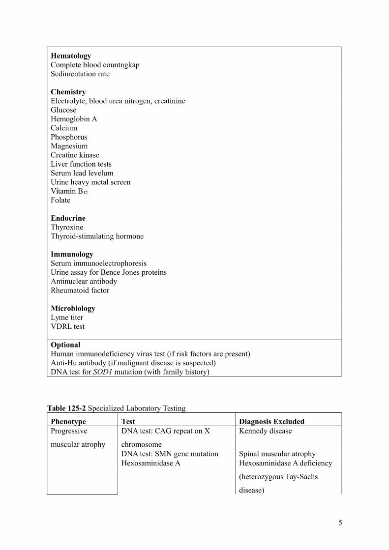
In most neuromuscular clinics, a routine panel of laboratory tests is performed for all patients
thought to have ALS. A suggested set of such tests is provided in Table 125-1. The rationale
behind the performance of this extensive battery of tests is to assess the general health of the
patient and to exclude treatable conditions. The differential diagnosis, developed after the
history and physical examination, may suggest that more specialized testing be performed.
Table 125-2 suggests additional tests that may warranted when the presentation is with the
progressive muscular atrophy, primary lateral sclerosis, or progressive bulbar palsy
phenotype. When there is a family history of motor neuron disease, SOD1 testing may be
considered.
Table 125-1 Suggested Laboratory Studies
4
HematologyComplete blood countngkapSedimentation rate
ChemistryElectrolyte, blood urea nitrogen, creatinineGlucoseHemoglobin ACalciumPhosphorusMagnesiumCreatine kinaseLiver function testsSerum lead levelumUrine heavy metal screenVitamin B12
Folate
EndocrineThyroxineThyroid-stimulating hormone
ImmunologySerum immunoelectrophoresisUrine assay for Bence Jones proteinsAntinuclear antibodyRheumatoid factor
MicrobiologyLyme titerVDRL test
OptionalHuman immunodeficiency virus test (if risk factors are present)Anti-Hu antibody (if malignant disease is suspected)DNA test for SOD1 mutation (with family history)
Table 125-2 Specialized Laboratory Testing
Phenotype Test Diagnosis ExcludedProgressive
muscular atrophy
DNA test: CAG repeat on X
chromosome
Kennedy disease
DNA test: SMN gene mutation Spinal muscular atrophyHexosaminidase A Hexosaminidase A deficiency
(heterozygous Tay-Sachs
disease)
5
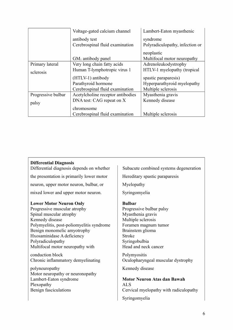
Voltage-gated calcium channel
antibody test
Lambert-Eaton myasthenic
syndromeCerebrospinal fluid examination Polyradiculopathy, infection or
neoplasticGM1 antibody panel Multifocal motor neuropathy
Primary lateral
sclerosis
Very long chain fatty acids AdrenoleukodystrophyHuman T-lymphotropic virus 1
(HTLV-1) antibody
HTLV-1 myelopathy (tropical
spastic paraparesis)Parathyroid hormone Hyperparathyroid myelopathyCerebrospinal fluid examination Multiple sclerosis
Progressive bulbar
palsy
Acetylcholine receptor antibodies Myasthenia gravisDNA test: CAG repeat on X
chromosome
Kennedy disease
Cerebrospinal fluid examination Multiple sclerosis
Differential DiagnosisDifferential diagnosis depends on whether
the presentation is primarily lower motor
neuron, upper motor neuron, bulbar, or
mixed lower and upper motor neuron.
Subacute combined systems degeneration
Hereditary spastic paraparesis
Myelopathy
Syringomyelia
Lower Motor Neuron Only BulbarProgressive muscular atrophy Progressive bulbar palsySpinal muscular atrophy Myasthenia gravisKennedy disease Multiple sclerosisPolymyelitis, post-poliomyelitis syndrome Foramen magnum tumorBenign monomelic amyotrophy Brainstem gliomaHxosaminidase A deficiency StrokePolyradiculopathy SyringobulbiaMultifocal motor neuropathy with
conduction block
Head and neck cancer
PolymyositisChronic inflammatory demyelinating
polyneuropathy
Oculopharyngeal muscular dystrophy
Kennedy diseaseMotor neuropathy or neuronopathyLambert-Eaton syndrome Motor Neuron Atas dan BawahPlexopathy ALSBenign fasciculations Cervical myelopathy with radiculopathy
Syringomyelia
6
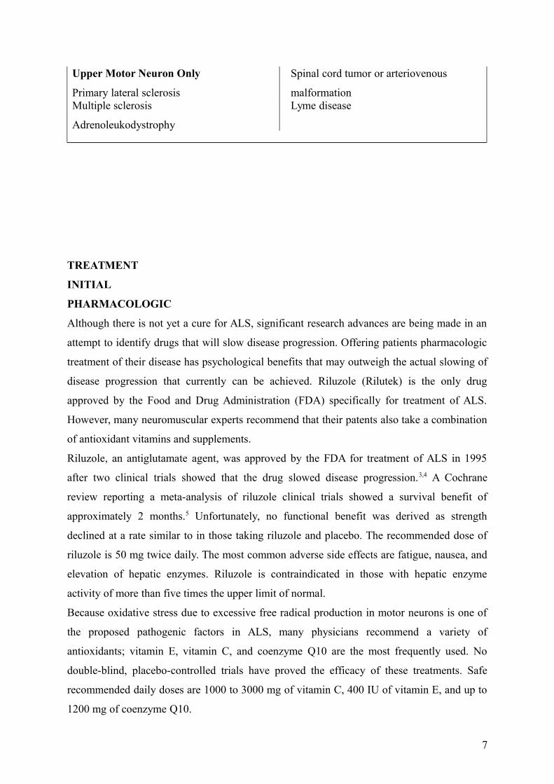
Upper Motor Neuron Only
Primary lateral sclerosis
Spinal cord tumor or arteriovenous
malformationMultiple sclerosis
Adrenoleukodystrophy
Lyme disease
TREATMENT
INITIAL
PHARMACOLOGIC
Although there is not yet a cure for ALS, significant research advances are being made in an
attempt to identify drugs that will slow disease progression. Offering patients pharmacologic
treatment of their disease has psychological benefits that may outweigh the actual slowing of
disease progression that currently can be achieved. Riluzole (Rilutek) is the only drug
approved by the Food and Drug Administration (FDA) specifically for treatment of ALS.
However, many neuromuscular experts recommend that their patents also take a combination
of antioxidant vitamins and supplements.
Riluzole, an antiglutamate agent, was approved by the FDA for treatment of ALS in 1995
after two clinical trials showed that the drug slowed disease progression.3,4 A Cochrane
review reporting a meta-analysis of riluzole clinical trials showed a survival benefit of
approximately 2 months.5 Unfortunately, no functional benefit was derived as strength
declined at a rate similar to in those taking riluzole and placebo. The recommended dose of
riluzole is 50 mg twice daily. The most common adverse side effects are fatigue, nausea, and
elevation of hepatic enzymes. Riluzole is contraindicated in those with hepatic enzyme
activity of more than five times the upper limit of normal.
Because oxidative stress due to excessive free radical production in motor neurons is one of
the proposed pathogenic factors in ALS, many physicians recommend a variety of
antioxidants; vitamin E, vitamin C, and coenzyme Q10 are the most frequently used. No
double-blind, placebo-controlled trials have proved the efficacy of these treatments. Safe
recommended daily doses are 1000 to 3000 mg of vitamin C, 400 IU of vitamin E, and up to
1200 mg of coenzyme Q10.
7
Recent negative ALS clinical trials have tested a number of growth factors, creatine,
glutamate antagonists, anti-inflammatory agents (celecoxib), calcium channel blockers, and
amino acids.6 At present, trials of neurotrophic factors, antioxidants, glutamate antagonists,
coenzyme Q10, and tamoxifen are ongoing. In the future, a cocktail approach to slowing of
disease progression may be the ideal treatment strategy.7
A number of drugs are useful for treatment of spasticity, sialorrhea, pseudobulbar affect,
depression, and anxiety.
Spasticity requires treatment only if it interferes with function. Nonpharmacologic
management involves teaching patients stretching exercises and positioning techniques that
decrease muscle tone. Baclofen (Lioresal) is the most effective pharmacologic agent,
followed by tizanidine (Zanaflex). Diazepam (Valium) should be avoided because it may
suppress respiration, and dantrolene (Dantrium) is not recommended because it causes
excessive muscle weakness. In general, pharmacologic management of spasticity is less
successful in ALS than in multiple sclerosis or spinal cord injury because the lower motor
neuron component of ALS makes patients extremely susceptible to the development of
excessive weakness.
Patients with bulbar dysfunction experience sialorrhea because they have difficulty
swallowing and managing the oral secretions they normally produce. A variety of
anticholinergic drugs may be used to dry the mouth. Tricyclic antidepressants are often tried
first but may not be tolerated because of adverse side effects (dry mouth, somnolence, urinary
retention). One benefit of the tricylcic antidepressants is that they may treat other ALS-related
symptoms, such as pseudobulbar affect (see next paragraph for definition), insomnia, and
pain. Tricyclic antidepressants are contraindicated in patients with cardiac arrhythmia or
conduction disorder. When tricyclic antidepressants are not tolerated, a scopolamine patch
(Transderm Scop) or glycopyrrolate (Robinul) may be helpful. If these treatments are
inadequate, patients may benefit from injection of botulinum toxin into the salivary glands.8
Pseudobulbar affect, also sometimes called emotional incontinence, refers to the patient’s
inability to accurately portray emotions being experienced. Patients laugh or cry when they
are experiencing sadness or happiness, respectively. They also may have an exaggerated
response to situationally appropriate feelings. Amitriptyline (Elavil), a combination of
dextromethorphan (Robitussin, Drixoral) and quinidine, or fluvoxamine (Luvox) may help
blunt the intensity of these inappropriate or exaggerated reactions. A combination preparation
of dextromethorphan and quinidine was found to be effective in a large randomized clinical
trial,9 but this combination has not yet been approved by the FDA.
8
Reactive depression and anxiety are both normal responses to a diagnosis of ALS.10 Patients
and their families may benefit from individual counseling and participation in ALS support
groups. Anxiety may be treated with benzodiazepines as long as the patient does not have
significant reduction of vital capacity. Undetected hypoventilation may produce or contribute
to preexisting feelings of anxiety. Depression should be treated pharmacologically because
not treating it may have a significant negative impact on the quality of life remaining.11,12
Selective serotonin reuptake inhibitors (SSRIs) are good first choices because of their
minimal side effects. However, the tricyclic antidepressants may be preferred if they are also
needed to treat other symptoms, such as sialorrhea or pseudobulbar affect.
REHABILITATION
Skeletal muscle weakness is the primary impairment in ALS and causes the majority of the
clinical problems. In the early stages of ALS, patients often inquire about the role of exercise
in preventing or forestalling the development of weakness. Later in the disease, rehabilitation
strategies must be used to maintain function and to compensate for muscle weakness.
Three forms of exercise training are relevant to patients with ALS: flexibility, strengthening,
and aerobic exercise. Flexibility training helps prevent the development of painful
contractures, nonpharmacologically decreases spasticity, and aborts painful muscle spasms.
Traditionally, physicians have been reluctant to recommend strengthening exercises because
they fear that overuse weakness will accelerate disability. This philosophy promotes the
development of disuse weakness and muscle deconditioning, which may compound the
weakness produced by ALS itself. The literature supporting the development of overwork
weakness in neuromuscular patients is anecdotal, and overuse weakness has not been
demonstrated in any controlled prospective studies. A single study of strength training has
been published in ALS. It demonstrated a slowing of decline in physical function and no
adverse effects.13 Studies of patients with more slowly progressive neuromuscular diseases
and post-poliomyelitis syndrome suggest that muscles only mildly affected by the disease
process can be strengthened by a moderate resistance strengthening program.14-16 High-
resistance eccentric exercise should be avoided because it may damage muscle.
I recommend that interested ALS patients begin or continue with a strengthening program to
maximize the strength of unaffected or mildly affected muscles in an attempt to delay the
time at which function becomes impaired. Weight training should be performed with a weight
that can be lifted 20 times, and the individual should be instructed to perform submaximal
sets of 10 to 15 repetitions; this ensures that the training is performed at a low to moderate
9
level of resistance. If an exercise regimen consistently produces muscle soreness or fatigue
lasting longer than a half an hour after exercise, it is too strenuous.
Aerobic exercise helps maintain cardiorespiratory fitness. A study of moderate aerobic
activity in patients with ALS demonstrated a short-term positive effect on disability, patients
who exercised remained in a milder disease state longer.17 In addition, studies in a transgenic
mouse model of ALS have shown that aerobic exercise prolongs survival.18-20 Given the lack
of any apparent contraindication, aerobic exercise training is recommended for patients with
ALS as long as it can be performed safely without a risk of falling or injury. In addition to the
physical benefist, strengthening and aerobic exercise may have a beneficial effect on mood,
psychological well-being, bone health, appetite, and sleep.
As ALS progresses, the rehabilitation focus shifts from exercise to maintenance of
independent mobility and function for as long as possible. Interventions include assistive
devices such as canes, walkers, braces, hand splints, wheelchairs, and scooters; home
equipment such as dressing aids, adapted utensils, grab bars, raised toilet seats, shower
benches, and lifts; home modifications (ramps, wide doorways); and automobile adaptations,
such as hand controls, environmental control units, and voice-activated software. These
rehabilitation interventions are best provided by a multidisciplinary team that includes a
physiatrists, physical therapists, occupational therapists, and orthotists.
Patients frequently develop musculoskeletal pain syndromes, such as adhesive capsulitis
(frozen shoulder), low back pain and neck pain due to muscle weakness and inability to
change positions. Measures to prevent adhesive capsulitis include range of motion exercises
and support of the arm as much as possible rather than allowing it to dangle at the side. Low
back pain can be triggered by an uncomfortable seating system. Preventive measures include
a lumbar support for the wheelchair, a good cushion on a solid seat, encouragement of
frequent weight shifts, and a reclining back or tilt-in-space wheelchair. Neck pain associated
with head drop is one of the most difficult musculoskeletal pain issues to remedy. A variety of
cervical collars may be tried. A head support on the wheelchair or a reclining lounge chair
may be more comfortable than a collar. Nonpharmacologic measures such as massage and
transcutaneous electrical nerve stimulation can also be used for pain control. Acetaminophen,
nonsteroidal anti-inflammatory drugs, Lidoderm patches, and, if necessary, opioids should be
used to alleviate musculoskeletal pain. Trancutaneous electrical nerve stimulation can also be
used. The major concerns with opiate use are respiratory depression and constipation. The
respiratory depression may be acceptable in the late stages of the disease, in fact, morphine is
good way to relieve air hunger in the terminal stage.
10
Adequate swallowing function is necessary to maintain the nutritional status of the patient
with ALS (unless a feeding tube is in place). If nutritional status is not properly maintained,
patients tend to use muscle as fuel and thus lose muscle mass and strength earlier than they
would otherwise.21 Swallowing dysfunction may also precipitate aspiration pneumonia or
respiratory failure. Early signs and symptoms of dysphagia are drooling, a wet voice,
coughing during or after drinking thin liquids, nasal regurgitations, and requiring an
excessive amount of time to complete meals. Patients should be referred to a speech
pathologist when the first signs of dysphagia develop. Those with mild swallowing
difficulties can be taught compensatory techniques to reduce the risk of aspiration and
choking.22 Recommendations may be given concerning modification of food consistencies.
The development of aspiration pneumona, loss of more than 10% of body weight, and
requiring an excessive amount of time to eat such that quality of life is impaired are
indications for feeding tube placement.
Early or mild dysarthria may be managed by having a speech therapist teach patients adaptive
strategies, such as overarticulation and slowing of the speaking rate. In patients with
hypernasal speech caused by palatal weakness and primarily lower motor neuron dysfunction,
a palatal lift or augmentation prosthesis may improve speech clarity.23 As dysarthria worsens,
patients require alternative forms of communication. Writing is a good alternative while hand
function is intact. For those unable to write, Low-technology interventions include letter and
word boards for written communication in tlose with good hand function. Higher technology
solutions are augmentative communication devices that may have a voice synthesizer. As
long as one muscle somewhere in the body can be voluntarily activated (including the
extraocular muscles), the patient should be able to operate a communication device. High-
technology solutions to communication problems are not suitable for all patients. Systems
must be flexible so that the method of access can be modified as weakness progresses.
Respiratory failure is the primary cause of death in ALS. In the absence of underlying
intrinsic pulmonary disease, the respiratory failure in ALS is purely mechanical. Because of
muscle weakness, the lungs do not inflate fully on inspiration. Most patients with ALS remain
asymptomatic until the FVC is less than 50% of predicted. Pulmonary function tests,
particularly the FVC, should be monitored every few months, depending on rate of disease
progression. The earliest symptoms of respiratory failure are caused by nocturnal
hypoventilation and include poor sleep with frequent awakening, nightmares, early morning
headaches, and excessive daytime fatigue and sleepiness. Another early sign of respiratory
muscle weakness is a weak cough and difficulty in clearing secretions.
11
The management of respiratory failure in ALS involves prevention of infection when possible
and provision of mechanical ventilatory assistance. All patients with ALS should receive a
pneumococcal vaccination and a yearly influenza vaccination. If the expiratory muscles are
too weak to generate an adequate cough, patients can be helped by either manually assisted
coughing or the Cough Assist (J.H. Emerson Co., Cambridge, Mass).24,25 Providing patients
wit supplemental oxygen can relieve symptoms of air hunger and dyspnea but also may
suppress respiratory drive, exacerbate alveolar hypoventilation, and ultimately lead to carbon
dioxide retention and respiratory arrest.26 Supplemental oxygen is recommended only for
patients with concomitant pulmonary disease or as a comfort measure for those who decline
assisted ventilation. Discussion concerning the possibility of respiratory failure should be
initiated soon after the diagnosis of ALS so that patients and their families can learn about
their choices and, ideally, make a decision about ventilator use in a noncrisis situation.
Noninvasive respiratory muscle aids are not a permanent solution to respiratory failure but fo
provide many patients with additional time to make a decision concerning tracheostomy.
Ultimately, less than 5% of ALS patients choose long-term ventilatory support by
tracheostomy.27,28
Noninvasive positive pressure ventilation (NIPPV) can be delivered through a variety of oral
or nasal masks and interfaces by use of bilevel positive airway pressure (BiPAP) machines or
portable volume-cycled ventilators. BiPAP is the most commonly used form of NIPPV.
Several studies suggest that use of NIPPV may prolong survival and slow the decline of
FVC.29,30 The American Academy of Neurology’s practice parameter on ALS recommends
that NIPPV be introduced when the FVC falls to 50% of predicted, or earlier if the patient is
symptomatic.31 However, others have suggested that earlier introduction of NIPPV may
improve survival and quality of life.32,33 Initially, NIPPV is used only at night. As FVC
continues to decline, ventilator use extends into the day for varying periods and eventually
becomes continuous.
12
PROCEDURES
MANAGEMENT OF SIALORRHEA
Transtympanic neurectomy, salivary duct ligation, and parotid gland irradiation have been
used to decrease saliva production but are associated with high failure and complication
rates.34 Botulinum toxin injection into salivary glands is a preferred method of management
for patients who do not respond to pharmacologic therapy.8
GASTROCTOMY TUBE
Either a percutaneous endoscopic gastrostomy (PEG) tube or a radiologically inserted
gastrostomy tube is recommended when a feeding tube is needed. The American Academy of
Neurology’s practice parameter on ALS states that the morbidity and mortality of PEG tube
placement increase when the FVC is below 50% of predicted31 and recommends placement
before that time. However, later studies have shown that PEG tubes can be safely placed with
BiPAP assistance in patients with lower FVCs.35,36 In addition, radiologically inserted
gastrostomy tubes may be preferable in patients with low FVCs; less sedation is required,
incidence of aspiration is lower, tube placement is more often successful.37 Two studies have
suggested longer survival in patients choosing PEG tube placement38,39 when they are
compared with comparable patients refusing PEG.
SURGERY
TRACHEOSTOMY
This is best performed on a planned basis when patients choose long-term ventilatory
support. However, it is more frequently performed in a crisis situation. A cuffless
tracheostomy tybe or a tube with a deflated cuff is preferred.
13
POTENTIAL DISEASE COMPLICATIONS
The potential disease complications are progressive weakness, joint contractures,
musculoskeletal pain syndromes, dysphagia, aspiration, dysarthria, depression, progressive
respiratory failure, and death.
POTENTIAL TREATMENT COMPLICATIONS
The potential treatment complications include drug reactions (e.g., to riluzole, tricyclic
antidepressants) and malfunction or infection of the PEG tube or radiologically inserted
gastrostomy tube.
Complications of long-term ventilation by tracheostomy are tracheomalacia, loss of
extraocular movements, totally locked-in state, and dementia.
REFERENCES
1. Rosenfeld J, Swash M. What’s in a name? Lumping or splitting ALS, PLS, PMA andother motor neuron diseases. Neurology 2006;66:624-625.
2. Brooks BR, Miller RG, Swash M, Munsat TL; World Federation of Neurology ResearchGroup on Motor Neuron Diseases. El Escorial revisited: revised criteria for the diagnosisof amyotrophic lateral sclerosis. ALS Other Motor Neuron Disord 2000;1:293-299.
3. Bensimon G, Lacomblez L, Meininger V, et al. A controlled trial of riluzole inamyotrophic lateral sclerosis. N Engl J Med 1994;330:585-591.
4. Lacomblez L, Bensimon G, Leigh PN, et al. Dose-ranging study of riluzole inamyotophic lateral sclerosis. Lancet 1996;347:1425-1431.
5. Miller R, Mitchell J, Lyon M, Moore D. Riluzole for amyotrophic lateral sclerosis(ALS)/motor neuron disease (MND). Cochrane Database Syst Rev 2002;2:CD001447.
6. de Carvalho M, Costa J, Swash M. Clinical trials in ALS: a review of the role of clinicaland neurophysiological measurements. ALS Other Motor Neuron Disord 2005;6:202-212.
7. Rosenfeld J. ALS combination treatment. Drug cocktails. ALS Other Motor NeuronDisord 2004;5:115-117.
8. Tan E. Botulinum toxin treatment of sialorrhea; comparing different therapeuticpreparations. Eur J Neurol 2006;13:60-64.
9. Brooks B, Thisted R, Appel S, et al. Treatment of pseudobulbar affect in ALS withdextromethorphan/quinidine: a randomized trial. Neurology 2004;63:1364-1370.
10. Ganzini L, Johnston WS, Hoffman WF. Correlated of suffering in amyotrophic lateralsclerosis. Neurology 1999;52:1434-1440.
11. Lou J, reeves A, Benice T, Sexton G. Fatigue and depression are associated with poorquality of life in ALS. Neurology 2003;60:122-123.
12. Kubler A, Winter S, Ludolph A, et al. Severity of depressive symptoms and quality of lifein patients with amyotrophic lateral sclerosis. Neurorehabil Neural Repair 2005;19:182-193.
13. Dal Bello-Haas V, Florence JM, Kloos AD, et al. A randomized controlled trial ofresistance exercise in individuals with ALS. Neurology 2007;68:2003-2007.
14
14. Lindeman E, Leffer P, Spaans F, et al. Strength training in patients with myotonicdystrophy and hereditary motor and sensory neuropathy: a randomized clinical trial. ArchPhys Med Rehabil 1995;76:612-620.
15. Aitkens SG, McCrory MA, Kilmer DD, Bernauer EM. Moderate resistance exerciseprogram: its effect in slowly progressive neuromuscular disease. Arch Phys Med Rehabil1993;74:711-715.
16. Agre JC, Rodriquez AA, Frankle TM. Strength, endurance, and work capacity aftermuscle strengthening exercise in postpolio subjects. Arch Phys Med Rehabil1997;78:681-686.
17. Drory V, Goltsman E, Reznik J, et al. The value of muscle exercise in beneficial to amouse model of amyotrophic lateral sclerosis. J Neurol Sci 2002;191:133-137.
18. Kirkenzos I, Hernandez D, Bradley W, Moraes C. Regular exercise is beneficial to amouse model of amyotrophic lateral sclerosis. Ann Neurol 2003;53:804-807.
19. Veldink J, Bar P, Joosten E, et al. Sexual differences in onset of disease and response toexercise in a transgenic model of ALS. Neuromuscul Disord 2003;13:737-743.
20. Kaspar B, Frost L, Christian L, et al. Synergy of insulin-like growth factor-1 and exercisein amyotrophic lateral sclerosis. Ann Neurol 20058;57:649-655.
21. Heffernan C, Jenkinson C, Holmes T, et al. Nutritional management in MND/ALSpatients: an evidence based review. ALS Other Motor Neuron Disord 2004;5:72-83.
22. Strand EA, Miller RM, Yorkston KM, Hillel AD. Management of oral-pharyngealdysphagia symptoms in amyotrophic lateral sclerosis. Dysphagia 1996;11:129-139.
23. Esposito S, Mitsumoto H, Shanks M. Use of palatal lift and palatal augmentationprotheses to improve dysarthria in patients with amyotrophic lateral sclerosiss: a caseseries. J Prosthet Dent 2000;83:90-98.
24. Bach J. Respiratory muscle aids for the prevention of morbidity and mortality. SeminNeurol 1995;15:72-83.
25. Winck J, Gonclaves M, Lourenco C, et al. Effects of mechanical insufflation-exsufflationon respiratory parameters for patients with chronic airway secretion encumbrance. Chest2004;126:774-780.
26. Gay P, Edmonds L. Severe hypercapnia after flow oxygen therapy in patients withneuromuscular disease and diaphragmatic dysfunction. Mayo Clin Proc 1995;70:327-330.
27. Moss A, Casey P, Stocking C, et al. Home ventilation for amyotrophic lateral sclerosispatients: outcomes, costs, and patient, family, and physician attitudes. Neurology1993;43:438-443.
28. Lechtzin N, Weiner C, Clawson L, et al. Use of noninvasive ventilation in patients withamyotrophic lateral sclerosis. ALS Other Motor Neuron Disord 2204;5:9-15.
29. Kleopa KA, Sher M, Neal B, et al. Bi PAP improves survival and rate of pulmonaryfunction decline in patients with ALS. J Neurol Scri 1999;164:82-88.
30. Aboussouan L, Khan S, Banerjee M, et al. Objective measures of the efficacy ofnoninvasive positive-pressure ventilation in amyotropic lateral sclerosis. Muscle Nerve2001;24:403-409.
31. Miller R, Rosenberg J, Gelinas D, et al. A preliminary evaluation of a prospective studyof pulmonary function studies and symptoms of hypoventilation in ALS/MND patients. JNeurol Sci 2001;191:75-78.
32. Jackson C, Rosenfeld J, Moore D, et al. A preliminary evaluation of a prospective studyof pulmonary function studies and symptoms of hypoventilation in ALS/MND patients. JNeurol SAci 2001;191:75-78.
33. Bourke S, Tomlinson M, Williams T, et al. Effects of non-invasive ventilation on survivaland quality of life in patients with amyotrophic lateral sclerosis: a randomised controlledtrial. Lancet Neurol 2006;5:40-147.
15
34. Yorkston KM, Miller RM, Strand EA. Management of Speech and Swallowing inDegenerative Diseases. Tucson, Communication Skill Builders, 1995:253.
35. Gregory S, Siderowf A, Golaszewski A, et al. Gastrostomy insertion in ALS patients withlow vital capacity: respiratory support and survival. Neurology 2002;58:485-487.
36. Boitano L, Jordan T, Benditt J. Noninvasive ventilation allows gastrostomy tubeplacement in patients with advanced ALS. Neurology 2001;56:413-414.
37. Thornton F, Fotheringham T, Alexander M, et al. Amyotrophic lateral sclerosis: enteralnutrition provision––endoscopic or radiologic gastrostomy? Radiology 2202;224:713-717.
38. Mathus-Vliegen L, Louwerse L, Merkus M, et al. Percutaneous endoscopic gastrostomyin patients with amyotrophic lateral sclerosis and impaired pulmonary function.Gastrointest Endosc 1994;40:463-469.
39. Mazini L, Corra T, Zaccala M, et al. Percutaneous endoscopic gastrostomy and enteralnutrition in amyotrophic lateral sclerosis. J Neurol 1995;242:695-698.
125
PENYAKIT NEURON MOTORIK
Lisa S. Krivickas, MD
DEFINISI
Istilah penyakit motor neuron merujuk pada gangguan neuromuskuler progresif di mana
neuron motorik atas atau bawah mengalami degenerasi. Bentuk yang paling umum dari
penyakit motor neuron adalah amyotrophic lateral sclerosis (ALS), yang merupakan fokus
utama pada bab ini. Prinsip-prinsip manajemen untuk bentuk lain dari penyakit motor neuron
adalah sama. Untuk memenuhi kriteria diagnostik ALS, seseorang harus memiliki kedua
disfungsi pada neuron motorik atas dan bawah. Jika hanya terdapat disfungsi pada neuron
motorik bawah, maka penyakit ini disebut atrofi otot progresif; jika hanya disfungsi neuron
motorik atas, maka disebut sklerosis lateral primer. Jika hanya terdapat disfungsi bulbar,
maka penyakit ini disebut progressive bulbar palsy. Kebanyakan pasien awalnya didiagnosis
memiliki atrofi otot progresif, sklerosis lateral primer, atau bulbar palsy progresif, yang pada
akhirnya menjadi kesatuan ALS. Mereka yang penyakitnya tidak menjadi ALS memiliki
tingkat perkembangan penyakit yang lebih lambat.
Sebagian besar kasus ALS adalah idiopatik. Namun, 5% sampai 10% dari pasien memiliki
riwayat kelaian pada keluarga, biasanya diturunkan dengan pola autosomal dominan. Pada
sekitar 20% dari kasus-kasus keluarga, mutasi di SOD1 (superoxide dismutase) dapat
ditemukan. Bentuk langka lainnya dari penyakit motor neuron yang diturunkan orang
16
dewasa, yaitu penyakit Kennedy (X-linked resesif) dan atrofi otot tulang belakang dewasa
(autosomal resesif), yang keduanya hanya memiliki disfungsi neuron motorik bawah.
ALS secara cepat menghasilkan kelemahan pada otot rangka, yang akhirnya mengarah pada
kebutuhan untuk bantuan ventilator atau kematian akibat gagal pernapasan. Timbulnya
kelemahan dapat dimulai pada tungkai manapun, otot bulbar, atau otot-otot pernapasan. Otot-
otot ekstraokular dan usus dan fungsi kandung kemih umumnya terhindar. Rata-rata
kelangsungan hidup, tanpa trakeostomi, adalah 3 tahun dari onset gejala, namun kisaran
mungkin kurang dari 1 tahun sampai lebih dari 20 tahun. Satu penjelasan untuk variabilitas
ekstrim dalam laju perkembangan penyakit adalah bahwa ALS mungkin kelompok heterogen
dari berbagai penyakit daripada sebuah penyakit tunggal.1 Usia rata-rata saat onset adalah
pada pertengahan 50-an, tetapi ALS dapat berkembang pada orang dewasa dari segala usia.
Penyebab penyakit ini tidak diketahui, tetapi teori terkemuka tentang patogenesis mengarah
pada eksisitotoksisiti glutamat, stres oksidatif, neuroinflamasi, aktivasi sel mikroglial,
apoptosis, dan disfungsi mitokondria.
17
LMN signs only ≥ 1 region
UMN signs only ≥ 1 region
LMN + UMN
1 region
Suspected ALS
Possible ALS
Identified DNA gene
Definite familial ALS
laboratory supported
LMN + UMN
1 region or UMN ≥ 1 region
Probable ALS
laboratory supported
EMG acute denervation
≥ 2 limbs
LMN + UMN
2 regions
Probable ALS
Definite ALS
LMN + UMN
3 regions
SKEMA
Kelemahan, atrofi, hiperrefleks, spastisitasPerkembangan dari waktu ke waktuEMG, NCV, neuroimaging, biopsy
Neuropatologi
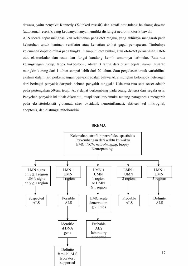
Gambar 125-1. Kriteria El Escorial untuk diagnosis ALS. EMG, electromyography; NCV,
nerve conduction velocity; LMN, lower motor neuron; UMN, upper motor neuron.
GEJALA
Pasien dengan sindrom neuron motorik atas mungkin dijumpai dengan spastisitas, hilangnya
ketangkasan, kekakuan, kelemahan akibat spastisitas, dan hilangnya kontrol motorik sadar.
Pasien dengan sindrom neuron motorik bawah dapat dijumpai dengan kelemahan, kedutan
otot, atrofi otot, dan kram otot. Gejala bulbar termasuk disartria, disfagia, sialorrhea (air liur),
aspirasi, dan pseudobulbar afek (tertawa atau menangis tidak sesuai dengan suasana hati).
Kegagalan pernafasan dan gejala konstitusional seperti penurunan berat badan dan kelelahan
dapat dijumpai. Gejala kognitif meliputi disfungsi perilaku atau eksekutif dan demensia
frontotemporal pada sebagian kecil pasien.
PEMERIKSAAN FISIK
Penekanan dari pemeriksaan fisik pasien yang diduga atau didiagnosis penyakit motor neuron
adalah pada sistem neurologis, muskuloskeletal, dan kardiorespirasi. Pada pemeriksaan
neurologis, salah satu pemeriksaan adalah mencari bukti disfungsi motorik neuron atas dan
bawah. Status mental, fungsi non-motor saraf kranial, pemeriksaan sensorik, dan temuan
pemeriksaan pada serebelum biasanya normal. Standar baku emas untuk diagnosis penyakit
neuron motorik atas adalah adanya refleks patologis-tanda Babinski, tanda Hoffmann, dan
brisk jaw jerk. Pasien dengan ALS mungkin hyperrefleksia atau hiporefleksia, tergantung
pada tahap perjalanan penyakit mereka dan apakah mereka memiliki dominasi pada penyakit
neuron motorik atas atau bawah. Bukti penyakit neuron motorik bawah termasuk kelemahan
otot, atrofi, hipotonia, hiporefleksia, dan kedutan otot. Atrofi sering muncul pertama kali di
otot intrinsic tangan. Meskipun kedutan bukan kriteria yang diperlukan untuk diagnosis ALS,
seseorang harus mempertanyakan diagnosis ketika tidak ditemukan kedutan. Lidah diperiksa
untuk kedutan dan atrofi, dan kekuatan lidah, serta dinilai rentang gerak lidah. Pemeriksaan
muskuloskeletal berfokus pada penilaian jangkauan gerak dan evaluasi nyeri sendi atau
struktur jaringan lunak. Karena gagal napas berkembang secara progresif, sistem
18
kardiorespirasi harus dinilai pada setiap kunjungan. Kapasitas vital paksa (FVC) dapat diukur
dengan spirometer genggam dalam lingkungan ruangan.
BATASAN FUNGSIONAL
Mayoritas keterbatasan fungsional yang berkembang pada pasien dengan ALS adalah
kelemahan otot secara langsung ataupun tidak langsung. Selama penyakit berlangsung, pasien
memiliki gangguan mobilitas dan kesulitan dengan kegiatan, bahkan kegiatan yang paling
dasar dari kehidupan sehari-hari, seperti makan sendiri. Kelemahan otot bulbar menghasilkan
disartria (kesulitan berbicara) dan disfagia (kesulitan menelan); yang pada akhirnya, beberapa
pasien menjadi anarthria dan tidak mampu menelan bahkan ludah mereka sendiri. Depresi
reaktif, kelelahan umum, dan nyeri muskuloskeletal lebih lanjut membatasi fungsi
kehidupan.
STUDI DIAGNOSTIK
Diagnosis ALS didasarkan pada pemeriksaan fisik yang sesuai dan temuan elektrodiagnostik
dan penggunaan neuroimaging dan hasil laboratorium klinik untuk mengeksklusi kondisi lain
yang mirip ALS. Semua pasien harus menjalani tes elektrodiagnostik. Kriteria El Escorial
yang telah direvisi saat ini digunakan untuk mendiagnosis ALS.2 Mereka mengklasifikasikan
tingkat kepastian diagnosis menjadi salah satu dari lima kategori: definite, probable, probable
dengan hasil laboratorium, possible, dan suspected (Gambar 125-1.).
Sistem motorik dibagi menjadi empat wilayah: bulbar, servikal, torakal, dan lumbosakral.
Bukti klinis penyakit neuron motorik atas dan bawah dicari di setiap daerah. Tingkat
kepastian diagnosis tergantung pada berapa banyak daerah yang menunjukan penyakit neuron
motorik atas atau neuron motorik bawah. Temuan elektropsikologi dapat digunakan baik
untuk mengkonfirmasi disfungsi neuron motorik bawah di daerah-daerah yang terkena
dampak klinis dan untuk mendeteksi disfungsi neuron motorik bawah di daerah klinis tidak
terkena dampak klinis.
Studi pencitraan digunakan untuk mengeksklusi kemungkinan lain selain penyakit motor
neuron dari diagnosis diferensial. Pencitraan resonansi magnetik adalah modalitas pencitraan
utama dalam evaluasi pasien yang diduga ALS. Hampir semua pasien harus memiliki
pencitraan resonansi magnetik dari tulang belakang servikal untuk mengeksklusi kompresi
spinal, syrinx, atau penyakit sumsum tulang belakang lainnya. Lokasi gejala akan
menentukan apakah daerah lain dari sumsum tulang belakang harus dicitrakan. Pada pasien
19
yang mengalami gejala bulbar, pencitraan resonansi magnetik pada otak harus dilakukan
untuk menyingkirkan stroke, tumor, syringobulbia, dan proses patologis lainnya.
Dalam kebanyakan praktek klinik neuromuskuler, sebuah panel rutin tes laboratorium
dilakukan untuk semua pasien yang dianggap mengalami ALS. Satu set tes yang disarankan
tersebut diberikan pada Tabel 125-1. Alasan di balik banyaknya tes yang dilakukan adalah
untuk menilai kesehatan umum pasien dan untuk mengeksklusi kondisi yang dapat diobati.
Diferensial diagnosis, dikembangkan setelah anamnesis dan pemeriksaan fisik, dapat
menunjukkan tes yang lebih khusus yang harus dilakukan. Tabel 125-2 menunjukkan tes
tambahan yang mungkin diperlukan saat ditemukan atrofi otot progresif, sklerosis lateral
primer, atau bulbar progresif fenotipe palsy. Ketika ada riwayat keluarga penyakit motor
neuron, pengujian SOD1 dapat dipertimbangkan.
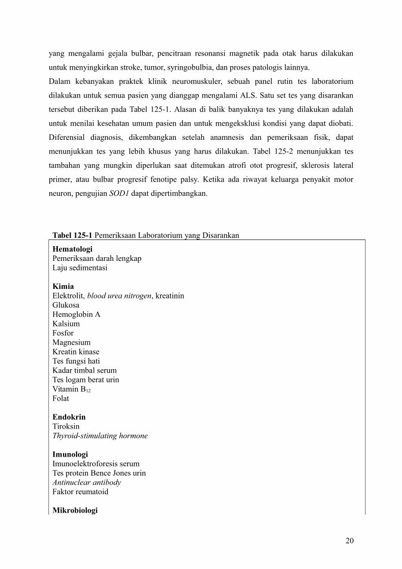
Tabel 125-1 Pemeriksaan Laboratorium yang Disarankan
HematologiPemeriksaan darah lengkapLaju sedimentasi
KimiaElektrolit, blood urea nitrogen, kreatininGlukosaHemoglobin AKalsiumFosforMagnesiumKreatin kinaseTes fungsi hatiKadar timbal serumTes logam berat urinVitamin B12
Folat
EndokrinTiroksinThyroid-stimulating hormone
ImunologiImunoelektroforesis serumTes protein Bence Jones urinAntinuclear antibodyFaktor reumatoid
Mikrobiologi
20
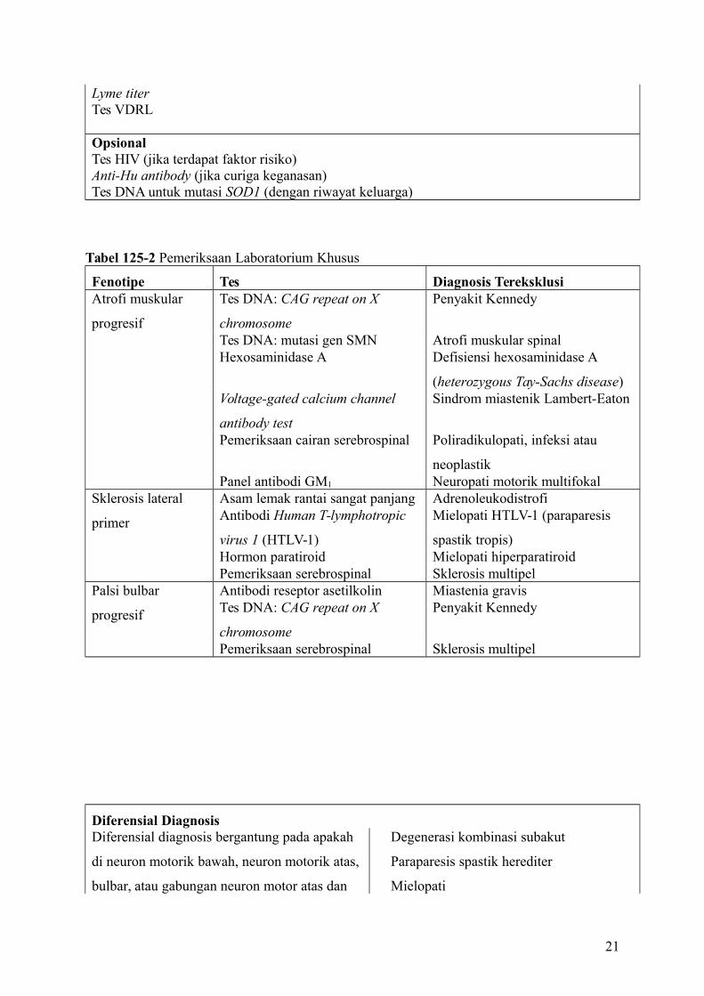
Lyme titerTes VDRL
OpsionalTes HIV (jika terdapat faktor risiko)Anti-Hu antibody (jika curiga keganasan)Tes DNA untuk mutasi SOD1 (dengan riwayat keluarga)
Tabel 125-2 Pemeriksaan Laboratorium Khusus
Fenotipe Tes Diagnosis TereksklusiAtrofi muskular
progresif
Tes DNA: CAG repeat on X
chromosome
Penyakit Kennedy
Tes DNA: mutasi gen SMN Atrofi muskular spinalHexosaminidase A Defisiensi hexosaminidase A
(heterozygous Tay-Sachs disease)Voltage-gated calcium channel
antibody test
Sindrom miastenik Lambert-Eaton
Pemeriksaan cairan serebrospinal Poliradikulopati, infeksi atau
neoplastikPanel antibodi GM1 Neuropati motorik multifokal
Sklerosis lateral
primer
Asam lemak rantai sangat panjang AdrenoleukodistrofiAntibodi Human T-lymphotropic
virus 1 (HTLV-1)
Mielopati HTLV-1 (paraparesis
spastik tropis)Hormon paratiroid Mielopati hiperparatiroidPemeriksaan serebrospinal Sklerosis multipel
Palsi bulbar
progresif
Antibodi reseptor asetilkolin Miastenia gravisTes DNA: CAG repeat on X
chromosome
Penyakit Kennedy
Pemeriksaan serebrospinal Sklerosis multipel
Diferensial DiagnosisDiferensial diagnosis bergantung pada apakah
di neuron motorik bawah, neuron motorik atas,
bulbar, atau gabungan neuron motor atas dan
Degenerasi kombinasi subakut
Paraparesis spastik herediter
Mielopati
21
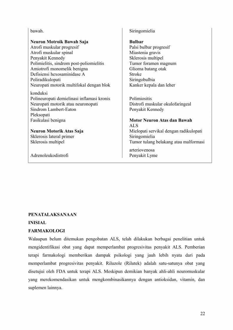
bawah. Siringomielia
Neuron Motroik Bawah Saja BulbarAtrofi muskular progresif Palsi bulbar progresifAtrofi muskular spinal Miastenia gravisPenyakit Kennedy Sklerosis multipelPolimielitis, sindrom post-poliomielitis Tumor foramen magnumAmiotrofi monomelik benigna Glioma batang otakDefisiensi hexosaminidase A StrokePoliradikulopati SiringobulbiaNeuropati motorik multifokal dengan blok
konduksi
Kanker kepala dan leher
Polineuropati demielinasi inflamasi kronis PolimiositisNeuropati motorik atau neuronopati Distrofi muskular okulofaringealSindrom Lambert-Eaton Penyakit KennedyPleksopatiFasikulasi benigna Motor Neuron Atas dan Bawah
ALSNeuron Motorik Atas Saja Mielopati servikal dengan radikulopatiSklerosis lateral primer SiringomieliaSklerosis multipel Tumor tulang belakang atau malformasi
arteriovenosaAdrenoleukodistrofi Penyakit Lyme
PENATALAKSANAAN
INISIAL
FARMAKOLOGI
Walaupun belum ditemukan pengobatan ALS, telah dilakukan berbagai penelitian untuk
mengidentifikasi obat yang dapat memperlambat progresivitas penyakit ALS. Pemberian
terapi farmakologi memberikan dampak psikologi yang jauh lebih nyata dari pada
memperlambat progresivitas penyakit. Riluzole (Rilutek) adalah satu-satunya obat yang
disetujui oleh FDA untuk terapi ALS. Meskipun demikian banyak ahli-ahli neuromuskular
yang merekomendasikan untuk mengkombinasikannya dengan antioksidan, vitamin, dan
suplemen lainnya.
22
Penggunaan Riluzole (agen antiglutamat) untuk terapi ALS telah disetujui pada 1995 setelah
adanya bukti penurunan progresivitas penyakit pada dua uji klinis.3,4 Cochrane melaporkan
meta-analisis dari uji klinis Riluzole menunjukkan manfaat kurang lebih 2 bulan.5 Sayangnya,
tidak ditemukan perbedaan manfaat fungsional dari mereka yang mengkonsumsi riluzole dan
placebo. Efek samping yang paling sering ditemukan adalah lelah, mual, dan peningkatan
enzim hepar. Riluzole dikontraindikasikan pada pasien dengan peningkatan aktivitas enzim
hepar lima kali di atas normal.
Karena stres oksidatif akibat produksi radikal bebas berlebih pada motor neuron adalah salah
satu faktor patogen pemicu pada ALS, para dokter menyarankan untuk mengkonsumsi
antioksidan seperti vitamin E, vitamin C, dan coenzyme Q10. Belum ada uji buta ganda
dengan placebo yang membuktikan keberhasilan terapi ini. Dosis aman harian yang
direkomendasikan adalah 1000 sampai 3000 mg untuk vitamin C, 400 IU untuk vitamin E,
dan sampai 1200 mg untuk coenzyme Q10.
Uji klinis negatif ALS terbaru menguji sejumlah faktor pertumbuhan, keratin, glutamat
antagonis, agen antiinflamasi (celecoxib), calcium channel blockers, dan asam amino.6 Kini,
sedang diuji mengenai neurotrophic factor, antioksidan, glutamate antagonis, coenzyme Q10,
dan tamoxifen. Di masa yang akan datang, pendekatan koktail mungkin merupakan strategi
terapi yang paling ideal untuk menurunkan progresivitas penyakit.7
Berbagai terapi farmakologi bermanfaat untuk mengatasi spastisitas, sialorrhea,
pseudobulbar affect, depresi, dan ansietas.
Spastisitas perlu diterapi hanya bila hal menimbulkan gangguan fungsi. Terapi
nonfarmakologi meliputi mengajari pasien mengenai latihan peregangan dan positioning
technique untuk mengurangi tonus otot. Baclofen (Lioresal) adalah terapi farmakologi yang
paling efektif, diikuti dengan tizanidine (Zanaflex). Diazepa, (Valium) perlu dihindari karena
dapat menyebabkan supresi pada system respirasi, dan dantrolene (Dantrium) juga tidak
direkomendasikan karena dapat menyebabkan kelemahan otot berlebih. Secara keseluruhan,
terapi farmakologi untuk spastisitas kurang berhasil pada pasien ALS dibandingkan dengan
pasien multiple sclerosis atau spinal cord injury karena komponen motor neuron bawah pada
ALS membuat penderitanya sangat rentan terhadap kelemahan otot yang sangat progresif.
Pasien dengan disfungsi bulbar mengalami sialorrhea karena adanya kesulitan menelan dan
mengatur sekresi oral dari produksi normal mereka. Beragam obat antikolinergik mungkin
digunakan untuk mengeringkan mulut. Antidepresan trisiklik sering menjadi obat pertama
yang dipilih namun tidak sebanding dengan efek sampingnya (mulut kering, somnolen,
retensi urin). Ketika antidepresan trisiklik tidak dapat ditolerir, scopolamine patch
23
(Transderm Scop) atau glycopyrrolate (Robinul) mungkin dapat digunakan sebagai
alternatifnya. Bila terapi tersebut belum adekuat, pasien dapat menggunakan injeksi toksin
botulinum pada kelenjar ludah.8
Pseudobulbar affect, yang sering disebut inkontinensi emosi merujuk pada ketidakmampuan
pasien untuk menggambarkan emosi yang sedang dirasakannya secara tepat. Pasien tertawa
saat sedih atau menangis saat bahagia. Mereka juga memberikan respon yang berlebihan saat
memiliki perasaan yang sesuai dengan situasi saat itu. Amitriptyline (Elavil), kombinasi
dextromethorphan (Robitussin, Drixoral) dan quinidine, atau fluvoxamine (Luvox) dapat
membantu mengurangi intensitas dari reaksi yang tidak sesuai atau berlebihan tersebut.
Kombinasi dextromethorphan dan quinidine terbukti efektif pada uji klinik acak bersampel
besar9 namun kombinasi ini belum disetujui oleh FDA
Depresi yang reaktif dan ansietas merupakan reaksi normal untuk diagnosis ALS.10 Pasien
dan keluarganya dapat diberikan konseling individu dan berpartisipasi dalam kelompok
pendukung ALS. Ansietas dapat diterapi dengan benzodiazepine selama pasien tidak
menunjukkan penurunan kapasitas vital yang signifikan. Hipoventilasi yang tidak terdeteksi
dapat menyebabkan dan memicu perasaan cemas. Depresi harus diterapi secara farmakologi
karena dapat memberikan dampak negatif pada kualitas hidup bila tidak diobati.11,12 Selective
serotonin reuptake inhibitors (SSRIs) adalah pilihan pertama yang baik karena efek
sampingnya sangatlah sedikit. Meskipun demikian, antidepresan triskilik mungkin lebih
disukai jika diperlukan juga terapi untuk gejala lain seperti sialorrhea dan pesudobulbar
affect.
REHABILITASI
Kelemahan otot rangka adalah gangguan utama pada ALS dan penyebab sebagian besar
masalah klinis. Pada tahap awal ALS, pasien sering bertanya tentang peran latihan dalam
mencegah perburukan pada kelemahan otot. Kemudian untuk penyakitnya sendiri, strategi
rehabilitasi harus digunakan untuk mempertahankan fungsi dan untuk mengkompensasi
kelemahan otot. Tiga bentuk latihan pelatihan yang relevan untuk penderita ALS: latihan
fleksibilitas, penguatan, dan aerobik. Latihan fleksibilitas membantu mencegah
perkembangan kontraktur yang menyakitkan, secara nonfarmakologi membantu mengurangi
kelenturan dan menghilangkan kejang otot yang berlebihan. Umumnya, para dokter enggan
24
untuk merekomendasikan latihan penguatan karena mereka takut bahwa pemakaian berlebih
pada kelemahan otot akan mempercepat terjadinya disabilitas. Filosofi ini mendorong
perkembangan kelemahan otot akibat tidak digunakan dan dekondisi otot yang dapat
memperparah kelemahan yang disebabkan oleh ALS tersebut. Literatur yang mendukung
mengenai perkembangan kelemahan otot akibat pemakaian berlebih pada pasien
neuromuskular tidaklah dapat dipercaya, dan kelemahan akibat pemakaian berlebihan tidak
dapat dibuktikan dalam studi kontrol prospektif manapun. Studi mengenai latihan penguatan
pada ALS menunjukkan adanya perlambatan penurunan fungsi fisik dan tidak ada efek
sampingnya.13 Studi pada pasien dengan penyakit neuromuskuler yang lebih lambat
progresivitasnya dan pasien sindrom post-poliomyelitis menggambarkan bahwa otot-otot
yang hanya sedikit terpengaruh oleh proses penyakit dapat diperkuat dengan program
penguatan beresistensi sedang.14-16 Latihan penguatan eksentrik beresistensi tinggi harus
dihindari karena dapat merusak otot.
Saya menyarankan pasien ALS yang berminat untuk memulai atau melanjutkan program
penguatan untuk memaksimalkan kekuatan otot-otot yang masih normal atau yang
mengalami kelemahan ringan guna menunda terjadinya kerusakan fungsi otot tersebut.
Latihan beban harus dilakukan dengan beban yang dapat diangkat 20 kali, dan individu harus
diinstruksikan untuk melakukan submaximal set dengan pengulangan kurang lebih 10-15
kali; hal ini untuk memastikan bahwa latihan tersebut dilakukan dalam level resistensi rendah
sampai sedang. Jika rejimen latihan terus menerus menyebabkan nyeri otot atau kelelahan
yang berlangsung lebih dari setengah jam setelah latihan, itu berarti latihan terlalu berat.
Latihan aerobik membantu mempertahankan kesehatan kardiorespirasi. Studi mengenai
aktivitas aerobik moderat pada pasien dengan ALS menunjukkan efek positif jangka pendek
pada disabilitas, pasien yang melakukan latihan akan lebih lama berada pada level penyakit
yang lebih ringan.17 Selain itu, studi dengan model tikus transgenik pada ALS telah
menunjukkan bahwa latihan aerobik memperpanjang kelangsungan hidup.18-20 Mengingat
kurangnya kontraindikasi yang jelas, latihan aerobik hanya dianjurkan untuk pasien dengan
ALS selama dapat dilakukan dengan aman tanpa risiko jatuh atau cedera. Selain dari manfaat
fisik, latihan penguatan dan aerobik memberikan efek positif pada suasana hati, kesejahteraan
psikologis, nafsu makan, dan tidur.
Seiring dengan progresivitas ALS, fokus rehabilitasi bergeser dari latihan ke pemeliharaan
mobilitas dan fungsi independen selama mungkin. Intervensi termasuk alat bantu seperti
tongkat, alat bantu berjalan, penyangga kaki, bidai tangan, kursi roda, dan skuter; peralatan
rumah seperti alat bantu untuk berpakaian, peralatan yang telah disesuaikan, palang
25
pegangan, raised toilet seats, bangku mandi, dan lift; modifikasi rumah (pintu yang landai,
lebar); dan mobil dengan adaptasi, seperti hand control, environmental control unit, dan
voice-activated software. Intervensi rehabilitasi ini paling baik disediakan oleh tim
multidisiplin yang mencakup psikiater, terapis fisik, terapis okupasi, dan ortotis.
Pasien sering mengalami sindrom nyeri muskuloskeletal seperti adhesive capsulitis (kaku
bahu), nyeri punggung bawah, dan nyeri leher akibat kelemahan otot dan ketidakmampuan
untuk mengubah posisi tubuhnya sendiri. Langkah-langkah untuk mencegah bahu kaku
meliputi latihan range of motion dan sebisa mungkin penggunaan penyangga tangan daripada
dibiarkan menjuntai di sisi tubuh. Nyeri pinggang bawah dapat ditimbulkan karena posisi
duduk yang tidak nyaman. Langkah-langkah pencegahan termasuk di dalamnya yaitu
penyangga lumbal pada kursi roda, bantal empuk pada dudukan kursi yang kokoh, frequent
weight shift, dan reclining back or tilt-in-space wheelchair. Neck pain associated with head
drop adalah salah satu masalah nyeri muskuloskeletal yang paling sulit untuk diatasi.
Penyangga kepala pada kursi roda atau kursi baring lebih baik dan nyaman daripada
penyangga leher. Asetaminofen, obat anti-inflamasi non steroid, Lidoderm patch, dan jika
perlu, opioid harus digunakan untuk meringankan nyeri muskuloskeletal. Transcutaneous
electrical nerve stimulation (TENS) juga dapat digunakan untuk mengontrol rasa sakit.
Kekhawatiran utama dalam penggunaan obat opiate adalah distres pernapasan dan sembelit.
Distres pernapasan mungkin muncul pada tahap akhir penyakit, namun pada kenyataannya,
morfin adalah cara yang baik untuk mengurangi kekurangan oksigen dalam tahap terminal.
Fungsi menelan yang adekuat diperlukan untuk mempertahankan status nutrisi pasien dengan
ALS (kecuali yang menggunakan selang makanan). Jika status gizi tidak benar-benar
diperhatikan, pasien cenderung menggunakan otot sebagai bahan bakar dan dengan demikian
pasien dapat kehilangan massa dan kekuatan otot lebih cepat dari biasanya.21 Disfungsi
menelan juga dapat memicu pneumonia aspirasi atau kegagalan pernafasan. Tanda dan gejala
awal disfagia adalah drooling, suara yang basah, batuk saat atau setelah minum air,
regurgitasi nasal, dan membutuhkan waktu yang lama untuk menghabiskan makanan. Pasien
harus dirujuk ke patologi wicara saat tanda-tanda pertama disfagia muncul. Pasien dengan
kesulitan menelan ringan dapat diajarkan teknik mengurangi risiko aspirasi dan tersedak.22
Rekomendasi mengenai cara modifikasi konsistensi makanan yang disajikan dapat diberikan.
Perkembangan pneumonia aspirasi, kehilangan lebih dari 10% berat badan, dan waktu yang
lama untuk makan sedemikian rupa sehingga gangguan kualitas hidup adalah indikasi unuk
pemberian makanan melalui selang.
26
Disartria awal atau ringan mungkin dapat diatasi oleh para terapis wicara yang mengajarkan
pasien dengan strategi adaptif, seperti overarticulation dan memperlambat kecepatan
berbicara. Pada pasien yang berbicara dengan hipernasal yang disebabkan oleh kelemahan
palatum dan terutama disfungsi neuron motorik bawah, pengangkatan atau augmentasi
prostesis palatum dapat meningkatkan kejelasan dalam berbicara.23 Seiring dengan
perburukan disartria, pasien memerlukan bentuk-bentuk alternatif komunikasi. Menulis
adalah alternatif yang baik untuk pasien yang memiliki fungsi tangan yang masih baik. Bagi
mereka yang tidak bisa menulis, intervensi berteknologi rendah berupa huruf dan word
boards. Solusi teknologi yang lebih maju adalah perangkat komunikasi augmentatif dengan
penghasil suara. Selama satu otot di tubuh dapat diaktifkan secara volunter (termasuk otot-
otot ekstraokular), pasien tentu mampu mengoperasikan perangkat komunikasi. Solusi
berteknologi tinggi belum tentu cocok untuk semua pasien dengan masalah komunikasi.
Sistem tersebut haruslah fleksibel sehingga cara pengaksesan dapat dimodifikasi seiring
dengan progresivitas kelemahan pada pasien.
Gagal nafas adalah penyebab utama kematian pada pasien ALS. Saat tidak ada penyakit
intrinsik paru yang mendasarinya, gagal nafas pada ALS murni adalah masalah mekanis.
Karena kelemahan otot, paru-paru tidak mengembang sepenuhnya pada fase inspirasi.
Kebanyakan pasien dengan ALS tetap asimtomatik sampai FVC kurang dari 50% dari yang
diprediksikan. Tes fungsi paru-paru, khususnya FVC, harus dipantau setiap beberapa bulan,
tergantung pada tingkat perkembangan penyakit. Gejala awal gagal napas disebabkan oleh
hipoventilasi nokturnal dan termasuk kurangnya tidur karena sering terbangun, mimpi buruk,
sakit kepala pada pagi hari, dan kelelahan serta kantuk yang berlebihan pada siang hari.
Tanda awal lainnya dari kelemahan otot pernapasan adalah batuk ringan dan kesulitan dalam
pembersihan sekresi.
Pengelolaan kegagalan pernafasan di ALS melibatkan pencegahan infeksi bila mungkin dan
pemberian bantuan ventilasi mekanis. Semua pasien dengan ALS harus menerima vaksinasi
pneumokokus dan vaksinasi influenza tahunan. Jika otot-otot ekspiratori terlalu lemah untuk
menghasilkan batuk yang memadai, pasien dapat dibantu baik bantuan batuk manual maupun
dengan Cough Assist (J.H. Emerson Co., Cambridge, Mass).24,25 Memberikan pasien bantuan
tambahan oksigen dapat meringankan gejala hipoksia dan dyspnea tetapi juga dapat menekan
hantaran pernafasan, memperburuk hipoventilasi alveolar, dan pada akhirnya menyebabkan
retensi karbondiksida dan henti nafas. 26
Oksigen tambahan hanya direkomendasikan untuk pasien dengan penyakit paru konkomitan
atau sebagai tolak ukur kenyamanan bagi mereka yang menolak ventilasi bantuan. Diskusi
27
mengenai kemungkinan kegagalan pernapasan harus dimulai segera setelah diagnosis ALS
sehingga pasien dan keluarga mereka dapat mempelajari pilihan mereka dan membuat
keputusan secara ideal mengenai penggunaan ventilator dalam situasi yang tidak kritis.
Bantuan otot pernapasan noninvasive bukan merupakan solusi permanen untuk kegagalan
pernapasan tetapi memang memberikan pasien tambahan wantu untuk membuat keputusan
mengenai trakeostomi. Pada akhirnya, kurang dari 5% ALS pasien memilih trakeostomi
sebagai pendukung ventilasi jangka panjang.27,28
Ventilasi tekanan positif non-invasif (NIPPV) dapat diberikan melalui berbagai macam
sungkup mulut dan/atau hidung dengan menggunakan mesin bilevel positive airway pressure
(BiPAP) atau portable volume-cycled ventilators. BiPAP adalah bentuk penggunaan paling
umum dari NIPPV. Beberapa penelitian menunjukkan bahwa penggunaan NIPPV dapat
memperpanjang kelangsungan hidup dan memperlambat penurunan FVC. 29,30
Parameter praktik American Academy of Neurology pada ALS merekomendasikan
penggunaan NIPPV ketika FVC turun sampai 50% dari perkiraan atau lebih awal pada pasien
simptomatik.31 Meskipun demikian, beberapa pihak lain menyatakan penggunaan NIPPV
yang lebih awal dapat meningkatkan kelangsungan dan kualitas hidup.32,33 Pada awalnya,
NIPPV digunakan hanya pada malam hari. Seiring FVC terus menurun, penggunaan
ventilator memanjang sampai satu hari dalam beragam kondisi dan akhirnya menjadi terus-
menerus.
PROSEDUR
MANAJEMEN SIALORRHEA
Transtympanic neurectomy, salivary duct ligation, dan parotid gland irradiation telah
digunakan untuk mengurangi produksi saliva namun memiliki tingkat kegagalan dan
komplikasi yang tinggi. Injeksi toksin botulinum pada kelenjar ludah juga sering digunakan
pada pasien yang tidak memberikan respon pada terapi farmakologi.
SELANG GASTROSTOMI
Percutaneous endoscopic gastrostomy (PEG) atau pemasangan selang gastrostomi dengan
bantuan radiologi direkomendasikan bila pasien membutuhkan selang makanan. Parameter
praktik American Academy of Neurology pada ALS menyatakan bahwa morbiditas dan
mortalitas pemasangan selang PEG meningkat ketika FVC di bawah 50% dari prediksi dan
direkomendasikan pemasangan sebelum hal tersebut terjadi. Namun, studi studi selanjutnya
menunjukkan bahwa selang PEG aman dipasang dengan bantuan BiPAP pada pasien dengan
FVC rendah. Selain itu, pemasangan selang gastrostomi dengan bantuan radiologi lebih
28
dipilih pada pasien dengan FVC rendah; lebih sedikit sedasi yang dibutuhkan, insiden
aspirasi lebih rendah, keberhasilan pemasangan selang lebih tinggi. Dua studi lainnya
menunjukkan angka kelangsungan hidup yang lebih baik pada pasien yang memilih
pemasangan selang PEG dibanding dengan pasien yang menolak pemasangan selang PEG.
PEMBEDAHAN
TRAKEOSTOMI
Paling baik dilakukan secara terencana ketika pasien memilih menggunakan bantuan
ventilator jangka panjang. Namun, tindakan ini lebih sering dilakukan saat situasi kritis.
Cuffless tracheostomy tube atau tube with deflated cuff lebih sering digunakan.
KOMPLIKASI POTENSIAL PENYAKIT
Komplikasi potensial penyakit adalah kelemahan progresif, kontraktur sendi, musculoskeletal
pain syndrome, disfagia, aspirasi, disartria, depresi, gagal nafas progresif, dan kematian.
KOMPLIKASI POTENSIAL TERAPI
Komplikasi potensial terapi meliputi reaksi farmakologi (seperti dengan riluzole,
antidepresan trisiklik) dan malfungsi atau infeksi pada selang PEG atau selang gastrostomi.
Komplikasi ventilasi jangka panjang dari trakeostomi adalah trakeomalasia, hilangnya
pergerakan ekstraokular, locked-in state, dan demensia.
REFERENSI
1. Rosenfeld J, Swash M. What’s in a name? Lumping or splitting ALS, PLS, PMA andother motor neuron diseases. Neurology 2006;66:624-625.
2. Brooks BR, Miller RG, Swash M, Munsat TL; World Federation of Neurology ResearchGroup on Motor Neuron Diseases. El Escorial revisited: revised criteria for the diagnosisof amyotrophic lateral sclerosis. ALS Other Motor Neuron Disord 2000;1:293-299.
3. Bensimon G, Lacomblez L, Meininger V, et al. A controlled trial of riluzole inamyotrophic lateral sclerosis. N Engl J Med 1994;330:585-591.
4. Lacomblez L, Bensimon G, Leigh PN, et al. Dose-ranging study of riluzole inamyotophic lateral sclerosis. Lancet 1996;347:1425-1431.
29
5. Miller R, Mitchell J, Lyon M, Moore D. Riluzole for amyotrophic lateral sclerosis(ALS)/motor neuron disease (MND). Cochrane Database Syst Rev 2002;2:CD001447.
6. de Carvalho M, Costa J, Swash M. Clinical trials in ALS: a review of the role of clinicaland neurophysiological measurements. ALS Other Motor Neuron Disord 2005;6:202-212.
7. Rosenfeld J. ALS combination treatment. Drug cocktails. ALS Other Motor NeuronDisord 2004;5:115-117.
8. Tan E. Botulinum toxin treatment of sialorrhea; comparing different therapeuticpreparations. Eur J Neurol 2006;13:60-64.
9. Brooks B, Thisted R, Appel S, et al. Treatment of pseudobulbar affect in ALS withdextromethorphan/quinidine: a randomized trial. Neurology 2004;63:1364-1370.
10. Ganzini L, Johnston WS, Hoffman WF. Correlated of suffering in amyotrophic lateralsclerosis. Neurology 1999;52:1434-1440.
11. Lou J, reeves A, Benice T, Sexton G. Fatigue and depression are associated with poorquality of life in ALS. Neurology 2003;60:122-123.
12. Kubler A, Winter S, Ludolph A, et al. Severity of depressive symptoms and quality of lifein patients with amyotrophic lateral sclerosis. Neurorehabil Neural Repair 2005;19:182-193.
13. Dal Bello-Haas V, Florence JM, Kloos AD, et al. A randomized controlled trial ofresistance exercise in individuals with ALS. Neurology 2007;68:2003-2007.
14. Lindeman E, Leffer P, Spaans F, et al. Strength training in patients with myotonicdystrophy and hereditary motor and sensory neuropathy: a randomized clinical trial. ArchPhys Med Rehabil 1995;76:612-620.
15. Aitkens SG, McCrory MA, Kilmer DD, Bernauer EM. Moderate resistance exerciseprogram: its effect in slowly progressive neuromuscular disease. Arch Phys Med Rehabil1993;74:711-715.
16. Agre JC, Rodriquez AA, Frankle TM. Strength, endurance, and work capacity aftermuscle strengthening exercise in postpolio subjects. Arch Phys Med Rehabil1997;78:681-686.
17. Drory V, Goltsman E, Reznik J, et al. The value of muscle exercise in beneficial to amouse model of amyotrophic lateral sclerosis. J Neurol Sci 2002;191:133-137.
18. Kirkenzos I, Hernandez D, Bradley W, Moraes C. Regular exercise is beneficial to amouse model of amyotrophic lateral sclerosis. Ann Neurol 2003;53:804-807.
19. Veldink J, Bar P, Joosten E, et al. Sexual differences in onset of disease and response toexercise in a transgenic model of ALS. Neuromuscul Disord 2003;13:737-743.
20. Kaspar B, Frost L, Christian L, et al. Synergy of insulin-like growth factor-1 and exercisein amyotrophic lateral sclerosis. Ann Neurol 20058;57:649-655.
21. Heffernan C, Jenkinson C, Holmes T, et al. Nutritional management in MND/ALSpatients: an evidence based review. ALS Other Motor Neuron Disord 2004;5:72-83.
22. Strand EA, Miller RM, Yorkston KM, Hillel AD. Management of oral-pharyngealdysphagia symptoms in amyotrophic lateral sclerosis. Dysphagia 1996;11:129-139.
23. Esposito S, Mitsumoto H, Shanks M. Use of palatal lift and palatal augmentationprotheses to improve dysarthria in patients with amyotrophic lateral sclerosiss: a caseseries. J Prosthet Dent 2000;83:90-98.
24. Bach J. Respiratory muscle aids for the prevention of morbidity and mortality. SeminNeurol 1995;15:72-83.
25. Winck J, Gonclaves M, Lourenco C, et al. Effects of mechanical insufflation-exsufflationon respiratory parameters for patients with chronic airway secretion encumbrance. Chest2004;126:774-780.
26. Gay P, Edmonds L. Severe hypercapnia after flow oxygen therapy in patients withneuromuscular disease and diaphragmatic dysfunction. Mayo Clin Proc 1995;70:327-330.
30
27. Moss A, Casey P, Stocking C, et al. Home ventilation for amyotrophic lateral sclerosispatients: outcomes, costs, and patient, family, and physician attitudes. Neurology1993;43:438-443.
28. Lechtzin N, Weiner C, Clawson L, et al. Use of noninvasive ventilation in patients withamyotrophic lateral sclerosis. ALS Other Motor Neuron Disord 2204;5:9-15.
29. Kleopa KA, Sher M, Neal B, et al. Bi PAP improves survival and rate of pulmonaryfunction decline in patients with ALS. J Neurol Scri 1999;164:82-88.
30. Aboussouan L, Khan S, Banerjee M, et al. Objective measures of the efficacy ofnoninvasive positive-pressure ventilation in amyotropic lateral sclerosis. Muscle Nerve2001;24:403-409.
31. Miller R, Rosenberg J, Gelinas D, et al. A preliminary evaluation of a prospective studyof pulmonary function studies and symptoms of hypoventilation in ALS/MND patients. JNeurol Sci 2001;191:75-78.
32. Jackson C, Rosenfeld J, Moore D, et al. A preliminary evaluation of a prospective studyof pulmonary function studies and symptoms of hypoventilation in ALS/MND patients. JNeurol SAci 2001;191:75-78.
33. Bourke S, Tomlinson M, Williams T, et al. Effects of non-invasive ventilation on survivaland quality of life in patients with amyotrophic lateral sclerosis: a randomised controlledtrial. Lancet Neurol 2006;5:40-147.
34. Yorkston KM, Miller RM, Strand EA. Management of Speech and Swallowing inDegenerative Diseases. Tucson, Communication Skill Builders, 1995:253.
35. Gregory S, Siderowf A, Golaszewski A, et al. Gastrostomy insertion in ALS patients withlow vital capacity: respiratory support and survival. Neurology 2002;58:485-487.
36. Boitano L, Jordan T, Benditt J. Noninvasive ventilation allows gastrostomy tubeplacement in patients with advanced ALS. Neurology 2001;56:413-414.
37. Thornton F, Fotheringham T, Alexander M, et al. Amyotrophic lateral sclerosis: enteralnutrition provision––endoscopic or radiologic gastrostomy? Radiology 2202;224:713-717.
38. Mathus-Vliegen L, Louwerse L, Merkus M, et al. Percutaneous endoscopic gastrostomyin patients with amyotrophic lateral sclerosis and impaired pulmonary function.Gastrointest Endosc 1994;40:463-469.
39. Mazini L, Corra T, Zaccala M, et al. Percutaneous endoscopic gastrostomy and enteralnutrition in amyotrophic lateral sclerosis. J Neurol 1995;242:695-698.
31