Embed Size (px)
Citation preview

2
13C Magnetic Resonance Spectroscopy in Neurobiology - Its Use in Monitoring Brain
Energy Metabolism and in Identifying Novel Metabolic Substrates and Metabolic Pathways
Bjørnar Hassel Department of Neurohabilitation, Oslo University Hospital-Ullevål, Oslo
Norwegian Defense Research Establishment, Kjeller Norway
1. Introduction
A wealth of information on brain metabolism has been gathered from studies in which 13C-labeled metabolic substrates (often glucose) have been administered to human subjects or experimental animals. 13C Magnetic resonance spectroscopy (MRS1) of the brain (or extracts of brain) has shown to what degree the 13C-labeled compound has been metabolized, and, to some extent, along which metabolic pathways. The latter interpretation derives from the fact that 13C MRS shows the 13C labeling not of individual compounds, e.g. amino acids, but of individual carbon positions in those different compounds (Fig. 1). Because some enzymes of amino acid metabolism have a cell-specific expression, it is possible to study the metabolic activity of individual cell types or the transfer of amino acids between them. Finally, 13C MRS allows studies of a host of substrates to examine their potential as metabolic substrates for the brain. An extensive review, which included an overview of technical aspects of 13C MRS in studies of brain and cultured brain cells, appeared recently (Rodrigues et al., 2009).
2. 13
C MRS for the visualization of brain energy metabolism
Glucose is considered the physiologic energy substrate of the brain and is used by all brain cells, neurons and glial cells alike. Glucose consumption is tightly coupled to the need for energy (ATP), which in the brain largely reflects neuronal activity, or more specifically, depolarization of neuronal cell membranes. The membrane potential derives from the ionic gradients created by ion pumps that transport sodium and potassium against their concentration gradients across cell membranes, an activity that is fuelled by ATP (Attwell and Laughlin, 2001). Therefore, alterations in neuronal activity will be reflected in changes in glucose consumption, and, conversely, constraints in the availability of glucose and oxygen will limit neuronal activity. 13C MRS after administration of 13C-labeled glucose to
1 Abbreviations:ATP: adenosine triphosphate, CoA: coenzyme A, GABA: -aminobutyrate, i.v. intravenously, MRS: magnetic resonance spectroscopy, PET; positron emission tomography, TCA cycle: tricarboxylic acid cycle.
www.intechopen.com

Magnetic Resonance Spectroscopy
30
human volunteers or to experimental animals is well suited to investigate the relationship between neuronal activity, energy requirements, and glucose metabolism, because several of the downstream metabolites of glucose (lactate, alanine, glutamate, glutamine GABA, aspartate – see Fig. 1) can be detected by 13C MRS.
Fig. 1. 13C MR spectrum of a brain extract from a mouse that received 150 µmol [U- 13C] glucose intravenously in the awake state and was sacrificed after 5 minutes. Peak numbers: 1, lactate C3; 2, glutamate C4; 3, GABA C2; 4, glutamine C4, 5, aspartate C3, 6: lactate C2; 7, alanine C3, 8: glutamate C3, which is labeled after a full turn of the TCA cycle. *Internal standard (dioxane). Note the strong 13C labeling of glutamate C4 and lactate C3 from [U-13C]glucose. Reprinted from Nguyen et al., 2003, with permission from the publisher.
Administration of 13C-labeled glucose intravenously (i.v.) or intraperitoneally leads to predominant labeling of glutamate in the brain (Figs. 1 and 2). This is so, because glutamate is present in concentrations that allow detection by 13C MRS, and because virtually all glucose that enters the brain is metabolized through glutamate: glutamate equilibrates with α-ketoglutarate of the tricarboxylic acid (TCA) cycle, through which glucose is metabolized oxidatively. Briefly, 13C-glucose is metabolized to 13C-acetyl-CoA, which reacts with oxaloacetate to form citrate. Acetyl-CoA gives rise to the 1st and 2nd carbons of citrate, which correspond to the 5th and 4th carbon positions in α-ketoglutarate and glutamate (Fig. 2). Logically, these two positions are the first to be labeled from glucose, provided that uniformly labeled glucose, [U-13C]glucose is being used. If [1-13C]glucose is used, which is often the case, the 2nd position of acetyl-CoA becomes labeled, and hence the 4th position of glutamate (e.g. Fitzpatrick et al., 1990; Shank et al., 1993; Mason et al., 1999; see Figs. 1 and 2).
However, when α-ketoglutarate is metabolized further through the TCA cycle, succinate is formed. Succinate has four carbon atoms and is symmetrical, so the label is now scrambled between the 1st + 2nd positions and the 3rd + 4th positions (or the 2nd and 3rd positions if [1-13C]glucose is being used). From succinate fumarate, malate, and oxaloacetate are formed before another molecule of acetyl-CoA enters the TCA cycle to form citrate. The labeling of the 2nd and 3rd positions in succinate, fumarate, malate, and oxaloacetate correspond to the 2nd and 3rd positions in glutamate. Labeling of these positions in glutamate reflects passage of 13C through the TCA cycle (Fig. 1 and 2). From the labeling of the 2nd and 3rd vs. the 4th positions in glutamate from [1-13C]glucose an impression of the TCA cycle rate may be gained, or the rate may even be calculated (Hassel et al., 1995a; 1997; Mason et al., 1999; Hyder et al., 2003).
www.intechopen.com

13C Magnetic Resonance Spectroscopy in Neurobiology - Its Use in Monitoring
Brain Energy Metabolism and in Identifying Novel Metabolic Substrates and Metabolic Pathways
31
Fig. 2. Simplified scheme of glycolysis and the TCA cycle. Glucose is metabolized glycolytically to pyruvate, which may undergo decarboxylation in the pyruvate dehydrogenase reaction to become acetyl-CoA. Acetyl-CoA enters the TCA cycle to condense with oxaloacetate (OAA) and form citrate. The two carbons from acetyl-CoA that contribute to citrate (and hence α-ketoglutarate, glutamate, glutamine and GABA) are shown in italics. Due to the symmetry of succinate these two carbons are distributed evenly between the C1+C2 positions and the C3+C4 positions in succinate and in fumarate, malate and oxaloacetate, which are formed downstream of succinate. Pyruvate may also become aminated to alanine or reduced to lactate.
Oxaloacetate equilibrates with aspartate, and whereas oxaloacetate at a tissue concentration
of ~10 nmol/g brain tissue (Siesjö, 1978) defies detection by 13C MRS, aspartate is readily detected, especially its 2nd and 3rd carbon positions, the labeling of which reflects the passage of 13C through the TCA cycle (Figs. 1 and 2). It has been shown with various techniques that aspartate is concentrated in GABAergic neurons (Ottersen and Storm-Mathisen, 1985; Hassel et al., 1992; 1995b). With 13C MRS this has been verified by blocking the TCA cycle of GABAergic neurons specifically; this leads to a marked reduction in the 13C labeling of aspartate from [1-13C]glucose (Hassel and Sonnewald, 1995a; Johannessen et al., 2001).
Inhibition of TCA cycle activity by drugs or toxins may reduce the 13C labeling of glutamate, GABA, or glutamine from [13C]glucose (e.g. Hassel and Sonnewald, 1995a). But because TCA cycle activity is closely coupled to neuronal activity, any influence that reduces brain activity (e.g. anesthetics, antiepileptic drugs) will reduce glucose metabolism in the brain
www.intechopen.com
Magnetic Resonance Spectroscopy
32
and hence 13C labeling of glucose metabolites. Therefore, a reduction in the labeling of cerebral metabolites from [13C]glucose must be interpreted with some caution. Such a reduction does not necessarily imply a direct effect, e.g. of a drug, on TCA cycle activity, for instance through enzyme inhibition.
Conversely, any influence that leads to increased activation of the brain may increase glucose metabolism and increase 13C labeling from [13C]glucose. Such effects have been seen in human subjects and in rats (Hyder et al., 1996; Patel et al., 2004). However, it should be noted that although previous studies on a variety of experimentally induced conditions, including epileptic seizures and stroke, have shown increased uptake of the glucose analogue deoxyglucose by positron emission tomography (PET) or autoradiographic methods, data obtained with 13C MRS seem to indicate that the oxidative metabolism of glucose may be only little affected or even reduced (Petroff et al., 2002; Eloqayli et al., 2004; Nguyen et al., 2007a; Pan et al., 2008; Håberg et al., 2009). The same is true for other conditions that would be expected to entail an increased energy demand, e.g. traumatic brain injury (Bartnik et al., 2007; Scafidi et al., 2009). Deoxyglucose is a substrate for the glucose transporter in the blood-brain barrier and in neurons, as well as for the first enzyme of glucose metabolism, hexokinase, which phopsphorylates glucose and deoxyglucose to their 6-phospho- derivatives. But once phophorylated deoxyglucose is not metabolized further and accumulates intracellularly; this allows its detection by PET or autoradiography. It follows that the deoxyglucose-based methods strictly detect the transport and initial phosphorylation of deoxyglucose, whereas 13C MRS detection of glutamate labeling from [13C]glucose reflects both glycolytic and oxidative metabolic activity in the brain.
The possibility of visualizing brain energy metabolism appeared to pave the way for studies in which brain activity, including sensory activation and thought processes, could be monitored by 13C MRS. It soon became clear, however, that the increase in glucose metabolism caused by various forms of physiological activation, was quite small, and not always readily detectable on the background of a high basal metabolic rate in the un-anesthetized brain (Shulman et al., 2004).
3. Visualization of the metabolic interplay between astrocytes and neurons
The cellular complexity of the brain makes it difficult to assess the metabolic activity of
individual cell types separately. However, because some metabolic pathways are unique to
certain cells, and because some metabolic substrates are metabolized selectively by certain
cell types, some degree of metabolic dissection of the brain is made possible. First we
consider the transfer of glutamine from astrocytes to neurons as a precursor for glutamate
and GABA in neurons. Thereafter we discuss the transfer of glutamate from neurons to
astrocytes during glutamatergic neurotransmission.
In the brain, acetate is metabolized oxidatively by astrocytes and not by neurons. Because astrocytes express glutamine synthetase (Martinez-Hernandez et al., 1977), isotopically labeled acetate (given i.v. or intracerebrally) leads to preferential labeling of glutamine (van den Berg et al., 1969; Hassel et al., 1997; Lebon et al., 2002; Deelchand et al., 2009). Glutamine is exported from astrocytes to the extracellular fluid, from where it is taken up by neurons, both glutamatergic and GABAergic. Inside neurons glutamine becomes deamidated to glutamate by phosphate-activated glutaminase (Kvamme et al., 2001), a mitochondrial enzyme, which in the brain appears to be expressed only by neurons. In GABAergic neurons
www.intechopen.com
13C Magnetic Resonance Spectroscopy in Neurobiology - Its Use in Monitoring
Brain Energy Metabolism and in Identifying Novel Metabolic Substrates and Metabolic Pathways
33
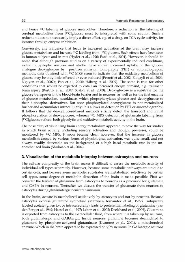
the glutamine-derived glutamate becomes decarboxylated to GABA by glutamic acid decarboxylase (GAD), an enzyme that in the brain is expressed in inhibitory GABAergic neurons (Fonnum et al., 1970). Therefore, when administration of 13C-labeled acetate to experimental animals leads to 13C labeling of GABA, this labeling illustrates a series of metabolic and transport-related events (Fig. 3) that highlights the role of (astrocytic) glutamine as a precursor of (neuronal) glutamate and GABA (Hassel et al., 1997).
Fig. 3. Metabolism of [13C]acetate in astrocytes with formation of [13C]glutamine, which is transferred to neurons, where it is deamidated to [13C]glutamate. In GABAergic neurons [13C]glutamate is decarboxylated to [13C]GABA. The spectrum is from a brain extract from a
mouse that received [1,2-13C]acetate, 150 mol i.v. and was sacrificed after 15 minutes. Peaks are: 1, alanine C-3; 2, lactate C-3; 3, GABA C-3; 5, glutamine C-3; 6, glutamate C-3; 7, glutamine C-4; 8, glutamate C-4; 10, GABA C-2; 11, aspartate C-3; 12, GABA C-4. Note the strong labeling of glutamine compared to results with [13C]glucose (Fig. 1) (From Hassel et al., 1997, with permission from the publisher). Thus, formation of [13C]GABA from [13C]acetate illustrates astrocytic uptake and oxidative metabolism of [13C]acetate, glutamine synthetase activity, the activity of glutamine transporters at the astrocytic and neuronal cell membranes, neuronal glutaminase activity, and glutamate decarboxylase activity.
www.intechopen.com

Magnetic Resonance Spectroscopy
34
An enigma of brain metabolism is why acetate is oxidized only by astrocytes and not by neurons. Waniewski and Martin (1998) provided evidence that acetate is taken up across the cell membrane only in astrocytes. This finding was unexpected, since acetate is a small monocarboxylate, for which neurons are richly equipped with transporters (Bergersen, 2007). Nguyen et al (2007b) later found that neurons take up propionate (which is closely related to acetate), which suggested that even acetate is taken up (but not metabolized) by neurons. Later it was found that administration of propionate to mice leads to an increase in brain GABA levels, suggesting an inhibitory action of propionate on the GABA-degrading enzyme GABA transaminase (Hassel et al., unpublished). GABA transaminase is an intramitochondrial enzyme in GABAergic neurons (Schousboe et al., 1974), and its inhibition by a small monocarboxylate such as propionate suggests that propionate (and acetate) is taken up by neurons and neuronal mitochondria, but that the latter are deficient in the enzyme that converts acetate and propionate into their mitochondrial CoA derivatives.
Because glutamate is readily labeled from 13C-labeled precursors, there has been considerable interest in identifying transmitter glutamate with 13C MRS. Glutamate is the main excitatory neurotransmitter in the central nervous system. While glutamate serves many functions in the brain, it has been estimated that 2-20% of the total amount of brain glutamate serves a neurotransmitter function (Hassel and Dingledine, 2006). After release from synaptic nerve endings glutamate is largely taken up by astrocytes that surround the synaptic cleft, and converted into glutamine (Danbolt, 2001). This process may be illustrated with 13C MRS. Administration of 13C-labeled glucose to experimental animals leads to rapid 13C labeling of glutamate and, after a delay, of glutamine. This labeling of glutamine to a large extent reflects uptake of (neuronal) transmitter glutamate into astrocytes. This was shown in an experiment in which mice had their astrocytic TCA cycle inhibited by the glia-specific metabolic inhibitor fluoroacetate (Hassel et al., 1997). The mice could not form glutamate or glutamine through their TCA cycle activity, but they still produced 13C-glutamine from 13C-glucose. Formation of glutamine from 13C-glucose was interpreted to reflect formation of 13C-glutamate from 13C-glucose in neurons, release of 13C-labeled transmitter glutamate from neurons, uptake into astrocytes of the released 13C-labeled transmitter glutamate and subsequent amidation by glutamine synthetase (Fig. 4).
The exchange between neurons and astrocytes of glutamate and glutamine has been the subject of many studies, in both human subjects and experimental animals (Shen et al., 1999; see Rothman et al., 1999; 2003, for review). Interpretation of 13C MRS after infusion of 13C-labeled glucose into awake humans indicated that the flux of glutamate from neurons to astrocytes was close to 50% of the cerebral metabolism of glucose. This would suggest a massive transfer of glutamate from neurons to astrocytes. However, if this is true, it is still not known whether all this glutamate is transmitter glutamate in the sense that it originates from presynaptic vesicles.
In 13C MRS studies of the brain, acetate is used as a glial substrate, and glucose is used as an energy substrate for all brain cells, but with a dominant contribution from (glutamatergic) neurons. As will become evident below ('Identification of alternative substrates for brain energy metabolism'), lactate and pyruvate have become regarded as more purely neuronal substrates that are used by glutamatergic and GABAergic neurons alike, whereas glycerol appears to be a substrate specifically used by GABAergic neurons (Nguyen et al., 2003). Thus, some degree of metabolic dissection of the brain is made possible by combining the administration of an appropriate 13C-labeled energy substrate with 13C MRS.
www.intechopen.com

13C Magnetic Resonance Spectroscopy in Neurobiology - Its Use in Monitoring
Brain Energy Metabolism and in Identifying Novel Metabolic Substrates and Metabolic Pathways
35
Fig. 4. Uptake of transmitter glutamate by astrocytes. In this experiment mice received the glia-specific metabolic inhibitor fluoroacetate, which blocks the astrocytic TCA cycle at the level of aconitase. Thus, the ability of astrocytes to form glutamate and hence glutamine from α-ketoglutarate from their own TCA cycle was blocked. Even so, glutamine was
www.intechopen.com

Magnetic Resonance Spectroscopy
36
formed from [13C]glucose, presumably because 13C-labeled transmitter glutamate was still being released from neurons and taken up by astrocytes with subsequent formation of [13C]glutamine. The upper spectrum is from the brain of a mouse that received [1,2-13C]acetate and [1-13C]glucose. The doubly labeled acetate gives rise to the double peaks in glutamine C4 (peak 7). The lower spectrum is from a mouse, whose astrocytic TCA ycle was blocked by fluoroacetate; here only glutamine labeled from [1-13C]glucose (single peak 7) can be seen (From Hassel et al., 1997, with permission from the publisher).
4. Identification of alternative substrates for brain energy metabolism
Serum glucose is considered the physiological energy substrate of the brain. Even so, a number of substrates may feed into the glycolytic pathway or the TCA cycle of brain cells. For example, lactate, which was shown by 13C MRS to be a substrate for brain metabolism in mice (Hassel and Bråthe, 2000), was recently shown by 13C MRS to contribute importantly to energy metabolism in the human brain under physiological conditions (Gallagher et al., 2009; Boumezbeur et al., 2010). The ability of a substrate to function as a metabolic substrate for brain cells depends on its ability to cross the blood-brain barrier, i.e. on the presence of specific transporter proteins in the barrier. Further, brain cells must have the necessary transporters to take the substrates up from the extracellular fluid as well as the enzymes necessary for their metabolism. A substrate that is not metabolized in the brain after i.v. injection may prove to be metabolized after intracerebral injection, a finding that points to transport limitations at the blood-brain barrier; this holds true for dicarboxylates like fumarate and malate (Hassel et al., 2002); if injected directly into the brain parenchyma, these substrates are taken up into astrocytes and rapidly metabolized through the TCA cycle to glutamine, indicating the presence of the plasma membrane dicarboxylate transporter predominantly in astrocytes. A similar situation exists for the monosaccharide fructose (Hassel et al., submitted), which hardly crosses the blood-brain barrier at all. This can be shown by giving mice [13C]fructose i.v.; in contrast to liver, the brain does not accumulate the injected [13C]fructose, as can be shown by 13C MRS of brain and liver extracts. However, if fructose is injected into the brain parenchyma, it becomes metabolized. The oxidation of fructose by brain tissue would explain the ability of fructose to support energy-requiring processes, such a axonal activity, in vitro (Meakin et al., 2007).
Even though a substrate crosses the blood-brain barrier, the enzymatic machinery required for its metabolism may not be present in the brain. An example is propionate, which readily crosses the blood-brain barrier, but hardly becomes metabolized in the brain (Nguyen et al., 2007b; Morland et al., in prep). The enzymes required for the initial metabolism of propionate, propionyl-CoA synthetase is expressed at low levels, making it difficult to detect propionate metabolites by 13C MRS after i.v. administration of [13C]propionate to experimental animals. Cerebral metabolism of propionate may be detected with radiolabeled propionate, however, owing to the higher sensitivity of radiodetective methods (scintillation counting) than of 13C MRS (Nguyen et al., 2007b).
With the use of 13C MRS -hydroxybutyrate (Künnecke et al., 1993; Pan et al., 2002; Andrews et al., 2009), lactate (Hassel and Bråthe, 2000; Tyson et al., 2003; Boumezbeur et al., 2010), and pyruvate (Gonzalez et al., 2005) have been shown to be energy substrates for the brain after i.v. injection, pointing to the existence of the appropriate monocarboxylate transporters at the blood-brain barrier. The predominant labeling of glutamate (over glutamine) points to
www.intechopen.com

13C Magnetic Resonance Spectroscopy in Neurobiology - Its Use in Monitoring
Brain Energy Metabolism and in Identifying Novel Metabolic Substrates and Metabolic Pathways
37
a predominantly neuronal metabolism of these subtrates (Fig. 5). Even octanoate has been shown to be metabolized by the brain (Ebert et al., 2003), suggesting a certain capacity for fatty acid metabolism in the brain; the preferential labeling of glutamine pointed to astrocytes as the main metabolic compartment.
Fig. 5. 13C MR spectra of brain extracts of mice that received [3-13C]pyruvate or [2-13C]pyruvate i.v. The (labeling of) individual carbon positions in certain metabolites is indicated. Wake mice received 9 mmoles/kg [3-13C]pyruvate (upper panel) or [2-13C]pyruvate (lower panel) i.v. Survival time was 5 min. [3-13C]Pyruvate labels the C3 of lactate (Lac) and alanine (Ala), the C4 of glutamate (Glu) and glutamine (Gln), and GABA C2 (but not glutamate C5 or the C1 of GABA or lactate; insert). [2-13C]Pyruvate labels lactate and alanine C2, and glutamate C5, GABA C1 and lactate C1 (insert). D is dioxane (internal standard). The peak corresponding to Glu C4 in lower spectrum represents naturally abundant 13C-glutamate. Reprinted from Gonzalez et al., 2005, with permission from the publisher.
13C-Labeled acetate also yields good labeling of cerebral amino acids, predominantly glutamine, after i.v. injection, a finding that points to their metabolism in astrocytes (see above, 3. Visualization of the metabolic interplay between astrocytes and neurons). The labeling of amino acids in the brain from [13C]acetate is on a level similar to that achieved with [13C]glucose (Hassel et al., 1995a;1997), which suggests that acetate could support brain energy metabolism; this has been shown in hypoglycemic mice (Urion et al., 1979).
www.intechopen.com

Magnetic Resonance Spectroscopy
38
An interesting example of the power of 13C MRS in elucidating cell-specific metabolism in the CNS is the avid labeling of GABA from [13C]glycerol (Nguyen et al., 2003); this labeling greatly exceeded that of glutamate and glutamine (Fig. 6), pointing to GABAergic neurons as the predominant cell compartment for glycerol metabolism. A later 13C MRS study showed that glycerol is being produced from glucose by both neurons and astrocytes and that this production increases during hypoxia or epileptic seizures (Nguyen et al., 2007c). The two findings put together (formation of glycerol in both neurons and astrocytes and its metabolism by GABAergic neurons) could point to a 'glycerol cycle' in the brain.
Fig. 6. 13C MR spectra of brain extracts from mice that received [U- 13C]glycerol i.v. Wake mice received 150 µmol [U-13C]glycerol and were killed at 5 or 15 min. Peak numbers: 1, lactate C3; 2, glutamate C4; 3, GABA C2; 4, glycerol C1 + C3. *Internal standard (dioxane). Note the strong labeling of GABA C2 relative to glutamate C4 at 15 min after injection of [U-13C]glycerol in comparison with the much higher labeling of glutamate from 13C-labeled glucose or pyruvate (Figs. 1 and 5). Reprinted from Nguyen et al., 2003, with permission from the publisher.
5. Identification of metabolic pathways from the 13
C labeling of specific carbon positions in glutamate and glutamine
In the second paragraph of this paper ('13C MRS for the visualization of brain energy metabolism') the 13C labeling of glutamate from [13C]glucose through TCA cycle activity is described. In fact, any 13C-labeled substrate that gives rise to 13C-labeled acetyl-CoA will label glutamate and glutamine in positions that reflect the entry of label into the TCA cycle and its subsequent turning in the cycle. This applies to 13C-labeled lactate, pyruvate, alanine, beta-hydroxybutyrate, acetate, butyrate, glycerol, and others. However, substrates that give
www.intechopen.com

13C Magnetic Resonance Spectroscopy in Neurobiology - Its Use in Monitoring
Brain Energy Metabolism and in Identifying Novel Metabolic Substrates and Metabolic Pathways
39
rise to 13C-labeled pyruvate in the brain (glucose, pyruvate, lactate, alanine, glycerol) may enter the TCA cycle through two different enzymatic routes. The first, which gives rise to acetyl-CoA, is pyruvate dehydrogenase, which removes a carboxylic group from pyruvate in the form of CO2. The second route involves the addition of a carboxylic group (from CO2 or HCO3-) to pyruvate to produce oxaloacetate or its immediate precursor malate (See upper part of Fig. 7); these reactions are known as pyruvate carboxylation. In astrocytes, the reaction is catalyzed by pyruvate carboxylase, in neurons, it may be catalyzed by malic enzyme. These reactions are 'anaplerotic', meaning that they function to 'fill up' the TCA cycle with intermediates. They are thought to compensate for the loss of α-ketoglutarate inherent in export of glutamine from astrocytes and for the similar loss of α-ketoglutarate inherent in glutamatergic and GABAergic neurotransmission when (See below) astrocytes take up glutamate or GABA from the synaptic cleft.
Fig. 7. Reversible pyruvate carboxylation and equilibration of malate with fumarate, which explains rapid formation of 13CO2 from [1-13C]pyruvate. [1-13C]Pyruvate undergoes carboxylation to [1-13C]malate, presumably by malic enzyme. [1-13C]Malate equilibrates with the symmetrical fumarate, so that both [1-13C]malate and [4-13C]malate are formed. Decarboxylation of these malates produces 13CO2 as well as unlabeled CO2. The 13C is represented in italics.
www.intechopen.com

Magnetic Resonance Spectroscopy
40
When pyruvate is 13C-labeled these reactions lead to the formation of 13C-labeled malate and oxaloacetate. Pyruvate labeled in the 3rd carbon (which originates from glucose labeled in the 1st position, [1-13C]glucose) leads to formation of malate or oxaloacetate labeled in the 3rd carbon. This carbon position corresponds to the 2nd carbon in glutamate and glutamine. In several studies the greater labeling of the 2nd than the 3rd carbon in glutamine has been taken as evidence of pyruvate carboxylase activity in astrocytes (Shank et al., 1993; Hassel et al., 1995a; Serres et al., 2008). A similar preferential labeling of the 2nd over the 3rd carbon in glutamate has not been consistently found, apparently pointing to the absence of pyruvate carboxylation in neurons. However, oxaloacetate and malate tend to equilibrate with the symmetrical fumarate, leading to scrambling of label between the two carbon position. 13C MRS evidence of such scrambling in neurons has been reported in both cultured neurons (Merle et al., 1996) and in vivo (Hassel et al., 2000; Gonzalez et al., 2005). Another way to study pyruvate carboxylation is to use glucose labeled in the 2nd position. Glucose labeled in this position ([2-13C]glucose) does not label acetyl-CoA, but it may label the 2nd and 3rd positions in glutamate and glutamine through pyruvate carboxylation. Mason et al (2007) showed in awake human subjects the flux through pyruvate carboxylation to be approximately 6% of that through pyruvate dehydrogenase (the activity of which was studied with [1-13C]glucose). This value was in good agreement with previous 13C MRS studies in rats (Shank et al., 1993) and with previous estimates based on radiolabeling studies (Van den Berg, 1973).
Formation of pyruvate from TCA cycle intermediates malate or oxaloacetate (through the decarboxylating activities of malic enzyme and phosphoenolpyruvate carboxykinase, respectively) is termed pyruvate recycling. This process takes place in the brain (Cerdan et al., 1990; Hassel and Sonnewald, 1995b; Cruz et al., 1998) as it does in liver. In one study this process was identified in brain by 13C MRS of brain extracts from rats that had received [13C]acetate, which labeled lactate (Hassel and Sonnewald, 1995b). Such labeling can only occur through formation of pyruvate from TCA cycle intermediates. When pyruvate thus formed (and labeled) enters the TCA cycle through pyruvate dehydrogenase a distinct labeling pattern may be seen in glutamate (Cerdan et al., 1990; Håberg et al., 1998). Valid criticism of the interpretation of previous studies has come from Serres et al. (2008) who pointed out that hepatic gluconeogenesis with 13C labeling of serum glucose from [13C]acetate may influence findings in the brain. However, pyruvate recycling also occurs in cultured brain cells (astrocytes), in which hepatic gluconeogenesis is not an issue (Sonnewald et al., 1996).
With the use of 13C MRS Gruetter and colleagues were able to determine the concentration and turnover of glycogen in the intact brain (van Heeswijk et al., 2010), and they also described the formation of N-acetyl-aspartate, which is present in the brain at high concentrations, from [13C]glucose, and they determined the turnover rate of this presumed 'inert' compound (Choi et al., 2004).
6. The use of hyperpolarization of 13
C to increase MRS sensitivity
A problem with 13C MRS is the low sensitivity of the method. This weakness implies that 13C-labeled metabolic substrates have to be given in large amounts to achieve detection of their metabolites. In contrast, radiolabeling studies are done with minute quantities of substrate. In an attempt to improve 13C MRS sensitivity, hyperpolarization of 13C-labeled
www.intechopen.com

13C Magnetic Resonance Spectroscopy in Neurobiology - Its Use in Monitoring
Brain Energy Metabolism and in Identifying Novel Metabolic Substrates and Metabolic Pathways
41
substrates has been done prior to i.v. injection. The MRS signal may thus be enhanced more than 10,000 times (Ardenkjaer-Larsen et al., 2003). In some studies pyruvate, which is labeled in the carboxylic position, [1-13C]pyruvate, has been used (Hurd et al., 2010; Marjańska et al., 2010; Mayer et al., 2011). The [13C]pyruvate is hyperpolarized immediately before i.v. injection. The hyperpolarization is lost within some seconds, however, and the detection of metabolism of pyruvate is restricted to 13C-labeled lactate, alanine, and CO2 (see Fig. 2). Formation of 13CO2 has been interpreted to reflect pyruvate dehydrogenase activity, i.e. the formation of acetyl-CoA, which may enter the TCA cycle to form citrate. But in the brain the activity of pyruvate dehydrogenase is quite low (Morland et al., 2007), and formation of TCA cycle intermediates and related amino acids takes several minutes (Gonzalez et al., 2005), so a more likely explanation for the formation of 13CO2 within a few seconds is reversible pyruvate carboxylation (Fig. 3): [13C]pyruvate is carboxylated to [13C]malate, which equilibrates with (the symmetrical) fumarate, so that 13C is distributed evenly between the two carboxylic groups of fumarate. [13C]Fumarate equilibrates with malate, which may become decarboxylated to pyruvate and CO2. Some of this CO2 will then be 13C-labeled. This series of reactions has been described in the brain with the use of [2-13C]pyruvate and [3-13C]pyruvate (Gonzalez et al., 2005), and they occur much more rapidly than the pyruvate dehydrogenase reaction. Another issue in studies that use hyperpolarized substrates is the use of anesthesia, which reduces brain metabolism of 13C-labeled substrates markedly (Shank et al., 1993).
Hyperpolarized [13C]pyruvate may prove useful in the diagnostic workup of malignant tumors, including brain tumors, which show a greater tendency to convert [13C]pyruvate into [13C]lactate than the normal surrounding tissue. Some recent studies even suggest the possibility of monitoring tumor response to therapy from the reduced conversion of pyruvate into lactate within a day’s time after irradiation or chemotherapy (Day et al., 2007; 2011; Park et al., 2011).
7. References
Andrews MT, Russeth KP, Drewes LR, Henry PG. (2009) Adaptive mechanisms regulate
preferred utilization of ketones in the heart and brain of a hibernating mammal
during arousal from torpor. Am. J. Physiol. Regul. Integr. Comp. Physiol. 296:R383-
393.
Ardenkjaer-Larsen JH, Fridlund B, Gram A, Hansson G, Hansson L, Lerche MH, Servin R,
Thaning M, Golman K. (2003) Increase in signal-to-noise ratio of > 10,000 times in
liquid-state NMR. Proc. Natl. Acad. Sci. U. S. A. 100:10158-10163.
Attwell D, Laughlin SB. (2001) An energy budget for signaling in the grey matter of the
brain. J. Cereb. Blood Flow Metab. 21:1133-1145.
Bartnik BL, Lee SM, Hovda DA, Sutton RL. (2007) The fate of glucose during the period of
decreased metabolism after fluid percussion injury: a 13C NMR study. J.
Neurotrauma. 24:1079-1092.
Bergersen LH. (2007) Is lactate food for neurons? Comparison of monocarboxylate
transporter subtypes in brain and muscle. Neuroscience 145:11-19.
Boumezbeur F, Petersen KF, Cline GW, Mason GF, Behar KL, Shulman GI, Rothman DL.
(2010) The contribution of blood lactate to brain energy metabolism in humans
www.intechopen.com

Magnetic Resonance Spectroscopy
42
measured by dynamic 13C nuclear magnetic resonance spectroscopy. J. Neurosci.
30:13983-13991.
Cerdan S, Künnecke B, Seelig J. (1990) Cerebral metabolism of [1,2-13C2]acetate as detected
by in vivo and in vitro 13C NMR. J. Biol. Chem. 265:12916-12926.
Choi IY, Gruetter R. (2004) Dynamic or inert metabolism? Turnover of N-acetyl aspartate
and glutathione from D-[1-13C]glucose in the rat brain in vivo. J. Neurochem.
91:778-787.
Cruz F, Scott SR, Barroso I, Santisteban P, Cerdán S. (1998) Ontogeny and cellular
localization of the pyruvate recycling system in rat brain. J. Neurochem. 70:2613-
2619.
Danbolt NC. (2001) Glutamate uptake. Prog. Neurobiol. 65:1-105.
Day SE, Kettunen MI, Gallagher FA, Hu DE, Lerche M, Wolber J, Golman K, Ardenkjaer-
Larsen JH, Brindle KM. (2007) Detecting tumor response to treatment using
hyperpolarized 13C magnetic resonance imaging and spectroscopy. Nat. Med.
13:1382-1387.
Day SE, Kettunen MI, Cherukuri MK, Mitchell JB, Lizak MJ, Morris HD, Matsumoto S,
Koretsky AP, Brindle KM. (2011) Detecting response of rat C6 glioma tumors to
radiotherapy using hyperpolarized [1- 13C]pyruvate and 13C magnetic resonance
spectroscopic imaging. Magn. Reson. Med. 65:557-563.
Deelchand DK, Shestov AA, Koski DM, Uğurbil K, Henry PG. (2009) Acetate transport and
utilization in the rat brain. J. Neurochem. 109 Suppl 1:46-54.
Ebert D, Haller RG, Walton ME. (2003) Energy contribution of octanoate to intact rat brain
metabolism measured by 13C nuclear magnetic resonance spectroscopy. J. Neurosci.
23:5928-5935.
Eloqayli H, Dahl CB, Götestam KG, Unsgård G, Sonnewald U. (2004) Changes of glial-
neuronal interaction and metabolism after a subconvulsive dose of
pentylenetetrazole. Neurochem. Int. 45:739-745.
Fitzpatrick SM, Hetherington HP, Behar KL, Shulman RG. (1990) The flux from glucose to
glutamate in the rat brain in vivo as determined by 1H-observed, 13C-edited NMR
spectroscopy. J. Cereb. Blood Flow Metab. 10:170-179.
Fonnum F, Storm-Mathisen J, Walberg F. (1970) Glutamate decarboxylase in inhibitory
neurons. A study of the enzyme in Purkinje cell axons and boutons in the cat. Brain
Res. 20:259-275.
Gallagher CN, Carpenter KL, Grice P, Howe DJ, Mason A, Timofeev I, Menon DK,
Kirkpatrick PJ, Pickard JD, Sutherland GR, Hutchinson PJ. (2009) The human brain
utilizes lactate via the tricarboxylic acid cycle: a 13C-labelled microdialysis and
high-resolution nuclear magnetic resonance study. Brain 132:2839-2849.
Gonzalez SV, Nguyen NH, Rise F, Hassel B. (2005) Brain metabolism of exogenous
pyruvate. J. Neurochem. 95:284-293.
Hassel B, Paulsen RE, Johnsen A, Fonnum F. (1992) Selective inhibition of glial cell
metabolism in vivo by fluorocitrate. Brain Res. 576:120-124.
Hassel B, Sonnewald U, Fonnum F. (1995a) Glial-neuronal interactions as studied by
cerebral metabolism of [2-13C]acetate and [1-13C]glucose: an ex vivo 13C NMR
spectroscopic study. J. Neurochem. 64:2773-2782.
www.intechopen.com

13C Magnetic Resonance Spectroscopy in Neurobiology - Its Use in Monitoring
Brain Energy Metabolism and in Identifying Novel Metabolic Substrates and Metabolic Pathways
43
Hassel B, Westergaard N, Schousboe A, Fonnum F. (1995b) Metabolic differences between
primary cultures of astrocytes and neurons from cerebellum and cerebral cortex.
Effects of fluorocitrate. Neurochem. Res. 20:413-420.
Hassel B, Sonnewald U. (1995a) Selective inhibition of the tricarboxylic acid cycle of
GABAergic neurons with 3-nitropropionic acid in vivo. J. Neurochem. 65:1184-
1191.
Hassel B, Sonnewald U. (1995b) Glial formation of pyruvate and lactate from TCA cycle
intermediates: implications for the inactivation of transmitter amino acids? J.
Neurochem. 65:2227-2234.
Hassel B, Bachelard H, Jones P, Fonnum F, Sonnewald U. (1997) Trafficking of amino acids
between neurons and glia in vivo. Effects of inhibition of glial metabolism by
fluoroacetate. J. Cereb. Blood Flow Metab. 17:1230-1238.
Hassel B, Bråthe A. (2000) Cerebral metabolism of lactate in vivo: evidence for neuronal
pyruvate carboxylation. J. Cereb. Blood Flow Metab. 20:327-336.
Hassel B, Dingledine R. (2006) Glutamate. In GJ Siegel (ed) Basic Neurochemistry. Elsevier,
pp. 267-290.
Hurd RE, Yen YF, Mayer D, Chen A, Wilson D, Kohler S, Bok R, Vigneron D, Kurhanewicz
J, Tropp J, Spielman D, Pfefferbaum A. (2010) Metabolic imaging in the
anesthetized rat brain using hyperpolarized [1-13C] pyruvate and [1-13C] ethyl
pyruvate. Magn. Reson. Med. 63:1137-1143.
Hyder F, Chase JR, Behar KL, Mason GF, Siddeek M, Rothman DL, Shulman RG. (1996)
Increased tricarboxylic acid cycle flux in rat brain during forepaw stimulation
detected with 1H[13C]NMR. Proc. Natl. Acad. Sci. U. S. A. 93:7612-7617.
Hyder F, Brown P, Nixon TW, Behar KL. (2003) Mapping cerebral glutamate 13C turnover
and oxygen consumption by in vivo NMR. Adv. Exp. Med. Biol. 530:29-39.
Håberg A, Qu H, Bakken IJ, Sande LM, White LR, Haraldseth O, Unsgård G, Aasly J,
Sonnewald U. (1998) In vitro and ex vivo 13C-NMR spectroscopy studies of
pyruvate recycling in brain. Dev. Neurosci. 20:389-398.
Håberg AK, Qu H, Sonnewald U. (2009) Acute changes in intermediary metabolism in
cerebellum and contralateral hemisphere following middle cerebral artery
occlusion in rat. J. Neurochem. 109 Suppl 1:174-181.
Johannessen CU, Qu H, Sonnewald U, Hassel B, Fonnum F. (2001) Estimation of aspartate
synthesis in GABAergic neurons in mice by 13 C NMR spectroscopy. Neuroreport
12:3729-3732.
Künnecke B, Cerdan S, Seelig J. (1993) Cerebral metabolism of [1,2-13C2]glucose and [U-13C4]3-hydroxybutyrate in rat brain as detected by 13C NMR spectroscopy. NMR
Biomed. 6:264-277.
Kvamme E, Torgner IA, Roberg B. (2001) Kinetics and localization of brain phosphate
activated glutaminase. J. Neurosci. Res. 66:951-958.
Lebon V, Petersen KF, Cline GW, Shen J, Mason GF, Dufour S, Behar KL, Shulman GI,
Rothman DL. (2002) Astroglial contribution to brain energy metabolism in humans
revealed by 13C nuclear magnetic resonance spectroscopy: elucidation of the
dominant pathway for neurotransmitter glutamate repletion and measurement of
astrocytic oxidative metabolism. J. Neurosci. 22:1523-1531.
www.intechopen.com

Magnetic Resonance Spectroscopy
44
Marjańska M, Iltis I, Shestov AA, Deelchand DK, Nelson C, Uğurbil K, Henry PG. (2010) In
vivo 13C spectroscopy in the rat brain using hyperpolarized [1-13C]pyruvate and [2-13C]pyruvate. J. Magn. Reson. 206:210-218.
Martinez-Hernandez A, Bell KP, Norenberg MD. (1977) Glutamine synthetase: glial
localization in brain. Science 195:1356-1358.
Mason GF, Pan JW, Chu WJ, Newcomer BR, Zhang Y, Orr R, Hetherington HP. (1999)
Measurement of the tricarboxylic acid cycle rate in human grey and white matter in
vivo by 1H-[13C] magnetic resonance spectroscopy at 4.1T. J. Cereb. Blood Flow
Metab. 19:1179-1188.
Mason GF, Petersen KF, de Graaf RA, Shulman GI, Rothman DL. (2007) Measurements of
the anaplerotic rate in the human cerebral cortex using 13C magnetic resonance
spectroscopy and [1-13C] and [2-13C] glucose. J. Neurochem. 100:73-86.
Mason GF, Petersen KF, Lebon V, Rothman DL, Shulman GI. (2006) Increased brain
monocarboxylic acid transport and utilization in type 1 diabetes. Diabetes 55:929-
934.
Mayer D, Yen YF, Takahashi A, Josan S, Tropp J, Rutt BK, Hurd RE, Spielman DM,
Pfefferbaum A. (2011) Dynamic and high-resolution metabolic imaging of
hyperpolarized [1-13C]-pyruvate in the rat brain using a high-performance gradient
insert. Magn. Reson. Med. 65:1228-1233.
Merle M, Martin M, Villégier A, Canioni P. (1996) [1-13C]glucose metabolism in brain cells:
isotopomer analysis of glutamine from cerebellar astrocytes and glutamate from
granule cells. Dev. Neurosci. 18:460-468.
Morland C, Henjum S, Iversen EG, Skrede KK, Hassel B. (2007) Evidence for a higher
glycolytic than oxidative metabolic activity in white matter of rat brain.
Neurochem. Int. 50:703-709.
Nguyen NH, Bråthe A, Hassel B. (2003) Neuronal uptake and metabolism of glycerol and
the neuronal expression of mitochondrial glycerol-3-phosphate dehydrogenase. J.
Neurochem. 85:831-842.
Nguyen N, Gonzalez SV, Rise F, Hassel B. (2007a) Cerebral metabolism of glucose and
pyruvate in soman poisoning. A 13C nuclear magnetic resonance spectroscopic
study. Neurotoxicology 28:13-18.
Nguyen NH, Morland C, Gonzalez SV, Rise F, Storm-Mathisen J, Gundersen V, Hassel B.
(2007b) Propionate increases neuronal histone acetylation, but is metabolized
oxidatively by glia. Relevance for propionic acidemia. J. Neurochem. 101:806-814.
Nguyen NH, Gonzalez SV, Hassel B. (2007c) Formation of glycerol from glucose in rat brain
and cultured brain cells. Augmentation with kainate or ischemia. J. Neurochem.
101:1694-1700.
Ottersen OP, Storm-Mathisen J. (1985) Different neuronal localization of aspartate-like and
glutamate-like immunoreactivities in the hippocampus of rat, guinea-pig and
Senegalese baboon (Papio papio), with a note on the distribution of gamma-
aminobutyrate. Neuroscience 16:589-606.
Pan JW, Williamson A, Cavus I, Hetherington HP, Zaveri H, Petroff OA, Spencer DD. (2008)
Neurometabolism in human epilepsy. Epilepsia. 49 Suppl 3:31-41.
www.intechopen.com

13C Magnetic Resonance Spectroscopy in Neurobiology - Its Use in Monitoring
Brain Energy Metabolism and in Identifying Novel Metabolic Substrates and Metabolic Pathways
45
Pan JW, de Graaf RA, Petersen KF, Shulman GI, Hetherington HP, Rothman DL. (2002) [2,4-13C2 ]-beta-Hydroxybutyrate metabolism in human brain. J. Cereb. Blood Flow
Metab. 22:890-898.
Park I, Bok R, Ozawa T, Phillips JJ, James CD, Vigneron DB, Ronen SM, Nelson SJ. (2011)
Detection of early response to temozolomide treatment in brain tumors using
hyperpolarized 13C MR metabolic imaging. J. Magn. Reson. Imaging 33:1284-1290.
Patel AB, de Graaf RA, Mason GF, Kanamatsu T, Rothman DL, Shulman RG, Behar KL.
(2004) Glutamatergic neurotransmission and neuronal glucose oxidation are
coupled during intense neuronal activation. J. Cereb. Blood Flow Metab. 24:972-
985.
Petroff OA, Errante LD, Rothman DL, Kim JH, Spencer DD. (2002) Glutamate-glutamine
cycling in the epileptic human hippocampus. Epilepsia 43:703-710.
Rodrigues TB, Fonseca CP, Castro MM, Cerdán S, Geraldes CF. (2009) 13C NMR tracers in
neurochemistry: implications for molecular imaging. Q. J. Nucl. Med. Mol. Imaging
53:631-645.
Rothman DL, Sibson NR, Hyder F, Shen J, Behar KL, Shulman RG. (1999) In vivo nuclear
magnetic resonance spectroscopy studies of the relationship between the
glutamate-glutamine neurotransmitter cycle and functional neuroenergetics.
Philos. Trans. R. Soc. Lond. B Biol. Sci. 354:1165-1177.
Rothman DL, Behar KL, Hyder F, Shulman RG. (2003) In vivo NMR studies of the glutamate
neurotransmitter flux and neuroenergetics: implications for brain function. Annu.
Rev. Physiol. 65:401-27.
Scafidi S, O'Brien J, Hopkins I, Robertson C, Fiskum G, McKenna M. (2009) Delayed cerebral
oxidative glucose metabolism after traumatic brain injury in young rats. J.
Neurochem. 109 Suppl 1:189-197.
Serres S, Bezancon E, Franconi JM, Merle M. (2007) Brain pyruvate recycling and peripheral
metabolism: an NMR analysis ex vivo of acetate and glucose metabolism in the rat.
J. Neurochem. 101:1428-1240.
Serres S, Raffard G, Franconi JM, Merle M. (2008) Close coupling between astrocytic and
neuronal metabolisms to fulfill anaplerotic and energy needs in the rat brain. J.
Cereb. Blood Flow Metab. 28:712-724.
Shank RP, Leo GC, Zielke HR. (1993) Cerebral metabolic compartmentation as revealed by
nuclear magnetic resonance analysis of D-[1-13C]glucose metabolism. J.
Neurochem. 61:315-323.
Shen J, Petersen KF, Behar KL, Brown P, Nixon TW, Mason GF, Petroff OA, Shulman GI,
Shulman RG, Rothman DL. (1999) Determination of the rate of the
glutamate/glutamine cycle in the human brain by in vivo 13C NMR. Proc. Natl.
Acad. Sci. U. S. A. 96:8235-8240.
Shulman RG, Rothman DL, Behar KL, Hyder F. (2004) Energetic basis of brain activity:
implications for neuroimaging. Trends Neurosci. 27:489-495.
Siesjö BK. (1978) Brain Energy Metabolism, New York.
Sonnewald U, Westergaard N, Jones P, Taylor A, Bachelard HS, Schousboe A. (1996)
Metabolism of [U-13C5] glutamine in cultured astrocytes studied by NMR
spectroscopy: first evidence of astrocytic pyruvate recycling. J. Neurochem.
67:2566-2572.
www.intechopen.com
Magnetic Resonance Spectroscopy
46
Tyson RL, Gallagher C, Sutherland GR. (2003) 13C-Labeled substrates and the cerebral
metabolic compartmentalization of acetate and lactate. Brain Res. 992:43-52.
Urion D, Vreman HJ, Weiner MW. (1979) Effect of acetate on hypoglycemic seizures in mice.
Diabetes 28:1022-1026.
Van den Berg, C. J. (1973) Metabolic compartmentation in the brain (Balázs R and Cremer JE,
eds), Macmillan, London, pp. 137–166.
van Heeswijk RB, Morgenthaler FD, Xin L, Gruetter R. (2010) Quantification of brain
glycogen concentration and turnover through localized 13C NMR of both the C1
and C6 resonances. NMR Biomed. 23:270-276.
Waniewski RA, Martin DL. (1998) Preferential utilization of acetate by astrocytes is
attributable to transport. J. Neurosci. 18:5225-5233.
www.intechopen.com

Magnetic Resonance SpectroscopyEdited by Prof. Dong-Hyun Kim
ISBN 978-953-51-0065-2Hard cover, 264 pagesPublisher InTechPublished online 02, March, 2012Published in print edition March, 2012
InTech EuropeUniversity Campus STeP Ri Slavka Krautzeka 83/A 51000 Rijeka, Croatia Phone: +385 (51) 770 447 Fax: +385 (51) 686 166www.intechopen.com
InTech ChinaUnit 405, Office Block, Hotel Equatorial Shanghai No.65, Yan An Road (West), Shanghai, 200040, China
Phone: +86-21-62489820 Fax: +86-21-62489821
Magnetic Resonance Spectroscopy (MRS) is a unique tool to probe the biochemistry in vivo providingmetabolic information non-invasively. Applications using MRS has been found over a broad spectrum ininvestigating the underlying structures of compounds as well as in determining disease states. In this book,topics of MRS both relevant to the clinic and also those that are beyond the clinical arena are covered. Thebook consists of two sections. The first section is entitled 'MRS inside the clinic' and is focused on clinicalapplications of MRS while the second section is entitled 'MRS beyond the clinic' and discusses applications ofMRS in other academic fields. Our hope is that through this book, readers can understand the broadapplications that NMR and MRS can offer and also that there are enough references to guide the readers forfurther study in this important topic.
How to referenceIn order to correctly reference this scholarly work, feel free to copy and paste the following:
Bjørnar Hassel (2012). 13C Magnetic Resonance Spectroscopy in Neurobiology - Its Use in Monitoring BrainEnergy Metabolism and in Identifying Novel Metabolic Substrates and Metabolic Pathways, MagneticResonance Spectroscopy, Prof. Dong-Hyun Kim (Ed.), ISBN: 978-953-51-0065-2, InTech, Available from:http://www.intechopen.com/books/magnetic-resonance-spectroscopy/13c-magnetic-resonance-spectroscopy-in-neurobiology-its-use-in-monitoring-brain-energy-metabolism-an