Embed Size (px)
Citation preview


Centro Congressi d’Ateneo “Federico II” – Via Partenope, NapoliJune 7-10, 2012
13th Naples Workshop on Bioactive Peptides
CONFORMATION AND ACTIVITY IN PEPTIDES:
RELATIONSHIPS AND INTERACTIONS
Organized by:Centro Interuniversitario di Ricerca sui Peptidi Bioattivi (CIRPeB)
Università di Napoli “Federico II” – Dipartimento delle Scienze BiologicheIstituto di Biostrutture e Bioimmagini del Consiglio Nazionale delle Ricerche
Istituto di Cristallografia del Consiglio Nazionale delle RicercheDFM Scarl
Under the auspicies of
Consiglio Nazionale delle RicercheEuropean Peptide Society

2
Digital reference: www.cirpeb.unina.it/naples2012
Official photographer: Ludovica MorelliArt & Design: Luca De Luca
3
Honorary Chair
Prof. Carlo Pedone
Co-Chairmen
Prof. Giancarlo MorelliDr. Michele Saviano
Organizing Committee
G. Morelli University of Naples “Federico II”
M. Saviano Institute of Crystallography, CNR
L. De Luca Institute of Biostructures and Bioimaging, CNR
M. Ruvo Institute of Biostructures and Bioimaging, CNR
P. Grieco University of Naples “Federico II”
D. Tesauro University of Naples “Federico II”
P.V. Pedone Second University of Naples
L. Vitagliano Institute of Biostructures and Bioimaging, CNR
R. Fattorusso Second University of Naples
Scientific Committee
F. Hudecz Eötvös L. University - Budapest, Hungary
C. Toniolo University of Padua - Padua, Italy
L. Moroder Max Planck Institute - Martinsried, Germany
D. Andreu Universitat Pompeu Fabra - Barcelona, Spain
G. Morelli University of Naples “Federico II” - Naples Italy
M. Saviano Institute of Crystallography, CNR - Bari, Italy

4
13th Naples Workshop on Bioactive Peptides — Conformation and Activity in Peptides: Relationships and Interactions
Acknowledgments
The Organizing Committee gratefully acknowledges the support and assistance of the following Institutions:
Università di Napoli ˝Federico II˝ – Dipartimento delle Scienze Biologiche
Centro Interuniversitario di Ricerca sui Peptidi Bioattivi (CIRPeB)
Istituto di Biostrutture e Bioimmagini del Consiglio Nazionale delle Ricerche
Istituto di Cristallografia del Consiglio Nazionale delle Ricerche
Ministero dell'Università e della Ricerca Scientifica (MIUR)
Ordine dei Farmacisti della Provincia di Napoli
DFM Scarl
Regione Campania Assessorato Università e Ricerca Scientifica
European Peptide Society (EPS)

5
Conformation and Activity in Peptides: Relationships and Interactions
The Organizing Committee gratefully acknowledges the contribution given to the organization of the event by the following companies

6
13th Naples Workshop on Bioactive Peptides — Conformation and Activity in Peptides: Relationships and Interactions

7
Conformation and Activity in Peptides: Relationships and Interactions
Content
Program 9
Plenary Lectures and Key Notes 15
Murray Goodman Young investigators’ session 33
Oral presentations 43
Poster presentations 59
List of titles 135
List of authors 139

8

PROGRAM

10
13th Naples Workshop on Bioactive Peptides — Conformation and Activity in Peptides: Relationships and Interactions
Thursday, June 7 th
2:00 pm -6:00 pm Registration
6:00 pm - 6:30 pm Welcome Addresses
6:30 pm - 7:30 pm PL1 M. Chorev Cambridge, MA, USAGlycated CD59 - A novel biomarker in diabetes
7:30 pm - 9:00 pm Welcome Party
Friday, June 8 th
SESSION I
The structure of peptides and their interaction with biomoleculesChairmen: L. Moroder - M. Saviano
8:45 am - 9:30 am PL2 D. Mierke Hanover, NH, USAModulating GPCR signaling by targeting the cytoplasmic domains
9:30 am - 10:00 am KN1 F. Formaggio Padua, ItalyThe fully-extended/310-helix molecular switch
10:00 am - 10:20 am O1 J. Seelig Basel, SwitzerlandCell penetrating peptides: how do they get through membranes?
10:20 am - 10:45 am Coffee break. Offered by by Avantech Srl
SESSION II
The structure of peptides and their interaction with biomoleculesChairmen: C. Pedone - L. Bracci
10:45 am - 11:30 am PL3 J. Lambris Philadelphia, PA, USAPeptide based complement inhibitors: from bench to bedside
11:30 am - 11:50 am O2 N. Doti Naples, ItalyInhibition of apoptosis inducing factor-mediated neuronal loss by a peptide antagonist of Cyclophilin A
11:50 am - 12:10 pm O3 M. Larregola Paris, FranceDevelopment of β-turn mimics to inhibit protein-protein interactions
12:10 pm - 12:30 pm O4 R. Fattorusso Caserta, ItalyNMR characterization of a potent angiogenic peptide functional mechanism in living cells

11
Program
SESSION IIIPeptides in nanotechnology and nanomedicine
Chairman: G. Morelli
12:30 pm - 1:00 pm KN2 G. Rosenman Tel Aviv, IsraelNanotechnology inspired by nature: basic physics and engineering of peptide supramolecular nanostructures
1:00 pm Lunch Break
Murray Goodman young investigators' sessionChairmen: M. Chorev - S. Galdiero
3:00 pm - 3:20 pm Y1 A. Cardoso Coimbra, PortugalEfficient siRNA delivery mediated by S4(13)-PV cell penetrating peptide-based systems: a comparative study
3:20 pm - 3:40 pm Y2 A. Sandomenico Naples, ItalyTargeting Cripto-containing complexes for therapeutic and diagnostic applications
3:40 pm - 4:00 pm Y3 J. Freire Lisbon, PortugalMolecular therapy using dengue virus proteins for peptide-mediated nucleic acid delivery
4:00 pm - 4:20 pm Y4 H. Memczak St. Ingbert, GermanyDirect detection of intact influenza viruses using peptide-based biosensors
4:20 pm - 4:40 pm Y5 R. Tarallo Naples, ItalyFunctionalization of a dendrimer with a membrane-interacting domain of Herpes Simplex virus Type 1: applications toward intracellular delivery
4:40 pm - 5:00 pm Y6 D. Roversi Rome, ItalyMembrane-perturbing effects of antimicrobial peptides: a systematic spectroscopic analysis
5:00 pm - 5:20 pm Y7 C. Avitabile Naples, ItalyTargeting pre-miRNA by PNAs: a New Strategy to Interfere in the miRNA Function
5:20 pm - 5:40 pm Coffee Break. Offered by by Inbios srl
5:40 pm - 7:30 pm Poster session - Discussion of posters with EVEN numbers
saTurday, June 9 th
SESSION IV
Peptides in nanotechnology and nanomedicineChairmen: C. Toniolo - A. Romanelli
8:30 am - 9:15 am PL4 A. Bianco Strasbourg, FrancePeptide and protein functionalized carbon nanotubes for cell interfacing and targeted drug delivery
9:15 am - 9:45 am KN3 J. De La Fuente Zaragoza, SpainMultifunctional gold nanoparticles functionalized with cell penetrating peptides and siRNA for gene silencing

12
13th Naples Workshop on Bioactive Peptides — Conformation and Activity in Peptides: Relationships and Interactions
9:45 am - 10:05 am O5 L. Bracci Siena, ItalyDevelopment of branched peptides as cancer theranostics
10:05 am - 10:25 am O6 A.S. Veiga Lisbon, PortugalArginine containing peptide-based hydrogels as injectable antibacterial materials
10:25 am - 11:00 am Coffee break. Offered by Shimadzu Italia
SESSION V
Peptides in nanotechnology and nanomedicineChairmen: A. Bianco - D. Tesauro
11:00 am - 11:30 am KN4 M. Venanzi Rome, ItalyPeptide self-assembled monolayers: a new platform for nanotechnology
11:30 am - 12:00 am KN5 D. Andreu Barcelona, SpainDiscovering antimicrobial peptide motifs in proteins: search tools and potential evolutionary mechanisms
12:00 am - 12:30 pm KN6 F. Hudecz Budapest, HungaryCell penetrating conjugates of calpastatin derived oligopeptides for cellular monitoring
12:30 pm - 12:50 pm O7 S. Galdiero Naples, ItalyViral peptide shuttles for intracellular delivery
12:50 pm - 1:10 pm O8 A. Moretto Padua, ItalyPeptide self-assembly and microstructure formation
1:10 pm Lunch Break
SESSION VI
Peptides for diagnostic and therapeutic applicationsChairmen: D. Andreu - P. Grieco
3:15 pm - 3:45 pm KN7 A.M. Papini Frorence, ItalyThe mimicry pathway from glycopeptides to specific protein antigens of autoimmune neurodegenerative diseases
3:45 pm - 4:05 pm O9 R. Falcioni Rome, ItalyInhibition of ErbB-3/p85 interaction overcomes trastuzumab resistance of human breast cancer
4:05 pm - 4:25 pm O10 P. Carvajal-Rondanelli Valparaiso, ChileModel cationic peptides and their antimicrobial activity
4:25 pm - 4:45 pm O11 M. De Zotti Padua, ItalyShort peptaibiotics as new antitumor agents: synthesis, conformational analysis and cytotoxicity evaluation of trichogin GA IV and selected analogs thereof

13
Program
SESSION VII
Peptide drug designChairman: D. Andreu
4:45 pm - 5:30 pm PL5 A. Ferrer-Montriel Alicante, SpainTRPducins: a new peptide-based paradigm for modulating ion channel activity
5:30 pm - 7:00 pm Poster session - Discussion of posters with ODD numbers(Coffee break will be served during this session)
9:00 pm - 00:30 am Gala Dinner
sunday, June 10 th
SESSION VIII
Peptide drug designChairmen: D. Mierke - A. M. Papini
9:00 am - 9:45 am PL6 G. Bulaj Salt Lake City, (UT) USAConverting neuroactive peptides into drug leads for pain and epilepsy
9:45 am - 10:05 am O12 D. Marasco Naples, ItalyNew mimetic peptides of kinase inhibitory region (KIR) of SOCS1 through focused peptide libraries for the modulation of the JAK-STAT pathway
10:05 am - 10:25 am O13 B. De Spiegeleer Gent, BelgiumBlood-brain barrier modelling of bioactive peptides
10:25 am - 11:00 am Coffee break. Offered by Delchimica Scientific Glassware srl
SESSION IX
Synthesis of peptides, proteins and related analogsChairmen: F. Hudecz - M. Ruvo
11:00 am - 11:30 am KN8 O. Seitz Berlin GermanyRapid chemical synthesis of immobilized SH3 domains
11:30 am - 11:50 am O14 R. Latajka Wroklaw, PolandPhosphinopeptides as inhibitors of cathepsin C- Design, synthesis and biological studies
11:50 am - 12:20 pm KN9 M. Marchetti-Deschmann Vienna, AustriaUsing mass spectrometry for localization and structure elucidation of surface/tissue associated peptides
12:20 pm - 12:50 pm KN10 F. Dettner Bubendorf, SwitzerlandFrom process research to large scale manufacturing of peptide APIs

14

PLENARY LECTURES AND KEY NOTES

16
13th Naples Workshop on Bioactive Peptides — Conformation and Activity in Peptides: Relationships and Interactions
PL1
Glycated CD59 – A novel biomarker in diabetesM. Chorev
Laboratory for Translational Research, Harvard Medical School, Cambridge, MA 02139Department of Medicine, Brigham and Women’s Hospital, Boston, MA 02115
The number of adults with diabetes has more than doubled since 1980. Diabetes affected more than 366 million people worldwide in 2011 and is expected to rise to 552 million by 2030. High blood glucose and diabetes causes more than 4.6 million deaths globally each year and the annual healthcare spending on diabetes has reached 465 billion USD. Importantly, it is estimated that 79 million Americans have pre-diabetes most of them undiagnosed. As such, there is an unmet clinical need to develop diagnostic tests that will enable more timely and accurate diagnosis of diabetics and pre-diabetics, monitor the glycemic state, and assess the risk of vascular diabetic complications. Our studies aim at the development of a novel diagnostic platform based on a disease-relevant biomarker sensitive to the glycemic state and intimately involved in the debilitating complications of diabetes. Targeting a post-translationally modified biomarker such as glycated CD59 presented some unique challenges and demanded untraditional solutions, which may be applicable to wide range of post-translationally modified biomarkers.

17
Plenary Lectures and Key Notes
PL2
Modulating GPCR Signaling by Targeting the Cytoplasmic Domains.
D. F. Mierke
Dept. Chemistry, 6128 Burke Hall, Dartmouth College, Hanover NH 03755
We will describe our efforts to develop molecular regulators of GPCR signaling by targeting the multiple interactions involving the cytoplasmic domains of these receptors. We have developed a number of model systems allowing for screening of chemical libraries using both high throughput and NMR-based methods. Results from our studies of the receptors for parathyroid hormone, endothelin, angiotensin, and glutamate (mGluR1/5) will be highlighted.

18
13th Naples Workshop on Bioactive Peptides — Conformation and Activity in Peptides: Relationships and Interactions
KN1
The fully-extended conformation/310-helix molecular switchF. Formaggio
ICB, Padova Unit, CNR, Department of Chemistry, University of Padova, 35131 Padova, Italy
Molecular switches based on peptides enjoy attractive features, such as a relatively easy synthesis, the chance to shape them into a desired 3D-structure, and the possibility to fine tuning spectroscopic and physical properties by conformational changes. The fundamental property for any type of molecular switch is the occurrence of an equilibrium between two different molecular conformers, which can be shifted toward either direction by means of an external stimulus or an environmental modification.In the last 30 years peptides rich in specific Cα-tetrasubstituted residues were exploited to characterize the fully-extended (C5) conformation and the related 2.05-helix. Multiple, consecutive C5 conformations were observed in homo-peptides based on residues which possess two side chains longer than a methyl (e.g., Deg, Cα,α-diethylglycine). Interestingly, the axial translation per residue in the 2.05-helix is the longest possible (about 3.85 Å) for a single amino acid, which makes this conformation extremely attractive for its use as a spacer or bridge. However, subtle perturbations of the chemical structure or of the environment may promote a conformational transition to the 310-helix (about 50% shorter), thus making these peptides of great interest as molecular switches as well. With the aim at detecting the most appropriate chemical structures and experimental conditions for the stabilization of the 2.05-helix, we synthesized and characterized several series of Deg homo-peptides, differing by the nature of the terminal protecting (or blocking) groups. We indeed observed that an N-terminal Tfa (trifluoroacetyl) group stabilizes the 2.05-helix; at the C-terminus esters or tertiary amides are compatible with the 2.05-helix, whereas primary and secondary amides favor the formation of a 310-helix because of their extra H-bonding donor NH groups which are unsatisfied in the 2.05-helix. We also noticed a dramatic influence of the nature of solvent on the 3D-structure of these peptides. In particular, we characterized a solvent-driven, 2.05-/310-helix, conformational switch by IR absorption, NMR, and fluorescence techniques.
Figure 1. A -(Deg)5- segment in the 310- and 2.05-helical conformations. The solvent-induced, spring-like behavior is highlighted.

19
Plenary Lectures and Key Notes
PL3
Compstatin: a complement inhibitor on its way to clinical application.
J. D. Lambris
Department of Pathology & Laboratory Medicine, University of Pennsylvania, Philadelphia, USA
Therapeutic modulation of the human complement system is considered a promising approach for treating a number of pathological conditions. Owing to its central position in the cascade, component C3 is a particularly attractive target for complement-specific drugs. Compstatin, a cyclic tridecapeptide, which was originally discovered from phage-display libraries, is a highly potent and selective C3 inhibitor that demonstrated clinical potential in a series of experimental models. A combination of chemical, biophysical, and computational approaches allowed a remarkable optimization of its binding affinity towards C3 and its inhibitory potency. I will discuss the mechanism by which Compstatin inhibits complement activation and novel design approaches for enhancing its stability, the inhibitory efficacy, tissue targeting, and plasma half-life of Compstatin to broaden treatment applications. Finally, I will present data on the efficacy of Compstatin to treating various diseases and clinical conditions.

20
13th Naples Workshop on Bioactive Peptides — Conformation and Activity in Peptides: Relationships and Interactions
KN2
Nanotechnology inspired by nature: basic physics and engineering of peptide supramolecular nanostructures
G. Rosenman
School of Electrical Engineering-Physical Electronics, Tel Aviv University, Tel Aviv, 69978, Israel
The emerging “bottom-up” nanotechnology reveals a new field of bioinspired nanomaterials composed from chemically synthesized biomolecules. They are formed from elementary constituents into supramolecular structures by the use of developed by nature self assembly mechanism.. The focus of this Lecture is on intrinsic fundamental physical properties of bioinspired peptide nanostructures and their small building units linked by weak noncovalent bonds. The observed exceptional optical properties indicate on phenomenon of quantum confinement in these supramolecular structures, which originates from nanoscale size of their elementary building blocks. The dimensionality of the confinement gives insight into intrinsic packing of peptide supramolecular nanomaterials. We also describe found at nanoscale ferroelectric and related properties based on original crystalline asymmetry of the nanoscale units, packing these structures. In this context, we reveal classic solid state physics phenomenon such as reconstructive phase transition observed in bioorganic peptide nanotubes. This irreversible phase transformation leads the drastic reshaping of their quantum structure from quantum dots to quantum wells, which is followed by variation of their space group symmetry from asymmetric to symmetric. We show that supramolecular origin of these bioinspired nanomaterials provide unique chance to be dissembled into elementary building blocks peptide nanodots of 1-2 nm size possessing unique electron, optical and ferroelectric properties. These multifunctional nanounits could lead to a new future step down in nanotechnology and nanoscale advanced devices in the fields of nanophotonics, nanobiomedicine, nanobiopiezotronics, etc.

21
Plenary Lectures and Key Notes
PL4
Peptide and protein functionalized carbon nanotubes for cell interfacing and targeted drug delivery
A. Bianco
CNRS, Institut de Biologie Moléculaire et Cellulaire, Laboratoire d’Immunologie et Chimie Thérapeutiques, 67000 Strasbourg, France
Carbon nanotubes (CNTs) are considered an advanced nanomaterial for different applications in the domain of materials science and nanomedicine. The potential of functionalized CNTs for therapeutic and diagnostic purposes is currently attracting great interests. CNTs are a form of carbon constituted of graphene layers rolled-up into a cylindrical shape. Discovered in the 1950s and structurally described at atomic level in 1991 by S. Iijima, they immediately appeared as interesting materials because of their chemico-physical features. Carbon nanotubes can be either composed by a single sheet of graphene (single-walled carbon nanotubes) or by multiple concentric layers (multi-walled carbon nanotubes). Their diameters are in the nanometer scale, while their lengths can reach several microns. The combination of their mechanical, thermal, chemical and electronic properties makes both single- and multi-walled CNTs unique materials. In the biomedical domain and in nanomedicine, they can be considered novel and innovative tools for the development of alternative methodologies for the delivery of therapeutic and diagnostic molecules. One major concern related to the extreme difficulty to manipulate this material due to the insolubility in all types of solvents, particularly in aqueous solutions, has been solved. In this context, we have explored the possibility to develop complex antigenic systems and novel vectors for peptides, nucleic acids, and drugs covalently linked or complexed to carbon nanotubes. This is mainly owing to the established capacity of CNTs to penetrate into the cells with remarkably reduced toxic effects. In this presentation we will describe the design and applications of peptide and protein (i.e. antibodies) functionalized carbon nanotubes as new supports for cell growth and as new targeted conjugates against cancer cells. We will also give a brief overview on the issues related to their possible toxic effects and their potential biodegradation.

22
13th Naples Workshop on Bioactive Peptides — Conformation and Activity in Peptides: Relationships and Interactions
KN3
Multifunctional Gold Nanoparticles Functionalized with Cell Penetrating Peptides and siRNA for Gene Silencing
V. Sanz,1 J. Conde,1,2 A. Ambrosone,3 Y. Hernández,1 V. Marchesano,3 G.G. Estrada,4 M.R. Ibarra,1 P.V. Baptista,2 F. Tian,4 C. Tortiglione3, J.M. de la Fuente1
1. Institute of Nanoscience of Aragon, University of Zaragoza, 50018, Zaragoza, Spain.2. Centro de Investigação em Genética Molecular Humana (CIGMH), Departamento de Ciências da Vida,
Faculdade de Ciências e Tecnologia, Universidade Nova de Lisboa, 2829-516 Caparica, Portugal.3. Istituto di Cibernetica “E.Caianiello”, Consiglio Nazionale delle Ricerche, 80078 Pozzuoli, Italy4. Helmholtz Zentrum München, 85764 Neuherberg, Munich, Germany.
The use of inorganic nanoparticles as drug release systems is nowadays gaining power. One of the most used nanoparticles for biomedical applications are gold nanoparticles (AuNPs). AuNPs provide non-toxic carriers and have been used in highly sensitive diagnostic assays, thermal ablation and as drug and gene delivery. Small interfering-RNAs (siRNA) show significant potential in new molecular approaches to down-regulate specific gene expression in cancerous or viral-infected cells. However, there are still significant obstacles to be overcome such as its short half-lives and degradation by RNases. We have developed effective conjugation strategies to combine, in a highly controlled way, biomolecules to the surface of AuNPs with specific functions such as cell penetrating peptides to overcome the cellular membrane barrier, quaternary ammonium to introduce stable positively charged in AuNPs surface and siRNA complementary to a master regulator gene, the proto-oncogene c-Myc. This gene is implicated in cell growth, proliferation, loss of differentiation and apoptosis. Two approaches were designed for the binding of all these molecules to the nanoparticles, the use of a thiolated siRNA for binding covalently to the surface of the nanoparticles; and by ionic interactions incorporation positive charge to the nanoparticles. These nanoparticles were characterized on their chemical functionalization, ease of uptake, cellular toxicity and knockdown of MYC protein expression in a cancer cell line. The results showed in human cells confirmed the high efficiency of these nanoparticles for silencing MYC expression. These results are in high concordance with the obtained results on other biological systems (Hydra and mice)

23
Plenary Lectures and Key Notes
KN4
Peptide self-assembled monolayers as a new tool for bio-inspired nanotechnology
M. Venanzi, E. Gatto, M. Caruso
University of Rome ‘Tor Vergata’ , Department of Chemical Sciences and Technologies, 00133 – Rome
Peptide-based self-assembled monolayers (SAMs) have been raising more and more interest for their versatility and possible applications as enantioselective sensors, biocompatible platforms for tissue engineering, and smart elements in hybrid devices.We have recently reported that a short hexapeptide, predominantly formed by Cα-tetrasubstituted residues, is able to form a densely packed SAM when chemically linked on a gold surface. This is because these non metabolic amino acids preferentially populate helical conformations and the latter were shown to promote the regular deposition of peptides on solid substrates, giving rise to densely packed SAMs almost vertically arranged with respect to the surface.[1] Stable bi-component SAMs have been also obtained by exploiting favorable helix-helix interactions between peptide building blocks suitably functionalized with thiol-containing terminal groups for chemisorption on gold substrates.[2]
In this contribution, we report on the electrochemical and photochemical properties of peptide SAMs containing electro- and/or photo-active groups. Results on two different peptide building blocks will be presented:- A photosensitive polypeptide chemisorbed on a gold surface via a Cys residue. The polypeptide is composed by a triblock polymer with the peptide sequence C-[(VPGVG)2(VPGE0.5G)(VPGVG)2]15, where E0.5 stands for 50% of glutamic acid residues functionalized with azobenzene units. Photocurrent generation across the polypeptide scaffold was investigated as a function of the photoinduced cis-trans isomerization of the azobenzene group.- Helical oligopeptides functionalized with a pyrene chromophore. These SAMs were shown to modulate the electronic properties of gold electrodes, paving the way for surface engineering of materials by peptide coating. The efficient conductive properties of helical peptides were characterized by electrochemical and photocurrent generation experiments. The influence of the electrostatic field associated to the helix macrodipole on the direction of electron transfer (ET) across the peptide chain and the effect of the peptide/metal junction on the ET efficiency will be also discussed at the conference.
References1. E. Gatto, M. Caruso, A. Porchetta, C. Toniolo, F. Formaggio, M. Crisma and M. Venanzi J. Pept. Sci. (2011), 17, 124.2. E. Gatto, A. Porchetta, M. Scarselli, M. De Crescenzi, F. Formaggio, C. Toniolo and M. Venanzi Langmuir (2012), 28, 2817.

24
13th Naples Workshop on Bioactive Peptides — Conformation and Activity in Peptides: Relationships and Interactions
KN5
Discovering antimicrobial peptide motifs in proteins: search tools and potential evolutionary mechanisms
D. Andreu1, D. Pulido2, M.V. Nogués2, E. Boix2, M. Torrent1,2
1. Department of Experimental & Health Sciences, Universitat Pompeu Fabra, Barcelona Biomedical Research Park, 08003 Barcelona, Spain
2. Department of Biochemistry and Molecular Biology, Universitat Autònoma de Barcelona, 08197 Bellaterra, Spain
While the growing interest in antimicrobial peptides (AMPs) has led to the development of various predictive tools [1], the in silico identification of protein regions with potential AMP activity has received much less attention. This presentation will describe AMPA [2], a recently introduced web tool for the discovery of AMP motifs in proteins that can be turned into novel AMP leads. AMPA results are shown by means of a user-friendly graphical interface and can be downloaded as raw data for later examination. Protein regions identified by AMPA as potentially antimicrobial can be conveniently validated by means of the corresponding synthetic AMPs. As an illustrative example in this regard, the putative antimicrobial motifs unveiled by AMPA at the N-terminal domains of the eight described human RNAses have been confirmed in all cases to possess significant –and in several cases yet unreported– antimicrobial properties. There is considerable evidence that some AMPs form amyloid–like structures and, conversely, that some amyloid proteins share with AMPs the ability to destabilize phospholipid bilayers and have even been described to possess some antimicrobial activity [3]. This potential cross-talk between AMPs and amyloid proteins led us to hypothesize that both amyloid propensity and antimicrobial activity are related in the sense that aggregation-prone regions may have served as templates from which AMPs were evolutionarily derived. The hypothesis was tested with a high (>75%) success rate by means of 24 de novo AMPs, derived from characteristic amyloid-forming human proteins by cationization with Lys residues at privileged positions[4]. The exercise demonstrated the likelihood of evolution of amyloid-prone protein regions, lacking antimicrobial action, into potent antimicrobial domains.
References1. Lata S, Sharma BK, Raghava GP. BMC Bioinformatics 2007, 8, 263; Fjell CD, Jenssen H, Hilpert K, Cheung WA, Panté N,
Hancock RE, Cherkasov A. J Med Chem. 2009 52, 2006-2615; Torrent M, Andreu D, Nogués VM, Boix E. PLoS One. 2011, 6(2):e16968.
2. Torrent M, Di Tommaso P, Pulido D, Nogués MV, Notredame C, Boix E, Andreu D. Bioinformatics 2012, 28, 130-131.3. Zhao H, Sood R, Jutila A, Bose S, Fimland G, Nissen-Meyer J, Kinnunen PK. Biochim Biophys Acta 2006, 1758, 1461-
1474; Mahalka AK, Kinnunen PK. Biochim Biophys Acta 2009, 1788, 1600-1609; Hertel C, Terzi E, Hauser N, Jakob-Rotne Y, Seelig J, Kemp JA. Proc Natl Acad Sci U S A 1997, 94, 9412-9416; Zhao H, Tuominen EK, Kinnunen PK. Biochemistry 2004, 43, 10302-10307.
4. Torrent M, Valle J, Nogués MV, Boix E, Andreu D. Angew Chem Int Ed Engl. 2011, 50, 10686-10689.

25
Plenary Lectures and Key Notes
KN6
Cell penetrating conjugates of calpastatin derived oligopeptides for cellular monitoring
Z. Bánóczi1 , L. E. Dókus1, A. Farkas 2, O. Tőke3, A. Alexa2, Á. Tantos2, I. Világi4, P. Friedrich2, F. Hudecz1,5
1. Research Group of Peptide Chemistry, Eötvös L. University, Hungarian Academy of Sciences, H-1518 Budapest Hungary
2. Institute of Enzymology, Biological Research Center, Hungarian Academic of Sciences, H-1518 Budapest, Hungary
3. Institute of Structural Chemistry, Chemical Research Center, Hungarian Academy of Sciences, H-1025 Budapest, Hungary
4. Department of Physiology and Neurobiology, Eötvös Loránd University, H-1117 Budapest, Hungary 5. Department of Organic Chemistry, Eötvös L. University, H-1117 Budapest, Hungary
Calpastatin is the only known native specific protein inhibitor of calpain, intracellular cysteine protease enzymes involved in crutial physiological and pathological events (e.g. disorder in Ca2+ homeostasis, Alzheimer and Huntington diseases, death of nervous cells after traumatic brain/spinal cord injury) [1]. In our laboratories we aimed to prepare, characterize set of cell permeable conjugates in which an oligopeptide with calpain activating, calpain inhibitory or calpain substrate propertsis is covalently attached to cell penetrating peptide (e.g. penetratin, oligarginine). In cell-penetrating calpastatin-peptide conjugates with the activating capacity of m-calpain intracellularly peptides related to the calpastatin A or C subunit was covalently conjugated to the C-terminal of penetratin via amide, thioether, or disulfide bond [2,3]. For the design of approipriate cell permeable substrates and also inhibitors, sequence (TPLKSPPPSPR) described by us [4,5] was utilized. Internalization experiments with fluorophore labeled conjugates show that these constructs are taken up by COS-7 cells and their functional properties were maineted not only in the persence of isolated enzyme, but also in the cytosol [6]. The performance of the conjugate was dependent on experimental conditions and the type of covalent linkage between the components. The ex vivo activating effect of octaarginine-calpastatin peptide cell-penetrating conjugates was demonstrated on rat hippocampus slices [7].These studies were supported by grants: OTKA PD-PD 83923, OTKA K-68285, GVOP-3.2.1-200404-0352/3.0. ZB thanks the Bolyai János Scholarship (Hungarian Acad. Sciences).
References1. Y. Ono, H, Sorimachi Biochim Biophys Acta (2012) 1824, 224.2. Z. Bánóczi, Á. Alexa, et al. Bioconjugate Chem. (2008) 19, 1375.3. O. Tőke, Z. Bánóczi, et al. J. Peptide Sci. (2009) 15, 404.4. P. Tompa, P. Buzder-Lantos, P. et al. J. Biol. Chem. (2004) 279, 20775.5. Z. Bánóczi, Á. Tantos et al. Bioconjugate Chem. (2007) 18, 130.6. Z. Bánóczi, et al. In: Peptides 2010, Proc.31st Eur. Pept. Symp, (Eds.: Lebl, M., et al.) EPS, (2010) 606.7. I. Világi, D.S. Kiss et al. Mol Cell Neurosci. (2008) 38, 629.

26
13th Naples Workshop on Bioactive Peptides — Conformation and Activity in Peptides: Relationships and Interactions
KN7
The mimicry pathway from glycopeptides to specific protein antigens of autoimmune neurodegenerative diseases
A. Papini, S. Pandey, D. Lambardi, E. Peroni, F. Lolli, F. Real-Fernandez, P. Rovero
Dipartimento di Chimica, Università di Firenze, 50019 Sesto Fiorentino (FI), Italy
Sophisticated approaches have recently led to the identification of novel autoantigens associated with Multiple Sclerosis, e.g. neurofascin, contactin, CNPase and other T cell receptor membrane anchored proteins. These putative antigens, although differing from the conventional myelin derivatives, are conceptually based on an animal model of experimental autoimmune encephalomyelitis. We describe herein the identification of putative antigens based on their recognition by autoantibodies isolated from Multiple Sclerosis patient serum following the “Chemical Reverse Approach” we developed at the Laboratory of Peptide & Protein Chemistry & Biology. Following this approach, we have previously shown that an N-glucosylated peptide probe, named CSF114(Glc), specifically identifies serum autoantibodies in a subset of Multiple Sclerosis patients, representing approximately 30% of the patient population. The autoantibodies, purified from Multiple Sclerosis patients’ sera, through CSF114(Glc) affinity chromatography, detected three immunoreactive protein bands present in the rat brain. Proteomic analysis of the immunoreactive bands, involving MALDI and MS/MS techniques, revealed the presence of four proteins distinguishable by their mass: alpha fodrin, alpha actinin 1, creatine kinase, and CNPase.The immunoreactive profile of these rat brain proteins was compared with that of commercially available standard proteins by challenging against either CSF114(Glc) purified Multiple Sclerosis autoantibodies, or monoclonal antibodies. Further discrimination among the rat brain proteins was provided by the following procedure: whereas monoclonal antibodies recognized all rat brain proteins, isolated Multiple Sclerosis specific antibodies recognize only Alpha actinin 1 as a putative antigen. In fact, Alpha actinin 1 displayed a robust immunoreactive response against all Multiple Sclerosis patients’ sera examined, whereas the other three bands were not consistently detectable. Thus, alpha actinin 1, a cytoskeleton protein implicated in inflammatory/degenerative autoimmune diseases (lupus nephritis and autoimmune hepatitis) might be regarded as a novel Multiple Sclerosis autoantigen, perhaps a prototypic biomarker for the inflammatory/degenerative process typical of the disease.The hypothesis is that the peptide probe CSF114(Glc) is mimicking aberrant N-glucosylation of protein antigens triggering specific IgMs autoantibodies (after viral and/or bacterial infections?). A correlation between specific N-glucosylated autoantibody response and an early immune-mediated neurodegeneration was investigated studying the naive immune system in Rett syndrome children.
AcknowledgmentsThis work was funded in part by the French Agence Nationale de la Recherche (Chaire d’Excellence 2009-2013), the Italian Ministero dell’Istruzione, dell’Università e della Ricerca (Progetti di Ricerca di Rilevante Interesse Nazionale 2008), and the Fondazione Ente Cassa di Risparmio di Firenze (Italy).

27
Plenary Lectures and Key Notes
PL5
TRPducins: a new peptide-based paradigm to modulate ion channel activity
A. Ferrer-Montiel
Instituto de Biología Molecular y Celular, Universidad Miguel Hernández, Alicante, Spain.
The transient receptor potential vanilloid 1 (TRPV1) channel is a thermosensory receptor implicated in diverse physiological and pathological processes. The TRP domain, a highly conserved region in the C-terminus adjacent to the internal channel gate, is critical for subunit tetramerization and channel gating. We found that peptides patterned after this protein domain block TRPV1 activity by binding to the intracellular side of the receptor and, presumably, interfering with protein-protein interactions at the level of the TRP domain that are essential for the conformational change that leads to gate opening. Palmitoylation of active peptides reveals that they are moderate and selective TRPV1 antagonists both in vitro and in vivo, blocking receptor activity in intact rat primary sensory neurons and their peripheral axons. The most potent lipidated peptide, TRP-p5, blocked all modes of TRPV1 gating with micromolar efficacy (IC50≤10 µM), without significantly affecting other thermoTRP channels. In contrast, its retrosequence or the corresponding sequences of other thermoTRP channels did not alter TRPV1 channel activity (IC50>100 µM). TRP-p5 display anit-hyperalgesic and anti-prurito activity in a model of chronic hepatic failure. Therefore, these palmitoylated peptides, that we coined TRPducins, are non-competitive, voltage-independent, sequence-specific TRPV1 blockers with in vivo activity. Our findings indicate that TRPducin-like peptides may embody a novel molecular strategy that can be exploited to generate a selective pharmacological arsenal for the TRP superfamily of ion channels, as well as other channel families.
Acknowledgements: Funded by MICINN, CONSOLIDER-INGENIO, GVA-PROMETEO, and Diverdrugs.
References: 1. Valente, P, Fernández-Carvajal A, Camprubí-Robles M, Gomis A, Fernandez-Ballester G, Viana F, Gonzalez Ros JM,
Belmonte C, Planells-Cases, R, Ferrer-Montiel. FASEB J. 25, 1628-1640. 2011.

28
13th Naples Workshop on Bioactive Peptides — Conformation and Activity in Peptides: Relationships and Interactions
PL6
Converting neuroactive peptides into drug leads for pain and epilepsy
G. Bulaj
Department of Medicinal Chemistry, College of Pharmacy, University of Utah, Salt Lake City, UT 84108
Neuroactive peptides are attractive sources of drug leads for the treatment of neurological diseases. Recent advances in molecular biology and peptide engineering start to accelerate discovery of novel neurotoxins with targeted pharmacological properties. Furthermore, new repertoires of chemical modifications that improve CNS bioavailability of neuroactive peptides are emerging. Our research on: (1) discovery and engineering analgesic conotoxins, and (2) developing blood-brain-barrier permeable galanin analogs for epilepsy and pain will be presented. These two projects illustrate the recent developments in transforming neuroactive peptides into future neurotherapeutics.

29
Plenary Lectures and Key Notes
KN8
Rapid chemical synthesis of immobilized SH3 domainsO. Seitz
Institut für Chemie, Humboldt-Universität zu Berlin, 12489 Berlin
Parallel formats of chemical peptide synthesis, such as peptides on arrays, have facilitated high-throughput studies of protein-peptide interactions. We are aiming for the development of a methodology that enables the rapid parallel synthesis of protein domains.Native chemical ligation provides access to protein domains. This fragment ligation technique draws upon the reaction of a peptide thioester with a cysteinyl peptide. Both fragments remain unprotected and the reaction proceeds in aqueous solution., Rapid access to the peptide thioesters is required if this reaction ought to be used in a parallel format. However, the commonly used methods for the Fmoc-based synthesis of peptide thioesters involve non-automated solution steps. This and the need for HPLC purification complicates the rapid synthesis of peptide thioesters. We recently reported a method that enables crude peptides to be used in divergent native chemical ligations.[1] This method combines an on-resin macrocyclization via a N-terminal protecting group with the thiolytic ring-opening at sulfonamide “safety-catch” resin. Only the full length product is detached. Owing to the resulting self-purification effect the crude products are obtained in high purity. The method is suitable for syntheses of a broad range of peptide thioesters.[2] We demonstrate the potential of this method by showing the cysteine scan of SH3 domains. This revealed cysteine substitutions that a) confer useful ligation yields, b) support correct folding and c) sustain protein function.
Scheme. Self-purification in Fmoc-based solid-phase peptide synthesis: mild acidic cleavable protecting groups enable automation of the synthesis. The crude peptide thioesters are used in the synthesis
of immobilized SH3 domains which are subsequently tested for biological activity
References1. F. Mende, O. Seitz, Angew. Chem. Int. Ed. 2007, 46, 4577-45802. F. Mende, O. Seitz, J. Am. Chem. Soc. 2010, 132, 11110-11118

30
13th Naples Workshop on Bioactive Peptides — Conformation and Activity in Peptides: Relationships and Interactions
KN9
Using MALDI Mass spectrometry for localization and structure elucidation of surface/tissue associated peptides
M. Marchetti-Deschmann, S. Fröhlich, G. Allmaier
Vienna University of Technology, Institute of Chemical Technologies and Analytics, 1060 Vienna, Austria
An important phase for understanding the biological activity of peptides is the determination of peptide distributions within tissue or on surfaces, respectively. Traditional imaging technologies visualize analyte distributions based on molecular or radioisotpic labels. These approaches are usually hypothesis driven investigations where the analyte is already identified and the structure known. However unbiased comparative approaches for visualization of peptide distributions as well as complete structural characterization of detected species can reveal not yet known interactions and modifications. Matrix-assisted laser desorption/ionization (MALDI) mass spectrometry (MS) imaging, MSI is a rapidly developing technique that uses spatially resolved data to visualize the distribution of hundreds of biomolecules directly from surfaces or tissue samples without prior labelling. Molecular peaks are correlated to underlying surface architecture and a virtual image is rebuilt with respect to the intensity of each differential molecular species.MSI was used to study the adsorbtion of biological substrates from synovial fluid on artificial joints. Ultra-high molecular weight polyethylene (UHMW-PE) is a material preferentially used for hip joint implants due to its high biocompatibility, low friction coefficient and high wear resistance. Nevertheless patients often suffer from implant failure. This shelf life aging is principally related to material oxidation, which is enhanced by adsorbing biomolecules in vitro. Consequently, understanding implant failure mechanisms on the molecular level requires identification and detailed structure elucidation of adsorbed biomolecules like peptides. Additionally the localization of adsorbed substances within areas of material failure are urgently needed.Desorbing peptides from polymer surfaces was also interesting for natural latex gloves to detect allergenic compounds in finalized products. Direct investigation of inner and outer surfaces of surgical latex gloves for large peptide/protein contamination allowed for the first time the detection and localization of major latex allergens, Minor Hevein (4.7 kDa) and Rubber Elongation Factor (13.6 and 14.6 kDa, respectively). Collision induced dissociation (CID) experiments for the desorbed peptides/proteins allowed to elucidate amino acid sequence relevant information for unambiguous identification which was not easily conducted in any other way (e.g. immunological assays). For bioactive peptides very often detailed sequence information has to be collected to clearly distinguish isobaric amino acids, i.e. leucine and isoleucine. For this a true high energy collision regime in tandem MS is unavoidable giving furthermore information on post-translational modifications in the case of peptides/proteins.

31
Plenary Lectures and Key Notes
KN10
From process research to large scale manufacturing of peptide APIs
F. Dettner
Research, Bachem AG, Bubendorf, Switzerland
The presentation gives an overview over the aspects of process development, which serves as a foundation for successful process transfer to production scale. Apart from high quality standard of starting materials (e.g. amino acid derivatives), modern analysis methods (e.g. U-HPLC and HRMS) as well as continuous research (e.g. elucidation of observed side reactions) are the basic requirements for scaling-up a process. The presentation covers the cGMP production of a small protein API on a multikilogram-scale and shows the systems established for SPPS, peptide cleavage and purification at Bachem. Finally, the present state and future perspectives in peptide manufacturing are addressed.

32

“MURRAY GOODMAN” YOUNG INVESTIGATORS' SESSION

34
13th Naples Workshop on Bioactive Peptides — Conformation and Activity in Peptides: Relationships and Interactions
Y1
Efficient siRNA delivery mediated by S413-PV cell penetrating peptide-based systems: a comparative study
A. M. Cardoso1, S. Trabulo1, A. L. Cardoso1, S. Maia2, P. Gomes2, A. S. Jurado1,3, M. C. Pedroso Lima1,3
1. CNC - Centre for Neuroscience and Cell Biology, University of Coimbra, Portugal2. CIQUP, Department of Chemistry and Biochemistry, University of Porto, Porto, Portugal3. Department of Life Sciences, University of Coimbra, Portugal
Cell penetrating peptides (CPPs) have been extensively used for nucleic acid delivery. Although several CPPs derive from transduction domains of viral proteins, others are designed and synthesised by introducing appropriate modifications of the natural occurring sequences. Following our previous work on the development of CPP-based vectors for nucleic acid delivery [1,2], comparative biological studies have been performed between the S413-PV peptide and three related peptides aiming at establishing efficient and safe siRNA delivery vectors: 1) S413-PVrev in which the NLS sequence of the S413-PV peptide is inverted; 2) S413-PVscr which has the same amino acid composition and overall charge as S413-PV but a distinct primary sequence; 3) H5-S413-PV in which a histidine sequence was added to the N terminal of the S413-PV peptide.Flow cytometry analysis of CPP-mediated GFP silencing showed that all the studied CPPs, except the scrambled peptide, efficiently deliver siRNAs to HT 1080 cells, suggesting that the ability to adopt an α-helix conformation (not shared by the scrambled peptide) should be an important feature for achieving that purpose. The NLS sequence seemed also to play a role in the delivery of siRNAs, as inferred from the lower efficiency of S413-PVrev to accomplish siRNA delivery to HT 1080 cells, when compared to that of the wild type S413-PV peptide. Additionally, the siRNA complexes formed with S413-PVrev were highly toxic. On the other hand, the presence of the histidine-tail in H5-S413-PV promoted higher silencing efficiency (69.2%) with respect to the wild type peptide S413-PV peptide (45.5%) with lesser non-specific effects.It was also possible to observe in two different types of human cancer cells, fibrosarcoma HT 1080 cells and adenocarcinoma A549 cells, that survivin, an anti-apoptotic protein overexpressed in cancer cells and essentially absent in normal cells, can be efficiently silenced by H5-S413-PV/siRNA systems, as assessed by qRT-PCR and Western Blot analysis but not by the wild type S413-PV peptide.The enhancement of gene silencing efficiency provided by the histidine sequence may be explained by the protonation of histidyl residues in the endosomal acidic environment, thus potentially eliciting a proton sponge effect similarly to poliethylenimine (PEI) [3].The results presented in this work demonstrate the importance of peptide structural features for siRNA delivery, and introduce an innovative, highly efficient and non-toxic system for gene silencing based on a novel CPP, which may be a promising candidate for pre-clinical and clinical applications.
References1. S. Trabulo, et al. J Gene Med. (2008), 10, 1210.2. S. Trabulo et al. J Control Release (2010), 145, 149.3. P. Midoux, M. Monsigny Bioconjug. Chem. (1999), 10, 406.

35
“Murray Goodman” Young Investigators' Session
Y2
Targeting Cripto-containing complexes for therapeutic and diagnostic applications.
A. Sandomenico
CNR, Istituto di Biostrutture e Bioimmagini, 80134, Naples, Italy
Human Cripto-1, the original member of the soluble and cell-bound growth factors known as EGF-CFC (epidermal growth factor (EGF)-cripto-1-FRL-1-cryptic (CFC)) family, is a small extracellular GPI-anchored signalling protein with important roles in embryonic development, stem cell functions and cancer progression [1]. High levels of Cripto-1 expression have been detected in a variety of human tumours, including breast and colon cancer, whereas it is expressed at low levels or is absent in adult tissues [2]. The level of Cripto-1 expression increases with the degree of dysplasia, therefore it is also a prognostic factor [3]. Cripto-1 is a developmental oncoprotein that serves as co-receptors of other growth factors of the TGF-β family, including Nodal, and signals via the activin receptor complex-Smad2/3 and MAPK/ERK, PI3K/Akt pathways [2]. Many efforts are currently spent to develop efficient modulators, including monoclonal antibodies [4] and small peptides [5]. It is well-established that targeting the CFC region of Cripto-1 allows to interfere with the Alk4 receptor-mediated Cripto-1 activity through the inhibition of activin B pro-apoptotic signalling and the cytostatic TGF-b1 effects [2]. Moreover, emerging evidences show that CFC domain of Cripto-1 also binds to GRP78 and this new complex modulates other oncogenic Cripto-1 signals [6,7] which can influence those involving activin receptors (Alk4-ActRIIB). In this context, we have used the synthetic human Cripto1 CFC domain (residues 112-150) to obtain anti-CFC specific Cripto-1 monoclonal antibodies which can be used as highly selective and potent Cripto-1 modulators. To select mAbs selectively recognizing the region involved in Alk4 binding (H120 and W123), we have performed clone screening using a variant of the domain mutated on these two residues [8]. We have thus selected mAbs which bind the antigen on different sites and one binding the full–length human Cripto-1 with very high affinity (around 0.1 nM) and selectivity and inhibiting the binding to Alk4. To further study the receptor complex containing Cripto-1 and to develop other tools for targeting the same signalling pathway, we have also generated a set of monoclonal antibodies against the CBR (Cripto-Binding-Region, encompass Glu49 and Glu50) of Nodal [9,10], which also is a prognostic marker for many cancers [7]. mAbs binding full–length human Nodal and inhibiting its interaction with Cripto-1 with high affinity (KD 1-5 nM) have been selected and are currently being tested – together with those against Cripto-1 - as specific diagnostic-prognostic molecular tools and as potential modulators of the activity of these proteins in several models of cancer [2].
References1. de Castro NP, et al. Future Oncol. 2010 Jul;6(7):1127-42.2. Bianco C, Salomon DS. Expert Opin Ther Pat. 2010 Dec;20(12):1739-49. 3. Bianco C, et al. Clin Cancer Res. 2006 Sep 1;12(17):5158-64.4. Adkins HB, et al. J Clin Invest. 2003; 112:575–587.5. Lonardo E,et al. Stem Cells. 2010 Aug;28(8):1326-37.6. Kelber JA, et al. Oncogene. 2009 Jun 18;28(24):2324-36.7. Gray PC, Vale W. FEBS Lett. 2012 Feb 1 - http://dx.doi.org/10.1016/j.febslet.2012.01.0518. Calvanese L,et al. J Pept Sci. 2009 Mar;15(3):175-83.9. De Luca A, et al. Br J Cancer. 2011 Sep 27;105(7):1030-810. Calvanese L, et al. Biopolymers. 2010 Nov; 93(11):1011-21.11. Strizzi L,et al. Expert Rev Dermatol. 2009;4(1):67-78

36
13th Naples Workshop on Bioactive Peptides — Conformation and Activity in Peptides: Relationships and Interactions
Y3
Molecular therapy using dengue virus proteins for peptide-mediated nucleic acid delivery
J.M. Freire1, I. Rego de Figueiredo1, A.S. Veiga1, W. Kowalczyk2, N. C. Santos1, D. Andreu2, M.A.R.B. Castanho1
1. Instituto de Medicina Molecular, Faculdade de Medicina da Universidade de Lisboa, 1649-028 Lisbon, Portugal.
2. Department of Experimental and Health Sciences, Pompeu Fabra University, Barcelona Biomedical Research Park, E-08003 Barcelona, Spain
There is huge demand for more specific and effective drugs. A plethora of drug delivery systems have been developed to improve pharmacological properties and availability of drug inside the cell. Cell Penetrating Peptides (CPP’s) can be used as delivery systems, and are currently being tested in numerous therapies. Since the discovery of HIV Trans-Activator of Transcription protein (TAT), natural sources have been screened to find equally effective peptide templates. We have studied two Dengue virus (DENV) Capsid (C) protein conserved domains, a hydrophobic one assigned to lipid membrane interactions (pepM), and a cationic one assigned to viral ssRNA binding (pepR), for CPP-like properties. In particular, the extent of their translocation across biological membranes and delivery of small nucleic acid molecules.A broad range of fluorescence spectroscopy techniques together with quantitative analysis using biophysical models were used to study the membrane interaction profile of both peptides. Confocal microscopy, at both 37º and 4º C, was also used on several cell lines (baby hamster kidney, hepatocytes and astrocytes). These studies allowed us to decipher and compare the mode of action of the non-arginine (pepM) vs. the arginine-rich (pepR) peptides. Large unilamelar vesicles (LUV) were used as cellular membrane models for the biophysical studies. In microscopy experiments, full length purified DENV C protein was used as control for transfection capability of the derived peptides. The study enabled conclusions on the molecular bases of the transfection capability of both peptides as well as the potential use of viral proteins as templates for novel delivery systems with the ability of carrying efficiently nucleic acid based drugs.

37
“Murray Goodman” Young Investigators' Session
Y4
Direct detection of intact influenza viruses using peptide-based biosensors
H. Memczak1,2, M. Hovestädt,1,2 E. Rümpel1, A. Christmann1, W.F.M. Stöcklein1, E. Ehrentreich-Förster1, F.F. Bier1,2
1. Fraunhofer Institute for Biomedical Engineering, Department of Molecular Bioanalytics and Bioelectronics, 14476 Potsdam, Germany
2. University of Potsdam, Institute of Biochemistry and Biology, 14476 Potsdam, Germany
The trimeric hemagglutinin glycoprotein (HA) represents 80% of the surface proteins of influenza viruses. Around 500 trimers are presented per virus particle, each containing three conserved sialic acid binding sites at its globular head. Targeting the natural binding site of HA by immobilized small molecules exploits multivalent interactions to whole virus particles. This makes HA a crucial target for the development of diagnostically useable influenza assays. Related to this a linear peptide derived from an inhibitory antibody[1] binding HA was developed. Binding properties of the peptide were studied using surface plasmon resonance (SPR) and microarray techniques. Specific binding to the sialic acid binding site was proven by competitive experiments. Peptides with different strain specificities (H1N1, H3N2) were obtained by an amino acid substitution analysis and by cyclization of peptides. Distinct inactivated viruses were bound using the peptide as ligand, while the structural integrity of bound viruses was confirmed by atomic force microscopy. Finally, the optimized peptide was used in a portable fluorescence-based biosensor device for influenza virus detection.
References1. D. Fleury, S.A. Wharton, J.J. Skehel, M. Knossow, T Bizebard. Nat. Struct. Biol. (1998), 5, 119-123.

38
13th Naples Workshop on Bioactive Peptides — Conformation and Activity in Peptides: Relationships and Interactions
Y5
Functionalization of a dendrimer with a membrane-interacting domain of Herpes Simplex Virus Type 1:
applications toward intracellular deliveryR. Tarallo,1 A. Falanga,1 T. P. Carberry,2 E. Finamore,3
M. Galdiero,3 M. Weck,2 S. Galdiero1
1. Department of Biological Sciences, Division of Biostructures and C.I.R.P.E.B - University of Naples “Federico II”, 80134, Napoli, Italy
2. Molecular Design Institute and Department of Chemistry New York University, New York 10003, USA3. Department of Experimental Medicine - II University of Naples, 80138, Naples, Italy
One of the major problems affecting delivery of therapeutics inside cells is the crossing of cellular membranes. A dendrimer is a perfectly branched macromolecule with a well-defined structure. Due to this branched architecture, dendrimers exhibit vastly different properties than linear polymers with the same composition and molecular weight.[1] They tend to exhibit globular structures with the many termini on the outside. Dendrimers have the benefit of a highly controlled synthesis, giving perfect control over the size, weight, and terminal functionalities of the resulting structure. Moreover, dendrimers are known to have extended lifetimes in vivo, whereas lipid complexes are large particles that are usually rapidly cleared from the circulation by splenic and hepatic phagocytes.[2] A poly(amide) based dendrimer was synthesized and functionalized with the membrane-interacting peptide gH625 derived from the Herpes simplex virus type 1 (HSV-1) envelope glycoprotein H. The peptide sequence has previously been shown to assist in delivering large cargoes across the cellular membrane. Herein, we show that the attachment of the gH625 peptide to the termini of a dendrimer allows the conjugate to penetrate into the cellular matrix, whereas the unfunctionalized dendrimer is excluded from translocation. Basic mechanistic studies of this interaction are also presented. These data suggest the peptidodendrimeric scaffold may be a promising start for an efficient drug-delivery engine.
References1. C. C. Lee, J. A. MacKay, J. M. J. Fréchet, F. C. Szoka, Nature Biotech. 2005, 23, 1517-15262. J. S. Zhang, F. Liu, L. Huang, Adv Drug Deliv Rev 2005, 57, 689-698.

39
“Murray Goodman” Young Investigators' Session
Y6
Membrane-perturbing effects of antimicrobial peptides: a systematic spectroscopic analysis
D. Roversi1, L. Giordano1, M. De Zotti2, G. Bocchinfuso1, A. Farrotti1, S. Bobone1, A. Palleschi1, Y. Park3, K.-S. Hahm4, F. Formaggio2, C. Toniolo2, L. Stella1,*
1. University of Rome “Tor Vergata”, Department of Chemical Sciences and Technologies, 00133- Rome, Italy 2. University of Padova, bICB, Padova Unit, CNR, Department of Chemistry, 35131- Padova, Italy 3. Chosun University, Department of Biotechnology, 501759- Gwangju, Korea 4. BioLeaders Corporation, 305500-Daejeon, Korea
Antimicrobial peptides (AMPs) exhibit a strong activity against a wide range of microorganisms, mainly by perturbing the permeability of bacterial membranes through the formation of pores. However, AMPs effects on membrane properties probably extend beyond pore-formation. We performed a systematic spectroscopic analysis of the effects on membrane structure and dynamics of two very different AMPs: the cationic PMAP-23, which creates pores according to the “carpet” model,[1] and alamethicin, which forms “barrel-stave” channels.[2] By using fluorescence anisotropy measurements on liposomes comprising probes localized at different depths in the bilayer, we measured peptide effects on membrane fluidity and order. Laurdan spectral shifts provided indications about water penetration in the bilayer. In the case of PMAP-23, it was possible to focus specifically on the lipids surrounding the peptide by following the membrane-probe fluorescence due to FRET from the peptide Trp residues. Finally, peptide-induced perturbation of lateral mobility and domain formation were determined by several methods. All experiments were compared with liposome-leakage measurements: while for PMAP-23 all membrane-perturbing effects are correlated with the vesicle leakage process, alamethicin does not significantly influence membrane dynamics at the concentrations in which it forms pores. Surprisingly, in all cases the most significant peptide-induced effect is a reduction in membrane fluidity.
References1. Orioni, B.; Bocchinfuso, G.; Kim, J.Y.; Palleschi, A.; Grande, G.; Bobone, S.; Park, Y.; Kim, J. I.; Hahm K.-S.; Stella, L.
Biochim. Biophys. Acta 2009, 1788, 1523-1533.2. Stella, L.; Burattini, M.; Mazzuca, C.; Palleschi, A.; Venanzi, M.; Baldini, C.; Formaggio, F.; Toniolo, C.; Pispisa, B. Chem.
Biodivers. 2007, 4, 1299-1312.

40
13th Naples Workshop on Bioactive Peptides — Conformation and Activity in Peptides: Relationships and Interactions
Y7
Targeting pre-miRNA by PNAs: a new strategy to interfere in the miRNA function
C. Avitabile1, M. Saviano2, L. D. D’ Andrea2, N. Bianchi3, E. Fabbri3, E. Brognara3, R. Gambari3, A. Romanelli1
1. Università di Napoli “Federico II”, Dipartimento delle Scienze Biologiche, 80134-Napoli 2. Istituto di Cristallografia, CNR, 70126-Bari 3. Università di Ferrara, Dipartimento di Biochimica e Biologia Molecolare, 44100-Ferrara
MicroRNA play a very important role in regulation of gene expression, being involved in numerous processes such as cell proliferation, cell differentiation, apoptosis and also in the progress of diseases as cancer, cardiovascular disorders and Alzheimer. miRNAs associated to diseases have recently become targets for the development of new drugs, based on antisense oligonucleotides or analogues, complementary to the chosen miRNA. Therapeutic silencing of miRNA has been also demonstrated in several animal disease model.[1]
In this work we propose a new approach to interfere in the miRNA function, based on Peptide Nucleic Acid oligomers designed to be complementary to selected regions of the miRNA precursor, the pre-miRNA. As in the pre-miRNA bases belonging to the stem are not perfectly complementary, we hypothesized that the mismatched duplex of the pre-miRNA could be easily opened by Peptide Nucleic Acids oligomers with the consequent inhibition of its maturation into miRNA.Two PNA sequences, targeting respectively the “sense region” and the “ 5’ end region” of the pre-miR210 were designed.
Figure 1. microRNA 210 precursor.
PNA oligomers were conjugated to different carrier peptides, the fragment 48-60 of the HIV TAT, R8, K4 and two Nuclear Localization Signal peptides (NLS and biNLS), in order to increase their cellular uptake. Furthermore, to verify the ability of the designed PNAs to give strand invasion on the pre-miRNA, we conjugated PNAs to the thiazole orange, a probe which lights-up upon hybridization.[2] Fluorescence experiments carried out with the TO-modified PNAs revealed that the oligomers efficiently perform strand invasion in vitro. Peptide-PNA conjugates were further conjugated to FITC and employed in FACS experiments for a preliminary evaluation of their cellular uptake. Finally the amount of miRNA produced in cells K562 treated with the antipre-miR was evaluated by quantitative RT-PCR.
References1. J. Stenvang, A. Petri, M. Lindow, S. Obad, S. Kauppinen,Silence (2012), 3: 1. 2. A. Tonelli , T. Tedeschi, A. Germini, S. Sforza, R. Corradini, M. C. Medici, et al. Mol Biosyst (2011), 7, 1684-92.

41
“Murray Goodman” Young Investigators' Session

42

ORAL PRESENTATIONS

44
13th Naples Workshop on Bioactive Peptides — Conformation and Antivity in Peptides: Relationships and Interactions
O1
Cell penetrating peptides: how do they get through membranes?J. Seelig
Biozentrum, University of Basel, CH-4056 Basel
Cell penetrating peptides (CPPs) are cationic peptides which, when linked to proteins, genes, or nanoparticles, facilitate the transport of these entities across the cell membrane. Different models have been suggested for their mode of action. One possibility is the binding of CPPs to negatively charged lipids and the formation of non-bilayer structures. We have studied the binding of two CPPs (R9, HIV-1 TAT) to model membranes with isothermal titration calorimetry and observe indeed a strong binding to the membrane. However, the bilayer remains intact and non-bilayer structures can be excluded based on 2H- and 31P-NMR spectroscopy. A second mechanism is the binding of CPPs to sulfated sugars at the membrane surface, followed by endocytosis. We have characterized the binding of a large variety of CPPs to heparin sulfate (HS), heparin, and related sulfated glycosaminoglycans (GAGs) and find binding constants in the order of 106 - 107 M-1 per binding site. An even stronger interaction is found between CPPs and DNA with binding constants in 107 - 108 M-1. We have further synthesized a fluorescent derivative of the HIV-1 TAT protein transduction domain (Fg-CPPTAT(PTD)) and have observed its uptake into non-fixated living fibroblasts with time-lapse confocal microscopy. We find that Fg-CPPTAT(PTD) enters the cytoplasm and nucleus of non-fixated fibroblasts within seconds. With differential interference contrast microscopy we furthermore detect dense aggregates on the cell surface. Several observations suggest that these aggregates consist of Fg-CPPTAT(PTD) bound to membrane-associated heparan sulfate (HS). Finally a pore-forming peptide, melittin, also binds to sulfated GAGs but not magainin 2 or nisin Z.

45
Oral Presentations
O2
Inhibition of apoptosis inducing factor-mediated neuronal loss by a peptide antagonist of Cyclophilin A
N. Doti1, C. Reuther2, P.L. Scognamiglio1, A.M. Dolga2, N. Plesnila3, C.Culmsee2, M.Ruvo1
1. Institute of Biostructure and Bioimaging-CNR, 80134-Naples, Italy 2. Institute of Pharmacology and Clinical Pharmacy University of Marburg, 35032-Marburg, Germany3. Institute of Stroke and Dementia Research, University of Munich Medical School, Munich, Germany
Cyclophilin A (CypA) plays a critical role in the pathogenesis of a wide range of diseases [1]. In neurons, CypA may mediate glutamate toxicity by a caspase-independent mechanism, interacting with the protein Apoptosis Inducing Factor (AIF). They have therefore been identified as promising therapeutic targets for a variety of neurological disorders, e.g. stroke [2]. Studies by gene silencing strategies convincingly demonstrated that the pro-apoptotic action of AIF in neurological disorders occurs following its release in the cytosol through mitochondrial membrane and its CypA-mediated translocation into the nucleus, where they induce chromatinolysis [2]. However, no evidence so far has been provided that the CypA/AIF interaction really occurs in neuronal cells during neurodegeneration. In an attempt to answer this question, in this study we have developed a peptide which is able to inhibit the association between the two targets. Starting from a predicted molecular model of the complex between AIF and CypA, a set of potential blocking peptides derived from the CypA-binding site on AIF have been designed and synthesized [3]. Peptide activities have been tested by competition and direct binding SPR experiments, mapping the precise domain of AIF involved in the interaction with CypA. The peptide mimicking this region is able to bind CypA and to block the AIF interaction with KD and IC50 values in the micromolar range. Peptide effects have been evaluated in a model of glutamate-induced oxytosis in HT22 cell lines, by MTT assays, using an xCELLigence impedance-based system, by mitochondrial morphoanalysis and by mitochondrial membrane potential assessment.Here we show, for the first time, that the inhibition of the AIF/CypA axis using an antagonist of CypA, inhibits apoptosis. This is supported by a numbers of findings: (i) the transduction of the peptide in HT22 cells blocks the AIF and CypA nuclear translocations and (ii) the peptide attenuates glutamate-induced oxidative stress. Surprisingly, these effects are accompanied by protective effects at the level of mitochondria, preventing mitochondrial depolarization and fragmentation. In line with this observation, the administration of the peptide reduces the intracellular concentration of calcium, reporting the pathologic environment to a physiologic one. Data obtained strongly demonstrate that the complex AIF/CypA plays a crucial role in caspase-independent mediated apoptosis and suggest, at the same time, that the glutamate toxicity could involve a novel CypA-dependent mechanism that works at mithocondrial level. Thus, the inhibition of CypA may provide strong neuroprotection effects in diseases, where loss of mitochondrial integrity and AIF release cause neuronal death.
References1. Lee J, et al, J. Int. Med. Res. (2010), 38(5):1561-742. Slemmer JE et al. Am. J. Pathology (2008), 173(6):1795-8053. Candè C et al, Oncogene (2004), 23:1514-21

46
13th Naples Workshop on Bioactive Peptides — Conformation and Antivity in Peptides: Relationships and Interactions
O3
Development of β-turn mimics to inhibit protein-protein interactions
M. Larregola,1,2 O. Lequin1, P. Karoyan1, D. Guianvarc’h1, S. Lavielle1
1. Laboratory of BioMolecules, UPMC-CNRS-ENS, Paris, France2. Current Affiliation: PeptLab-SOSCO, EA 4505, University of Cergy-Pontoise, France.
As protein-protein interactions are implied in many biological processes, the development of small molecules to modulate these interactions is an interesting way to discover new therapeutic agents. β-Turn is a common recognition motif between proteins, thus we synthesized mimics of this structure using as a model the complex between α-amylase and one of its peptide inhibitors, tendamistat. Indeed, the crystallographic structure of the complex shows that the main contact area between the two partners is composed of a βI-turn Ser-Trp-Arg-Tyr of tendamistat.[1]
Various peptide structures were synthesized by solid or solution phase strategy and their propensity to mimic the tendamistat β-turn was evaluated by NMR studies. Peptides containing series of prolinoamino acids and N-methylamino acids,[2]prolinoamino acids and cyclopropylamino acids,[3] or β-amino acids[4] were compared to different cyclic peptides.Finally, their capacity to interact with α-amylase was estimated thanks to an enzymatic inhibition test.[5]
Figure 1.Tendamistat βI-turn and the various peptide sequences synthesized to mimic this turn. X and Y correspond to variable amino acids.
References1. G. Wiegand, O. Epp et al.J. Mol. Biol. (1995), 247, 992. C. Mothes, M. Larregola et al.ChemBioChem (2010), 11, 55.3. K. Guitot, M. Larregola et al.ChemBioChem (2011), 12, 1039.4. R. Moumné, M. Larregola et al. Tetrahedron Lett. (2008), 49, 4704.5. M. Larregola, O. Lequin et al. J. Pept. Sci. (2011), 17, 632.
HN
Ser
CHN
HNTyr
O
Trp Arg
O
NH
X
O
NH
O
Y
Arg
Trp
NC
O
OX
N
O
HNY
ArgTrp
NC
OX
HN
O
HNY
Trp
Arg
Tendamistat I-turn sequence
-amino acids containing sequences
Use of prolinohomotryptophan(ProHTrp) and N-methylarginine
Series of ProHTrp and cyclopropylarginine
Cyclic peptides
Trp Arg
Ser Tyr
YX
β

47
Oral Presentations
O4
NMR characterization of a potent angiogenic peptide functional mechanism in living cells
D. Diana1, R. Di Stasi1, V. Celentano1, D. Capasso1, S. Di Gaetano1, L.D. D’Andrea1, R. Fattorusso2
1. C.N.R. , Istituto di Biostrutture e Bioimmagini, 80134 – Napoli2. Seconda Università di Napoli, Dipartimanto di Scienze Ambientali, 81100 – Caserta
Angiogenesis, the process of the growing new blood vessels from an already established vasculature, is a fundamental biological mechanism, whose disregulation results in relevant pathological conditions.[1] The Vascular Endothelial Growth Factor (VEGF), is a homodimeric protein and has been characterized as a prime regulator of angiogenesis and vasculogenesis; when cells lose the ability to control the synthesis of VEGF, angiogenic disease ensues. In vitro studies show that VEGF is a potent and specific angiogenic factor involved in the development of the vascular system and in the differentiation of endothelial cells. VEGF biological function is mediated through binding to two receptor tyrosine kinases: the kinase domain receptor (KDR or VEGFR-2) and the Fms-like tyrosine kinase (Flt-1 or VEGFR-1) [2], which are localized on the cell surface of various endothelial cell types. This binding activates signal transduction and can regulate both physiological and pathological angiogenesis. In fact, VEGF and its receptors are overexpressed in pathological angiogenesis, making this system a potential target for therapeutic and diagnostic applications.[3]
In the last years we have reported the structural characterization and biologic properties of novel designed VEGF mimicking peptide[4,5], reproducing a region of the VEGF binding interface: the helix region 17-25. In this work, we have carried out a NMR study, by STD and trNOESY techniques, to elucidate the structural requirements for the interaction of this bioactive peptide with intact cells in which the receptor VEGFR-2 is highly expressed.
References1. Folkman, J. Nat. Med. (1999), 1, 27-31.2. Ferrara, N. Biochem. Biophys. Res. Commun. (1989), 161, 851-858.3. Ferrara, N. and Davis-Smyth, T., Endocrinol. Res., (1997), 18, 4-25.4. D’Andrea, L.D. ; Iaccarino, G. ; Fattorusso, R.; Sorriento, D.; Carranante, C.; Capasso, D.; Trimarco, C.; Pedone, C. Proc.
Natl. Acad. Sci. USA, (2005), 102, 14215-20.5. Diana D., Ziaco B., Colombo G., Scarabelli G., Romanelli A., Pedone C., Fattorusso R., D’Andrea L.D. Chemistry.
(2008),14, 4164-6.

48
13th Naples Workshop on Bioactive Peptides — Conformation and Antivity in Peptides: Relationships and Interactions
O5
Development of branched peptides as cancer theranosticsC.Falciani1, J. Brunetti1, B. Lelli1, B, N. Ravenni1, L. Lozzi1, L. Depau1,
A.Pini1, A. Accardo2, D. Tesauro2, G. Morelli2, L. Bracci1
1. Department of Molecular Biology, Laboratory of Molecular Biotechnology University of Siena, 53100 Siena, Italy
2. CIRPeB, Department of Biological Sciences & IBB CNR University of Naples “Federico II”, 80134 Napoli Italy
Novel branched peptide-based theranostics have been designed and validated for selective diagnosis, imaging and therapy of different human carcinomas.The proof of concept for this innovative approach, which is based on totally new molecules as selective targeting agents, was obtained with tetra-branched peptides (NT4) containing the sequence of human neurotensin (NT), whose receptors are selectively expressed or over-expressed by different human tumors. By using NT4 conjugated to functional units for either cell imaging or therapy, we validated NT4 receptors as targetable tumor markers in human colon, pancreas and urinary bladder carcinomas, where NT4 peptides selectively recognize cancer cells, with respect to healthy tissues, in a very high percentage of patients.Many different drug-armed NT4 molecules were constructed and validated on human tissues, human cancer cell lines and in animal models, where at least 50% reduction of tumor growth was obtained.Branched NT peptides were coupled to DOPC liposomes carrying doxorubicine. NT4-functionalized liposomes resulted more efficient than nude liposomes in both drug internalization and cytotoxicity experiments. We are developing completely new peptide-based molecules for selective tumor targeting, which can work as theranostics, allowing both cancer cell tracing and killing. By means of a translational approach we have validated both the target and the targeting agents in different human carcinomas.

49
Oral Presentations
O6
Arginine containing peptide-based hydrogels as injectable antibacterial materials
A.S. Veiga1,2, C. Sinthuvanich1,3, J.P. Schneider1
1. Chemical Biology Lab, National Cancer Institute, National Institutes of Health, Frederick, MD 21702, USA2. Instituto de Medicina Molecular, Faculdade de Medicina da Universidade de Lisboa, 1649-028 Lisboa, Portugal3. Chemistry and Biochemistry Department, University of Delaware, Newark, DE 19176, USA
Bacterial infections are a common problem associated with dermal wounds. These infections can prolong or impair wound healing. Hydrogel materials that display inherent activity against bacteria can be used to treat accessible wounds to prevent or kill existing infection. In this work, we describe the design and utilization of injectable gels prepared from self-assembling β-hairpin peptides having a high content of arginine. These gels were found to be extremely effective at killing both gram-positive and gram–negative bacteria, including multi-drug resistant P. aeruginosa. Live-dead assays suggest that the mechanism of action involves membrane disruption that occurs when cells come in contact with the gel’s surface. Importantly, no added antibacterial agents are necessary since the nanostructure of the gel, itself, is the active agent. Using self-assembling peptides for material construction allows facile structure-activity relationships to be determined since changes in peptide sequence at the monomer level are directly transposed to the bulk material’s antibacterial properties. Structure-activity relationships studies show that arginine content largely influences the hydrogel’s antibacterial activity, and influences their bulk rheological properties. These studies culminated in an optimized gel, composed of the peptide PEP6R. PEP6R gels prepared at 1.5 wt % or higher concentration, demonstrate high potency against bacteria, but are cytocompatible towards mammalian mesenchymal stem cells. Rheological studies indicate that the gel is moderately stiff and displays shear-thin recovery behavior, allowing its delivery via simple syringe.

50
13th Naples Workshop on Bioactive Peptides — Conformation and Antivity in Peptides: Relationships and Interactions
O7
Viral peptide shuttles for intracellular deliveryS. Galdiero
University of Naples “Federico II”, Department of Biological Science, 80134, Napoli, Italy
Over the past decade, an increasing number of potential drugs have been suggested for therapeutic application. Often bio-macromolecules show a limited ability to cross the plasma membrane resulting in poor cellular access, which largely prevents them from reaching intracellular targets and from crossing epithelial or endothelial barriers. Several approaches have been proposed to overcome such limitations, including micro-injection, electroporation, viral delivery systems, liposomes, encapsulation in polymers, and receptor mediated endocytosis. Unfortunately, these approaches are often plagued with limited efficiency, and high cellular toxicity. The discovery of several peptides with the ability to cross the plasma membrane of eukaryotic cells by a possibly receptor- and endocytosis-independent mechanism, has opened a new avenue in biomedical research. Among the so-called cell penetrating peptides (CPPs), Tat, penetratin and VP22 have been widely used and have shown to be entrapped in intracellular organelles. Thus, one of the main goals of recent research is the obtainment of novel delivery systems that are able to cross membranes without being entrapped in intracellular organelles1. Viral derived peptides, and in particular those derived by viral entry proteins, may be useful as delivery vehicles due to their intrinsic properties of inducing membrane perturbation.The present talk will describe results obtained with the use of a peptide derived from Herpes simplex virus type 1 for the delivery of bioactive molecules or fluorescent dyes inside the host cell. In particular, its use for the intracellular delivery of quantum dots (QDs)2, liposomes3 and nanoparticles will be addressed.
Figure 1. Proposed mechanisms of cellular internalization pathways.
References1. S. Galdiero, M.Vitiello, A. Falanga, M. Cantisani, N. Incoronato, M. Galdiero, Curr Drug Metab (2012) 13, 93.2. A. Falanga, M. Vitiello, M. Cantisani, R. Tarallo, D. Guarnieri, E. Mignogna, P. Netti, C. Pedone, M.Galdiero, S. Galdiero
Nanomedicine (2011) 7, 925.3. R. Tarallo, A. Accardo, A. Falanga, D. Guarnieri, G. Vitiello, P. Netti, G. D’Errico, G. Morelli, S. Galdiero, Chemistry (2011)
17, 12659.

51
Oral Presentations
O8
Peptide self-assembly and microstructure formationA. Moretto
ICB, Padova Unit, Department of Chemistry, University of Padova, 35131, Padova Italy
We recently developed a new family of nano- and microstructures, based on water-compatible peptides, able to incorporate a variety of molecules and release them under appropriate stimuli. Applications include: (i) new drug-delivery platforms, (ii) imaging and (iii) artificial photosynthetic systems. We showed that short (2-10 residue) peptides containing non-coded α-amino acids spontaneously self-assemble in water. They form vesicle-like aggregates (peptosomes) that may range from 50 nm to over 1 μm in diameter, depending on processing conditions and amino acid composition. Moreover, we were able to load these peptosomes with a variety of molecules, including drugs. Because of the presence of non-coded amino acids, the peptosomes should be able to evade the body’s immune system and “cryptically” deliver the enclosed substances to the appropriate cellular targets. Also, we studied an interesting photoswitch moiety: a Cα-tetrasubstituted α-amino acid, namely bis[p-(phenylazo)benzyl]glycine (pazoDbg), which may reversibly change from the cis- to the trans-isomer. The two different spatial arrangements are endowed with significantly distinct physical and chemical properties, including dipole moments. Based on these findings, we synthesized the dipeptide Fmoc-L-Phe-pazoDbg-OH and the derivative H-(pazoDbg)-O-(CH2)2-NH-Boc to build up light-driven, self-assemblies in water. Our preliminary data in water revealed: (i) formation of microspheres, (ii) transitions to different microstructures upon photo-illumination or dilution in water, (iii) reversibility, (iv) encapsulation of metallic nanoparticles or drugs, (iv) release of encapsulated molecules. Finally, we applied the ‘mechanochemistry’ concept to solid reactions. We recently set up a method by which, surprisingly enough, grinding together water-soluble, microstructure-forming peptides with water-insoluble compounds (e.g. organic chromophores), we were able to solubilize the insoluble part of the mixture in water. In this connection, we also were able to drive, under external stimuli, the abovementioned solutions into well-ordered microstructures (fibres, tapes, ribbons) in water.

52
13th Naples Workshop on Bioactive Peptides — Conformation and Antivity in Peptides: Relationships and Interactions
O9
Inhibition of ErbB-3/p85 interaction overcomes Trastuzumab resistance of human breast cancer
Folgiero V.1, Di Carlo S.E.1, Spugnin E.P.2, Bon G.1, Accardo A.3, Mottolese M.4, Morelli G.3, Falcioni R.1
1. Regina Elena Cancer Institute, Department of Experimental Oncology, 00158 Rome, Italy, 2. Regina Elena Cancer Institute SAFU, 00158 Rome, Italy, 3. Department of Biological Sciences, CIRPeB, University of Naples “Federico II” & IBB CNR, 80134
Naples, Italy4. Department of Pathology, Regina Elena Cancer Institute, 00144 Rome, Italy.
Amplification of the ErbB-2 oncogene occurs in almost 25% of breast cancers and is associated with poor patient outcome. Trastuzumab therapy induces positive clinical responses in the majority of primary mammary tumors overexpressing ErbB-2. However, most metastatic breast tumors escape Trastuzumab treatment despite ErbB-2 expression. It has been suggested that in these tumors activation of the PI3K/Akt pathway by the ErbB-2/ErbB-3 heterodimer could counteract the effects of Trastuzumab. Here we demonstrate that inhibition of the interaction between ErbB-3 and p85, the regulatory subunit of PI3K, by a phosphopeptide interfering with the complex formation, induces cell death and sensitizes responsiveness to Trastuzumab in resistant JIMT-1 and KPL-4 ErbB-2-overexpressing breast cancer cell lines. Noteworthy, phosphopeptide delivery in vivo by electroporation or liposomes significantly inhibits tumor growth and metastasis formation respectively, improving responsiveness to Trastuzumab. Overall, these results indicate that the ErbB-3/p85 complex is a limiting and clinically relevant factor in predicting the responsiveness to Trastuzumab therapy.

53
Oral Presentations
O10
Model cationic peptides and their antimicrobial activityF. Guzmán1, C. Ojeda1, F. Albericio2, S. Marshall1,3 P. Carvajal-Rondanelli4
1. Biotechnology Nucleus of Curauma, Pontificia Universidad Católica de Valparaíso, Valparaíso Chile2. Department of Organic Chemistry, Universitat de Barcelona, Barcelona, Spain.3. Biology Institute, Pontificia Universidad Católica de Valparaíso, Valparaíso, Chile4. School of Food Engineering , Pontificia Universidad Católica de Valparaíso, Valparaíso, Chile
Cationic peptides have a broad spectrum of antimicrobial activity, not only against bacteria, but also against antibiotic-resistant bacterial strains, fungi, and viruses. The effects of size, composition, and structure have not been easy to deconvolute. In essence, the study of cationic antimicrobial peptides mechanism using heterogeneous peptides is limited since every amino acid in the peptide sequence could influence in a different manner the antimicrobial action. One simplification of the problem is using simple repeat sequences of different length. We synthesized homopeptides of 7 to 14 residues of lysine, arginine, and proline and investigated the antimicrobial activity of these peptides against a wide range of microorganisms including bacteria, fungi, and the IPN virus. All homopeptides showed a distinct poly-L-proline type II (PPII) structure demonstrated by circular dichroism (CD) analysis. Lysine homopeptides showed the strongest antibacterial effect compared to arginine homopeptides of the same size at a level of 100 µM. Among the chains lengths studied, the 11 or 13-residue peptides inhibited the growth of 11 out of 15 bacteria tested. Proline homopeptides did not exhibit antimicrobial activity, which is indicative that the PPII-like secondary structure is not the sole determinant of antibacterial action, suggesting a more flexible peptide backbone for antibacterial action is required. The stronger antimicrobial effect of lysine homopeptides indicates that a lesser dispersed positive amine charge and a more hydrophobic stem compared to arginine side chains enhance the antibacterial activity effect. However, none of the peptides showed any antifungal and antivirus activity.
Figure 1. CD spectra of lysine homopeptides of 7 to 14 residues in 20 mM PBS.
References1. R. Woody J. Am. Chem. Soc. (2009), 131, 8232.2. M. Kelly et al. Biochem. (2001), 40, 14376.

54
13th Naples Workshop on Bioactive Peptides — Conformation and Antivity in Peptides: Relationships and Interactions
O11
Short peptaibiotics as new antitumor agents: synthesis, conformational analysis and cytotoxicity evaluation
of trichogin GA IV and selected analogs thereofM. De Zotti1, B. Biondi1, R. Tavano2, C. Peggion1, F. Formaggio1, E. Papini2, C. Toniolo1
1. ICB, Padova Unit, CNR, Department of Chemistry, University of Padova, 35131 Padova, Italy2. CRIBI and Department of Biomedical Sciences, University of Padova, 35121 Padova, Italy
Trichogin GA IV, isolated from the fungus Trichoderma longibrachiatum,[1] is the prototype of lipopeptaibols, a sub-class of short-length peptaibiotics exhibiting membrane-modifying properties. Its primary structure is as follows:
n-Oct-Aib1-Gly-Leu-Aib-Gly-Gly-Leu-Aib-Gly-Ile10-Lol
where n-Oct is n-octanoyl, Aib is α-aminoisobutyric acid, and Lol is the 1,2-amino alcohol leucinol.This peptaibol is predominantly folded in a mixed 310-/α-helical conformation with a clear, albeit modest, amphiphilic character.[2] In this work, we synthesized by solution and solid-phase methodologies a set of trichogin GA IV analogs in which the four Gly residues, lying on the poorly hydrophilic face of the helical structure, are substituted by one (or more) strongly hydrophilic Lys residues. Moreover, we synthesized another set of analogs where one (or more) Aib residues are replaced by Leu. The conformational preferences of these analogs were assessed by X-ray diffraction, CD, and 2D-NMR techniques.[3] We tested the role played by the substitutions on the peptide bioactivity, e.g. protease resistance, cytotoxicity, and hemolysis. Cytotoxicity was tested using three in vitro cell-based assays: (i) human red-blood cells lysis; (ii) cell mortality in total human blood leukocytes and in separate sub-populations; (iii) cell mortality in three tumor-derived stable cell lines (HeLa, A541, and A431). Our data show that some of our trichogin analogs are active against tumor cells, leaving the leukocytes unaffected.
References1. C. Auvin-Guette, S. Rebuffat, Y. Prigent, B. Bodo J. Am. Chem. Soc. (1992), 114, 2170.2. C. Peggion, F. Formaggio, M. Crisma, R.F. Epand, R.M. Epand, C. Toniolo J. Pept. Sci. (2003), 9, 679.3. M. De Zotti, B. Biondi, Y. Park, K.-S. Hahm, M. Crisma, C. Toniolo, F. Formaggio Amino Acids (2012), 42, in press, DOI:
10.1007/s00726-012-1261-7.

55
Oral Presentations
O12
New mimetic peptides of Kinase Inhibitory Region (KIR) of SOCS1 through focused peptide libraries
for the modulation of the JAK-STAT pathway1D. Marasco1,2, N. Doti2, P. L. Scognamiglio1,2, S. Madonna3 C. Albanesi3
1. University “Federico II”, Department of Biological Sciences, 80134, Naples, Italy 2. IBB-CNR, 80134, Naples, Italy3. Istituto Dermopatico dell’Immacolata, Lab Experimental Immunology, 00167 Rome, Italy
Suppressor Of Cytokine Signalling (SOCS) proteins are negative feedback regulators of the Janus Kinase (JAK) and Signal Transducer and Activator of Transcription (STAT) pathway. Their expression levels are low in physiological conditions, but they are up-regulated in response to cytokine stimulation in many immune and inflammatory processes. Overexpression of SOCS1 in keratinocyte clones abrogates the IFN-γ-induced expression of many pro-inflammatory genes and the release of related chemokines by blocking the JAK-STAT pathway. SOCS1 inhibits JAK2 kinase activity by binding the catalytic site of JAK2, with its Kinase Inhibitory Region (KIR) acting as a pseudo-substrate of the enzyme. Through the screening of a focused combinatorial peptide library of KIR to identify new peptides able to mimic its function with an improved affinity towards the JAK2 catalytic site. Using an Ala-scanning method, KIR residues that are crucial for the interaction with JAK2 were unveiled. In this way the KIR sequence was restricted to a shorter segment and “non-essential” residues were substituted with different amino acids following a simplified combinatorial approach. We selected a new unnatural sequence able to bind to JAK2 with KD values in the nanomolar range. This peptide was tested in human keratinocyte cultures and reduced the phosphorylation of STAT1 and the expression level of the transcription interferon regulatory factor-1 (IRF-1).
References1. N. Doti, P. L. Scognamiglio et al, Biochem. Journ. (2012), 443, 231.

56
13th Naples Workshop on Bioactive Peptides — Conformation and Antivity in Peptides: Relationships and Interactions
O13
Blood-brain barrier modelling of bioactive peptidesB. De Spiegeleer, M. D’Hondt, S. Stalmans, S. Van Dorpe
Ghent University, Faculty of Pharmaceutical Sciences, DruQuaR-group, B-9000 Gent, Belgium.
Peptides are able to a varying degree to cross the blood–brain barrier (BBB) through various mechanisms, opening new diagnostic and therapeutic avenues [1]. Our goal is to establish relations between peptide descriptors and its BBB behaviour.The currently used mainstream chemical descriptors are developed for small molecules, and are less appropriate for peptides. We introduce here a new in-silico derived amino acid (AA) descriptor:
AAdescriptor = 3.41 + 3.28Mor23v + 11.73Mor32v + 2.73Mor25p + 2.22aLogP
Its usefulness was demonstrated by the quantitative retention modelling of peptides, containing also unnatural AAs, on different fused-core HPLC stationary phases, with following equation [2]:
RTpeptide = b0 + b1 logAAdescriptor + b2 cLogP + b3 logSv + b4 lognHDon + b5 lognHAcc
Figure 1. Peptide fused-core retention model (overall mean R2 = 0.82)
Next, we evaluated the relationship between the chromatographic retention times of peptides on different fused-core HPLC systems with the BBB-influx properties using PCA:
Figure 2. PCA score plot (R2 of PC2 with BBB-Kin = 0.91)
A first relationship is established between the chromatographic properties on different fused-core chemistry-stationary phases and the blood-brain barrier (BBB) influx rate constant Kin.
References1. S. Van Dorpe et al. Brain Struct Funct (2012) DOI: 10.1007/s00429-011-0375-0.2. M. D’Hondt et al. J. Chromatogr. A (2012) Submitted for publication.

57
Oral Presentations
O14
Phosphinopeptides as inhibitors of cathepsin C: design, synthesis and biological studies
R. Latajka1, M. Jewginski1, K. Haremza1 , M. Drag1, J.M. de los Santos2
1. Wroclaw University of Technology , Department of Bioorganic Chemistry, 50-370 Wroclaw, Poland2. University of the Basque Country, Department of Organic Chemistry I, 01080 Vitoria-Gasteiz, Spain
Cathepsin C (dipeptidyl dipeptidase I; EC 3.4.14.1) belongs to papain family of proteases[1] and sequentially removes dipeptides from the free N-termini of proteins and peptides. It has a broad substrate specificity being able to hydrolyse out nearly every possible dipeptide unit, with exception of those containing basic amino acids (Arg or Lys) at N-terminal position or Pro on either side of the scissile bond. It is also quite unusual in that it requires the presence of halide ions for its activity. Elevated activity of these enzymes in serum or the extracellular matrix often signifies a number of gross pathological conditions. Cathepsin-mediated diseases include: Alzheimer’s disease, numerous types of cancer, autoimmune related diseases like arthritis and the accelerated breakdown of bone structure seen with osteoporosis. The application of phosphinic analogues of peptides as inhibitors of proteases is based on the concept of the resemblance of the phosphinic moiety to the high-energy tetrahedral transition state of the amide bond hydrolysis[2-5]. Based on substrate specificity of the enzyme, we have designed (by use of molecular modeling) and synthesized a group of phosphinopeptides, which have been found as the most promising inhibitors of the enzyme. The results of activity of this group of compounds towards cathepsin C will be also presented.
References1. B. Turk, I. Dolenc and V. Turk, Handbook of Proteolytic Enzymes, Barrett A.J. (ed), Academic Press (1998), 6312. H.M. Holden, D.E. Tronrud, A.F. Monzingo, L.H. Weaver, B.W. Matthews, Biochemistry 1987, 26, 8542; P. Scott, V. Adels J.
Appl. Crystallogr. (2007), 35, 24.3. D.E. Tronrud, H.M. Holden, B.W. Matthews, Science (1987), 571.4. F. Grams, V. Dive, A. Yiotakis, I. Yiallouros, S. Vassiliou, R. Zwilling, W. Bode, W. Stocker, Nat. Struct.Biol. (1996), 3, 671.5. A.L. Gall, M.Ruff, R. Kannan, P. Cuniasse, A. Yiotakis, V. Dive, M.C. Rio, P. Basset, D. Moras, J. Mol.Biol. (2001), 307, 577.

58

POSTER PRESENTATIONS

60
13th Naples Workshop on Bioactive Peptides — Conformation and Activity in Peptides: Relationships and Interactions
P1
Molecular interactions of a proline-rich peptide with the SH3 domain of tyrosine-protein kinase: a computational approach
D. Pirolli1, A. Vitali2, G. Radicioni1, B. Giardina1 M. Castagnola1, M. C. De Rosa2
1. Institute of Biochemistry and Clinical Biochemistry, Faculty of Medicine, Catholica University, I-00168, Rome, Italy
2. Institute for the Chemistry of Molecular Recognition, National Research Council, I-00168, Rome, Italy
Protein kinases play a key role in signal transduction pathways, and altered kinase activity has been observed in many diseases. In particular Src protein-tyrosine kinases family has been found to be overexpressed in several types of human cancer and neoplasms. Some members of Src family are also involved in the pathogenic mechanisms of AIDS [1].Recently a 1932 Da salivary proline-rich peptide (p1932), has been patented for its HIV antiretroviral activity [Pat. n° PCT/IB2012/050415]. This peptide is also under study as it shows a modulatory activity on intracellular calcium release upon progesterone stimulus in an oral cancer cell line. It is well known that Proline Recognition SH3 domains of kinases (PRS), are involved in the molecular mechanisms of HIV proliferation, binding the retroviral protein Nef [2]. It has also been reported the ability of Hck, Src and Fyn SH3 domains to bind progesterone receptor [3]. We hypothesize that this salivary peptide may fulfills its activities by directly binding the SH3 domain of kinases through its proline stretches PxxP, thus displacing its natural targets.In this study we investigated, by molecular modeling, docking and dynamics simulations, the molecular interactions between p1932 and the SH3 domains of Hck, Src and Fyn kinases with the aim to:
▪ build putative three-dimensional structures of potential complexes ▪ evaluate the different binding affinities to better understand the underlying molecular recognition mechanism ▪ define the structural basis for a rational drug design study aimed to discover new and more potent p1932 derivatives.
References1. R. Roskoski Jr., Biochem. Biophys. Res. Comm. (2004), 324, 1155.2. K. Saksela, G. Cheng, D. Baltimore, EMBO J. (1995), 14, 484.3. V. Boonyaratanakornkit, M. Porter Scott, V. Ribon, L. Sherman, S. M Anderson, J. L. Maller, W.T. Miller, D. P Edwards,
Mol. Cell (2001), 8, 269.

61
Poster Presentations
P2
gH625 modified polystyrene nanoparticles: uptake and permeation across blood brain endothelium
A. Falanga1, D. Guarnieri 2, R. Tarallo1, O. Muscetti2, S. Fusco2, M. Cantisani1, M. Galdiero3, P. Netti2, S. Galdiero1
1. Department of Biological Sciences, Division of Biostructures and CIRPeB, University of Naples “Federico II”, Napoli, Italy
2. Center for Advanced Biomaterials for Health Care@CRIB, Istituto Italiano di Tecnologia, 80125 Napoli 3. Department of Experimental Medicine - II University of Naples, 80138, Naples, Italy
Neurological disorders contribute significantly to the global burden of disease and are likely to increase in the coming years due to an aging population; thus, significant efforts have been devoted towards the development of improved therapies for central nervous system (CNS) diseases. However many therapeutic drugs are excluded from entering the brain, due to their lack of transport through the blood-brain-barrier (BBB)1. The development of new strategies for enhancing drug delivery to the brain is of great importance in diagnostics and therapeutics of central nervous diseases. Nanocarriers are emerging as a promising class of drug delivery systems that can be easily tailored to delivery drugs to various parts of the body, including the brain; they are unique because of their size and the possibility of modifying their surface properties. A key mechanism to further enhance nanocarrier delivery is to improve their transport by modifying their surface with cell-penetrating peptides (CPPs)2. CPPs refer to a group of relatively short peptides that are able to penetrate cell membranes and transport a large variety of cargo molecules/materials into cells. However, the general application of CPPs-functionalized nanocarriers in cellular delivery has been hampered by the controversy regarding the uptake mechanism used by these systems. The present study aims at evaluating the uptake and permeation of 100 nm fluorescent polystyrene nanoparticles (NPs) conjugated with a sequence, derived from the glycoprotein gH of Herpes simplex type 1 virus (gH625), across the BBB model and exploring the underlying mechanisms. We demonstrated that gH625 peptide increases NP uptake in mouse brain endothelial bEnd3 cells, modifies NP intracellular trajectories and enhances the permeability of bEnd3 cell confluent monolayer to NPs. The results of this study suggest that these effects might depend on the better interaction of NPs with cell membrane, mediated by gH625 peptide, than non-functionalized NPs.
References1. Abbott, N. J.; Ronnback, L.; Hansson, E., Astrocyte-endothelial interactions at the blood-brain barrier. Nat Rev Neurosci
2006, 7 (1), 41-53..2. Zorko, M.; Langel, U., Cell-penetrating peptides: mechanism and kinetics of cargo delivery. Adv Drug Deliv Rev 2005, 57
(4), 529-45.

62
13th Naples Workshop on Bioactive Peptides — Conformation and Activity in Peptides: Relationships and Interactions
P3
Radiolabeled bombesin derivatives for GRP-R imagingA. Accardo,1 R. Mansi,2 A. Morisco,3 D. Tesauro,1 M. Aurilio,3 L. Aloj,3 G. Morelli1
1. Department of Biological Sciences, CIRPeB, University of Naples “Federico II”, & IBB CNR, 80134 Naples, Italy
2. Department of Nuclear Medicine Istituto Nazionale per lo Studio e la Cura dei Tumouri, Fondazione “G. Pascale”, 80131, Naples (Italy)
3. Division of Radiological Chemistry, University Hospital Basel, Basel, Switzerland;
The Bombesin receptor family is a well know important target for diagnostic and therapeutic applications. It consists of four receptor subtypes (BB1, GRP, BB3 and BB4). The first two are widely diffused in normal tissues and on cells of several human tumors (ovarian cancers, breast cancers and prostate cancers). Many radiolabeled bombesin derivatives have been investigated by different groups using the full-length peptide, bombesin(7-14) or analogues modified to bind all the four receptors of the bombesin receptor family. Most of them differ not only for the peptide sequence but also for the chelating agent, the radioactive metal ion and the spacer between the radioactive center and the bioactive sequence. In order to compare different bombesin analogues for their binding and stability properties, we have synthesized peptide derivatives all having the following general formula DOTA-Peg4-peptide (Table 1) in which DOTA chelating agent has been introduced on the N-terminal end of bombesin like peptide and a Peg4 residue has been used to space the active sequence from the DOTA chelating agent. The peptide conjugates were synthesized in solid-phase and labelled with 177Lu or 111In in order to study stability in human serum and binding to PC3 cells overexpressing the GRP receptor.
# Peptide conjugates Rt/min MW [M+2H+]/2
a DOTA-Peg4-Gln-Trp-Ala-Val-Gly-His-Leu-Nle-NH2 12.91 1595 798
b DOTA-Peg4-Gln-Trp-Ala-Val-Gly-His-Cha-Nle-NH2 13.66 1630 816
c DOTA-Peg4-Gln-Trp-Ala-Val-Gly-His-Sta-Leu-NH2 12.56 1636 819
d DOTA-Peg4-Gln-Trp-Ala-Val-NMeGly-His-Sta-Leu-NH2 12.46 1649 825
e DOTA-Peg4-DPhe-Gln-Trp-Ala-Val-Gly-His-Cha-Nle-NH2 14.35 1777 889
f DOTA-Peg4-DPhe-Gln-Trp-Ala-Val-Gly-His-Sta-Leu-NH2 13.19 1783 892
g DOTA-Peg4-D-Phe-Gln-Trp-Ala-Val-NMeGly-His-Sta-Leu-NH2 13.13 1796 899
h DOTA-Peg4-Gln-Trp-Ala-Val-Gly-His-Cha-Nle-Glu-NH2 13.55 1759 880
Table 1: Peptide conjugate sequences, their HPLC retention time and molecular weight.
Peptide g results to be the more stable with a half-life of 414.1 h while peptide a is the less stable with a half-life of only 15.5h. Peptide g also showed the highest affinity with a Kd value of 28.2 ± 16.2 nM. Therefore the Sta13-Leu14 C-terminal sequence, and N-methyl-glycine (NMeGly) in the place of natural glycine increase peptide stabilty in serum, mantaining high binding properties to GRP receptors.

63
Poster Presentations
P4
A novel class of ligand-based CXCR4-inhibitor cyclic peptides impairs metastases dissemination in murine solid tumors.
L. Portella1, R. M. Vitale2, , S. De Luca3, C. D’Alterio1, C. Ieranò1, M. Napolitano1, L. Monfregola3, M. N. Polimeno1, A. Barbieri2, A.
Luciano2, C. Arra2, G. Castello5, P. Amodeo3, S. Scala1
1. Oncological Immunology, National Cancer Institute Naples “G. Pascale” Foundation, Naples, Italy. 2. ICB-CNR, CNR Pozzuoli, Italy 3. IBB-CNR, CNR, Naples, Italy4. Animal Facility, National Cancer Institute of Naples “G. Pascale” Foundation, Naples, 5. CROM, Mercogliano (AV), Italy
CXCR4/CXCL12 axis plays a multifaceted role in cancer, stem cell mobilization and chemosensitization. Although it is a suitable therapeutic target there are not CXCR4 inhibitors for anticancer therapy. A rational design approach was taken based on a structural motif present in CXCL12 and in the viral chemokine inhibitor, v-MIPII. A 19-unit cyclic peptide library was generated and evaluated for activity on CXCR4 receptor in CCRF-CEM, T-leukemia cell lines, and in PES43, human melanoma cell line through receptor binding, cell migration, P-Erk1/2-induction and calcium efflux. Four peptides were identified as promising CXCR4 antagonists and evaluated in in vivo studies. B16-CXCR4 mice melanoma cells were inoculated into the tail vein and mice were treated once a day for 10 days with peptides, I, R and S versus AMD3100 intraperitoneally(ip). A dramatic reduction in lung metastases was detected. Balb/C mice were injected with K7M2 mice osteosarcoma cells and treated for 3 weeks with peptides, I, R and S versus AMD3100 ip and again, a net reduction in pulmonary metastases was detected. Moreover, the peptides I, R and S, reduced primary growth in a model of human renal cancer cells SN12C-EGFP. These results strongly encourage clinical development of the new class of CXCR4 inhibitors.

64
13th Naples Workshop on Bioactive Peptides — Conformation and Activity in Peptides: Relationships and Interactions
P5
Bombesin labelled liposomes as target selective delivery system for doxorubicin
A. Accardo,1 B. Ziaco,2 G. Salzano,3 A. Morisco,4 D. Tesauro,1 L. Aloj,4 A. Parisi,5 F. Maione,5 C. Cicala,5 G. De Rosa,3 G. Morelli1
1. Department of Biological Sciences, CIRPeB, University of Naples “Federico II”, & IBB - CNR, 80134 Naples, Italy
2. Invectors srl, 111 80131 Napoli (Italy)3. Department of Pharmaceutical Chemistry, University of Naples “Federico II”, 80131 Napoli (Italy)4. Department of Nuclear Medicine Istituto Nazionale per lo Studio e la Cura dei Tumouri, Fondazione “G.
Pascale”, Naples (Italy) 5. Department of Experimental Pharmacology, University of Naples “Federico II”, Napoli (Italy)
Doxorubicin (DOX) is a cytotoxic agent utilized for treatment of many cancer types, in particular breast cancer. However, DOX encapsulation into liposomes was proposed to reduce the cardiotoxicity associated with conventional doxorubicin. In fact, liposomes can alter the tissue distribution and pharmacokinetics of these agents with the objective of maintaining efficacy and improving the therapeutic index. All liposomal doxorubicin formulations presently marketed are based on non-specific liposomes. To increase therapeutic efficacy of the encapsulated drug and reduce potential toxic side effects on non-target cells, we designed nanovectors decorated by bombesin peptides able to deliver a constant dose of chemo-therapeutic agent directly and selectively to cancer cells over an extended period of time. The Bombesin receptor subtype 2 (GRP-R) has been found overexpressed by tumor cell lines of several human tumors (ovarian cancers, breast cancers and prostate cancer). Many studies demonstrate that both the fourteen-residues Bombesin peptide (BN) and its eight-residues C-terminal peptide sequence ([7-14]BN) can be used to target these receptors. For liposome preparation, the novel amphiphilic peptide derivative MonY-BN was synthesized. MonY-BN contains the [7-14]bombesin peptide, the diethylenetriaminepentaacetate (DTPA) chelating agent, a hydrophobic moiety with two C18 alkyl chains, and polyethylenglycole (PEG) spacers. By co-aggregation of MonY-BN with 1,2-distearoyl-sn-glycero-3-phosphocholine (DSPC), liposomes, externally functionalized with the bioactive sequence of the bombesin peptide and with the chelating agent, are obtained and studied as new target selective drug delivery systems.Liposomes were structurally characterized by DLS. High doxorubicin (Dox) loading was obtained. Specific preferential binding to PC-3 cells of DSPC/MonY-BN liposomes was observed, compared to the pure DSPC liposomes. Incubation of cells with DSPC/MonY-BN/Dox showed significantly lower cell survival compared to DSPC/(C18)2DTPA/Dox treated cells, in presence of 100 ng/ml and 300 ng/ml drug amounts, in cytotoxicity experiments. PC-3 xenograft-bearing mice treatment with DSPC/MonY-BN/Dox at 10mg/kg Dox dose produced a tumour growth inhibition respect to the treatment with saline buffer, and compared to mice treated with unspecific DSPC/Dox liposomes at the same Dox dose.

65
Poster Presentations
P6
Peptidic inhibitors of the 20S proteasome with the allosteric activity mode
J. Stoj1, M. Sosnowska1, P. Karpowicz1, M. Gaczynska2, P. Osmulski2, E. Jankowska1
1. University of Gdansk, Department of Medicinal Chemistry, 80-952 Gdansk, Poland2. University of Texas Health Science Center, Department of Molecular Medicine, San Antonio, TX 78245, USA
Proteins play crucial roles in nearly all biological processes and the equilibrium between their synthesis and degradation is responsible for cellular homeostasis. One of the two main proteolytic systems in a cell is ubiquitin-proteasome pathway, in which protein degradation is catalysed by the proteasome, a giant multienzyme complex. Defects in functioning of this system play a causal role in a number of diseases, including muscular dystrophy, inflammation, autoimmune diseases and various cancers. The 26S proteasome, which is responsible for ATP-dependent proteolysis of ubiquitin-tagged proteins, consists of a barrel-like core particle – the 20S proteasome, and attached to it two regulatory particles 19S. The core particle is composed of four rings (αββα). The inner β-rings harbour active sites (in Eukaryota two of each kind: chymotrypsin-like, trypsin-like and peptidylglutamyl) [1]. The N-terminal residues of α subunits create a kind of a gated channel leading to the catalytic chamber, which opens after binding the activators such as 11S, 19S or PA200. Since proteasome dysfunction is connected with development of a number of diseases, possibilities of governing its activity have been thoroughly studied and many small competitive inhibitors have been already discovered. However, they are not selective enough and block all active centers in the enzyme causing cell apoptosis. We believe that allosteric modulators may allow to gain more selectivity and what is more, they can give a unique opportunity to selectively enhance the proteasome activity.We focus our searching on biomolecules which bind to the α-ring and send allosteric signals to the catalytic chamber, influencing the proteasome activity. One of the natural proteasome regulators is HIV-1 Tat protein [2]. We designed peptides based on the sequence of the basic domain of this protein[3]. To investigate the importance of individual amino acids in the most active peptides we synthesized their alanine scans. We exchanged not only each single residue but also several adjacent amino acids in blocks, and tested the influence of these changes on the proteasome activity. We also investigated the peptides’ structure using Fourier-transform infrared spectroscopy.
Acknowledgement: The work was supported by grant UMO-2011/01/B/ST5/06616 and DS/8440-4-0172-2.
References1. A. J. Marques et al., Chem. Rev. (2009), 109, 1509.2. X. Huang et al., J. Mol. Biol. (2002), 323, 771.3. E. Jankowska et al., Biopolymers (2010), 93, 481.

66
13th Naples Workshop on Bioactive Peptides — Conformation and Activity in Peptides: Relationships and Interactions
P7
Conformational studies of short peptides containing two dehydroamino acid residues
R. Latajka1, M. Jewginski1, J. Krzciuk1, M. Makowski3
1. Wroclaw University of Technology , Department of Bioorganic Chemistry, 50-370 Wroclaw, Poland2. University of Opole, Faculty of Chemistry, 45-052 Opole, Poland
Dehydroamino acid residues in peptides have been found to influence the main-chain and side-chain dramatically, due to the presence of Cα=Cβ double bond[1]. For example, (Z)-dehydrophenylalanine exerts a β-turn conformation in short peptides[2] and 310-helical conformation in the case of peptides with longer main-chain[3-5]. It suggests, that dehydroamino acid residues exert a powerful conformational influence, independent on other constraints. Thus, introduction of dehydroamino acid residues into bioactive peptide sequences has become a useful tool to study structure-function relationship and to provide new analogues of enhanced activity.In our former studies, we have undertook investigations of pentapeptides containing two dehydroamino acid (respectively ΔZPhe and ΔEPhe) residues. The results suggested that they had bent conformation and existed in two predominant conformations[4]. In current work, we were focused on influence of introduction of such an amino acid residues like Val and His on conformation of pentapeptides containing dehydroamino acids in positions 2 and 4 in the peptide chain To define structure and conformation of investigated peptides we were used different methods of NMR spectroscopy
References1. O. Pieroni, A. Fissi, R.M. Jain, V.S. Chauhan, Biopolymers (1996), 38, 1412. K.R. Rajashankar, S. Ramakumar, V.S. Chauhan J. Am. Chem. Soc. (1992), 114, 92253. M.R. Ciajolo, A. Tuzi, C.R. Pratesi, A. Fissi, O. Pieroni, Biopolymers (1990), 30, 9114. R. Latajka, M. Jewginski, M. Makowski, M. Pawelczak, T. Huber, N. Sewald, P. Kafarski, J. Peptide Sci. (2008), 14, 10, 1084

67
Poster Presentations
P8
Protein dynamic properties: essential dynamics method vs. NMR backbone dynamics
L. Calvanese1, L. Falcigno1,2, G. D’Auria1,2
1. Department of Chemical Sciences, University of Naples “Federico II”, Complesso Universitario MSA, 80126, Naples (IT).
2. Institute of Biostructures and Bioimaging- CNR, 80134, Naples (IT).
Proteins are dynamic systems whose internal motions and resulting conformational changes are essential for their functional skills. While the rigidity is required to maintain the structure, flexibility is needed to perform the function. The study of protein flexibility can be handled by both experimental, such as NMR backbone dynamics in solutions, and computational methods, such as molecular dynamics simulations. The major problem with molecular dynamics simulations is due to the conformational sampling efficiency that requires long times of calculation. In recent decades a new computational approach, based on the essential dynamics sampling (EDS) [1] has been applied to the study of protein flexibility, folding etc. In essential dynamics sampling, an usual molecular dynamics simulation is performed, but only those steps, not increasing the distance from a target structure, are accepted. This method offers the possibility of representing protein dynamics in the essential subspace only, so reducing the complex protein dynamics to its essential degrees of freedom[2]. In this work we apply ED simulations to identify flexible regions in two protein systems previously studied by NMR backbone dynamics. The results obtained by the two approaches are compared and discussed.
References1. A. Amadei, A.B.M. Linssen, B.L. de Groot, D.M. van Aalten and H.J.C. Berendsen. 1996. An efficient method for sampling
the essential subspace of proteins. J. Biomol. Struct. Dyn., 13, 615–625.2. A. Amadei, A.B.M. Linssen and H.J.C. Berendsen. 1993. Essential Dynamics of Proteins. Proteins, 17, 412-425.
68
13th Naples Workshop on Bioactive Peptides — Conformation and Activity in Peptides: Relationships and Interactions
P9
Octreotide labelled aggregates as multidrug delivery toolD.Tesauro1, A.Accardo1, L. Paduano2, G. Morelli1
1. Department of Biological Sciences, CIRPeB University of Naples “Federico II” & IBB CNR,80134 – Napoli (Italy)
2. Department of Chemical Sciences University of Naples “Federico II” 80127– Napoli (Italy)
The standard treatment for most advanced cancers is multidrug therapy. Most of the drugs used to fight the tumours show many relevant side effects. These effects could be reduced delivering chemiotherapic drugs directly on cancer cells. One of the most innovative approaches, is based on the drugs encapsulation in liposomes. Currently, doxorubicin, an antracicline chemioterapic drug, is clinically administered in stabilized PEGylated “stealth” liposomal formulations[1]. The research are pursuing the same way to delivery cis-platinum very active cytoxic molecule in many tumors [2]. A liposomal formulation may overcome platinum resistance by delivering a high dose of the drug at the tumor site and introducing the complex in the cells following different pathway. In the last years we developed peptide containing mixed aggregates able to deliver contrast agents and drugs to tumor cells overexpressing peptide receptors. Currently our goal is to delivery at same time platinum complexes and doxorubicin as chemiotherapeutic drugs by using the same liposomal formulation. The new liposomal aggregates were designed and obtained by co-assembling two amphiphilic monomers: a first monomer containing the octreotide bioactive peptide able to recognize somatostatin receptors, sstr2 and sstr5, overexpressed in a wide number of solid tumors and a hydrophobic tail based on two hydrocarbon chains with eighteen carbon atoms each. The second monomer contains the same lipophilic moiety and a lysine residue bearing on Nα and Nε amino functions an N-ethylglycine platinum complex and a PEG 1500 chain. This monomer allows to carry the platinum drug on the bilayer shell in innovative. The aggregates were physic-chemically characterized by dynamic light scattering (DLS) and SANS measurements. These techniques indicate the kind of aggregates, the size and shape. Afterwards, the aggregates are loaded with doxorubicin in their aqueous inner compartment. Biological assays on cells overexpressing somatostatin receptors will followed in order to demonstrate the specificity of the aggregates and the efficacy of the delivered drugs.
References1. A. A. Gabizon, In Nanoparticulates as Drug Carriers; V. P. Torchilin, , Eds.; World Scientific Publishing Co Inc.:
Hackensack, NJ, 2006; pp 437–4622. P. Scott, V. Adels J. Appl. Crystallogr. (2007), 35, 24.

69
Poster Presentations
P10
Design of new phosphonopeptidic inhibitors of tyrosinaseR. Latajka, M. Jewginski, E. Mrozinska
Wroclaw University of Technology, Department of Bioorganic Chemistry, 50-370 Wroclaw, Poland
Tyrosinase is a copper-containing enzyme involved in melanogenesis[1] This enzyme is also known as polyphenol oxidase (PPO) and it is widely distributed in nature and involved in the first two steps of the melanin biosynthesis in which L-tyrosine is hydroxylated to 3,4-dihydroxyphenylalanine L-DOPA (monophenolase activity) and the latter is subsequently oxidated to dopaquinone (diphenolase activity) [2,3]. Excessive accumulation of melanin, due to the overexpression of tyrosinase leads to skin disorders such as age spots, freckles, melasma and malignant melanoma [4,5].Structural differences between carboxylic and phosphonic acid groups do not prevent aminophosphonic acids from serving as substrates of some enzymes that normally utilize amino acids. For that reason we have decided to focus on phosphonopeptides as a potential group of inhibitors of tyrosinase. Based on molecular modeling we have designed new class of potential inhibitors which structures are presented below.
R1,R2,R3,R4=H, OH, OMeR5,R6=OH, OMe, CH3
R7=H, Tyr, Phe, TyrPhe, PheTyr
References1. K. Bao, Y. Dai, Z. –B. Zhu, F.-J. Tu, W. –G Zhang, X.-S. Yao, Bioorg. Med. Chem. (2010), 18, 6708.2. U. Ghani, N. Ullah, Bioorg. Med. Chem. (2010), 18, 4042.3. M.E. Chiari, M.B. Joray, G. Ruiz, S.M. Palacios, M.C. Carpinella, Food Chem. (2010), 120, 104. M. Khan, Top. Heterocycl. Chem. (2007), 9, 119.5. M.E. Chiari, D.M.A. Vera, S.M. Palacios, M.C.Carpinella Bioorg. Med. Chem. (2011), 19, 3474

70
13th Naples Workshop on Bioactive Peptides — Conformation and Activity in Peptides: Relationships and Interactions
P11
Interaction of cisplatin with a CCHC zinc fingerM.A. Castiglione-Morelli1, A. Ostuni1, P. Cristinziano1, D. Tesauro2 , A. Bavoso1
1. Department of Chemistry University of Basilicata, 85100, Potenza, Italy 2. Department of Biological Sciences, CIRPeB University of Naples “Federico II” & IBB CNR,80134,
Napoli Italy
Cis-diamminedichloroplatinum(II) (cisplatin) and many of its analogs are currently used in the treatment of testicular and ovarian cancers and increasingly in other types of solid tumours [1]. Largest amount of cisplatin is known to bind extra- and intracellular proteins, reacting with methionine and cysteine residues [2]. These interactions, explored only marginally, are probably responsible for the relevant toxic side-effects caused by platinum drugs, and might also play some role in the anticancer mechanism. Here we report the studies carried out about the interaction between Cisplatin and an 18-residues (H-QTCYNCGKPGHLSSQCRA-OH) CCHC zinc finger motif derived from a retroviral nucleocapsid protein. The 4-(Pyridyl-2-azo)-resorcinol, a chelating agent able to complex zinc, allows to reveal the slow displacement of this ion by cis platinum using the UV-VIS spectrometry.The effect of the Zn2+ and Pt2+ on the secondary structures of the peptide has been investigated by CD and 2D 1HNMR spectroscopies.Cisplatin irreversibly blocks the cysteine zinc binding groups in the free peptide. After an initial binding with two cysteine residues and the formation of the peptide-Platinum-(NH3)2 complex, a release of one ammonia molecule occurs due to trans labilization and the third cysteine is coordinated, leading to a mixture of isomers and/or conformers of the peptide-Platinum-NH3 complex. The identity of the complexes is confirmed by ESI-MS spectrometry. The results suggest possible interaction of cisplatin with the cellular nucleic acid binding proteins (CNBS). Moreover the platinum(II) compounds could have a potential antiretroviral activity.
References1. B. Lippert Cisplatin: Chemistry and Biochemistry of a Leading Anticancer Drug. Wiley-VCH, Weinheim, 1999..2. J. Reedijk Chem. Rev. (1999), 99, 2499.

71
Poster Presentations
P12.
Biophysical characterization and membrane interaction of the two fusion loops of glycoprotein B from herpes simplex type I virus
M. Cantisani1,2, A. Falanga1,2, R. Tarallo1, E. Perillo1, G. Vitiello3, M. Vitiello4, G. D’Errico3, M. Galdiero4, S. Galdiero1,2,5
1. Division of Biostructures, Department of Biological Sciences, University of Naples ‘‘Federico II’’, Napoli, Italy, – Naples
2. Centro Interuniversitario di Ricerca sui Peptidi Bioattivi, University of Naples ‘‘Federico II’’, Napoli, Italy3. Department of Chemistry, University of Naples ‘‘Federico II’’ and Consorzio per lo Studio dei Sistemi a
Grande Interfase, CSGI, Monte Sant’Angelo, Napoli, Italy4. Department of Experimental Medicine, II University of Naples, Napoli, Italy5. Istituto di Biostrutture e Bioimmagini, CNR, Napoli, Italy
The molecular mechanism of entry of herpesviruses requires a multicomponent fusion system. Cell invasion by Herpes simplex virus (HSV) requires four virally encoded glycoproteins: namely gD, gB and gH/gL. The role of gB has remained elusive until recently when the crystal structure of HSV-1 gB became available and the fusion potential of gB was clearly demonstrated. Although much information on gB structure/function relationship have been gathered in recent years, the elucidation of the nature of the fine interactions between gB fusion loops and the membrane bilayer may help to understand the precise molecular mechanism behind herpesvirus-host cell membrane fusion. We report the first biophysical study on the two fusion peptides of gB[1], with a particular focus on the effects determined by both peptides on lipid bilayers of various compositions. The two fusion loops constitute a structural subdomain wherein key hydrophobic amino acids form a ridge that is supported on both sides by charged residues. When used together the two fusion loops have the ability to significantly destabilize the target membrane bilayer, notwithstanding their low bilayer penetration when used separately[2]. These data support the model of gB fusion loops insertion into cholesterol enriched membranes.
References1. Galdiero S, Vitiello M, D’Isanto M, Falanga A, Cantisani M, et al. (2008) The identification and characterization of
fusogenic domains in herpes virus glycoprotein B molecules. Chembiochem 9: 758–7672. Biophysical characterization and membrane interaction of the two fusion loops of glycoprotein B from herpes simplex type
I virus. A. Falanga, R. Tarallo, G. Vitiello, M. Vitiello, E. Perillo, M. Cantisani, G. D’Errico, M. Galdiero, S. Galdiero. Plos One. 2012; 7 (2).

72
13th Naples Workshop on Bioactive Peptides — Conformation and Activity in Peptides: Relationships and Interactions
P13
An NMR method to discriminate between the 2.05-helix and the 310-helix of a spacer peptide
C. Peggion, M. Crisma, F. Formaggio, C. Toniolo
ICB, Padova Unit, CNR, Department of Chemistry, University of Padova, 35131 Padova, Italy
The ideal fully-extended, α-peptide conformation, also known as 2.05-helix, is characterized by φ = ψ = ω = 180° torsion angles. The repeating motif of this foldamer is a pentagonal (pseudo)cyclic structure (called C5), stabilized by an intraresidue H-bond. The N-H and C=O groups in the 2.05-helix are not involved in intermolecular H-bonds. Multiple C5 conformations were observed in homo-peptides made up of Cα,α-dialkylated glycines with both side chains longer than a methyl. This is the case for Cα,α-diethylglycine (Deg), the residue studied in this work. It is known that Deg homo-peptides can adopt the 2.05-helix[1] or the 310-helix depending on environmental factors and N- and/or C-terminal moieties.[2,3]
In this communication, we introduce an NMR method to discriminate between the 2.05-helix and the 310-helix based on the observation of cross-peak intensities in the NOESY spectrum.[4] A NH(i)→βCH(i-1) cross peak more intense than the NH(i)→βCH(i) cross peak is observed when the peptide adopts the 2.05-helixl conformation. By contrast, an opposite trend of intensities of the same NOE cross peaks indicates the occurrence of a 310-helical conformation.By using this method, we were also able to determine that polar solvents, such as MeCN or MeOH, may induce a 310-helical structure in Deg homo-peptides which otherwise adopt the 2.05-helix conformation in CDCl3. This finding allows us to rapidly switch these peptide series from “short” (310-helix) to “long” (2.05-helix) by changing the solvent only, thus making them extremely attractive tools.
References1. C. Toniolo, M. Crisma, F. Formaggio, C. Peggion, Biopolymers (Pept. Sci.) (2001), 60, 396.2. F. Formaggio, M. Crisma, C. Peggion, A. Moretto, M. Venanzi, C. Toniolo, J. Org. Chem. (2012), 167.3. F.Formaggio, M. Crisma, G. Ballano, C. Peggion, M. Venanzi, C. Toniolo, Org. Biomol. Chem. (2012), 10, 2413.4. C. Peggion, M. Crisma, C. Toniolo, F. Formaggio, Tetrahedron (2012), 68, in press.

73
Poster Presentations
P14
Nucleoamino acid-based compounds for biomedical strategiesG. N. Roviello1, D. Musumeci1,2, E. M. Bucci1, A. Ricci1, A. Di Napoli1, C. Pedone1
1. Consiglio Nazionale delle Ricerche, IBB-CNR, I-80134, Naples2. Federico II University of Naples , Department of Chemical Sciences, 80126, Naples
Nucleobase-containing amino acids (nucleoamino acids) and peptides (nucleopeptides) are interesting molecular tools to be used in biomedical application due not only to their biomolecular recognition properties towards natural targets such as proteins and nucleic acids [1], but also to their good resistance to enzymatic degradation [2]. Indeed, the ability of nucleoamino acids and nucleopeptides to bind DNA, RNA and proteins is a remarkable characteristic for the possibility to modulate those biochemical processes in which nucleic acids and proteins play a key role. On the other hand, their stability in sero is another appreciable feature of nucleoamino acid-based molecules, also in consideration of the scarce resistance to enzymatic degradation of natural oligonucleotides. Even if some of these molecules are natural, such as the antimicrobial peptidyl nucleosides and willardiine-containing peptides, recovered from vegetal sources, many examples are also known of artificial nucleobase-containing amino acids and peptides which were realized by chemical synthesis both in solution and in solid phase as previously reported in literature [2]. In this work, we report some examples of nucleoamino acid-containing molecules whose possible utilization in biotechnological and medical strategies was previously investigated. More particularly, we describe not only their structural characteristics but also a possible synthetic route to a nucleobase-based molecule, as well as some interesting properties of molecular recognition which could be beneficial in the development of innovative biomedical strategies.
AcknowledgmentsWe thank Consiglio Nazionale delle Ricerche (CNR) for the research grant received under the bilateral CNR, Italy – SRNSF, Georgia research Program (2012-2013).
References1. G. N. Roviello, S. Di Gaetano, et al. J. Med. Chem. (2011), 54, 2095.2. G. N. Roviello, E. Benedetti, C. Pedone, E. M. Bucci Amino Acids (2010), 39, 45.

74
13th Naples Workshop on Bioactive Peptides — Conformation and Activity in Peptides: Relationships and Interactions
P15
Cellular uptake mechanism of cationic branched polypeptides with polylysine backbone
R. Szabó1, G. Mező1, Sz. Bősze1, K. Horváti1, F. Hudecz1,2
1. Research Research Group of Peptide Chemistry, Hungarian Academy of Sciences at Eötvös L.University, 112, H-1518, Budapest, Hungary
2. Department of Organic Chemistry, Eötvös L. University, H-1518 Budapest 112, Hungary
Polylysine based polypeptides with cationic character proved to be effective carriers for drugs like daunomycin[1] and methotrexate[2]. Earlier we demonstrated that polyanionic polypeptides enter murine macrophages and macrophage cell line J774 via class A scavenger receptor[3,4]. It is known that histidylated polylysine forms a complex with plasmid DNA and delivers it into HepG2 human hepatoma cells[5]. In the present study we investigated the cellular uptake mechanism of poly[l-lysine] and five structurally related branched cationic polypeptides labelled with 5(6)-carboxyfluorescein on HT-29 human colon carcinoma and HepG2 human hepatoma cell lines. Survival of the cells was tested by MTT assay. Cellular uptake was studied by flow cytometry and the mechanism of the internalisation was investigated using endocytosis inhibitors for different types of endocytosis. Intracellular localisation of the labelled polypeptides following the uptake was visualized by fluorescent microscopy. Results indicate that polypeptides with highly cationic character (poly[Lys] and poly[Lys(His0,56)]) were toxic on the cells over 25 µg/ml concentration. Four polypeptides (poly[Lys], poly[Lys(His0.56)], poly[Lys(dl-Ala4.5)] and poly[Lys(dl-Ala3.0-Leu0,97)] were effectively internalised. We observed that in case of these four polypeptides an endocytotic process was involved. This study was supported by grants from OTKA K-68285, and GVOP-3.2.1-2004-04-0352/3.0.
References1. J. Reményi, G.Csík, P.Kovács, F. Reig, F. Hudecz Biochim. Biophys. Acta (2006) 1758, 280 – 2892. G. Kóczán, A.C. Ghoose, A. Mookerjee, F. Hudecz Bioconjugate Chem. (2002) 13, 518-5243. R. Szabó, L. Peiser, A. Plüddemann, Sz. Bősze, S. Heinsbroek, S. Gordon, F. Hudecz. Bioconjug. Chem. (2005) 16, 1442-14504. Szabó R, Mezö G, Pállinger E, Kovács P, Köhidai L, Bösze S, Hudecz F. Bioconjug. Chem. (2008) 19, 1078-1086 5. M. Bello Roufaï, P. Midoux, Bioconjug. Chem. (2001) 12, 92-99

75
Poster Presentations
P16
Conjugated Platinum(II)-Peptide Complexes for Targeting Integrin Receptors
D.Tesauro1, L. Zaccaro1, A.Accardo1, A. Del Gatto1, C. Rozzo2, G. Palmieri2, G. Morelli1
1. Department of Biological Sciences, CIRPeB University of Naples “Federico II” & IBB CNR,80134, Napoli Italy
2. Institute of Biomolecular Chemistry (IBC), C.N.R.07100 , Sassari Italy
Targeted drug delivery is currently an active area of research in cancer therapy. It offers two major advantages over traditional chemotherapy: (1) it reduces the severe side effects avoiding damage to the normal tissue, and (2) it can prevent drug resistance which is a real limit for cytoxic platinum complexes applications [1]. The selective delivery could be reach driving the drugs through a bioactive or macromolecular vector. Peptides able to recognize receptors are very amazing tools to perform this task. They must contain a coordinating arm capable of binding the cytotoxic Pt-moiety without losing the capability to bind receptors. The integrins αvβ3 and αvβ5 are highly expressed in tumor-induced angiogenesis [2], making them attractive targets for therapeutic intervention. We have designed, synthesized and tested a new platinum complex conjugated to RGD cyclic sequence c(RGDfK). The platinum was anchored to the peptide moiety through a bidentate chelating agent linked to a hydrophilic ethoxylic linker. Both, the synthesis of the peptide moiety and the platinum coordination were carried out on solid phase adapting the Fmoc strategy. The conjugate was purified by recrystallization and characterized by LC and ESI mass spectrometry. Biological assays on different human melanoma line cells overexpressing integrin receptors will be carried out in order to demonstrate the increase of the efficacy of the platinum tethered complex versus the free complex.
Figure1: Platinum RGD Conjugate
References1. J. Reedijk, Chem. Rev., (1999), 99, 2499. 2. P.C. Brooks, R.A. Clark, D.A. Cheresh Science (1994), 264, 569.

76
13th Naples Workshop on Bioactive Peptides — Conformation and Activity in Peptides: Relationships and Interactions
P17
Peptaibiotic folding and bioactivity: role of backbone endothioamide linkages
M. De Zotti1, B. Biondi1, C. Peggion1, M. De Poli1, H. Fathi1, S. Oancea2, F. Formaggio1, C. Toniolo1
1. ICB, Padova Unit, CNR, Department of Chemistry, University of Padova, 35131 Padova, Italy2. Department of Biochemistry and Toxicology, University “Lucian Blaga”, 550012 Sibiu, Romania
A common tool to bias the conformation of linear peptides is the insertion of side-chain modified amino acids or side-chain/main-chain conformationally restricted building blocks. An alternative approach is a simple backbone modification. In this connection, backbone amide replacements with (almost) isosteric surrogates were extensively used. These modifications may impart resistance to enzymatic degradation and better bioavailability to the peptides, but also influence the secondary structure. A thioamide (ψ[CS-NH]) is perhaps the closest structural mimic of an amide. However, it possesses different and attractive features: (i) Its NH group forms stronger hydrogen bonds, being more acidic than that of the amide. (ii) Its C-N bond undergoes cis/trans isomerization by irradiation at 260 nm (π→π* transition). (iii) It may act as a “minimalist” fluorescence quencher.For all these reasons, we started a program aimed at exploring how the endothioamide bond affects peptide folding and bioactivity. In this communication, we describe the synthesis and conformational results of the three analogs of the membrane-active peptaibiotic trichogin GA IV[1] listed below:
n-octanoyl-Aib-Gly-ψ[CS-NH]-Leu-Aib-Gly-Gly-Leu-Aib-Gly-Ile-Leu-OMe (2/3)n-octanoyl-Aib-Gly-Leu-Aib-Gly-ψ[CS-NH]-Gly-Leu-Aib-Gly-Ile-Leu-OMe (5/6)n-octanoyl-Aib-Gly-Leu-Aib-Gly-Gly-Leu-Aib-Gly-ψ[CS-NH]-Ile-Leu-OMe (9/10)
The syntheses of the three peptides were accomplished in solution according to a fragment condensation approach. Appropriate thioamide-containing tri- or tetrapeptides were prepared by treating the corresponding all-amide precursors with the Lawesson reagent. FT-IR absorption, 2D-NMR and CD conformational investigations on the three analogs were conducted in comparison with the [Leu11-OMe] analog of the naturally occurring peptaibiotic. All three mono-thionated analogs maintain the capability to interact with the DOPE/DOPG model phospholipid membranes and exhibit a comparable bioactivity against S. aureus.
References1. C. Peggion, F. Formaggio, M. Crisma, R.F. Epand, R.M. Epand, C. Toniolo J. Pept. Sci. (2003), 9, 679.

77
Poster Presentations
P18
Membrane thickness and the mechanism of action of the short peptaibol trichogin GA IV
S. Bobone1, Y. Gerelli2, M. De Zotti3, G. Bocchinfuso1, B. Orioni1, A. Palleschi1, F. Sebastiani2, E. Latter4, J. Penfold4, R. Senesi5, F. Formaggio3, C. Toniolo3, G. Fragneto2, L. Stella1
1. University of Rome “Tor Vergata”, Department of Chemical Sciences and Technologies, 00133- Rome, Italy 2. Institut Laue-Langevin, 38000-Grenoble, France3. University of Padova, bICB, Padova Unit, CNR, Department of Chemistry, 35131- Padova, Italy4. Rutherford-Appleton Laboratory, Harwell Oxford Didcot OX11 0QX, UK 5. University of Rome “Tor Vergata”, Department of Physics, 00133- Rome, Italy
Trichogin GA IV (Tric) is an antimicrobial peptide belonging to the peptaibol family, the most studied member of which is alamethicin (Alm). It acts by perturbing membrane permeability. Previous data showed that pore formation is linked to Tric aggregation and insertion in the hydrophobic core of the membrane.1-3 This behavior is similar to that of Alm and is in agreement with a barrel-stave mechanism in which transmembrane oriented peptides aggregate to form a channel. However, while the 19-residue Alm has a length comparable to the membrane thickness, Tric is just 10-residue long. Therefore, its helix is about half the bilayer thickness. Molecular dynamics simulations predicted that in its transmembrane orientation Tric interacts strongly with the polar phospholipid headgroups, drawing them towards its N- and C-termini. Thus, using this mechanism Tric might be able to span the entire bilayer. Indeed, neutron reflectivity measurements on a POPC bilayer and a deuterated Tric analog indicated that the peptide inserts in the hydrophobic region of the membrane causing a significant thinning (from about 30 Å to 20 Å). Finally, vesicle leakage experiments demonstrated that Tric activity is significantly higher with thinner membranes, becoming similar to that of Alm when the bilayer thickness is comparable to its size. Overall, these data indicate that a barrel-stave mechanism of pore formation might be possible for Tric despite its relatively small size.
References1. 1.Stella, L.; Mazzuca, C.; Venanzi, M.; Palleschi, A.; Didonè, M.; Formaggio, F.; Toniolo, C.; Pispisa, B. Biophys. J.
2004, 86, 936-945.2. 2.Mazzuca, C.; Stella, L.; Venanzi, M.; Formaggio, F.; Toniolo, C.; Pispisa, B. Biophys. J. 2005, 88, 3411-3421.3. 3.Salnikov, E.S.; Erilov, D.A.; Milov, A.D.; Tsvetkov, Yu.D.; Peggion, C.; Formaggio, F.; Toniolo, C.; Raap, J.;
Dzuba, S.A. Biophys. J. 2006, 91, 1532-1540.

78
13th Naples Workshop on Bioactive Peptides — Conformation and Activity in Peptides: Relationships and Interactions
P19
Design and characterization of a conformational constrained ApoA-I mimetic peptide
L. De Rosa1, D. Diana1, R. Di Stasi1, M.S. Spagnuolo2, B. Maresca2, R. Fattorusso3, L. Cigliano2, L. D. D’Andrea1
1. Istituto di Biostrutture e Bioimmagini, CNR, 80134, Napoli (Italy)2. Dipartimento delle Scienze Biologiche, Università di Napoli Federico II, 80134, Napoli (Italy)3. Dipartimento di Scienze Ambientali, Seconda Università di Napoli, 81100, Caserta (Italy)
Apolipoprotein A-I (ApoA-I), the major protein component of HDL, plays a key role in reverse cholesterol transport, mainly by stimulating the efflux of cholesterol and activating the enzyme LCAT. LCAT converts cholesterol into cholesteryl esters and addresses them to HDL for transport into the circulation. The binding of haptoglobin (Hpt) to ApoA-I is associated with inhibition of LCAT activity.[1] On the basis of the above information, high levels of Hpt, as occurring during the acute phase of inflammation,[2] were suggested to be a major cause of both poor cholesterol removal from peripheral cells and low level of HDL cholesterol in the circulation. Previously, we showed that an ApoA-I mimetic peptide (P2a), with amino acid sequence overlapping the stimulatory site for LCAT, was effective in displacing Hpt from ApoA-I, and able to rescue in vitro and in vivo the stimulatory function of ApoA-I in the presence of high Hpt levels.[3, 4]
P2a peptide reproduce the ApoA-I helical sequence 141-164. In this work we designed a conformational constrained peptide, and compared its biological activity with peptide P2a. Furthermore, the peptide solution structure and the peptide binding determinant to Hpt were analyzed by NMR. The designed peptide assumes a well-folded helical conformation in solution, is able to interact with Hpt, displaces Hpt from HDL, and restores LCAT activity in presence of Hpt.
References1. Balestrieri, M., Cigliano, L., De Simone, M.L., Dale, B., Abrescia, P. Mol. Reprod. Dev. (2001), 59, 186.2. Langlois, M.R., Delanghe, J.R. Clin Chem (1996), 42,1589.3. Spagnuolo, M.S., Cigliano, L., D’Andrea, L.D., Pedone, C., Abrescia, P. J. Biol. Chem. (2005), 280, 1193.4. Bucci, M., Cigliano, L., Vellecco, V., D’Andrea, L.D., Ziaco, B., Rossi, A., Sautebin, L., Carlucci, A., Abrescia, P., Pedone,
C., Ianaro, A., Cirino, G. J Pharmacol Exp. Ther. (2012), 340, 716.

79
Poster Presentations
P20
Dissecting the stability of a β-hairpin that folds in water: structural and thermodynamic analysis
D. Diana1, L. De Rosa1, A. Russomanno1, G. Colombo2, L. D. D’Andrea1, R. Fattorusso3
1. C.N.R. , Istituto di Biostrutture e Bioimmagini, 80134 – Napoli2. C.N.R. , Istituto di Chimica del Riconoscimento Molecolare, 20131 – Milano3. Seconda Università di Napoli, Dipartimento di Scienze Biologiche, 81100 – Caserta
The protein folding represents one of the most intensively studied phenomena of recent times. To this end, peptide models have provided key insights into understanding the relationship between sequence, folded structure and stability. While short peptides have been widely and successfully adopted in probing the stability of α-helical[1,2] and β-turn structures, only recently have sequences been identified that fold autonomously in water to form monomeric β-sheet as a major component of regular secondary structure in proteins. A β-hairpin represents a context-free model for examining the nature of β-sheet stabilizing interactions relevant to protein stability and initiation events during folding. To address the issue of the origin of β-hairpin stability in water solution, we present an analysis of the structure and thermodynamic stability of a designed β-hairpin, that we have recently shown by CD and NMR to fold to a significant degree (≈ 70%) in water without the need for incorporation of non-natural amino acids or disulphide bonds.[3] Here we have reported an analysis of the temperature dependence of this β-hairpin peptide has enabled us to determine in detail its thermodynamic profile and to examine the nature of the β-sheet stabilizing interactions relevant to protein stability and protein folding events.
References1. D. Diana, B. Ziaco, G. Colombo, G. Scarabelli, A. Romanelli, C. Pedone, R. Fattorusso, L.D. D’Andrea. Chemistry - a
European Journal (2008), 14, 4164.2. D. Diana, B. Ziaco, G. Scarabelli , C. Pedone , G. Colombo, L.D. D’Andrea, R. Fattorusso. Chemistry - a European Journal
(2010), 16, 5400.3. D. Diana, A. Basile, L. De Rosa, R. Di Stasi, S. Auriemma, C. Arra, C. Pedone, M.C. Turco, R. Fattorusso, L.D. D’Andrea. J
Biol Chem (2011), 48, 41680.

80
13th Naples Workshop on Bioactive Peptides — Conformation and Activity in Peptides: Relationships and Interactions
P20
The importance of a “Chemical Reverse Approach” to detect biomarkers of immune-mediated diseases
M. Larregola1, A.M. Papini2
1. PeptLab-SOSCO , University of Cergy-Pontoise (France) and University of Florence (Italy)2. Dipartimento di Chimica, Università di Firenze, 50019 Sesto Fiorentino (FI), Italy
Biomarkers are decision-making tools at the basis of clinical diagnostics and essential for guiding therapeutic treatments.[1] Autoantibodies fluctuating in biological fluids can be used as immune-mediated disease biomarkers and they can be, thus, detected by diagnostic immunoassays using native autoantigens.[2] However, it is now accepted that post-translational modifications may affect the immunogenicity of self-protein antigens, triggering an autoimmune response and creating neo-antigens.[3] In this case, as antibodies recognize a minimal part of a protein corresponding to a linear or conformational epitope, modified peptides represent a more valuable tool with respect to isolated or recombinant proteins. In fact, synthetic peptides can be specifically modified to mimic neo-antigens and selectively detect autoantibodies as disease biomarkers.[4] Therefore, a ‘Chemical Reverse Approach’ to select synthetic peptides, bearing specific post-translational modifications, able to fishing out autoantibodies from patients’ biological fluids, can be successfully applied for the development of specific in vitro diagnostic/prognostic assays of autoimmune diseases.[5] Such innovative approach is based on β<-turn peptide structures optimally exposing aberrant post-translational modifications. These peptides compared to putative protein antigens[6] were already demonstrated to be able to identify biomarkers in different autoimmune diseases, such as multiple sclerosis, systemic lupus erythematosus, rheumatoid arthritis, primary biliary cirrhosis, and Rett syndrome.[7-10] This multidisciplinary approach is possible thanks to the interaction between the numerous coworkers - chemists, immunologists, and clinicians - with the final aim of developing efficient and reliable peptide-based diagnostic/prognostic/theranostic assays for immune-mediated diseases.
References1. M. Baker Nat.Biotechnol. (2005), 23, 297.2. D. Leslie, P. Lipsky, et al. J.Clin. Invest. (2001), 108, 1417.3. HA. Doyle, MJ. Mamula Trends Immunol. (2001), 22, 443.4. MC. Alcaro, F. Lolli, et al. Chem Today (2007), 25(3), 14.5. A.M. Papini Nature Medicine (2005), 11(1), 13.6. F. Gori, B. Mulinacci, et al. J. Neuroimmunol. (2011), 233, 216.7. A.M. Papini J. Pept. Sci. (2009), 15, 621.8. F. Lolli, B. Mulinacci, et al. Proc. Natl. Acad. Sci., U.S.A. (2005), 102(29), 10273.9. F. Nuti, E. Peroni, et al. Biopolymers (Peptide Science) (2010), 94, 791.10. E. Innocenti, E. Peroni et al. J. Pept. Sci. (2008), 14(8), 133.

81
Poster Presentations
P22
The role of short peptides in the amyloidogenesis of serum amyloid A protein
M. Sosnowska, Z. Szmidke, A. Papkov, E. Jankowska
University of Gdansk, Department of Medicinal Chemistry, 80-952 Gdansk, Poland
Human serum amyloid A (hSAA) is a small apolipoprotein (12 kDa) produced by hepatocytes under regulation by interleukin (IL)-1, IL-6, and tumor necrosis factor.The SAA family contain a number of differentially expressed apolipoproteins divided into two main classes: acute-phase A-SAA and constitutive C-SAA, on the base of their responsiveness to inflammatory stimuli. It has been clearly established that SAA is the serum precursor of the amyloid A protein. The N-terminal 76-amino acids fragment of SAA is the main component of amyloid deposits found in amyloidotic tissues, predominately in the kidneys, liver, and spleen.[1]
The ability of SAA to form amyloid plaques seems to be connected mainly with the N-terminal portion of the molecule. The capacity of short synthetic peptides derived from the human and mice SAA N-terminus, to form fibrils in vitro proves that the most amyloidogenic region is within the first 10-15 amino acids.[2] In this study, we tested the hypothesis that short peptidic sequences derived from the parent aggregating protein would interact with the analogous region in the full-length serum amyloid A molecule and block its assembly into oligomers and amyloid fibrils.We designed and synthesized a short peptide with the sequence (1RSFFS5) derived from the human SAA primary structure, and then tested it as a potential inhibitor of the aggregation process of SAA protein. The hypothesis about the role of aromatic interactions in amyloid fibril formation led us to test another peptide: 17LVFF20, which is derived from the sequence of Aβ. It is known that the longer analog of this peptide (16KLVFF20) mediates the intermolecular interactions between Aβ monomers during formation of amyloid fibrils,[3] where Lys16, Leu18 and Phe20 are critical for binding to Aβ and inhibition of Aβ fibril formation.[4] I tested propensity of the N-terminal segments of human (1-11, 1-12, 1-15) and mice (1-11, 1-15) SAA for amyloid fibrils formation, either alone or incubated together with potential inhibitors. Thioflavin T (ThT) fluorescence test was used to detect amyloid fibrils formation. This kind of research will help to identify the critical region in the protein sequence, responsible for its aggregation. The results of this work will be presented.
AcknowledgementThe work was supported by grant UMO-2011/01/B/ST5/06616.
References1. A. Solomon, T. Richey, C. L. Murphy, D. T. Weiss, J. S. Wall, G. T. Westermark, P. Westermark PNAS (2007), 104,
10998–110012. G.T. Westermark, U. Engstrom, P. Westermark Biochem. Biophys. Res. Commun. (1992) 182, 27-333. E. Gazit FEBS J. (2005), 272, 5971-59784. L. O. Tjernberg, J. Näslund, F. Lindqvist, J. Johansson, A. R. Karlström, J. Thyberg. L Terenius , C. Nordstedt Am. Soc.
Biochem. Mol. Biol. (1996), 271, 8545-8548

82
13th Naples Workshop on Bioactive Peptides — Conformation and Activity in Peptides: Relationships and Interactions
P23
Biochemical and structural analysis of a new carbonic anhydrase from the Thermophilic Bacterium
Sulfurihydrogenibium sp. YO3AOP1.A. Di Fiore1, C. Capasso2, V. De Luca2, S.M. Monti1, G. Ascione1,
C.T. Supuran3, A. Scozzafava3, C. Pedone1, M. Rossi2, G. De Simone1
1. Istituto di Biostrutture e Bioimmagini-CNR, 80134 - Napoli.2. Istituto di Biochimica delle Proteine-CNR, 80131 - Napoli.3. Università degli Studi di Firenze, 50019 - Sesto Fiorentino (Firenze).
Carbonic anhydrases (CAs) are ubiquitous metalloenzymes, which catalyze the reversible hydration of carbon dioxide to bicarbonate ion and proton. These proteins are encoded by five evolutionarily unrelated gene families: the α-class (present in vertebrates, bacteria, algae and cytoplasm of green plants), the β-class (present predominantly in bacteria algae and chloroplasts), the g-class (present in archaea and some bacteria) and the d- and z-class (present in marine diatoms).[1] All human CAs belong to the a-class and 15 isoforms have been presently identified in different tissues and organs. Since at these sites CAs play a crucial role in various physiological processes, they have recently become interesting targets for pharmaceutical research. For this reason a large number of kinetic and structural studies on different human a-CA isozymes have been reported in order to provide a scientific basis for the rational drug design of more selective and effective molecules with clinical applications.[1] On the contrary very few information is so far available on a-CAs from bacterial source.[2] In particular, few bacterial α-CAs have been cloned and characterized so far from pathogenic bacteria (i.e. Neisseria gonorrhoeae, Helicobacter pylori and Neisseria sicca and related species), whereas only the α-CA from N. gonorrhoeae has been structurally characterized.Recently, a new α-Carbonic Anhydrase has been isolated from the thermophilic bacterium Sulfurihydrogenibium sp. YO3AOP1 (SspCA).[3] In this work, recombinant SspCA has been functionally and structurally characterized. Biochemical studies revealed that this isozyme acts like a very efficient catalyst for the CO2 hydration reaction, while the analysis of its crystallographic structure gives additional insights into the catalytic mechanism of the CA enzyme family. Furthermore, these results have provided important prospects for future biotechnological applications.
References1. V. Alterio, A. Di Fiore, K. D’Ambrosio, C.T. Supuran, G. De Simone. Drug Design of Zinc-Enzyme Inhibitors, 73-138,
Wiley PRESS, Unites States of America, 2009.2. C.T. Supuran. Front Pharmacol. (2011), 2:34. 3. C. Capasso, V. De Luca, V. Carginale, P. Caramuscio, C.F.N. Cavalheiro, R. Cannio, M. Rossi. Chemical Engineering
Transactions. (2012), 27.

83
Poster Presentations
P24
Conformational properties of the spin-labeled tylopeptin B and heptaibin peptaibiotics based on PELDOR spectroscopy data
M. De Zotti1, M. Gobbo1, B. Biondi1, A.D. Milov2, Yu.D. Tsvetkov2, A.G. Maryasov2, F. Formaggio1, C. Toniolo1
1. ICB, Padova Unit, CNR, Department of Chemistry, University of Padova, 35131 Padova, Italy2. Institute of Chemical Kinetics and Combustion, 630090 Novosibirsk, Russian Federation
In this work, we extracted 3D-structural information on newly synthesized, medium-length, double spin-labeled peptaibiotics using PELDOR spectroscopy. We investigated the magnetic dipole-dipole interactions between spin labels and the orientation selectivity effects. In particular, the medium-length peptaibiotics tylopeptin B[1,2] and heptaibin,[3] double spin-labeled with the nitroxyl probe TOAC (4-amino-1-oxyl-2,2,6,6-tetramethylpiperidine-4-carboxylic acid), were studied by means of X-Band PELDOR spectroscopy. This study was conducted on tylopeptin labeled at positions 3 and 13 (T313) and heptaibin labeled at positions 2 and 14 (H214) in frozen glassy methanol solutions at 77 К. PELDOR data analysis was carried out using the theory developed for short interspin distances. The distance distribution functions between spin labels for Т313 (maximum at 1.78 nm, half-width of 0.08 nm) and Н214 (maximum at 2.30 nm, half-width of 0.05 nm) were determined. The intramolecular distances observed between the labels allowed us to assign an essentially α-helical conformation to Т313 and a largely prevailing 310-helical structure to Н214 under the aforementioned experimental conditions. These results agree well with those extracted from our FT-IR absorption, 2D-NMR and CD spectroscopic analyses.
References1. M. Gobbo, C. Poloni, M. De Zotti, C. Peggion, B. Biondi, G. Ballano, F. Formaggio, C. Toniolo, Chem. Biol. Drug Des.
(2010), 75, 169.2. M. Gobbo, E. Merli, B. Biondi, S. Oancea, A. Toffoletti, F. Formaggio, C. Toniolo, J. Pept. Sci. (2012), 18, 37.3. M. De Zotti, B. Biondi, C. Peggion, Y. Park, K.-S. Hahm, F. Formaggio, C. Toniolo, J. Pept. Sci. (2011), 17, 585.

84
13th Naples Workshop on Bioactive Peptides — Conformation and Activity in Peptides: Relationships and Interactions
P25
Synthesis and characterization of a novel L-tyrosine-based nucleopeptide for biomedical applications
D. Musumeci1,2, G. Roviello1, E. M. Bucci1, C. Pedone3
1. Istituto di Biostrutture e Bioimmagini, CNR, 80134, Napoli2. Università di Napoli “Federico II”, Dipartimento di Scienze Chimiche, 80126, Napoli3. Università di Napoli “Federico II”, Dipartimento di Scienze Biologiche, 80134, Napoli
In the last decades, several studies have demonstrated that the sugar phosphodiester linkage of nucleic acids can be variously modified affording oligonucleotide analogs with interesting properties.[1] For example, an extensive work was performed by many research teams on derivatives with real peptide skeleton as an alternative to the oligonucleotide-like linkage, and chiral alpha-nucleopeptides with interesting properties were obtained in several cases.[2]-[4]
Here we present the synthesis and characterization of a novel Fmoc-protected thymine nucleoamino acid, based on L-tyrosine, carrying the DNA nucleobase on the aromatic hydroxyl group by means of an ester bond. This building block was used for the solid phase assembly of a thymine-functionalized tetrapeptide composed of nucleobase-containing and underivatized L-tyrosine moieties alternated in the backbone. Future efforts will be directed towards the study of the properties of both the nucleoamino acid and the obtained nucleopeptide in view of their potential applications in biomedicine.
Figure 1. The repeating unit of the L-tyrosine-based nucleopeptide
References1. N.M. Bell, J. Micklefield Chembiochem (2009), 17, 2691.2. U. Diederichsen Angew. Chem. Int. Ed. (1996), 35, 445.3. G.N. Roviello, D. Musumeci, A. De Cristofaro, D. Capasso, S. Di Gaetano, E.M. Bucci, C. Pedone Mol. Biosyst. (2010), 6, 189.4. G.N. Roviello, E. Benedetti, C. Pedone, E.M. Bucci Amino Acids (2010), 39, 45.

85
Poster Presentations
P26
Antagonistic effect of a salivary proline-rich peptide on the cytosolic Ca2+concentration induced by
progesterone in an oral squamous cancer cells lineA. Vitali1, M. Mazzoni2,G. Radicioni3, A. Molinari4, A. Stringaro4,
I. Messana5, M. Castagnola3, C.A. Palmerini2
1. Institute for the Chemistry of Molecular Recognition, National Research Council, 00168 Rome, Italy2. Department of Internal Medicine, Laboratory of Biochemistry, University of Perugia, 06122, Perugia, Italy 3. Institute of Biochemistry and Clinical Biochemistry, Faculty of Medicine, Catholic University, Rome 00168, Italy. 4. Superior Health Institute (ISS), Department of Technologies and Health, 00161,Rome, Italy5. Department of Sciences Applied to Biosystems, University of Cagliari, Monserrato 09042, Cagliari, Italy
A proteomic study performed by our group in these years on human saliva, has disclosed the presence of several peptides deriving from post-translational proteolytic processes, accounting for more than 200 characterized sequences [1]. The class of the proline-rich peptides (PRPs) is significantly represented, deriving from the post-translational and secretory processing of the Acidic- and Basic Proline-Rich Proteins, the main specific components of human salivary fluid [2]. The biological role for the majority of these peptides is unknown and the question is complicated by the highly-superimposable sequences shared among them. In order to shed light on the roles played in oral environment by PRPs, we have selected among others, a 1932 Da antifungal and antiviral [3] peptide (p1932) as a model to be used in a series of biological trials. This study focuses on the modulatory activity exerted by p1932 on intracellular calcium signalling upon hormone stimulation in an oral cancer cell line. In order to understand the molecular mechanisms at the basis of this activity, aimed structural and functional studies have been carried out. Circular dichroism and FT-IR spectroscopy and molecular dynamics approaches have been employed to describe the conformational arrangements of the peptide. Cell penetrating ability of p1932 has been also explored and demonstrated by means of FACS and confocal microscopy. SH3 domains, important scaffolds in many cellular metabolic pathways [4], have been considered as potential molecular targets of this peptide.
References1. M. Castagnola, T. Cabras, A.Vitali, M.T. Sanna, I. Messana, Trends Biotechnol. (2011) 29, 409.2. I. Messana, T. Cabras, E. Pisano et al., Mol. Cell. Proteomics (2008) 7, 911.3. Pat. activity n° PCT/IB2012/0504154. T. Kaneko, L. Li, S.S. Li, Front. Biosci. (2008)13,4938.

86
13th Naples Workshop on Bioactive Peptides — Conformation and Activity in Peptides: Relationships and Interactions
P27
Hollow crescent based on backbone-rigidified oligopeptidesE. Fenude1, P. Amodeo1, C. Isernia2, M.Saviano3
1. ICB, Department Chemical Science and Material Technology, CNR, 07100 – Sassari (Italy)2. Dipartimento di Scienze Ambientali Seconda Università di Napoli, Caserta (Italy)3. IC, Department Chemical Science and Material Technology, CNR, 70126 – Bari (Italy)
Hollow structures containing pockets and pores formed by oligopeptides and proteins are involved in numerous biological processes. Except for a small number of hollows associated with secondary structures, most voids in nature are associated with tertiary and quaternary structures of proteins. One of the most important aspect of natural hollow structures is the exquisite complementarity between their sizes and functions and those of the corresponding guest molecules, process, and reactions. With their complementarity, natural cavities and pores provide micro-enviroments that lead to a specific binding, catalysis, transportation, and other functions. Since the discovery of crown ether, many macrocycles have been created as host for various guests. The majority of synthetic macrocycles and their acyclic analogous have flexible backbones and thus collapsible cavities. Looking to the example by nature pore-, or cavity-containing secondary structures, work described in this paper stemmed from the development of synthetic peptides containing any cyclic residues insert in well-defined positions of the main chain. This cyclic unit immobilize the curvature into the corresponding backbones, leads an enforced helical (or ring with cyclic peptides) conformation. As a result, a variety of reliably folded, modifiable scaffold can now be constructed. The well-defined crescent helical conformation contain non collassable internal cavities having multiple, introverted amide bonds. Changing the backbone curvature by tuning the cyclic unit (geometry and/or position) leads to crescents ring dimension or helical with cavities of tunable sizes. We synthesized a series of molecules inserting cyclic units in well-defined position obtaining the designed, natural-like hollow structure. The computational and spectroscopic integrated study of these models allowed us to identify different new structures that will be discussed.
References1. E. Fenude, A.M. Roggio SardiniaChem (2006), Cagliari 31 Maggio 2. E. Fenude, S. Dedola, M. Fais “Complex Systems: structure, properties, reactivity and dynamics” (2004) Alghero, 3. M. Saviano, L. Zaccaria, A. Lombardi,C. Pedone, B. Di Blasio, X. Sun, G.P. Lorenzi, J.of Inclusion Phenomena and
Molecular Recognition in Chemistry, (1994) 18, 274. E. Navarro, E. Fenude, B. Celda Biopolymers, (2004) 73, 229.

87
Poster Presentations
P28
Application of the MxeGyrA intein as fusion partner for the expression in E. coli of two isotopically
labeled bioactive peptides for NMR studiesL. De Rosa1,2, A. Russomanno1, D. Diana3, A. Romanelli2, R. Fattorusso3, L. D. D’Andrea1
1. Istituto di Biostrutture e Bioimmagini, CNR, 80134, Napoli, Italy2. Dipartimento delle Scienze Biologiche, Università degli Studi di Napoli Federico II, 80134, Napoli, Italy3. Dipartimento di Scienze Ambientali, Seconda Università degli Studi di Napoli, 81100, Caserta, Italy
Angiogenesis is a fundamental physiological process involving the formation of a blood vessel from a pre-existing one. Angiogenesis is tightly regulated by several angiogenic factors, such as the Vascular Endothelial Growth Factor (VEGF). VEGF possesses several homologs, including the Placental Growth Factor (PlGF). VEGF and its homologs elicit their biological activity through the interaction with membrane receptors, VEGF receptor 1 (VEGFR1/Flt-1) and VEGF receptor 2 (VEGFR2/KDR), which then transfer the signal to cell interior inducing specific biochemical pathways. The impairment of signalling pathways activated by VEGF and its homologs may contribute to the onset, development and progression of several common and lethal human diseases [1, 2]. Therefore, VEGF receptors have been the target of intense research aimed to develop molecules able to inhibit or stimulate angiogenesis and to characterize the way they interact with target molecules [3]. We recently reported the design, the structural and the biological characterization of two peptides respectively mimicking the β-hairpin region 87-100 of PlGF (HPLW) [4] and the α-helix region 17-25 of VEGF (QK) [5], being both regions part of the interaction surface with VEGFR1. The two peptides were demonstrated to assume the expected structure in solution, to bind to the receptor and to interfere with VEGF-dependent angiogenesis. Here we describe an efficient procedure based on the use of recombinant DNA technologies for the preparation of isotope labeled HPLW and QK. We used the self-cleaving MxeGyrA mini-intein as fusion partner for the expression of QK and HPLW in bacterial hosts and we developed a purification scheme which allowed the isolation of homogeneously isotope labeled peptides. The availability of isotope labeled HPLW and QK opens the way to further NMR studies aimed to characterize the folding dynamics of the two peptides and their structures in complex with VEGF receptor.
References1. Y. Cao, Sci. Signal. 2009, 2(59).2. J. A. Nagy, Annu. Rev. Pathol. Mech. Dis., 2007, 2, 251-257.3. L. D. D’Andrea et al., Curr. Pharm. Des., 2009,15, 2414-2429.4. Diana D. et al, J Biol. Chem. (2011) 286(48), 41680-91.5. Diana D. et al, Chemistry. (2008) 14(14), 4164-6.

88
13th Naples Workshop on Bioactive Peptides — Conformation and Activity in Peptides: Relationships and Interactions
P29
β-hairpin stabilization through an interstrand triazole bridgeV. Celentano1, D. Diana1, L. De Rosa1, C. Di Salvo1,
A. Romanelli2, R. Fattorusso3, L. D. D’Andrea1
1. Istituto di Biostrutture e Bioimmagini, CNR, 80134 Napoli, Italy2. Dipartimento delle Scienze Biologiche, Università di Napoli “Federico II”, 80134 Napoli, Italy3. Dipartimento di Scienze Ambientali, Seconda Università di Napoli, 81100 Caserta, Italy
β-hairpin is an important secondary structural element used by many proteins for biomolecular recognition, and thus is an attractive tool for mimetic design [1]. A β-hairpin motif consists of two antiparallel strands linked by a turn region. In this work we aimed to stabilize a β-hairpin conformation by an interstrand covalent bridge, made through a click chemistry reaction, which yields 1,4-disubstituted 1,2,3, triazole, starting from alkyne and azide reactive groups. In particular, we explored the conformational stability of a series of β-hairpin peptides containing a 1,4-disubstituted 1,2,3 triazole bridge with variable length. We employed the well structurally characterized trpzip2 peptide as a convenient β-hairpin model system [2]. As alkyne amino acids were introduced propargylglycine (Pra), homopropargylglycine (Hpg) or bishomopropargylglycine (Bpg), while the following azide amino acids were inserted: L-β-azidoalanine (Dap (N3)), L-g-azidohomoalanine (Dab (N3)), and L-d-azidoornitine (Orn (N3)). All peptides were synthesized and purified. Successively, the linear unprotected peptides were cyclized in solution by the Cu(I)-catalyzed azide alkyne cycloaddition (CuAAC) reaction and purified prior to the spectroscopic characterization. Linear and cyclic peptides were analyzed by a combination of CD and NMR techniques [3].
References1. J. A. Robinson, S. De Marco, F. Gombert, K. Moehle, D. Obrecht Drug Discov. Today (2008), 13, 944.2. A. G. Cochran, N. J. Skelton, M. A. Starovasnik Proc. Natl. Acad. Sci. USA (2001), 98, 5578.3. V. Celentano, D. Diana, L. De Rosa, A. Romanelli, R. Fattorusso, L. D. D’Andrea Chem. Commun. (2012), 48, 762-764.

89
Poster Presentations
P30
The antimicrobial peptide M33A. Pini1, C. Falciani, J. Brunetti2, S. Bindi, B. Lelli2, S. Scali, L. Lozzi, L. Bracci2
1. Department of Biotechnology, University of Siena, 53100 Siena, 2. SetLance srl, 53100 Siena, Italy.
The antimicrobial peptide M33 derives from the random selection of a combinatorial library incubated with E. coli cells and a rational optimization phase that produced a peptide with the typical features of antimicrobial peptides].M33 peptide is synthesized in tetra-branched form, a structure that makes peptides highly resistant to circulating peptidases, thus becoming particularly suitable for in vivo use. M33 peptide is predominantly active against Gram-negative bacteria with MICs comparable to many traditional antibacterial agents currently used in the clinic. Its mechanism of action has been characterized for membrane interaction, pore formation, biofilm eradication, DNA binding and LPS neutralization. Neutralization of LPS, demonstrated as a reduction in TNF-a production by macrophages, is a crucial aspect because it suggests that in vivo the peptide is involved not only in the direct killing of bacteria but possibly also in the reduction of cytokines that generate inflammation. This aspect, along with the low MIC shown by M33 against clinical isolates of P. aeruginosa from patients with Cystic Fibrosis or sepsis, increases the interest of this molecule as a new drug for these diseases where inflammation triggered by bacterial infection is a major element of pathology progression.The current preclinical development of peptide M33 consists of efficacy experiments in animal models of sepsis, pneumonia and skin infection. Here we present results where strong reduction of bacterial load in-vivo and high animal survival rates are obtained through M33 administration.

90
13th Naples Workshop on Bioactive Peptides — Conformation and Activity in Peptides: Relationships and Interactions
P31
Translocation of Kyotorphin-derived peptides across an in vitro blood-brain barrier model
I. Serrano, J. Freire, M. Castanho
Instituto de Medicina Molecular, Faculdade de Medicina da Universidade de Lisboa, 1649-028, Lisboa, Portugal
Kyotorphin (KTP) is an endogenous dipeptide (L-Tyr-L-Arg) found in the brain of mammals, with an analgesic activity when directly delivered into the central nervous system. When KTP is administered systemically it has no significant analgesic effect, probably due to its inability to cross the blood-brain barrier (BBB). KTP derivatives with increased lipophilicity have analgesic activity when systematically injected in rats, particularly amidated KTP conjugated with ibuprofen, suggesting ability to cross the blood-brain barrier.To evaluate the passage of KTP derivatives to the brain we used an established in vitro BBB model based on a mono-culture of bEnd.3 endothelial cell line from mouse brain. KTP, amidated KTP (KTP-NH2), and amidated or non-amidated KTP conjugated with ibuprofen (ibKTP-NH2 and ibKTP-OH) were added to the apical compartment of a transwell filter and their fluorescence was periodically sampled. The fluorescence intensity of ibuprofen-conjugated KTP decreased in the apical compartment during the first 5 minutes and then remained constant, whereas KTP and KTP-NH2 displayed a flat apical fluorescence. These results suggest that ibuprofen-conjugated KTP (ibKTP-NH2 and ibKTP-OH) translocate cells efficiently, which is in agreement with the highest lipophilicity conferred by ibuprofen.

91
Poster Presentations
P32
Just a click: triazole urea-derivates as inhibitors of APEHA. Sandomenico, V.Celentano, D.L. D’Andrea, M. Ruvo
CNR, Istitute of Biostructure and Bioimaging, 80134, Naples, Italy
Acyl peptide hydrolase (APEH), an ubiquitous serine protease belonging to the prolyl oligopeptidase (POP) class of enzymes, catalyzes the removal of acetyl-amino acids from the N-terminus of short peptides and cytoplasmic proteins and is attracting an ever increasing interest for its role as a key player of the complex mechanism of regulation of protein turnover [1-3]. The identification of APEH inhibitors is thereby becoming a major need, because they can serve as tools to elucidate the enzyme downstream effects and in therapeutic applications where a down regulation of its activity is required [4,5]. Recently, 1,2,3-triazole urea-derivatives, generated by click reactions, have been identified as pharmacologically privileged scaffolds for the inhibition of APEH and other serine hydrolases [5]. Exploiting click chemistry, we have generated a set of 1,4-disubstituted 1,2,3-triazole-containing short peptides, whereby the number and position of substituent methylene units within the triazole ring and the attached peptide chains are varied [6]. Therefore aiming to develop new selective APEH inhibitors, we have tested these molecules by a robust photometric assay [7] using the recombinant enzyme and the specific substrate N-acetyl-L-alanine p-nitroanilide (AANA). We found that, amongst those tested, only one click compound, named NHB3-3, was able to inhibit the enzyme activity in a dose-dependent manner and with an IC50 of 10.5 ± 1.7 µM. Further studies are underway to determine the structure and to improve the activity of NHB3-3. The mechanism of action and the specificity for the target enzyme are also being investigated. This triazole-containing peptide can be seen as an attractive precursor scaffold for the development of new selective and potent APEH inhibitors.
References1. Perrier, J.; Durand, A.; Giardina, T.; Puigserver, A.Catabolism of intracellular N-terminal acetylated proteins: involvement of
acylpeptide hydrolase and acylase Biochimie. 2005, 87, 673– 6852. Forte, G. M.; Pool, M. R.; Stirling, C. J.N-terminal acetylation inhibits protein targeting to the endoplasmic reticulum PLoS
Biol. 2011, 9, e1001073. 3. Palmieri, G.; Bergamo, P.; Luini, A.; Ruvo, M.; Gogliettino, M.; Langella, E.; Saviano, M.; Hegde, R.; Sandomenico, A.;
Rossi, M. Acyl Peptide hydrolase Inhibition as targeted strategy to induce proteasomal dysfunction. PLoS One 2011, 6(10), e25888.
4. Olmos, C.; Sandoval, R.; Rozas, C.; Navarro, S.; Wyneken, U.; Zeise, M.; Morales, B.; Pancetti, F. Effect of short-term exposure to dichlorvos on synaptic plasticity of rat hippocampal slices: involvement of acylpeptide hydrolase and alpha(7) nicotinic receptors. Toxicol. Appl. Pharmacol. 2009, 238, 37-46.
5. Adibekian A, Martin BR, Wang C, Hsu KL, Bachovchin DA, Niessen S, Hoover H, Cravatt BF.Click-generated triazole ureas as ultrapotent in vivo–active serine hydrolase inhibitors. Nat. Chem. Biol. 2012 Mar;8(3):318.
6. Celentano V, Diana D, De Rosa L, Romanelli A, Fattorusso R, D’Andrea LD. β-Hairpin stabilization through an interstrand triazole bridge. Chem. Commun. (Camb). 2012 Jan 18;48(5):762-4
7. Sandomenico A, Russo A, Palmieri G, Bergamo P, Gogliettino M, Falcigno L, Ruvo M. Small Peptide inhibitors of acetyl-Peptide hydrolase having an uncommon mechanism of inhibition and a stable bent conformation. J. Med. Chem. 2012 Mar 8;55(5):2102-11

92
13th Naples Workshop on Bioactive Peptides — Conformation and Activity in Peptides: Relationships and Interactions
P33
Structural and functional characterization of a new class 3 intein from M. Smegmatis
L. De Rosa,1, 2 K. Tori, 3 F. Perler,3 L. D. D’Andrea1,2, A. Romanelli1,2
1. Istituto di Biostrutture e Bioimmagini, CNR, 80134, Napoli – Italy2. Dipartimento delle Scienze Biologiche, Università degli Studi di Napoli Federico II, 80134, Napoli – Italy3. New England Biolabs, 01938, Ipswich – Massachusetts (USA)
Protein splicing is a natural post-translational modification process by which an internal segment of a precursor protein (referred to as intein) catalyzes its self-excision from the polypeptide chain and the joining of the two flanking portions (termed exteins). Inteins can thus be considered as protein analogues of RNA introns. Inteins occur in proteins from organisms of all three kingdoms of life as well as in viral proteins and are predominantly found in enzymes involved in DNA replication and repair [1]. Inteins have recently been grouped into 3 classes based on their sequence and splicing mechanism [2]. Class 1 comprises the great majority of all the identified inteins. Class 1 inteins are characterized by the presence of a N-terminal nucleophile amino acid (Cys or Ser or Thr), which catalyzes the first reactive step of the splicing process [3]. The N-terminal nucleophile is, instead, absent in class 2 and class 3 inteins. Thus, such inteins follow different reaction pathways. Although the biological role of protein splicing remains an open question, the process has been extensively exploited in the area of biotechnology and chemical biology for its useful applications. Thanks to their self-removing nature, the use of inteins tagged with an affinity appendage as fusion partner generates a protease-free purification system that can be exploited for isolating recombinant proteins without the need to remove any sort of tag by the use of specific protease. Furthermore, protein expressed as an in-frame N-terminal fusion partner with an engineered inteins can be cleaved by thiols and released as a C-terminal thioester. Importantly, C-terminal thioester proteins prepared by this route can be used as building blocks for protein semi-synthesis by Expressed Protein Ligation, which is a useful tool to incorporate into proteins any desired probe or post-translational modification [4]. In this study we analyze a new intein belonging to class 3, the Mycobacterium smegmatis DnaB1 intein (MsmDnaB1). The intein was successfully cloned and expressed in bacterial hosts. We report a preliminary structural and functional study of MsmDnaB1 which opens the way to a more precise characterization of its splicing mechanism.
References1. Perler F.B., Nucl. Acids Res., 2000, 28, 344-345.2. Tori, K. et al., J. Biol. Chem., 2010, 285, 2515–2526.3. Romanelli A. et al., Proc. Natl. Acad. Sci. USA, 2004, 101, 6397-64024. De Rosa L. et al., Org. Biomol. Chem., 2012, 10(2), 273-80.

93
Poster Presentations
P34
Fluorescence-based detection of intact influenza viruses by a peptidic capture molecule in a point-of-care approach
M. Hovestädt,1,2 H. Memczak,1,2 E. Rümpel1, A. Christmann1, W.F.M. Stöcklein1, E. Ehrentreich-Förster1, F.F. Bier1,2
1. Fraunhofer Institute for Biomedical Engineering, Department of Molecular Bioanalytics and Bioelectronics, 14476 Potsdam, Germany
2. University of Potsdam, Institute of Biochemistry and Biology, 14476 Potsdam, Germany
Influenza virus particles are coated with about 500 trimeric hemagglutinin glycoproteins (HA). Three conserved sialic acid binding sites per trimer make HA to a crucial target for the multivalent detection of influenza viruses. A linear peptide derived from an inhibitory antibody paratope-sequence [1] binding HA was developed and in further experiments optimized and characterized.The HA binding peptide was successfully transferred to a portable fluorescence-based biosensor for influenza detection. The peptide was covalently immobilized on a sheated glass fibre, various trivalent and monovalent vaccine samples as well as structurally intact viruses were labelled and detected by fluorescence measurement. The fluorescence-based biosensor technique may enable a point-of-care detection of various pathogens in future.This work was supported by BMBF (03IS2201A) in context of “Taschentuch-Labor”.
References1. D. Fleury, S.A. Wharton, J.J. Skehel, M. Knossow, T Bizebard. Nat. Struct. Biol. (1998), 5, 119-123.

94
13th Naples Workshop on Bioactive Peptides — Conformation and Activity in Peptides: Relationships and Interactions
P35
Anti-hNodal monoclonal antibodies as diagnostic tools for melanoma
A. Sandomenico1, A. Focà1, L. Sanguigno2, G. Focà1, M.C Sini3 , G. Palmieri3, A. Leonardi2, M. Ruvo1
1. CNR, Istituto di Biostrutture e Bioimmagini, 80134, Naples, Italy2. Università Federico II di Napoli, Dipartimento di Biologia e Patologia Cellulare e Molecolare, Napoli3. CNR, Istituto di Chimica Biomolecolare, 07100 Sassari, Italy.
Nodal is an embryonic morphogen belonging to the TGF-β superfamily. It binds the Alk4/ActRIIB receptor complex only in the presence of the co-receptor Cripto-1, which also directly associates with Alk4 and to ActRIIB via Nodal. Nodal expression is physiologically restricted to a specific time interval of the embryonic life, whereas it is not detectable in normal adult tissues, including melanocytes [2,3]. An intense and persistent Nodal expression is however observed in many tumors, especially in primary and metastatic melanomas, an aggressive skin cancer associated with poor prognosis. The protein expression profile is thus being studied as a prognostic hallmark of this disease. It has been also shown that Nodal signaling plays an important role in the aggressive progression of metastatic melanoma, indeed inhibition of the Nodal pathway also blocks the tumorigenic capacity and the plasticity of aggressive human melanoma cells [1]. Recently, also Cripto-1 expression has been correlated to the pathogenesis and progression of human melanoma tumor [4]. These findings suggest the hypothesis that inhibition of the Nodal-Cripto-1 signaling, which is supported by a direct binding between the two proteins, is as a valid therapeutic approach and increase the interest for Nodal not only as a diagnostic or prognostic marker but also as a potential new therapeutic target for melanoma. With the aim to produce antibodies able to recognize Nodal and/or to block its signaling by preventing its association with Cripto-1, we have generated a set of monoclonal antibodies targeting one of the CBR (Cripto-Binding-Region) sites which encompass residues around Glu49 and Glu50 of Nodal [5]. To select monoclonal antibodies (mAbs) neutralizing the binding of Nodal to Cripto-1, hybridomas have been screened using synthetic peptides corresponding to hNodal[43-69] and the Glu49Ala/Glu50Ala-variant, choosing only those able to discriminate between the wild-type and the mutant. We have, in this way, selected a mAb that binds full–length human Nodal with high affinity (KD 1-5 nM) and can detect the protein in ELISA and immunoblotting assays. As a proof-of-principle of their use in diagnostic-prognostic applications, the selected mAb has also been successfully tested on human melanoma tissues by immunohistochemistry. The inhibiting capacity of the mAb has been tested by SPR against a variety of Nodal interactions, including its interaction with Cripto, Alk4, and ActRIIB.
References1. Strizzi L, Postovit LM, Margaryan NV, Lipavsky A, Gadiot J, Blank C, Seftor RE, Seftor EA, Hendrix MJ. Nodal as
a biomarker for melanoma progression and a new therapeutic target for clinical intervention. Expert Rev Dermatol. 2009;4(1):67-78.
2. Schier AF. Nodal signaling in vertebrate development. Annu. Rev. Cell. Dev. Biol. 2003;19:589–621. 3. Postovit LM, Seftor EA, Seftor RE, Hendrix MJ Targeting Nodal in malignant melanoma cells Expert Opin Ther. Targets
2007; 11(4):497-5054. De Luca A, Lamura L, Strizzi L, Roma C, D’Antonio A, Margaryan N, Pirozzi G, Hsu MY, Botti G, Mari E, Hendrix MJ,
Salomon DS, Normanno N. Expression and functional role of CRIPTO-1 in cutaneous melanoma. Br J Cancer. 2011 Sep 27;105(7):1030-8
5. Calvanese L, Marasco D, Doti N, Saporito A, D’Auria G, Paolillo L, Ruvo M, Falcigno L. Structural investigations on the Nodal-Cripto binding: a theoretical and experimental approach. Biopolymers. 2010 Nov; 93(11):1011-21.

95
Poster Presentations
P36
Cyclosporin A reverses the metabolic effects of methylene blue in a model of multidrug resistance
C. Esposito, P. Äänismaa, A. Seelig
Biophysical Chemistry, Biozentrum, University of Basel, CH-4056 Basel, Switzerland
The P-glycoprotein (P-gp/MDR1/ABCB1) is a clinically-relevant drug-exporter of the ATP cassette superfamily eliciting multidrug-resistance in cancerous cells [1]. In this work, we employ P-gp overexpressing cells as model of multidrug resistance in vitro to characterize the intracellular uptake of methylene blue (MB). MB is a cationic phenothiazinium dye currently tested as photosensitizers in Photodynamic Therapy (PDT) for cancer treatment [2]. By combining measurement of P-gp ATPase activity in inside-out vesicles and in living cells we elucidate the dose-dependent activation of the transporter upon MB stimulation. The real-time monitoring of metabolic parameters including oxygen consumption, extracellular acidification and cell adhesion displays attenuated MB-induced mitochondrial activity and cytotoxicity in P-gp transfected cells as compared to the parental cells, pointing out to the protective role of the transporter. Strikingly, the co-treatment with cyclosporin A, a competitive transport substrate for P-gp, enhances the intracellular response of MB in P-gp overexpressing cells in a way resembling the metabolic profile of parental cells. The observed modulation of the drug pharmacokinetics may be relevant for the chemotherapeutic use of the present dye.
References1. Linton KJ. Structure and function of ABC transporters. Physiology (2007) 22, p.122. 2. Wainwright M, Crossley KB. Methylene Blue-a therapeutic dye for all seasons? J Chemother. (2002) 14, p.431.

96
13th Naples Workshop on Bioactive Peptides — Conformation and Activity in Peptides: Relationships and Interactions
P37
Highly selective monoclonal antibodies against the synthetic CFC domain of human Cripto neutralize the binding to Alk4
A. Sandomenico1, G. Focà1, L. Sanguigno2, A. Focà1, S. Iaconis3,G. Minchiotti3, A. Leonardi2, M. Ruvo1
1. CNR, Istituto di Biostrutture e Bioimmagini, 80134, Naples, Italy2. Università Federico II di Napoli, Dipartimento di Biologia e Patologia Cellulare e Molecolare, Napoli3. CNR, Istituto di Genetica e Biofisica “A. Buzzati-Traverso,” 80131 Naples, Italy.
Monoclonal antibodies (mAbs) are seen as highly efficient and effective diagnostic and therapeutic agents for the treatment of a variety of diseases. Because of their high selectivity and affinity in recognizing antigens or markers on the surface of tumor cells, mAbs have broad immunotherapeutic applications in cancer [1].Cripto-1, the founding member of the extra-cellular EGF-CFC family of growth factors, is a cell surface protein over-expressed in different types of human carcinomas, including 75–80% of human breast, colon, and lung cancers, while it is poorly expressed or even absent in adult and normal tissues [2]. It is now well established that Cripto-1 performs key functions in embryonic development and in cell differentiation. Several studies have also demonstrated its oncogenic activity in vitro and in vivo, suggesting Cripto-1 as an attractive target for cancer immunotherapy and as potential prognostic factor, especially in breast cancer patients [4]. Many efforts are therefore currently spent to develop efficient modulators, including monoclonal antibodies [4] and small peptides [5]. It has been reported that targeting the CFC region of Cripto-1 allows to interfere with the Alk4 receptor-mediated Cripto-1 activity, which inhibits the Activin B pro-apoptotic signalling in tumor cells [3,4]. The CFC region is a domains of about 40 residues with three internal disulfide bridges, that binds Alk4 essentially via the side chains of His120 and Trp123 [6]. To obtain Cripto-1-blocking mAbs, we have used the synthetic human CFC domain (residues 112-150) of Cripto-1 to immunize BALB/c mice and to generate Alk4-binding neutralizing mAbs which selectively recognize the protein around His120 and Trp123. For this purpose, hybridoma supernatants have been initially screened by ELISA for binding to the wild-type hCFC domain [112-150], but not binding to the doubly-mutated His120Ala-Trp123Ala-hCFC domain [112-150]. In this way, we have selected two mAbs which recognize the antigen on different sites and bind the full–length human Cripto-1 with very high affinity (around 0.1 nM) and selectivity (no binding to mouse Cripto-1). The selected mAb can detect very efficiently the protein in ELISA, immunoblotting and FACS assays, and, at the same time, are able to potently inhibit the interaction of the protein with the receptor Alk4.
References1. Weiner LM, Murray JC, Shuptrine CW. Antibody-based immunotherapy of cancer. Cell. 2012 Mar 6;148(6):1081-4.2. Wechselberger C, Ebert AD, Bianco C, Khan NI, Sun Y,Wallace-Jones B, Montesano R, and Salomon DS. Cripto-1 enhances
migration and branching morphogenesis of mouse mammary epithelial cells. Exp Cell Res 2001; 266: 95-105.3. Bianco C, Salomon DS. Targeting the embryonic gene Cripto-1 in cancer and beyond. Expert Opin Ther
Pat. 2010 Dec;20(12):1739-49. 4. Adkins HB, Bianco C, Schiffer SG, Rayhorn P, Zafari M, Cheung AE, et al. Antibody blockade of the Cripto CFC domain
suppresses tumor cell growth in vivo. J Clin Invest. 2003; 112:575–587.5. Lonardo E, Parish CL, Ponticelli S, Marasco D, Ribeiro D, Ruvo M, De Falco S, Arenas E, Minchiotti G. A small synthetic
cripto blocking Peptide improves neural induction, dopaminergic differentiation, and functional integration of mouse embryonic stem cells in a rat model of Parkinson’s disease.Stem Cells. 2010 Aug;28(8):1326-37.
6. Calvanese L, Saporito A, Oliva R, D’ Auria G, Pedone C, Paolillo L, Ruvo M, Marasco D, Falcigno L. Structural insights into the interaction between the Cripto CFC domain and the ALK4 receptor. J Pept Sci. 2009 Mar;15(3):175-83.

97
Poster Presentations
P38
The Interaction of Lipid-Like Detergents and Lipids with the Efflux Transporter P-glycoprotein
X. Li-Blatter A. Seelig
Biophysical Chemistry, Biozentrum, University of Basel, Basel, Switzerland
Lipids and lipid-like detergents are often used as excipients to deliver or encapsulate drug molecules for cancer therapy to circumvent their extrusion by efflux transporters such as P-glycoprotein. It is known that certain lipids interact with the efflux transporter P-glycoprotein. If their affinity to P-glycoprotein is higher than that of the co-administered drug they act as modulators or inhibitors of P-glycoprotein and enhance drug absorption which could cause severe, unforeseen side effects. We therefore systematically investigated different lipid-like detergents and lipids (including Fos-Choline-m, lyso-phosphatidylcholines, phosphatidylcholines of different chain lengths and their charged analogs) with respect to their membrane binding propensity and their ability to bind to P-glycoprotein. We conclude that the lipid-like detergents and lipids are allocrits for P-glycoprotein and bind to the transporter via hydrogen bond formation as shown previously for drugs (1-3). The positive charge of the allocrits enhanced the transport rate of the compound by P-glycoprotein, and the negative charge reduced it. Short chain analogs are more prone to an interaction with P-glycoprotein than very long analogs.
Fig.1. P-gp-ATPase activity measured in plasma membrane vesicles of NIH-MDR1-G185 cells as a function of the detergent Fos-Choline-m concentration. Measurements were performed at pH 7.0 and 37°C. Detergent concentrations remained always below 0.5*CMC. Data are expressed as the average of two measurements. Solid lines represent fits to data using modified michaelis menten equation with two binding sites model (ref.1).
References:1. Gatlik-Landwojtowicz E., Aanismaa P., Seelig A. 2006. Quantification and characterization of P-glycoprotein-substrate
interactions. Biochemistry 45:3020-32.2. Li-Blatter X., Seelig A. 2010. Exploring the P-glycoprotein binding cavity with polyoxyethylene alkyl ethers. Biophys J
99:3589-98.3. Li-Blatter X., Seelig A. 2012. P-Glycoprotein-ATPase Modulation: The Molecular Mechanisms. Biophys. J. 102:1383-1393.

98
13th Naples Workshop on Bioactive Peptides — Conformation and Activity in Peptides: Relationships and Interactions
P39
RiDOM interaction with DNA, membrane lipids and heparan sulfate
G. Klocek, J. Seelig
University of Basel, Biozentrum, Division of Biophysical Chemistry, CH-4056 Basel
The gene therapy is seen as a potential method to treat genetic diseases by insertion of genes into individual cells or tissues. However the free DNA is not taken up via cell plasma membrane, because of its size and negative charge. Therefore the research is concentrated on systems, which can complex DNA by neutralizing its charge and compacting its size leading to an efficient delivery into to the cell [1]. Covalent binding of cationic peptides to a lipid moiety can create a stable and efficient gene delivery system. RiDOM is a hybrid molecule in which a lipid is coupled to a retro-inverso analog of melittin. RiDOM has a higher resistance to enzymatic degradation and a lower toxicity than melittin, and mediates efficient transfection. Due to its positive charge riDOM will bind to negatively charged DNA by electrostatic interaction. Efficient internalization can be due to insertion into and perturbation of lipid membranes by the lipopeptide as shown also for cell penetrating peptides [2]. The uptake of cationic molecules can however also be mediated by anionic glycosaminoglycans (GAGs), which are present on almost all cell surfaces [2, 3].We have used high-sensitivity isothermal titration calorimetry (ITC), circular dichroism (CD) spectroscopy, dynamic light scattering (DLS) and leakage assay to characterize interaction and binding affinities of riDOM to DNA, anionic lipid vesicles, and heparan sulfate (HS) – one of the specific GAG of physiological significance and to shed some light on the transfection mechanism of cationic gene delivery systems.ITC shows that the interaction of riDOM with DNA, anionic lipid vesicles and HS are characterized by large binding constants of ~ 5 x 10⁷ M-1, ~107 M-1, and ~106 M-1, respectively. The reaction enthalpies, ΔH, per riDOM molecule are quite small with 0.5 to 2.2 kcal/mol for DNA and HS and -0.5 to -2.2 kcal/mol for POPG. The small reaction enthalpies together with high binding affinities indicate that the binding reaction is essentially entropy-driven, even though electrostatic interactions play an essential role. The effective electric charge of riDOM, seen in ζ-potential measurements, in all three interactions is small and varies between z = 1.0 to 1.9, which is clearly smaller than the expected nominal charge of z = 4 – 5 per molecule.
References1. M.A. Minzer and E.E. Simanek Chem. Rev. (2009), 109, 259.2. A. Ziegler Adv. Drug Deliv. Rev. (2008), 60, 5803. A. Ziegler and J. Seelig Biochemistry (2011), 50, 4650.

99
Poster Presentations
P40
Cyclopeptoid as a molecular water millC. Tedesco1, L. Erra2, G. Cerasuolo1, C. De Cola1, B. Nardone, I. Izzo1, F. De Riccardis1
1. Università di Salerno, Dipartimento di Chimica e Biologia, 84084 – Fisciano2. European Synchrotron Radiation Facility, 38000 – Grenoble
The design and synthesis of protein-like systems is a fundamental challenge in materials science. A new class of promising building blocks for the development of self-assembled solid-state supramolecular architectures could be represented by cyclopeptoids. At the University of Salerno cyclic tri-, tetra-, and hexa-N-substituted glycines were prepared and characterized.[1] It has been demonstrated that cyclic peptoids promote the transport across a phospholipid membrane, probably via a carrier mechanism. Thus, they may represent new motifs on which to base artificial ionophoric antibiotics. [2] The biological assays indicated in some cases antifungal activity and no toxicity toward red blood cells. [3] Since aromatic-aromatic interactions may play a central role in the self-assembly of both biological and synthetic molecules, hexa-N-benzylglycine 1 (Fig.1) was recently synthesized and characterized by thermal analysis, single crystal and PXRD. As synthesized 1 crystallizes in a hydrate monoclinic form featuring a rather big cell parameter, after liophilization it crystallizes in an anhydrous triclinic form. As demonstrated by single crystal X-ray diffraction data, the molecular structure of the compound in the two crystal forms differs only for the conformation of two benzyl groups. This seems to suggest a facile conversion between the two crystal forms due to the mobility of the benzyl moieties. The compound would act as a molecular water mill, where the benzyl moieties are the paddles.Thermal analysis and PXRD data suggest that both anhydrous triclinic and hydrate monoclinic form transform into a third phase, which could be the key to understand the polymorphism of compound 1 and pave the way to exploit the mobility of the benzyl moieties to form inclusion compounds.
Figure 1. hexa-N-benzylglycine 1
References1. N. Maulucci, I. Izzo, G. Bifulco, A. Aliberti, C. De Cola, D. Comegna, C. Gaeta, A. Napolitano, C. Pizza, C. Tedesco, D.
Flot, F. De Riccardis Chem. Comm. (2008), 3927.2. C. De Cola, S. Licen, D. Comegna, E. Cafaro, G. Bifulco, I. Izzo, P. Tecilla, F. De Riccardis Org.& Biomol. Chem. (2009),
7, 2851.3. D. Comegna, M. Benincasa , R. Gennaro, I. Izzo, F. De Riccardis Bioorg. Med. Chem. (2010), 18, 2010.

100
13th Naples Workshop on Bioactive Peptides — Conformation and Activity in Peptides: Relationships and Interactions
P41
Tyr-dephoshorylation as molecular switch to induce membrane leakage of a cell-penetrating peptide
R. Sauder, J. Seelig, A. Ziegler1
Biozentrum University of Basel, Biophysical Chemistry, 4058 – Basel
Cell-penetrating peptides (CPPs) are polycationic peptides that enter biological cells at low micromolar concentrations. Furthermore, they are able to carry various (macro)molecules along with, such as nucleic acids or even proteins. These properties make them promising vectors for intracellular drug delivery.In vitro studies propose endosomal pathways as major uptake mechanism. However, in order to reach the cytoplasm, the CPPs have to escape from the endosome before lysosomal degradation. Unilamellar vesicles provide a reproducible model to investigate such a translocation across a lipid bilayer.We have investigated dephosphorylation of a CPP as a molecular switch to trigger the conversion of the membrane inactive CPP into its active form. Phosphorylated Tyr was introduced by substitution of Ala10 in p2AL, a more hydrophobic mutant of the well known CPP penetratin (pAntp).Our results show that p2AL‘s destabilizing effect on the lipid bilayer was modulated by the phosphorylated Tyr residue, resulting in the peptide termed pA(pY)L, which showed considerably reduced membrane permeation. However, its unphosphorylated counterpart, pAYL, still induced membrane permeation similar to p2AL. Importantly, the membrane permeation of pAYL was restored by enzymatic dephosphorylation of pA(pY)L, using an alkaline phosphatase. Thus, Tyr-phosphorylation of pAYL is a powerful molecular switch to trigger the conversion of the membrane-inactive peptide pA(pY)L into its membrane-lytic form pAYL.

101
Poster Presentations
P42
Insights into Carbonic Anhydrase IX interactome in normoxic and hypoxic conditions
P. Buanne1, G. Renzone2, F. Monteleone1, B. Crifò1,3, M. Vitale1,3, M. Sabatella1,3, E. Sasso1,3, S. M. Monti4, C. T. Supuran5, S. Pastorekova6, A. Scaloni2, G. De Simone4, N. Zambrano1,3
1. CEINGE Biotecnologie Avanzate SCaRL, Naples, Italy2. Proteomics and Mass Spectrometry Laboratory, ISPAAM, National Research Council, Naples, Italy3. Dipartimento di Biochimica e Biotecnologie Mediche, Università degli Studi di Napoli Federico II, Italy4. Istituto di Biostrutture e Bioimmagini, National Research Council, Naples, Italy5. Laboratorio di Chimica Bioinorganica, Università degli Studi di Firenze, Italy 6. Department of Molecular Medicine, Institute of Virology, Slovak Academy of Sciences, Bratislava, Slovak
Republic
Among carbonic anhydrases, CA IX is a transmembrane member of this family of proteins, which is often overexpressed in cancer.[1,2] Other than being a negative prognostic factor, CA IX has also become a clinically relevant target for new anti-cancer drugs. Currently, two main classes of drugs targeting CA IX are under investigation: the monoclonal antibodies and the small molecule inhibitors. [1,2] The therapeutic treatment with small molecule inhibitors is however impaired by off-target inhibition of intracellular CAs. In order to identify novel inhibitors of CA IX function, mimicking its protein-protein interactions in cancer cells, we are currently characterizing the CA IX interactome.To this aim, we are implementing biochemical approaches based on affinity chromatography of epitope-tagged CA IX protein and protein domains. Preliminary experiments show specific binding of CA IX to cellular proteins, both in normoxic and hypoxic human cells. However, selective CA IX interactors are also captured from cells exposed to anoxic stimuli. Protein identifications by mass spectrometry are currently in progress, to define the CA IX interactome.
References1. Supuran, CT. Nat. Rev. Drug Discov., 7, 168-181, 2008.2. Monti, SM, Supuran, CT, De Simone, G. Curr Med Chem.19, 821-830, 2012.

102
13th Naples Workshop on Bioactive Peptides — Conformation and Activity in Peptides: Relationships and Interactions
P43
Bioactive polyurethanes for supporting clinical applicationA. Caporale1, S. Sartori 2, M. Boffito2, F. Boccafoschi3, G. Ciardelli2,4
1. Dip. Scienze Molecolari e Nanosistemi, Università Ca’ Foscari di Venezia, Calle Larga S. Marta Dorsoduro Venezia, Italy
2. Department of Mechanical and Aerospace Engineering, Politecnico di Torino, 10129 Torino, Italy3. Department of Experimental and Clinical Medicine, University of Piemonte Orientale, Novara, Italy4. CNR-IPCF UOS Pisa, 56124 Pisa
Polyurethanes (PURs) represent a main class of synthetic elastomers applied for long-term medical implants and scaffolds in tissue engineering [1]. They present a lot of useful criteria: they must be biocompatible and bioresorbable, support cell adhesion and proliferation, provide the mechanical support necessary to maintain tissue structure [2]. Their use as biodegradable materials in the biomedical field is often discouraged by releasing toxic diamines from conventional aromatic diisocyanates which can be overcome using an L-lysine derived diisocyanate (LDI) or 1,4-diisocyanatobutane (BDI) and convenient peptide sequences as hard segment [3].In this work a family of biodegradable PURs were synthesized by a two steps procedure, in inert atmosphere, using polycaprolactone (PCL) diol as soft segment, 1,4-diisocyanatobutane and a diamine peptide sequence as chain extender [4,5]. We designed the chain extenders containing the proteolitically degradable Ala-Ala sequence in order to modulate the degradation kinetic and create a synthetic Extracellular Matrix, able to mimic the native tissue. PURs were successfully synthesized as confirmed by ATR-FT-IR spectra that showed PUR characteristic peaks (urethane C=O peak at 1690-1630 cm-1 and amide I and II at 1670 and 1560 cm-1, respectively). The molecular weights (encompassing from 15.000 to 30.000 g/mol) were determined by Size Exclusion Chromatography (SEC). From the thermal point of view, PURs were characterized by Differential Scanning Calorimetry (DSC) and Dynamic Thermo-Mechanical Analysis (DMTA). In the series, differences in crystallinity were observed showing the influence of the chain extender on the polymer crystallization process. DSC and DMTA analysis show melting points above physiological temperature.Thermogravimetric Analysis (TGA) demonstrated that the introduction of peptide sequences in the polymer chain did not negatively affect PURs thermal degradation. PURs containing peptide sequences exhibited a good thermal stability and their thermal degradation started above 200°C, proving that the synthesized PURs could be handled in a wide range of temperature without alteration of their properties. DMTA analysis showed that these PURs have mechanical properties suitable for soft tissue engineering. Finally, all the synthesized PURs showed a hydrophobic or moderate hydrophobic surface, which is a key parameter to promote cell adhesion [6].
References1. Lambda NMK, Woodhouse KA, Cooper SL. Polyurethanes in Biomedical Applications. CRC Press: New York, 1998; 205.2. Zdrahala RJ, Zdrahala IJ. J. Biomater. Appl. 1999; 14, 67.3. Guan J, Sacks MS, Beckman EJ, et al. Biomaterials 2005; 26, 3961.4. Ciardelli G, Rechichi A, Cerrai P, Tricoli M, Barbani N, Giusti P, Macromolecular Symposia, 2006; 218, 261.5. Ciardelli G, Rechichi A, Sartori S, D’Acunto M, Caporale A, Peggion E, Vozzi G, Giusti P. Polym Adv. Technol., 2006; 17, 786.6. Lee JH, Khang G, Lee JW, Lee HB, Journal of Colloid and Interface Science, 1998; 205, 323.

103
Poster Presentations
P44
The function of the ATP binding cassette transporters P-glycoprotein and BCRP in hormone and
peptide transport across the membraneE. Egido, X. Li-Blatter, R. Müller, A. Seelig
Biophysical Chemistry, Biozentrum, University of Basel, Basel CH-4056, Switzerland
The Breast Cancer Resistance Protein (BCRP, ABCG2) and P-glycoprotein (P-gp, MDR1, ABCB1) are ATP-binding cassette (ABC) transporters. They bind their substrates in the cytosolic leaflet of the plasma membrane and move them to the outer leaflet which prevents their intrusion into the cytosol. The two transporters exhibit overlapping substrate specificity and recognize and bind a large number of chemically unrelated compounds, including many toxins, drugs and several peptides, based on hydrogen bond acceptor patterns (Seelig 1998). Both transporters are highly expressed in several membrane barriers such as the intestinal barrier and the blood-brain barrier but also in the kidney and liver and are clinically important for drug absorption, distribution and elimination. Moreover, BCRP, and P-gp are expressed in many tumor cells, where they give rise to multidrug resistance. Here we show that net substrate transport by BCRP and P-gp across the bilayer membrane (Seelig, 2007) has to be assessed as the sum of active efflux and passive influx. Active efflux was determined by measuring the ATPase activity as a function of the substrate concentration by means of a colorimetric phosphate release assay and passive influx across the lipid membrane was determined on the basis of surface activity measurements. As substrates we used steroid hormones and peptides. These results were then correlated with published data from bidirectional transport assays which measure the apparent flux across the membrane. We conclude that substrates for P-gp and BCRP can be reliably predicted by combining results from ATPase activity assays with results from surface activity measurements. Inhibitors of P-gp and BCRP can be derived directly from ATPase assays. This approach is less labor-intensive than other in vitro models and yields detailed molecular insight into the transporter function.Supported by Junta de CyL (European Social Found) and the Swiss National Science Foundation (grant No. 3100AO-107793).
References1. Seelig, A. (1998) Eur. J. Biochem. 251,2522. Seelig, A, (2007), J Mol Neurosci 33, 32

104
13th Naples Workshop on Bioactive Peptides — Conformation and Activity in Peptides: Relationships and Interactions
P45
Monitoring peptide folding in membrane-active peptides: a time-resolved spectroscopic studyE. Gatto,1 S. Lopes, 2 L. Stella,1 G. Bocchinfuso,1 A. Palleschi,1
C. Serpa,2 F. Formaggio,3 C. Toniolo3, M. Venanzi1
1. Universiy of Rome Tor Vergata , Department of Chemical Sciences and Technologies, 00133-Rome, Italy 2. University of Coimbra, Coimbra Chemistry Center, 3004-535-Coimbra, Portugal 3. ICB, Padova Unit, CNR, Chemistry Department, University of Padova, 35131-Padova, Italy
Trichogin GA IV (nOct-Aib-Gly-Leu-Aib-Gly-Gly-Leu-Aib-Gly-Ile-Lol, in which nOct is n-octanoyl and Lol is leucinol) is an antimicrobial peptide member of the lipopeptaibol family, a unique group of membrane-active compounds of fungal origin, characterized by a high content of the nonproteinogenic Ca,a-disubstituted glycine Aib (a-aminoisobutyric acid). Owing to the gem-dimethyl substitution on the Ca atom, Aib exhibits a strong propensity to induce 310/a-helical conformations in peptides, and β-turns as well.We have previously reported on a fluorescent analogue of trichogin GA IV,[1,2] which primary structure (and acronym) was:
Fmoc-Aib-Gly-Leu-Aib-Gly-Gly-Leu-TOAC-Gly-Ile-Leu-OMe (F0T8)where Fmoc is fluorenyl-9-methyloxycarbonyl, Aib is α-aminoisobutyric acid, TOAC is 2,2,6,6-tetramethylpiperidine-1-oxyl-4-amino-4-carboxylic acid, and OMe is methoxy. The double substitution of an energy donor (Fmoc) at the N-terminus and an acceptor (TOAC) in the trichogin sequence enabled us to make use of time-resolved optical spectroscopies, spanning from the nanosecond to the microsecond time regime, to investigate the conformational propensity and the dynamical features of F0T8.Experimental and computational results indicated that the structural and dynamical properties of F0T8 are characterized by a transition from an elongated helical conformation to a more compact structure mimicking a helix-turn-helix motif. To further investigate the role of the Gly5-Gly6 central motif we have synthesized a new trichogin analogue having the Gly6 residue substituted by Aib:
Fmoc-Aib-Gly-Leu-Aib-Gly-Aib-Leu-TOAC-Gly-Ile-Leu-OMe (F0A6T8)Experimental and computational results indicated that also the F0A6T8 peptide populate two conformations, the dynamics of which have been studied at different temperatures, using time resolved spectroscopic measurements.This replacement has been demonstrated to stiffen the peptide backbone, reducing the flexibility around the crucial -Gly5-Gly6- dipeptide unit.
References1. M. Venanzi, E. Gatto, G. Bocchinfuso, A. Palleschi, L. Stella, C. Baldini, F. Formaggio and C. Toniolo J. Phys. Chem. B
(2006), 110, 22834-22841.2. M. Venanzi, E. Gatto, G. Bocchinfuso, A. Palleschi, L. Stella, F. Formaggio and C. Toniolo ChemBioChem (2006), 7, 43-45.

105
Poster Presentations
P46
Structural insights into Carbonic Anhydrase CDCA1 from marine diatom Thalassiosira Weissflogii
V. Alterio,1 E. Langella,1 F. Viparelli,1 D. Vullo,2 G. Ascione,1 N. A. Dathan,1 F. M. M. Morel,3 C. T. Supuran,2 G. De Simone,1 S. M. Monti1
1. Istituto di Biostrutture e Bioimmagini-CNR, 80134 Naples, Italy.2. Dipartmento di Chimica Ugo Schiff, Università degli Studi di Firenze, 50019 - Sesto Fiorentino (Firenze), Italy.3. Department of Geosciences, Princeton University, Princeton, NJ, 08544 USA.
The limited capacity of CO2(gas) to be retained by water as well as the slow diffusion rate of CO2,(aq) which is 10-4 times that in the atmosphere,[1] makes the marine environment poor in CO2 which is required for photosynthesis. To compensate for the low availability of CO2 in water, many phytoplankton species have developed a Carbon Concentrating Mechanism (CCM) that increases the concentration of CO2 at the site of fixation. Carbonic anhydrases (CAs) play a key role in this process catalyzing the dehydration of HCO3
- to CO2 required in the first step of Calvin cycle.[1,2] CAs are ubiquitous metallo-enzymes which were originally classified in three genetically distinct classes: a, β and g.[2] The a- and β-CAs contain Zn(II) ion at the active site, while the g-CAs are probably Fe(II) enzymes (but they are active also with bound Zn(II) or Co(II) ions)[3]. Recently, in the marine diatom Thalassiosira weissflogii a novel enzyme, CDCA1, naturally using Cd in its active site, has been isolated and categorized in a new CA class, namely z-CA.[4] This enzyme, which consists of three repeats (R1, R2 and R3), is a cambialistic carbonic anhydrase that can spontaneously exchange Zn or Cd at its active center, presumably an adaptative advantage for diatoms that grow fast in the metal-poor environment of the surface ocean.[4] The X-ray crystal structures of two of the three repeats, i.e., R1 and R2, were also solved.[4] Here we report the X-ray structure of the third repeat of CDCA1, CDCA1-R3, in its cadmium-bound form and present a model of the full length protein obtained by docking approaches.[5] Results show that CDCA1 has a quite compact not symmetric structure, characterized by two covalently linked R1-R2 and R2-R3 interfaces and a small non-covalent R1-R3 interface. The three dimensional arrangement shows that most of the non-conserved aminoacids of the three repeats are located at the interface regions and that the active sites are far from each other and completely accessible to the substrate.
References1. Satoh, D, Hiraoka, Y, Colman, B, Matsuda, Y. Plant Physiol., 126, 1459-1470, 2001.2. Supuran, CT. Nat. Rev. Drug Discov., 7, 168-181, 2008.3. Ferry, JF. Biochim. Biophys. Acta, 1804, 374-381, 2010.4. Xu, Y, Feng, L, Jeffrey, PD, Shi, Y, Morel, FMM. Nature, 452, 56-61, 2008.5. Alterio, V, Langella, E, Viparelli, F, Vullo, D, Ascione, G, Dathan NA, Morel, F, Supuran, CT, De Simone, G, Monti, SM.
Biochimie, 94, 1232-1241, 2012.

106
13th Naples Workshop on Bioactive Peptides — Conformation and Activity in Peptides: Relationships and Interactions
P47
Thermodynamics of thermal unfolding at the example of apolipoprotein A-1: peptide`s theory used for proteins.
F. Zehender1, A. Ziegler1, H.-J. Schönfeld2, J. Seelig1
1. Division of Biophysical Chemistry, Biozentrum, University of Basel, CH-4056 Basel, Switzerland2. F. Hoffman-La Roche Ltd., CH-4070 Basel, Switzerland
Atherosclerosis is a clinical syndrome affecting a large number of people worldwide. Since blood levels of high-density lipoprotein (HDL) are inversely correlated to cardiovascular diseases, factors leading to increased HDL levels are atheroprotective. Apolipoprotein A-1, a 28.2 kDa protein, is the main protein constituent of HDL and facilitates its transport through the bloodstream by stabilizing the hydrophobic lipid core within the aqueous phase.Although a lot of research has been done in this field, the existing literature reveals diverging answers on a number of elementary structural problems. We investigated thermal protein unfolding and used highly purified recombinant human Apo A-1 for our studies. Circular dichroism spectroscopy (CD) was used to determine secondary structure and shows predominantly alpha-helical structure between 25 - 85 °C. After cooling down the sample from 85 °C to 25 °C the spectrum was identical to the first one at this temperature, meaning the protein refolds reversibly.By heating up, approximately 33 % of the helical structure is lost, while the fraction of beta-sheet and random coiled increases from 26 % to 50 %, respectively from 19 % to 28 %. Thermodynamics of the helix -> sheet/random coiled transition was measured using differential scanning calorimetry (DSC). We found the maximum of molar heat capacity at 53 °C and a transition enthalpy of ΔU
NH = 430 kJ/mol (102.7 kcal/mol). This is in excellent agreement with a theoretical analysis using the Zimm-Bragg model, which is normally used only for peptides. In this cooperative model, the nucleation parameter for the first step of transition and the length of the secondary structural element plays the main role. The nucleation parameter was determined to be σ = 2.7 x 10⁻⁵, and the number of residues involved in the transition was calculated as n = 85. Analog to the CD data the DSC curves were completely reversible up to 80 °C.

107
Poster Presentations
P48
Chemoselective postsynthetic modifications of peptidesE. Calce, L. Monfregola, M. Leone, S. De Luca
Istituto di Biostrutture e Bioimmagini, Consiglio Nazionale delle Ricerche, 80138 Napoli.
A novel, chemoselective and mild method for performing postsynthetic modifications of peptide sequences is presented. It requires only activated molecular sieves in the presence of an alkyl halide in order to N-alkylate lysine side chains. The aim has been to provide the means to easily insert a wide variety of substituents into peptides, which is an essential prerequisite for the development of novel peptide-based drugs.After having performed a fine tuning of several reaction parameters, we found out that the proposed methodology is compatible with most of the functionalities encountered in the very sensitive naturally occurring peptides. Moreover, it discriminates the reactivity of differently protected lysines, and proceeds in good yield. The mild conditions employed were further proved by performing the N-alkylation of a peptide containing a disulfide bridge.We believe that our versatile and straightforward approach will find wide application in the field of peptide-based drug development. In fact, to the best of our knowledge a protocol that allows generating a large number of analogs by performing the same reaction, with different alkylating agents, on a single presynthesized peptide sequence, has not been reported in literature. The proposed procedure can find wide applications in medicinal chemistry, particularly for the introduction of novel pharmacologically relevant functionalities into peptide side chains.
References1. Monfregola L, Leone M, Calce E, De Luca S. Org Lett. 2012 Mar 12. [Epub ahead of print]2. Monfregola L, De Luca S. Amino Acids. 2011 Oct;41(4):981-90.3. De Luca, S.; Della Moglie, R.; De Capua, A.; Morelli, G. Tetrahedron Lett. 2005, 46, 6637–6640.

108
13th Naples Workshop on Bioactive Peptides — Conformation and Activity in Peptides: Relationships and Interactions
P49
Design and synthesis of carbonic anhydrase inhibitors incorporating two zinc binding groupsK. D’Ambrosio1, E. Langella1, F.-Z. Smaine2, F. Carta3,
G. De Simone1, J.-Y. Winum2, C.T. Supuran3
1. Consiglio Nazionale delle Ricerche, Istituto di Biostrutture e Bioimmagini (IBB), 80134 – Napoli2. Institut des Biomolécules Max Mousseron (IBMM) UMR 5247 CNRS-UM1-UM2, 34296 – Montpellier
Cedex, France.3. Università degli Studi di Firenze, Laboratorio di Chimica Inorganica e Bioinorganica, 50121 – Firenze
Sulfonamides constitute the main class of inhibitors of the metalloenzyme carbonic anhydrase (CA, EC 4.2.1.1).[1] Compounds incorporating isosteric moieties with the sulfonamide zinc binding group (ZBG), such as sulfamates and sulfamides, also act as potent CA inhibitors (CAIs) and some of them have clinical applications.[2,3] Here we report a series of new CAIs incorporating both sulfonamide and sulfamide as ZBGs. Crystallographic studies on the complex of hCA II with the lead compound of this series, namely 4-sulfamido-benzenesulfonamide, revealed the binding of two molecules in the enzyme active site: the first one canonically coordinated to the zinc ion by means of the sulfonamide group, and the second one located at the entrance of the cavity (Figure 1).[4] This observation led to the design of elongated molecules incorporating these two ZBGs, separated by a linker of proper length, to allow the simultaneous binding to these different sites. The “long” inhibitors indeed showed around 10 times better enzyme inhibitory properties compared to the shorter molecules against four physiologically relevant human (h) isoforms, hCA I, II, IX and XII. Thus, these data show an interesting case of drug design of CAIs based on a serendipitous observation of two molecules of 4-sulfamido-benzenesulfonamide bound within the active site of hCA II.
Figure 1. Ribbon diagram of the hCA II-4-sulfamido-benzenesulfonamide complex. The two inhibitor molecules (green scaffold) bound within the active site, together with their interacting residues, are represented in ball-and-stick
References1. Alterio, V., Di Fiore, A., D’Ambrosio, K., Supuran, C.T., De Simone, G. Drug Design of Zinc-Enzyme Inhibitors, 73-138,
Wiley PRESS, Unites States of America, 2009.2. Winum, J.Y.; Vullo, D.; Casini, A.; Montero, J.L.; Scozzafava, A.; Supuran C.T. J. Med. Chem., 46, 2197-2204, 20033. Abbate, F.; Winum, J.Y.; Potter, B.V.; Casini, A.; Montero, J.L.; Scozzafava, A.; Supuran, C.T. Bioorg. Med. Chem. Lett., 14,
231-234, 20044. D’Ambrosio, K., Smaine, F.-Z., Carta, F., De Simone, G., Winum, J.-Y., Supuran, C. T. J. Med. Chem., submitted for
publication.

109
Poster Presentations
P50
Optimized expression and biochemical characterization of native Psychrobacter sp.TA144 hormone-sensitive lipase
G. Ascione1, D. de Pascale2, C. De Santi2, C. Pedone1, E. Benedetti1, N.A. Dathan1, S.M. Monti1
1. Istituto di Biostrutture e Bioimmagini, CNR, I-80134, Napoli, Italy2. Istituto di Biochimica delle proteine, CNR, I-80131, Napoli, Italy
Psychrobacter, a micro-organism originally isolated from Antarctic sea water, expresses an extremely active hormone-sensitive lipase (HSL) which catalyzes the hydrolysis of fatty acid esters at very low temperature and is therefore of great potential industrial and pharmaceutical interest[1]. An insoluble form of the entire enzyme has previously been cloned and expressed in E.coli, subsequently refolded and shown to be active, whilst a shorter, but completely inactive version, lacking the N-terminal 98 amino acids has been expressed in soluble form[2]. In this study the entire enzyme has been expressed as a fully soluble protein in E.coli in the presence of either the osmolyte trehalose, plus high salt concentration, or the membrane fluidizer benzyl alcohol[3,4]. The resultant recombinant protein proved to be more stable and active than its previously expressed counterpart, as confirmed by CD and enzymatic activity data. Light scattering analysis revealed a monomeric protein. The increased stability of the full length native protein will help in understanding the structure of PsyHSL and the role of its regulatory N-terminal for eventual application in the biotechnological industries.
References1. J. Goldberg, J. Lipid Res. (1996) 37, 693-7072. A. Houde, A. Kademi, D. Leblanc, Applied Biochemistry and Biotechnology (2004) 118, 155-1703. T. Schultz, J. Liu, P. Capasso, A de Marco, Biochem. Biophys. Res. Commun. (2007) 355, 234-2394. A. de Marco, L. Vigh, S. Diamant, P. Goloubinoff, Cell Stress Chaperones (2005) 10, 329-339

110
13th Naples Workshop on Bioactive Peptides — Conformation and Activity in Peptides: Relationships and Interactions
P51
Human Carbonic Anhydrase VII protects cells from oxidative damage
D. M. Monti1, R. Del Giudice1, A. Sandomenico3, A. Arciello1, C. T. Supuran2, A. Di Fiore3, V. Alterio3, G. De Simone3, S. M. Monti3
1. Dipartimento di Biologia Funzionale e Strutturale, Università degli Studi di Napoli Federico II, Scuola di Scienze Biotecnologiche, 80126 Napoli, Italy
2. Università degli Studi di Firenze, Sesto Fiorentino (Firenze), Italy3. Istituto di Biostrutture e Bioimmagini-CNR, 80134 - Napoli, Italy
Human Carbonic Anhydrase (hCA) VII is a cytosolic enzyme with high carbon dioxide hydration activity. It is expressed in some brain tissues where it contributes to neuronal excitability [1] as well as in other tissues such as stomach, duodenum, colon, skeletal muscle and liver [2] where its function has still to be investigated. 3D Structure of hCAVII was recently solved by X-ray crystallography showing that, as observed for other α-CAs, its structure consists of a central ten-stranded β-sheet surrounded by several helices and additional β -strands. The active site is located in a conical cavity, with the catalytic zinc ion at the bottom [3]. However, little is known about hCAVII physiological role, thus a deep biochemical investigation on hCAVII function was started.When producing the recombinant hCAVII protein an accidental S-gluthationylation was observed due to the presence of high GSH concentration in the buffer and to the presence of two defined reactive cysteines which were identified by LC-MS/MS analysis. As this phenomenon has already been reported in vivo [4] for CAIII, we investigated whether this modification observed in vitro for hCAVII could affect its catalytic efficiency and if the enzyme could have a scavenger role in vivo. Kinetic results showed that S-glutathionylation does not affect any of the investigated catalytic activities of the enzyme, which, on the contrary, showed an unexpected degree of phosphatase and esterase activity. In order to investigate whether hCAVII could have a protective role towards mild oxidative stress in vivo, HeLa cells were transiently transfected with wild type hCAVII, and the effects of the enzyme overexpression, in response to oxidative stress, was evaluated. Nuclei were stained with a fluorescent dye and immunofluorescence analyses indicated that the number of apoptotic cells in cells overexpressing wild type protein was reduced with respect to parental cells. The same result was obtained when procaspase3 activation was evaluated by western blotting. Results clearly show that hCAVII functions as an oxyradical scavenger thus protecting cells from oxidative damage.
References1. E. Ruusuvuori, H. Li, K. Huttu, J.M. Palva, S. Smirnov, C. Rivera, K. Kaila, J. Voipio, J. Neurosci. (2004), 24, 2699.2. F. Bootorabi, J. Jänis, et al., Biochimie (2010), 92, 1072.3. A. Di Fiore, E. Truppo, et al., Bioorg. Med. Chem. Lett. (2010), 20, 5023.4. R. J. Mallis, B.W. Poland, et. al., FEBS Lett. (2000), 482, 237.5. P. Roy, E. Reavey, et al., FEBS Lett. (2009), 441, 277.

111
Poster Presentations
P52
Delivery of proteins into living cells by gH625-644 peptide G. Smaldone1, S. Galdiero1, A. Falanga1, M. Zollo2, L. Pirone5, S. Correale1,
D. Capasso1, D. Guarnieri4, P. Netti3, E. Pedone3, S. Di Gaetano3
1. Dip.to delle Scienze Biologiche, Università degli studi di Napoli “Federico II” 80134 Napoli, Italia2. Ceinge , 80145, Napoli, Italia3. Istituto Biostrutture e Bioimmagini CNR, 80134 Napoli, Italia4. Center for Advanced Biomaterials for Health Care@CRIB, Istituto Italiano di Tecnologia, 80125 Napoli,
Italia5. Istituto di Cristallografia, 70126 Bari Italy
The ability to transfer complex molecules, like proteins, within cells represents a field of considerable scientific interest for both basic and applied research. The direct transfer of macromolecules in animal cells, obtained through the so-called “cell-penetrating peptides” (CPP), was characterized by the property to move across cell membranes. This study is aimed at obtaining a construct of the C-terminal domain of protein h-prune as a fusion product with a peptide for cellular internalization. The human protein h-prune, belonging to the DHH phosphodiesterase protein family, induces cell motility and enhances cancer metastases and through its C-terminal region (prune-C) it interacts with many partners [1]. This strategy involved the use of a portion (aa 625-644) of the glycoprotein H of herpes simplex virus type 1 (HSV-1) involved in the complex mechanism of fusion between the virus envelope and host cell membrane. Biochemical and NMR spectroscopy studies showed that alpha-helical nature of this peptide is important for the interaction of the membranes and that the aromatic residues in the sequence are essential to ensure the merger [2]. The nucleotide sequence of the peptide was amplified from the genome of HSV-1 and then fused to the sequence encoding the C-terminal domain of h-prune. Different cell strains of E. coli, conditions of temperature, time of induction were examined to optimize the expression levels. Size exclusion chromatography, ESI-MS spectrometry and CD analyses were used to characterize gH625-644-prune-C. Based on the demonstration that the peptide gH625-644 crosses membrane bilayers mainly through a translocation mechanism, the properties of internalization of the new fusion protein were evaluated using Small Unilamellar Vesicles (SUVs), structures that mimic the cell membranes [3]. Incubating the fusion protein with different SUVs marked in different positions it was possible to understand that the internalization occurred through the fusion between the membrane phospholipid and the protein. The internalization in tumor cells was evaluated by FACS and confocal microscopy.It was demonstrated, for the first time, that the peptide gH625-644 is able to convey through the cytoplasmic membrane a protein. In addition this study will allow to undergo novel experimental approaches such as drug discovery and/or interaction studies in vivo.
References1. Galasso A, Zollo M. Mol. Cell Biochem. 20092. Falanga A. et al. Nanomedicine. 2011 3. Galdiero S.et al. 2005 JBC

112
13th Naples Workshop on Bioactive Peptides — Conformation and Activity in Peptides: Relationships and Interactions
P53
New structural and functional insigths in SOCSs/kinases interactions
P. Brandi, P.L. Scognamiglio, D. Marasco
University of Naples “Federico II”, Naples, Italy
Several pathways involved in immune response, are controlled by negative feedback regulators that belong to the family of Suppressor Of Cytokine Signalling (SOCS) [1] . There are many similarities as well as some intriguing differences between SOCS1 and SOCS3. Both can block signalling by direct inhibition of JAK enzymatic activity but they play their role through different anchoring points within the receptor complex. The SOCS3–SH2 domain interact with Y1007 in JAK2 but also, with higher affinity, with pY residues located within receptor subunits. On the contrary the primary SOCS1 interaction is within the JAK catalytic loop, but it can also interact with pY[2] residues in the IFNαR1 and IFNgR1 subunits in a JAK1-independent manner [3]. Small regions at the N-termini of the SOCS1 and SOCS3–SH2 domains, and at the C-terminus of the SOCS3–SH2 domain have been identified, by mutagenesis studies, as critical for phosphotyrosine binding. In order to gain insights in molecular discriminants for the interaction of both SOCS1 and SOCS3 toward JAK2 and TYK2 we have set up a new binding assay to evaluate the affinities of complexes formation and performed CD structural studies in other to evaluate peptides conformational properties. Then we have designed new peptide sequences through an Ala-scanning approach. These sequences contained un-natural amino acids that better recognize wild sequences and whole proteins. These new sequences can have a potential application as modulators of disorders involving SOCs overexpression.
References 1. Yoshimura, A., T. Naka, et al. Nat. Rev. Immunol. 2007, 7, 454.2. Doti, N., Scognamiglio, P.L., et al. Biochem J. 20113. Zeng, B., Li H,, et al.Cancer Res 2008, 68, 5397–404

113
Poster Presentations
P54
Short peptides as ligands of maltose binding protein: a theoretical and experimental approach
P.L. Scognamiglio1 , R.P. Hong Enriquez2, A. Laio2, D. Marasco1, G. Scoles2
1. Department of Biological Sciences, University of Naples “Federico II”, Naples, Italy2. SISSA/ISAS - International School for Advanced Studies, Trieste 34100, Italy
Peptides possess several attractive features when compared to small molecule and protein therapeutics, such as high structural compatibility with target proteins, the ability to disrupt protein–protein interfaces, and small size. However, structural insights into the architecture of protein–peptide interfaces have recently culminated in several computational approaches for the rational design of peptides that target proteins. These methods provide a valuable alternative to experimental high-resolution structures of target protein–peptide complexes, bringing closer the dream of in silico designed peptides for therapeutic applications[1]. Here we present a theoretical method for designing short peptide sequences able to bind to a chosen target protein with high affinity. As first step it implies the optimization of the primary sequence of the peptide to maximize its binding affinity; the method explores simultaneously the sequence and conformational spaces of the peptide using a combination of molecular dynamics and semi-flexible docking within the framework of Replica Exchange Monte Carlo[2]. Here this method is applied to design octapeptides able to bind Maltose Binding Protein (MBP). MBP is a monomeric, soluble periplasmic protein in Gram-negative bacteria involved in the transport of maltose. It is able to bind maltose, other linear maltodextrins, and cyclodextrins with high affinity. The sugar binding site is located in a cleft between the two domains of the protein; maltose binding induces a large structural change from an “open” unligated conformer to a “closed” structure of the complex. From the analysis of the high-resolution crystal structures of apo and holo forms of MBP three sites on the surface of the protein can be identified that undergo local structural changes in concert with ligand-mediated global domain movement[3]. In this study, new small peptides were designed to bind to maltose site and the other two allosteric sites. The theoretical binding affinity have been confirmed by fluorescence and SPR-based dose-response experiments showing KD values of the peptides-MBP complexes in the nanomolar range.
References1. Vanhee, P., et al. Trends Biotechnol. (2011) 29, 231–239.2. R.P. H. Enriquez, et al. J. Chem. Theory Comput. (2012) in press.3. Marvin, J.S., et al. Proc. Natl. Acad. Sci. (1997 ) 94: 4366–4371.

114
13th Naples Workshop on Bioactive Peptides — Conformation and Activity in Peptides: Relationships and Interactions
P55
Insights into the mechanism of interaction between trehalose-derived Beta-sheetbreakers peptides and
AB(1- 42) fibrils by molecular dynamics simulationsI. Autiero1, E. Langella1, M. Saviano2
1. Institute of Biostructure and Bioimaging, National Research Council (CNR-IBB), Napoli, Italy2. Institute of Crystallography, National Council of Research of Italy (CNR-IC), Bari, Italy
Beta-sheet breakers represent a key for new therapeutic strategies aimed to contrast the neuronal dysfunction associated with the accumulation of insoluble protein fibril and Alzheimer’s disease1.Although various experimental data have supported the hypothesis that this ligands act inhibiting the accumulation of amyloid ABeta peptide, their mechanism of action has not been yet precisely understood.Recently, new trehalose-conjugated pentapeptides inhibitors of AB(1-42), suggested to act like beta-sheet breakers peptides, were proposed by De Bona et al.2.These compounds (ThCT and ThNT) were designed to combine the inhibitory effects of the peptide portion, made of the well known beta-sheet breaker peptide LPFFD3, and of the trehalose group, that was shown to inhibit AB(1-42) aggragation by itself4. The experimental results on these compounds suggested that they are more effective inhibitors of AB(1-42) aggregation with respect to the pentapeptide LPFFD alone, and demonstrated a higher stability toward proteolytic degradation.In order to investigate the mechanism of interaction between these compounds (ThCT and ThNT) and the AB(1-42)5 system at the atomic level, we have performed all-atom molecular dynamics simulation with explicit waters. More in detail, we have analysed the complexes between these compounds and the AB(1-42) fibrils, using the 3D structure determined by NMR spectroscopy by Luhrs el al.. This structure remains the most reliable and detailed 3D amyloid fibril structure with the characteristics cross-β structure fibril forms, containing parallel in-register β-sheets. In our investigation, we have found some insights on: i) fundamental anchor points of the complexes, ii) the role of the disaccharide group, iii) the influence of the ligands on the stability of the fibril structures. These results represent a good starting point for the design of novel, more effective, trehalose-derived inhibitors of AB(1-42).
References1. Bieler S. Curr Drug Targets2. De Bona P, Peptide 20083. Soto C., Nat. Med., 19984. Liu R., Neurobiol. Dis, 20055. Luhrs T., PNAS, 2005

115
Poster Presentations
P56
Overlapping membrane-interacting peptides of HSV-1 glycoprotein H that inhibit infectivity
M. Vitiello1, A. Falanga2, E. Finamore1, A. Kampanaraki1, M. Cantisani2, E.Mignogna1, N. Incoronato1, L. Russo1, S. Galdiero2,3, C. Pedone2,3, M.Galdiero1,3
1. Department of Sperimental medicine, Microbiology and Clinical Microbiology department, Second University of Study of Naples - Napoli
2. Department of Biological Science, University of Naples “Federico II” - Napoli 3. CIRPEB - University of Naples “Federico II” - Naples
Herpes simplex virus (HSV), the prototype of the alphaherpesviruses, is a human pathogen that infects epithelial cells before spreading to the peripheral nervous system to establish infection. It is an enveloped DNA virus, and must therefore fuse its membrane with a cellular membrane to enter cells.The minimal fusion machinery in HSV is composed of gD, gB, and gH/gL, which are all essential for the entry process.HSV initiate fusion by binding of glycoprotein gD to a cell receptor. A conformational change then trigger gB and gH/gL to effect virus-cell and cell-cell fusion. Both, the formation of a multiprotein fusion complex or a stepwise process have been considered for the involvement of gB and gH/gL in the fusion process.The fusogenic properties of gB are well established, in fact, the crystal structure of HSV-1 gB revealed that the protein displays characteristic features of Class III membrane-fusion proteins, while gH/gL may be involved in fusion as a second fusogenic protein or a regulator of the activity of gB.We have investigated the inhibitory activity through the synthesis of overlapping peptides of both glycoproteins, namely gH and gL. We have performed an analysis of the whole ectodomains of gH/gL in order to explore the inhibitory activity of peptides modelled on these glycoproteins against HSV-1 infection. We have identified a set of membrane-interacting peptides, which are located in the ectodomain of gH that are able to inhibit virus entry at an early stage of the infectious process. The identification of membrane interacting regions, which are capable of modifying the biophysical properties of phospholipids membranes, support the direct involvement of such domains in membrane fusion, therefore, these findings are of relevance to the potential development of novel therapeutic compounds to prevent HSV-1 infections.
References1. Galdiero S, Falanga A, Vitiello G, Vitiello M, Pedone C, D’Errico G, Galdiero M. Role of membranotropic sequences from
herpes simplex virus type I glycoproteins B and H in the fusion process. Biochim Biophys Acta. (2010);1798(3):579-91 2. Galdiero S, Falanga A, Vitiello M, Browne H, Pedone C, Galdiero M. Fusogenic domains in herpes simplex virus type 1
glycoprotein H. J Biol Chem. (2005) ;280(31):28632-43

116
13th Naples Workshop on Bioactive Peptides — Conformation and Activity in Peptides: Relationships and Interactions
P57
Activity optimization of analogues of Myxinidin, a novel antimicrobial peptide derived from the epidermal mucus of hagfish
E. Mignogna1, M. Cantisani2, A. Kampanaraki1, N. Incoronato1, A. Falanga2, E. Perillo2, M.E. Della Pepa1,V. D’Oriano1, S. Galdiero2, M. Galdiero1
1. Department of Sperimental medicine, Microbiology and Clinical Microbiology department, “Second University of Study of Naples” - Napoli
2. Department of Biological Science, University of Naples “Federico II” - Napoli
A key innate immune component is the mucus layer on the surface of fish, which is secreted by mucus cells in the epidermis and functions as a physical and biochemical barrier between fish and its aquatic environment. The antimicrobial peptides (AMPs) are one of the major components of the innate defences in protecting from infections. Considering the fact that the microbial challenge in the marine environment includes many human pathogens, it is not unreasonable to wonder whether fishes could display a conserved panel of AMPs able to contrast mammalian pathogens. A large number of AMPs have been isolated from a wide number of fish species during recent years, among which pleurocidin from winter flounder, cathelicidins from rainbow trout, defensins from zebrafish, piscidins from hybrid striped bass, dicentracin from sea bass, hepcidin from channel catfish and myxinidin from hagfish. Fish epidermal mucus AMPs have shown a broad spectrum of activity that is 12–100 times more potent than that of amphibian AMPs against various fish and human pathogens, and their bactericidal effect is generally retained in the presence of sodium salt and divalent cations. These observations suggest that AMPs derived from fish epidermal mucus could be attractive candidates for different therapeutic approaches. We have synthesized a peptide with the following sequence: GLY-ILE-HIS-ASP-ILE-LEU-LYS-TYR-GLY-LYS-PRO-SER and performed an ALA scanning mutagenesis on it to analyse structural requirements needed for optimization of its antimicrobial activity. Several Gram-positive and Gram-negative bacteria were highly sensitive to myxinidin with minimum bactericidal concentrations ranging from 1.5 to 10 μg/mL in absence of detectable hemolytic activity at concentrations higher than its antimicrobial activity. These properties of myxinidin suggest that it may be beneficial in human health-related applications, representing a significant addition to the current drug repertoire to combat bacterial infections and the emergence of antibiotic-resistant bacteria.
References1. Subramanian S, Ross NW, MacKinnon SL, Myxinidin, a novel antimicrobial peptide from the epidermal mucus of hagfish,
Myxine glutinosa L. Mar Biotechnol (NY) (2009) 11(6):748-57.2. Scudiero O, Galdiero S, Cantisani M, Di Noto R, Vitiello M, Galdiero M, Naclerio G, Cassiman JJ, Pedone C, Castaldo G,
Salvatore F. Novel synthetic, salt-resistant analogs of human beta-defensins 1 and 3 endowed with enhanced antimicrobial activity. Antimicrob Agents Chemother. (2010) 54(6):2312-22.

117
Poster Presentations
P58
Design and novel synthetic strategies of peptidyl vinyl sulfones as inhibitors of RipA
A. Angelastro, F. Squeglia, M. Leone, A. Ruggiero, R. Berisio, D. Marasco
1. Department of Biological Sciences-Biostructure Section, University of Naples “Federico II”, I-80134, Naples, Italy
2. Institute of Biostructures and Bioimaging, CNR, I-80134, Naples, Italy
Cysteine proteases are a large class of endo- and eso-peptidases involved in various pathogenic mechanisms, which mediates protein hydrolysis via nucleophilic attack on the carbonyl carbon of a susceptible peptide bond[1]. The resuscitation promoting factor interacting protein (RipA) is a key enzyme secreted by M. tuberculosis during its cell division[2], it is a cysteine protease which hydrolyzes peptidoglycan’s cross-links and represents an excellent target for the development of a new class of antitubercular drugs. On the basis of its crystal structure [3] we have designed a series of potential inhibitors of RipA based on vinyl sulfone moieties, that are known as irreversible inactivators of cysteine proteases[4]. In order to synthesize a wide collection of both peptide and non-peptide vinyl sulfone compounds, firstly we have optimized the synthetic strategy in order to make it more flexible and versatile respect to those traditionally described. Within our approach, that combines solid and liquid phase methodologies, we have introduced new procedures to improve synthetic yields with respect to those previously reported. Each synthetic intermediate has been characterized through NMR and ESI mass spectrometry. The efficacy of covalent conjugation of the obtained vinyl sulfones was first checked by employing a Cys containing peptide, then the inhibitory activity toward several RipA mutants was analysed through a fluorescence-based assay.
References1. M. W. Robinson, J. P. Dalton Cysteine Proteases of Pathogenic Organisms (2011)2. E.C. Hett, E.J. Rubin Microbiol. Mol. Biol. Rev. 72, 126-1563. A. Ruggiero, D. Marasco, F. Squeglia, S. Soldini, E. Pedone, C. Pedone and R. Berisio Structure (2010), 18, 1184-1190.4. G. Wang, and S. Q. Yao Organic Letters (2003), Vol. 5, No. 23, 4437-4440
118
13th Naples Workshop on Bioactive Peptides — Conformation and Activity in Peptides: Relationships and Interactions
P59
Spectroscopic studies on the structural and functional role of acetylation of Lys residues in the APE1 N-terminal domain
R. Pastore1, P.L. Scognamiglio1, C. Vascotto2, M. Poletto2, M. Leone3, G.Tell2, D. Marasco1,3
1. Department of Biological Sciences, University of Naples “Federico II”, Naples, Italy2. Department of Medical and Biological Sciences, University of Udine, Udine, Italy3. Institute of Biostructures and Bioimaging, National Research Council, Naples, Italy
Progress in identifying cancer-associated molecules and targeting these biomarkers has advanced clinical cancer diagnosis and therapeutics. Post-translational modifications (PTMs) of nuclear proteins are involved in activating and inactivating critical signaling pathways in cellular processes related to malignant transformation and progression. Acetylation, a prominent PTM in mammalian cells, occurs on lysine residues in proteins and has been demonstrated to regulate cellular functions [1]. These modified proteins have diverse functions ranging from the coordination of cell signaling, protein–protein and protein–DNA interactions. Acetylation of lysine is a reversible posttranslational modification (PTM), which neutralizes the positive charge of this amino acid, changing protein function in diverse ways. APE1/Ref1 (apurinic -apirimidinic / Redox effector factor1) is a mammalian multifunctional protein that plays both DNA Repair and Transcriptional regulatory activities and has a pleiotropic role in controlling cellular response to oxidative stress [2]. APE1 is the main abasic endonuclease in BER pathway of DNA lesions caused by oxidation/alkylation in mammalian cells; within nucleoli, it interacts with nucleophosmin and rRNA through N-terminal K27/K31/K32/K35 residues that may undergo acetylation in vivo [3]. Here we present spectroscopic studies of peptides bearing different levels of acetylation covering several regions of the un-structured N-terminal domain of APE1; these sequences represent valuable tools for the comprehension of the role of acetylation in the modulation of APE1 biological functions exerted by its N-terminal arm. The impact of acetylation on peptide conformation was evaluated through structural analysis of these sequences in solution by far-UV CD and NMR spectroscopy. Furthermore through SPR experiments we characterized new antibodies raised against these peptides determining their kinetic and thermodynamic parameters.Our data strongly support the notion that these critical K residues may indeed constitute a new and important structural switch for APE1 functional regulation.
References1. Choudhary C, Kumar C, Gnad F, Nielsen ML, Rehman M, Walther TC, Olsen JV, Mann M. 2009. Lysine acetylation targets
protein complexes and coregulates major cellular functions. Science 325:834–840.2. Tell, G., Fantini, D. and Quadrifoglio, F. (2010) Understanding different functions of mammalian AP endonuclease (APE1)
as a promising tool for cancer treatment. Cell Mol Life Sci., 67, 3589-3608.3. Fantini, D., Vascotto, C., Marasco, D., D’Ambrosio, C., Romanello, M., Vitagliano, L., Pedone, C., Poletto, M., Cesaratto,
L., Quadrifoglio, F., et al. (2010) Critical lysine residues within the overlooked N-terminal domain of human APE1 regulate its biological functions. Nucleic Acids Res., 38, 8239-8256.

119
Poster Presentations
P60
Sam domains heterotypic interactions: functional and structural characterization of minimum interacting protein regions
C. Di Natale1, F. Mercurio1, P.L. Scognamiglio1,2, L. Pirone4, E. M. Pedone2, M. Pellecchia3, D. Marasco1,2, M. Leone2
1. Department of Biological Sciences, University of Naples “Federico II”, Naples, Italy2. Institute of Biostructures and Bioimaging, National Research Council, Naples, Italy3. Sanford-Burnham Medical Research Institute, La Jolla, California 92037, United States4. Institute of Cristallography, National Research Council, 70126 Bari Italy
The sterile alpha motif (SAM) domain is a protein module of ~70 amino acids that is found in a variety of signaling molecules [1]. Sam domains of Odin, a member of the ANKS (ankyrin repeat and sterile alpha motif domain containing) family of proteins, seem to be implicated in the mechanism of EphA2 endocytosis and in particular in attenuation of receptor degradation in cancer cells. EphA2 receptor plays key roles in many physiological and pathological events, including cancer [2]. Recently, we reported solution structure studies of the first Sam domain of Odin (Odin-Sam1) and binding studies with the Sam domain of EphA2 (EphA2-Sam). Through a variety of assays relying on NMR, surface plasmon resonance (SPR), and ITC techniques, we clearly demonstrate that Odin-Sam1 and EphA2-Sam interact with low micromolar affinity and a 1:1 stoichiometry. NMR chemical shift perturbation experiments and molecular modeling studies point out that the two Sam domains interact with a head-to-tail topology characteristic of several Sam−Sam complexes [3]. To further investigate structural determinants of proteins recognition, we designed and characterized peptide sequences mimicking both Odin and EphA2 Sam domain regions. Their conformational properties were investigated through CD and NMR spectroscopies. Our studies confirmed a clear tendency of these regions to adopt helical structures. Moreover, through preliminary SPR experiments we have identified limited binding interfaces contributing to the interaction between whole proteins that can thus be used as selective target for the design of effective antagonists.
References1. C. A. Kim, J. U Bowie. Trends Biochem.(2003),625−628.2. J. Kim, H. Lee. Mol Cell Biol.(2010), 1582-92.3. F.A. Mercurio, D. Marasco. Biochemistry. (2012),2136-45.

120
13th Naples Workshop on Bioactive Peptides — Conformation and Activity in Peptides: Relationships and Interactions
P61
Neuropeptide S analogues substituted in position 5, 6 and 7 with L and D proline
R. Guerrini1, L. Del Zoppo1, E. Marzola1, C. Trapella1, D. Malfaccini2, G. Calò2, S. Salvadori1
1. Department of Pharmaceutical Sciences, University of Ferrara, 44100 Ferrara, Italy2. Department of Experimental and Clinical Medicine, Section of Pharmacology, University of Ferrara,
44100 Ferrara, Italy
Neuropeptide S (NPS)[1] is a 20 amino acid peptide which has been identified as the natural ligand of a previously orphan GPCR now named NPS receptor (NPSR). The supraspinal administration of NPS in rodents produces a rather unique pattern of actions: stimulation of wakefulness and anxiolytic-like effects. NPS has been also reported to inhibit food intake, facilitate memory, elicit antinociceptive effects, and recent evidence suggests an involvement of the NPS/NPSR system in drug addiction[2]. In the past years we contributed to increase knowledge about the medicinal chemistry, in vitro and in vivo pharmacology, and biological actions modulated by the NPS/NPSR system. In particular we discovered that the NPS N-terminal domain Phe2-Arg3-Asn4 is crucial for NPSR binding, the sequence Gly5-Val6-Gly7 behaves as a flexible region important for inducing and/or stabilizing NPS bioactive conformations and the C-terminal NPS domain is essential for in vivo bioactivity. In addition, replacement of Gly5 with different natural and non natural amino acids with L or D chirality demonstrated that peptide efficacy is not modified by L amino acid residues while being strongly reduced or even abolished by D residues. These data suggest that modifications of the relative spatial disposition of the N- and C-terminal domains of NPS induced by chirality changes in position 5 are important for NPSR activation. To deeply investigate the role played by the flexible Gly5-Val6-Gly7 NPS region for bioactivity we synthesized a small panel of compounds by replacing these positions with L and D proline. Novel derivatives were pharmacologically evaluated in a calcium mobilization assay using HEK293 cells stably expressing the mouse NPSR. [D-Pro5]NPS behaved as a NPSR agonist 10-fold less potent than the natural peptide while replacement of position 6 and 7 generated potent partial agonists.
References1. Y.L. Xu, et al. Neuron (2004), 43, 487.2. R. Guerrini, et al. Med. Res. Rev. (2010), 30, 751.
121
Poster Presentations
P62
“Minimum bias” molecular dynamics simulations to determine peptide orientation in membranes
G. Bocchinfuso1, A. Farrotti1, A. Palleschi1, B. Bechinger2, L. Stella1
1. University of Rome “Tor Vergata”, Department of Chemical Sciences and Technologies, 00133- Rome, Italy 2. Université de Strasbourg/Centre National de la Recherche Scientifique, Institut de Chimie, F-67000
Strasbourg, France.
The development of more effective antimicrobial peptides (AMPs), to fight drug-resistant bacteria, requires a detailed understanding of their mechanism of membrane perturbation, and of their position and orientation inside lipid bilayers. Molecular dynamics simulations can predict such information, but the simulation time-scales are often too short for a peptide to attain its most stable position in a preformed membrane. To overcome this limitation, the so-called “minimum-bias” approach was developed,1,2 in which the simulation starts from a random mixture of lipids, water and a peptide molecule, and a bilayer self-assembles during the trajectory. The high fluidity of the system during this process ensures that the peptide can find its minimum free energy configuration in the final membrane. In this work we provide a very stringent test of the reliability of this approach, by using the artificial AMP LAH4, which contains no cationic amino acids, but comprises four His residues, which change their protonation state with pH. Solid-state NMR experiments demonstrated that at acidic pH values this peptide is always located on the membrane surface, while at pH where the His residues are neutral the peptide inserts in a transmembrane orientation.3 Several independent simulations were performed for both the charged and neutral peptide state (for a cumulative trajectory length of almost 2 μs). In all cases the charged LAH4 remained in contact with the phospholipid headgroups, while in the neutral state it became embedded in the acyl tails, in agreement with the experimental data. These simulations provide the possibility to analyze how peptide orientation and charge influence its membrane-perturbing properties.
References1. 1.Esteban-Martìn, S.; Salgado, S. Biophys. J. 2007, 92, 903-912.2. 2.Bocchinfuso, G.; Palleschi, A.; Orioni, B.; Grande, G.; Formaggio, F. Toniolo, C.; Park, Y.; Hahm, K.S.; Stella, L. J. Pept.
Sci. 2009, 15, 550-558.3. 3.Georgescu, J.; Munhos, V.H.; Bechinger, B. Biophys. J. 2010, 99, 2507-2515.

122
13th Naples Workshop on Bioactive Peptides — Conformation and Activity in Peptides: Relationships and Interactions
P63
Identification, synthesis and characterization of a novel antimicrobial peptide from Solanum lycopersicum
M.M. Rigano1, A. Romanelli 2, A. Fulgione1, N. Nocerino1, N. D’Agostino3, C. Avitabile2, A. Barone1, R. Capparelli1
1. University of Naples “Federico II”, School of Biotechnological Sciences, Department of Soil, Plant, Environmental and Animal Production Sciences, 80055, Portici
2. University of Naples “Federico II”, School of Biotechnological Sciences, Department of Biological Sciences, 80134, Naples
3. Italian “Agricultural Research Council”, Research Centre for Vegetable Crops, 84098 Pontecagnano (SA)
During the last 200 years Solanum lycopersicum (tomato) has become one of the most important vegetable crops. Several studies have demonstrated that regular consumption of tomato fruits improves human health and wellbeing and is associated with lower risk of several types of cancer and vascular disease [1]. Tomato fruits are particularly rich of nutritional compounds such as vitamin C, polyphenols, and carotenoids (such as lycopene and β-carotene). However, up to date, little attention has been paid to proteins and peptides presents in tomato plants that may have antimicrobial activity. The recent released of the complete tomato genome, that is now available on the site of the SOL Genomics Network (http://solgenomics.net), allows for the search of genes that encode for proteins with potential antimicrobial activity. In this work we identified 17 putative plant defensins by bioinformatics analysis. All the identified sequences contain an highly conserved γ-motif (GxCx3-9C) that consists of two antiparallel β sheets connected through a loop. Recent studies demonstrated that the γ-motif is the main responsible for the antimicrobial activity of plant defensins [2]. We synthesized the γ-motif of one putative tomato defensin composed of 16 amino acids and characterized by a total net charge of +5. The antimicrobial activity of the peptide has been tested against Gram-positive (Staphylococcus aureus A170, Staphylococcus epidermidis, Listeria monocytogenes) and Gram-negative bacteria (Salmonella enterica serovar Paratyphi, Escherichia coli and Helicobacter pylori). Preliminary data suggested that the synthesized peptide has a bactericidal activity against Gram-negative bacteria (MIC: 15 µg/ml) and a bacteriostatic activity against Gram-positive bacteria (MIC: 40 µg/ml). In addition, the antibacterial activity of the synthesized peptide tested against probiotic bacteria (Lactobacillus Plantarum, Lactobacillus Paracasei) is much lower than that of gentamicin. Finally, the peptide at the concentration of 50 µg/ml did not display haemolytic activity and tested on line J774 murine macrophage at concentrations of 60 µg/mL and 120 µg/mL, showed no cytotoxic effects. Preliminary analysis were also carried out to investigate the effect of the peptide on mRNA levels of TNF-α, IFN- γ and NOD2 in the monocytic cell line THP-1 through qReal-Time PCR. This analysis showed that the plant peptide has an anti-inflammatory activity.
References1. C. Cavallini, M. Trettene, M. Degan, P. Delva, B. Molesini, P. Minuz, T. Pandolfini. British Journal of Pharmacology. (2011)
162, 1261.2. U.S. Sagarm. R. Pandurangi, J. Kaur, T.J. Smith , D.M. Shah. PLOS One (2011), 6, e18550.

123
Poster Presentations
P64
Structure-activity relationship studies of GRK2 inhibitors peptides E. Vernieri1, I. M. Gomez-Monterrey2, M. Sala1, G. Iaccarino3,
A. Carotenuto2, A. Limatola2, P. Grieco2, E. Novellino2, P. Campiglia1
1. Department of Pharmaceutical and Biomedical Science, University of Salerno, 84084-Fisciano (SA) Italy2. Department of Pharmaceutical and Toxicological Chemistry, University of Naples “Federico II” 80131,
Naples, Italy3. Department of Clinical Medicine Cardiovascular and Immunological Sciences, University of Naples
“Federico II” 80131, Naples, Italy
G protein-coupled receptor kinase 2 (GRK2) regulates cell signaling by promoting agonist-specific desensitization of several metabolism-related GPCRs, including the β-adrenergic receptors, endothelin, and glucose-dependent insulinotropic polypeptides. The upregulation of GRK2 and corresponding desensitization of these metabolism-related GPCRs seem play an important role in the onset or progression of diseases such as heart failure, myocardial ischemia, hypertension, Type 2 diabetes and in cell cycle progression. Recent studies suggest that GRK2 participates in the regulatory network controlling cell cycle arrest and survival in such conditions [1].Understanding of the molecular mechanisms leading to altered GRK2 levels, as well as the identification of GRK2 inhibitors is a very active field of research. As reported in literature, two short peptides KRX107 (GLLRrHS) and KRX124 (GLLRrHSI), derived from HJ loop of GRK2/3[2], show a positive effect on glucose metabolism in animal models of Type 2 diabetes, increasing insuline sensitivity and improving glucose homeostasis and emerge as a valuable starting point for the development of a novel class of GRK2 inhibitors.Thus, in this communication we report the preliminary results obtained with a small library of short analogues of KRX107 and KRX124.
References1. P. Penela, V. Rivas, A. Salcedo et al., Cell Biology (2010), 17, 1118-1123. 2. Y. Anis, O. Leshem, H. Reuveni et al., Diabetologia (2004), 47, 1232-1244.

124
13th Naples Workshop on Bioactive Peptides — Conformation and Activity in Peptides: Relationships and Interactions
P65
Identification of a selective peptide CaMKII inhibitorM. Sala1, I.M. Gomez-Monterrey2, E. Vernieri1, M. Illario3, M.R. Rusciano3,
A. Carotenuto2, G. Iaccarino4, P. Grieco2, E. Novellino2, P. Campiglia1
1. Department of Pharmaceutical and Biomedical Science, University of Salerno, 84084, Fisciano (SA), Italy2. Department Pharmaceutical and Toxicological Chemistry, University of Naples “Federico II”, 80131,
Naples, Italy 3. Department of Experimental Pharmacology, University of Naples “Federico II”, 80131, Naples, Italy4. Department of Clinical Medicine, Cardiovascular & Immunologic Sciences, University of Naples “Federico
II”, 80131, Naples, Italy
Ca2+ /calmodulin (CaM)-dependent protein kinase is a multifunctional Ser/Thr protein kinase that plays an important role in many cellular function including cell division, differentiation, cardiac contraction and synaptic plasticity[1].Over the past decade, several CaMKII inhibitors have been reported to study CaMKII function. Most of these compounds showed low potency and absence of highly specific inhibition. In the research of major selectivity the natural CaMKII inhibitor protein, CaM-KIIN, provides a promising alternative, because it potently inhibits CaMKII but not CaMKI, CaMKIV, PKA or PKC. CaM-KIIN-derived peptides could provide superior CaMKII inhibitors, especially if they are short enough to be synthesized easily. COOH-terminal truncations of CaM-KIIN indicated that its inhibitory potency and activities resided largely in a 27 aminoacid residues. This peptide, named CaM-KNtide (KRPPKLGQIGR SKRVVIEDDRIDDVLK) had a similar IC50 of 50 nM for both the total and the Ca2+-independent activities of CaM-KII[2]. As part of our current interest in the study of CaMKII-dependent cell signaling that regulates some many physiological function, we directed our efforts toward the identification of a novel peptides CaMKII inhibitors. So, analogues of CaM-KNtide were prepared to explore new structural requiriments for the inhibitory activity. The 1-17 fragment of CaM-KNtide (KRPPKLGQIGRAKRVVI), emerged as the most potent compounds of this series.
References1. Tsui, J.; Inagaki, M.; Schulman, H. J Biol Chem. (2005), Mar 11, 280(10), 9210-6. 2. Chang B.H., Mukherji S., Soderling T.R. Proc. Natl. Acad. Sci. USA (1998), 95, 10890-10895.

125
Poster Presentations
P66
Modulation of cell migration by distinct subregions of Urokinase connecting peptide
P. Grieco1, A. Carotenuto1, C. Marcozzi1, P. Franco2, G. Votta2, C. Sarno2, I. Iaccarino2, A. Limatola1, D. Brancaccio1, E. Novellino1, P. Stoppelli2
1. Dept. of Pharmaceutical and Tox. Chemistry, University “Federico II”, 80131-Naples, Italy.2. Institute of Genetics and Biophysics “Adriano Buzzati-Traverso”, CNR, 80131, Naples, Italy.
Functional analysis of isolated protein domains may uncover cryptic activities otherwise missed. The serine protease urokinase (uPA) has a clearcut motogen activity that is catalytic-independent and resides in its amino-terminal growth factor domain (GFD, residues 1-49) and connecting peptide (CP, residues 132-158), the latter being retained by the CPp peptide (residues 135-158).[1,2] To functionally dissect the CP region, we analysed the biological activity of two synthetic peptides corresponding to the N-terminal and C-terminal subregions of CPp, (uPA-(135-143) and uPA-(144-158)). Most of the CPp chemotactic activity for HEK-293/uPAR-25 cells is retained by uPA-(144-158), at nanomolar concentrations. In contrast, uPA-(135-143) lacks chemotactic activity, but inhibits basal, CPp-, vitronectin- and fibronectin-induced migration. Radioreceptor binding assays on intact HEK-293 cells revealed that uPA-(135-143) and uPA-(144-158) are both able to compete with 125I-CPp, albeit with different affinities. As previously reported, S138 can be naturally phosphorylated in the full uPA and, if phosphorylated or substituted with a glutamic acid residue mimicking phospho-serine, prevents uPA chemotactic activity.[3] Hence, the consequences of S138E replacement were studied using [138E]uPA-(135-158) and [138E]uPA-(135-143) peptides. Unlike CPp, [138E]uPA-(135-158) exhibits inhibitory properties, whereas the extent of inhibition by uPA-(135-143) and [138E]uPA-(135-143) are similar. Finally, analysis of the peptides conformational preferences allowed to identify several secondary structure elements exclusively characterising the inhibitory peptides. These findings shed light on the cryptic activities of uPA and provide mechanistic support to the stimulatory activities of CPp and uPA-(144-158) as well as to the remarkable inhibitory activity of uPA-(135-143), [138E]uPA-(135-158) and [138E]uPA-(135-143) peptides. In particular, the knowledge of CPp-related peptides conformational preferences may aid in the design of novel anti-migratory peptides or peptidomimetics, thus developing new weapons for anticancer therapy.
References1. H.W. Smith, C.J. Marshall, Nat. Rev. Mol. Cell Biol. (2010), 11, 23. 2. P. Franco, I. Vocca, M.V. Carriero, D. Alfano, L. Cito, I. Longanesi-Cattani, P. Grieco, L. Ossowski and M.P. Stoppelli3. Vocca, P. Franco, D. Alfano, G. Votta, M.V. Carriero, Y. Estrada, P.A. Netti, L. Ossowski, M.P. Stoppelli Int. J. Cancer
(2009), 124, 316.

126
13th Naples Workshop on Bioactive Peptides — Conformation and Activity in Peptides: Relationships and Interactions
P67
Intrinsic flexibility of the nerve growth factor analyzed by essential dynamics: implications for receptor recognition
L. Vitagliano1, F. Stanzione1, A. Merlino2, L. Esposito1
1. Istituto di Biostrutture e Bioimmagini, CNR, 80134, Napoli2. Dipartimento di Scienze Chimiche, Università degli Studi di Napoli “Federico II”, 80126, Napoli.
NGF and other neurotrophins (NTs) are key factors in many processes of the nervous system. Their potential as candidates for therapeutic treatments of different types of neurological disorders has been proven. However, their concrete use is limited by their poor pharmacological properties. Therefore, many efforts are currently devoted to the search of small molecules with NT agonist/antagonists activities. A profound knowledge of the intrinsic structural and dynamic properties of these proteins is an important requisite for the design of peptide-based NT mimetics. Despite the extensive work performed on this system, studies aimed at unveiling dynamic properties of these proteins are very limited. Contributions hitherto reported are related to the complex between NGF and TrkA and to small fragments of NGF/NT4 N-termini. One of the most striking features of these proteins emerged from both comparative analysis of different crystallographic studies and from independent MD simulations is the high intrinsic flexibility of NTs regions directly involved in partner recognition. Indeed, receptor binding by NTs is frequently mediated by high flexible regions such as the N-termini and the loop regions. In this scenario, it is evident that a detailed characterization of the intrinsic dynamic properties of the protein represents an important step for understanding the structural basis of partner recognition by NTs. We have recently characterized the intrinsic conformational properties of NT4/NGF N-termini [1]. Here, the dynamic behaviour of the main body (residues 10-115) of NGF has been described for the first time by performing molecular dynamics simulations coupled with essential dynamics analyses. The MD analysis confirms the high motility of the loop regions. Several distinct indicators (RMSF and RMSD values, eigenvectors of principal component analysis, etc.) demonstrate that NGF motions are highly symmetrical. Indeed, even high flexible loop regions of the two monomers within the dimer display concerted motions. The implications of these findings for the mechanism of p75NTR recognition by NGF will be discussed.
References1. F. Stanzione, L. Esposito, A. Paladino, C. Pedone, G. Morelli, L. Vitagliano. Biophys J (2010) 99:2273-2278.

127
Poster Presentations
P68
Variability of bond distances and planarity in peptide bonds does not strictly follow the classical resonance model
L. Esposito, L. Vitagliano, R. Improta
Istituto di Biostrutture e Bioimmagini, CNR, 80134, Napoli, Italy
Understanding the physico-chemical principles underlying protein structures is a major issue in structural biology. Protein folded states often combine structural complexity with an intrinsic fragility, which is essential for functionality and turnover. Therefore, a full comprehension of the principles that dictate protein structures cannot be reached without the quantification/definition of all factors involved. In this scenario, the elucidation of peptide bond geometrical properties has proven to be crucial for the prediction of basic elements of protein structure. Indeed, the seminal work by Pauling on peptide bond planarity developed by the application of the resonance model has been crucial for predicting the structure of protein secondary structure elements. More recently, the analysis of high resolution protein structures has highlighted an intricate picture of the interplay of peptide bond geometrical parameters [1-5]. By combining quantum-mechanical analysis on very small model compounds and statistical surveys of protein/peptide structure databases we have recently unveiled the stereoelectronic effects associated with peptide group distortions in peptides and proteins [6]. Here, these analyses have been extended to more subtle structural features as bond length and bond angles variability detected in peptide bonds. The excellent agreement between computed and statistical data suggests that peptide bond variability is essentially driven by local effects. Moreover, the analysis of the variability of bond distances and planarity in peptide bonds shows that simple interpretative models based on ‘Lewis like’ pictures, as those used in the classical resonance notation, cannot give full account of subtle structural details observed in real protein structures.
References1. Esposito, L.; Vitagliano, L.; Zagari, A.; Mazzarella, L. Protein Sci. 2000, 9, 2038-2042.2. Esposito, L.; Vitagliano, L.; Zagari, A.; Mazzarella, L. Protein Eng. 13, 825-828.3. Esposito, L.; De Simone, A.; Zagari, A.; Vitagliano, L. J. Mol. Biol. 2005, 347, 483-487.4. Berkholz, D.S.; Shapovalov, M.V.; Dunbrack, R.L., Jr.; Karplus, P.A. Structure 2009, 17, 1316-1325.5. Berkholz, D.S.; Driggersa, C.M.; Shapovalovc, M.V.; Dunbrack, R.L.; Karplus, P.A. PNAS 2012, 109, 449-453.6. Improta, R.; Vitagliano, L.; Esposito, L. PLoS One. 2011, 6, e24533.

128
13th Naples Workshop on Bioactive Peptides — Conformation and Activity in Peptides: Relationships and Interactions
P69
Cell growth regulation in pathogenic bacteriaM. Romano1,2, P. De Simone1, P. Chirafisi1, F. Squeglia1,3, A. Ruggiero1, R. Berisio1
1. Institute of Biostructure and Bioimaging, 80134 Napoli, Italy.2. Seconda Università degli Studi di Napoli, 81100 Caserta3. University Federico II of Naples, 80126 Napoli, Italy.
Post-translational modifications are a ubiquitous means of rapidly and reversibly modifying the physico-chemical properties of a protein, triggering a number of possible consequences: change of enzyme activity, oligomerisation state, interaction with other proteins, sub-cellular localization or half-life. Signal transduction through reversible protein phosphorylation is a key regulatory mechanism of both prokaryotes and eukaryotes. Phosphorylation frequently occurs in response to environmental signals and is mediated by specific protein kinases. Recent studies reported that the eukaryotic-type serine/threonine kinase PrkC from Bacillus subtilis is also involved in bacterial exit from dormancy [1]. Under conditions of nutritional limitation, B. subtilis produces dormant spores, which are resistant to harsh environmental conditions and can survive in a dormant state for years[1]. Generally, growing bacteria release muropeptides in the surrounding environment, due to cell wall peptidoglycan remodelling associated to cell growth and division [1-6]. Therefore, the presence of muropeptides in the close environment of dormant spores is a clear signal that conditions are optimal for growth. The process of bacterial cell growth and resuscitation regulation in pathogenic bacteria will be discussed.
References1. I. M. Shah, M. H. Laaberki, D. L. Popham, and J. Dworkin (2008) A eukaryotic-like Ser/Thr kinase signals bacteria to exit
dormancy in response to peptidoglycan fragments. Cell, 135, 486-962. A. Ruggiero, B. Tizzano, E. Pedone, C. Pedone, M. Wilmanns and R. Berisio (2009), J. Mol. Biol. 385, 153-62.3. A. Ruggiero, D. Marasco, F. Squeglia, S. Soldini, E. Pedone, C. Pedone and R. Berisio. (2010) Structure and functional
regulation of RipA, a mycobacterial enzyme essential for daughter cell separation. Structure, 18, 1184-1190.4. A. Ruggiero, F. Squeglia, L. Pirone, S. Correale and R. Berisio. (2011) Expression, purification, crystallization and
preliminary X-ray crystallographic analysis of a major fragment of the resuscitation-promoting factor RpfB from Mycobacterium tuberculosis. Acta Crystallogr Sect F Struct Biol Cryst Commun., 67, 164-168.
5. A. Ruggiero, F. Squeglia, D. Marasco, R. Marchetti, A. Molinaro and R. Berisio. (2011) X-ray structural studies of the entire extra-cellular region of the Ser/Thr kinase PrkC from Staphylococcus aureus. Biochem J., 435, 33-41.
6. F. Squeglia, R. Marchetti, A. Ruggiero, R. Lanzetta, D. Marasco, J. Dworkin, M. Petoukhov, A. Molinaro, R. Berisio, A. Silipo. (2011) Chemical Basis of Peptidoglycan Discrimination by PrkC, a Key Kinase Involved in Bacterial Resuscitation from Dormancy. J Am Chem Soc,133(51), 20676-9.

129
Poster Presentations
P70
Molecular determinants of inactivation of the resuscitation promoting factor B from Mycobacterium tuberculosis
P. De Simone1, P. Chirafisi1, M. Romano1, V. Makarov2, A. Ruggiero1, R. Berisio1
1. Institute of Biostructure and Bioimaging, 80134 Napoli, Italy.2. Institute of Biochemistry, 119071, Moscow, Russia
Inactivation of revival of Mycobacterium tuberculosis from dormancy is one of the main goals of the WHO Global Plan to stop TB 2011-2015, given the huge reservoir of latently infected individuals. This process requires a group of secreted proteins, denoted as Resuscitation promoting factors (Rpfs) [1-4]. Of these, RpfB is the sole member indispensable for resuscitation in vivo. The first class of inhibitors of RpfB was identified among 2-nitrophenylthiocyanates [1]. However, their inactivation mechanism is hitherto not known. By coupling crystallographic and computational studies, we identified key determinants of RpfB inactivation by this class of molecules. We evidenced that an important role is played by the thiocyanate group. Consistently, we prove that the substitution of this group implies a complete loss of RpfB inactivation. Our results provide valuable information for modifications of NPT7 structure to enhance its binding affinity to RpfB, with the final aim of developing second-generation inhibitors of therapeutic interest in TB eradication strategy.
References1. G. R. Demina, V.A. Makarov, V. D. Nikitushkin, O. B. Ryabova, G. N. Vostroknutova, E. G. Salina, M. O. Shleeva, A. V.
Goncharenko, & A. S. Kaprelyants (2009). Finding of the low molecular weight inhibitors of resuscitation promoting factor enzymatic and resuscitation activity. PloS one 4: e8174.
2. A. Ruggiero, B. Tizzano, E. Pedone, C. Pedone, M. Wilmanns, R. Berisio (2009), J. Mol. Biol. 385, 153-62.3. A. Ruggiero, D. Marasco, F. Squeglia, S. Soldini, E. Pedone, C. Pedone, R. Berisio. (2010) Structure and functional regulation
of RipA, a mycobacterial enzyme essential for daughter cell separation. Structure, 18, 1184-1190.4. A. Ruggiero, F. Squeglia, L. Pirone, S. Correale, R. Berisio (2011). Expression, purification, crystallization and preliminary
X-ray crystallographic analysis of a major fragment of the resuscitation-promoting factor RpfB from Mycobacterium tuberculosis. Acta Crystallogr Sect F Struct Biol Cryst Commun. 67, 164-168.

130
13th Naples Workshop on Bioactive Peptides — Conformation and Activity in Peptides: Relationships and Interactions
P71
Novel Enfomorphin-2 analogues: design, synthesis and biological evaluation
A. Mollica1, F. Pinnen1, A. Stefanucci2, R. Costante1, I. Cacciatore1, E. Fornasari1, S. Pieretti3
1. Dipartimento di Scienze del Farmaco, Università di Chieti-Pescara “G. d’Annunzio”, 66100 Chieti, Italy2. Dipartimento di Chimica, Facoltà di S.M.F.N., “Sapienza” Università di Roma, 00185, Rome, Italy3. Department of Therapeutic Research and Medicines Evaluation, Istituto Superiore di Sanità, Rome, Italy
Endomorphin-1 (Tyr-Pro-Trp-Phe-NH2; EM1) and Endomorphin-2 (Tyr-Pro-Phe-Phe-NH2; EM2) are endogenous neuropeptides originally isolated from bovine brain, and subsequently from human brain cortex by Zadina and Hackler. [1] Because of their particularly high affinity and selectivity towards m-opioid receptor, compared with other important natural opioid peptides, these compounds play a key role in acute analgesic response and continue to attract considerable attention in the field of the analgesic opioid peptides.[2]
In order to obtain additional information about structure-activity relationships, we synthesized novel EM2 analogues, in which N-methylation was performed on Phe residues, and etanolamide C-terminal moiety was introduced.Affinity of EM2 analogues for opioid receptors was determined by radioligand binding assay. Although they do not reach the same binding potency of the parent compound, the most potent analogue shows a
high affinity and selectivity for m-opioid receptor (Kim = 34 nM), comparable to that of the EM2.
Figure 1. Structure and binding affinity of EM2 analogue.
References1. J. E. Zadina, L. Hackler, L. J. Ge, A. J. Kastin Nature (1997), 386, 499.2. J. Fichna, A. Janecka, J. Costentin, J. C. Do Rego Pharmacol. Rev. (2007), 59, 88.
���
��
�
�
�
�
�
�
�� �
��
�� � �� � � ��

131
Poster Presentations
P72
Synthesis of GPE analogues as potential neuroprotective agents E. Fornasari, I. Cacciatore, L. Baldassarre, A. Mollica, R. Costante, F. Pinnen
University “G. d’Annunzio” of Chieti-Pescara, Department of Drug Science, Italy
Glycine-proline-glutamate (GPE) is the naturally cleaved N-terminal tripeptide of insulin-like growth factor-1 (IGF-1) in brain tissues. Although GPE does not bind to IGF-1 receptors and its mode of action is not completely clear, in vitro studies have demonstrated its ability to stimulate acetylcholine and dopamine release[1]. More importantly, GPE has shown to have potent neuroprotective effects in numerous animal models of hypoxic-ischemic brain injury and neurodegenerative diseases such as Parkinson’s, Alzheimer’s and Huntington’s diseases. As a consequence, GPE was suggested to be a potential target for the rational design of neuroprotective agents. In fact, the molecule of GPE undoubtedly appears to be an attractive and promising pharmacological tool for the control of neurological disorders but, due to a poor stability and a low bioavailability, its delivery to the CNS is limited. The facility of enzymatic hydrolysis of GPE and the difficulty in crossing cell membranes limit its therapeutic potential[2].For this purpose, we have recently focused our attention on different experimental approaches to transform GPE into a potential drug with improved bioavailability retaining neuroprotective activity. Following general approaches for structural modification of peptides we introduced the aminomethylene CH2NH group in order to confer unique properties within the GPE sequence, other than the expected enzyme resistance and increased flexibility. We also paid particular attention to the replacement and/or modification of the amino acids of the GPE sequence. We introduced α-alkyl-proline in place of the native residue of proline and at the same time, we investigated the role of glutamate, which belongs to the group of amino acids with charged polar side chain. Thus, we replaced glutamate with basic amino acids such as arginine, lysine, and histidine. The synthesis and a preliminary biological evaluation of the synthesized peptides will be discussed during the poster session.
Figure 1. Structures of the synthesized GPE analogues.
References1. J. Guan, P. D. Gluckman. Br. J. of Pharmacol. (2009), 157, 881.2. I. Cacciatore, C. Cornacchia, L. Baldassarre, E. Fornasari, A. Mollica, A. Stefanucci , F. Pinnen. Mini Rev. Med. Chem.
(2012), 12, 13.

132
13th Naples Workshop on Bioactive Peptides — Conformation and Activity in Peptides: Relationships and Interactions
P73
Stapled peptides mimicking Cullin3 binding region to KCTDs: design, structural and functional studiesI. de Paola,1 L. Pirone,2 E. Pedone,1 S. Di Gaetano,1 G. De Simone, 1 S. Correale,1 L. Vitagliano,1 R. Fattorusso,3 G. Malgieri,3 L. Zaccaro1
1. Institute of Biostructures and Bioimaging-CNR, Naples, Italy.2. Institute of Crystallography- CNR, 70126 Bari, Italy3. Department of Environmental Sciences, Second University of Naples, Caserta, Italy
Helical segments of proteins participate in several biologically significant protein–protein interactions. Effective design of peptide mimicking protein helical segments is a great challenge. An intriguing approach to improve the helical content in a peptide sequence is achieved through the “peptide stapling” strategy using a hydrocarbon cross-linker (staple)1.In this study we have investigated the effect of the introduction and location of staples on secondary structure of sequences derived from a reported peptide, mimicking Cullin3 binding region to KCTD112,3, to gain insights into the design of new molecular entities able to modulate the KCTD11 tumor suppressor activity. Indeed KCTD11 plays a crucial role in the ubiquitination of HDAC1 by acting, in complex with Cullin3, as an E3 ubiquitin ligase, thereby affecting Hedgehog activity and medulloblastoma growth4. In the designed peptides the stapling has been achieved by ring-closing olefin metathesis between two S-2-(4’-pentenyl) alanine residues incorporated at i and i+4 positions and into different sites within the peptide chains. The stapling position has been chosen on the basis of Cullin3-KCTD11 complex model2.Circular dichroism studies have highlighted that the stapled peptides exhibit remarkable and different changes in secondary structure contents in comparison with the linear precursors and the unmodified sequence. A NMR conformational analysis will allow the determination of the conformational preferences of the stapled peptides at atomic resolution with also relevant implications for the KCTD-Cullin3 recognition. ITC experiments will be also performed to evaluate the binding affinity of the stapled peptides to KCTD11 and to other recently characterized KCTDs 5,6 able to bind Cullin3.
References1. Schafmeister, C. E.; Po, J.; Verdine, G. L. J. Am. Chem. Soc. 2000, 122, 5891-5892.2. Pirone, L.; Correale, S.; de Paola, I.; Zaccaro L.; De Simone, G.; Vitagliano, L.; Pedone, E.; Di Gaetano S. J Pept Sci. 2011,
17, 373-376. 3. Correale, S.; Pirone, L.; Di Marcotullio, L.; De Smaele, E.; Greco, A.; Mazzà, D.; Moretti, M.; Alterio, V.; Vitagliano, L.; Di
Gaetano, S.; Gulino, A.; Pedone E.M. Biochimie 2011, 93, 715-724. 4. Canettieri G, Di Marcotullio L, Greco A, Coni S, Antonucci L, Infante P, Pietrosanti L, De Smaele E, Ferretti E, Miele E,
Pelloni M, De Simone G, Pedone EM, Gallinari P, Giorgi A, Steinkühler C, Vitagliano L, Pedone C, Schinin ME, Screpanti I, Gulino A. Nat Cell Biol. 2010 Feb;12(2):132-42.
5. Bayón Y, Trinidad AG, de la Puerta ML, Del Carmen Rodríguez M, Bogetz J, Rojas A, De Pereda JM, Rahmouni S, Williams S, Matsuzawa S, Reed JC, Crespo MS, Mustelin T, Alonso A. FEBS J. 2008 Aug;275(15):3900-10.
6. De Smaele E, Di Marcotullio L, Moretti M, Pelloni M, Occhione MA, Infante P, Cucchi D, Greco A, Pietrosanti L, Todorovic J, Coni S, Canettieri G, Ferretti E, Bei R, Maroder M, Screpanti I, Gulino A. Neoplasia. 2011 Apr;13(4):374-85
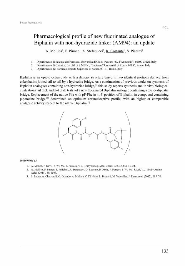
133
Poster Presentations
P74
Pharmacological profile of new fluorinated analogue of Biphalin with non-hydrazide linker (AM94): an update
A. Mollica1, F. Pinnen1, A. Stefanucci2, R. Costante1, S. Pieretti3
1. Dipartimento di Scienze del Farmaco, Università di Chieti-Pescara “G. d’Annunzio”, 66100 Chieti, Italy2. Dipartimento di Chimica, Facoltà di S.M.F.N., “Sapienza” Università di Roma, 00185, Rome, Italy3. Dipartimento del Farmaco, Istituto Superiore di Sanità, 00161, Rome, Italy
Biphalin is an opioid octapeptide with a dimeric structure based in two identical portions derived from enkephalins joined tail to tail by a hydrazine bridge. As a continuation of previous works on synthesis of Biphalin analogues containing non-hydrazine bridge,[1] this study reports synthesis and in vivo biological evaluation (tail flick and hot plate tests) of a new fluorinated Biphalin analogue containing a cyclo-aliphatic bridge. Replacement of the native Phe with pF-Phe in 4, 4’ position of Biphalin, in compound containing piperazine bridge,[2] determined an optimum antinociceptive profile, with an higher or comparable analgesic activity respect to the native Biphalin.[3]
References1. A. Molica, P. Davis, S-Wu Ma, F. Porreca, V. J. Hruby Bioog. Med. Chem. Lett. (2005), 15, 2471.2. A. Mollica, F. Pinnen, F. Feliciani, A. Stefanucci, G. Lucente, P. Davis, F. Porreca, S-Wu Ma, J. Lai, V. J. Hruby Amino
Acids (2011), 40, 1503.3. S. Leone, A. Chiavaroli, G. Orlando, A. Mollica, C. Di Nisio, L. Brunetti, M. Vacca Eur. J. Pharmacol. (2012), 685, 70.

134

LIST OF PARTICIPANTS

136
AAccardo Antonella ItalyAlterio Vincenzo ItalyAndreu David SpainAngelastro Antonio ItalyAscione Giuseppina ItalyAssmus Frauke SwitzerlandAutiero Ida ItalyAvitabile Concetta ItalyAy Bernhard Germany
BBavoso Alfonso ItalyBerisio Rita ItalyBianco Alberto FranceBracci Luisa ItalyBrandi Paola ItalyBulaj Grzegorz United States
CCacciatore Ivana ItalyCalce Enrica ItalyCantisani Marco ItalyCapasso Domenica ItalyCaporale Andrea ItalyCardoso Ana PortugalCarvajal-Rondanelli Patricio ChileCelentano Veronica ItalyChirafisi Paola ItalyChorev Michael United StatesComegna Daniela ItalyCostante Roberto ItalyCostantini Susan Italy
DD'Ambrosio Katia ItalyD'Andrea Luca Domenico ItalyDathan Nina ItalyDe Antonellis Pasqualino Italyde la Fuente Jesus M. SpainDe Luca Luca ItalyDe Luca Stefania ItalyDe Paola Ivan ItalyDe Rosa Lucia ItalyDe Simone Giuseppina ItalyDe Simone Paola ItalyDe Spiegeleer Bart BelgiumDe Zotti Marta ItalyDel Zoppo Luisa ItalyDettner Frank SwitzerlandDi Fiore Anna Italy
Di Napoli Antonella ItalyDi Natale Concetta ItalyDi Stasi Rossella ItalyDiana Donatella ItalyDoti Nunzianna Italy
EEgger Christine SwitzerlandEgido de Frutos Estefania SwitzerlandEsposito Cinzia SwitzerlandEsposito Carla ItalyEsposito Luciana Italy
FFalanga Annarita ItalyFalciani Chiara ItalyFalcioni Rita ItalyFenude Emma ItalyFerrer-Montiel Antonio SpainFesta Luisa ItalyFocà Giuseppina ItalyFocà Annalia ItalyFormaggio Fernando ItalyFornasari Erika ItalyFreire João Portugal
GGaldiero Massimiliano ItalyGaldiero Stefania ItalyGatto Emanuela ItalyGrieco Paolo ItalyGuerrini Remo Italy
HHossein Nejad Ariani Hanieh PolandHovestädt Marc GermanyHudecz Ferenc Hungary
IIsernia Carla Italy
KKlocek Gabriela Switzerland

137
LLambris John United StatesLarregola Maud FranceLatajka Rafal PolandLeone Marilisa ItalyLi Blatter Xiaochun SwitzerlandLiguori Lucia ItalyLiguoro Annamaria ItalyLimatola Antonio ItalyLucietto Pierluigi Italy
MMangoni Maria Luisa ItalyMarasco Daniela ItalyMariniello Raffaella ItalyMemczak Henry GermanyMercurio Flavia Anna ItalyMerlino Francesco ItalyMerlino Antonello ItalyMierke Dale United StatesMignogna Eleonora ItalyMollica Adriano ItalyMonti Daria Maria ItalyMonti Simona Maria ItalyMorelli Giancarlo ItalyMoretto Alessandro ItalyMoroder Luis GermanyMusumeci Domenica Italy
OOlah-Szabo Rita Hungary
PPalmieri Maddalena ItalyPapini Anna Maria ItalyPastore Raffaele ItalyPerillo Emiliana ItalyPinnen Francesco ItalyPirone Luciano Italy
RRajik Mohamed MalaysiaRicci Andrea ItalyRigano Maria Manuela ItalyRomanelli Alessandra ItalyRomano Maria ItalyRosenman Gil IsraelRossi Filomena ItalyRoversi Daniela Italy
Roviello Giovanni ItalyRuggiero Alessia ItalyRussomanno Anna ItalyRuvo Menotti Italy
SSaccone Irene ItalySala Marina ItalySandomenico Annamaria ItalySanseverino Marina ItalySauder Reto SwitzerlandSaviano Michele ItalyScognamiglio Liana P. ItalySeelig Joachim SwitzerlandSeelig Anna SwitzerlandSeitz Oliver GermanySerrano Isa PortugalSmaldone Giovanni ItalySosnowska Marta PolandStella Lorenzo ItalyStueber Werner Germany
TTarallo Rossella ItalyTedesco Consiglia ItalyTesauro Diego ItalyToniolo Claudio ItalyTornesello Anna Lucia Italy
VVancolen Annick BelgiumVeiga Ana Salome PortugalVenanzi Mariano ItalyVernieri Ermelinda ItalyVilasi Silvia ItalyVitagliano Luigi ItalyVitali Alberto Italy
YYousif Ali Munaim Italy
ZZaccaro Laura ItalyZehender Fabian Switzerland

138

LIST OF AUTHORS

140
AÄänismaa P. 95Accardo A. 48, 52, 62, 64, 68, 75Albanesi C. 55Albericio F. 53Alexa A. 25Allmaier G. 30Aloj L. 62, 64Alterio V. 105, 110Ambrosone A. 22Amodeo P. 63, 86Andreu D. 24, 36Angelastro A. 117Arciello A. 110Arra C. 63Ascione G. 82, 105, 109Aurilio M. 62Autiero I. 114Avitabile C. 40, 122
BBaldassarre L. 131Bánóczi Z. 25Baptista Y. 22Barbieri A. 63Barone A. 122Bavoso A. 70Bechinger B. 121Benedetti E. 109Berisio R. 117, 128, 129Bianchi N. 40Bianco A. 21Bier F.F. 37, 93Bindi S. 89Biondi B. 54, 76, 83Bobone S. 39, 77Boccafoschi F. 102Bocchinfuso G. 39, 77, 104, 121Boffito M. 102Boix E. 24Bon G 52Bősze S. 74Bracci L. 48, 89Brancaccio D. 125Brandi P. 112Brognara E. 40Brunetti J. 48, 89Buanne P. 101Bucci E.M. 73, 84Bulaj G. 28
CCacciatore I. 130, 131
Calce E. 107Calò G. 120Calvanese L. 67Campiglia P. 123, 124Cantisani M. 61, 71, 115, 116Capasso C. 82Capasso D. 47, 111Caporale A. 102Capparelli R. 122Carberry T.P. 38Cardoso A.L. 34Cardoso A.M. 34Carotenuto A. 123, 124, 125Carta F. 108Caruso M. 23Carvajal-Rondanelli P. 53Castagnola M. 60, 85Castanho M.A.R.B. 36, 90Castello G. 63Castiglione-Morelli M.A. 70Celentano V. 47, 88, 91Cerasuolo G. 99Chirafisi P. 128, 129Chorev M. 16Christmann A. 37, 93Ciardelli G. 102Cicala C. 64Cigliano L. 78Colombo G. 79Conde J. 22Correale S. 111, 132Costante R. 130, 131, 133Crifò B. 101Crisma M. 72Cristinziano P. 70Culmsee C. 45
DD’Agostino N. 122D’Alterio C. 63D’Ambrosio K. 108D’Andrea L.D. 40, 47, 78, 79, 87, 88, 91,
92Dathan N.A. 105, 109D’Auria G. 67De Cola C. 99de la Fuente J.M. 22Del Gatto A. 75Del Giudice R. 110Della Pepa M.E. 116de los Santos J.M. 57De Luca S. 63, 107De Luca V. 82Del Zoppo L. 120de Paola I. 132

141
de Pascale D. 109Depau L. 48De Poli M. 76De Riccardis F. 99De Rosa G. 64De Rosa L. 78, 79, 87, 88, 92De Rosa M.C. 60D’Errico G. 71De Santi C. 109De Simone G. 82, 101, 105, 108, 110,
132De Simone P. 128, 129De Spiegeleer B. 56Dettner F. 31De Zotti M. 39, 54, 76, 77, 83D’Hondt M. 56Diana D. 47, 78, 79, 87, 88Di Carlo S.E. 52Di Fiore A. 82, 110Di Gaetano S. 47, 111, 132Di Napoli A. 73Di Natale C. 119Di Salvo C. 88Di Stasi R. 47, 78Dókus L.E. 25Dolga A.M. 45D’Oriano V. 116Doti N. 45, 55Drag M. 57
EEgido E. 103Ehrentreich-Förster E. 37, 93Erra L. 99Esposito C. 95Esposito L. 126, 127Estrada G.G. 22
FFabbri E. 40Falanga A. 38, 61, 71, 111, 115, 116Falciani C. 48, 89Falcigno L. 67Falcioni R. 52Farkas A. 25Farrotti A. 39, 121Fathi H. 76Fattorusso R. 47, 78, 79, 87, 88, 132Fenude E. 86Ferrer-Montiel A. 27Finamore E. 38, 115Focà A. 94, 96Focà G. 94, 96Folgiero V. 52
Formaggio F. 18, 39, 54, 72, 76, 77, 83, 104
Fornasari E. 130, 131Fragneto G. 77Franco P. 125Freire J.M. 36, 90Friedrich P. 25Fröhlich S. 30Fulgione A. 122Fusco S. 61
GGaczynska M. 65Galdiero M. 38, 61, 71, 115, 116Galdiero S. 38, 50, 61, 71, 111, 115,
116Gambari R. 40Gatto E. 23, 104Gerelli Y. 77Giardina B. 60Giordano L. 39Gobbo M. 83Gomes P. 34Gomez-Monterrey I.M. 123, 124Grieco P. 123, 124, 125Guarnieri D. 61, 111Guerrini R. 120Guianvarc’h D. 46Guzmán F. 53
HHahm K.-S. 39Haremza K. 57Hernández 22Hong Enriquez R.P. 113Horváti K. 74Hovestädt M. 37, 93Hudecz F. 25, 74
IIaccarino G. 123, 124Iaccarino I. 125Iaconis S. 96Ibarra M.R. 22Ieranò C. 63Illario M. 124Improta R. 127Incoronato N. 116Isernia C. 86Izzo I. 99

142
JJankowska E. 65, 81Jewginski M. 57, 66, 69Jurado A.S. 34
KKampanaraki A. 115, 116Karoyan P. 46Karpowicz P. 65Klocek G. 98Kowalczyk W. 36Krzciuk J. 66
LLaio A. 113Lambardi D. 26Lambris J.D. 19Langella E. 105, 114Larregola M. 46, 80Latajka R. 57, 66, 69Latter E. 77Lavielle S. 46Lelli B. 48, 89Leonardi A. 94, 96Leone M. 107, 117, 118, 119Lequin O. 46Li-Blatter X. 97, 103Limatola A. 123, 125Lolli F. 26Lopes S. 104Lozzi L. 48, 89Luciano A. 63
MMadonna S. 55Maia S. 34Maione F. 64Makarov V. 129Makowski M. 66Malfaccini D. 120Malgieri G. 132Mansi R. 62Marasco D. 55, 112, 113, 117, 118, 119Marchesano V. 22Marchetti-Deschmann M. 30Marcozzi C. 125Maresca B. 78Marshall S. 53Maryasov A.G. 83Marzola E. 120Mazzoni M. 85
Memczak H. 37, 93Mercurio F. 119Merlino A. 126Messana I. 85Mező G. 74Mierke D.F. 17Mignogna E. 115, 116Milov A.D. 83Minchiotti G. 96Molinari A. 85Mollica A. 130, 131, 133Monfregola L. 63, 107Monteleone F. 101Monti D.M. 110Monti S.M. 82, 101, 105, 109, 110Morel F.M.M. 105Morelli G. 48, 52, 62, 64, 68, 75Moretto A. 51Morisco A. 62, 64Mottolese M. 52Mrozinska E. 69Müller R. 103Muscetti O. 61Musumeci D. 73, 84
NNapolitano M. 63Nardone B. 99Netti P. 61, 111Nocerino N. 122Nogués M.V. 24Novellino E. 123, 124, 125
OOancea S. 76Ojeda C. 53Orioni B. 77Osmulski P. 65Ostuni A. 70
PPaduano L. 68Palleschi A. 39, 77, 104, 121Palmerini C.A. 85Palmieri G. 75, 94Pandey S. 26Papini A.M. 26, 54, 80Papkov A. 81Parisi A. 64Park Y. 39Pastorekova S. 101Pastore R. 118

143
Pedone C. 73, 82, 84, 109, 115Pedone E.M. 111, 119, 132Pedroso Lima M.C. 34Peggion C. 54, 72, 76Pellecchia M. 119Penfold J. 77Perillo E. 71, 116Perler F. 92Peroni E. 26Pieretti S. 130, 133Pini A. 48, 89Pinnen F. 130, 131, 133Pirolli D. 60Pirone L. 111, 119, 132Plesnila N. 45Poletto M. 118Polimeno M.N. 63Portella L. 63Pulido D. 24
RRadicioni G. 60, 85Ravenni N. 48Real-Fernandez F. 26Rego de Figueiredo I. 36Renzone G. 101Reuther C. 45Ricci A. 73Rigano M.M. 122Romanelli A. 40, 87, 88, 92, 122Romano M. 128, 129Rosenman G. 20Rossi M. 82Rovero P. 26Roversi D. 39Roviello G. 73, 84Rozzo C. 75Ruggiero A. 117, 128, 129Rümpel E. 37, 93Rusciano M.R. 124Russo L. 115Russomanno A. 79, 87Ruvo M. 45, 91, 94, 96
SSabatella M. 101Sala M. 123, 124Salvadori S. 120Salzano G. 64Sandomenico A. 35, 91, 94, 96, 110Sanguigno L. 94, 96Santos N.C. 36Sanz V. 22Sarno C. 125
Sartori S. 102Sasso E. 101Sauder R. 100Saviano M. 40, 86, 114Scala S. 63Scali S. 89Scaloni A. 101Schneider J.P. 49Schönfeld H.-J. 106Scognamiglio P.L. 45, 55, 112, 113, 118, 119Scoles G. 113Scozzafava 82Sebastiani F. 77Seelig A. 95, 97, 103Seelig J. 44, 98, 100, 106Seitz O. 29Senesi R. 77Serpa C. 104Serrano I. 90Sini M.C. 94Sinthuvanich C. 49Smaine F.-Z. 108Smaldone G. 111Sosnowska M. 65, 81Spagnuolo M.S. 78Spugnin E.P. 52Squeglia F. 117, 128Stalmans S. 56Stanzione F. 126Stefanucci A. 130, 133Stella L. 39, 77, 104, 121Stöckleinm W.F.M. 37, 93Stoj J. 65Stoppelli P. 125Stringaro A. 85Supuran C.T. 82, 101, 105, 108, 110Szabó R. 74Szmidke Z. 81
TTantos Á 25Tarallo R. 38, 61, 71Tavano R. 54Tedesco C. 99Tell G. 118Tesauro D. 48, 62, 64, 68, 70, 75Tian F. 22Tőke O. 25Toniolo C. 39, 54, 72, 76, 77, 83, 104Tori K. 92Torrent M. 24Tortiglione C. 22Trabulo S. 34Trapella C. 120Tsvetkov Y.D. 83

144
VVan Dorpe S. 56Vascotto C. 118Veiga A.S. 36, 49Venanzi M. 23, 104Vernieri E. 123, 124Világi I. 25Viparelli F. 105Vitagliano L. 126, 127, 132Vitale M. 101Vitale R.M. 63Vitali A. 60, 85Vitiello G. 71Vitiello M. 71, 115Votta G. 125Vullo D. 105
WWeck M. 38Winum J.-Y. 108
ZZaccaro L. 75, 132Zambrano N. 101Zehender F. 106Ziaco B. 64Ziegler A. 100, 106Zollo M. 111