Embed Size (px)
Citation preview
IntroductionThe LKB1 protein kinase was originally identified as a genemutated in the inherited Peutz Jeghers Syndrome (PJS), inwhich subjects are predisposed to developing benign andmalignant tumours (Hemminki et al., 1998; Jenne et al., 1998).Subsequent work indicated that LKB1 forms a complex withthe regulatory proteins STRAD and MO25 (Baas et al., 2003;Boudeau et al., 2003), and is likely to function as a tumoursuppressor by regulating cell proliferation and polarity(reviewed by Baas et al., 2004). The first identifiedphysiological substrate of LKB1 was the AMP-activatedprotein kinase (AMPK) (Hawley et al., 2003; Shaw et al.,2004b; Woods et al., 2003), which functions as a regulator ofcellular energy (Hardie, 2004). LKB1 activates AMPK byphosphorylating Thr172 in the T-loop of this enzyme. The twoisoforms of AMPK (AMPK�1 and AMPK�2) are associatedwith AMPK� and AMPK� regulatory subunits, enablingAMPK to be activated under low energy conditions by bindingof 5�-AMP to the AMPK� subunit (Hardie, 2004). The bindingof AMPK to 5�-AMP also promotes the phosphorylation andactivation of AMPK by the LKB1 complex. Tumour formationin LKB1-deficient cells could result from deregulation ofpathways involving the tuberous sclerosis complex/mTOR
(Corradetti et al., 2004; Shaw et al., 2004a) or p53 (Jones etal., 2005), which are reportedly regulated by AMPK.
LKB1 also phosphorylates and activates at least 12 otherprotein kinases that are closely related to AMPK, namelyQSK, SIK, QIK, MARK1, MARK2, MARK3/Par-1A/C-TAK1, MARK4, NUAK1/ARK5, NUAK2/SNARK,BRSK1/SAD-A, BRSK2/SAD-B and SNRK (Jaleel et al.,2005; Lizcano et al., 2004). The most studied of these enzymesare the MARK isoforms, which regulate anterior-posterior cellpolarity development at the one-cell stage of embryonicdevelopment in C. elegans (Guo and Kemphues, 1995) andDrosophila (Shulman et al., 2000). The MARK enzymes alsocontrol gastrulation in Xenopus (Kusakabe and Nishida, 2004).In mammals, MARK isoforms phosphorylate Tau (Drewes etal., 1997), thereby priming it for hyperphosphorylation by thekinases GSK-3 and Cdk5, an event that induces theaggregation of Tau into the toxic filaments and tangles that areobserved in patients with Alzheimer’s disease (Nishimura etal., 2004). Recent studies employing mice lacking BRSK1 andBRSK2, which are exclusively expressed in the brain, indicatethat these enzymes play a key role in regulating neuronalpolarization (Kishi et al., 2005). Less is known regarding thefunction of other AMPK-related kinases, i.e. QSK, QIK, SIK,
5661
The LKB1 tumour suppressor kinase phosphorylates andactivates a number of protein kinases belonging to theAMP-activated protein kinase (AMPK) subfamily. We haveused a modified tandem affinity purification strategy toidentify proteins that interact with AMPK��, as well as thetwelve AMPK-related kinases that are activated by LKB1.The AMPK�� and AMPK�� regulatory subunits wereassociated with AMPK��, but not with any of the AMPK-related kinases, explaining why AMP does not influence theactivity of these enzymes. In addition, we identified novelbinding partners that interacted with one or more of theAMPK subfamily enzymes, including fat facets/ubiquitinspecific protease-9 (USP9), AAA-ATPase-p97, adeninenucleotide translocase, protein phosphatase 2Aholoenzyme and isoforms of the phospho-protein bindingadaptor 14-3-3. Interestingly, the 14-3-3 isoforms bounddirectly to the T-loop Thr residue of QSK and SIK, afterthese were phosphorylated by LKB1. Consistent with this,the 14-3-3 isoforms failed to interact with non-
phosphorylated QSK and SIK, in LKB1 knockout muscleor in HeLa cells in which LKB1 is not expressed. Moreover,mutation of the T-loop Thr phosphorylated by LKB1,prevented QSK and SIK from interacting with 14-3-3 invitro. Binding of 14-3-3 to QSK and SIK, enhancedcatalytic activity towards the TORC2 protein and theAMARA peptide, and was required for the cytoplasmiclocalization of SIK and for localization of QSK to punctatestructures within the cytoplasm. To our knowledge, thisstudy provides the first example of 14-3-3 binding directlyto the T-loop of a protein kinase and influencing its catalyticactivity and cellular localization.
Supplementary material available online athttp://jcs.biologists.org/cgi/content/full/118/23/5661/DC1
Key words: AMPK, MARK/Par1, Cell polarity, Mass spectrometry,Tandem affinity purification.
Summary
14-3-3 cooperates with LKB1 to regulate the activityand localization of QSK and SIKAbdallah K. Al-Hakim1,*, Olga Göransson1, Maria Deak1, Rachel Toth1, David G. Campbell1, Nick A. Morrice1,Alan R. Prescott2 and Dario R. Alessi11MRC Protein Phosphorylation Unit, MSI/WTB complex, University of Dundee, Dow Street, Dundee, DD1 5EH, UK2Division of Cell Biology and Immunology, MSI/WTB complex, University of Dundee, Dow Street, Dundee, DD1 5EH, UK*Author for correspondence (e-mail: [email protected])
Accepted 31 August 2005Journal of Cell Science 118, 5661-5673 Published by The Company of Biologists 2005doi:10.1242/jcs.02670
Research Article
Jour
nal o
f Cel
l Sci
ence
5662
NUAK1, NUAK2 and SNRK. RNAi-mediated knockdown ofQSK in Drosophila cells resulted in mitotic defects thatincluded spindle and chromosome alignment abnormalities(Bettencourt-Dias et al., 2004). SIK (salt-inducible kinase),was first cloned from the adrenal glands of rats fed a high saltdiet (Wang et al., 1999) and its mRNA is induced bymembrane depolarization in the brain (Feldman et al., 2000).The mRNA expressing QIK (SIK2) is highest in adiposetissue and, in overexpression studies, QIK was reportedto phosphorylate human IRS1 at the same residuephosphorylated by AMPK (Horike et al., 2003). QIK alsophosphorylates the CREB co-activator TORC2, inunstimulated cells, to sequester it in the cell cytoplasm,thereby inhibiting CREB-dependent gene-expression(Screaton et al., 2004). In overexpression studies, NUAK1(ARK5) suppressed apoptosis induced by certain stimuli,including nutrient starvation (Suzuki et al., 2003a), and wasalso suggested to regulate Caspase-6 activity (Suzuki et al.,2004b). NUAK2 (SNARK) was shown to be most highlyexpressed in the kidney, and its activity was reportedlystimulated by glucose starvation of cells (Lefebvre et al., 2001;Suzuki et al., 2003b). Expression of NUAK2 was alsoupregulated by CD95 and TNF in apoptosis-resistant tumourcell lines, and it plays a role in protecting these cells fromapoptosis (Legembre et al., 2004). SNRK mRNA was inducedthreefold when granule neurons were cultured in lowpotassium, indicating that it could play a role in regulatingsurvival responses in these cells (Yoshida et al., 2000). SNRKis also highly expressed in testis (Jaleel et al., 2005).
To date, little is known regarding the mechanism ofregulation of AMPK-related kinases. Our analysis hasindicated that none of the AMPK-related kinases is activatedby agonists that stimulate AMPK, such as AICA-riboside orphenformin, or by muscle contraction (Lizcano et al., 2004;Sakamoto et al., 2004). However, it has also been reported thatthe AICA-riboside AMP mimetic, activated NUAK2 in ahepatocarcinoma-derived cell line, suggesting that NUAK2-like AMPK possesses an AMP-binding subunit (Lefebvre andRosen, 2005).
In order to determine whether any of the AMPK-relatedkinases were complexed to AMPK� and AMPK� regulatorysubunits or associated with other binding partners, we haveperformed a tandem affinity purification analysis with each ofthe LKB1-activated AMPK-related kinases. Our findingsindicate that the 14-3-3 phospho-protein binding adaptors(Mackintosh, 2004) cooperate with LKB1 to regulate theactivity and localization of QSK and SIK.
Materials and MethodsMaterialsProtein G-Sepharose, calmodulin-Sepharose 4B, glutathione-Sepharose, streptavidin-Sepharose, [�-32P]ATP and enhancedchemiluminescence reagent were purchased from AmershamBioscience; protease-inhibitor cocktail tablets, and precast SDSpolyacrylamide Bis-Tris gels were from Invitrogen; Tween-20, rabbitIgG-agarose and dimethyl pimelimidate were from Sigma; NP-40 wasfrom Fluka; and phosphocellulose P81 paper was from Whatman. Thehexahistidine-tagged TEV protease was expressed in E. coli by G.Kular and purified using nickel agarose affinity chromatography andgel filtration. All peptides were synthesized by Graham Bloomberg atthe University of Bristol.
AntibodiesThe following antibodies were raised in sheep and affinity purified onthe appropriate antigen: anti-LKB1 (raised against mouse protein,used for immunoblotting), anti-QSK (residues 1349-1369 of humanQSK, TDILLSYKHPEVSFSMEQAGV, used for immunoblotting andimmunoprecipitation), Phospho-anti-T-loop QSK/SIK (residues 175-189 of human SIK, KSGEPLST(P)WCGSPPY phosphorylated atThr183, used for immunoblotting), anti-SIK (residues 1-20 of humanSIK, MVIMSEFSADPAGQGQGQQK, used for immunoblottingand immunoprecipitation), Phospho-anti-T-loop MARK (residues204-218 of human MARK3 phosphorylated at Thr211,TVGGKLDT(P)FCGSPPY, used for immunoblotting) and the anti-GST (raised against the glutathione S-transferase protein, used forimmunoblotting). Anti-MARK3 antibody used for immunoblottingwas from Upstate Biotech (anti c-TAK #05-680), polyclonal antibodyrecognizing 14-3-3 isoforms used for immunoblotting was purchasedfrom Santa Cruz (#sc629), mouse monoclonal [4D11] recognizing thehexahistidine affinity tag were from Abcam (#ab5000-100),monoclonal antibodies recognizing HA epitope tag was purchasedfrom Roche (#1666606), and secondary antibodies coupled tohorseradish peroxidase used for immunoblotting were obtained fromPierce.
General methodsTissue culture, transfection, immunoblotting, restriction enzymedigests, DNA ligations, and other recombinant DNA procedures wereperformed using standard protocols. All mutagenesis was carried outusing the Quick-Change site-directed mutagenesis method(Stratagene). DNA constructs used for transfection were purified fromE. coli DH5� using Qiagen plasmid Mega or Maxi kit according tothe manufacturer’s protocol. All DNA constructs were verified byDNA sequencing, which was performed by The Sequencing Service,School of Life Sciences, University of Dundee, UK, using DYEnamicET terminator chemistry (Amersham Biosciences) on AppliedBiosystems automated DNA sequencers. The generation and cultureof HeLa cells stably expressing wild-type or kinase-inactiveLKB1[D179A] has been described previously (Sapkota et al., 2002).
BuffersLysis buffer contained: 50 mM Tris-HCl pH 7.5, 1 mM EGTA, 1 mMEDTA, 1% (w/v) NP-40, 1 mM sodium orthovanadate, 10 mMsodium-�-glycerophosphate, 50 mM sodium fluoride, 5 mM sodiumpyrophosphate, 0.27 M sucrose, 1 mM dithiothreitol (DTT) andcomplete proteinase inhibitor cocktail (one tablet/50 ml). Buffer Acontained: 50 mM Tris/HCl pH 7.5, 0.1 mM EGTA, 0.27 M sucroseand 1 mM DTT. Buffer B contained: 50 mM Tris-HCl pH 7.5, 0.15M NaCl, 0.27 M sucrose, 1% (w/v) NP-40 and 1 mM DTT. Buffer Ccontained: 50 mM Tris pH 7.5, 0.15 M NaCl, 1 mM MgCl2, 1 mMimidazole, 2 mM CaCl2, 0.27 M sucrose and 1 mM DTT. Buffer Dcontained: 10 mM Tris pH 7.5, 1 mM imidazole, 20 mM EGTA, and1 mM DTT. TBS-Tween buffer contained: 50 mM Tris/HCl pH 7.5,0.15 M NaCl and 0.2% (v/v) Tween-20.
PlasmidsThe generation of wild-type and mutant epitope-tagged AMPK-related kinases in the pEBG2T vector encoding for the expression ofGST-fusion proteins in eukaryotic cells have been describedpreviously (Lizcano et al., 2004). The wild-type and mutant QSKwere subcloned into the pEGFP-C1 vector (Clontech) as a BamHI-BamHI fragment from the pEBG2T vector. The wild-type and mutantSIK were subcloned into NotI-modified pEGFP-C1 vector as BamHI-NotI fragment from pEBG2T vector. A cDNA of full-length N-terminally Myc-tagged rat AMPK�1[T172E], was kindly provided byG. Hardie, and subcloned into the EcoRI-KpnI sites of the pEGFP-
Journal of Cell Science 118 (23)
Jour
nal o
f Cel
l Sci
ence
5663Regulation of SIK and QSK by 14-3-3
C2-TAP vector. 14-3-3� (GenBank NM_145690) was amplified fromIMAGE EST clone 2988020 with oligonucleotides introducing aBamHI restriction site at both the 5� and 3� ends and was subclonedinto a pGEX6P-1 vector. TORC2 (GenBank NM_181715) wasamplified from IMAGE EST clone 6188068 with oligonucleotidesintroducing a NotI restriction site at both the 5� and 3� ends andsubcloned into pGEX6P-2 vector. The kinase-inactive SIK[D167A],T-loop mutant SIK[T182A], kinase inactive QSK[D206A] and T-loopmutant QSK[T221A] have been described previously (Lizcano et al.,2004).
Generation of the pEBGFP-C2-TAP vectorBased on the original Tandem Affinity Purification strategy (Rigautet al., 1999), we constructed a modified expression vector describedin Fig. 1A that incorporates a green fluorescent protein (GFP) tagbefore the TAP tag to enable more facile selection of stable cell linesthat express TAP-tagged proteins. In addition we have also insertedan additional precision protease cleavage site immediately after thecalmodulin binding motif to enable cleavage of the TAP-taggedprotein from the calmodulin-Sepahrose without using EDTA or SDSelution methods and to permit removal of the entire tag of theexpressed TAP-tagged protein after purification if necessary. Togenerate this vector we used a PCR based strategy employing as a
template a vector encoding two tandem protein A IgG-bindingdomains (IGG1 and IGG2) and a calmodulin-binding motif that waskindly provided by B. Seraphin (EMBL, Heidelberg, Germany) andmodified by M. Stark (School of Life Sciences, Dundee). The primersused for this PCR (5�-gccaccatggacacaagtgcccacgatgaagc-3� and 5�-gcggccgcggtaccagatctggatcccaggggcccctggaacagaacttccagagcagttgg-aatatcataatcaagtgcccc-3�) incorporated a precision protease site and aBamHI and NotI restriction enzyme site after the calmodulin-bindingdomain. The resulting PCR product was ligated into pCR2.1.TOPOvector (Invitrogen). This was then subcloned in-frame into thepolylinker region of pEGFP-C2 vector as an EcoRI-EcoRI fragment.The upstream EcoRI site and the downstream BamHI site in thepolylinker region of the resulting vector were then eliminated byPCR mutagenesis. The resulting vector described in Fig. 1A istermed pEGFP-C2-TAP, and enables proteins to be tagged at theirN-terminus with a GFP-TAP tag. The cloning of HA-epitope-tagged AMPK-related kinases has been described previously(Lizcano et al., 2004) and the full-length coding regions of theseenzymes were appropriately subcloned as BamHI-BamHI,BamHI/EcoRI, NotI/NotI or BamHI-NotI fragments into the pEGFP-C2-TAP vector to incorporate an N-terminal GFP-TAG. Human 14-3-3� was also subcloned in to the pEGFP-C2-TAP as a NotI-NotIfragment.
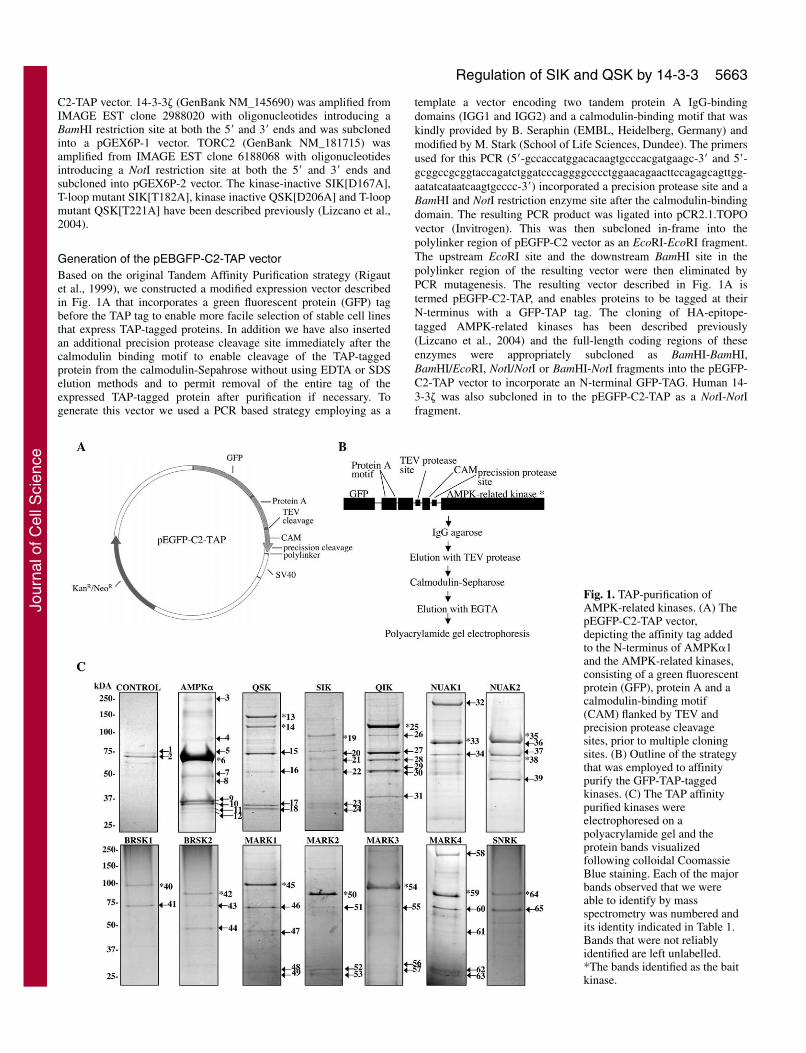
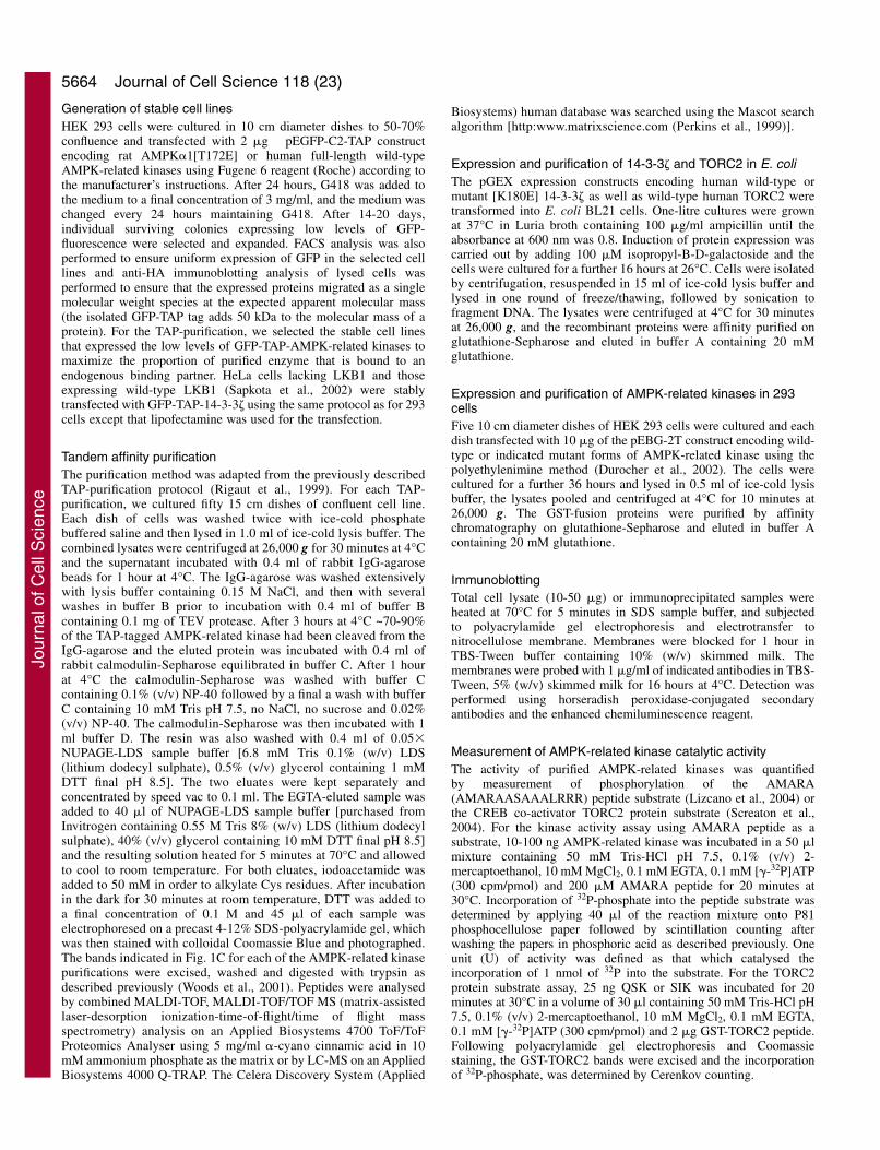
Fig. 1. TAP-purification ofAMPK-related kinases. (A) ThepEGFP-C2-TAP vector,depicting the affinity tag addedto the N-terminus of AMPK�1and the AMPK-related kinases,consisting of a green fluorescentprotein (GFP), protein A and acalmodulin-binding motif(CAM) flanked by TEV andprecision protease cleavagesites, prior to multiple cloningsites. (B) Outline of the strategythat was employed to affinitypurify the GFP-TAP-taggedkinases. (C) The TAP affinitypurified kinases wereelectrophoresed on apolyacrylamide gel and theprotein bands visualizedfollowing colloidal CoomassieBlue staining. Each of the majorbands observed that we wereable to identify by massspectrometry was numbered andits identity indicated in Table 1.Bands that were not reliablyidentified are left unlabelled.*The bands identified as the baitkinase.
Jour
nal o
f Cel
l Sci
ence
5664
Generation of stable cell linesHEK 293 cells were cultured in 10 cm diameter dishes to 50-70%confluence and transfected with 2 �g pEGFP-C2-TAP constructencoding rat AMPK�1[T172E] or human full-length wild-typeAMPK-related kinases using Fugene 6 reagent (Roche) according tothe manufacturer’s instructions. After 24 hours, G418 was added tothe medium to a final concentration of 3 mg/ml, and the medium waschanged every 24 hours maintaining G418. After 14-20 days,individual surviving colonies expressing low levels of GFP-fluorescence were selected and expanded. FACS analysis was alsoperformed to ensure uniform expression of GFP in the selected celllines and anti-HA immunoblotting analysis of lysed cells wasperformed to ensure that the expressed proteins migrated as a singlemolecular weight species at the expected apparent molecular mass(the isolated GFP-TAP tag adds 50 kDa to the molecular mass of aprotein). For the TAP-purification, we selected the stable cell linesthat expressed the low levels of GFP-TAP-AMPK-related kinases tomaximize the proportion of purified enzyme that is bound to anendogenous binding partner. HeLa cells lacking LKB1 and thoseexpressing wild-type LKB1 (Sapkota et al., 2002) were stablytransfected with GFP-TAP-14-3-3� using the same protocol as for 293cells except that lipofectamine was used for the transfection.
Tandem affinity purificationThe purification method was adapted from the previously describedTAP-purification protocol (Rigaut et al., 1999). For each TAP-purification, we cultured fifty 15 cm dishes of confluent cell line.Each dish of cells was washed twice with ice-cold phosphatebuffered saline and then lysed in 1.0 ml of ice-cold lysis buffer. Thecombined lysates were centrifuged at 26,000 g for 30 minutes at 4°Cand the supernatant incubated with 0.4 ml of rabbit IgG-agarosebeads for 1 hour at 4°C. The IgG-agarose was washed extensivelywith lysis buffer containing 0.15 M NaCl, and then with severalwashes in buffer B prior to incubation with 0.4 ml of buffer Bcontaining 0.1 mg of TEV protease. After 3 hours at 4°C ~70-90%of the TAP-tagged AMPK-related kinase had been cleaved from theIgG-agarose and the eluted protein was incubated with 0.4 ml ofrabbit calmodulin-Sepharose equilibrated in buffer C. After 1 hourat 4°C the calmodulin-Sepharose was washed with buffer Ccontaining 0.1% (v/v) NP-40 followed by a final a wash with bufferC containing 10 mM Tris pH 7.5, no NaCl, no sucrose and 0.02%(v/v) NP-40. The calmodulin-Sepharose was then incubated with 1ml buffer D. The resin was also washed with 0.4 ml of 0.05�NUPAGE-LDS sample buffer [6.8 mM Tris 0.1% (w/v) LDS(lithium dodecyl sulphate), 0.5% (v/v) glycerol containing 1 mMDTT final pH 8.5]. The two eluates were kept separately andconcentrated by speed vac to 0.1 ml. The EGTA-eluted sample wasadded to 40 �l of NUPAGE-LDS sample buffer [purchased fromInvitrogen containing 0.55 M Tris 8% (w/v) LDS (lithium dodecylsulphate), 40% (v/v) glycerol containing 10 mM DTT final pH 8.5]and the resulting solution heated for 5 minutes at 70°C and allowedto cool to room temperature. For both eluates, iodoacetamide wasadded to 50 mM in order to alkylate Cys residues. After incubationin the dark for 30 minutes at room temperature, DTT was added toa final concentration of 0.1 M and 45 �l of each sample waselectrophoresed on a precast 4-12% SDS-polyacrylamide gel, whichwas then stained with colloidal Coomassie Blue and photographed.The bands indicated in Fig. 1C for each of the AMPK-related kinasepurifications were excised, washed and digested with trypsin asdescribed previously (Woods et al., 2001). Peptides were analysedby combined MALDI-TOF, MALDI-TOF/TOF MS (matrix-assistedlaser-desorption ionization-time-of-flight/time of flight massspectrometry) analysis on an Applied Biosystems 4700 ToF/ToFProteomics Analyser using 5 mg/ml �-cyano cinnamic acid in 10mM ammonium phosphate as the matrix or by LC-MS on an AppliedBiosystems 4000 Q-TRAP. The Celera Discovery System (Applied
Biosystems) human database was searched using the Mascot searchalgorithm [http:www.matrixscience.com (Perkins et al., 1999)].
Expression and purification of 14-3-3� and TORC2 in E. coliThe pGEX expression constructs encoding human wild-type ormutant [K180E] 14-3-3� as well as wild-type human TORC2 weretransformed into E. coli BL21 cells. One-litre cultures were grownat 37°C in Luria broth containing 100 �g/ml ampicillin until theabsorbance at 600 nm was 0.8. Induction of protein expression wascarried out by adding 100 �M isopropyl-B-D-galactoside and thecells were cultured for a further 16 hours at 26°C. Cells were isolatedby centrifugation, resuspended in 15 ml of ice-cold lysis buffer andlysed in one round of freeze/thawing, followed by sonication tofragment DNA. The lysates were centrifuged at 4°C for 30 minutesat 26,000 g, and the recombinant proteins were affinity purified onglutathione-Sepharose and eluted in buffer A containing 20 mMglutathione.
Expression and purification of AMPK-related kinases in 293cellsFive 10 cm diameter dishes of HEK 293 cells were cultured and eachdish transfected with 10 �g of the pEBG-2T construct encoding wild-type or indicated mutant forms of AMPK-related kinase using thepolyethylenimine method (Durocher et al., 2002). The cells werecultured for a further 36 hours and lysed in 0.5 ml of ice-cold lysisbuffer, the lysates pooled and centrifuged at 4°C for 10 minutes at26,000 g. The GST-fusion proteins were purified by affinitychromatography on glutathione-Sepharose and eluted in buffer Acontaining 20 mM glutathione.
ImmunoblottingTotal cell lysate (10-50 �g) or immunoprecipitated samples wereheated at 70°C for 5 minutes in SDS sample buffer, and subjectedto polyacrylamide gel electrophoresis and electrotransfer tonitrocellulose membrane. Membranes were blocked for 1 hour inTBS-Tween buffer containing 10% (w/v) skimmed milk. Themembranes were probed with 1 �g/ml of indicated antibodies in TBS-Tween, 5% (w/v) skimmed milk for 16 hours at 4°C. Detection wasperformed using horseradish peroxidase-conjugated secondaryantibodies and the enhanced chemiluminescence reagent.
Measurement of AMPK-related kinase catalytic activityThe activity of purified AMPK-related kinases was quantifiedby measurement of phosphorylation of the AMARA(AMARAASAAALRRR) peptide substrate (Lizcano et al., 2004) orthe CREB co-activator TORC2 protein substrate (Screaton et al.,2004). For the kinase activity assay using AMARA peptide as asubstrate, 10-100 ng AMPK-related kinase was incubated in a 50 �lmixture containing 50 mM Tris-HCl pH 7.5, 0.1% (v/v) 2-mercaptoethanol, 10 mM MgCl2, 0.1 mM EGTA, 0.1 mM [�-32P]ATP(300 cpm/pmol) and 200 �M AMARA peptide for 20 minutes at30°C. Incorporation of 32P-phosphate into the peptide substrate wasdetermined by applying 40 �l of the reaction mixture onto P81phosphocellulose paper followed by scintillation counting afterwashing the papers in phosphoric acid as described previously. Oneunit (U) of activity was defined as that which catalysed theincorporation of 1 nmol of 32P into the substrate. For the TORC2protein substrate assay, 25 ng QSK or SIK was incubated for 20minutes at 30°C in a volume of 30 �l containing 50 mM Tris-HCl pH7.5, 0.1% (v/v) 2-mercaptoethanol, 10 mM MgCl2, 0.1 mM EGTA,0.1 mM [�-32P]ATP (300 cpm/pmol) and 2 �g GST-TORC2 peptide.Following polyacrylamide gel electrophoresis and Coomassiestaining, the GST-TORC2 bands were excised and the incorporationof 32P-phosphate, was determined by Cerenkov counting.
Journal of Cell Science 118 (23)
Jour
nal o
f Cel
l Sci
ence
5665Regulation of SIK and QSK by 14-3-3
Immunoprecipitation of endogenous QSK and SIKQuadriceps mouse muscle was isolated and homogenized as describedpreviously (Sakamoto et al., 2005). HeLa cells were grown toconfluence on 10 cm diameter dishes and lysed in 1 ml of lysis buffer.The QSK and SIK antibodies were covalently coupled to protein G-Sepharose in a ratio of 1 mg antibody to 1 ml resin using a dimethylpimelimidate cross-linking procedure. Clarified quadriceps muscleextract or HeLa cell extract containing 1 mg total cell protein, wereincubated at 4°C for 1 hour on a vibrating platform with 5 �l ofthe QSK or SIK-antibody-protein G-Sepharose conjugate. Theimmunoprecipitates were washed twice with 1 ml of lysis buffercontaining 0.15 M NaCl and twice with 1 ml of buffer A. Theimmunoprecipitates were either subjected to protein kinase activityassay as described above or analysed by immunoblotting. Prior toimmunoblotting the beads were washed in buffer A lacking DTT andresuspended in a volume of 20 �l of buffer A lacking DTT to which5 �l of SDS sample buffer lacking DTT was added. The samples weresubjected to electrophoresis and then immunoblotted as describedabove.
Affinity purification of 14-3-3 binding proteinsClarified lysates of 293 cells containing 1 mg total cell protein, wereincubated at 4°C for 1 hour on a vibrating platform with 20 �lglutathione-Sepharose conjugated to 20 �g GST-14-3-3� expressed inE. coli as described above. The beads were washed twice with 1 mlof lysis buffer containing 0.15 M NaCl, twice with 1 ml of buffer Aand then subjected to electrophoresis and immunoblotting asdescribed above.
Affinity purification of 14-3-3 isoforms with QSK T-loop peptide293 cell lysate (3 mg protein) was incubated for 30 minutes at4°C with 10 �g N-terminal Biotin-conjugated QSK-Phospho-T-loop peptide TPGQLIKT(P)WCGSPPY or QSK-T-loop peptideTPGQLIKTWCGSPPY. 10 �l Streptavidin-Sepharose, previouslyequilibrated in lysis buffer, was added to the lysates that were thenincubated for another 1 hour on a vibrating platform at 4°C. The beadswere washed twice with 1 ml lysis buffer containing 0.15 M NaCl,twice with 1 ml buffer A and then resuspended in a volume of 20 �lof buffer A to which 5 �l of SDS sample buffer was added. Thesamples were then subjected to electrophoresis and immunoblottedfor 14-3-3 as described above.
Dissociation of 14-3-3 isoforms from QSK and SIKGST fusion proteins of QSK and SIK were expressed in 293 cells andabsorbed onto glutathione-Sepharose and washed with buffer A asdescribed above. Aliquots of 20 �l of glutathione-Sepharose, stillconjugated to GST-QSK or GST-SIK, were incubated with 200 �l ofbuffer A containing either no peptide or 10 �g of the QSK-Phospho-T-loop peptide (TPGQLIKT(P)WCGSPPY) or 10 �g of the QSK-T-loop peptide (TPGQLIKTWCGSPPY). After incubation for 30minutes at 4°C on a vibrating platform, the beads were washed twicewith 1 ml buffer A, and GST-SIK and GST-QSK was eluted in 20 �lbuffer A containing 20 mM glutathione. Activity and immunoblottinganalysis was performed on the eluted protein as described above.
14-3-3 overlay assayWild-type and T-loop mutant forms of GST-QSK, GST-SIK and wild-type GST-MARK3 (0.5 �g) were subjected to polyacrylamide gelelectrophoresis and electrotransfer to nitrocellulose membrane.Overlay assays were undertaken using a previously described method(Moorhead et al., 1996). Briefly, membranes were blocked for 1 hourin TBS-Tween buffer containing 10% (w/v) skimmed milk and up to0.5 M NaCl. The membranes were incubated with 5 �g/ml total His-BMH1 and His-BMH2 (yeast 14-3-3 isoforms), in TBS-Tween
containing 1 mg/ml BSA and up to 0.5 M NaCl for 16 hours at 4°C.The membranes were washed six times for 5 minutes with TBS-Tween containing up to 0.5 M NaCl. The membrane was probed with1:5000 dilution of anti-His antibody in TBS-Tween, 5% (w/v)skimmed milk containing up to 0.5 M NaCl for 1 hour at roomtemperature. Detection was performed using horseradish-peroxidase-conjugated secondary antibodies and the enhancedchemiluminescence reagent.
Binding of 14-3-3� to peptides1 �g of the indicated N-terminal biotin-linked peptides wereconjugated for 15 minutes at 4°C with 5 �l of streptavidin-Sepharose,previously equilibrated in buffer A containing 0.2 mg/ml BSA. Thebeads were incubated with 100 ng or 300 ng of GST-14-3-3� purifiedfrom E. coli in a total volume of 0.1 ml in buffer A containing 0.2mg/ml BSA, for 30 minutes at 4°C on a vibrating platform. The beadswere washed twice with buffer A containing 0.15 M NaCl, twice withbuffer A and then resuspended in a volume of 20 �l of buffer A towhich 5 �l of SDS sample buffer was added. The samples were thensubjected to electrophoresis and immunoblotted for 14-3-3 asdescribed above.
Mapping SIK and QSK phosphorylation sites on TORC2TORC2 (1 �g) was incubated for 10 minutes with 10 ng of GST-QSKor GST-SIK isolated from 293 cells that is associated with 14-3-3 orGST-QSK or GST-SIK from which 14-3-3 was dissociated asdescribed. Phosphorylation reactions were performed in buffer Acontaining 5 mM Mg-acetate and 100 �M [�-32P]ATP (2500cpm/pmol) in a total reaction volume of 25 �l. The reaction wasterminated by adding SDS to a final concentration of 1% w/v anddithiothreitol to 10 mM and heated at 100°C for 1 minute. The sitesphosphorylated on TORC2 were mapped after trypsin digestion andreverse phase HPLC, using an Applied Biosystems 4700 ProteomicsAnalyser (MALDI-TOF-TOF) and solid-phase Edman degradation onan Applied Biosystems 494C sequencer of the peptide coupledto Sequelon-AA membrane (Milligen) as described previously(Campbell and Morrice, 2002).
Localization studiesThe different HeLa cell lines used in Fig. 5 were cultured to 50%confluence on glass coverslips (no. 1.5) in 60 mm diameter dishes andtransfected with 0.4 �g of pEGFP construct encoding wild-type orindicated QSK or SIK mutants, using Fugene-6 transfection reagent(Roche) according to the manufacturer’s protocol. A duplicate set ofdishes was used for each condition. The cells were washed with PBS20 hours post-transfection, and were fixed for 10 minutes in freshlyprepared 3% (v/v) paraformaldehyde in PBS (Oxoid Limited,#BR0014G). The cells were then washed twice with PBS andpermeabilized for 10 minutes with 1% (v/v) NP40 in PBS, followedby 1% (v/v) NP40 containing 0.5 �g/ml 4�,6�-diamidino-2-phenylindole dihydrochloride (DAP1 purchased from Fluka). Thecells were viewed using a Zeiss LSM 510 META or Leica Sp2 AOBSconfocal microscope.
ResultsIdentification of proteins that bind to AMPK�1 andAMPK-related kinasesWe generated human embryonic kidney 293 cell lines thatstably express AMPK�1 and 12 of the AMPK-related kinasesthat are activated by LKB1, each possessing a modified tandemaffinity purification (TAP) tag (Puig et al., 2001; Rigaut et al.,1999) at their N-terminus that incorporates a GFP moiety (Fig.1A). The presence of a GFP tag permits visual and FACS
Jour
nal o
f Cel
l Sci
ence
5666
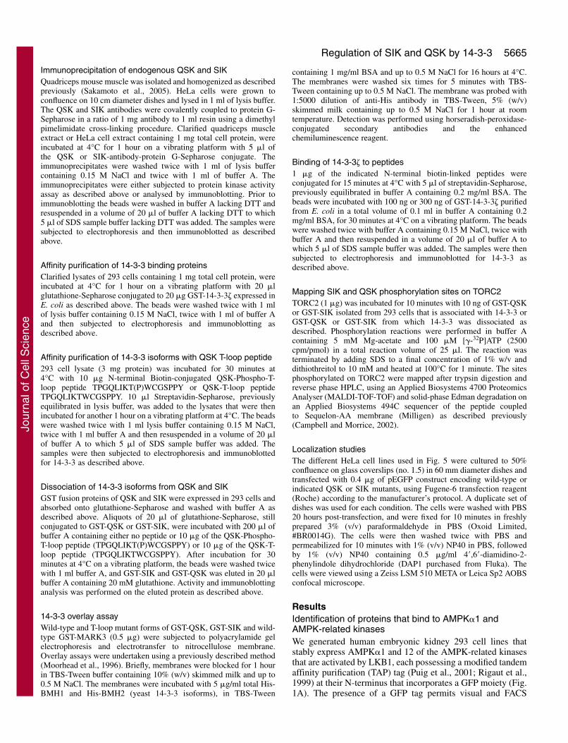
sorting of stable cell lines that express low levels of the GFP-TAP-proteins, considerably facilitating the isolation/selectionof these cells. The TAP tag also contains protein-A andcalmodulin-binding motifs to permit facile purification oftagged proteins on IgG-agarose and calmodulin-Sepharose,respectively. We performed successive chromatography onagarose and Sepharose resins in order to prevent contaminationof purified samples with Sepharose- or agarose-bindingproteins. We selected stable cell lines in which the GFP-TAP-tagged fusion proteins were expressed in a non-proteolysedform and at as low as possible levels, as determined byimmunoblot analysis of each of the selected stable cell lines.Each affinity purification was performed from fifty 15 cmdiameter dishes of confluent cells, using the strategy outlinedin Fig. 1B. The purified preparations were subjected toelectrophoresis on a polyacrylamide gel, which was thenstained with colloidal Coomassie Blue (Fig. 1C). The identityof the major colloidal Coomassie Blue-stained bands in eachpreparation was established by tryptic peptide mass-spectralfingerprinting procedures (Table 1).
In all samples, AMPK�1 and the AMPK-related kinase baitswere detected as the major staining band, migrating at theirpredicted molecular size (Fig. 1C). For QSK and NUAK2,additional proteolysed forms of the bait were also detected. In
most samples, including the control cell line expressing onlyGFP-TAP, tubulin and Hsp70 were identified, which indicatesthat they were likely to comprise non-specific contaminants ofour purification. Importantly, AMPK�1 was found to beassociated with its regulatory AMPK�1, AMPK�2 andAMPK�1 subunits (Fig. 1C, Table 1), thus validating thisapproach to identify physiological binding partners. Bycontrast, AMPK� and AMPK� subunits were not associatedwith any of the AMPK-related kinases. Several other proteinswere present at significant stoichiometry relative to the bait(Table 1). These included the 280 kDa fat facets/ubiquitin-specific protease 9 (USP9), which was associated withMARK4 and NUAK1, the AAA-ATPase-p97, as well as thecatalytic subunit of protein phosphatase 2A (PP2A) and its tworegulatory A and B subunits that bound to QIK. NUAK2 andAMPK�1 were associated with a chaperone heterodimerconsisting of Cdc37 and Hsp90, known to interact withnumerous kinases, whereas the adenine nucleotide translocasebound AMPK�1. The 14-3-3 and 14-3-3� phospho-bindingprotein adapters (Mackintosh, 2004) were associated with QSKand SIK as well as with all four MARK isoforms.
We also undertook TAP purifications from stable cell linesexpressing different levels of TAP-tagged MARK4(supplementary material Fig. S1). 14-3-3�, 14-3-3 and USP9
Journal of Cell Science 118 (23)
Table 1. Identification of proteins associated with AMPK-related kinases
Sample Sequence Protein Swiss-Prot ID* Protein coverage (%) score† acc. no.
1 GRP78 30 100 P110212 Hsp70 35 140 P081073 Carbamoyl phosphate 10 691 Q6F8M7
synthetase4 Hsp90 28 741 P079005 Hsp70 14 262 P081076 AMPK� 43 1089 P546457 Tubulin 41 675 P052098 Cdc37 15 219 Q165439 AMPK�1 47 300 Q9Y47810 AMPK�1 43 391 P5461911 AMPK�2 51 385 O4374112 Adenine nucleotide 11 179 PO5141
translocase 213 Qsk 28 244 Q9Y2K214 Qsk 33 128 Q9Y2K215 Hsp70 37 248 P0810716 Tubulin 28 96 P0520917 14-3-3� 40 178 P6310418 14-3-3 42 121 P6225819 Sik 19 127 Q86YJ220 Hsp70 37 131 P0810721 Sik 25 116 Q86YJ222 Tubulin 34 119 P0520923 14-3-3� 30 98 P6310424 14-3-3 38 120 P6225825 Qik 30 260 Q9H0K126 VCP 33 210 P5507227 Hsp70 52 369 P0810728 PP2A reg. subunit A 38 199 Q8NHV829 Tubulin 24 98 P0520930 PP2A reg. subunit B 49 145 P6315131 PP2A catalytic 49 203 P67775
subunit
Sample Sequence Protein Swiss-Prot ID* Protein coverage (%) score† acc. no.
32 USP9 22 448 Q9300833 Nuak1 48 409 O6028534 Hsp70 20 52 P0810735 Nuak2 24 192 Q9H09336 Hsp90 22 221 P0790037 Hsp70 36 157 P0810738 Nuak2 33 124 Q9H09339 cdc37 16 92 Q1654340 Brsk1 42 640 Q8TDC241 Hsp70 57 389 P0810742 Brsk2 52 397 Q8IWQ343 Hsp70 39 256 P0810744 Tubulin 32 147 P0520945 Mark1 34 176 Q9P0L246 Hsp70 33 111 P0810747 Tubulin 32 98 P0520948 14-3-3� 25 83 P6310449 14-3-3 34 140 P6225850 Mark2 49 518 Q1552451 Hsp70 42 120 P0810752 14-3-3� 25 83 P6310453 14-3-3 32 127 P6225854 Mark3 34 178 P2744855 Hsp70 25 83 P0810756 14-3-3� 31 98 P6310457 14-3-3 34 104 P6225858 USP9 14 204 Q9300859 Mark4 56 565 Q96GZ360 Hsp70 25 122 P0810761 Tubulin 25 98 P0520962 14-3-3� 35 104 P6310463 14-3-3 36 120 P6225864 Snrk 11 91 Q9NRH265 Hsp70 34 125 P08107
*The colloidal Coomassie-Blue-stained bands that were labelled as indicated in Fig. 1C were excised from the gel, digested in-gel with trypsin, and theiridentities were determined by tryptic peptide mass-spectral fingerprint as described in Materials and Methods.
†Mascot protein score where a value >63 is considered significant (P<0.05).
Jour
nal o
f Cel
l Sci
ence
5667Regulation of SIK and QSK by 14-3-3
were clearly associated with MARK4 derived from stable celllines expressing a low and a five- to tenfold higher level ofMARK4. Interestingly, however, MARK4 isolated from 293cells expressing higher levels of MARK4 resulting fromtransient transfection, revealed the presence of only 14-3-3isoforms associated with MARK4, but not USP9(supplementary material Fig. S1).
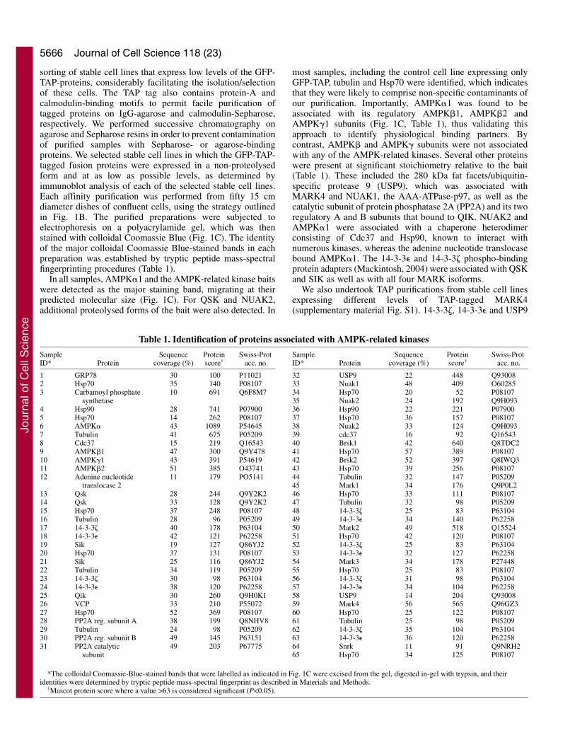
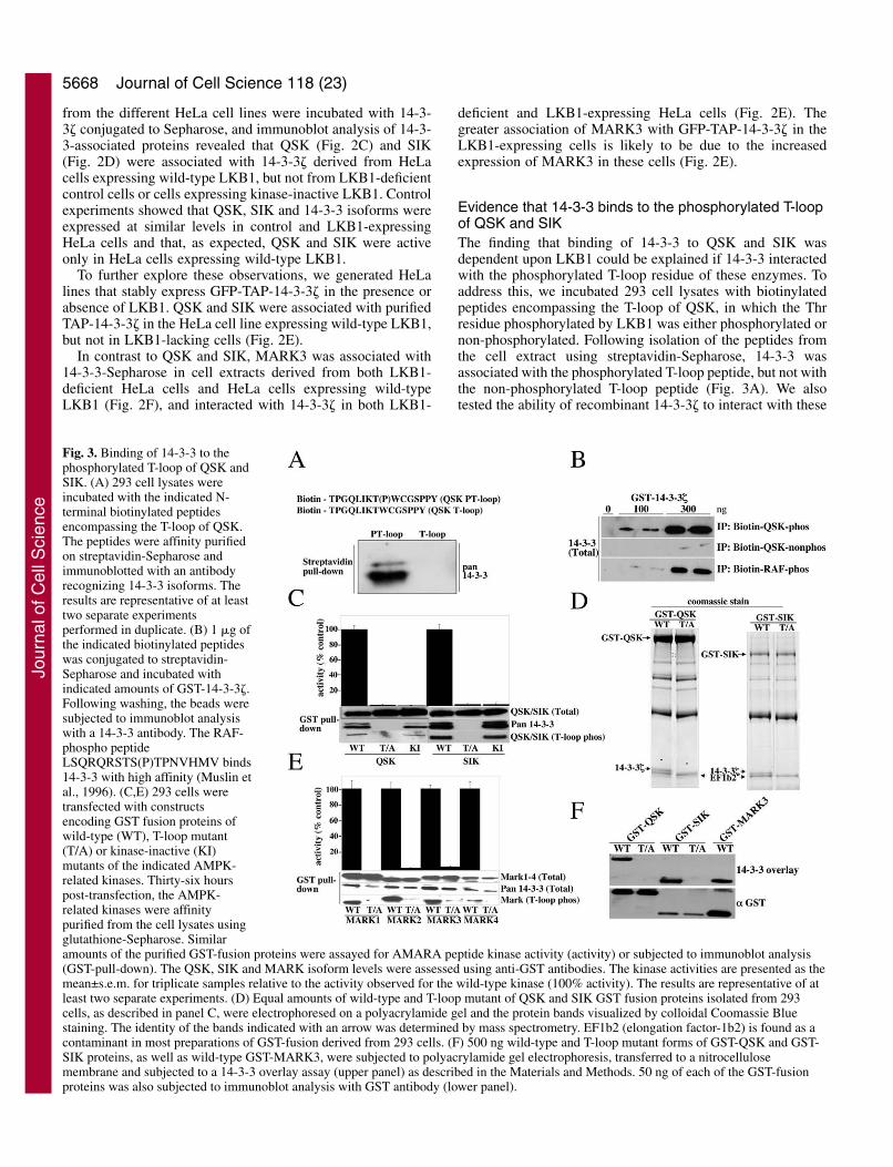
Binding of QSK and SIK to 14-3-3 is LKB1-dependentWe next explored whether the interaction of AMPK-relatedkinases with 14-3-3 was regulated by LKB1. We have recentlydescribed the generation of mice that lack expression of LKB1in skeletal muscle (Sakamoto et al., 2005). Strikingly, QSKimmunoprecipitated from LKB1-knockout muscle was not
associated with 14-3-3, whereas QSK derived from wild-typemuscle was bound to 14-3-3 (Fig. 2A). QSK was markedly lessactive in LKB1-deficient muscle, consistent with it not beingphosphorylated at its T-loop. As SIK was not detected in mouseskeletal muscle (AKA, data not shown) we could not testwhether it was associated with 14-3-3 in this system.
We next addressed whether AMPK-related kinasesinteracted with 14-3-3 isoforms in cell extracts derived fromcontrol HeLa cells that do not express LKB1 or HeLa cellsstably expressing wild-type or kinase-inactive LKB1. Asreported previously (Hawley et al., 2003; Lizcano et al., 2004),the kinase-inactive LKB1 is expressed at lower levels thanwild-type LKB1 in the HeLa cell lines, but is still expressed athigher levels than endogenously expressed LKB1 in 293 cellsor in mouse skeletal muscle (Fig. 2B). Cell extracts derived
Fig. 2. Binding of 14-3-3 to QSK and SIK,but not MARK3 is dependent on LKB1.(A) QSK was immunoprecipitated from thequadriceps muscle derived from wild-typemice (LKB1+/+) and muscle-specific LKB1-knockout (LKB1–/–) mice (Sakamoto et al.,2005). As a control, immunoprecipitationfrom LKB1+/+ muscle was also performedwith a preimmune antibody. Theimmunoprecipitates (IP) were subjected toimmunoblot analysis and also assayed usingthe AMARA peptide. Muscle samplesderived from five LKB1–/– and LKB+/+ micewere analysed and similar results obtainedfor each one. The QSK activity is themean±s.d. for each muscle sample assayedin duplicate. Pre-imm, pre-immuneantibody. (B) Cell lysates (20 �g) derivedfrom 293 cells, control (CT) HeLa cells thatlack LKB1 expression, or HeLa cells thatstably express wild-type (WT) or kinase-inactive (KI) LKB1, as well as skeletalmuscle derived from wild-type (WT) andmuscle-specific LKB1-knockout (KO)mice, were immunoblotted with an antibodyrecognizing LKB1. (C,D,F) Cell lysatesderived from control (CT) HeLa cells thatlack LKB1 expression or HeLa cells thatstably express wild-type (WT) or kinase-inactive (KI) LKB1 were incubated withglutathione-Sepahrose conjugated to 14-3-3� (14-3-3 pull-down). After washing, thebeads were immunoblotted with theindicated antibodies. The activity of theindicated AMPK-related kinases towardsthe AMARA peptide was also measuredfollowing immunoprecipitation. The resultsare presented as the mean±s.d. from two separate experiments each assayed intriplicate. Cell lysates were also subjected to the immunoblot analysis (lysate). SIKwas immunoblotted after its immunoprecipitation from the cell lysate. The results arerepresentative of at least two separate experiments. (E) The indicated HeLa cellsstably expressing GFP-TAP-14-3-3� were generated, and the GFP-TAP-14-3-3� wasaffinity purified and analysed by immunoblotting with the indicated antibodies (TAP-eluate). Cell lysates derived from the HeLa cells were also immunoblotted (lysate).As GFP-TAP-14-3-3� migrates to the same position as endogenous MARK3, thereby interfering with immunoblot analysis, the controlMARK3 immunoblot blot in cell lysate was performed using the parental HeLa cell line that does not express GFP-TAP-14-3-3�. The upperband in 14-3-3 blots of the TAP-eluate represents exogenously expressed 14-3-3� and the lower doublet represents co-purified endogenous 14-3-3 and 14-3-3�. The results are representative of three separate experiments.
Jour
nal o
f Cel
l Sci
ence
5668
from the different HeLa cell lines were incubated with 14-3-3� conjugated to Sepharose, and immunoblot analysis of 14-3-3-associated proteins revealed that QSK (Fig. 2C) and SIK(Fig. 2D) were associated with 14-3-3� derived from HeLacells expressing wild-type LKB1, but not from LKB1-deficientcontrol cells or cells expressing kinase-inactive LKB1. Controlexperiments showed that QSK, SIK and 14-3-3 isoforms wereexpressed at similar levels in control and LKB1-expressingHeLa cells and that, as expected, QSK and SIK were activeonly in HeLa cells expressing wild-type LKB1.
To further explore these observations, we generated HeLalines that stably express GFP-TAP-14-3-3� in the presence orabsence of LKB1. QSK and SIK were associated with purifiedTAP-14-3-3� in the HeLa cell line expressing wild-type LKB1,but not in LKB1-lacking cells (Fig. 2E).
In contrast to QSK and SIK, MARK3 was associated with14-3-3-Sepharose in cell extracts derived from both LKB1-deficient HeLa cells and HeLa cells expressing wild-typeLKB1 (Fig. 2F), and interacted with 14-3-3� in both LKB1-
deficient and LKB1-expressing HeLa cells (Fig. 2E). Thegreater association of MARK3 with GFP-TAP-14-3-3� in theLKB1-expressing cells is likely to be due to the increasedexpression of MARK3 in these cells (Fig. 2E).
Evidence that 14-3-3 binds to the phosphorylated T-loopof QSK and SIKThe finding that binding of 14-3-3 to QSK and SIK wasdependent upon LKB1 could be explained if 14-3-3 interactedwith the phosphorylated T-loop residue of these enzymes. Toaddress this, we incubated 293 cell lysates with biotinylatedpeptides encompassing the T-loop of QSK, in which the Thrresidue phosphorylated by LKB1 was either phosphorylated ornon-phosphorylated. Following isolation of the peptides fromthe cell extract using streptavidin-Sepharose, 14-3-3 wasassociated with the phosphorylated T-loop peptide, but not withthe non-phosphorylated T-loop peptide (Fig. 3A). We alsotested the ability of recombinant 14-3-3� to interact with these
Journal of Cell Science 118 (23)
Fig. 3. Binding of 14-3-3 to thephosphorylated T-loop of QSK andSIK. (A) 293 cell lysates wereincubated with the indicated N-terminal biotinylated peptidesencompassing the T-loop of QSK.The peptides were affinity purifiedon streptavidin-Sepharose andimmunoblotted with an antibodyrecognizing 14-3-3 isoforms. Theresults are representative of at leasttwo separate experimentsperformed in duplicate. (B) 1 �g ofthe indicated biotinylated peptideswas conjugated to streptavidin-Sepharose and incubated withindicated amounts of GST-14-3-3�.Following washing, the beads weresubjected to immunoblot analysiswith a 14-3-3 antibody. The RAF-phospho peptideLSQRQRSTS(P)TPNVHMV binds14-3-3 with high affinity (Muslin etal., 1996). (C,E) 293 cells weretransfected with constructsencoding GST fusion proteins ofwild-type (WT), T-loop mutant(T/A) or kinase-inactive (KI)mutants of the indicated AMPK-related kinases. Thirty-six hourspost-transfection, the AMPK-related kinases were affinitypurified from the cell lysates usingglutathione-Sepharose. Similaramounts of the purified GST-fusion proteins were assayed for AMARA peptide kinase activity (activity) or subjected to immunoblot analysis(GST-pull-down). The QSK, SIK and MARK isoform levels were assessed using anti-GST antibodies. The kinase activities are presented as themean±s.e.m. for triplicate samples relative to the activity observed for the wild-type kinase (100% activity). The results are representative of atleast two separate experiments. (D) Equal amounts of wild-type and T-loop mutant of QSK and SIK GST fusion proteins isolated from 293cells, as described in panel C, were electrophoresed on a polyacrylamide gel and the protein bands visualized by colloidal Coomassie Bluestaining. The identity of the bands indicated with an arrow was determined by mass spectrometry. EF1b2 (elongation factor-1b2) is found as acontaminant in most preparations of GST-fusion derived from 293 cells. (F) 500 ng wild-type and T-loop mutant forms of GST-QSK and GST-SIK proteins, as well as wild-type GST-MARK3, were subjected to polyacrylamide gel electrophoresis, transferred to a nitrocellulosemembrane and subjected to a 14-3-3 overlay assay (upper panel) as described in the Materials and Methods. 50 ng of each of the GST-fusionproteins was also subjected to immunoblot analysis with GST antibody (lower panel).
Jour
nal o
f Cel
l Sci
ence
5669Regulation of SIK and QSK by 14-3-3
peptides. The phosphorylated QSK T-loop peptide interacted with 14-3-3� to amarkedly greater extent than did the non-phosphopeptide (Fig. 3B). Under theassay conditions employed, thephosphorylated QSK T-loop peptideinteracted with 14-3-3� to an extentsimilar to that of a well characterized14-3-3 binding phosphopeptideencompassing Ser259 of the c-Rafprotein kinase (Muslin et al., 1996).
We next expressed, in 293 cells, wild-type QSK or SIK as well as mutants ofthese enzymes that were catalyticallyinactive or in which the T-loop Thrphosphorylated by LKB1 was changed toAla. The wild-type and catalyticallyinactive QSK and SIK associated withendogenous 14-3-3 proteins and werefound to be phosphorylated at theirT-loop residue, as revealed byimmunoblotting with a phosphospecificantibody that recognizes thisphosphorylation site (Fig. 3C). Analysisof a Coomassie-stained polyacrylamidegel of purified QSK and SIK indicatedthat a significant proportion of theseenzymes were associated with 14-3-3(Fig. 3D). By contrast, the QSK and SIKT-loop mutants failed to interact with 14-3-3 (Fig. 3C,D). All four MARKisoforms were associated with 14-3-3when expressed in 293 cells (Fig. 3E).Consistent with the notion that LKB1does not regulate binding of theseenzymes to 14-3-3, mutation of theMARK T-loop Thr to Ala, althoughabolishing catalytic activity, did notaffect binding to 14-3-3 (Fig. 3E).
14-3-3 binding to proteins has alsobeen previously studied using a FarWestern overlay approach, in which 14-3-3 binding to denatured proteins on anitrocellulose membrane can be detected.Using this assay, we confirm that 14-3-3isoforms were capable of interacting withwild-type QSK and SIK, but not withmutants of these enzymes in which the T-loop Thr phosphorylated by LKB1 waschanged to Ala (Fig. 3F). This providesfurther evidence that 14-3-3 isoforms caninteract directly with the phosphorylatedT-loop of QSK and SIK.
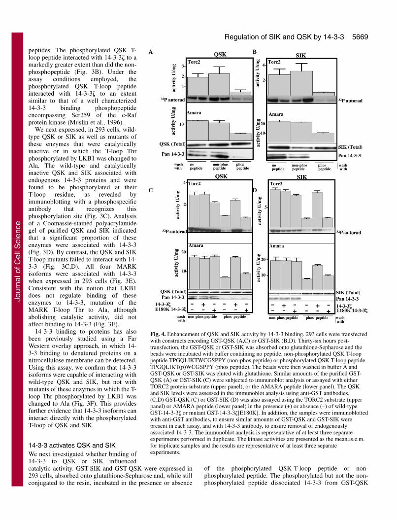
14-3-3 activates QSK and SIKWe next investigated whether binding of14-3-3 to QSK or SIK influencedcatalytic activity. GST-SIK and GST-QSK were expressed in293 cells, absorbed onto glutathione-Sepharose and, while stillconjugated to the resin, incubated in the presence or absence
of the phosphorylated QSK-T-loop peptide or non-phosphorylated peptide. The phosphorylated but not the non-phosphorylated peptide dissociated 14-3-3 from GST-QSK
Fig. 4. Enhancement of QSK and SIK activity by 14-3-3 binding. 293 cells were transfectedwith constructs encoding GST-QSK (A,C) or GST-SIK (B,D). Thirty-six hours post-transfection, the GST-QSK or GST-SIK was absorbed onto glutathione-Sepharose and thebeads were incubated with buffer containing no peptide, non-phosphorylated QSK T-looppeptide TPGQLIKTWCGSPPY (non-phos peptide) or phosphorylated QSK T-loop peptideTPGQLIKT(p)WCGSPPY (phos peptide). The beads were then washed in buffer A andGST-QSK or GST-SIK was eluted with glutathione. Similar amounts of the purified GST-QSK (A) or GST-SIK (C) were subjected to immunoblot analysis or assayed with eitherTORC2 protein substrate (upper panel), or the AMARA peptide (lower panel). The QSKand SIK levels were assessed in the immunoblot analysis using anti-GST antibodies.(C,D) GST-QSK (C) or GST-SIK (D) was also assayed using the TORC2 substrate (upperpanel) or AMARA peptide (lower panel) in the presence (+) or absence (–) of wild-typeGST-14-3-3� or mutant GST-14-3-3�[E180K]. In addition, the samples were immunoblottedwith anti-GST antibodies, to ensure similar amounts of GST-QSK and GST-SIK werepresent in each assay, and with 14-3-3 antibody, to ensure removal of endogenouslyassociated 14-3-3. The immunoblot analysis is representative of at least three separateexperiments performed in duplicate. The kinase activities are presented as the mean±s.e.m.for triplicate samples and the results are representative of at least three separateexperiments.
Jour
nal o
f Cel
l Sci
ence
5670
(Fig. 4A) or GST-SIK (Fig. 4B). We assayedQSK and SIK kinase activity using theCREB co-activator protein TORC2, arecently identified substrate for QIK(Screaton et al., 2004), which is alsophosphorylated by QSK and SIK with anefficiency similar to that of QIK in vitro(A.K.A., unpublished). We also assayedQSK and SIK by employing the shortAMARA peptide substrate (Lizcano et al.,2004). Dissociation of 14-3-3 from QSK(Fig. 4A) or SIK (Fig. 4B) resulted in a five-to sixfold reduction in activity when assayedwith the TORC2 protein and a ~2.5-foldreduction in catalytic activity of theseenzymes when assayed with the AMARApeptide substrate. Consistent with thereduction in activity resulting from a loss of14-3-3 binding, addition of recombinantwild-type 14-3-3� to GST-QSK (Fig. 4C) orGST-SIK (Fig. 4D) from which 14-3-3 hadbeen dissociated, restored activity. Bycontrast, addition of 14-3-3� [E180K]mutant, which is unable to interact withphosphopeptides (Chang and Rubin, 1997),failed to restore QSK or SIK activity.
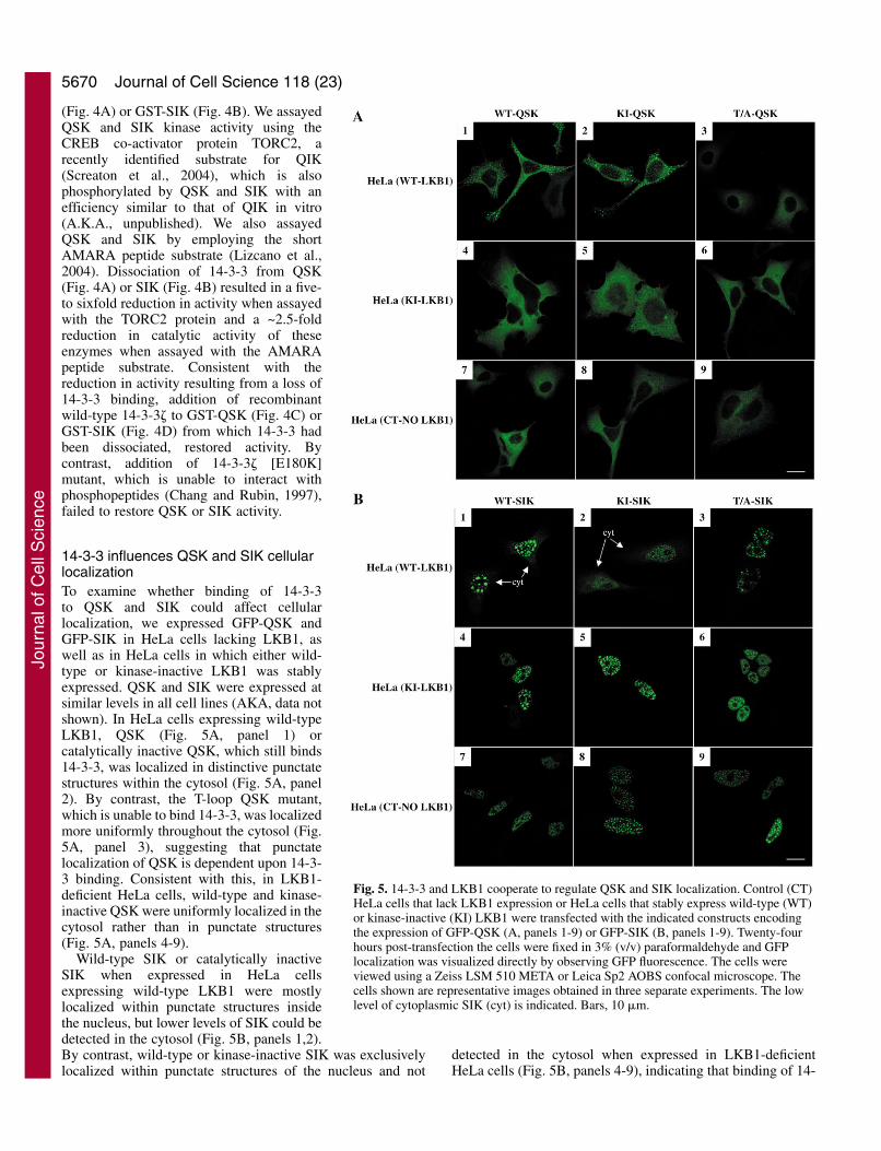
14-3-3 influences QSK and SIK cellularlocalizationTo examine whether binding of 14-3-3to QSK and SIK could affect cellularlocalization, we expressed GFP-QSK andGFP-SIK in HeLa cells lacking LKB1, aswell as in HeLa cells in which either wild-type or kinase-inactive LKB1 was stablyexpressed. QSK and SIK were expressed atsimilar levels in all cell lines (AKA, data notshown). In HeLa cells expressing wild-typeLKB1, QSK (Fig. 5A, panel 1) orcatalytically inactive QSK, which still binds14-3-3, was localized in distinctive punctatestructures within the cytosol (Fig. 5A, panel2). By contrast, the T-loop QSK mutant,which is unable to bind 14-3-3, was localizedmore uniformly throughout the cytosol (Fig.5A, panel 3), suggesting that punctatelocalization of QSK is dependent upon 14-3-3 binding. Consistent with this, in LKB1-deficient HeLa cells, wild-type and kinase-inactive QSK were uniformly localized in thecytosol rather than in punctate structures(Fig. 5A, panels 4-9).
Wild-type SIK or catalytically inactiveSIK when expressed in HeLa cellsexpressing wild-type LKB1 were mostlylocalized within punctate structures insidethe nucleus, but lower levels of SIK could bedetected in the cytosol (Fig. 5B, panels 1,2).By contrast, wild-type or kinase-inactive SIK was exclusivelylocalized within punctate structures of the nucleus and not
detected in the cytosol when expressed in LKB1-deficientHeLa cells (Fig. 5B, panels 4-9), indicating that binding of 14-
Journal of Cell Science 118 (23)
Fig. 5. 14-3-3 and LKB1 cooperate to regulate QSK and SIK localization. Control (CT)HeLa cells that lack LKB1 expression or HeLa cells that stably express wild-type (WT)or kinase-inactive (KI) LKB1 were transfected with the indicated constructs encodingthe expression of GFP-QSK (A, panels 1-9) or GFP-SIK (B, panels 1-9). Twenty-fourhours post-transfection the cells were fixed in 3% (v/v) paraformaldehyde and GFPlocalization was visualized directly by observing GFP fluorescence. The cells wereviewed using a Zeiss LSM 510 META or Leica Sp2 AOBS confocal microscope. Thecells shown are representative images obtained in three separate experiments. The lowlevel of cytoplasmic SIK (cyt) is indicated. Bars, 10 �m.
Jour
nal o
f Cel
l Sci
ence
5671Regulation of SIK and QSK by 14-3-3
3-3 to SIK may be required for low levels of cytosoliclocalization of SIK. Consistent with this, the T-loop SIKmutant was localized exclusively within the nucleus whenexpressed in HeLa cells expressing wild-type LKB1 (Fig. 5B,panel 3).
DiscussionWe have used the Tandem Affinity purification strategy toidentify potential binding partners of the AMPK-relatedkinases in 293 cells. We have modified the TAP vector toinclude a GFP tag that facilitates the isolation of cell coloniesexpressing low levels of bait protein. We believe thatgeneration of stable cell lines expressing low levels of baitproteins maximizes the proportion of the bait that is bound toendogenous binding partners, whose expression may belimiting. Thus, when MARK4 was expressed in 293 cells athigh levels using a transient transfection approach we wereable to detect the binding of MARK4 to 14-3-3 isoforms thatare abundantly expressed in cells. However, under theseconditions, we were unable to detect the interaction of MARK4with USP9, which is likely to be expressed at lower levels than14-3-3 isoforms (supplementary material Fig. S1). We alsovalidated this approach by demonstrating that TAP-taggedAMPK�1 was associated with its known AMPK� andAMPK� regulatory subunits. We also identified severalproteins that bind to some of the AMPK-related kinases andfurther work will be needed to investigate the physiologicalsignificance of these interactions.
Our previous work indicates that the AMPK-related kinasesmay be regulated in a distinct manner to that of AMPK, as noneof the AMPK-related kinases were stimulated in cell lines orskeletal muscle by the AMP mimetic AICA-riboside orfollowing treatments that elevate 5�-AMP (Lizcano et al., 2004;Sakamoto et al., 2004). The finding that the AMPK-relatedkinases, when purified from cells, are not associated with theAMPK� or AMPK� regulatory subunits, which are requiredfor the activation of AMPK by 5�AMP, is likely to account forthe inability of the AMPK-related kinases to be stimulated by5�-AMP. Two pairs of CBS domains located within theAMPK� subunit mediate the binding of 5�-AMP to AMPK.None of the proteins that were associated with the AMPK-related kinases possess predicted CBS domains, includingNUAK2, which was reported to be stimulated with AICAR(Lefebvre and Rosen, 2005) in hepatocarcinoma cells. We havepreviously shown in mouse embryonic fibroblast cells thatNUAK2 activity is not increased with AICAR (Lizcano et al.,2004). It may be worthwhile to investigate whether NUAK2could interact with AMPK� or another CBS-domain-containing proteins in hepatocarcinoma cells.
14-3-3 proteins interact dynamically with many intracellularproteins, which exerts a widespread influence on diversecellular processes. They operate by binding to specificphosphorylated residues on target proteins. In C. elegans, the14-3-3 protein, termed Par-5, was originally identified as agene that played a crucial role in the early events leading topolarization of the zygote (Morton et al., 2002). Par-5 was alsorequired for the asymmetric cortical localization of Par-1/MARK to the posterior of the embryo, as well as locating theother polarity regulators, Par-3 and Par-6/PKC�, to the cellanterior (Morton et al., 2002). Work performed in Drosophila
(Benton et al., 2002) and in mammalian cells (Brajenovic etal., 2003), also indicated that the Par-1/MARK enzymesinteracted directly with 14-3-3 isoforms. Recent studiessuggest that phosphorylation of Par-1/MARK by the Par-6/PKC� kinase, at residue(s) lying C-terminal to the catalyticdomain, induced a relocalization of Par-1/MARK from theplasma membrane and enhanced the binding of 14-3-3 to Par-1/MARK (Hurov et al., 2004; Kusakabe and Nishida, 2004;Suzuki et al., 2004a). Consistent with the 14-3-3 binding site(s)being located C-terminal to the catalytic domain, we observethat 14-3-3 isoforms bound to MARK3 in LKB1-deficient cells(Fig. 2F) and mutation of the T-loop Thr phosphorylated byLKB1 did not affect the ability of any MARK isoform to bind14-3-3 (Fig. 3E). By contrast, binding of 14-3-3 to QSK andSIK was mediated by LKB1, as QSK and SIK failed to bind14-3-3 in LKB1-deficient cells or skeletal muscle (Fig. 2).Moreover, mutation of the T-loop LKB1 phosphorylation siteon QSK and SIK abolished 14-3-3 binding (Fig. 3C,F). The T-loops of QSK and SIK do not conform to the classical 14-3-3binding motif (Mackintosh, 2004), but the finding that 14-3-3isoforms expressed in cell lysates (Fig. 3A) or recombinant 14-3-3� (Fig. 3B) could interact directly with a phosphorylated(but not non-phosphorylated) peptide encompassing the T-loopof QSK (Fig. 3A) indicates that this motif has the intrinsicability to bind 14-3-3.
The binding of 14-3-3 to many proteins, including Par-1/MARK (Hurov et al., 2004; Kusakabe and Nishida, 2004)and TORC2 (Screaton et al., 2004), serves to localize theseproteins in the cell cytosol. Our results also show that thebinding of 14-3-3 isoforms to SIK and QSK plays a role inregulating the localization of these enzymes. In the case of SIK,binding to 14-3-3 induces a moderate redistribution of SIKfrom the nucleus to the cytosol (Fig. 5B) whereas, for QSK,interaction with 14-3-3 results in localization within punctatestructures in the cytosol (Fig. 5A). Further work is required tocharacterize the punctate structures that QSK and SIK interactwith and the mechanism by which 14-3-3 anchors QSK topunctate structures in the cytosol and localizes SIK in thecytoplasm. It would also be important to develop sensitiveantibodies to study the localization of endogenously expressedQSK and SIK in wild-type and LKB1-deficient cell lines toensure that the localization patterns are similar to thoseobserved in the overexpressed studies.
Most interestingly, our results suggest that 14-3-3 binding toQSK and SIK enhances their catalytic activity (Fig. 4). As theT-loop of a kinase is a key motif in controlling intrinsiccatalytic activity, binding of 14-3-3 to this region of QSK andSIK could induce a structural change that stabilizes theseenzymes in an active conformation. Moreover, the effect of 14-3-3 binding on catalytic activity is more pronounced using theTORC2 protein substrate than the AMARA peptide substrate(Fig. 4). It has previously been reported that phosphorylationof TORC2 at Ser171 by the QIK AMPK-related kinase enabledTORC2 to bind 14-3-3 (Screaton et al., 2004). An intriguingpossibility is that binding of QSK and SIK to 14-3-3 enhancesthe ability of QSK/SIK to interact with and phosphorylate 14-3-3-binding substrates such as TORC2. We found that QSKand SIK phosphorylated TORC2 at Ser171 as well as atleast two additional residues, namely Ser70 and Ser348(supplementary material Fig. S2). Dissociation of 14-3-3 fromQSK and SIK reduced the extent of phosphorylation of Ser70,
Jour
nal o
f Cel
l Sci
ence
5672
Ser171 and Ser348 to a similar extent for each site(supplementary material Fig. S2), indicating that the presenceof 14-3-3 does not influence the ability of QSK/SIK tophosphorylate specific residues on TORC2. It is also possiblethat the greater stimulation of QSK and SIK activity towardsthe TORC2 substrate than the AMARA peptide (Fig. 4), resultsfrom 14-3-3� binding directly to TORC2 and converting it intoa better substrate for QSK/SIK. It is unlikely that 14-3-3 wouldbind to the short AMARA peptide, and thus the ability of 14-3-3� to stimulate of QSK and SIK AMARA peptide kinaseactivity is likely to result from the direct binding of 14-3-3� toQSK/SIK.
In conclusion, we demonstrate that catalytic activity andlocalization of QSK and SIK are regulated by 14-3-3 bindingto the T-loop of these enzymes following their phosphorylationby LKB1. To our knowledge, this is the first report of 14-3-3binding to the T-loop of a protein kinase and directlyinfluencing its catalytic activity. This represents a novelmechanism by which 14-3-3 can regulate biological function.14-3-3 isoforms are dimers that possess two substrate-bindingpockets per dimer (reviewed by Mackintosh, 2004). It ispossible that if QSK/SIK occupied only one of the binding siteson the 14-3-3 dimer, then the other site might be available forthe recruitment of a substrate of QSK/SIK. In this way, 14-3-3 isoforms could function as a scaffolding complex to facilitatethe interaction of QSK/SIK with their 14-3-3-bindingsubstrates. It is likely that many of the substrates of QSK/SIK,as well as other AMPK-related kinases, comprise 14-3-3-binding proteins and/or are converted to 14-3-3 bindingproteins following their phosphorylation, a feature that couldbe exploited to identify substrates for these enzymes.
We thank Kei Sakamoto for providing the LKB1-deficient muscleextracts, Carol MacKintosh for helpful advice, Mark Peggie forcloning and generation of expression constructs for 14-3-3� andTORC2, Agnieszka Kieloch for tissue culture, Rosie Clarke for FACSanalysis, Grahame Hardie for the AMPK�1 cDNA, the SequencingService (School of Life Sciences, University of Dundee, UK) for DNAsequencing, the Post Genomics and Molecular Interactions Centre forMass Spectrometry facilities (School of Life Sciences, University ofDundee) and the protein production and antibody purification teams[Division of Signal Transduction Therapy (DSTT), University ofDundee] co-ordinated by Hilary McLauchlan and James Hastie forexpression and purification of antibodies and the TORC2 protein.A.K.A. is supported by a Moffat Charitable Trust studentship andO.G. is supported by a Wenner-Gren Foundation fellowship. We thankthe Association for International Cancer Research, Diabetes UK, theMedical Research Council, the Moffat Charitable Trust and thepharmaceutical companies supporting the Division of SignalTransduction Therapy Unit (AstraZeneca, Boehringer-Ingelheim,GlaxoSmithKline, Merck & Co., Merck KgaA and Pfizer) forfinancial support.
ReferencesBaas, A. F., Boudeau, J., Sapkota, G. P., Smit, L., Medema, R., Morrice,
N. A., Alessi, D. R. and Clevers, H. C. (2003). Activation of the tumoursuppressor kinase LKB1 by the STE20-like pseudokinase STRAD. EMBOJ. 22, 3062-3072.
Baas, A. F., Smit, L. and Clevers, H. (2004). LKB1 tumor suppressor protein:PARtaker in cell polarity. Trends Cell. Biol. 14, 312-319.
Benton, R., Palacios, I. M. and St Johnston, D. (2002). Drosophila 14-3-3/PAR-5 is an essential mediator of PAR-1 function in axis formation. Dev.Cell 3, 659-671.
Bettencourt-Dias, M., Giet, R., Sinka, R., Mazumdar, A., Lock, W. G.,
Balloux, F., Zafiropoulos, P. J., Yamaguchi, S., Winter, S., Carthew, R.W. et al. (2004). Genome-wide survey of protein kinases required for cellcycle progression. Nature 432, 980-987.
Boudeau, J., Baas, A. F., Deak, M., Morrice, N. A., Kieloch, A.,Schutkowski, M., Prescott, A. R., Clevers, H. C. and Alessi, D. R. (2003).MO25alpha/beta interact with STRADalpha/beta enhancing their ability tobind, activate and localize LKB1 in the cytoplasm. EMBO J. 22, 5102-5114.
Brajenovic, M., Joberty, G., Kuster, B., Bouwmeester, T. and Drewes, G.(2003). Comprehensive proteomic analysis of human Par protein complexesreveals an interconnected protein network. J. Biol. Chem. 279, 12804-12811.
Campbell, D. G. and Morrice, N. A. (2002). Identification of proteinphosphorylation sites by a combination of mass spectrometry and solidphase edman sequencing. J. Biomol. Techn. 13, 121-132.
Chang, H. C. and Rubin, G. M. (1997). 14-3-3 epsilon positively regulatesRas-mediated signaling in Drosophila. Genes Dev. 11, 1132-1139.
Corradetti, M. N., Inoki, K., Bardeesy, N., DePinho, R. A. and Guan, K.L. (2004). Regulation of the TSC pathway by LKB1: evidence of amolecular link between tuberous sclerosis complex and Peutz-Jegherssyndrome. Genes Dev. 18, 1533-1538.
Drewes, G., Ebneth, A., Preuss, U., Mandelkow, E. M. and Mandelkow, E.(1997). MARK, a novel family of protein kinases that phosphorylatemicrotubule-associated proteins and trigger microtubule disruption. Cell 89,297-308.
Durocher, Y., Perret, S. and Kamen, A. (2002). High-level and high-throughput recombinant protein production by transient transfection ofsuspension-growing human 293-EBNA1 cells. Nucleic Acids Res. 30, E9.
Feldman, J. D., Vician, L., Crispino, M., Hoe, W., Baudry, M. andHerschman, H. R. (2000). The salt-inducible kinase, SIK, is induced bydepolarization in brain. J. Neurochem. 74, 2227-2238.
Guo, S. and Kemphues, K. J. (1995). par-1, a gene required for establishingpolarity in C. elegans embryos, encodes a putative Ser/Thr kinase that isasymmetrically distributed. Cell 81, 611-620.
Hardie, D. G. (2004). The AMP-activated protein kinase pathway – newplayers upstream and downstream. J. Cell Sci. 117, 5479-5487.
Hawley, S. A., Boudeau, J., Reid, J. L., Mustard, K. J., Udd, L., Makela,T. P., Alessi, D. R. and Hardie, D. G. (2003). Complexes between theLKB1 tumor suppressor, STRADalpha/beta and MO25alpha/beta areupstream kinases in the AMP-activated protein kinase cascade. J. Biol. 2,28.
Hemminki, A., Markie, D., Tomlinson, I., Avizienyte, E., Roth, S., Loukola,A., Bignell, G., Warren, W., Aminoff, M., Hoglund, P. et al. (1998). Aserine/threonine kinase gene defective in Peutz-Jeghers syndrome. Nature391, 184-187.
Horike, N., Takemori, H., Katoh, Y., Doi, J., Min, L., Asano, T., Sun, X.J., Yamamoto, H., Kasayama, S., Muraoka, M. et al. (2003). Adipose-specific expression, phosphorylation of Ser794 in insulin receptor substrate-1, and activation in diabetic animals of salt-inducible kinase-2. J. Biol.Chem. 278, 18440-18447.
Hurov, J. B., Watkins, J. L. and Piwnica-Worms, H. (2004). Atypical PKCphosphorylates PAR-1 kinases to regulate localization and activity. Curr.Biol. 14, 736-741.
Jaleel, M., McBride, A., Lizcano, J. M., Deak, M., Toth, R., Morrice, N.A. and Alessi, D. R. (2005). Identification of the sucrose non-fermentingrelated kinase SNRK, as a novel LKB1 substrate. FEBS Lett. 579, 1417-1423.
Jenne, D. E., Reimann, H., Nezu, J., Friedel, W., Loff, S., Jeschke, R.,Muller, O., Back, W. and Zimmer, M. (1998). Peutz-Jeghers syndrome iscaused by mutations in a novel serine threonine kinase. Nat. Genet. 18, 38-43.
Jones, R. G., Plas, D. R., Kubek, S., Buzzai, M., Mu, J., Xu, Y., Birnbaum,M. J. and Thompson, C. B. (2005). AMP-activated protein kinase inducesa p53-dependent metabolic checkpoint. Mol. Cell 18, 283-293.
Kishi, M., Pan, Y. A., Crump, J. G. and Sanes, J. R. (2005). MammalianSAD kinases are required for neuronal polarization. Science 307, 929-932.
Kusakabe, M. and Nishida, E. (2004). The polarity-inducing kinase Par-1controls Xenopus gastrulation in cooperation with 14-3-3 and aPKC. EMBOJ. 23, 4190-4201.
Lefebvre, D. L. and Rosen, C. F. (2005). Regulation of SNARK activity inresponse to cellular stresses. Biochim. Biophys. Acta 1724, 71-85.
Lefebvre, D. L., Bai, Y., Shahmolky, N., Sharma, M., Poon, R., Drucker,D. J. and Rosen, C. F. (2001). Identification and characterization of a novelsucrose-non-fermenting protein kinase/AMP-activated protein kinase-related protein kinase, SNARK. Biochem. J. 355, 297-305.
Legembre, P., Schickel, R., Barnhart, B. C. and Peter, M. E. (2004).
Journal of Cell Science 118 (23)
Jour
nal o
f Cel
l Sci
ence
5673Regulation of SIK and QSK by 14-3-3
Identification of SNF1/AMP kinase-related kinase as an NF-kappaB-regulated anti-apoptotic kinase involved in CD95-induced motility andinvasiveness. J. Biol. Chem. 279, 46742-46747.
Lizcano, J. M., Goransson, O., Toth, R., Deak, M., Morrice, N. A.,Boudeau, J., Hawley, S. A., Udd, L., Makela, T. P., Hardie, D. G. et al.(2004). LKB1 is a master kinase that activates 13 kinases of the AMPKsubfamily, including MARK/PAR-1. EMBO J. 23, 833-843.
Mackintosh, C. (2004). Dynamic interactions between 14-3-3 proteins andphosphoproteins regulate diverse cellular processes. Biochem. J. 381, 329-342.
Moorhead, G., Douglas, P., Morrice, N., Scarabel, M., Aitken, A. andMacKintosh, C. (1996). Phosphorylated nitrate reductase from spinachleaves is inhibited by 14-3-3 proteins and activated by fusicoccin. Curr. Biol.6, 1104-1113.
Morton, D. G., Shakes, D. C., Nugent, S., Dichoso, D., Wang, W., Golden,A. and Kemphues, K. J. (2002). The Caenorhabditis elegans par-5 geneencodes a 14-3-3 protein required for cellular asymmetry in the earlyembryo. Dev. Biol. 241, 47-58.
Muslin, A. J., Tanner, J. W., Allen, P. M. and Shaw, A. S. (1996). Interactionof 14-3-3 with signaling proteins is mediated by the recognition ofphosphoserine. Cell 84, 889-897.
Nishimura, I., Yang, Y. and Lu, B. (2004). PAR-1 kinase plays an initiatorrole in a temporally ordered phosphorylation process that confers tautoxicity in Drosophila. Cell 116, 671-682.
Perkins, D. N., Pappin, D. J., Creasy, D. M. and Cottrell, J. S. (1999).Probability-based protein identification by searching sequence databasesusing mass spectrometry data. Electrophoresis 20, 3551-3567.
Puig, O., Caspary, F., Rigaut, G., Rutz, B., Bouveret, E., Bragado-Nilsson,E., Wilm, M. and Seraphin, B. (2001). The tandem affinity purification(TAP) method: a general procedure of protein complex purification.Methods 24, 218-229.
Rigaut, G., Shevchenko, A., Rutz, B., Wilm, M., Mann, M. and Seraphin,B. (1999). A generic protein purification method for protein complexcharacterization and proteome exploration. Nat. Biotechnol. 17, 1030-1032.
Sakamoto, K., Goransson, O., Hardie, D. G. and Alessi, D. R. (2004).Activity of LKB1 and AMPK-related kinases in skeletal muscle; effects ofcontraction, phenformin and AICAR. Am J. Physiol. Endocrinol. Metab.287, E310-E317.
Sakamoto, K., McCarthy, A., Smith, D., Green, K. A., Hardie, D. G.,Ashworth, A. and Alessi, D. R. (2005). Deficiency of LKB1 in skeletalmuscle prevents AMPK activation and glucose uptake during contraction.EMBO J. 24, 1810-1820.
Sapkota, G. P., Deak, M., Kieloch, A., Morrice, N., Goodarzi, A. A.,Smythe, C., Shiloh, Y., Lees-Miller, S. P. and Alessi, D. R. (2002).Ionizing radiation induces ataxia telangiectasia mutated kinase (ATM)-
mediated phosphorylation of LKB1/STK11 at Thr-366. Biochem. J. 368,507-516.
Screaton, R. A., Conkright, M. D., Katoh, Y., Best, J. L., Canettieri, G.,Jeffries, S., Guzman, E., Niessen, S., Yates, J. R., 3rd, Takemori, H. etal. (2004). The CREB coactivator TORC2 functions as a calcium- andcAMP-sensitive coincidence detector. Cell 119, 61-74.
Shaw, R. J., Bardeesy, N., Manning, B. D., Lopez, L., Kosmatka, M.,DePinho, R. A. and Cantley, L. C. (2004a). The LKB1 tumor suppressornegatively regulates mTOR signaling. Cancer Cell 6, 91-99.
Shaw, R. J., Kosmatka, M., Bardeesy, N., Hurley, R. L., Witters, L. A.,DePinho, R. A. and Cantley, L. C. (2004b). The tumor suppressor LKB1kinase directly activates AMP-activated kinase and regulates apoptosis inresponse to energy stress. Proc. Natl. Acad. Sci. USA 101, 3329-3335.
Shulman, J. M., Benton, R. and St Johnston, D. (2000). The Drosophilahomolog of C. elegans PAR-1 organizes the oocyte cytoskeleton and directsoskar mRNA localization to the posterior pole. Cell 101, 377-388.
Suzuki, A., Kusakai, G., Kishimoto, A., Lu, J., Ogura, T. and Esumi, H.(2003a). ARK5 suppresses the cell death induced by nutrient starvation anddeath receptors via inhibition of caspase 8 activation, but not bychemotherapeutic agents or UV irradiation. Oncogene 22, 6177-6182.
Suzuki, A., Kusakai, G., Kishimoto, A., Lu, J., Ogura, T., Lavin, M. F. andEsumi, H. (2003b). Identification of a novel protein kinase mediating Aktsurvival signaling to the ATM protein. J. Biol. Chem. 278, 48-53.
Suzuki, A., Hirata, M., Kamimura, K., Maniwa, R., Yamanaka, T.,Mizuno, K., Kishikawa, M., Hirose, H., Amano, Y., Izumi, N. etal. (2004a). aPKC acts upstream of PAR-1b in both the establishmentand maintenance of mammalian epithelial polarity. Curr. Biol. 14, 1425-1435.
Suzuki, A., Kusakai, G., Kishimoto, A., Shimojo, Y., Miyamoto, S., Ogura,T., Ochiai, A. and Esumi, H. (2004b). Regulation of caspase-6 and FLIPby the AMPK family member ARK5. Oncogene 23, 7067-7075.
Wang, Z., Takemori, H., Halder, S. K., Nonaka, Y. and Okamoto, M.(1999). Cloning of a novel kinase (SIK) of the SNF1/AMPK family fromhigh salt diet-treated rat adrenal. FEBS Lett. 453, 135-139.
Woods, A., Johnstone, S. R., Dickerson, K., Leiper, F. C., Fryer, L. G.,Neumann, D., Schlattner, U., Wallimann, T., Carlson, M. and Carling,D. (2003). LKB1 is the upstream kinase in the AMP-Activated proteinkinase cascade. Curr. Biol. 13, 2004-2008.
Woods, Y. L., Rena, G., Morrice, N., Barthel, A., Becker, W., Guo, S.,Unterman, T. G. and Cohen, P. (2001). The kinase DYRK1Aphosphorylates the transcription factor FKHR at Ser329 in vitro, a novel invivo phosphorylation site. Biochem. J. 355, 597-607.
Yoshida, K., Yamada, M., Nishio, C., Konishi, A. and Hatanaka, H. (2000).SNRK, a member of the SNF1 family, is related to low K(+)-inducedapoptosis of cultured rat cerebellar granule neurons. Brain Res. 873, 274-282.
Jour
nal o
f Cel
l Sci
ence