Embed Size (px)
Citation preview
17-1
Chapter 17
Lecture Outline
See PowerPoint Image Slides
for all figures and tables pre-inserted into
PowerPoint without notes.
Copyright (c) The McGraw-Hill Companies, Inc. Permission required for reproduction or display.
17-2
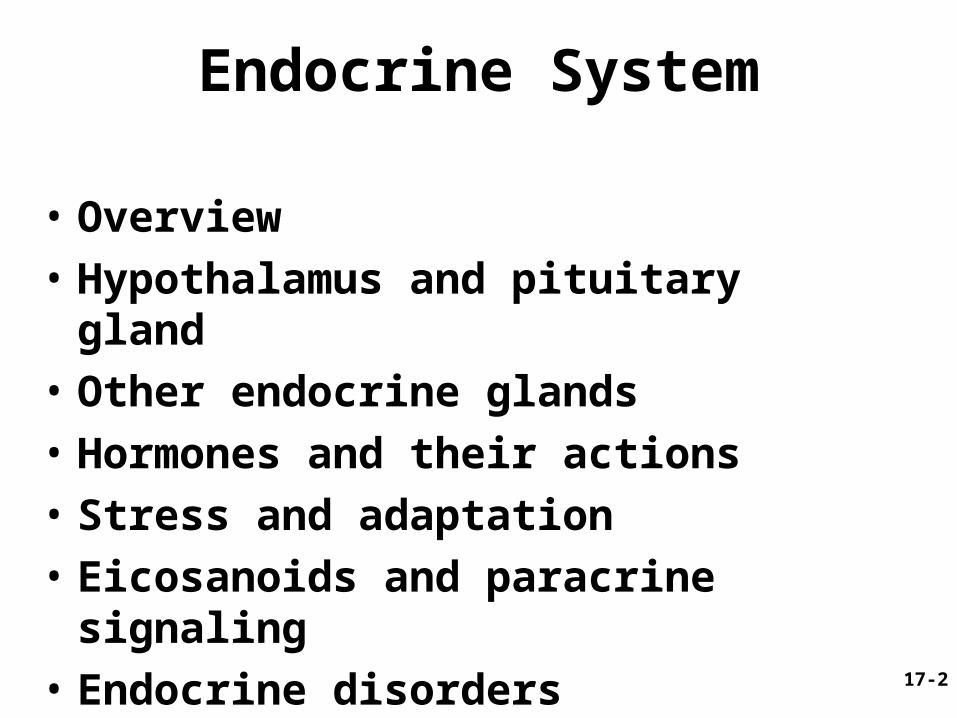
Endocrine System
• Overview
• Hypothalamus and pituitary gland
• Other endocrine glands
• Hormones and their actions
• Stress and adaptation
• Eicosanoids and paracrine signaling
• Endocrine disorders
17-3
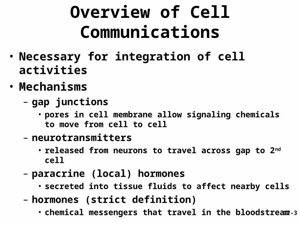
Overview of Cell Communications
• Necessary for integration of cell activities• Mechanisms
– gap junctions• pores in cell membrane allow signaling chemicals to move
from cell to cell
– neurotransmitters• released from neurons to travel across gap to 2nd cell
– paracrine (local) hormones• secreted into tissue fluids to affect nearby cells
– hormones (strict definition)• chemical messengers that travel in the bloodstream

17-4
• Hormone– chemical messenger secreted
into bloodstream, stimulates response in another tissue or organ
• Target cells– have receptors
for hormone
• Endocrine glands– produce hormones
• Endocrine system– endocrine organs (thyroid,
pineal, etc)– hormone producing cells in
organs (brain, heart and small intestine)
Endocrine System Components
17-5
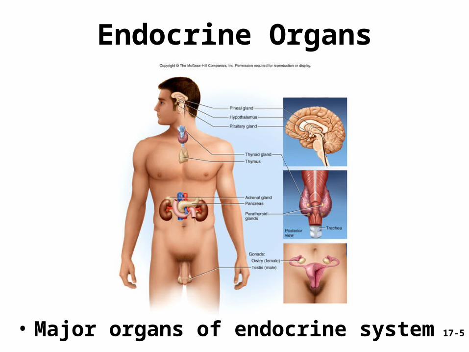
Endocrine Organs
• Major organs of endocrine system

17-6
Endocrine vs. Exocrine Glands
• Exocrine glands– ducts carry secretion to a surface or organ
cavity– extracellular effects (food digestion)
• Endocrine glands– no ducts, release hormones into tissue fluids,
have dense capillary networks to distribute hormones
– intracellular effects, alter target cell metabolism

17-7
Nervous vs. Endocrine Systems
• Communication– nervous - both electrical and chemical– endocrine - only chemical
• Speed and persistence of response– nervous - reacts quickly (1 - 10 msec), stops quickly– endocrine - reacts slowly (hormone release in
seconds or days), effect may continue for weeks• Adaptation to long-term stimuli
– nervous - response declines (adapts quickly)– endocrine - response persists
• Area of effect– nervous - targeted and specific (one organ)– endocrine - general, widespread effects (many
organs)

17-8
Communication by the Nervous and Endocrine Systems

17-9
Nervous and Endocrine Systems
• Several chemicals function as both hormones and neurotransmitters– NE, cholecystokinin, thyrotropin-releasing hormone,
dopamine and ADH
• Some hormones secreted by neuroendocrine cells (neurons)– oxytocin and catecholamines
• Both systems with overlapping effects on same target cells– NE and glucagon cause glycogen hydrolysis in liver
• Systems regulate each other– neurons trigger hormone secretion– hormones stimulate or inhibit neurons
17-10
Hypothalamus
• Shaped like a flattened funnel, forms floor and walls of third ventricle
• Regulates primitive functions from water balance to sex drive
• Many functions carried out by pituitary gland
17-11
Pituitary Gland (Hypophysis)
• Suspended from hypothalamus by stalk (infundibulum)
• Location and size– housed in sella turcica of sphenoid bone– 1.3 cm diameter
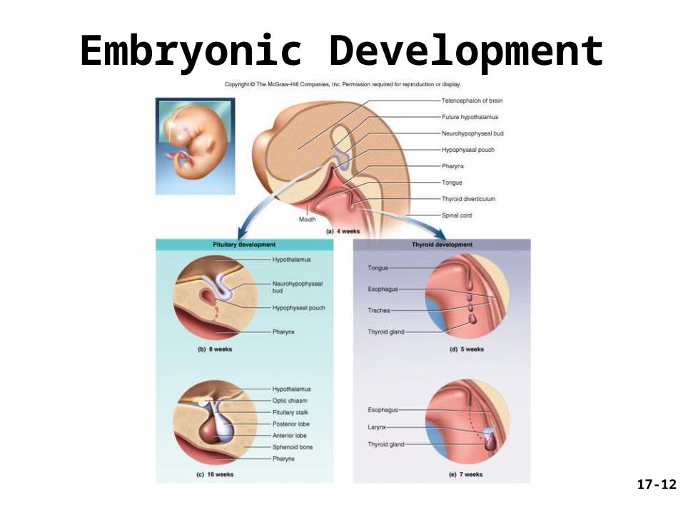
• Adenohypophysis (anterior pituitary)– arises from hypophyseal pouch (outgrowth of
pharynx)
• Neurohypophysis– arises from brain
17-12
Embryonic Development
17-13
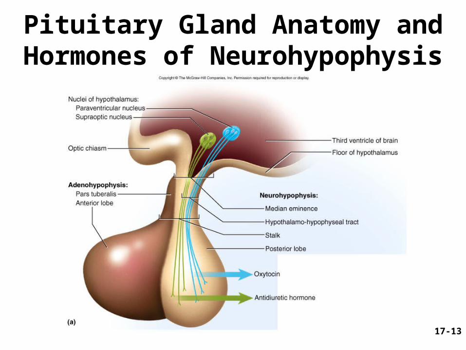
Pituitary Gland Anatomy and Hormones of Neurohypophysis
17-14
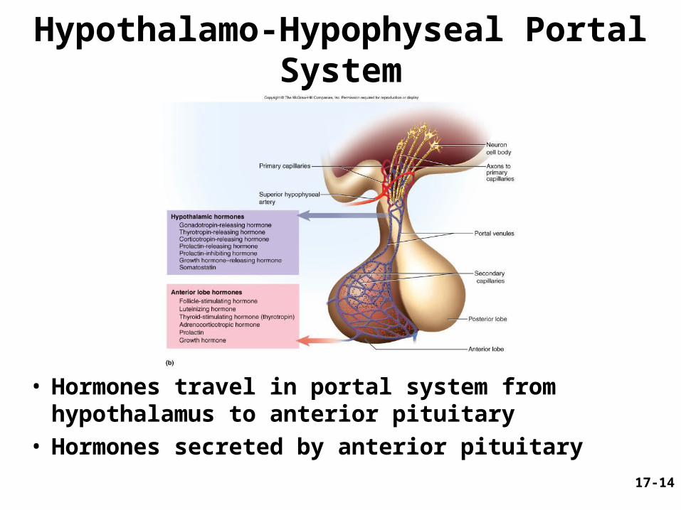
Hypothalamo-Hypophyseal Portal System
• Hormones travel in portal system from hypothalamus to anterior pituitary
• Hormones secreted by anterior pituitary

17-15
Histology of Pituitary Gland

17-16
• Tropic hormones target other endocrine glands– gonadotropins target gonads
• FSH (follicle stimulating hormone) • LH (luteinizing hormone)
– TSH (thyroid stimulating hormone)– ACTH (adrenocorticotropic hormone)
• PRL (prolactin)
• GH (growth hormone)
Pituitary Hormones - Anterior Lobe

17-17
Anterior Pituitary Hormones
• Principle hormones and target organs shown• Axis - refers to way endocrine glands interact
17-18
• Present in fetus; absent in adult
• Remnant cells– produce POMC (pro-opiomelanocortin)
• processed into ACTH and endorphins
• Produces MSH in animals influencing pigmentation of skin, hair or feathers– not apparently present/functioning in
humans
Pituitary Hormones - Pars Intermedia
17-19
• OT (oxytocin) and ADH – produced in hypothalamus– transported by hypothalamo-hypophyseal
tract to posterior lobe (stores then releases hormones)
Pituitary Hormones - Posterior Lobe
17-20
Hormone Actions: Anterior Lobe
• FSH (secreted by gonadotrope cells)– stimulates production of egg or sperm
cells
• LH (secreted by gonadotrope cells)– mainly stimulates hormone production
• females - stimulates ovulation and corpus luteum to secrete progesterone and estrogen
• males - stimulates interstitial cells of testes to secrete testosterone
• TSH (secreted by thyrotropes)– stimulates growth of gland and secretion of
TH
17-21
• ACTH or corticotropin (secreted by corticotropes)– regulates response to stress, stimulates
adrenal cortex • corticosteroids regulate glucose, fat and protein
metabolism
• PRL (secreted by lactotropes)– female - milk synthesis after delivery– male - LH sensitivity, thus testosterone
secretion
• GH or somatotropin – see next two slides
Hormone Actions: Anterior Lobe

17-22
• Secreted by somatotropes of anterior pituitary• Promotes tissue growth
– mitosis and cellular differentiation– stimulates liver to produce IGF-I and II
• protein synthesis – DNA transciption for mRNA production, proteins
synthesized– enhances amino acid transport into cells, protein catabolism
• lipid metabolism – stimulates FFA and glycerol release from adipocytes, protein
sparing
• CHO metabolism– glucose sparing effect = less glucose used for energy
• Electrolyte balance– promotes Na+, K+, Cl- retention, Ca 2+ absorption
Growth Hormone

17-23
• Childhood and adolescence– bone, cartilage and muscle growth– Stimulates growth at epiphyseal plates
• Adulthood– increase osteoblastic activity and appositional
growth affecting bone thickening and remodeling– blood concentration decrease by age 75 to ¼ of that
of adolescent
• Levels of GH (fluctuates throughout day)– higher during deep sleep, after high protein meals,
after vigorous exercise– lower after high CHO meals
Growth Hormone and Aging
17-24
Hormone Actions: Posterior Lobe
• ADH– targets kidneys
water retention, reduce urine
– also functions as neurotransmitter
• Oxytocin– labor contractions, lactation– possible role in
• sperm transport• emotional bonding

17-25
Control of Pituitary: Hypothalamic and Cerebral Control
• Anterior lobe control - releasing hormones and inhibiting hormones of hypothalamus
• Posterior lobe control - neuroendocrine reflexes – hormone release in response to nervous system
signals• suckling infant stimulates nerve endings
hypothalamus posterior lobe oxytocin milk ejection
– hormone release in response to higher brain centers
• milk ejection reflex can be triggered by a baby's cry
17-26
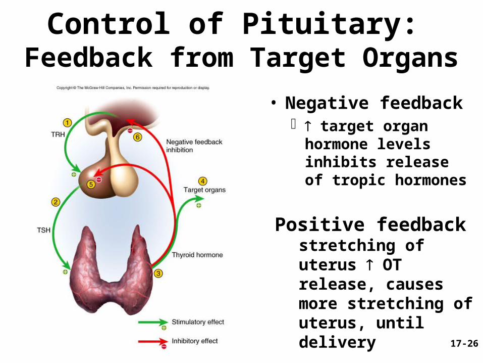
Control of Pituitary: Feedback from Target Organs
• Negative feedback target organ
hormone levels inhibits release of tropic hormones
Positive feedback stretching of uterus OT release, causes more stretching of uterus, until delivery

17-27
Pineal Gland
• Peak secretion ages 1-5; by puberty 75% lower
• Produces serotonin by day, converts it to melatonin at night
• May regulate timing of puberty in humans
• Melatonin in SAD + PMS; by phototherapy– depression, sleepiness, irritability and
carbohydrate craving
17-28
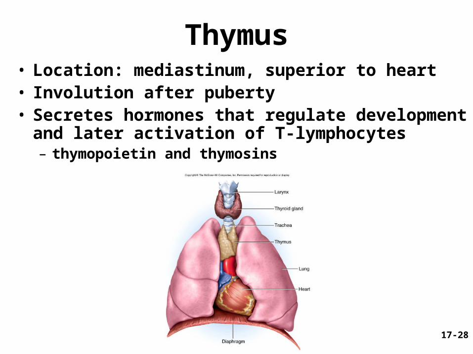
Thymus• Location: mediastinum, superior to heart• Involution after puberty• Secretes hormones that regulate development
and later activation of T-lymphocytes– thymopoietin and thymosins
17-29
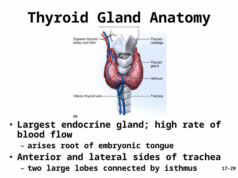
Thyroid Gland Anatomy
• Largest endocrine gland; high rate of blood flow– arises root of embryonic tongue
• Anterior and lateral sides of trachea– two large lobes connected by isthmus
Fig. 17.9a

17-30
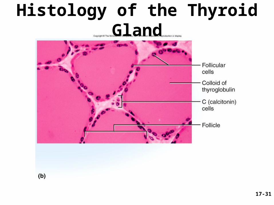
Thyroid Gland• Thyroid follicles
– filled with colloid and lined with simple cuboidal epithelial (follicular cells) that secretes two hormones, T3 and T4
– thyroid hormone body’s metabolic rate and O2 consumption• calorigenic effect - heat production heart rate and contraction strength respiratory rate• stimulates appetite and breakdown CHO, lipids and
proteins
• C (calcitonin or parafollicular) cells– produce calcitonin that blood Ca2+ , promotes Ca2+
deposition and bone formation especially in children
17-31
Histology of the Thyroid Gland
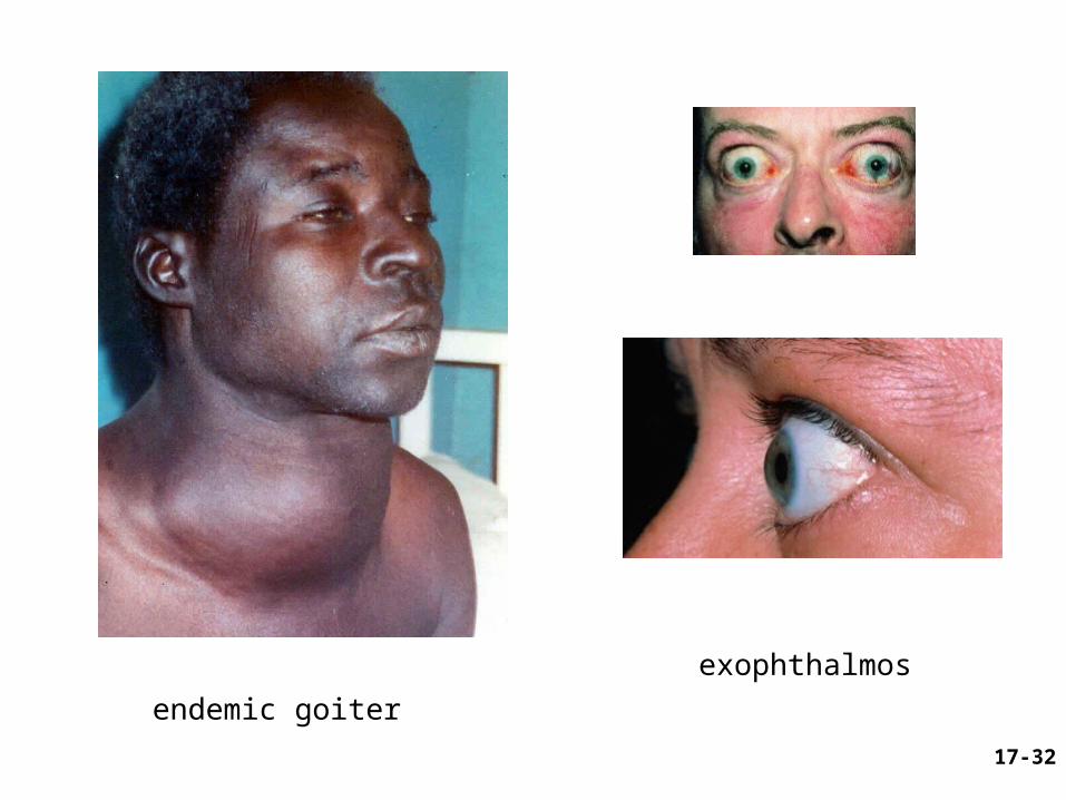
17-32
exophthalmos
endemic goiter

17-33
Grave’s disease

17-34
Parathyroid Glands
• PTH release blood Ca2+ levels– promotes synthesis of
calcitriol absorption of Ca2+
urinary excretion bone resorption
17-35
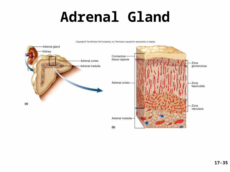
Adrenal Gland

17-36
Adrenal Medulla
• Sympathetic ganglion innervated by sympathetic preganglionic fibers – consists of modified neurons called chromaffin
cells– stimulation causes release of catecholamines
(epinephrine, NE)• Hormonal effect is longer lasting
– Increases alertness, anxiety, or fear– increases BP, heart rate and air flow– raises metabolic rate
• inhibits insulin secretion• stimulates gluconeogenesis and glycogenolysis
• Stress causes medullary cells to stimulate cortex

17-37
Adrenal Cortex
• Layers– zona glomerulosa (outer) – zona fasciculata (middle) – zona reticularis (inner)
17-38
Adrenal Cortex• Corticosteroids
– mineralocorticoids (zona glomerulosa)• control electrolyte balance, aldosterone promotes Na+
retention and K+ excretion
– glucocorticoids (zona fasciculata)• especially cortisol, stimulates fat and protein catabolism,
gluconeogenesis (from a.a.’s and FA’s) and release of fatty acids and glucose into blood
• anti-inflammatory effect becomes immune suppression with long-term use
– sex steroids (zona reticularis)• androgen (including DHEA which other tissues convert to
testosterone) and estrogen (important after menopause)
17-39
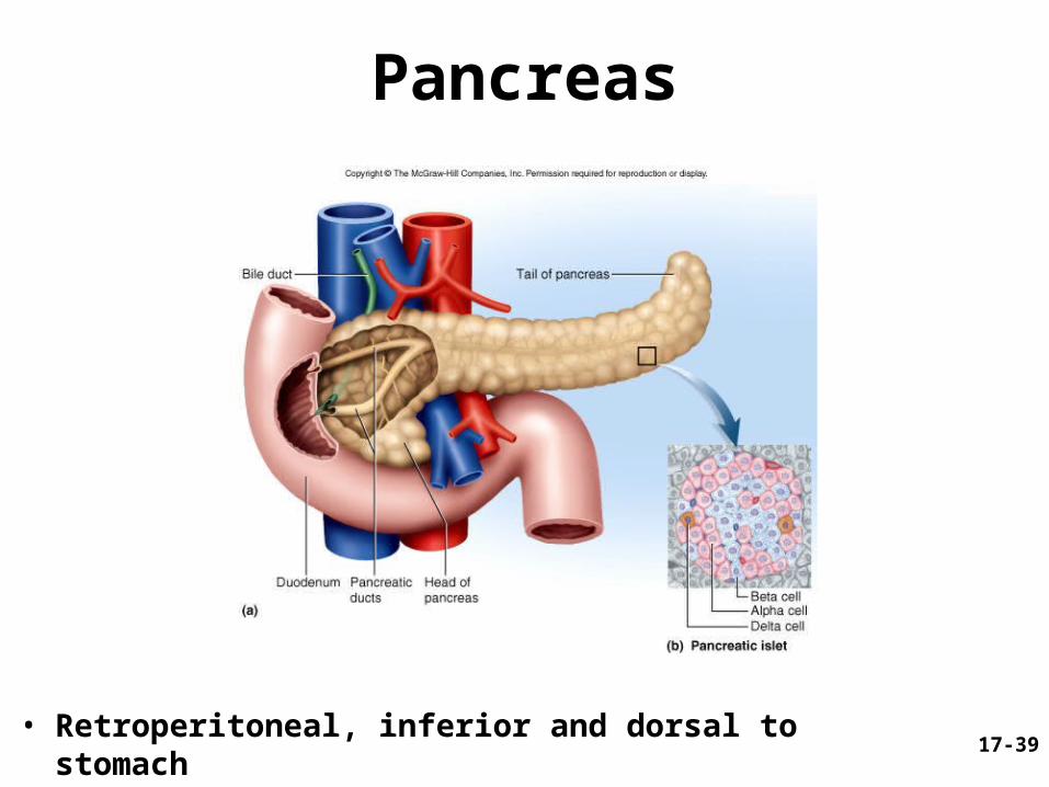
Pancreas
• Retroperitoneal, inferior and dorsal to stomach

17-40
Pancreatic Hormones
• 1-2 million islets produce hormones– 98% of organ produces digestive enzymes
(exocrine)
• Insulin (from cells)– secreted after meal with carbohydrates raises
glucose blood levels– stimulates glucose and amino acid uptake– nutrient storage effect (stimulates glycogen,
fat and protein synthesis)– antagonizes glucagon

17-41
Pancreatic Hormones
• Glucagon (from cells)– secreted in very low carbohydrate and high
protein diet or fasting– stimulates glycogenolysis, fat catabolism
(release of FFA’s) and promotes absorption of amino acids for gluconeogenesis
• Somatostatin (from delta () cells)– secreted with rise in blood glucose and
amino acids after a meal– paracrine secretion = inhibits secretion of
insulin, glucagon by and cells
17-42
Pancreatic Hormones
• Hyperglycemic hormones raise blood glucose– GH, epinephrine, NE, cortisol and
corticosterone• Hypoglycemic hormones lower blood
glucose– insulin
17-43
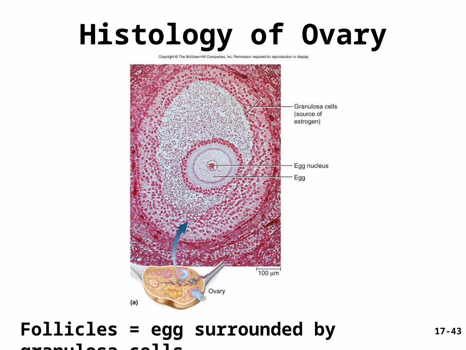
Histology of Ovary
Follicles = egg surrounded by granulosa cells

17-44
Ovary
• Granulosa cells in wall of ovarian follicle– produces estradiol, first half of menstrual cycle
• Corpus luteum: follicle after ovulation– produces estradiol and progesterone for 12 days or 8-
12 weeks with pregnancy
• Functions of estradiol and progesterone– development of female reproductive system and
physique including bone growth– regulate menstrual cycle, sustain pregnancy– prepare mammary glands for lactation
• Both secrete inhibin: suppresses FSH secretion

17-45
Testes
• Interstitial cells (between seminiferous tubules)– produce testosterone and estrogen
• Functions– development of male reproductive system and
physique– sustains sperm production and sex drive
• Sustentacular sertoli cells– secrete inhibin which suppresses FSH
secretion which stabilizes sperm production rates
17-46
Endocrine Functions of Other Organs
• Heart – – atrial natriuretic peptide released with an increase in
BP blood volume and BP by Na+ and H2O loss by
kidneys
• Skin - helps produce D3• Liver
– 15% of erythropoietin (stimulates bone marrow)– angiotensinogen (a prohormone)
• precursor of angiotensin II
– source of IGF-I (works with GH)– converts vitamin D3 to calcidiol– Hepcidin – promotes intestinal absorption of iron
17-47
Endocrine Functions of Other Organs • Kidneys
– produces 85% of erythropoietin – • stimulates bone marrow to produce RBC’s
– convert angiotensinogen to angiotensin I– converts calcidiol to calcitriol (active form of vitamin D)
absorption by intestine and inhibits loss in the urine• more Ca2+ available for bone deposition
• Stomach and small intestines (10 enteric hormones)– coordinate digestive motility and secretion
• Placenta– secretes estrogen, progesterone and others
• regulate pregnancy, stimulate development of fetus and mammary glands

17-48
Hormone Chemistry• Steroids
– derived from cholesterol– sex steroids,
corticosteroids• Peptides and
glycoproteins– OT, ADH; all releasing
and inhibiting hormones of hypothalamus; most of anterior pituitary hormones
• Monoamines (biogenic amines)– derived from amino
acids• catecholamines
(norepinephrine, epinephrine, dopamine) and thyroid hormones

17-49
Oxytocin and ADH

17-50
Hormone Synthesis: Steroid Hormones
• Synthesized from cholesterol – differs in functional groups attached to 4-ringed steroid backbone

17-51
Hormone Synthesis: Peptides
• Cellular steps– RER removes
segment, forms prohormone
– Golgi complex further modifies it into hormone
– e.g. insulin formation
• preproinsulin converted to proinsulin in RER
• proinsulin split into insulin and C peptide in golgi complex
17-52
Hormone Synthesis: Monoamines
• All are synthesized from tyrosine– except melatonin which is synthesized from
tryptophan
• Thyroid hormone is unusual– composed of two tyrosine molecules– requires a mineral, iodine

17-53
Thyroid Hormone Synthesis
Fig. 17.18
17-54
T3 and T4 Synthesis
• Follicular cells – absorb I- from blood and store in lumen as I-
– synthesize thyroglobulin and store in lumen • contains tyrosine
– tyrosine and Iodine form T3 and T4
• TSH – stimulates follicular cells to remove T3 and
T4 from thyroglobulin for release into plasma

17-55
Chemistry of Thyroid Hormone
Fig. 17.19
MIT contains one iodine atom, DIT has twoT3 = combination of MIT plus DITT4 = combination of two DITs

17-56
Hormone Transport• Monoamines and peptides are hydrophilic
– mix easily with blood plasma
• Steroids and thyroid hormone are hydrophobic – must bind to transport proteins for transport– bound hormone - attached to transport protein,
• prolongs half-life to weeks• protects from enzymes and kidney filtration
– unbound hormone leaves capillary to reach target cell (half-life a few minutes)
• Transport proteins in blood plasma– albumin, thyretin and TGB (thyroxine binding
globulin) bind to thyroid hormone– steroid hormones bind to globulins (transcortin)– aldosterone - no transport protein, 20 min. half-life
17-57
Hormone Receptors
• Located on plasma membrane, mitochondria, other organelles, or in nucleus
• Usually thousands for given hormone– hormone binding turns metabolic
pathways on or off
• Exhibit specificity and saturation
17-58
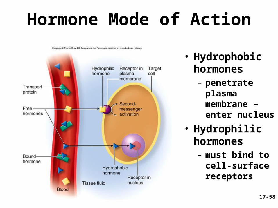
Hormone Mode of Action
• Hydrophobic hormones– penetrate
plasma membrane – enter nucleus
• Hydrophilic hormones – must bind to
cell-surface receptors

17-59
Thyroid Hormone Effects• TH binds to
receptors on – mitochondria
rate of aerobic respiration
– ribosomes and chromatin protein
synthesis
• Na+-K+ ATPase produced – generates heat

17-60
Hydrophilic Hormones: Mode of Action cAMP as Second Messenger
1) Hormone binding activates G protein
2) Activates adenylate cyclase
3) Produces cAMP
4) Activates kinases
5) Activates enzymes
6) Metabolic reactions: – synthesis– secretion– change membrane
potentials

17-61
Hydrophilic Hormones: Mode of Action Other 2nd and 3rd Messengers
Hormones may use different second messengers in different tissues.

17-62
Enzyme Amplification

17-63
Hormone Clearance
• Hormone signals must be turned off
• Take up and degraded by liver and kidney
• Excreted in bile or urine
• Metabolic clearance rate (MCR)
• Half-life - time required to clear 50% of hormone
17-64
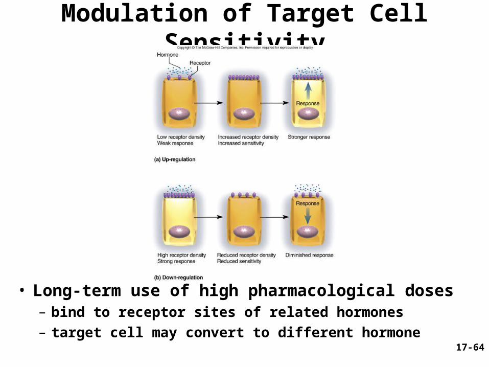
Modulation of Target Cell Sensitivity
• Long-term use of high pharmacological doses– bind to receptor sites of related hormones– target cell may convert to different hormone

17-65
Hormone Interactions
• Most cells sensitive to more than one hormone and exhibit interactive effects
• Synergistic effects
• Permissive effects– one hormone enhances response to a
second hormone
• Antagonistic effects
17-66
Stress and Adaptation
• Stress– caused by any situation that upsets
homeostasis and threatens one’s physical or emotional well-being
• General adaptation syndrome – way body reacts to stress– occurs in 3 stages
1. alarm reaction
2. stage of resistance
3. stage of exhaustion
17-67
Alarm Reaction
• Initial response epinephrine and norepinephrine levels HR and BP blood glucose levels
• Sodium and water retention (aldosterone)

17-68
Stage of Resistance
• After a few hours, glycogen reserves gone
ACTH and cortisol levels
• Fat and protein breakdown
• Gluconeogenesis
• Depressed immune function
• Susceptibility to infection and ulcers

17-69
Stage of Exhaustion
• Stress that continues until fat reserves are gone
• Protein breakdown and muscle wasting • Loss of glucose homeostasis• Hypertension and electrolyte imbalances
(loss of K+ and H+)• Hypokalemia and alkalosis leads to death

17-70
Paracrine Secretions• Chemical messengers that diffuse short
distances and stimulate nearby cells– unlike neurotransmitters not produced in neurons– unlike hormones not transported in blood
• Examples and their functions– histamine
• from mast cells in connective tissue • causes relaxation of blood vessel smooth muscle
– nitric oxide • from endothelium of blood vessels, causes vasodilation
– somatostatin • from gamma cells, inhibits secretion of alpha and beta cells
– catecholamines • diffuse from adrenal medulla to cortex

17-71
Eicosanoids: a Paracrine Secretion• Leukotrienes
– converted from arachidonic acid (by lipoxygenase) – mediates allergic and inflammatory reactions
• Prostacyclin (by cyclooxygenase) – inhibits blood clotting and vasoconstriction
• Thromboxanes (by cyclooxygenase) – produced by blood platelets after injury; override
prostacyclin, stimulates vasoconstriction and clotting
• Prostaglandins (by cyclooxygenase): diverse; includes– PGE: relaxes smooth muscle in bladder, intestines,
bronchioles, uterus and stimulates contraction of blood vessels
– PGF: opposite effects

17-72
Eicosanoid Synthesis
17-73
Endocrine Disorders
• Variations in hormone concentration and target cell sensitivity have noticeable effects on body
• Hyposecretion – inadequate hormone release– tumor or lesion destroys gland
• head trauma affects pituitary gland’s ability to secrete ADH– diabetes insipidus = chronic polyuria
• Hypersecretion – excessive hormone release– tumors or autoimmune disorder
• toxic goiter (graves disease) – antibodies mimic effect of TSH on the thyroid

17-74
Pituitary Disorders• Hypersecretion of growth hormones
– acromegaly– thickening of the bones and soft tissues– problems in childhood or adolescence
• gigantism if oversecretion• dwarfism if hyposecretion
17-75
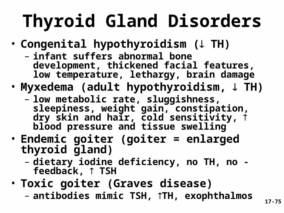
Thyroid Gland Disorders• Congenital hypothyroidism ( TH)
– infant suffers abnormal bone development, thickened facial features, low temperature, lethargy, brain damage
• Myxedema (adult hypothyroidism, TH)– low metabolic rate, sluggishness, sleepiness,
weight gain, constipation, dry skin and hair, cold sensitivity, blood pressure and tissue swelling
• Endemic goiter (goiter = enlarged thyroid gland)– dietary iodine deficiency, no TH, no - feedback,
TSH• Toxic goiter (Graves disease)
– antibodies mimic TSH, TH, exophthalmos

17-76
Parathyroid Disorders
• Hypoparathyroid– surgical excision during thyroid surgery– fatal tetany 3-4 days
• Hyperparathyroid = excess PTH secretion– tumor in gland– causes soft, fragile and deformed bones blood Ca2+
– renal calculi

17-77
Adrenal Disorders• Cushing syndrome - excess cortical secretion
– hyperglycemia, hypertension, weakness, edema– muscle and bone loss occurs with protein catabolism– buffalo hump and moon face = fat
deposition between shoulders or in face
• Adrenogenital syndrome (AGS)– adrenal androgen hypersecretion; accompanies
Cushing– enlargement of external sexual organs in children and
early onset of puberty– masculinizing effects on women (deeper voice and
beard growth)

17-78
Diabetes Mellitus
• Signs and symptoms of hyposecretion of insulin– polyuria, polydipsia, polyphagia– hyperglycemia, glycosuria, ketonuria– osmotic diuresis
• blood glucose levels rise above transport maximum of kidney tubules, glucose remains in urine (ketones also present)
• increased osmolarity draws water into urine
17-79
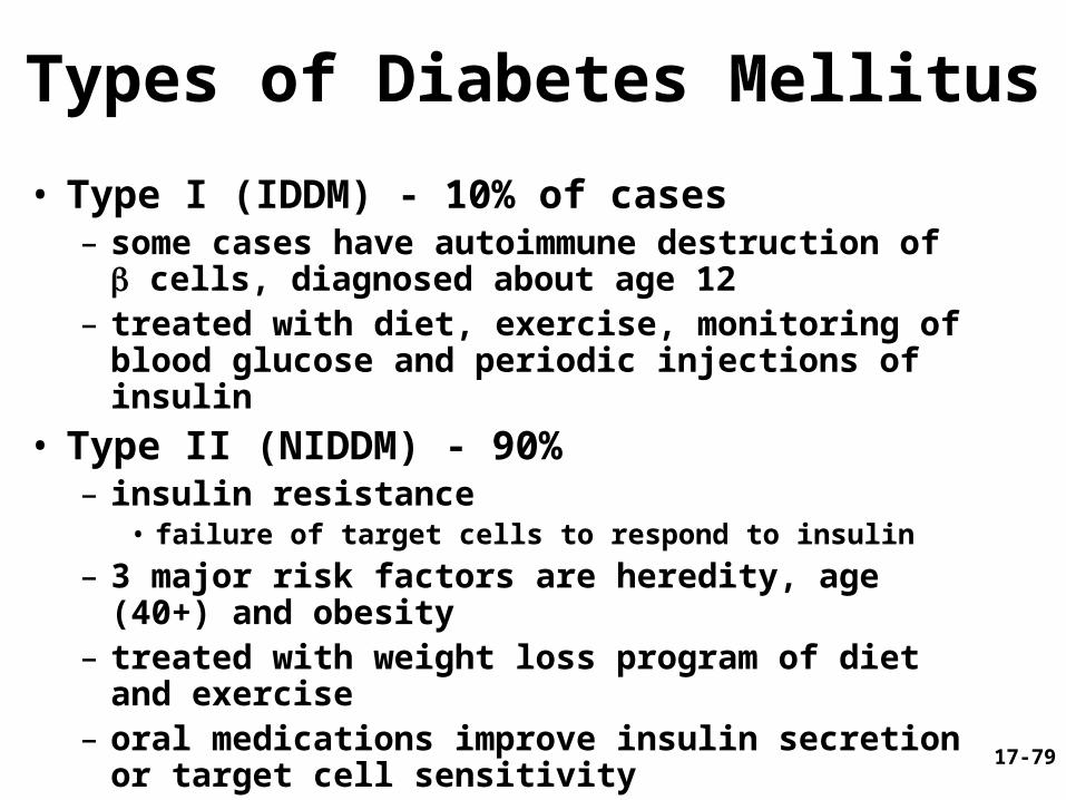
Types of Diabetes Mellitus
• Type I (IDDM) - 10% of cases– some cases have autoimmune destruction of
cells, diagnosed about age 12– treated with diet, exercise, monitoring of blood
glucose and periodic injections of insulin
• Type II (NIDDM) - 90%– insulin resistance
• failure of target cells to respond to insulin
– 3 major risk factors are heredity, age (40+) and obesity
– treated with weight loss program of diet and exercise
– oral medications improve insulin secretion or target cell sensitivity

17-80
Pathology of Diabetes• Acute pathology: cells cannot absorb glucose,
rely on fat and proteins (weight loss, weakness)– fat catabolism FFA’s in blood and ketone bodies– ketonuria promotes osmotic diuresis, loss of Na+
and K+
– ketoacidosis occurs as ketones blood pH• if continued causes dyspnea and eventually diabetic coma
• Chronic pathology– chronic hyperglycemia leads to neuropathy and
cardiovascular damage from atherosclerosis• retina and kidneys (common in type I), atherosclerosis
leads to heart failure (common in type II), and gangrene
17-81
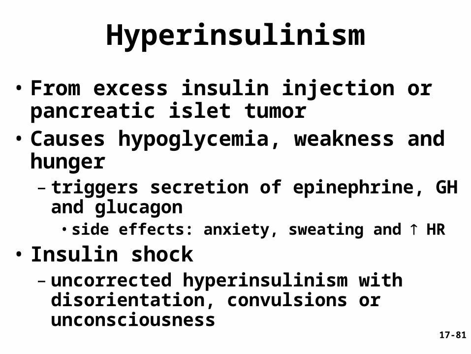
Hyperinsulinism
• From excess insulin injection or pancreatic islet tumor
• Causes hypoglycemia, weakness and hunger– triggers secretion of epinephrine, GH and
glucagon• side effects: anxiety, sweating and HR
• Insulin shock– uncorrected hyperinsulinism with
disorientation, convulsions or unconsciousness