Embed Size (px)
Citation preview
Cannabinoid receptors in submandibular acinar cells:Functional coupling between saliva fluid andelectrolytes secretion and Ca2+ signalling
Olga Kopach1, Juliana Vats2, Olga Netsyk2, Nana Voitenko1,2, Andrew Irving3 and Nataliya Fedirko2,*1State Key Laboratory of Molecular and Cellular Biology, Bogomoletz Institute of Physiology, Bogomoletz Str. 4, Kiev 01024, Ukraine2Department of Human and Animal Physiology, Biological Faculty, Ivan Franko National University of Lviv, Grushevskogo Str.4, Lviv 79005, Ukraine3Division of Neuroscience, University of Dundee, Ninewells Hospital, Dundee DD1 SY, UK
*Author for correspondence ([email protected])
Accepted 12 December 2011Journal of Cell Science 125, 1884–1895� 2012. Published by The Company of Biologists Ltddoi: 10.1242/jcs.088930
SummaryCannabinoid receptors (CBRs) belong to the G protein-coupled receptor superfamily, and activation of CBRs in salivary cells inhibits
agonist-stimulated salivation and modifies saliva content. However, the role of different CBR subtypes in acinar cell physiology and inintracellular signalling remains unclear. Here, we uncover functional CB1Rs and CB2Rs in acinar cells of rat submandibular gland andtheir essential role in saliva secretion. Pharmacological activation of CB1Rs and CB2Rs in the submandibular gland suppressed salivaoutflow and modified saliva content produced by the submandibular gland in vivo. Using Na+-selective microelectrodes to record
secretory Na+ responses in the lumen of acini, we observed a reduction in Na+ transport following the activation of CBRs, which wascounteracted by the selective CB1R antagonist AM251. In addition, activation of CB1Rs or CB2Rs caused inhibition of Na+-K+-ATPaseactivity in microsomes derived from the gland tissue as well as in isolated acinar cells. Using a Ca2+ imaging technique, we showed that
activation of CB1Rs and CB2Rs alters [Ca2+]cyt signalling in acinar cells by distinct pathways, involving Ca2+ release from theendoplasmic reticulum (ER) and store-operated Ca2+ entry (SOCE), respectively. Our data demonstrate the expression of CB1Rs andCB2Rs in acinar cells, and their involvement in the regulation of salivary gland functioning.
Key words: CB1Rs and CB2Rs, Saliva fluid and electrolytes secretion, [Ca2+]cyt signalling, Acinar cells, Submandibular salivary gland
IntroductionCannabinoids, the terpenophenolic compounds derived from the
plant Cannabis sativa, have been used for centuries as
psychotropic drugs and medicinal agents with diverse systemic
effects. The actions of cannabinoids in mammalian tissues are
primarily mediated by two main types of cannabinoid receptor
(CBR): CB1 and CB2 (Kano et al., 2009; Matsuda et al., 1990;
Matsuda, 1997). CB1 receptors (CB1Rs) are expressed at high
levels in the central and peripheral nervous systems, where their
main role is to modulate neurotransmitter release (Freund et al.,
2003). CB2 receptors (CB2Rs) are found in immune cells as
well as within the cardiovascular system and gastrointestinal
tract (Pertwee, 2001; Sanger, 2007; Wright et al., 2008). CB2Rs
are thought to regulate abnormal gut motility, intestinal
inflammation, visceral sensitivity and pain (Duncan et al.,
2008; Kikuchi et al., 2008; Wright et al., 2008); however, their
physiological role remains controversial.
Although Cannabis gains access to the systemic circulation
within minutes of penetration, the oral cavity and gastrointestinal
tract are points of first contact and, therefore, represent sites of
considerable impact. Cannabinoids reduce enteric nerve activity
and intestinal motility (Izzo et al., 2000; Izzo et al., 2001;
Makwana et al., 2010; Mathison et al., 2004), inhibit saliva
secretion (McConnell et al., 1978; Prestifilippo et al., 2006) and
fluid and gastric acid secretion (Adami et al., 2002; Coruzzi et al.,
2006; Hornby and Prouty, 2004; Izzo and Coutts, 2005) and
prevent stimulated ion transport across the mucosa of the intestine,
thus reducing water accumulation (Izzo et al., 2003). All these
effects have been largely attributed to the activation of CB1Rs.
Although it was demonstrated that cannabinoids affect salivary
gland function (Prestifilippo et al., 2006), the precise mechanisms
of action remain unknown. Expression of both CB1Rs and CB2Rs
in the submandibular gland is suggested from preliminary
immunohistochemical studies, in which CB1Rs were found
mainly in the ductal system, whereas CB2Rs were found in the
acini (Prestifilippo et al., 2006). The high-affinity, endogenous
CBR agonist arachidonyl ethanolamide (anandamide, AEA)
markedly inhibited stimulated saliva secretion when injected into
the submandibular gland (Prestifilippo et al., 2006) or into the
lateral ventricle of the rat brain (Fernandez-Solari et al., 2009),
acting either directly on the receptors in the gland or through
presynaptic inhibition of neurotransmitter release. In contrast to
the effects on submandibular gland, AEA induced amylase release
in parotid glands that correlated with increased cAMP content, and
also inhibited Na+-K+-ATPase activity (Busch et al., 2004).
Among other salivary glands, the submandibular gland
provides a main source for secretion of saliva fluid and
electrolytes, continual secretion of which is required for the
moistening of the oral cavity (Ambudkar, 2000; Melvin et al.,
2005). Salivary fluid and electrolyte secretion is a two-stage
process: saliva is initially formed in the acinar lumen (primary
fluid) and then is modified in the salivary ducts by removal of
1884 Research Article
Journ
alof
Cell
Scie
nce
Na+ and Cl2 and addition of K+ and HCO32 to produce the final
saliva that flows out the ducts (Turner and Sugiya, 2002). Inacinar cells, the secretion of fluid and electrolytes is triggeredby a complex cytosolic Ca2+ ([Ca2+]cyt) signal, originating
from Ca2+ release from the endoplasmic reticulum (ER) withsubsequent activation of store-operated Ca2+ entry (SOCE),which is required for synchronised activation of spatiallyseparated Ca2+-dependent Cl2 and K+ channels (Ambudkar,
2000; Melvin et al., 2005). In endothelial cells, AEA was shownto initiate [Ca2+]cyt signalling via CB2R-mediated activation ofphospholipase C, 1,4,5-inositol trisphosphate receptor (IP3R)-
mediated Ca2+ release from the ER and accompanied Ca2+ influx(Waldeck-Weiermair et al., 2008; Zoratti et al., 2003). The CBRagonists, however, inhibited glucose-induced [Ca2+]cyt increase
and [Ca2+]cyt oscillations in pancreatic b-cells (Nakata and Yada,2008) and suppressed Ca2+ mobilisation in smooth muscle cells(Chataigneau et al., 1998; Zygmunt et al., 1997). Despite the
functional significance of CBR-mediated regulation of salivation,their signalling pathways in acinar cells as the primary sites forsaliva secretion have not been elucidated.
In the present study, we initially aimed to explore in vivo the
effects of different CBR agonists on outflow and content of salivaproduced by the rat submandibular gland. In addition, fast CBR-induced extracellular Na+ ([Na+]e) transients were recorded in the
lumen of acini in the submandibular gland. Ca2+ imagingtechniques were then used to characterise CB1R- and CB2R-induced [Ca2+]cyt signalling in isolated submandibular acinar cells.
ResultsCB1Rs and CB2Rs inhibit saliva outflow in vivo and modifythe content of final saliva
Because the submandibular gland provides mainly a secretion of
saliva fluid and electrolytes, we initially determined whethercannabinoids effect submandibular gland salivation. For this, weanalysed in vivo the main parameters of salivation, such as saliva
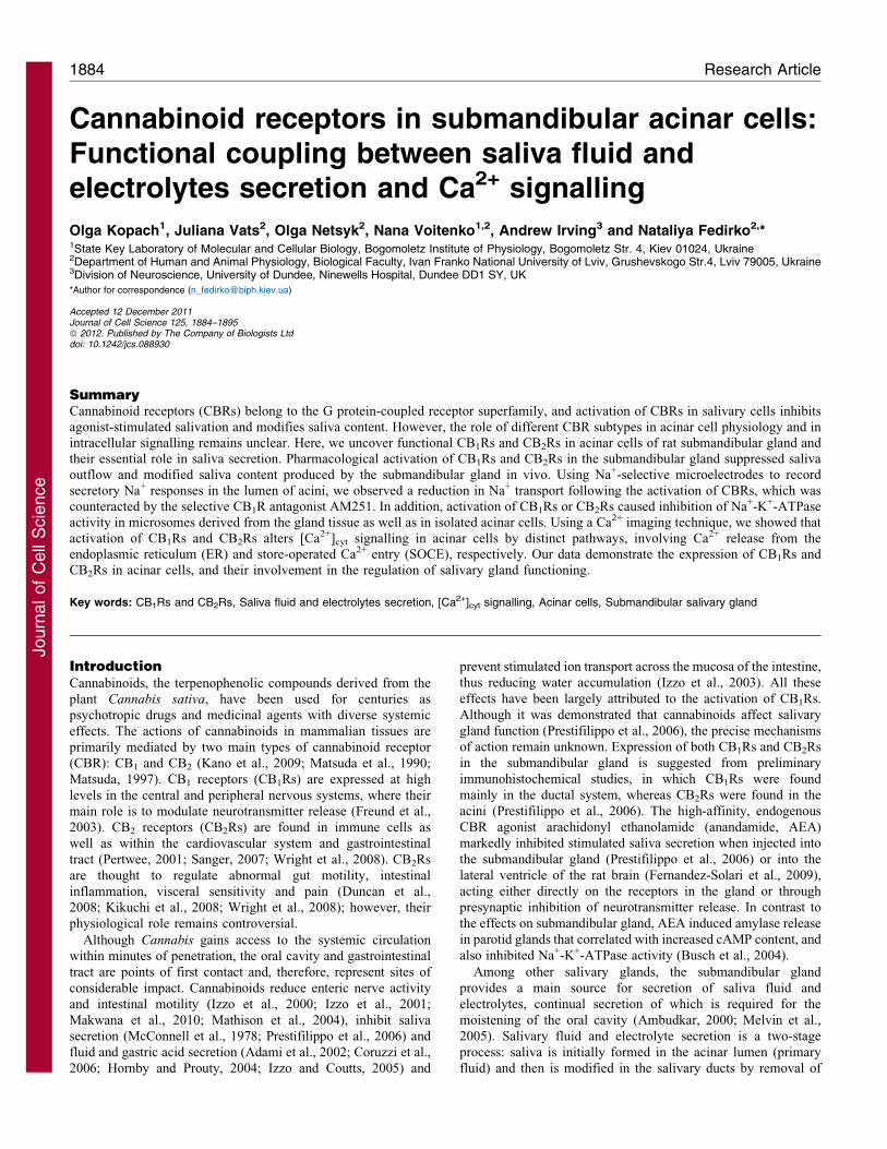
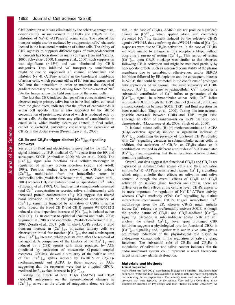
flow rate, total protein and electrolyte content of saliva secretedby the gland, before and after administration of cannabinoids.The potent, broad-spectrum CBR agonist WIN55,212-2 (5 mM),but not saline or vehicle, produced a marked decrease of saliva
flow rate (4366% decrease; n58, P,0.001) at 5 minutes after anintraglandular injection when compared with vehicle. At thesame time, no significant changes were observed either in total
Ca2+ or in protein concentration in secreted saliva (Fig. 1A).Similarly, (R)-(+)-methanandamide (5 mM), a metabolicallystable AEA analogue and selective agonist of the CB1Rs,
produced a significant decrease of saliva flow rate (2464%decrease; n57, P,0.001) and did not significantly affect totalCa2+ and protein concentrations (Fig. 1A). Another endogenouscannabinoid virodhamine (Porter et al., 2002) (30 mM) displayed
an inhibitory effect on saliva flow rate similar to that induced by(R)-(+)-methanandamide (2262% decrease; n57, P,0.001) butalso decreased total Ca2+ (1461% decrease; n57, P,0.05) and
protein concentration (3262% decrease; n57, P,0.01; Fig. 1A).Because of the complex pharmacology of virodhamine (Porteret al., 2002; Ryberg et al., 2007; Kozl9owska et al., 2006), we also
tested JWH015, a selective agonist of the CB2Rs (Pertwee,1999). At a dose of 100 nM, JWH015 produced a decrease ofsaliva flow rate (3465% decrease; n512, P,0.001) with no
significant effect on either total Ca2+ or protein concentrations(Fig. 1A). Thus, both CB1Rs and CB2Rs are likely to be involvedin the downregulation of salivation, markedly reducing saliva
outflow from the submandibular gland. The fact that the effect ofthe broad-spectrum CBR agonist WIN55,212-2 is greater than
that of selective CB1R or CB2R agonists alone suggests apotential additive effect in CBR-mediated downregulation ofsalivation.
To investigate more persistent effects of cannabinoids on
outflow and content of saliva, we performed repetitiveapplications of agonist to the gland (every 5 minutes),mimicking the conditions of single or persistent cannabis intake.
Consistent with the previous results (Fig. 1A), we observed asignificant reduction of saliva flow rate after a single applicationof WIN55,212-2 (5 mM), but not saline, which developed over 5
minutes (42611% of the corresponding time point in the saline-treated group; n55, P,0.05), reached a peak level at 10 minutes(4569% decrease; n58, P,0.001) and recovered to basal levelwithin 15–20 minutes (Fig. 1Ci). No significant changes of total
Ca2+ and proteins concentration in saliva were observed aftersingle administration of WIN55,212-2 (Fig. 1Cii–iii). By contrast,repetitive applications of WIN55,212-2 at the same dose (5 mM)
produced marked and constant inhibition of saliva flow rate,which began at 5 minutes (3967% of the corresponding timepoint in the saline-treated group; n57, P,0.05) and lasted for at
least 30 minutes (59611% of the saline-treated group at 20minutes; n510, P,0.001; Fig. 1Ci). Repetitive applications ofWIN55,212-2 (5 mM) also induced an increase of both Ca2+ andprotein concentrations in saliva. An increase of total protein
concentration began at 10 minutes (5269% of corresponding timepoint in the saline-treated group; n510, P,0.05) and wasmaintained for the whole period tested (7269% of the saline-
treated group at 15 minutes; n59, P,0.01; Fig. 1Cii). Asignificant increase of Ca2+ concentration was detected at 20minutes (4765% of the saline-treated group at 20 minutes; n58,
P,0.05) and lasted for 30 minutes during a repetitiveWIN55,212-2 application (Fig. 1Ciii). As expected, saline didnot affect salivation after either single or repetitive application
(Fig. 1Ci–iii). On further analysis of the electrolyte content ofsaliva, we found that repetitive application of WIN55,212-2 didnot significantly change K+ and P2+ concentrations in the finalsaliva during the period tested (P.0.05; Table 1). Taken together,
our results suggest that persistent activation of CB1Rs and CB2Rsin the submandibular gland causes a significant decrease in salivaflow rate, a concomitant increase in total protein content and Ca2+
concentration with no changes in other electrolytes tested in finalsaliva.
To confirm that the effects of cannabinoid ligands are CBR
specific, we investigated the effects of the CB1R- or CB2R-selective antagonists AM251 and AM630, respectively. Theinhibitory effect of either (R)-(+)-methanandamide (5 mM) orJWH015 (100 nM) on the parameters of salivation was
completely eliminated by AM251 (1 mM) or AM630 (1 mM),injected intraglandularly 20-30 minutes prior to the agonistadministration (Fig. 1B). Interestingly, AM251 alone stimulated
the saliva flow rate (4867% increase; n55, P,0.05 comparedwith vehicle) and slightly and insignificantly increased total Ca2+
and protein concentrations in secreted saliva (Fig. 1B). Selective
inhibition of CB2Rs by AM630 (1 mM) did not produce anysignificant changes in total protein and Ca2+ concentrations insaliva and did not alter saliva flow rate (Fig. 1B). To elucidate
the possible physiological role of CB1Rs and CB2Rs in regulationof salivation, we studied the time dependence of the effects oftheir selective antagonists. At a concentration of 1 mM, AM251
CBRs in submandibular acinar cells 1885
Journ
alof
Cell
Scie
nce
significantly potentiated saliva outflow when injected every 5
minutes for all periods tested. An increase of saliva flow rate
started within 10–15 minutes of AM251 administration (49610%
increase, P,0.05 compared with vehicle [time point ‘0’)] and
was maintained over 30 minutes (Fig. 1Di). Neither total protein
nor Ca2+ concentration was significantly changed during the
period of observation (Fig. 1Dii–iii). Selective blockade of
CB2Rs by AM630 (1 mM) did not significantly alter the main
parameters of salivation when compared with vehicle (time point
‘0’; Fig. 1Di–iii).
Taken together, our data indicate that cannabinoids cause a
significant decrease in saliva flow rate and modify the content of
final saliva through activation of CB1Rs and CB2Rs in the
submandibular gland.
CB1Rs decrease Na+ transport into the acinar lumen of the
submandibular gland
As cannabinoids do not affect K+ and P2+ concentrations in the
final saliva, we tested next whether cannabinoids alter
electrolytes secreted in the primary saliva fluid. For this, we
Table 1. Effect of saline or WIN55,212-2 (5 mM) on electrolyte concentration in saliva secreted by the submandibular gland
Electrolyteconcentration,normalised value
Saline WIN 55,212-2
Naive 0 minutes 10 minutes 20 minutes 30 minutes Naive 0 minutes 10 minutes 20 minutes 30 minutes
K+ 1.060.07(n57)
1.4160.05(n56)
1.4060.13(n56)
1.4360.13(n57)
1.4460.14(n57)
1.060.1(n57)
1.3360.07(n58)
1.4060.09(n57)
1.5460.16(n57)
1.6560.15(n58)
P2+ 1.060.18(n56)
1.4160.11(n56)
1.3360.14(n56)
1.3160.18(n56)
1.3460.11(n55)
1.060.32(n57)
1.4360.19(n56)
1.5560.38(n57)
1.5060.26(n57)
1.7660.37(n56)
Values are mean 6 s.e.m. (total number of cells). Statistical analysis was conducted by one-way and two-way ANOVA tests compared with time point ‘0’.
Fig. 1. The CB1Rs and CB2Rs in the submandibular gland affect saliva outflow and modify the content of final saliva. (A) Statistical summary of
normalised saliva flow rate and total Ca2+ and protein concentrations in saliva secreted by the gland 5 minutes after an intraglandular injection of vehicle or the
following cannabinoids: WIN55,212-2 (5 mM), (R)-(+)-methanandamide (5 mM), virodhamine (30 mM) or JWH015 (100 nM). *P,0.05, **P,0.01, ***P,0.001
versus the vehicle group (one-way ANOVA with Dunnett’s post-hoc test). (B) Effects of selective inhibition of CB1Rs or CB2Rs by AM251 (1 mM) or AM630
(1 mM), respectively, on (R)-(+)-methanandamide-induced or JWH015-induced changes in saliva flow rate and total Ca2+ and protein concentrations in final
saliva. *P,0.05 versus vehicle (one-way ANOVA with Bonferroni’s post-hoc correction). (C) Pooled data for saliva flow rate (Ci) and total protein (Cii) and Ca2+
(Ciii) concentrations (normalised to mean value before animal surgery) versus time of single or repetitive (every 5 minutes) WIN55,212-2 (5 mM) application
locally to the submandibular gland. *P,0.05, **P,0.01, ***P,0.001 versus the vehicle-treated group (two-way ANOVA followed by Bonferroni’s post-hoc
correction). (D) Pooled data for normalised saliva flow rate (Di) and total protein (Dii) and Ca2+ (Diii) concentrations versus time for experiments in which
AM630 (1 mM, black trace) or AM251 (1 mM, red trace) was injected alone every 5 minutes intraglandularly. *P,0.05 versus vehicle (time point ‘0’) (two-way
ANOVA followed by Bonferroni’s post-hoc correction). Error bars represent s.e.m.
Journal of Cell Science 125 (8)1886
Journ
alof
Cell
Scie
nce
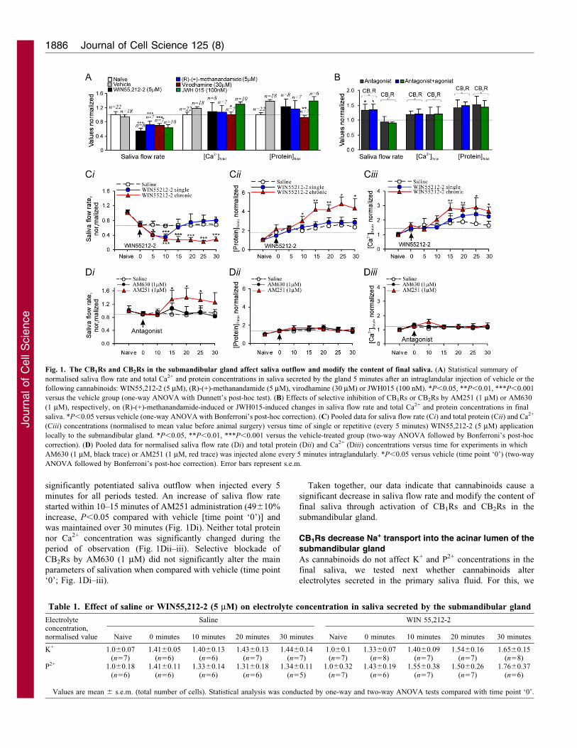
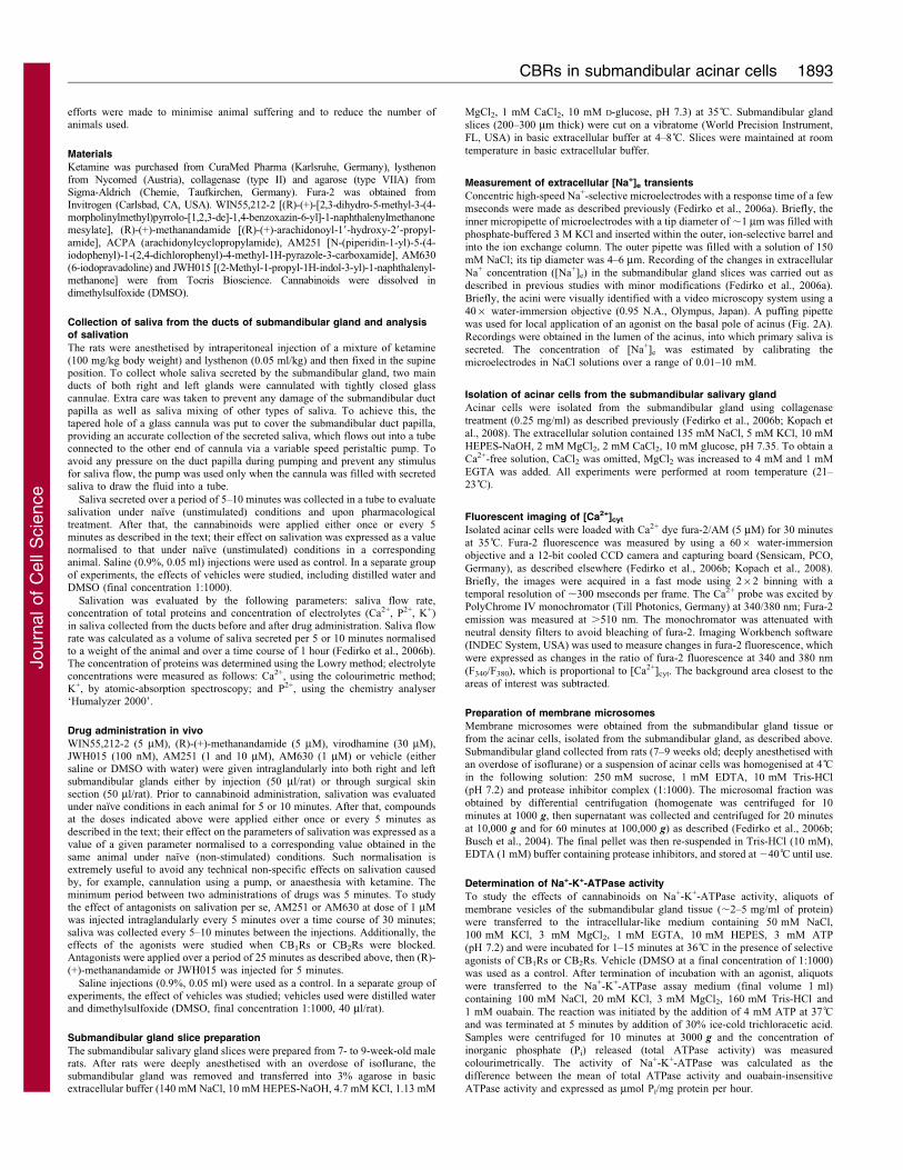
utilised a microelectrode technique for recording fast secretory
responses in submandibular gland slices in vitro (Fedirko et al.,
2006a). Using concentric Na+-selective microelectrodes, we
measured the changes of [Na+]e in the acinar lumen in response
to an agonist applied closely to the basal pole of acinus (Fig. 2A).
Acetylcholine (ACh, 5 mM), a principal neurotransmitter that
stimulates secretion of fluid and electrolytes, evoked a substantial
increase of [Na+]e in the lumen of the acinus (Fig. 2B). This
increase of [Na+]e reflects Na+ leakage into the lumen and
indicates an increased secretory response. The typical [Na+]e
response to ACh consisted of a fast initial transient rise in [Na+]e
(half-rise time was 201625 mseconds; n514; Fig. 2B,E)
followed by a much slower (within several seconds) recovery
to the baseline. The average amplitude of the ACh-induced
[Na+]e transient was 120612 mM (n514). By contrast,
WIN55,212-2 evoked a transient decrease in [Na+]e when
applied to the basal pole of acinus at different concentrations
(Fig. 2C). The EC50 for the decrease in [Na+]e induced by
WIN55,212-2 was 2.861027 M (Fig. 2F). The average
amplitudes of WIN55,212-2-induced [Na+]e decreases were
–266632 mM for 1 mM (n55) and –396616 mM for 10 mM
WIN55,212-2 (n514) (P,0.01); the half-time of decrease in
[Na+]e was 103626 mseconds (n55) and 134612 mseconds
(n514) for 1 mM and 10 mM WIN55,212-2, respectively
(Fig. 2E). These data indicate that CBRs inhibit Na+ transport
in the acinar lumen of the submandibular gland and alter
electrolyte content in primary saliva fluid.
In order to establish the contribution of the different CBR
subtypes in mediating this effect we used the selective CB1R
antagonist AM251 (10 mM). The decrease in [Na+]e induced by
WIN55,212-2 (10 mM) was strongly inhibited in the presence of
AM251 (9562% decrease; n57, P,0.001; Fig. 2D). This
suggests that it is CB1Rs that predominantly contribute to the
inhibition of Na+ transport into the acinar lumen.
Cannabinoids inhibit Na+-K+-ATPase activity in acinar cells
of the submandibular gland
The Na+-K+-ATPase present in the basolateral membrane of
polarised acinar cells represents one of the core regulatory
systems for fluid secretion driven by an osmotic gradient. To
elucidate a possible mechanism for reduced fluid secretion upon
activation of CBRs in the submandibular gland, we studied the
effect of CB1R and CB2R agonists on Na+-K+-ATPase activity. A
microsomal fraction was prepared from the submandibular gland
tissue. We found that an agonist of either CB1Rs or CB2Rs
markedly decreased Na+-K+-ATPase activity in gland tissue
(Fig. 3A). In particular, the decrease in Na+-K+-ATPase activity
was 47610% (n56, P,0.05) after 10 minutes incubation with
(R)-(+)-methanandamide (5 mM) compared with the vehicle, and
5068% (n57, P,0.05) after incubation with JWH015 (100 nM).
Furthermore, this effect was time dependent: the decrease in Na+-
K+-ATPase activity started at 1 minute of agonist presence
[3762% decrease, n55, P,0.05 for (R)-(+)-methanandamide;
42610% decrease, n56, P,0.05 for JWH015], reached a
maximum at 5 minutes [5965% decrease, n56, P,0.01 for
(R)-(+)-methanandamide; 60611% decrease, n56, P,0.01 for
JWH015], and lasted for all periods tested (Fig. 3B). The rate of
inhibition of Na+-K+-ATPase activity, calculated as the
difference in Na+-K+-ATPase activity between vehicle and
CBR agonist for the period of incubation, was the highest at 1
Fig. 2. CB1R-mediated decrease of Na+ transport into the
lumen of acini in the submandibular gland. (A) Transmitted light
image of the position of the puffing electrode (white arrow) and
Na+-selective concentric microelectrode (black arrow) in the acini
of a submandibular gland slice. Scale bar: 10 mm.
(B–D) Recordings of agonist-induced changes in extracellular Na+
concentration ([Na+]e) in acini lumen of the submandibular gland
when acinar cells were stimulated with ACh (5 mM; B),
WIN55,212-2 (1 and 10 mM; C) and WIN55,212-2 (10 mM) with
CB1R-selective antagonist, AM251 (10 mM; D). Arrows indicate
application of drug. (E,F) Statistical summary of WIN55,212-2-
induced [Na+]e transients amplitude and kinetics (E), and
concentration-response curve for WIN55,212-2-induced [Na+]e
decrease in acini lumen. **P,0.01, NS, non significant, Student’s
unpaired t-test. Error bars represent s.e.m.
CBRs in submandibular acinar cells 1887
Journ
alof
Cell
Scie
nce
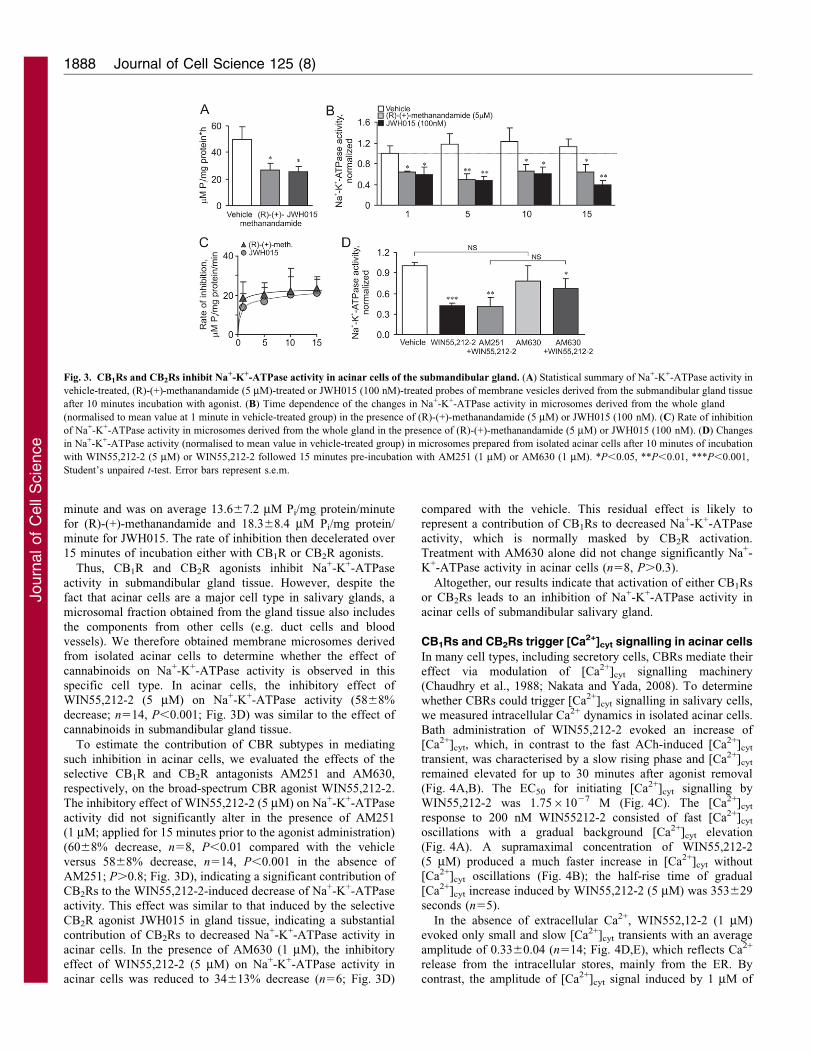
minute and was on average 13.667.2 mM Pi/mg protein/minute
for (R)-(+)-methanandamide and 18.368.4 mM Pi/mg protein/
minute for JWH015. The rate of inhibition then decelerated over
15 minutes of incubation either with CB1R or CB2R agonists.
Thus, CB1R and CB2R agonists inhibit Na+-K+-ATPase
activity in submandibular gland tissue. However, despite the
fact that acinar cells are a major cell type in salivary glands, a
microsomal fraction obtained from the gland tissue also includes
the components from other cells (e.g. duct cells and blood
vessels). We therefore obtained membrane microsomes derived
from isolated acinar cells to determine whether the effect of
cannabinoids on Na+-K+-ATPase activity is observed in this
specific cell type. In acinar cells, the inhibitory effect of
WIN55,212-2 (5 mM) on Na+-K+-ATPase activity (5868%
decrease; n514, P,0.001; Fig. 3D) was similar to the effect of
cannabinoids in submandibular gland tissue.
To estimate the contribution of CBR subtypes in mediating
such inhibition in acinar cells, we evaluated the effects of the
selective CB1R and CB2R antagonists AM251 and AM630,
respectively, on the broad-spectrum CBR agonist WIN55,212-2.
The inhibitory effect of WIN55,212-2 (5 mM) on Na+-K+-ATPase
activity did not significantly alter in the presence of AM251
(1 mM; applied for 15 minutes prior to the agonist administration)
(6068% decrease, n58, P,0.01 compared with the vehicle
versus 5868% decrease, n514, P,0.001 in the absence of
AM251; P.0.8; Fig. 3D), indicating a significant contribution of
CB2Rs to the WIN55,212-2-induced decrease of Na+-K+-ATPase
activity. This effect was similar to that induced by the selective
CB2R agonist JWH015 in gland tissue, indicating a substantial
contribution of CB2Rs to decreased Na+-K+-ATPase activity in
acinar cells. In the presence of AM630 (1 mM), the inhibitory
effect of WIN55,212-2 (5 mM) on Na+-K+-ATPase activity in
acinar cells was reduced to 34613% decrease (n56; Fig. 3D)
compared with the vehicle. This residual effect is likely to
represent a contribution of CB1Rs to decreased Na+-K+-ATPase
activity, which is normally masked by CB2R activation.
Treatment with AM630 alone did not change significantly Na+-
K+-ATPase activity in acinar cells (n58, P.0.3).
Altogether, our results indicate that activation of either CB1Rs
or CB2Rs leads to an inhibition of Na+-K+-ATPase activity in
acinar cells of submandibular salivary gland.
CB1Rs and CB2Rs trigger [Ca2+]cyt signalling in acinar cells
In many cell types, including secretory cells, CBRs mediate their
effect via modulation of [Ca2+]cyt signalling machinery
(Chaudhry et al., 1988; Nakata and Yada, 2008). To determine
whether CBRs could trigger [Ca2+]cyt signalling in salivary cells,
we measured intracellular Ca2+ dynamics in isolated acinar cells.
Bath administration of WIN55,212-2 evoked an increase of[Ca2+]cyt, which, in contrast to the fast ACh-induced [Ca2+]cyt
transient, was characterised by a slow rising phase and [Ca2+]cyt
remained elevated for up to 30 minutes after agonist removal
(Fig. 4A,B). The EC50 for initiating [Ca2+]cyt signalling by
WIN55,212-2 was 1.7561027 M (Fig. 4C). The [Ca2+]cyt
response to 200 nM WIN55212-2 consisted of fast [Ca2+]cyt
oscillations with a gradual background [Ca2+]cyt elevation
(Fig. 4A). A supramaximal concentration of WIN55,212-2
(5 mM) produced a much faster increase in [Ca2+]cyt without
[Ca2+]cyt oscillations (Fig. 4B); the half-rise time of gradual
[Ca2+]cyt increase induced by WIN55,212-2 (5 mM) was 353629
seconds (n55).
In the absence of extracellular Ca2+, WIN552,12-2 (1 mM)
evoked only small and slow [Ca2+]cyt transients with an average
amplitude of 0.3360.04 (n514; Fig. 4D,E), which reflects Ca2+
release from the intracellular stores, mainly from the ER. By
contrast, the amplitude of [Ca2+]cyt signal induced by 1 mM of
Fig. 3. CB1Rs and CB2Rs inhibit Na+-K+-ATPase activity in acinar cells of the submandibular gland. (A) Statistical summary of Na+-K+-ATPase activity in
vehicle-treated, (R)-(+)-methanandamide (5 mM)-treated or JWH015 (100 nM)-treated probes of membrane vesicles derived from the submandibular gland tissue
after 10 minutes incubation with agonist. (B) Time dependence of the changes in Na+-K+-ATPase activity in microsomes derived from the whole gland
(normalised to mean value at 1 minute in vehicle-treated group) in the presence of (R)-(+)-methanandamide (5 mM) or JWH015 (100 nM). (C) Rate of inhibition
of Na+-K+-ATPase activity in microsomes derived from the whole gland in the presence of (R)-(+)-methanandamide (5 mM) or JWH015 (100 nM). (D) Changes
in Na+-K+-ATPase activity (normalised to mean value in vehicle-treated group) in microsomes prepared from isolated acinar cells after 10 minutes of incubation
with WIN55,212-2 (5 mM) or WIN55,212-2 followed 15 minutes pre-incubation with AM251 (1 mM) or AM630 (1 mM). *P,0.05, **P,0.01, ***P,0.001,
Student’s unpaired t-test. Error bars represent s.e.m.
Journal of Cell Science 125 (8)1888
Journ
alof
Cell
Scie
nce
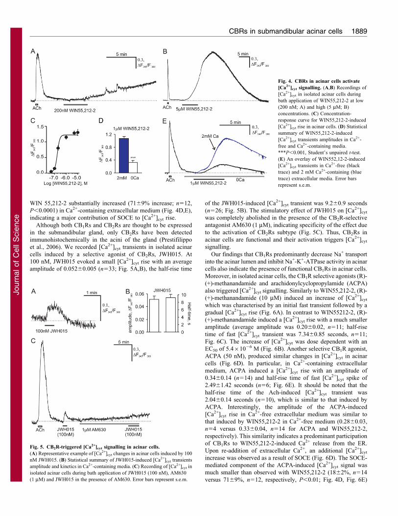
WIN 55,212-2 substantially increased (7169% increase; n512,
P,0.0001) in Ca2+-containing extracellular medium (Fig. 4D,E),
indicating a major contribution of SOCE to [Ca2+]cyt rise.
Although both CB1Rs and CB2Rs are thought to be expressed
in the submandibular gland, only CB2Rs have been detected
immunohistochemically in the acini of the gland (Prestifilippo
et al., 2006). We recorded [Ca2+]cyt transients in isolated acinar
cells induced by a selective agonist of CB2Rs, JWH015. At
100 nM, JWH015 evoked a small [Ca2+]cyt rise with an average
amplitude of 0.05260.005 (n533; Fig. 5A,B), the half-rise time
of the JWH015-induced [Ca2+]cyt transient was 9.260.9 seconds
(n526; Fig. 5B). The stimulatory effect of JWH015 on [Ca2+]cyt
was completely abolished in the presence of the CB2R-selective
antagonist AM630 (1 mM), indicating specificity of the effect due
to the activation of CB2Rs subtype (Fig. 5C). Thus, CB2Rs in
acinar cells are functional and their activation triggers [Ca2+]cyt
signalling.
Our findings that CB1Rs predominantly decrease Na+ transport
into the acinar lumen and inhibit Na+-K+-ATPase activity in acinar
cells also indicate the presence of functional CB1Rs in acinar cells.
Moreover, in isolated acinar cells, the CB1R selective agonists (R)-
(+)-methanandamide and arachidonylcyclopropylamide (ACPA)
also triggered [Ca2+]cyt signalling. Similarly to WIN55,212-2, (R)-
(+)-methanandamide (10 mM) induced an increase of [Ca2+]cyt
which was characterised by an initial fast transient followed by a
gradual [Ca2+]cyt rise (Fig. 6A). In contrast to WIN55212-2, (R)-
(+)-methanandamide induced a [Ca2+]cyt rise with a much smaller
amplitude (average amplitude was 0.2060.02, n511; half-rise
time of fast [Ca2+]cyt transient was 7.3460.85 seconds, n511;
Fig. 6C). The increase of [Ca2+]cyt was dose dependent with an
EC50 of 5.461026 M (Fig. 6B). Another selective CB1R agonist,
ACPA (50 nM), produced similar changes in [Ca2+]cyt in acinar
cells (Fig. 6D). In particular, in Ca2+-containing extracellular
medium, ACPA induced a [Ca2+]cyt rise with an amplitude of
0.3460.14 (n514) and half-rise time of fast [Ca2+]cyt spike of
2.4961.42 seconds (n56; Fig. 6E). It should be noted that the
half-rise time of the Ach-induced [Ca2+]cyt transient was
2.0460.14 seconds (n510), which is similar to that induced by
ACPA. Interestingly, the amplitude of the ACPA-induced
[Ca2+]cyt rise in Ca2+-free extracellular medium was similar to
that induced by WIN55,212-2 in Ca2+-free medium (0.2860.03,
n54 versus 0.3360.04, n514 for ACPA and WIN55,212-2,
respectively). This similarity indicates a predominant participation
of CB1Rs to WIN55,212-2-induced Ca2+ release from the ER.
Upon re-addition of extracellular Ca2+, an additional [Ca2+]cyt
increase was observed as a result of SOCE (Fig. 6D). The SOCE-
mediated component of the ACPA-induced [Ca2+]cyt signal was
much smaller than observed with WIN55,212-2 (1862%, n514
versus 7169%, n512, respectively, P,0.01; Fig. 4D, Fig. 6E)
Fig. 4. CBRs in acinar cells activate
[Ca2+]cyt signalling. (A,B) Recordings of
[Ca2+]cyt in isolated acinar cells during
bath application of WIN55,212-2 at low
(200 nM; A) and high (5 mM; B)
concentrations. (C) Concentration-
response curve for WIN55,212-2-induced
[Ca2+]cyt rise in acinar cells. (D) Statistical
summary of WIN55,212-2-induced
[Ca2+]cyt transients amplitudes in Ca2+-
free and Ca2+-containing media.
***P,0.001, Student’s unpaired t-test.
(E) An overlay of WIN552,12-2-induced
[Ca2+]cyt transients in Ca2+-free (black
trace) and 2 mM Ca2+-containing (blue
trace) extracellular media. Error bars
represent s.e.m.
Fig. 5. CB2R-triggered [Ca2+]cyt signalling in acinar cells.
(A) Representative example of [Ca2+]cyt changes in acinar cells induced by 100
nM JWH015. (B) Statistical summary of JWH015-induced [Ca2+]cyt transients
amplitude and kinetics in Ca2+-containing media. (C) Recording of [Ca2+]cyt in
isolated acinar cells during bath application of JWH015 (100 nM), AM630
(1 mM) and JWH015 in the presence of AM630. Error bars represent s.e.m.
CBRs in submandibular acinar cells 1889
Journ
alof
Cell
Scie
nce
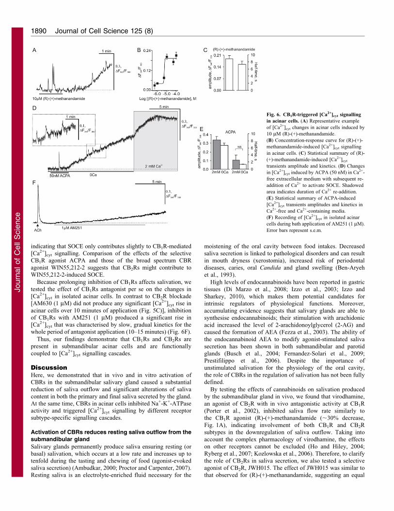
indicating that SOCE only contributes slightly to CB1R-mediated
[Ca2+]cyt signalling. Comparison of the effects of the selective
CB1R agonist ACPA and those of the broad spectrum CBR
agonist WIN55,212-2 suggests that CB2Rs might contribute to
WIN55,212-2-induced SOCE.
Because prolonging inhibition of CB1Rs affects salivation, we
tested the effect of CB1Rs antagonist per se on the changes in
[Ca2+]cyt in isolated acinar cells. In contrast to CB2R blockade
[AM630 (1 mM) did not produce any significant [Ca2+]cyt rise in
acinar cells over 10 minutes of application (Fig. 5C)], inhibition
of CB1Rs with AM251 (1 mM) produced a significant rise in
[Ca2+]cyt that was characterised by slow, gradual kinetics for the
whole period of antagonist application (10–15 minutes) (Fig. 6F).
Thus, our findings demonstrate that CB1Rs and CB2Rs are
present in submandibular acinar cells and are functionally
coupled to [Ca2+]cyt signalling cascades.
DiscussionHere, we demonstrated that in vivo and in vitro activation of
CBRs in the submandibular salivary gland caused a substantial
reduction of saliva outflow and significant alterations of saliva
content in both the primary and final saliva secreted by the gland.
At the same time, CBRs in acinar cells inhibited Na+-K+-ATPase
activity and triggered [Ca2+]cyt signalling by different receptor
subtype-specific signalling cascades.
Activation of CBRs reduces resting saliva outflow from thesubmandibular gland
Salivary glands permanently produce saliva ensuring resting (or
basal) salivation, which occurs at a low rate and increases up to
tenfold during the tasting and chewing of food (agonist-evoked
saliva secretion) (Ambudkar, 2000; Proctor and Carpenter, 2007).
Resting saliva is an electrolyte-enriched fluid necessary for the
moistening of the oral cavity between food intakes. Decreased
saliva secretion is linked to pathological disorders and can result
in mouth dryness (xerostomia), increased risk of periodontal
diseases, caries, oral Candida and gland swelling (Ben-Aryeh
et al., 1993).
High levels of endocannabinoids have been reported in gastric
tissues (Di Marzo et al., 2008; Izzo et al., 2003; Izzo and
Sharkey, 2010), which makes them potential candidates for
intrinsic regulators of physiological functions. Moreover,
accumulating evidence suggests that salivary glands are able to
synthesise endocannabinoids; their stimulation with arachidonic
acid increased the level of 2-arachidonoylglycerol (2-AG) and
caused the formation of AEA (Fezza et al., 2003). The ability of
the endocannabinoid AEA to modify agonist-stimulated saliva
secretion has been shown in both submandibular and parotid
glands (Busch et al., 2004; Fernandez-Solari et al., 2009;
Prestifilippo et al., 2006). Despite the importance of
unstimulated salivation for the physiology of the oral cavity,
the role of CBRs in the regulation of salivation has not been fully
defined.
By testing the effects of cannabinoids on salivation produced
by the submandibular gland in vivo, we found that virodhamine,
an agonist of CB2R with in vivo antagonistic activity at CB1R
(Porter et al., 2002), inhibited saliva flow rate similarly to
the CB1R agonist (R)-(+)-methanandamide (,30% decrease,
Fig. 1A), indicating involvement of both CB1R and CB2R
subtypes in the downregulation of saliva outflow. Taking into
account the complex pharmacology of virodhamine, the effects
on other receptors cannot be excluded (Ho and Hiley, 2004;
Ryberg et al., 2007; Kozlowska et al., 2006). Therefore, to clarify
the role of CB2Rs in saliva secretion, we also tested a selective
agonist of CB2R, JWH015. The effect of JWH015 was similar to
that observed for (R)-(+)-methanandamide, suggesting an equal
Fig. 6. CB1R-triggered [Ca2+]cyt signalling
in acinar cells. (A) Representative example
of [Ca2+]cyt changes in acinar cells induced by
10 mM (R)-(+)-methanandamide.
(B) Concentration-response curve for (R)-(+)-
methanandamide-induced [Ca2+]cyt signalling
in acinar cells. (C) Statistical summary of (R)-
(+)-methanandamide-induced [Ca2+]cyt
transients amplitude and kinetics. (D) Changes
in [Ca2+]cyt induced by ACPA (50 nM) in Ca2+-
free extracellular medium with subsequent re-
addition of Ca2+ to activate SOCE. Shadowed
area indicates duration of Ca2+ re-addition.
(E) Statistical summary of ACPA-induced
[Ca2+]cyt transients amplitudes and kinetics in
Ca2+-free and Ca2+-containing media.
(F) Recording of [Ca2+]cyt in isolated acinar
cells during bath application of AM251 (1 mM).
Error bars represent s.e.m.
Journal of Cell Science 125 (8)1890
Journ
alof
Cell
Scie
nce
contribution of CB1R and CB2R in the downregulation ofsalivation. In support of these observations, highly potent,
broad-spectrum CBR agonist, WIN55,212-2, demonstrated apronounced decrease in saliva flow rate, indicating an additiveaction of CB1R and CB2R in reduced saliva outflow.The inhibition of salivation observed upon cannabinoid
administration is mediated directly by CBR activation as theinhibitory effect of either the CB1R or the CB2R agonist wascompletely eliminated by their selective antagonists. When
administered alone, the CB1R antagonist had effects on salivaoutflow, which might indicate a regulatory role for CBRs incontinuous secretion of saliva fluid under unstimulated (basal)
conditions. Interestingly, the effect of CB1R blockade wasopposite to those produced by the receptor agonists: aprolonged inhibition of CB1R leads to increased salivation,whereas activation of CB1R markedly decreases salivation. A
similar effect was not clearly observed with the CB2R antagonist,which did not show any significant changes in saliva flow rate.Consistent with our findings, an antagonist of CB1R was reported
to augment electrically evoked acetylcholine release in ileum andto evoke contraction and peristalsis (Coutts and Pertwee, 1997;Izzo et al., 2000), whereas the CB2R-selective antagonist AM630
alone potentiated electrically stimulated relaxation of the ratfundus (Storr et al., 2002). By contrast, CB1R and CB2Rantagonists alone did not alter agonist-induced fluid secretion in
the submandibular gland (Fernandez-Solari et al., 2009), butwhen injected into the submandibular gland simultaneously(inhibiting both CB1R and CB2R) they increased salivationinduced by the low dose of an agonist (Prestifilippo et al., 2006).
Our present findings support the current clinical observationsof reduced saliva flow rate in people addicted to marijuana(Verstraete, 2005). By mimicking the conditions of single or
repetitive cannabis intake, we found that a reduction of salivaoutflow reached its maximum level within 10 minutes andrecovered briefly after a single administration of cannabinoid but
was reduced for prolonged periods if the agonist was appliedrepetitively (Fig. 1C).
Our data are consistent with the results demonstrating aninhibitory effect of CB1R on water accumulation in
gastrointestinal tract (Izzo et al., 2003; Sanger, 2007).Unstimulated (basal) salivation is maintained by low doses of aneurotransmitter released from the nerves innervating the gland
(Ambudkar, 2000), CB1Rs might reduce excitatory cholinergicneurotransmission in the intestine (Hinds et al., 2006; Storr et al.,2004) by presynaptic inhibition of evoked and spontaneous ACh
release from myenteric nerves (Coutts and Pertwee, 1997),resulting in reduced peristalsis and decreased gastrointestinalmotility and transit in vivo (Izzo et al., 2001). CBR-induceddecrease of electrically stimulated salivary flow is also mediated
by diminishing ACh release (McConnell et al., 1978). Reducedautonomic neurotransmission to the submandibular gland(Fernandez-Solari et al., 2009) might reduce the blood supply
of the gland (McConnell et al., 1978) and lead to decreased waterflow to the acini. However, evidence of CB2R-mediatedinhibition of salivation is still elusive because of limited
functional evidence for a role of CB2Rs in saliva secretion. Thefact that CB2Rs are thought to be expressed in submandibularacinar cells points to a potential role of this receptor subtype in
saliva secretion. Indeed, our findings demonstrated a reducedsaliva outflow upon activation of CB2Rs that might be caused byCB2R-mediated regulation of saliva release from the acinar cells
to the salivary ducts due to their predominant locationperipherally to the acini (Prestifilippo et al., 2006).
CB1Rs and CB2Rs alter saliva content in both primary andfinal saliva
The submandibular salivary gland is composed of acinar cells,
which secrete proteins and electrolytes (primary fluid), and ductalcells, which modify the electrolyte content producing final saliva(Ambudkar, 2000; Turner and Sugiya, 2002). We did not find any
significant changes in K+ and P+ concentrations in the finalsaliva, collected from the gland ducts, in response to repetitiveadministration of cannabinoid in vivo. However, we found a
significant increase of total protein concentration, which started10–15 minutes after WIN55,212 administration (time point ofmaximal inhibition of saliva outflow) and was maintained for theentire period if the agonist was applied repetitively (Fig. 1Cii).
Although we only measured the total fraction of saliva proteins,our data are consistent with previous reports demonstratingCB1R-induced amylase release from rat parotid glands (Busch
et al., 2004) and CB1R-mediated stimulation of insulin andglucagon secretion by the pancreas (Bermudez-Silva et al., 2008).In salivary gland cells, two major signal transduction pathways
are implicated in protein secretion: cAMP generation and theCa2+-phosphoinositide messenger system (Rubin and Adolf,1994). The enhancement of protein secretion induced by CBRactivation might be mediated through cAMP-induced Ca2+
signalling. Indeed, the cannabinoid-induced increase of proteinconcentration was associated with an increase of total Ca2+
concentration in the final saliva (Fig. 1Cii–iii), suggesting an
extrusion of Ca2+ during exocytosis of secretory vesicles(Gerasimenko et al., 1996) and supporting the hypothesis for arole of Ca2+ in CBR-induced secretion of salivary proteins.
In agreement with this assumption, in the case of singleadministration of cannabinoids, any significant changes in bothtotal protein and Ca2+ concentrations were found in final saliva
after 10 minutes of drug presence (Fig. 1A). Also, any effects ofthe antagonists of CB1Rs and CB2Rs alone on either basal totalprotein or Ca2+ concentrations were observed in final salivaduring all periods tested (30 minutes). This is consistent with the
observations of others showing an absence of an effect ofantagonists per se, used at the same doses, on basal level ofamylase release from parotid glands (Busch et al., 2004) as well
as basal or even KCl-stimulated amylase release from acinar cellsof rat pancreatic glands (Linary et al., 2009).
Contrary to observations for the final saliva, cannabinoids
dramatically altered the ionic content of the primary saliva. Usinga fast concentric microelectrode technique, we measured [Na+]e
in the acinar lumen close to the acinar cell apical membrane.WIN55,212-2 application to the basal pole of acini resulted in a
transient drop of [Na+]e in the acinar lumen reflecting inhibitedtransport of Na+ ions, which might, in turn, lead to the suppressedsaliva flow rate observed in vivo. The fact that the WIN55,212-2-
induced drop of [Na+]e was significantly abolished by theselective inhibition of CB1R (Fig. 2D) indicates not only CB1Rsfunctioning in acinar cells, but also their significant role in
regulation of Na+ transport. The inhibited Na+ transport is likelyto be mediated by decreased Na+-K+-ATPase activity, significantinhibition of which by cannabinoids has been directly
demonstrated either in whole gland or in isolated acinar cellsby ourselves (Fig. 3) and by others (Busch et al., 2004). Theeffect of cannabinoids on Na+-K+-ATPase activity is mediated by
CBRs in submandibular acinar cells 1891
Journ
alof
Cell
Scie
nce
CBR activation as it was eliminated by the selective antagonists,
demonstrating an involvement of CB1Rs and CB2Rs in theinhibition of Na+-K+-ATPases in acinar cells. The reduced iontransport might also be mediated by a suppression of K+ channels
located in the basolateral membrane of acinar cells. The ability ofCBR agonists to suppress different types of voltage-dependentK+ currents has been shown in many cell types (Fan and Yazulla,2003; Schweitzer, 2000; Hampson et al., 2000); such suppression
was significant (,45%) and was eliminated by CB1Rantagonists. Thus, inhibited Na+ transport by cannabinoidsmight be due to suppressed K+ channel conductance and
inhibited Na+-K+-ATPase activity in the basolateral membraneof acinar cells, which prevents efflux of K+ ions and extrusion ofNa+ into the interstitium in order to maintain the electrical
gradient necessary to cause a driving force for movement of Na+
into the lumen across the tight junctions of the acinar cells.
The fact that CBR-induced changes of ion concentrations wereobserved only in primary saliva but not in the final saliva, collected
from the gland ducts, indicates that the effect of cannabinoids isacinar cell specific. This is also supported by the alteredconcentration of proteins, secretion of which is produced only by
acinar cells. At the same time, any effects of cannabinoids onductal cells, which modify electrolyte content in final saliva,cannot be excluded, particularly considering the expression of
CB1Rs in the ductal system (Prestifilippo et al., 2006).
CB1Rs and CB2Rs trigger distinct [Ca2+]cyt signallingpathways
Secretion of fluid and electrolytes is governed by the [Ca2+]cyt
signal initiated by IP3R-mediated Ca2+ release from the ER andsubsequent SOCE (Ambudkar, 2000; Melvin et al., 2005). The[Ca2+]cyt signal also functions as a cellular messenger in
regulation of salivary protein secretion (Rubin and Adolf,1994). Previous studies have shown that CB2Rs induce[Ca2+]cyt mobilisation from the intracellular stores in
endothelial cells (Waldeck-Weiermair et al., 2008; Zoratti et al.,2003) whereas CB1R stimulation evokes capacitative Ca2+ entry(Filipeanu et al., 1997). Our findings that cannabinoids increased
total Ca2+ concentration in secreted saliva simultaneously withincreased protein concentration (Fig. 1C) suggest that alteredbasal salivation might be the physiological consequence of[Ca2+]cyt signalling triggered by activation of CBRs in acinar
cells. Indeed, the broad CB1R and CB2R agonist WIN55212-2induced a dose-dependent increase of [Ca2+]cyt in isolated acinarcells (Fig. 4). In contrast to epithelial (Nakata and Yada, 2008;
Sugiura et al., 2000) and endothelial (Waldeck-Weiermair et al.,2008; Zoratti et al., 2003) cells, in which CBRs evoked a rapidtransient increase in [Ca2+]cyt, in acinar salivary cells we
observed an initial fast transient [Ca2+]cyt rise and a subsequentslow [Ca2+]cyt increase, which persists even after the removal ofthe agonist. A comparison of the kinetics of the [Ca2+]cyt rise
induced by a CBR agonist with those produced by ACh(mediated by activation of muscarinic G-protein-coupledreceptors, GPCRs), showed a similarity of the half-rise timeof fast [Ca2+]cyt spikes induced by JWH015 or (R)-(+)-
methanandamide and ACPA to those induced by ACh,suggesting that the responses were due to a typical GPCR-mediated InsP3-evoked increase in [Ca2+]cyt.
Testing the effects of both CB1R (AM251) and CB2R(AM630) antagonists on the agonist-induced changes in[Ca2+]cyt as well as the effects of antagonists alone, we found
that, in the case of CB2Rs, AM630 did not produce significant
change in [Ca2+]cyt when applied alone, and completely
prevented [Ca2+]cyt transient induced by the selective CB2Rs
agonist JWH015, thus confirming that JWH015-induced [Ca2+]cyt
responses were due to CB2Rs activation. In the case of CB1Rs,
we were unable to antagonise this receptor subtype without
observing a run-up of resting [Ca2+]cyt. This run-up of resting
[Ca2+]cyt upon CB1R blockage was similar to that observed
following CB1R activation and might be mediated partially by
non-specific effects, such as increased permeability of the plasma
membrane due to cannabinoid adhesiveness and/or SERCA
inhibition followed by ER depletion and the consequent increase
in SOCE, that could be promoted in the conditions of prolonged
bath application of an agonist. The great sensitivity of CBR-
induced [Ca2+]cyt increase to extracellular Ca2+ indicates a
substantial contribution of Ca2+ influx to generation of the
[Ca2+]cyt signal. In submandibular acinar cells, Ca2+ influx
represents SOCE through the TRP1 channel (Liu et al., 2003) and
a strong correlation between SOCE, TRP1 and fluid secretion has
been established (Singh et al., 2000). Taking this into account,
possible cross-talk between CBRs and TRP1 might exist,
although an effect of cannabinoids on TRP1 has also been
demonstrated (Maccarrone et al., 2008; Singh et al., 2000).
In isolated acinar cells, (R)-(+)-methanandamine and ACPA
(CB1R-selective agonist) induced a significant increase of
[Ca2+]cyt, confirming the presence of functional CB1Rs coupled
with Ca2+ signalling cascades in submandibular acinar cells. In
addition, the activation of CB1Rs or CB2Rs alone or in
combination resulted in different amplitudes of SOCE-mediated
[Ca2+]cyt rise, suggesting that these receptors activate distinct
signalling pathways.
Overall, our data suggest that functional CB1Rs and CB2Rs are
expressed in submandibular acinar cells and their activation
inhibits Na+-K+-ATPase activity and triggers [Ca2+]cyt signalling,
which might underlie their effects on salivation and saliva
content. Although the overall effects of CB1R and CB2R
activation on salivary gland function are similar, there are
differences in their effects at the cellular level. CB2Rs appear to
be more important for regulation of Na+-K+-ATPase activity,
whereas CB1Rs markedly effect Ca2+ signalling by distinct
intracellular mechanisms. CB1Rs trigger intracellular Ca2+
mobilisation from the ER, whereas CB2Rs might initially
induce Ca2+ release but preferentially activate SOCE. Although
the precise nature of CB1R- and CB2R-mediated [Ca2+]cyt
signalling cascades in submandibular acinar cells are still
unclear, the altered [Ca2+]cyt level upon prolonged CBR
inhibition suggests a physiological role for functional CBRs in
[Ca2+]cyt signalling and, together with our in vivo data, give a
preliminary indication of the physiological role played by
endogenous cannabinoids in the regulation of salivary gland
functions. The substantial role of CB1Rs and CB2Rs in
modulation of salivation and saliva content indicates that the
endocannabinoid system could represent a novel therapeutic
target in salivary glands dysfunction.
Materials and MethodsAnimal preparation
Male Wistar rats (150-200 g) were housed in cages on a standard 12:12 hours light/dark cycle. Water and food were available ad libitum until rats were transported tothe laboratory before experiments. The animals were used in accordance withprotocols that were approved by the Animal Care and Use Committee at theBogomoletz Institute of Physiology and Ivan Franko National University. All
Journal of Cell Science 125 (8)1892
Journ
alof
Cell
Scie
nce
efforts were made to minimise animal suffering and to reduce the number ofanimals used.
Materials
Ketamine was purchased from CuraMed Pharma (Karlsruhe, Germany), lysthenonfrom Nycomed (Austria), collagenase (type II) and agarose (type VIIA) fromSigma-Aldrich (Chemie, Taufkirchen, Germany). Fura-2 was obtained fromInvitrogen (Carlsbad, CA, USA). WIN55,212-2 [(R)-(+)-[2,3-dihydro-5-methyl-3-(4-morpholinylmethyl)pyrrolo-[1,2,3-de]-1,4-benzoxazin-6-yl]-1-naphthalenylmethanonemesylate], (R)-(+)-methanandamide [(R)-(+)-arachidonoyl-19-hydroxy-29-propyl-amide], ACPA (arachidonylcyclopropylamide), AM251 [N-(piperidin-1-yl)-5-(4-iodophenyl)-1-(2,4-dichlorophenyl)-4-methyl-1H-pyrazole-3-carboxamide], AM630(6-iodopravadoline) and JWH015 [(2-Methyl-1-propyl-1H-indol-3-yl)-1-naphthalenyl-methanone] were from Tocris Bioscience. Cannabinoids were dissolved indimethylsulfoxide (DMSO).
Collection of saliva from the ducts of submandibular gland and analysisof salivation
The rats were anesthetised by intraperitoneal injection of a mixture of ketamine(100 mg/kg body weight) and lysthenon (0.05 ml/kg) and then fixed in the supineposition. To collect whole saliva secreted by the submandibular gland, two mainducts of both right and left glands were cannulated with tightly closed glasscannulae. Extra care was taken to prevent any damage of the submandibular ductpapilla as well as saliva mixing of other types of saliva. To achieve this, thetapered hole of a glass cannula was put to cover the submandibular duct papilla,providing an accurate collection of the secreted saliva, which flows out into a tubeconnected to the other end of cannula via a variable speed peristaltic pump. Toavoid any pressure on the duct papilla during pumping and prevent any stimulusfor saliva flow, the pump was used only when the cannula was filled with secretedsaliva to draw the fluid into a tube.
Saliva secreted over a period of 5–10 minutes was collected in a tube to evaluatesalivation under naıve (unstimulated) conditions and upon pharmacologicaltreatment. After that, the cannabinoids were applied either once or every 5minutes as described in the text; their effect on salivation was expressed as a valuenormalised to that under naıve (unstimulated) conditions in a correspondinganimal. Saline (0.9%, 0.05 ml) injections were used as control. In a separate groupof experiments, the effects of vehicles were studied, including distilled water andDMSO (final concentration 1:1000).
Salivation was evaluated by the following parameters: saliva flow rate,concentration of total proteins and concentration of electrolytes (Ca2+, P2+, K+)in saliva collected from the ducts before and after drug administration. Saliva flowrate was calculated as a volume of saliva secreted per 5 or 10 minutes normalisedto a weight of the animal and over a time course of 1 hour (Fedirko et al., 2006b).The concentration of proteins was determined using the Lowry method; electrolyteconcentrations were measured as follows: Ca2+, using the colourimetric method;K+, by atomic-absorption spectroscopy; and P2+, using the chemistry analyser‘Humalyzer 2000’.
Drug administration in vivo
WIN55,212-2 (5 mM), (R)-(+)-methanandamide (5 mM), virodhamine (30 mM),JWH015 (100 nM), AM251 (1 and 10 mM), AM630 (1 mM) or vehicle (eithersaline or DMSO with water) were given intraglandularly into both right and leftsubmandibular glands either by injection (50 ml/rat) or through surgical skinsection (50 ml/rat). Prior to cannabinoid administration, salivation was evaluatedunder naıve conditions in each animal for 5 or 10 minutes. After that, compoundsat the doses indicated above were applied either once or every 5 minutes asdescribed in the text; their effect on the parameters of salivation was expressed as avalue of a given parameter normalised to a corresponding value obtained in thesame animal under naıve (non-stimulated) conditions. Such normalisation isextremely useful to avoid any technical non-specific effects on salivation causedby, for example, cannulation using a pump, or anaesthesia with ketamine. Theminimum period between two administrations of drugs was 5 minutes. To studythe effect of antagonists on salivation per se, AM251 or AM630 at dose of 1 mMwas injected intraglandularly every 5 minutes over a time course of 30 minutes;saliva was collected every 5–10 minutes between the injections. Additionally, theeffects of the agonists were studied when CB1Rs or CB2Rs were blocked.Antagonists were applied over a period of 25 minutes as described above, then (R)-(+)-methanandamide or JWH015 was injected for 5 minutes.
Saline injections (0.9%, 0.05 ml) were used as a control. In a separate group ofexperiments, the effect of vehicles was studied; vehicles used were distilled waterand dimethylsulfoxide (DMSO, final concentration 1:1000, 40 ml/rat).
Submandibular gland slice preparation
The submandibular salivary gland slices were prepared from 7- to 9-week-old malerats. After rats were deeply anesthetised with an overdose of isoflurane, thesubmandibular gland was removed and transferred into 3% agarose in basicextracellular buffer (140 mM NaCl, 10 mM HEPES-NaOH, 4.7 mM KCl, 1.13 mM
MgCl2, 1 mM CaCl2, 10 mM D-glucose, pH 7.3) at 35 C. Submandibular gland
slices (200–300 mm thick) were cut on a vibratome (World Precision Instrument,
FL, USA) in basic extracellular buffer at 4–8 C. Slices were maintained at room
temperature in basic extracellular buffer.
Measurement of extracellular [Na+]e transients
Concentric high-speed Na+-selective microelectrodes with a response time of a few
mseconds were made as described previously (Fedirko et al., 2006a). Briefly, the
inner micropipette of microelectrodes with a tip diameter of ,1 mm was filled with
phosphate-buffered 3 M KCl and inserted within the outer, ion-selective barrel andinto the ion exchange column. The outer pipette was filled with a solution of 150
mM NaCl; its tip diameter was 4–6 mm. Recording of the changes in extracellular
Na+ concentration ([Na+]e) in the submandibular gland slices was carried out as
described in previous studies with minor modifications (Fedirko et al., 2006a).
Briefly, the acini were visually identified with a video microscopy system using a
406 water-immersion objective (0.95 N.A., Olympus, Japan). A puffing pipette
was used for local application of an agonist on the basal pole of acinus (Fig. 2A).
Recordings were obtained in the lumen of the acinus, into which primary saliva issecreted. The concentration of [Na+]e was estimated by calibrating the
microelectrodes in NaCl solutions over a range of 0.01–10 mM.
Isolation of acinar cells from the submandibular salivary gland
Acinar cells were isolated from the submandibular gland using collagenasetreatment (0.25 mg/ml) as described previously (Fedirko et al., 2006b; Kopach et
al., 2008). The extracellular solution contained 135 mM NaCl, 5 mM KCl, 10 mM
HEPES-NaOH, 2 mM MgCl2, 2 mM CaCl2, 10 mM glucose, pH 7.35. To obtain a
Ca2+-free solution, CaCl2 was omitted, MgCl2 was increased to 4 mM and 1 mM
EGTA was added. All experiments were performed at room temperature (21–
23 C).
Fluorescent imaging of [Ca2+]cyt
Isolated acinar cells were loaded with Ca2+ dye fura-2/AM (5 mM) for 30 minutes
at 35 C. Fura-2 fluorescence was measured by using a 606 water-immersion
objective and a 12-bit cooled CCD camera and capturing board (Sensicam, PCO,
Germany), as described elsewhere (Fedirko et al., 2006b; Kopach et al., 2008).
Briefly, the images were acquired in a fast mode using 262 binning with a
temporal resolution of ,300 mseconds per frame. The Ca2+ probe was excited byPolyChrome IV monochromator (Till Photonics, Germany) at 340/380 nm; Fura-2
emission was measured at .510 nm. The monochromator was attenuated with
neutral density filters to avoid bleaching of fura-2. Imaging Workbench software
(INDEC System, USA) was used to measure changes in fura-2 fluorescence, which
were expressed as changes in the ratio of fura-2 fluorescence at 340 and 380 nm
(F340/F380), which is proportional to [Ca2+]cyt. The background area closest to the
areas of interest was subtracted.
Preparation of membrane microsomes
Membrane microsomes were obtained from the submandibular gland tissue or
from the acinar cells, isolated from the submandibular gland, as described above.
Submandibular gland collected from rats (7–9 weeks old; deeply anesthetised withan overdose of isoflurane) or a suspension of acinar cells was homogenised at 4 C
in the following solution: 250 mM sucrose, 1 mM EDTA, 10 mM Tris-HCl
(pH 7.2) and protease inhibitor complex (1:1000). The microsomal fraction was
obtained by differential centrifugation (homogenate was centrifuged for 10
minutes at 1000 g, then supernatant was collected and centrifuged for 20 minutes
at 10,000 g and for 60 minutes at 100,000 g) as described (Fedirko et al., 2006b;
Busch et al., 2004). The final pellet was then re-suspended in Tris-HCl (10 mM),
EDTA (1 mM) buffer containing protease inhibitors, and stored at 240 C until use.
Determination of Na+-K+-ATPase activity
To study the effects of cannabinoids on Na+-K+-ATPase activity, aliquots of
membrane vesicles of the submandibular gland tissue (,2–5 mg/ml of protein)were transferred to the intracellular-like medium containing 50 mM NaCl,
100 mM KCl, 3 mM MgCl2, 1 mM EGTA, 10 mM HEPES, 3 mM ATP
(pH 7.2) and were incubated for 1–15 minutes at 36 C in the presence of selective
agonists of CB1Rs or CB2Rs. Vehicle (DMSO at a final concentration of 1:1000)
was used as a control. After termination of incubation with an agonist, aliquots
were transferred to the Na+-K+-ATPase assay medium (final volume 1 ml)
containing 100 mM NaCl, 20 mM KCl, 3 mM MgCl2, 160 mM Tris-HCl and
1 mM ouabain. The reaction was initiated by the addition of 4 mM ATP at 37 Cand was terminated at 5 minutes by addition of 30% ice-cold trichloracetic acid.
Samples were centrifuged for 10 minutes at 3000 g and the concentration of
inorganic phosphate (Pi) released (total ATPase activity) was measured
colourimetrically. The activity of Na+-K+-ATPase was calculated as the
difference between the mean of total ATPase activity and ouabain-insensitive
ATPase activity and expressed as mmol Pi/mg protein per hour.
CBRs in submandibular acinar cells 1893
Journ
alof
Cell
Scie
nce
Data analysis
All data are expressed as mean 6 s.e.m. Statistical significance was calculatedusing paired and unpaired two-tailed Student’s t-test as appropriate. The resultsobtained in vivo were analysed by one-way ANOVA followed by Dunnett’spost-hoc test or Bonferroni’s post-hoc correction and two-way ANOVA withBonferroni’s post-hoc correction where appropriate comparing with the effect ofvehicle. A P value less than 0.05 was considered to be statistically significant.Each trace shown is representative of at least four independent experiments.
AcknowledgementsThe authors thank David Brown and Alexei Verkhratsky for theireditorial assistance. The authors declare no conflict of interests.
FundingThis work was supported by the National Academy of Sciences ofUkraine (NASU) Biotechnology [grant number CP-10-01] to N.V.,the NASU Grant for Young Scientists [grant number Gr16.02.2011/12] to O.K., and The State Fund Fundamental Research (DFFD)program [grant number F46.2/001] to N.V.
ReferencesAdami, M., Frati, P., Bertini, S., Kulkarni-Narla, A., Brown, D. R., de Caro, G., Coruzzi,
G. and Soldani, G. (2002). Gastric antisecretory role and immunohistochemicallocalization of cannabinoid receptors in the rat stomach. Br. J. Pharmacol. 135, 1598-1606.
Ambudkar, I. S. (2000). Regulation of calcium in salivary gland secretion. Crit. Rev.
Oral Biol. Med. 11, 4-25.
Ben-Aryeh, H., Serouya, R., Kanter, Y., Szargel, R. and Laufer, D. (1993). Oralhealth and salivary composition in diabetic patients. J. Diabetes Complications 7, 57-62.
Bermudez-Silva, F. J., Suarez, J., Baixeras, E., Cobo, N., Bautista, D., Cuesta-
Munoz, A. L., Fuentes, E., Juan-Pico, P., Castro, M. J., Milman, G. et al. (2008).Presence of functional cannabinoid receptors in human endocrine pancreas.Diabetologia 51, 476-487.
Busch, L., Sterin-Borda, L. and Borda, E. (2004). Expression and biological effects ofCB1 cannabinoid receptor in rat parotid gland. Biochem. Pharmacol. 68, 1767-1774.
Chataigneau, T., Feletou, M., Thollon, C., Villeneuve, N., Vilaine, J. P., Duhault, J.
and Vanhoutte, P. M. (1998). Cannabinoid CB1 receptor and endothelium-dependent hyperpolarization in guinea-pig carotid, rat mesenteric and porcinecoronary arteries. Br. J. Pharmacol. 123, 968-974.
Chaudhry, A., Thompson, R. H., Rubin, R. P. and Laychock, S. G. (1988).Relationship between delta-9-tetrahydrocannabinol-induced arachidonic acid releaseand secretagogue-evoked phosphoinositide breakdown and Ca2+ mobilization ofexocrine pancreas. Mol. Pharmacol. 34, 543-548.
Coruzzi, G., Adami, M., Guaita, E., Menozzi, A., Bertini, S., Giovannini, E. and
Soldani, G. (2006). Effects of cannabinoid receptor agonists on rat gastric acidsecretion: discrepancy between in vitro and in vivo data. Dig. Dis. Sci. 51, 310-317.
Coutts, A. A. and Pertwee, R. G. (1997). Inhibition by cannabinoid receptor agonists ofacetylcholine release from the guinea-pig myenteric plexus. Br. J. Pharmacol. 121,1557-1566.
Di Marzo, V., Capasso, R., Matias, I., Aviello, G., Petrosino, S., Borrelli, F.,
Romano, B., Orlando, P., Capasso, F. and Izzo, A. A. (2008). The role ofendocannabinoids in the regulation of gastric emptying: alterations in mice fed a high-fat diet. Br. J. Pharmacol. 153, 1272-1280.
Duncan, M., Mouihate, A., Mackie, K., Keenan, C. M., Buckley, N. E., Davison,J. S., Patel, K. D., Pittman, Q. J. and Sharkey, K. A. (2008). Cannabinoid CB2receptors in the enteric nervous system modulate gastrointestinal contractility inlipopolysaccharide-treated rats. Am. J. Physiol. Gastrointest. Liver Physiol. 295, G78-G87.
Fan, S. F. and Yazulla, S. (2003). Biphasic modulation of voltage-dependent currents ofretinal cones by cannabinoid CB1 receptor agonist WIN 55212-2. Vis. Neurosci. 20,177-188.
Fedirko, N., Svichar, N. and Chesler, M. (2006a). Fabrication and use of high-speed,concentric h+- and Ca2+-selective microelectrodes suitable for in vitro extracellularrecording. J. Neurophysiol. 96, 919-924.
Fedirko, N. V., Kruglikov, I. A., Kopach, O. V., Vats, J. A., Kostyuk, P. G. andVoitenko, N. V. (2006b). Changes in functioning of rat submandibular salivary glandunder streptozotocin-induced diabetes are associated with alterations of Ca2+
signaling and Ca2+ transporting pumps. Biochim. Biophys. Acta 1762, 294-303.
Fernandez-Solari, J., Prestifilippo, J. P., Vissio, P., Ehrhart-Bornstein, M.,
Bornstein, S. R., Rettori, V. and Elverdin, J. C. (2009). Anandamide injectedinto the lateral ventricle of the brain inhibits submandibular salivary secretion byattenuating parasympathetic neurotransmission. Braz. J. Med. Biol. Res. 42, 537-544.
Fezza, F., Dillwith, J. W., Bisogno, T., Tucker, J. S., Di Marzo, V. and Sauer, J. R.
(2003). Endocannabinoids and related fatty acid amides, and their regulation, in thesalivary glands of the lone star tick. Biochim. Biophys. Acta 1633, 61-67.
Filipeanu, C. M., de Zeeuw, D. and Nelemans, S. A. (1997). Delta 9-tetrahydrocannabinolactivates [Ca2+]i increases partly sensitive to capacitative store refilling. Eur. J.
Pharmacol. 336, 1.
Freund, T. F., Katona, I. and Piomelli, D. (2003). Role of endogenous cannabinoids insynaptic signaling. Physiol. Rev. 83, 1017-1066.
Gerasimenko, O. V., Gerasimenko, J. V., Belan, P. V. and Petersen, O. H. (1996).Inositol trisphosphate and cyclic ADP-ribose-mediated release of Ca2+ from singleisolated pancreatic zymogen granules. Cell 84, 473-480.
Hampson, R. E., Mu, J. and Deadwyler, S. A. (2000). Cannabinoid and kappa opioidreceptors reduce potassium K current via activation of G(s) proteins in culturedhippocampal neurons. J. Neurophysiol. 84, 2356-2364.
Hinds, N. M., Ullrich, K. and Smid, S. D. (2006). Cannabinoid 1 (CB1) receptorscoupled to cholinergic motorneurones inhibit neurogenic circular muscle contractilityin the human colon. Br. J. Pharmacol. 148, 191-199.
Ho, W. S. and Hiley, C. R. (2004). Vasorelaxant activities of the putativeendocannabinoid virodhamine in rat isolated small mesenteric artery. J. Pharm.
Pharmacol. 56, 869-875.
Hornby, P. J. and Prouty, S. M. (2004). Involvement of cannabinoid receptors in gutmotility and visceral perception. Br. J. Pharmacol. 141, 1335-1345.
Izzo, A. A. and Coutts, A. A. (2005). Cannabinoids and the digestive tract. Handb. Exp.
Pharmacol. 168, 573-598.
Izzo, A. A. and Sharkey, K. A. (2010). Cannabinoids and the gut: new developmentsand emerging concepts. Pharmacol. Ther. 126, 21-38.
Izzo, A. A., Mascolo, N., Tonini, M. and Capasso, F. (2000). Modulation of peristalsisby cannabinoid CB(1) ligands in the isolated guinea-pig ileum. Br. J. Pharmacol. 129,984-990.
Izzo, A. A., Fezza, F., Capasso, R., Bisogno, T., Pinto, L., Iuvone, T., Esposito, G.,Mascolo, N., Di Marzo, V. and Capasso, F. (2001). Cannabinoid CB1-receptormediated regulation of gastrointestinal motility in mice in a model of intestinalinflammation. Br. J. Pharmacol. 134, 563-570.
Izzo, A. A., Capasso, F., Costagliola, A., Bisogno, T., Marsicano, G., Ligresti, A.,
Matias, I., Capasso, R., Pinto, L., Borrelli, F. et al. (2003). An endogenouscannabinoid tone attenuates cholera toxin-induced fluid accumulation in mice.Gastroenterology 125, 765-774.
Kano, M., Ohno-Shosaku, T., Hashimotodani, Y., Uchigashima, M. and Watanabe,
M. (2009). Endocannabinoid-mediated control of synaptic transmission. Physiol. Rev.
89, 309-380.
Kikuchi, A., Ohashi, K., Sugie, Y., Sugimoto, H. and Omura, H. (2008).Pharmacological evaluation of a novel cannabinoid 2 (CB2) ligand, PF-03550096,in vitro and in vivo by using a rat model of visceral hypersensitivity. J. Pharmacol.
Sci. 106, 219-224.
Kopach, O., Kruglikov, I., Pivneva, T., Voitenko, N. and Fedirko, N. (2008).Functional coupling between ryanodine receptors, mitochondria and Ca(2+) ATPasesin rat submandibular acinar cells. Cell Calcium 43, 469-481.
Kozłowska, H., Baranowska, M., Schlicker, E., Kozłowski, M., Laudanski, J. andMalinowska, B. (2008). Virodhamine relaxes the human pulmonary artery throughthe endothelial cannabinoid receptor and indirectly through a COX product. Br. J.
Pharmacol. 155, 1034-1042.
Linari, G., Agostini, S., Amadoro, G., Ciotti, M. T., Florenzano, F., Improta, G.,
Petrella, C., Severini, C. and Broccardo M. (2009). Involvement of cannabinoidCB1- and CB2-receptors in the modulation of exocrine pancreatic secretion.Pharmacol. Res. 59, 207-214.
Liu, X., Singh, B. B. and Ambudkar, I. S. (2003). TRPC1 is required for functionalstore-operated Ca2+ channels. Role of acidic amino acid residues in the S5-S6 region.J. Biol. Chem. 278, 11337-11343.
Maccarrone, M., Rossi, S., Bari, M., De Chiara, V., Fezza, F., Musella, A., Gasperi,
V., Prosperetti, C., Bernardi, G., Finazzi-Agro, A. et al. (2008). Anandamideinhibits metabolism and physiological actions of 2-arachidonoylglycerol in thestriatum. Nat. Neurosci. 11, 152-159.
Makwana, R., Molleman, A. and Parsons, M. E. (2010). Evidence for both inverseagonism at the cannabinoid CB1 receptor and the lack of an endogenous cannabinoidtone in the rat and guinea-pig isolated ileum myenteric plexus-longitudinal musclepreparation. Br. J. Pharmacol. 160, 615-626.
Mathison, R., Ho, W., Pittman, Q. J., Davison, J. S. and Sharkey, K. A. (2004).Effects of cannabinoid receptor-2 activation on accelerated gastrointestinal transit inlipopolysaccharide-treated rats. Br. J. Pharmacol. 142, 1247-1254.
Matsuda, L. A. (1997). Molecular aspects of cannabinoid receptors. Crit. Rev.
Neurobiol. 11, 143-166.
Matsuda, L. A., Lolait, S. J., Brownstein, M. J., Young, A. C. and Bonner, T. I.
(1990). Structure of a cannabinoid receptor and functional expression of the clonedcDNA. Nature 346, 561-564.
McConnell, W. R., Dewey, W. L., Harris, L. S. and Borzelleca, J. F. (1978). A studyof the effect of delta 9-tetrahydrocannabinol (delta 9-THC) on mammalian salivaryflow. J. Pharmacol. Exp. Ther. 206, 567-573.
Melvin, J. E., Yule, D., Shuttleworth, T. and Begenisich, T. (2005). Regulation offluid and electrolyte secretion in salivary gland acinar cells. Annu. Rev. Physiol. 67,445-469.
Nakata, M. and Yada, T. (2008). Cannabinoids inhibit insulin secretion and cytosolicCa2+ oscillation in islet beta-cells via CB1 receptors. Regul. Pept. 145, 49-53.
Pertwee, R. G. (2001). Cannabinoids and the gastrointestinal tract. Gut 48, 859-867.
Porter, A. C., Sauer, J. M., Knierman, M. D., Becker, G. W., Berna, M. J., Bao, J.,Nomikos, G. G., Carter, P., Bymaster, F. P., Leese, A. B. et al. (2002).Characterization of a novel endocannabinoid, virodhamine, with antagonist activity atthe CB1 receptor. J. Pharmacol. Exp. Ther. 301, 1020-1024.
Prestifilippo, J. P., Fernandez-Solari, J., de la Cal, C., Iribarne, M., Suburo, A. M.,
Rettori, V., McCann, S. M. and Elverdin, J. C. (2006). Inhibition of salivary
Journal of Cell Science 125 (8)1894
Journ
alof
Cell
Scie
nce
secretion by activation of cannabinoid receptors. Exp. Biol. Med. (Maywood) 231,1421-1429.
Proctor, G. B. and Carpenter, G. H. (2007). Regulation of salivary gland function byautonomic nerves. Auton. Neurosci. 133, 3-18.
Rubin, R. P. and Adolf, M. A. (1994). Cyclic AMP regulation of calcium mobilizationand amylase release from isolated permeabilized rat parotid cells. J. Pharmacol. Exp.
Ther. 268, 600-606.Ryberg, E., Larsson, N., Sjogren, S., Hjorth, S., Hermansson, N. O., Leonova, J.,
Elebring, T., Nilsson, K., Drmota, T. and Greasley, P. J. (2007). The orphanreceptor GPR55 is a novel cannabinoid receptor. Br. J. Pharmacol. 152, 1092-1101.
Sanger, G. J. (2007). Endocannabinoids and the gastrointestinal tract: what are the keyquestions? Br. J. Pharmacol. 152, 663-670.
Schweitzer, P. (2000). Cannabinoids decrease the K(+) M-current in hippocampal CA1neurons. J. Neurosci. 20, 51-58.
Singh, B. B., Liu, X. and Ambudkar, I. S. (2000). Expression of truncated transientreceptor potential protein 1alpha (Trp1alpha): evidence that the Trp1 C terminusmodulates store-operated Ca2+ entry. J. Biol. Chem. 275, 36483-36486.
Storr, M., Gaffal, E., Saur, D., Schusdziarra, V. and Allescher, H. D. (2002). Effectof cannabinoids on neural transmission in rat gastric fundus. Can. J. Physiol.
Pharmacol. 80, 67-76.Storr, M., Sibaev, A., Marsicano, G., Lutz, B., Schusdziarra, V., Timmermans, J. P.
and Allescher, H. D. (2004). Cannabinoid receptor type 1 modulates excitatory andinhibitory neurotransmission in mouse colon. Am. J. Physiol. Gastrointest. Liver
Physiol. 286, G110-G117.
Sugiura, T., Kondo, S., Kishimoto, S., Miyashita, T., Nakane, S., Kodaka, T., Suhara,
Y., Takayama, H. and Waku, K. (2000). Evidence that 2-arachidonoylglycerol
but not N-palmitoylethanolamine or anandamide is the physiological ligand for
the cannabinoid CB2 receptor. Comparison of the agonistic activities of various
cannabinoid receptor ligands in HL-60 cells. J. Biol. Chem. 275, 605-612.
Turner, R. J. and Sugiya, H. (2002). Understanding salivary fluid and protein
secretion. Oral Dis. 8, 3-11.
Verstraete, A. G. (2005). Oral fluid testing for driving under the influence of drugs:
history, recent progress and remaining challenges. Forensic Sci. Int. 150, 143-
150.
Waldeck-Weiermair, M., Zoratti, C., Osibow, K., Balenga, N., Goessnitzer, E.,
Waldhoer, M., Malli, R. and Graier, W. F. (2008). Integrin clustering enables
anandamide-induced Ca2+ signaling in endothelial cells via GPR55 by protection
against CB1-receptor-triggered repression. J. Cell Sci. 121, 1704-1717.
Wright, K. L., Duncan, M. and Sharkey, K. A. (2008). Cannabinoid CB2 receptors in
the gastrointestinal tract: a regulatory system in states of inflammation. Br. J.
Pharmacol. 153, 263-270.
Zoratti, C., Kipmen-Korgun, D., Osibow, K., Malli, R. and Graier, W. F. (2003).
Anandamide initiates Ca(2+) signaling via CB2 receptor linked to phospholipase C in
calf pulmonary endothelial cells. Br. J. Pharmacol. 140, 1351-1362.
Zygmunt, P. M., Hogestatt, E. D., Waldeck, K., Edwards, G., Kirkup, A. J. and
Weston, A. H. (1997). Studies on the effects of anandamide in rat hepatic artery. Br.
J. Pharmacol. 122, 1679-1686.
CBRs in submandibular acinar cells 1895
Journ
alof
Cell
Scie
nce
![Symphony No.2 [D.125] - Sheet music · Title: Symphony No.2 [D.125] Author: Schubert, Franz Peter - Arrangeur: Brahms , Johannes - Editeur: Leipzig: Breitkopf & Härtel, 1884. Plate](https://img.pdfslide.net/doc/110x75/60cd5e01241dcb3ed520af0b/symphony-no2-d125-sheet-music-title-symphony-no2-d125-author-schubert.jpg)


![THE EXPLOSIVES ACT, 1884 (IV of 1884) - The … EXPLOSIVES ACT, 1884.doc... · TEXT 1THE 2[* *] EXPLOSIVES ACT, 1884 (IV of 1884) [1st March, 1884] An Act to regulate the manufacture,](https://img.pdfslide.net/doc/110x75/5b8906817f8b9a851a8c9cf3/the-explosives-act-1884-iv-of-1884-the-explosives-act-1884doc-text.jpg)