Embed Size (px)
Citation preview
International Journal of Nuclear Medicine Research, Special Issue, July-2017, 107-113 107
E-ISSN: 2408-9788/17 © 2017 Cosmos Scholars Publishing House
188Re Tailor Made Skin Patch for the Treatment of Skin Cancers and Keloid: Overview and Technical Considerations
Jaya Shukla* and Bhagwant Rai Mittal
Department of Nuclear Medicine and PET, Post Graduate Institute of Medical Education and Research, Chandigarh 160012 India
Abstract: The incident of non-melanoma skin cancer and keloid is increasing and is associated with pain, itch and discomfort. Several methods have been used for the treatment but are associated with high recurrence rate. Tailor made Rhenium-188 (188Re) skin patch is convenient, non-invasive and safe method for the treatment. The recurrence is not noted during follow up period of more than three years.
Keywords: Keloid, BCC, SCC, Re-188 tin colloid, Skin patch.
INTRODUCTION
The skin is in direct contact with the atmosphere and its changes. The exposure to UV radiation, particularly the UV-B is responsible for non-melanoma skin cancer: basal cell carcinoma (BCC), squamous cell (SCC) and represents approximately 95% of all skin cancers [1]. However, some artificial sources of sunlight like tanning booths, sunlamps may also cause skin cancer. BCC is painless, shiny raised area with ulcer, usually non-metastatic but is life-threatening if becomes metastatic [2]. SCC usually presents as a hard lump with a scaly cover. However, it may also be ulcerative. SCC can often be metastatic [3].
Keloids are benign dermal fibro proliferative scars developed during the process of healing at the site of surgery or trauma. The Keloids develop in humans only; however the incident in male and female genders is equal. But the incident is more in the individuals or races with high skin pigmentation. These can be formed in due course after the injury (>1 year) and often enlarge beyond the original scar margins. The most common sites for keloid are high skin tension areas that include anterior chest and neck, deltoid region and shoulders [4]. Pain and pruritus are the common complaints in patients bearing keloids.
MANAGEMENT
Skin cancers and keloid are managed in a similar way. Several treatment options are available. In general, the patients were informed about the available treatment options and related problems. Based on the choice of patients the therapies are given.
*Address correspondence to this author at the Dept. of Nuclear Medicine and PET, Post Graduate Institute of Medical Education and Research, Chandigarh160012, India ; Tel: 911722756724; E-mail: [email protected]
Invasive Therapies
Surgical resection is curative and the choice of treatment for BCC and SCC. The surgical excision of lesion along with an adequate margin has high cure rate. The lesions often recommended for surgery are nodular, well delineated with tumor size up to 4 cm. For aggressive tumors, the excision beyond the margins (usually 1 cm) is also considered. Mohs’ micrographic surgery is the best choice and consists of real-time and progressive histological examination of tissue to evaluate the extent of tumor (the gold standard for diagnosis of cancer tissue). This helps in preserving the healthy tissue [5]. This procedure is tedious and expensive, therefore recommended for patients with special criteria. These patients include young, large tumor size, immuno-compromised, indistinct tumor margins, tumor originated in burns, histologically proven recurrent and aggressive etc [6, 7]. High success rate of Mohs’ micrographic surgery (>90%) has been reported for BCC, and SCC [8].
The areas where surgical excision is not possible either due to location (ear, eyelids, nose) or if the lesion is large and healthy tissue is not sufficient for sutures, plastics reconstruction surgery is often recommended. However relapse in these lesions is difficult to manage. The relapse may also occur in patients with keloids as the skin has the tendency to develop keloid at surgery or trauma site. In both conditions, a combinational therapy i.e. surgery with radiotherapy or radionuclide therapy, is preferred for better outcome.
Local therapies
The laser therapy eliminates excessive inflammatory and cancerous tissue either by inducing collagen shrinkage or inhibiting the production of
108 International Journal of Nuclear Medicine Research, 2017 Shukla and Mittal
collagen, microvascular thrombosis and by selectively inhibiting the metabolism [9-12]. The mechanical compression therapy using silicone gel, silicone occlusive dressings results in reduction of tissue metabolism, fibroblast proliferation and scar regression [13]. The photodynamic therapy is done using tumor-localizing photosensitizing agents like 5-amino-livulinic acid and methylaminolaevulinate followed by activation with visible light. These agents may be transported via Na+/Cl- dependent cell membrane activity or via active or passive transmembrane diffusion selectively by hyper-proliferative cells and cause cell death by variety of biochemical pathways. Photosentitizers mixed with liposomes are also used to improve the transmem- brane delivery [14]. The photodynamic therapy is less efficacious for the treatment of SCC. The recurrence rate is also high (~50%) [15-21]. The cryo-therapy using liquid nitrogen is to freeze the cancerous cells and collagen tissue followed by cell anoxia. This procedure is often repeated and may also be used in combination with intra-regional steroids administered for better outcome [9-12]. However, relapse due to the presence of tumor in the margins on histopathology, hyperpigmentation, pain, edema and blistering has been reported as adverse events [22]. The topical therapies are also been used for the treatment of keloid and tumors located in critical or inoperable patients e.g. patients with systemic underlying diseases like cardiomyopathy and pulmonary insufficiency. These include an immune response modifier, imiquimod [16-18]. Interferon therapy is done either by inducing the production of local interferon (verapamil, cyclosporine, D-penicillamine, retinoic acid, topical zinc, etc [23, 24] or by intralesional injection of interferons into the papillary dermis or into the fresh lesion to inhibit collagenase activity using drugs like tamoxifen, 5-fluorouracil alone or in combination excision until the disappearance of lesions [25-28]. These therapies often lead to development of hypopigmentation, pain, edema, blistering, dyschromia, scaring in treated lesions. Moreover, all these therapies are beneficial for lesions smaller in size and with non-recurrent histology.
Radiotherapy
Radiotherapy is associated with cell killing by DNA damage. It includes electron beam, X-rays, neutron beam and brachytherapy. The external radiation therapy is a non-invasive procedure but is associated with risk involving long-term cosmetic problems and the high recurrence rate. The therapy is beneficial in patients when surgical intervention is contraindicated. Superficial irradiation or orthovoltage X-rays are used
for the treatment [29]. Erythema, pigmentation, and moist reaction may occur as acute radiation effects however epidermal atrophy, scaling, pigmentation, fibrosis, and necrosis are some chronic effects [30-31]. This treatment is not recommended where penetrating radiations can be harmful (e.g face, eyes). There are disadvantages like cost, frequent and repeated visit to hospital. The special care is to be taken for the fractionation of dosage to protect underlying organs.
Radioactive Skin Patch
The Beta (β) particles have high linear energy transfer (LET), and rapid fall of. This unique property allows localized radiation to the superficial skin lesions and spares the underlying soft tissue and bone when treated with radioactive skin patch. Moreover, the procedure is non-invasive, painless, low cost and patient convenient. The surgical excision in combination with high-dose patch therapy is highly recommended [32]. The pure beta (β) or beta/gamma (β/γ) emitters are used as the sources of radiation for the treatment [33-36]. Different radionuclides and patch materials have been used for patch preparation [37-41].
The patch, a tailor made radiation source, is prepared incorporating radioactive beta-emitting isotopes on the inert matrix. The source is applied on the surface of the lesion to limit irradiation to the area and depth affected by the tumor / keloid. The radioactivity is spread uniformly on to the matrix and the patch prepared is laminated to prevent direct contact with skin. The patch containing the radiation source can easily adapt to skin surface without contamination. The dose is delivered to lesion and spares the healthy tissue. The therapy can be used for large sized lesions, relapsing or recurrent lesions and also for multifocal lesions. The treatments can be performed as a day care procedure in the Nuclear Medicine Department. The tailor made radioactive patch can be applied superficially on the lesion to treat skin diseases like basal cell carcinoma, SCC and keloids.
Tailor Made 188Re Patch: Technical Considerations
The patches and creams prepared with Re-188 for the treatment of skin cancers in animal model are reported in the literature [42-44]. Re-188 isotope is generator produced β/γ emitter with maximum ß energy (Eßmax) 2.12 MeV, Eßavg 764 keV and half-life is 16.9 h. The γ-photon energy is 155 keV (15%) that allows the accurate measurement of activity to incorporate on the patch. The W-188/Re-188 generator
188Re Tailor Made Skin Patch for the Treatment of Skin Cancers and Keloid International Journal of Nuclear Medicine Research, 2017 109
is commercially available for clinical use. The high-energy ß-particles of 188Re are effective at short range and more than 90% is deposited in 2-3 mm therefore spare healthy tissue irradiation unlike external beam radiotherapy. The availability of generator offers the ease to use the isotope on routine basis; hence the therapy can easily be planned.
Patch Preparation
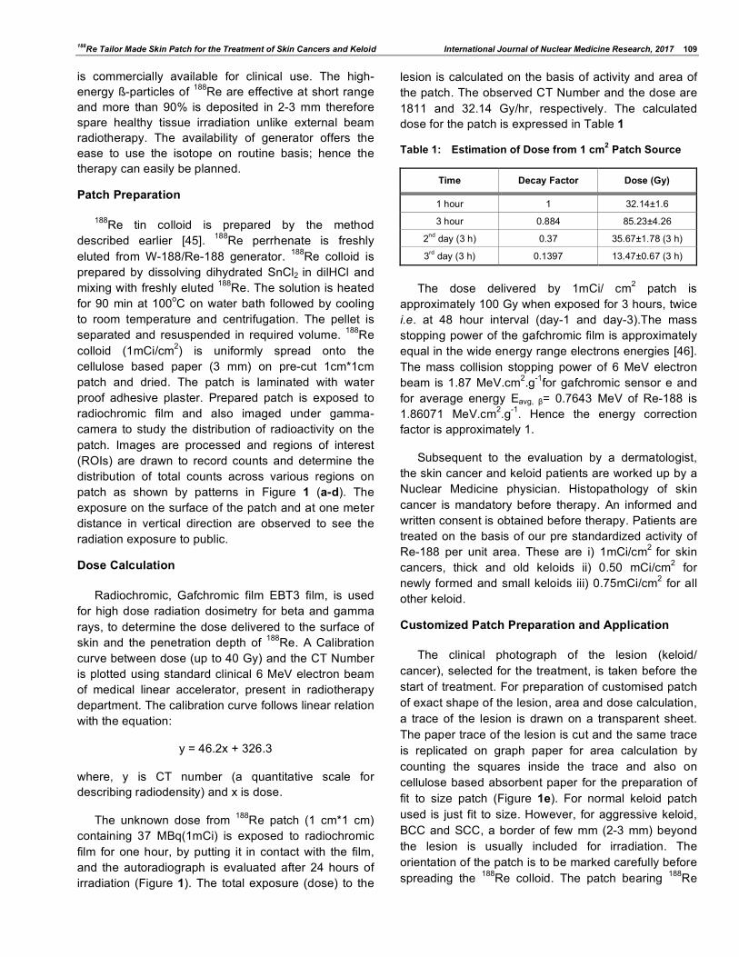
188Re tin colloid is prepared by the method described earlier [45]. 188Re perrhenate is freshly eluted from W-188/Re-188 generator. 188Re colloid is prepared by dissolving dihydrated SnCl2 in dilHCl and mixing with freshly eluted 188Re. The solution is heated for 90 min at 100oC on water bath followed by cooling to room temperature and centrifugation. The pellet is separated and resuspended in required volume. 188Re colloid (1mCi/cm2) is uniformly spread onto the cellulose based paper (3 mm) on pre-cut 1cm*1cm patch and dried. The patch is laminated with water proof adhesive plaster. Prepared patch is exposed to radiochromic film and also imaged under gamma-camera to study the distribution of radioactivity on the patch. Images are processed and regions of interest (ROIs) are drawn to record counts and determine the distribution of total counts across various regions on patch as shown by patterns in Figure 1 (a-d). The exposure on the surface of the patch and at one meter distance in vertical direction are observed to see the radiation exposure to public.
Dose Calculation
Radiochromic, Gafchromic film EBT3 film, is used for high dose radiation dosimetry for beta and gamma rays, to determine the dose delivered to the surface of skin and the penetration depth of 188Re. A Calibration curve between dose (up to 40 Gy) and the CT Number is plotted using standard clinical 6 MeV electron beam of medical linear accelerator, present in radiotherapy department. The calibration curve follows linear relation with the equation:
y = 46.2x + 326.3
where, y is CT number (a quantitative scale for describing radiodensity) and x is dose.
The unknown dose from 188Re patch (1 cm*1 cm) containing 37 MBq(1mCi) is exposed to radiochromic film for one hour, by putting it in contact with the film, and the autoradiograph is evaluated after 24 hours of irradiation (Figure 1). The total exposure (dose) to the
lesion is calculated on the basis of activity and area of the patch. The observed CT Number and the dose are 1811 and 32.14 Gy/hr, respectively. The calculated dose for the patch is expressed in Table 1
Table 1: Estimation of Dose from 1 cm2 Patch Source
Time Decay Factor Dose (Gy)
1 hour 1 32.14±1.6
3 hour 0.884 85.23±4.26
2nd day (3 h) 0.37 35.67±1.78 (3 h)
3rd day (3 h) 0.1397 13.47±0.67 (3 h)
The dose delivered by 1mCi/ cm2 patch is
approximately 100 Gy when exposed for 3 hours, twice i.e. at 48 hour interval (day-1 and day-3).The mass stopping power of the gafchromic film is approximately equal in the wide energy range electrons energies [46]. The mass collision stopping power of 6 MeV electron beam is 1.87 MeV.cm2.g-1for gafchromic sensor e and for average energy Eavg, β= 0.7643 MeV of Re-188 is 1.86071 MeV.cm2.g-1. Hence the energy correction factor is approximately 1.
Subsequent to the evaluation by a dermatologist, the skin cancer and keloid patients are worked up by a Nuclear Medicine physician. Histopathology of skin cancer is mandatory before therapy. An informed and written consent is obtained before therapy. Patients are treated on the basis of our pre standardized activity of Re-188 per unit area. These are i) 1mCi/cm2 for skin cancers, thick and old keloids ii) 0.50 mCi/cm2 for newly formed and small keloids iii) 0.75mCi/cm2 for all other keloid.
Customized Patch Preparation and Application
The clinical photograph of the lesion (keloid/ cancer), selected for the treatment, is taken before the start of treatment. For preparation of customised patch of exact shape of the lesion, area and dose calculation, a trace of the lesion is drawn on a transparent sheet. The paper trace of the lesion is cut and the same trace is replicated on graph paper for area calculation by counting the squares inside the trace and also on cellulose based absorbent paper for the preparation of fit to size patch (Figure 1e). For normal keloid patch used is just fit to size. However, for aggressive keloid, BCC and SCC, a border of few mm (2-3 mm) beyond the lesion is usually included for irradiation. The orientation of the patch is to be marked carefully before spreading the 188Re colloid. The patch bearing 188Re
110 International Journal of Nuclear Medicine Research, 2017 Shukla and Mittal
colloid is dried and laminated with water proof transparent adhesive tape in order to prevent the direct contact of radioactive matrix with the skin surface (epidermis). The exposure above the patch surface and at one meter has to be recorded.
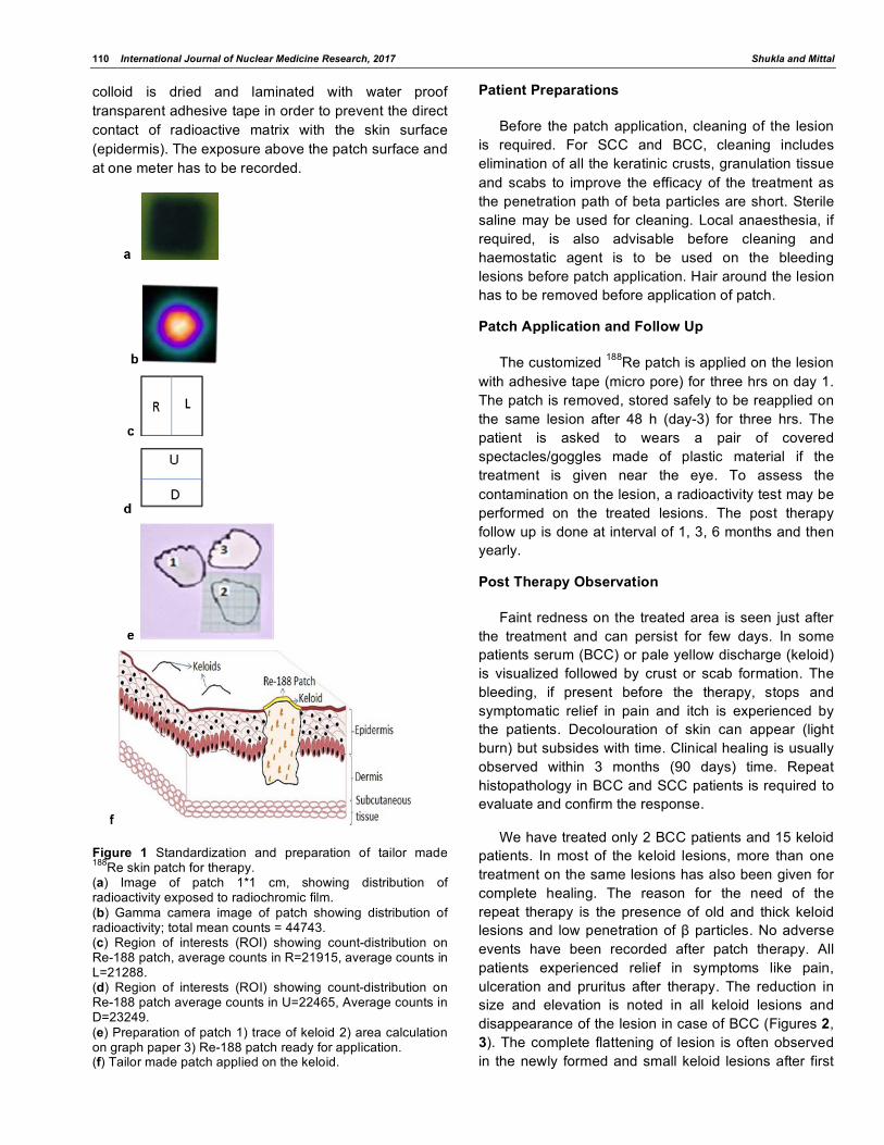
Figure 1 Standardization and preparation of tailor made 188Re skin patch for therapy. (a) Image of patch 1*1 cm, showing distribution of radioactivity exposed to radiochromic film. (b) Gamma camera image of patch showing distribution of radioactivity; total mean counts = 44743. (c) Region of interests (ROI) showing count-distribution on Re-188 patch, average counts in R=21915, average counts in L=21288. (d) Region of interests (ROI) showing count-distribution on Re-188 patch average counts in U=22465, Average counts in D=23249. (e) Preparation of patch 1) trace of keloid 2) area calculation on graph paper 3) Re-188 patch ready for application. (f) Tailor made patch applied on the keloid.
Patient Preparations
Before the patch application, cleaning of the lesion is required. For SCC and BCC, cleaning includes elimination of all the keratinic crusts, granulation tissue and scabs to improve the efficacy of the treatment as the penetration path of beta particles are short. Sterile saline may be used for cleaning. Local anaesthesia, if required, is also advisable before cleaning and haemostatic agent is to be used on the bleeding lesions before patch application. Hair around the lesion has to be removed before application of patch.
Patch Application and Follow Up
The customized 188Re patch is applied on the lesion with adhesive tape (micro pore) for three hrs on day 1. The patch is removed, stored safely to be reapplied on the same lesion after 48 h (day-3) for three hrs. The patient is asked to wears a pair of covered spectacles/goggles made of plastic material if the treatment is given near the eye. To assess the contamination on the lesion, a radioactivity test may be performed on the treated lesions. The post therapy follow up is done at interval of 1, 3, 6 months and then yearly.
Post Therapy Observation
Faint redness on the treated area is seen just after the treatment and can persist for few days. In some patients serum (BCC) or pale yellow discharge (keloid) is visualized followed by crust or scab formation. The bleeding, if present before the therapy, stops and symptomatic relief in pain and itch is experienced by the patients. Decolouration of skin can appear (light burn) but subsides with time. Clinical healing is usually observed within 3 months (90 days) time. Repeat histopathology in BCC and SCC patients is required to evaluate and confirm the response.
We have treated only 2 BCC patients and 15 keloid patients. In most of the keloid lesions, more than one treatment on the same lesions has also been given for complete healing. The reason for the need of the repeat therapy is the presence of old and thick keloid lesions and low penetration of β particles. No adverse events have been recorded after patch therapy. All patients experienced relief in symptoms like pain, ulceration and pruritus after therapy. The reduction in size and elevation is noted in all keloid lesions and disappearance of the lesion in case of BCC (Figures 2, 3). The complete flattening of lesion is often observed in the newly formed and small keloid lesions after first
188Re Tailor Made Skin Patch for the Treatment of Skin Cancers and Keloid International Journal of Nuclear Medicine Research, 2017 111
therapy. The patch therapy may be repeated after 3 months and separate lesions in same individual may be treated after one month. No recurrence has been
observed (both BCC and keloid) during the follow-up of more than three years.
(A)
(B)
(C)
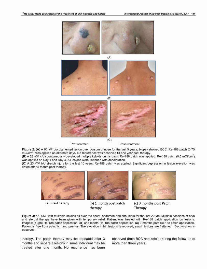
Pre-treatment Post-treatment Figure 2: (A) A 60 y/F c/o pigmented lesion over dorsum of nose for the last 5 years, biopsy showed BCC. Re-188 patch (0.75 mCi/cm2) was applied on alternate days. No recurrence was observed till one year post therapy. (B) A 23 y/M c/o spontaneously developed multiple keloids on his back. Re-188 patch was applied. Re-188 patch (0.5 mCi/cm2) was applied on Day 1 and Day 3. All lesions were flattened with decoloration. (C) A 23 Y/M h/o stretch injury for the last 10 years. Re-188 patch was applied. Significant depression in lesion elevation was noted after 5 month post therapy.
Figure 3: 45 Y/M with multople keloids all over the chest, abdomen and shoulders for the last 20 yrs. Multiple sessions of cryo and steroid therapy have been given with temporary relief. Patient was treated with Re-188 patch application on lesions. Images: (a) pre Re-188 patch application; (b) one month Re-188 patch application; (c) 3 months post Re-188 patch application. Patient is free from pain, itch and pruritus. The elevation in big lesions is reduced; small lesions are flattened . Decoloration is observed.
112 International Journal of Nuclear Medicine Research, 2017 Shukla and Mittal
General Precautions
To avoid leakage or leaching of radionuclide, the stability of the particles and lamination of patch is done before patch application. It will help in safe handling by personnel involved in the application of the patch to the patients. The radiation dermatitis may occur if the patch is mis-fit to the lesion or displaced during therapy period.
SALIENT FEATURES OF RE-188 PATCH THERAPY
The measurement of activity and the preparation of patch with 188Re is simple. The 188Re patch therapy has many advantages – 1) multiple lesions can be treated at a time, 2) the treatment of lesions where excision or external radiotherapy is not possible, 3) short tissue penetration and high linear energy transfer spares the radiation damage to underlying bone and soft tissue, 4) patients convenience is considered due to the presence of in-house generator.
REFERENCES
[1] Ramirez CC, Federman DG, Kirsner RS. Skin cancer as an occupational disease: the effect of ultraviolet and other forms of radiation. Int J Dermatol 2005; 44(2): 95-100. https://doi.org/10.1111/j.1365-4632.2005.02301.x
[2] Rubin AI, Chen EH and Ratner D. Basal-cell carcinoma. N Engl J Med 2005; 353: 2262-9. https://doi.org/10.1056/NEJMra044151
[3] Madan V, Lear JT and Szeimies RM. Non-melanoma skin cancer. Lancet 2010; 375: 673-85 https://doi.org/10.1016/S0140-6736(09)61196-X
[4] Babu M, Meenakshi J, Jayaraman V, Ramakrishnan KM. Keloids and hypertrophic scars: A review. Indian J Plas Surg 2005; 38: 75-9. https://doi.org/10.4103/0970-0358.19796
[5] Petrovic D, Visnjic M, Mihailovich D, Petrovic S, Pesic Z. Margin size in basocellular skin carcinoma resection: impact on relapse. Acta Fac Med Naiss 2004; 21: 195-200.
[6] Cumberland L, Dana A, Liegeois N. Mohs micrographic surgery for the management of nonmelanoma skin cancers. Facial Plast Surg Clin North Am 2009; 17: 325-35. https://doi.org/10.1016/j.fsc.2009.06.001
[7] Minton TJ. Contemporary Mohs surgery applications. Curr Opin Otolaryngol Head Neck Surg 2008; 16: 376-80. https://doi.org/10.1097/MOO.0b013e3283079cac
[8] Snow SN, Madjar DD. Mohs surgery in the management of cutaneous malignancies. Clin Dermatol 2001; 19: 339-47. https://doi.org/10.1016/S0738-081X(01)00169-9
[9] Alster TS, Handrick C. Laser treatment of hypertrophic scars, keloids, and striae. Semin Cutan Med Surg 2000; 19: 287-92. https://doi.org/10.1053/sder.2000.18369
[10] Tanzi EL, Alster TS. Laser treatment of scars. Skin Therapy 2004; Lett, 9: 4-7.
[11] Rusciani L, Rossi G, Bono R. Use of cryotherapy in the treatment of keloids. J Dermatol Surg Oncol 1993; 19: 529-34. https://doi.org/10.1111/j.1524-4725.1993.tb00386.x
[12] Har-Shai Y, Amar M, Sabo E. Intralesional cryotherapy for enhancing the involution of hypertrophic scars and keloids.
Plast Reconstr Surg 2003; 111: 1841-52. https://doi.org/10.1097/01.PRS.0000056868.42679.05
[13] Kwon SY, Park SD, Park K. Comparative effect of topical silicone gel and topical tretinoin cream for the prevention of hypertrophic scar and keloid formation and the improvement of scars. J Eur Acad Dermatol Venereol 2014; 28: 1025-33. https://doi.org/10.1111/jdv.12242
[14] Calzavara-Pinton PG, Venturini M, Sala R. Photodynamic therapy: update 2006. Part 1: photochemistry and photobiology. J Eur Acad Dermatol Venereol 2007; 21: 293-302. https://doi.org/10.1111/j.1468-3083.2006.01902.x
[15] Gauglitz GG, Korting HC, Pavicic T, Ruziicka T, Jeschke MG. Hypertrophic scarring and keloids: Pathomechanisms and current and emerging treatment strategies. Mol Med 2011; 17: 113-25. https://doi.org/10.2119/molmed.2009.00153
[16] Peris K, Campione E, Micantonio T. Imiquimod treatment of superficial and nodular basal cell carcinoma: 12 week open label trial. Dermatol Surg 2005; 31: 318-23. https://doi.org/10.1097/00042728-200503000-00012
[17] Geisse J, Caro I, Lindholm J. Imiquimod 5% cream for the treatment of superficial basal cell carcinoma: results from two phase III, randomized, vehicle controlled studies. J Am Acad Dermatol 2004; 50: 722-33. https://doi.org/10.1016/j.jaad.2003.11.066
[18] Bianchi L, Orlandi A, Campione E. Topical treatment of basal cell carcinoma with tazarotene: a clinicopathological study on a large series of cases. Br J Dermatol 2004; 151: 148-56. https://doi.org/10.1111/j.1365-2133.2004.06044.x
[19] Peng Q, Warloe T, Berg K, Moan J, Kongshaug M, Giercksky KE et al. 5-Aminolevulinic acid-based photodynamic theraphy: clinical research and future challenges. Cancer 1997; 79: 2282-308. https://doi.org/10.1002/(SICI)1097-0142(19970615)79:12<2282::AID-CNCR2>3.0.CO;2-O
[20] Oseroff AR, Blumenson LR, Wilson BD, Mang TS, Bellnier DA, Parsons JC et al. A dose ranging study of photodynamic therapy with porfimer sodium (Photofrin) for treatment of basal cell carcinoma. Lasers Surg Med 2006; 38: 417-26. https://doi.org/10.1002/lsm.20363
[21] Sandberg C, Stenquist B, Rosdahl I, Ros AM, Synnerstad I, Karlsson M et al. Important factors for pain during photodynamic therapy for actinic keratosis. Acta Derm Venereol 2006; 86: 404-8. https://doi.org/10.2340/00015555-0098
[22] Fleming I, Amonette MD, Managhan T, Fleming I. Principles of management of basal and squamous cell carcinoma of the skin. Cancer 1995; 75 (suppl no 2): 699-704 https://doi.org/10.1002/1097-0142(19950115)75:2+<699::AID-CNCR2820751413>3.0.CO;2-Q
[23] Andrews J, Marttala J, Macarak E, Rosenbloom J, Uitto J. Keloids: The paradigm of skin fibrosis-Pathomechanisms and treatment. Matrix Biology 2016; 51: 37-46. https://doi.org/10.1016/j.matbio.2016.01.013
[24] Arno AI, Gauglitz GG, Barett JP, Jeschke MG. Up-to-date approach to manage keloids and hypertrophic scars: A useful guide. Burns 2014; 40: 1255-66. https://doi.org/10.1016/j.burns.2014.02.011
[25] Kontochristopoulos G, Stefanaki C, Panagiotopoulos A, Stefanaki K, Argryakos T, Petridis A et al. Intralesional 5-fluorouracil in the treatment of keloids: an open clinical and histopathologic study. J Am Acad Dermatol 2005; 52: 474-9. https://doi.org/10.1016/j.jaad.2004.09.018
[26] Darougheh A, Asilian A, Shariati F. Intralesional triamcinolone alone or in combination with 5-fluorouracil for the treatment of keloid and hypertrophicscars. Clin Exp
188Re Tailor Made Skin Patch for the Treatment of Skin Cancers and Keloid International Journal of Nuclear Medicine Research, 2017 113
Dermatol 2009; 34: 219-23. https://doi.org/10.1111/j.1365-2230.2007.02631.x
[27] Lee JH, Kim SE, Lee AY. Effects of interferon-alpha2b on keloid treatment with triamcinolone acetonideintralesional injection. Int J Dermatol 2008; 47: 183-6. https://doi.org/10.1111/j.1365-4632.2008.03426.x
[28] Mikulec AA, Hanasono MM, Lum J, Kadleck JM, Kita M, Koch RJ. Effect of tamoxifen on transforming growth factor beta1 production by keloid and fetal fibroblasts. Arch Facial Plast Surg 2001; 3: 111-4. https://doi.org/10.1001/archfaci.3.2.111
[29] Goldschmidt H, Panizzon R. Modern dermatologic radiation therapy. Springer Verlag, New York. 1991. https://doi.org/10.1007/978-1-4613-9041-1
[30] Vora SA, Garner SL. Role of radiation therapy for facial skin cancers. Clin Plast Surg 2004; 31: 33-8. https://doi.org/10.1016/S0094-1298(03)00119-6
[31] Archambeau J, Pezner R, Wasserman T. Pathophysiology of irradiated skin and breast. Int J Radiat Oncol, BiolPhys1995; 31: 1171-85. https://doi.org/10.1016/0360-3016(94)00423-I
[32] Kal HB, Veen RE. Biologically effective doses of postoperative radiotherapy in the prevention of keloids. Dose-effect relationship. Strahlenther Onkol 2005; 181: 717-23. https://doi.org/10.1007/s00066-005-1407-6
[33] Vivante H, Salgueiro MJ, Ughetti R, Nicolini J, Zubillaga M. P-32 Patch contact brachyradiotherapy in the management of recalcitrant keloid and hypertrophic scars. Indian J Dermatol Venereol Leprol 2007; 73: 336-9. https://doi.org/10.4103/0378-6323.35736
[34] Salgueiro MJ, Duran H, Palmieri aff3M, Pirchio R, Nicolini J, Ughetti R et al. Design and bioevaluation of a P-32 patch for brachytherapy of skin diseases. Appl RadiatIsotop 2008; 66: 303-9. https://doi.org/10.1016/j.apradiso.2007.09.008
[35] Lee JD, Park KK, Lee MG, Kim EH, Rhim KJ, Lee JT et al. Radionuclide Therapy of Skin Cancers and Bowen's Disease Using a Specially Designed Skin Patch J Nucl Med 1997; 38: 697-702.
[36] Chung YL, Lee JD, Bang D, Lee JB, Park KB, Lee MG. Treatment of Bowen's disease with especially designed radioactive skin patch. Eur J Nucl Med 2000; 27: 842-6.
https://doi.org/10.1007/s002590000262 [37] Pandey U, Saxena SK, Sarma HD, Tandon P, Ram R,
Samuel G et al. Bioevaluation studies of P-32 incorporated mould brachytherapy sources for potential application in treatment of superficial tumors. Nucl Med Commun 2008; 29: 717-23. https://doi.org/10.1097/MNM.0b013e3282f813b4
[38] Saxena KS, Kumar Y, Dash A. P-32 patches for superficial brachytherapy applications. Cancer Biother Radiopharma 2012; 27: 276-86.
[39] Park KB, Kim J-R, Lee J-D. Radioactive patch/film and process for preparation. US patent 1999.
[40] Shukla J, Chaudhary V, Bhusari P, De D, Reddy R, Mittal B. Cost-effective Custom Made P-32 Incorporated Alginate Gel Patches for the Treatment of Basal Cell Carcinoma. World J Nucl Med 2013; 12 (Suppl 2): 227-8.
[41] Munaweera I, Levesque-Bishop D, Shi Y, Anthony Pasqua JD, Balkus KJ. Radiotherapeutic Bandage Based on electrospunpolyacrylonitrilecontaining holmium-166 Iron garnet nanoparticles for the treatment of Skin Cancer. App. Mater Interfaces 2014; 6: 22250-6. https://doi.org/10.1021/am506045k
[42] Shukla J, Bhusari P, Vatsa R, De D, Kumaran S, Handa S, Mittal BR. Tailor made Re-188 skin patch for radionuclide therapy of keloids. World J Nucl Med 2016; 15: S38.
[43] Mukherjee A, Pandey U, Sarma HD, Gupta SK, Ingle AD, Pillai MR et al. Bioevaluation of radioactive bandages in a murine model of melanoma. Int J Radiat Biol 2003; 79: 839-45. https://doi.org/10.1080/09553000310001610989
[44] Jeong JM, Lee YJ, Kim EH, Chang YS, Kim YJ, Son M et al. Preparation of 188Re-labeled paper for treating skin cancer. Appl RadiatIsot 2003; 58: 551-5 https://doi.org/10.1016/s0969-8043(03)00063-0
[45] Shukla J, Bandopadhyaya GP, Shamim SA, Kumar R. Characterization of Re-188-Sn microparticles used for synovitis treatment. Int J Pharmaceutics 2007; 338: 43-7. https://doi.org/10.1016/j.ijpharm.2007.01.014
[46] McLaughlin WL, Yun-Dong C, Soures CG. Sensitometry of the response of a new radiochromic film dosimeter to gamma radiation and electron beams. Nucl Instr Meth Phys Res1991; 302: 1165-76. https://doi.org/10.1016/0168-9002(91)90506-l
Received on 08-03-2017 Accepted on 07-04-2017 Published on 31-07-2017 http://dx.doi.org/10.15379/2408-9788.2017.10
© 2017 Shukla and Mittal; Licensee Cosmos Scholars Publishing House. This is an open access article licensed under the terms of the Creative Commons Attribution Non-Commercial License (http://creativecommons.org/licenses/by-nc/3.0/), which permits unrestricted, non-commercial use, distribution and reproduction in any medium, provided the work is properly cited.