Embed Size (px)
Citation preview
New Therapeutic Approach: Diphenyl DiselenideReduces Mitochondrial Dysfunction in Acetaminophen-Induced Acute Liver FailureNelson R. Carvalho1, Edovando F. da Rosa1, Michele H. da Silva1, Cintia C. Tassi1, Cristiane L. Dalla
Corte1, Sara Carbajo-Pescador2, Jose L. Mauriz2, Javier Gonzalez-Gallego2, Felix A. Soares1*
1 Departamento de Quımica, Centro de Ciencias Naturais e Exatas, Universidade Federal de Santa Maria, Campus UFSM, Santa Maria, Rio Grande do Sul, Brasil, 2 Institute
of Biomedicine (IBIOMED) and Centro de Investigacion Biomedica en Red de Enfermedades Hepaticas y Digestivas (CIBERehd), University of Leon, Leon, Spain
Abstract
The acute liver failure (ALF) induced by acetaminophen (APAP) is closely related to oxidative damage and depletion ofhepatic glutathione, consequently changes in cell energy metabolism and mitochondrial dysfunction have been observedafter APAP overdose. Diphenyl diselenide [(PhSe)2], a simple organoselenium compound with antioxidant properties,previously demonstrated to confer hepatoprotection. However, little is known about the protective mechanism onmitochondria. The main objective of this study was to investigate the effects (PhSe)2 to reduce mitochondrial dysfunctionand, secondly, compare in the liver homogenate the hepatoprotective effects of the (PhSe)2 to the N-acetylcysteine (NAC)during APAP-induced ALF to validate our model. Mice were injected intraperitoneal with APAP (600 mg/kg), (PhSe)2
(15.6 mg/kg), NAC (1200 mg/kg), APAP+(PhSe)2 or APAP+NAC, where the (PhSe)2 or NAC treatment were given 1 hfollowing APAP. The liver was collected 4 h after overdose. The plasma alanine and aspartate aminotransferase activitiesincreased after APAP administration. APAP caused a remarkable increase of oxidative stress markers (lipid peroxidation,reactive species and protein carbonylation) and decrease of the antioxidant defense in the liver homogenate andmitochondria. APAP caused a marked loss in the mitochondrial membrane potential, the mitochondrial ATPase activity, andthe rate of mitochondrial oxygen consumption and increased the mitochondrial swelling. All these effects were significantlyprevented by (PhSe)2. The effectiveness of (PhSe)2 was similar at a lower dose than NAC. In summary, (PhSe)2 provided asignificant improvement to the mitochondrial redox homeostasis and the mitochondrial bioenergetics dysfunction causedby membrane permeability transition in the hepatotoxicity APAP-induced.
Citation: Carvalho NR, da Rosa EF, da Silva MH, Tassi CC, Dalla Corte CL, et al. (2013) New Therapeutic Approach: Diphenyl Diselenide Reduces MitochondrialDysfunction in Acetaminophen-Induced Acute Liver Failure. PLoS ONE 8(12): e81961. doi:10.1371/journal.pone.0081961
Editor: Anna Alisi, Bambino Gesu’ Children Hospital, Italy
Received March 26, 2013; Accepted October 18, 2013; Published December 11, 2013
Copyright: � 2013 Carvalho et al. This is an open-access article distributed under the terms of the Creative Commons Attribution License, which permitsunrestricted use, distribution, and reproduction in any medium, provided the original author and source are credited.
Funding: This study was supported by the FINEP research grant ‘‘Rede Instituto Brasileiro de Neurociencia (IBN-Net)’’ #01.06.0842-00 and "Instituto Nacional deCiencia e Tecnologia (INCT)" for excitotoxicity and neuroprotection—MCT/CNPq. F.A.A.S. received a fellowship by CNPq. M.H.S. and N.R.C. received a fellowship byCAPES. S.C.P. is funded by the Consejerıa de Educacion (Junta de Castilla y Leon, Spain) and European Social Fund. Additional support was given by CNPq/FAPERGS/DECIT/SCTIE-MS/PRONEM #11/2029-1. CIBERehd is funded by the Instituto de Salud Carlos IIII, Spain. The funders had no role in study design, datacollection and analysis, decision to publish, or preparation of the manuscript.
Competing Interests: The authors have declared that no competing interests exist.
* E-mail: [email protected]
Introduction
Acetaminophen (N-acetyl-p-aminophenol; APAP) is a drug
widely employed as an analgesic and antipyretic that can induce
acute liver failure (ALF) when high doses are ingested [1]. Recent
data suggest a dramatic increase in ALF, liver transplants and
considerable morbidity and mortality associated with APAP
overdoses in the United States and many other countries [2,3].
During overdoses, APAP is mainly metabolized in the liver by
cytochrome P450, resulting in a highly reactive intermediate, N-
acetyl-p-benzoquinone imine (NAPQI). NAPQI reacts directly
with glutathione (GSH), causing a depletion of GSH in the liver.
This redox imbalance in the liver has been shown to play a major
role in ALF associated with APAP [3]. If glutathione is not
replenished, NAPQI begins to form covalent bonds with cellular
proteins, modifying their structure and function [4,5]. In addition,
the accumulation of neutrophils and Kuppfer cells contribute to
the inflammatory process and reactive species (RS) generation in
the hepatocytes [5,6,7]. The hepatic injury is associated with
damage to subcellular organelles including mitochondria, because
mitochondria are responsible for cellular energy metabolism and
represent a remarkable source of intracellular RS generation in
mammalian cells, effects on this organelle are critical with regard
to APAP-mediated liver injuries [8].
The compound N-acetylcysteine (NAC) is the treatment of
choice for acute poisoning with APAP [2]. NAC administration is
beneficial for preventing or reducing ALF by increasing GSH and
thiols levels, reduces the histological changes caused by oxidative
stress induced by APAP overdose [1,9,10]. The efficacy of NAC
and the prognosis are dependent on three factors, the type of
APAP ingestion (acute vs. chronic), the dose of APAP ingestion
and the elapsed time from APAP ingestion to the initiation of
NAC treatment [9]. In clinical situations, NAC is administered
after the occurrence of an APAP overdose, making the study of
alternative therapies attractive.
PLOS ONE | www.plosone.org 1 December 2013 | Volume 8 | Issue 12 | e81961
Considering the fact that NAC efficacy is limited to a narrow
window of time and situations [10,11]. The use of organoselenium
compounds could emerge as an alternative therapy. Several
studies have demonstrated both the antioxidant and the anti-
inflammatory properties of organoselenium compounds such as
diphenyl diselenide [(PhSe)2] and ebselen (Ebs) [12,13,14]. In
particular, (PhSe)2, the simplest of diaryl diselenides and a
lipophilic organic compound of selenium, has demonstrated low
toxicity in different experimental models. For example, the
calculated LD50 in mice is 655 mg/kg when administered
intraperitoneally [15]. The hepatoprotective is associated with
the biochemical and pharmacological properties of the organose-
lenium compounds to scavenge hydrogen peroxide and other
organic hydroperoxides originate from the powerful nucleophile
intermediates that involve the selenol-selenolate group, which play
critical roles in their glutathione peroxidase– and thioredoxin
reductase–like activities [13,15]. Earlier work from our laboratory
has shown that (PhSe)2 is effective for the treatment of cellular
damage caused by APAP [16,17]. However our study uses for the
first time the (PhSe)2 as a possible target to the mitochondrial
dysfunction in hepatic failure caused by APAP in a new
therapeutic approach.
Previously, the APAP toxicity was shown to consist of two
crucial phases: the initial GSH depletion and covalent binding of
NAPQI to target proteins and the subsequent increase in the
mitochondrial permeability transition (MPT) and nitration of
proteins [7]. In this condition, the impairment of GSH antioxidant
system has been noticed to enhance the susceptibility to
mitochondrial dysfunction from oxidant stress and resulting in
the collapse of mitochondrial membrane potential (Dym) and ATP
depletion [18]. It should be noted that MPT is mediated by
oxidant stress [19]. Therefore, MPT occurs with the release of
superoxide, which in turn can lead to peroxynitrite (ONOO2)
production and tyrosine nitration, a lethal event for the cell [8].
Moreover, both oxidative damage and NAPQI have been
reported to produce MPT through the oxidation of critical thiols
to disulfides, which appears to be a prerequisite for membrane
permeabilization [20]. Currently, it has been suggested that
organoselenium present modulatory effects in relation to mito-
chondrial oxidative stress; however, these studies were conducted
using in vitro models [21,22]. Thus, there is no evidence in the
literature demonstrating the effects of (PhSe)2 on liver mitochon-
drial dysfunction caused by APAP intoxication.
Thus, considering that relatively few studies have focused on the
mechanisms by which these organoselenium compounds exert
their pharmacological effects on APAP-induced ALF [16,17], this
study was designed to evaluate the benefits of the (PhSe)2treatment under the mitochondrial dysfunction, and subsequently,
compare in liver homogenate the hepatoprotective effects with N-
acetylcysteine (NAC) during APAP-induced ALF to validate our
model. This work may contribute to a better understanding of the
(PhSe)2 mechanism of action and open new perspectives for its
application.
Materials and Methods
Materials(PhSe)2 (98%), thiobarbituric acid (TBA), 29-79-dichlorofluor-
escein (DCFH), trichloroacetic acid (TCA) and nucleotides were
purchased from Sigma Chemical Co. (St. Louis, MO). All other
chemicals were of analytical grade and obtained from standard
commercial suppliers.
AnimalsSeven-week-old male adult Swiss albino mice (30–40 g) from
our own breeding colony were used. The animals were kept on a
separate animal room, on at 12 h light/dark cycle, at temperature
of 2262uC, with free access to food and water. Mice were
acclimated for 7 days before initiation of any procedures. This
study was approved by the Ethics and Animal Welfare Committee
of Federal University of Santa Maria, Brazil.
Experimental procedureBriefly, the mice were randomly divided into the following
groups: Control (vehicle); diphenyl diselenide [(PhSe)2]; N-
acetylcysteine (NAC); acute liver failure (induced by APAP); acute
liver failure treated with diphenyl diselenide [APAP+(PhSe)2] and
acute liver failure treated with N-acetylcysteine (APAP+NAC). All
the solutions were administered by the intraperitoneal (i.p.) route.
Injections were administered at 9:00 a.m. in order to remove any
confounding factors of circadian rhythm. The APAP, (PhSe)2 and
NAC doses were described earlier [9,16,17]. Each group
contained 7 different mice/group. Mice in the control, (PhSe)2and NAC groups were injected intraperitoneally (i.p.) with saline
0.9% (20 ml/Kg), and mice in the APAP, APAP+(PhSe)2 and
APAP+NAC groups were injected i.p. with 600 mg/Kg APAP
(20 ml/Kg in saline 0.9%). One hour after saline and APAP
treatment, mice were injected i.p. with 15.6 mg/Kg (PhSe)2(2.5 mL/Kg in canola oil) in the (PhSe)2 and APAP+(PhSe)2. In
addition, studies were done with a higher dose of NAC to validate
our model. One hour after saline and APAP treatment, mice were
injected i.p. with 1200 mg/kg NAC (20 ml/kg in saline 0.9%) in
the NAC and APAP+NAC. The feed was available ad libitum and
animals were not fasted prior to dosing. The biochemical analyses
were carried out 4 h as previous studies had shown toxicity was
apparent at this time [16,17]. The animals were killed by cervical
dislocation and blood was collected by cardiac puncture using
heparin-rinsed 1-mL syringes (20-gauge needles) and centrifuged.
The plasma was used for determination of alanine aminotransfer-
ase (ALT) and aspartate aminotransferase (AST) activities using a
commercial kit (LabtestH, Diagnostica S.A., Minas Gerais, Brazil).
SurvivalFor survival studies, mice were injected with 600 mg/kg and
after 1 h treated with (PhSe)2 or NAC. Then, thirty minutes later
the mice were returned to their cages and fed with food and water
ad libitum. To determine the effect of (PhSe)2 and NAC on
mortality of APAP-administrated mice, the survival rate after
APAP administration was evaluated for 48 h.
Liver homogenates preparationAt the end of the treatment period the liver was removed and
quickly dissected, placed on ice, and immediately homogenized in
cold 10 mM Tris–HCl pH 7.4. Homogenates were centrifuged at
2,0006g for 10 min to yield the low-speed supernatant fractions
that were used for different biochemical assays in all trials. Besides,
aliquots of liver preparations were frozen (220uC) for posterior
analysis.
Isolation of liver mitochondriaMice liver mitochondria were isolated at 4uC by differential
centrifugation [23]. The animals were sacrificed by cervical
dislocation. The livers were rapidly removed (within 1 min) and
immersed in ice-cold ‘‘Ionic Medium’’ containing 100 mM
Sucrose, 10 mM EDTA, 46 mM KCl and 100 mM Tris-HCl,
pH 7.4. The tissue was minced using surgical scissors and then
Diphenyl Diselenide and APAP Hepatotoxicity
PLOS ONE | www.plosone.org 2 December 2013 | Volume 8 | Issue 12 | e81961
extensively washed. The tissue was then homogenized in a power-
driven, tight-fitting Potter Elvehjem homogenizer with Teflon
pestle. The resulting suspension was centrifuged for 7 min at
2,0006g in a Hitachi CR 21E centrifuge. After centrifugation, the
supernatant was recentrifuged for 10 min at 12,0006g. The pellet
was resuspended in ‘‘Ionic Medium+BSA’’ containing 100 mM
sucrose, 10 mM EDTA, 46 mM KCl, 0.1% bovine serum
albumin free fatty acid and 100 mM Tris-HCl, pH 7.4, and
recentrifuged at 12,0006g for 10 min. The supernatant was
decanted, and the final pellet was gently washed and resuspended
in ‘‘Suspension Medium’’ containing 230 mM mannitol, 70 mM
sucrose, and 20 mM Tris-HCl, pH 7.4. The pellet was washed
three times with ice suspension medium buffer to get intact
mitochondria, which it has been suggested that after three
washings, influence of contaminating microsomes on NAD(P)H
oxidation, reactive oxygen species (ROS) production and mem-
brane permeabilization becomes negligible [24]. An aliquot of the
resulting mitochondrial suspension were separated and rapidly
frozen at 280uC for later biochemical analysis of GSH content,
TBARS, protein carbonyls and mitochondrial enzymes.
Measurement of lipid peroxidation (LPO)Liver homogenate and mitochondrial membrane LPO were
quantified measuring the malondiadehyde (MDA). In summary,
liver homogenate and mitochondria protein were incubated in
300 ml of a medium consisting of 175 mM KCl and 10 mM Tris-
HCl, pH 7.4, and then, were added to color reaction. Thiobarbi-
turic acid reactive substances (TBARS) levels were measured at
532 nm using a standard curve of MDA [25].
Measurement of ROS productionROS generation was determined spectrofluorimetrically in liver
homogenate and mitochondria, using H2DCF-DA levels as an
index of the peroxide production by cellular components (1 mM)
[26]. Briefly, the liver homogenate and mitochondria were added
to standard medium and the fluorescence was determined at
488 nm for excitation and 525 nm for emission, with slit widths of
3 nm.
Measurement of reduced glutathione (GSH)GSH levels were determined in liver homogenate and
mitochondria with fluorescence detection after reaction of the
supernatants from deproteinized containing H3PO4/NaH2PO4–
EDTA, with O-Phthalaldehyde (OPT) [27]. In brief, 250 mg of
liver were homogenized in 3.75 mL phosphate–EDTA buffer
(100 mM NaH2PO4, 5 mM EDTA, pH 8.0) plus 1 mL H3PO4
(25%), and isolated liver mitochondria (0.5 mg protein) resus-
pended in 1.5 mL phosphate-EDTA buffer and 500 ml H3PO4
(4.5%) were rapidly centrifuged at 100,0006g (Hitachi, TL-100
ultracentrifuge) for 30 min. For GSH determination, 100 ml of
supernatant was added to 1.8 ml phosphate buffer and 100 ml
OPT. After thorough mixing and incubation at room temperature
for 15 min, the solution was transferred to a quartz cuvette and the
fluorescence was measured at 420 and 350 nm emission and
excitation wavelengths, respectively. GSH contents were deter-
mined from comparisons with a linear GSH standard curve.
Measurement of antioxidant enzyme activitiesThe activities of antioxidant enzymes, total superoxide dis-
mutase (SOD), catalase (CAT), glutathione S-transferase (GST),
glutathione reductase (GR), and glutathione peroxidase (GPx)
have been measured in liver homogenates.
Liver homogenate total SOD activity was measured by the
capacity of inhibiting auto-oxidation of adrenaline to adreno-
chrome at 480 nm [28]. The liver supernatant (5 mg protein) was
added to a medium containing 2 mM EDTA, 50 mM NaHCO3/
Na2CO3 buffer (pH 10.3) and adrenaline (4 mM).
The CAT enzyme activity was determined in liver homogenate
in according to the method previously proposed [29]. Liver
homogenate (5 mg protein) was added to a medium containing
potassium phosphate buffer (50 mM KH2PO4, 50 mM K2HPO4;
pH 7.4) and H2O2 (1 mM). The kinetic analysis of CAT was
started after H2O2 addition. The CAT activity was determined
using the molar extinction coefficient 36 M21cm21 and the
reaction was measured at 240 nm.
Glutathione-S-transferase (GST) activity was determined spec-
trophotometrically [30]. GST activity was quantified in liver
homogenates (5 mg protein) in a reaction mixture containing
1 mM 1-chloro-2,4-dinitrobenzene (CDNB), and 1 mM glutathi-
one as substrates in 0.1 M sodium phosphate buffer, pH 6.5, at
37uC. Enzyme activity was calculated by the change in the
absorbance value from the slope of the initial linear portion of the
absorbance time curve at 340 nm for 5 min. Enzyme activity was
determined using the molar extinction coefficient 9,6 mM21cm21
and expressed as nmol CDNB/min/mg Prot.
Glutathione peroxidase (GPx) activity was determined spectro-
photometrically at 340 nm by NADPH consumption for 2 min at
30uC [31]. The liver homogenate supernatant (5 mg protein) was
added to medium containing 0.1 M phosphate buffer (0.1 M
KH2PO4, 0.1 M K2HPO4 and 5 mM EDTA, pH 7.0), 1 mM
GSH, 0.15 mM NADPH, 0.1 U/mL GR and 1 mM sodium
azide. So, the reaction was initiated by adding the H2O2 to a final
concentration of 0.4 mM. The GPx activity was determined using
the molar extinction coefficient 6220 M21cm21 and expressed as
nmol/min/mg protein.
For the measurement activity of glutathione reductase (GR)
activity, the liver homogenate supernatant (5 mg protein) was
added to medium containing 0.15 M phosphate buffer (0.15 M
K2HPO4 and 1.5 mM EDTA, pH 7.0) and 0.15 mM NADPH.
The measurements were made at 340 nm and initiated with
addition of 20 mM GSSG, at 30uC for 2 min [32]. GR activity
was determined using the molar extinction coefficient 6220 M21
cm21 and expressed as nmol/min/mg protein.
Measurement of mitochondrial protein carbonylsThe oxidative damage to proteins was measured by the
quantification of carbonyl groups based on the reaction with
dinitrophenylhidrazine (DNPH) assay [33]. The mitochondria
were divided into two portions containing 1 mg of protein in each.
To one portion, 1 ml of 2 N HCl was added and incubated at
room temperature shaking intermittently for 1 h. The other
portion was treated with 1 ml of 10 mM DNPH in 2 N HCl and
incubated by shaking intermittently for 1 h at room temperature.
After incubation the mixture was precipitated with 10% TCA and
centrifuged. The precipitate was washed thrice with 1 ml of
ethanol:ethyl acetate (1:1). The final protein precipitate was
dissolved in denaturation buffer (3% SDS and 150 mM
NaH2PO4; pH 6.8) and the absorption at 370 nm (DNPH-treated
sample minus sample blank) was determined. Carbonyl content
was calculated using the molar extinction coefficient of
22,000 M21 cm21 and expressed as nmol carbonyls/mg mito-
chondrial protein.
Mitochondrial transmembrane electrical potential (Dym)Mitochondrial Dym was estimated by fluorescence changes
in Safranine – O (10 mM) recorded by RF-5301 Shimadzu
Diphenyl Diselenide and APAP Hepatotoxicity
PLOS ONE | www.plosone.org 3 December 2013 | Volume 8 | Issue 12 | e81961
spectrofluorometer (Kyoto, Japan) operating at excitation and
emission wavelengths of 495 and 586, with slit widths of 5 nm
[34]. The mitochondria (0.5 mg protein) were added and 30
second latter mitochondrial respiration was induced by the
addition of succinate and glutamate. Mitochondrial preparation,
which was held on ice, was well maintained and did not change
over the course of 5–6 hours, as determined by their ability to
maintain a stable transmembrane potential in the presence of
oxidizable substrates.
Mitochondrial swellingMeasurement of mitochondrial swelling was performed in RF-
5301 Shimadzu spectrofluorometer at 600 nm and slit 1.5 nm for
excitation and emission. The mitochondria (0.1 mg protein) were
incubated in the presence of 100 mM Ca2+ [19].
Oxygen consumption of liver mitochondriaThe oxygen consumption of liver mitochondria was measured
using an oxymeter (Hansatech model with a Clark-type electrode)
at 30uC. The cuvete containing aerated medium consisting of
225 mM mannitol, 75 mM sucrose, 10 mM KCl, 10 mM Tris-
HCl, 10 mM K2HPO4, 5 mM MgCl2, 0.1 mM EDTA (pH 7.4)
was added 0.1 mg mitochondrial protein. Pyruvate (5 mM),
glutamate (5 mM) and succinate (5 mM) were placed in the
medium to increase the respiratory state.
Assessment of mitochondrial activity (MTT reductionassay)
This assay is based on the ability of mitochondrial enzymes to
metabolize MTT into formazan, a reaction that takes place only in
functionally intact mitochondria. The mitochondrial samples
(0.1 mg protein) were incubated with 20 mM succinate at 30uCfor 1 hour. After that, color was quenched with DMSO, and
readings were reported as the difference in absorbance between
570 and 630 nm, and then, expressed in percent of the control
[35].
Measurement of mitochondrial antioxidant enzymeactivities
The activities of antioxidant enzymes in liver mitochondria were
measured by the same methods described above. The enzyme
activities in isolated mitochondria were measured after disruption
of mitochondria by freeze-thawing (3x), following centrifugation at
2,000xg for 1 minute at 4uC, and the mitochondrial supernatant
(0.1 mg protein/mL) was add to reaction medium.
Mitochondrial MnSOD activity was measured as described
previously [28]. The isolated mitochondria were assayed after
incubation with 1 mM KCN. At this concentration cyanide
inhibits the CuZnSOD isoform of the enzyme, but does not affect
the MnSOD isoform [36].
Mitochondrial GPx activity was measured as described previ-
ously [31].
For the GR activity measurement, the mitochondria superna-
tant was added to reaction medium as described previously [32].
Mitochondrial complex I and complex II assaysThe samples were frozen and thawed three times, and
mitochondrial electron transfer chain activity detection was
performed as described below.
The activity of complex I (NADH dehydrogenase) was
measured by following the oxidation of NADH [37,38]. Approx-
imately 0.1 mg protein of mitochondria was added to a solution
containing 35 mM potassium phosphate buffer (pH 7.4) and
1.3 mM 2,6 dichloroindophenol (DCIP) in a final volume of 1 mL.
The reaction was initiated with the addition of 0.15 mM NADH.
Absorbance at 600 nm was monitored for 2 min to follow the rate
of oxidation of NADH, and the activity was determined using an
extinction coefficient of 6.22 mM21 cm21. After thawing, the
mitochondria were found to be completely permeable to NADH.
The activity of complex II (succinate dehydrogenase) was
determined by following the reduction of DCIP by succinate [39].
The reaction mixture consisted of 50 mM potassium phosphate
buffer pH 7.0, 1 mM KCN, 0.05 mM DCIP, 16 mM succinate
and 0.1–0.5 mg protein of mitochondrial. Absorbance changes
were followed at 600 nm, using an extinction coefficient of
19.1 mM21 cm21 for dichloroindophenol.
Mitochondrial ATPase activityThe mitochondrial ATPase activity was measured as the
hydrolysis rate of ATP to ADP + Pi [40]. Mitochondria were
incubated in buffer consisting of 50 mM Tris-HC1, pH 7.4,
75 mM KCl and 0.4 mM EDTA; 6.0 mM MgC12. After pre-
incubating 0.2–0.25 mg protein of mitochondrial in the reaction
mixture for 2 min at 37uC, the reaction was started by adding
6.0 mM ATP and carried out for 10 min. At the end of the
incubation period, the reaction was terminated by adding 0.1 ml
of 5% (w/v) sodium dodecyl sulphate [41]. A control was
performed in same conditions in order to obtain the non-
enzymatic hydrolysis of ATP. Inorganic phosphate (Pi) production
was measured using the method based on the determination of the
Pi released to the reaction medium by the hydrolysis of the ATP
[42]. The activity was measured spectrophotometrically at
405 nm. The values were calculated in relation to a standard
curve constructed with Pi at known concentration sand also
corrected by the protein content.
Protein DeterminationProtein content was determined using bovine serum albumin
(BSA) as standard [43].
Statistical analysisStatistical analysis was performed using GraphPad (version 5.0
for Macintosh OSX, GraphPad Software, San Diego, CA).
Significance was assessed by one-way analysis of variance
(ANOVA), followed by Newman–Keuls’s Test for post-hoc
comparison. Values of p,0.05 were considered statistically
significant.
Results
Effects of (PhSe)2 and NAC on Survival after APAPoverdose
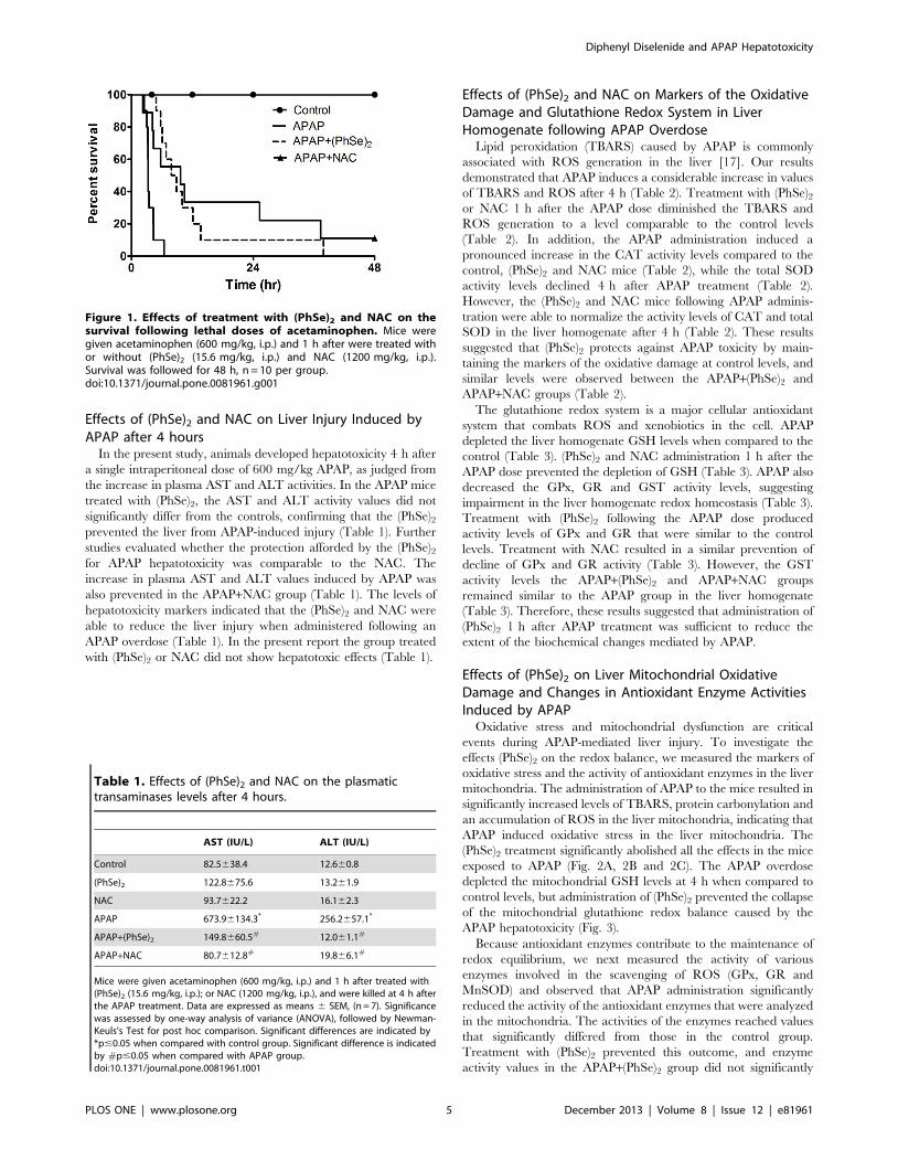
Mice were monitored for 48 h to determine the effects of
(PhSe)2 and NAC on the survival of mice following an APAP
overdose. The mice received 600 mg/kg APAP intraperitoneally
in a single dose. The APAP group mortality was pronounced when
compared to the control group, which was 100% in approximately
8 h (Fig. 1). Treatment with (PhSe)2 dramatically extended the
percent survival after the lethal APAP dose. All (PhSe)2 mice
receiving APAP survived up to 37.5 h after treatment. A similar
protection was reported following administration of NAC 1 h after
the acute APAP overdose (Fig. 1). The NAC mortality was 78%
compared to the control group. It is important to note that neither
the (PhSe)2 and NAC controls altered the mice survival during the
experimental period (data not shown).
Diphenyl Diselenide and APAP Hepatotoxicity
PLOS ONE | www.plosone.org 4 December 2013 | Volume 8 | Issue 12 | e81961
Effects of (PhSe)2 and NAC on Liver Injury Induced byAPAP after 4 hours
In the present study, animals developed hepatotoxicity 4 h after
a single intraperitoneal dose of 600 mg/kg APAP, as judged from
the increase in plasma AST and ALT activities. In the APAP mice
treated with (PhSe)2, the AST and ALT activity values did not
significantly differ from the controls, confirming that the (PhSe)2prevented the liver from APAP-induced injury (Table 1). Further
studies evaluated whether the protection afforded by the (PhSe)2for APAP hepatotoxicity was comparable to the NAC. The
increase in plasma AST and ALT values induced by APAP was
also prevented in the APAP+NAC group (Table 1). The levels of
hepatotoxicity markers indicated that the (PhSe)2 and NAC were
able to reduce the liver injury when administered following an
APAP overdose (Table 1). In the present report the group treated
with (PhSe)2 or NAC did not show hepatotoxic effects (Table 1).
Effects of (PhSe)2 and NAC on Markers of the OxidativeDamage and Glutathione Redox System in LiverHomogenate following APAP Overdose
Lipid peroxidation (TBARS) caused by APAP is commonly
associated with ROS generation in the liver [17]. Our results
demonstrated that APAP induces a considerable increase in values
of TBARS and ROS after 4 h (Table 2). Treatment with (PhSe)2or NAC 1 h after the APAP dose diminished the TBARS and
ROS generation to a level comparable to the control levels
(Table 2). In addition, the APAP administration induced a
pronounced increase in the CAT activity levels compared to the
control, (PhSe)2 and NAC mice (Table 2), while the total SOD
activity levels declined 4 h after APAP treatment (Table 2).
However, the (PhSe)2 and NAC mice following APAP adminis-
tration were able to normalize the activity levels of CAT and total
SOD in the liver homogenate after 4 h (Table 2). These results
suggested that (PhSe)2 protects against APAP toxicity by main-
taining the markers of the oxidative damage at control levels, and
similar levels were observed between the APAP+(PhSe)2 and
APAP+NAC groups (Table 2).
The glutathione redox system is a major cellular antioxidant
system that combats ROS and xenobiotics in the cell. APAP
depleted the liver homogenate GSH levels when compared to the
control (Table 3). (PhSe)2 and NAC administration 1 h after the
APAP dose prevented the depletion of GSH (Table 3). APAP also
decreased the GPx, GR and GST activity levels, suggesting
impairment in the liver homogenate redox homeostasis (Table 3).
Treatment with (PhSe)2 following the APAP dose produced
activity levels of GPx and GR that were similar to the control
levels. Treatment with NAC resulted in a similar prevention of
decline of GPx and GR activity (Table 3). However, the GST
activity levels the APAP+(PhSe)2 and APAP+NAC groups
remained similar to the APAP group in the liver homogenate
(Table 3). Therefore, these results suggested that administration of
(PhSe)2 1 h after APAP treatment was sufficient to reduce the
extent of the biochemical changes mediated by APAP.
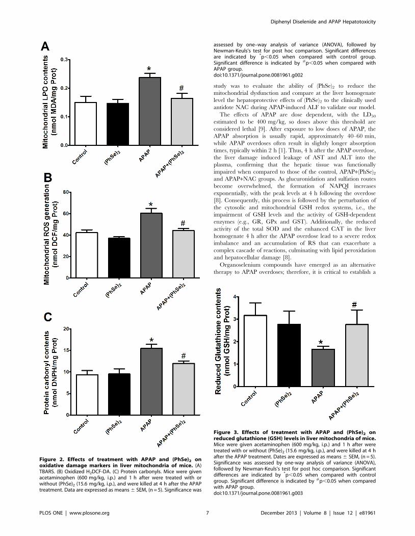
Effects of (PhSe)2 on Liver Mitochondrial OxidativeDamage and Changes in Antioxidant Enzyme ActivitiesInduced by APAP
Oxidative stress and mitochondrial dysfunction are critical
events during APAP-mediated liver injury. To investigate the
effects (PhSe)2 on the redox balance, we measured the markers of
oxidative stress and the activity of antioxidant enzymes in the liver
mitochondria. The administration of APAP to the mice resulted in
significantly increased levels of TBARS, protein carbonylation and
an accumulation of ROS in the liver mitochondria, indicating that
APAP induced oxidative stress in the liver mitochondria. The
(PhSe)2 treatment significantly abolished all the effects in the mice
exposed to APAP (Fig. 2A, 2B and 2C). The APAP overdose
depleted the mitochondrial GSH levels at 4 h when compared to
control levels, but administration of (PhSe)2 prevented the collapse
of the mitochondrial glutathione redox balance caused by the
APAP hepatotoxicity (Fig. 3).
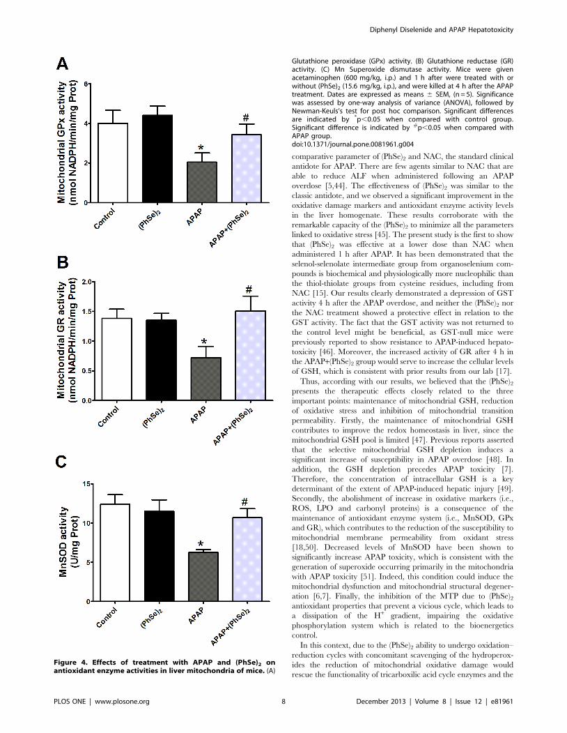
Because antioxidant enzymes contribute to the maintenance of
redox equilibrium, we next measured the activity of various
enzymes involved in the scavenging of ROS (GPx, GR and
MnSOD) and observed that APAP administration significantly
reduced the activity of the antioxidant enzymes that were analyzed
in the mitochondria. The activities of the enzymes reached values
that significantly differed from those in the control group.
Treatment with (PhSe)2 prevented this outcome, and enzyme
activity values in the APAP+(PhSe)2 group did not significantly
Figure 1. Effects of treatment with (PhSe)2 and NAC on thesurvival following lethal doses of acetaminophen. Mice weregiven acetaminophen (600 mg/kg, i.p.) and 1 h after were treated withor without (PhSe)2 (15.6 mg/kg, i.p.) and NAC (1200 mg/kg, i.p.).Survival was followed for 48 h, n = 10 per group.doi:10.1371/journal.pone.0081961.g001
Table 1. Effects of (PhSe)2 and NAC on the plasmatictransaminases levels after 4 hours.
AST (IU/L) ALT (IU/L)
Control 82.5638.4 12.660.8
(PhSe)2 122.8675.6 13.261.9
NAC 93.7622.2 16.162.3
APAP 673.96134.3* 256.2657.1*
APAP+(PhSe)2 149.8660.5# 12.061.1#
APAP+NAC 80.7612.8# 19.866.1#
Mice were given acetaminophen (600 mg/kg, i.p.) and 1 h after treated with(PhSe)2 (15.6 mg/kg, i.p.); or NAC (1200 mg/kg, i.p.), and were killed at 4 h afterthe APAP treatment. Data are expressed as means 6 SEM, (n = 7). Significancewas assessed by one-way analysis of variance (ANOVA), followed by Newman-Keuls’s Test for post hoc comparison. Significant differences are indicated by*p#0.05 when compared with control group. Significant difference is indicatedby #p#0.05 when compared with APAP group.doi:10.1371/journal.pone.0081961.t001
Diphenyl Diselenide and APAP Hepatotoxicity
PLOS ONE | www.plosone.org 5 December 2013 | Volume 8 | Issue 12 | e81961
differ from the control values (Fig. 4A, 4B and 4C). Therefore,
effects of (PhSe)2 on the antioxidant enzyme activities contributed
to the maintenance of the redox equilibrium in the liver
mitochondria.
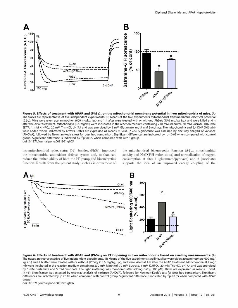
Effects of (PhSe)2 on APAP-Induced Liver MitochondrialDysfunction
Next, we analyzed the effects of (PhSe)2 on APAP-induced liver
mitochondria dysfunction. Because mitochondrial respiration and
ATP production depend on the transmembrane electrical
potential and mitochondrial membrane integrity, the Dym and
mitochondrial swelling were analyzed. A marked decrease of Dym
and considerable swelling were observed in the liver mitochondria
of the mice exposed to APAP compared to the control group.
Treatment with (PhSe)2 after APAP exposure prevented the loss of
Dym and prevented the mitochondrial swelling (Fig. 5 and Fig. 6,
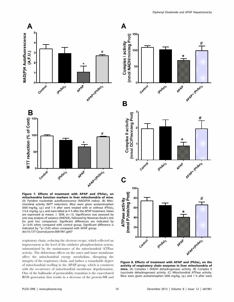
respectively). To determine whether APAP overdoses cause
changes in the mitochondrial bioenergetics function, the NAD(P)H
redox and mitochondrial activity were measured. APAP admin-
istration caused a significant decrease of mitochondrial NAD(P)H
redox status and mitochondrial activity, but (PhSe)2 treatment
following the APAP exposure prevented those effects (Fig. 7).
To further elucidate the mechanism by which APAP impairs the
mitochondrial bioenergetics function, the impact of the hepato-
toxicity was assessed with regard to the electron transport chain
(complex I and II) and the mitochondrial ATPase activity. The
activities of complex I (NADH dehydrogenase), complex II
(succinate dehydrogenase) and mitochondrial ATPase were
significantly reduced in the mice with APAP-induced liver injuries,
while the activity values of these enzymes did not significantly
differ from the control in the APAP+(PhSe)2 group (Fig. 8).
Because of the observed changes in the mitochondrial electron
transport chain, the mitochondrial aerobic capacity could also be
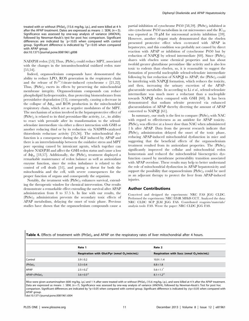
affected upon exposure to APAP. Therefore, the rates of
glutamate/pyruvate and succinate-supported O2 consumption in
liver mitochondria preparations were monitored (Table 4). APAP
administration caused a significant depletion of the rate of
mitochondrial oxygen consumption induced for the substrates of
complex I (glutamate and pyruvate) and the substrate of complex
II (succinate). The treatment with (PhSe)2 was able to restore the
oxygen consumption to values that did not significantly differ from
those in the control group (Table 4). Overall, these experiments
suggested that mitochondrial dysfunction plays a crucial role in
mitochondrial swelling, which is consistent with the changes that
were observed in the membrane potential, NADH redox state, and
mitochondrial activity and O2 consumption, indicating that
mitochondria undergo permeabilization following APAP-induced
hepatotoxicity. However, the treatment with (PhSe)2 even after 1 h
was able to reduce the mitochondrial dysfunction.
Discussion
(PhSe)2 delivers a hepatoprotective effect against APAP toxicity,
but the mechanism remains unclear [17]. The aim of the present
Table 2. Effects of (PhSe)2 and NAC on oxidative damage markers in liver homogenate after 4 hours.
TBARS (nmol MDA/mg Prot) ROS (mmol DCF/mg Prot) CAT (mmol H2O2/min/mg Prot)SOD(U/mg Prot)
Control 0.360.10 4.160.7 170.369.9 141.2611.4
(PhSe)2 0.260.03 4.961.1 186.9621.6 171.1615.8
NAC 0.360.03 3.560.7 149.8624.1 144.969.7
APAP 1.460.2* 8.860.3* 254.4613.9* 70.663.3*
APAP+(PhSe)2 0.860.1# 4.760.8# 190.3621.4# 113.567.3#
APAP+NAC 0.560.1# 4.360.9# 170.1629.4# 158.8617.3#
Mice were given acetaminophen (600 mg/kg, i.p.) and 1 h after treated with (PhSe)2 (15.6 mg/kg, i.p.); or NAC (1200 mg/kg, i.p.), and were killed at 4 h after the APAPtreatment. Data are expressed as means 6 SEM, (n = 7). Significance was assessed by one-way analysis of variance (ANOVA), followed by Newman-Keuls’s Test for posthoc comparison. Significant differences are indicated by *p#0.05 when compared with control group. Significant difference is indicated by #p#0.05 when comparedwith APAP group.doi:10.1371/journal.pone.0081961.t002
Table 3. Effects of (PhSe)2 and NAC on the Glutathione redox system in liver homogenate after 4 hours.
GSH (nmol GSH/mg Prot)GPx (nmol NADPH/min/mgProt)
GR (nmol NADPH/min/mgProt) GST (nmol CDNB/min/mg Prot)
Control 36.160.4 381.4634.0 28.163.1 566.1693.4
(PhSe)2 32.361.9 400.3624.7 29.863.1 588.2696.1
NAC 27.762.9 372.7647.2 32.165.2 658.5630.7
APAP 10.761.5* 147.7619.1* 15.362.1* 298.8620.7*
APAP+(PhSe)2 30.161.8# 365.0637.5# 30.663.3# 372.7626.3*
APAP+NAC 33.562.8# 335.1632.5# 32.163.8# 385.2634.6*
Mice were given acetaminophen (600 mg/kg, i.p.) and 1 h after treated with (PhSe)2 (15.6 mg/kg, i.p.); or NAC (1200 mg/kg, i.p.), and were killed at 4 h after the APAPtreatment. Data are expressed as means 6 SEM, (n = 7). Significance was assessed by one-way analysis of variance (ANOVA), followed by Newman-Keuls’s Test for posthoc comparison. Significant differences are indicated by *p#0.05 when compared with control group. Significant difference is indicated by #p#0.05 when comparedwith APAP group.doi:10.1371/journal.pone.0081961.t003
Diphenyl Diselenide and APAP Hepatotoxicity
PLOS ONE | www.plosone.org 6 December 2013 | Volume 8 | Issue 12 | e81961
study was to evaluate the ability of (PhSe)2 to reduce the
mitochondrial dysfunction and compare at the liver homogenate
level the hepatoprotective effects of (PhSe)2 to the clinically used
antidote NAC during APAP-induced ALF to validate our model.
The effects of APAP are dose dependent, with the LD50
estimated to be 400 mg/kg, so doses above this threshold are
considered lethal [9]. After exposure to low doses of APAP, the
APAP absorption is usually rapid, approximately 40–60 min,
while APAP overdoses often result in slightly longer absorption
times, typically within 2 h [1]. Thus, 4 h after the APAP overdose,
the liver damage induced leakage of AST and ALT into the
plasma, confirming that the hepatic tissue was functionally
impaired when compared to those of the control, APAP+(PhSe)2and APAP+NAC groups. As glucuronidation and sulfation routes
become overwhelmed, the formation of NAPQI increases
exponentially, with the peak levels at 4 h following the overdose
[8]. Consequently, this process is followed by the perturbation of
the cytosolic and mitochondrial GSH redox systems, i.e., the
impairment of GSH levels and the activity of GSH-dependent
enzymes (e.g., GR, GPx and GST). Additionally, the reduced
activity of the total SOD and the enhanced CAT in the liver
homogenate 4 h after the APAP overdose lead to a severe redox
imbalance and an accumulation of RS that can exacerbate a
complex cascade of reactions, culminating with lipid peroxidation
and hepatocellular damage [8].
Organoselenium compounds have emerged as an alternative
therapy to APAP overdoses; therefore, it is critical to establish a
Figure 2. Effects of treatment with APAP and (PhSe)2 onoxidative damage markers in liver mitochondria of mice. (A)TBARS. (B) Oxidized H2DCF-DA. (C) Protein carbonyls. Mice were givenacetaminophen (600 mg/kg, i.p.) and 1 h after were treated with orwithout (PhSe)2 (15.6 mg/kg, i.p.), and were killed at 4 h after the APAPtreatment. Data are expressed as means 6 SEM, (n = 5). Significance was
assessed by one–way analysis of variance (ANOVA), followed byNewman-Keuls’s test for post hoc comparison. Significant differencesare indicated by *p,0.05 when compared with control group.Significant difference is indicated by #p,0.05 when compared withAPAP group.doi:10.1371/journal.pone.0081961.g002
Figure 3. Effects of treatment with APAP and (PhSe)2 onreduced glutathione (GSH) levels in liver mitochondria of mice.Mice were given acetaminophen (600 mg/kg, i.p.) and 1 h after weretreated with or without (PhSe)2 (15.6 mg/kg, i.p.), and were killed at 4 hafter the APAP treatment. Dates are expressed as means 6 SEM, (n = 5).Significance was assessed by one-way analysis of variance (ANOVA),followed by Newman-Keuls’s test for post hoc comparison. Significantdifferences are indicated by *p,0.05 when compared with controlgroup. Significant difference is indicated by #p,0.05 when comparedwith APAP group.doi:10.1371/journal.pone.0081961.g003
Diphenyl Diselenide and APAP Hepatotoxicity
PLOS ONE | www.plosone.org 7 December 2013 | Volume 8 | Issue 12 | e81961
comparative parameter of (PhSe)2 and NAC, the standard clinical
antidote for APAP. There are few agents similar to NAC that are
able to reduce ALF when administered following an APAP
overdose [5,44]. The effectiveness of (PhSe)2 was similar to the
classic antidote, and we observed a significant improvement in the
oxidative damage markers and antioxidant enzyme activity levels
in the liver homogenate. These results corroborate with the
remarkable capacity of the (PhSe)2 to minimize all the parameters
linked to oxidative stress [45]. The present study is the first to show
that (PhSe)2 was effective at a lower dose than NAC when
administered 1 h after APAP. It has been demonstrated that the
selenol-selenolate intermediate group from organoselenium com-
pounds is biochemical and physiologically more nucleophilic than
the thiol-thiolate groups from cysteine residues, including from
NAC [15]. Our results clearly demonstrated a depression of GST
activity 4 h after the APAP overdose, and neither the (PhSe)2 nor
the NAC treatment showed a protective effect in relation to the
GST activity. The fact that the GST activity was not returned to
the control level might be beneficial, as GST-null mice were
previously reported to show resistance to APAP-induced hepato-
toxicity [46]. Moreover, the increased activity of GR after 4 h in
the APAP+(PhSe)2 group would serve to increase the cellular levels
of GSH, which is consistent with prior results from our lab [17].
Thus, according with our results, we believed that the (PhSe)2presents the therapeutic effects closely related to the three
important points: maintenance of mitochondrial GSH, reduction
of oxidative stress and inhibition of mitochondrial transition
permeability. Firstly, the maintenance of mitochondrial GSH
contributes to improve the redox homeostasis in liver, since the
mitochondrial GSH pool is limited [47]. Previous reports asserted
that the selective mitochondrial GSH depletion induces a
significant increase of susceptibility in APAP overdose [48]. In
addition, the GSH depletion precedes APAP toxicity [7].
Therefore, the concentration of intracellular GSH is a key
determinant of the extent of APAP-induced hepatic injury [49].
Secondly, the abolishment of increase in oxidative markers (i.e.,
ROS, LPO and carbonyl proteins) is a consequence of the
maintenance of antioxidant enzyme system (i.e., MnSOD, GPx
and GR), which contributes to the reduction of the susceptibility to
mitochondrial membrane permeability from oxidant stress
[18,50]. Decreased levels of MnSOD have been shown to
significantly increase APAP toxicity, which is consistent with the
generation of superoxide occurring primarily in the mitochondria
with APAP toxicity [51]. Indeed, this condition could induce the
mitochondrial dysfunction and mitochondrial structural degener-
ation [6,7]. Finally, the inhibition of the MTP due to (PhSe)2antioxidant properties that prevent a vicious cycle, which leads to
a dissipation of the H+ gradient, impairing the oxidative
phosphorylation system which is related to the bioenergetics
control.
In this context, due to the (PhSe)2 ability to undergo oxidation–
reduction cycles with concomitant scavenging of the hydroperox-
ides the reduction of mitochondrial oxidative damage would
rescue the functionality of tricarboxilic acid cycle enzymes and the
Figure 4. Effects of treatment with APAP and (PhSe)2 onantioxidant enzyme activities in liver mitochondria of mice. (A)
Glutathione peroxidase (GPx) activity. (B) Glutathione reductase (GR)activity. (C) Mn Superoxide dismutase activity. Mice were givenacetaminophen (600 mg/kg, i.p.) and 1 h after were treated with orwithout (PhSe)2 (15.6 mg/kg, i.p.), and were killed at 4 h after the APAPtreatment. Dates are expressed as means 6 SEM, (n = 5). Significancewas assessed by one-way analysis of variance (ANOVA), followed byNewman-Keuls’s test for post hoc comparison. Significant differencesare indicated by *p,0.05 when compared with control group.Significant difference is indicated by #p,0.05 when compared withAPAP group.doi:10.1371/journal.pone.0081961.g004
Diphenyl Diselenide and APAP Hepatotoxicity
PLOS ONE | www.plosone.org 8 December 2013 | Volume 8 | Issue 12 | e81961
intramitochondrial redox status [52], besides, (PhSe)2 improved
the mitochondrial antioxidant defense system and, so that can
reduce the limited ability of both the H+ pump and bioenergetics
function. Results from the present study, such as improvement of
the mitochondrial bioenergetics function (Dym, mitochondrial
activity and NAD(P)H redox status) and normalization of oxygen
consumption at sites 1 (glutamate/pyruvate) and 2 (succinate)
supports the idea of an improved energy coupling of the
Figure 5. Effects of treatment with APAP and (PhSe)2 on the mitochondrial membrane potential in liver mitochondria of mice. (A)The traces are representative of five independent experiments. (B) Means of the five experiments mitochondrial transmembrane electrical potential(Dym). Mice were given acetaminophen (600 mg/kg, i.p.) and 1 h after were treated with or without (PhSe)2 (15.6 mg/kg, i.p.), and were killed at 4 hafter the APAP treatment. Mitochondria (0.5 mg/ml) were incubated in the reaction medium containing 230 mM Mannitol, 70 mM Sucrose, 0.02 mMEDTA, 1 mM K2HPO4, 20 mM Tris-HCl, pH 7.4 and was energized by 5 mM Glutamate and 5 mM Succinate. The mitochondria and 2,4 DNP (100 mM)were added where indicated by arrows. Dates are expressed as means 6 SEM, (n = 5). Significance was assessed by one-way analysis of variance(ANOVA), followed by Newman-Keuls’s test for post hoc comparison. Significant differences are indicated by *p,0.05 when compared with controlgroup. Significant difference is indicated by #p,0.05 when compared with APAP group.doi:10.1371/journal.pone.0081961.g005
Figure 6. Effects of treatment with APAP and (PhSe)2 on PTP opening in liver mitochondria based on swelling measurements. (A)The traces are representative of five independent experiments. (B) Means of the five experiments swelling. Mice were given acetaminophen (600 mg/kg, i.p.) and 1 h after were treated with or without (PhSe)2 (15.6 mg/kg, i.p.), and were killed at 4 h after the APAP treatment. Mitochondria (0.1 mg/ml) were incubated in the reaction medium containing 230 mM Mannitol, 70 mM Sucrose, 1 mM K2HPO4, 20 mM Tris-HCl, pH 7.4 and was energizedby 5 mM Glutamate and 5 mM Succinate. The light scattering was monitored after adding CaCl2 (100 mM). Dates are expressed as means 6 SEM,(n = 5). Significance was assessed by one-way analysis of variance (ANOVA), followed by Newman-Keuls’s test for post hoc comparison. Significantdifferences are indicated by *p,0.05 when compared with control group. Significant difference is indicated by #p,0.05 when compared with APAPgroup.doi:10.1371/journal.pone.0081961.g006
Diphenyl Diselenide and APAP Hepatotoxicity
PLOS ONE | www.plosone.org 9 December 2013 | Volume 8 | Issue 12 | e81961
respiratory chain, reducing the electron escape, which reflected an
improvement at the level of the oxidative phosphorylation system,
substantiated by the maintenance of the mitochondrial ATPase
activity. The deleterious effects on the outer and inner membrane
affect the mitochondrial energy metabolism, disrupting the
integrity of the respiratory chain, and induce a remarkable degree
of mitochondrial swelling in the APAP group, which is consistent
with the occurrence of mitochondrial membrane depolarization.
One of the hallmarks of permeability transition is the exacerbated
ROS generation that results in a decrease of the protein-SH and
Figure 7. Effects of treatment with APAP and (PhSe)2 onmitochondria function markers in liver mitochondria of mice.(A) Pyridine nucleotide autofluorescence (NAD(P)H redox). (B) Mito-chondrial activity (MTT reduction). Mice were given acetaminophen(600 mg/kg, i.p.) and 1 h after were treated with or without (PhSe)2
(15.6 mg/kg, i.p.), and were killed at 4 h after the APAP treatment. Datesare expressed as means 6 SEM, (n = 5). Significance was assessed byone-way analysis of variance (ANOVA), followed by Newman-Keuls’s testfor post hoc comparison. Significant differences are indicated by*p,0.05 when compared with control group. Significant difference isindicated by #p,0.05 when compared with APAP group.doi:10.1371/journal.pone.0081961.g007
Figure 8. Effects of treatment with APAP and (PhSe)2 on theactivity of respiratory chain enzymes in liver mitochondria ofmice. (A) Complex I (NADH dehydrogenase) activity. (B) Complex II(succinate dehydrogenase) activity. (C) Mitochondrial ATPase activity.Mice were given acetaminophen (600 mg/kg, i.p.) and 1 h after were
Diphenyl Diselenide and APAP Hepatotoxicity
PLOS ONE | www.plosone.org 10 December 2013 | Volume 8 | Issue 12 | e81961
NAD(P)H redox [53].Thus, (PhSe)2 could reduce MPT, associated
with the changes in the intramitochondrial oxidized redox state
[53,54].
Indeed, organoselenium compounds have demonstrated the
ability to reduce LPO, ROS generation in the respiratory chain
and the release of Fe2+/citrate-induced cytochrome c [21,22].
Thus, (PhSe)2 exerts its effects by preserving the mitochondrial
membrane integrity. Organoselenium compounds can reduce
phospholipid hydroperoxides, thus protecting biomembranes from
peroxidative degradation [15], consequently, causing a decrease in
the collapse of Dym and ROS production in the mitochondrial
respiratory chain, which act as negative modulators of the MPT.
The mechanism of action involved in the hepatoprotective effect of
(PhSe)2 is related to its thiol peroxidase-like activity, i.e., its ability
to react with peroxide after its transformation to the selenol-
selenolate intermediate via either a direct interaction with GSH or
another reducing thiol or by its reduction via NADPH-catalyzed
thioredoxin reductase activity [55,56]. The mitochondrial dys-
function is a consequence during the ALF induced by APAP and
there is an interrelationship between the oxidative stress and MPT
pore opening caused by intoxicant agents, which together can
deplete NAD(P)H and affect the GSH redox status and cause a loss
of Dym [18,57]. Additionally, the (PhSe)2 treatment displayed a
remarkable maintenance of redox balance as well as antioxidant
enzyme function, since the redox imbalance is related to the
control of cell death [51], and posing a threat for both the
mitochondria and the cell, with severe consequences for the
proper function of organs and consequently the organism.
Notably, the treatment with (PhSe)2 enhances survival, extend-
ing the therapeutic window for chemical intervention. Our results
demonstrate a remarkable effect extending the survival after APAP
administration from 8 to 37.5 h. In line with our results, the
(PhSe)2 administration prevents the secondary toxic effects of
APAP metabolism, delaying the onset of toxic phase. Previous
studies have shown that the organoselenium compounds cause a
partial inhibition of cytochrome P450 [58,59]. (PhSe)2 inhibited in
vitro cytochrome P450 metabolism in rat microsomes and the IC50
was reported as 78 mM for microsomal activity inhibition [59].
However, another elegant study demonstrated that the ebselen
presented protective effect when co-treated with APAP in
hepatocytes, and this condition was probably not caused by direct
reaction with APAP or inhibition of cytochrome P450 but by
reduction of NAPQI by selenol intermediate [60]. Since (PhSe)2shares with ebselen some chemical properties and has about
twofold greater glutathione peroxidase–like activity and is also less
toxic to rodents than ebselen, so, it is reasonable to suggest the
formation of powerful nucleophile selenol-selenolate intermediate
following by fast reduction of NAPQI to APAP, the (PhSe)2 could
be interfering with NAPQI formation, which reduces the toxicity,
and then, increasing the urinary excretion of the APAP-
glucuronide metabolite. In according to Li et al., selenol-selenolate
intermediate was much more a reductant than a nucleophile
towards NAPQI when compared with GSH [60]. It has been
demonstrated that sodium selenite protected via enhanced
glucuronidation of APAP thereby diverting the amount of APAP
converted to NAPQI [61].
In summary, our study is the first to compare (PhSe)2 with NAC
with regard to effectiveness as an antidote for APAP toxicity.
(PhSe)2 was effective at a lower dose than NAC when administered
1 h after APAP. Data from the present research indicate that
(PhSe)2 administration delayed the onset of the toxic phase,
reducing APAP-induced mitochondrial dysfunction in mice and
suggesting that the beneficial effects of the organoselenium
treatment resulted from its antioxidant properties. The (PhSe)2significantly improved the cellular and mitochondrial redox
homeostasis and reduced the mitochondrial bioenergetics dys-
function caused by membrane permeability transition associated
with APAP overdose. These results may help to better understand
the role of mitochondrial dysfunction in APAP hepatotoxicity and
support the possibility that organoselenium (PhSe)2 could be used
as an adjuvant therapy to protect the liver from APAP-induced
injuries.
Author Contributions
Conceived and designed the experiments: NRC FAS JGG CLDC.
Performed the experiments: NRC EFdR MHdS CCT. Analyzed the data:
NRC CLDC SCP JLM JGG FAS. Contributed reagents/materials/
analysis tools: FAS. Wrote the paper: NRC CLDC JGG FAS.
treated with or without (PhSe)2 (15.6 mg/kg, i.p.), and were killed at 4 hafter the APAP treatment. Dates are expressed as means 6 SEM, (n = 5).Significance was assessed by one-way analysis of variance (ANOVA),followed by Newman-Keuls’s test for post hoc comparison. Significantdifferences are indicated by *p,0.05 when compared with controlgroup. Significant difference is indicated by #p,0.05 when comparedwith APAP group.doi:10.1371/journal.pone.0081961.g008
Table 4. Effects of treatment with (PhSe)2 and APAP on the respiratory rates of liver mitochondrial after 4 hours.
Rate 1 Rate 2
Respiration with Glut/Pyr (nmol O2/min/mL) Respiration with Succ (nmol O2/min/mL)
Control 3.960.2 10.961.4
(PhSe)2 3.360.4 8.861.8
APAP 2.560.2* 5.661.1*
APAP+(PhSe)2 3.660.5# 8.161.2#
Mice were given acetaminophen (600 mg/kg, i.p.) and 1 h after were treated with or without (PhSe)2 (15.6 mg/kg, i.p.), and were killed at 4 h after the APAP treatment.Data are expressed as means 6 SEM, (n = 7). Significance was assessed by one-way analysis of variance (ANOVA), followed by Newman-Keuls’s Test for post hoccomparison. Significant differences are indicated by *p#0.05 when compared with control group. Significant difference is indicated by #p#0.05 when compared withAPAP group.doi:10.1371/journal.pone.0081961.t004
Diphenyl Diselenide and APAP Hepatotoxicity
PLOS ONE | www.plosone.org 11 December 2013 | Volume 8 | Issue 12 | e81961
References
1. Larson AM, Polson J, Fontana RJ, Davern TJ, Lalani E, et al. (2005)
Acetaminophen-induced acute liver failure: results of a United Statesmulticenter, prospective study. Hepatology 42: 1364–1372.
2. Nourjah P, Ahmad SR, Karwoski C, Willy M (2006) Estimates of
acetaminophen (paracetamol)-associated overdoses in the United States.
Pharmacoepidemiology and Drug Safety 15: 398–405.3. de Achaval S, Suarez-Almazor M (2011) Acetaminophen overdose: a little
recognized public health threat. Pharmacoepidemiology and Drug Safety 20:
827–829.
4. Moyer AM, Fridley BL, Jenkins GD, Batzler AJ, Pelleymounter LL, et al. (2011)Acetaminophen-NAPQI hepatotoxicity: a cell line model system genome-wide
association study. Toxicological Sciences 120: 33–41.
5. Brown JM, Ball JG, Hogsett A, Williams T, Valentovic M (2010) Temporalstudy of acetaminophen (APAP) and S-adenosyl-L-methionine (SAMe) effects on
subcellular hepatic SAMe levels and methionine adenosyltransferase (MAT)expression and activity. Toxicology and Applied Pharmacology 247: 1–9.
6. Jaeschke H (1990) Glutathione disulfide formation and oxidant stress during
acetaminophen-induced hepatotoxicity in mice in vivo: the protective effect ofallopurinol. Journal of Pharmacology and Experimental Therapeutics 255: 935–
941.
7. Jaeschke H, Bajt ML (2006) Intracellular signaling mechanisms of acetamino-
phen-induced liver cell death. Toxicological Sciences 89: 31–41.8. Jaeschke H, McGill MR, Ramachandran A (2012) Oxidant stress, mitochondria,
and cell death mechanisms in drug-induced liver injury: lessons learned from
acetaminophen hepatotoxicity. Drug Metabolism Reviews 44: 88–106.
9. Chan KM, Han XD, Kan YW (2001) An important function of Nrf 2 incombating oxidative stress: Detoxification of acetaminophen. Proceedings of the
National Academy of Sciences of the United States of America 98: 4611–4616.
10. San-Miguel B, Alvarez M, Culebras JM, Gonzalez-Gallego J, Tunon MJ (2006)N-acetyl-cysteine protects liver from apoptotic death in an animal model of
fulminant hepatic failure. Apoptosis 11: 1945–1957.
11. Woodhead JL, Howell BA, Yang Y, Harrill AH, Clewell HJ 3rd, et al. (2012) Ananalysis of N-acetylcysteine treatment for acetaminophen overdose using a
systems model of drug-induced liver injury. The Journal of Pharmacology and
Experimental Therapeutics 342: 529–540.12. Meotti FC, Stangherlin EC, Zeni G, Nogueira CW, Rocha JBT (2004)
Protective role of aryl and alkyl diselenides on lipid peroxidation. Environmental
Research 94: 276–282.
13. Brandao R, Santos FW, Oliveira R, Roman SS, Nogueira CW (2009)Involvement of non-enzymatic antioxidant defenses in the protective effect of
diphenyl diselenide on testicular damage induced by cadmium in mice. Journalof Trace Elements in Medicine and Biology 23: 324–333.
14. Borges LP, Nogueira CW, Panatieri RB, Rocha JBT, Zeni G (2006) Acute liver
damage induced by 2-nitropropane in rats: Effect of diphenyl diselenide onantioxidant defenses. Chemico-Biological Interactions 160: 99–107.
15. Nogueira CW, Zeni G, Rocha JBT (2004) Organoselenium and organotellurium
compounds: Toxicology and pharmacology. Chemical Reviews 104: 6255–
6285.16. Da Silva MH, Da Rosa EJ, De Carvalho NR, Dobrachinski F, Da Rocha JB,
et al. (2011) Acute Brain Damage Induced by Acetaminophen in Mice: Effect of
Diphenyl Diselenide on Oxidative Stress and Mitochondrial Dysfunction.Neurotoxicity research 21: 334–344.
17. da Rosa EJ, da Silva MH, Carvalho NR, Bridi JC, da Rocha JB, et al. (2012)
Reduction of acute hepatic damage induced by acetaminophen after treatmentwith diphenyl diselenide in mice. Toxicologic Pathology 40: 605–613.
18. Bajt ML, Ramachandran A, Yan HM, Lebofsky M, Farhood A, et al. (2011)
Apoptosis-inducing factor modulates mitochondrial oxidant stress in acetamin-ophen hepatotoxicity. Toxicological Sciences 122: 598–605.
19. Votyakova TV, Reynolds IJ (2005) Ca2+-induced permeabilization promotes
free radical release from rat brain mitochondria with partially inhibited complexI. Journal of Neurochemistry 93: 526–537.
20. Kim JY, Park JH (2003) ROS-dependent caspase-9 activation in hypoxic cell
death. Febs Letters 549: 94–98.
21. Boireau A, Dubedat P, Bordier F, Coimbra M, Meunier M, et al. (1999) Effectsof ebselen, a glutathione peroxidase mimic, in several models of mitochondrial
dysfunction. Annals of the New York Academy of Sciences 893: 254–257.
22. Boireau A, Marechal PM, Meunier M, Dubedat P, Moussaoui S (2000) The
anti-oxidant ebselen antagonizes the release of the apoptogenic factorcytochrome c induced by Fe2+/citrate in rat liver mitochondria. Neuroscience
letters 289: 95–98.
23. Bhattacharya SK, Thakar JH, Johnson PL, Shanklin DR (1991) Isolation ofSkeletal-Muscle Mitochondria from Hamsters Using an Ionic Medium
Containing Ethylenediarninetetraacetic Acid and Nagarse. Analytical Biochem-istry 192: 344–349.
24. Kruglov AG, Teplova VV, Saris NE (2007) The effect of the lipophilic cation
lucigenin on mitochondria depends on the site of its reduction. Biochemical
Pharmacology 74: 545–556.25. Ohkawa H, Ohishi N, Yagi K (1979) Assay for Lipid Peroxides in Animal-
Tissues by Thiobarbituric Acid Reaction. Analytical Biochemistry 95: 351–358.
26. Dionisio N, Garcia-Mediavilla MV, Sanchez-Campos S, Majano PL, Benedicto
I, et al. (2009) Hepatitis C virus NS5A and core proteins induce oxidative
stress-mediated calcium signalling alterations in hepatocytes. Journal ofHepatology 50: 872–882.
27. Hissin PJ, Hilf R (1976) Fluorometric Method for Determination of Oxidized
and Reduced Glutathione in Tissues. Analytical Biochemistry 74: 214–226.
28. Misra HP, Fridovich I (1972) The generation of superoxide radical during the
autoxidation of hemoglobin. Journal of Biological Chemistry 247: 6960–6962.
29. Aebi H (1984) Catalase in vitro. Methods in enzymology 105: 121–126.
30. Habig WH, Pabst MJ, Jakoby WB (1974) Glutathione S-transferases. The firstenzymatic step in mercapturic acid formation. The Journal of Biological
Chemistry 249: 7130–7139.
31. Flohe L, Gunzler WA (1984) Assays of glutathione peroxidase. Methods inEnzymology 105: 114–121.
32. Carlberg I, Mannervik B (1985) Glutathione reductase. Methods in Enzymology
113: 484–490.
33. Levine RL, Garland D, Oliver CN, Amici A, Climent I, et al. (1990)
Determination of Carbonyl Content in Oxidatively Modified Proteins. Methodsin Enzymology 186: 464–478.
34. Akerman KEO, Wikstrom MKF (1976) Safranine as a Probe of Mitochondrial-
Membrane Potential. Febs Letters 68: 191–197.
35. Bernas T, Dobrucki J (2002) Mitochondrial and nonmitochondrial reduction ofMTT: interaction of MTT with TMRE, JC-1, and NAO mitochondrial
fluorescent probes. Cytometry 47: 236–242.
36. Geller BL, Winge DR (1984) Subcellular distribution of superoxide dismutases inrat liver. Methods in Enzymology 105: 105–114.
37. Bottje W, Iqbal M, Tang ZX, Cawthon D, Okimoto R, et al. (2002) Association
of mitochondrial function with feed efficiency within a single genetic line of male
broilers. Poultry science 81: 546–555.
38. Galante YM, Hatefi Y (1978) Resolution of complex I and isolation of NADHdehydrogenase and an iron—sulfur protein. Methods in Enzymology 53: 15–21.
39. Fischer JC, Ruitenbeek W, Berden JA, Trijbels JM, Veerkamp JH, et al. (1985)
Differential investigation of the capacity of succinate oxidation in human skeletalmuscle. Clinica Chimica Acta 153: 23–36.
40. Morin C, Zini R, Simon N, Charbonnier P, Tillement JP, et al. (2000) Low
glucocorticoid concentrations decrease oxidative phosphorylation of isolated rat
brain mitochondria: an additional effect of dexamethasone. Fundamental &Clinical Pharmacology 14: 493–500.
41. Katyare SS, Satav JG (1989) Impaired mitochondrial oxidative energy
metabolism following paracetamol-induced hepatotoxicity in the rat. BritishJournal of Pharmacology 96: 51–58.
42. Atkinson A, Gatenby AD, Lowe AG (1973) The determination of inorganic
orthophosphate in biological systems. Biochimica Et Biophysica Acta 320: 195–204.
43. Bradford MM (1976) A rapid and sensitive method for the quantitation of
microgram quantities of protein utilizing the principle of protein-dye binding.
Analytical Biochemistry 72: 248–254.
44. McGill MR, Williams CD, Xie Y, Ramachandran A, Jaeschke H (2012)Acetaminophen-induced liver injury in rats and mice: comparison of protein
adducts, mitochondrial dysfunction, and oxidative stress in the mechanism oftoxicity. Toxicology and Applied Pharmacology 264: 387–394.
45. Nogueira CW, Rocha JB (2011) Toxicology and pharmacology of selenium:
emphasis on synthetic organoselenium compounds. Archives of Toxicology 85:
1313–1359.
46. Arakawa S, Maejima T, Fujimoto K, Yamaguchi T, Yagi M, et al. (2012)Resistance to acetaminophen-induced hepatotoxicity in glutathione S-transfer-
ase Mu 1-null mice. Journal of Toxicological Sciences 37: 595–605.
47. Femandez-Checa JC, Kaplowitzc N (2005) Hepatic mitochondrial glutathione:transport and role in disease and toxicity. Toxicology and Applied Pharmacol-
ogy 204: 263–273.
48. Zhao P, Kalhorn TF, Slattery JT (2002) Selective mitochondrial glutathione
depletion by ethanol enhances acetaminophen toxicity in rat liver. Hepatology36: 326–335.
49. Vendemiale G, Grattagliano I, Altomare E, Turturro N, Guerrieri F (1996)
Effect of acetaminophen administration on hepatic glutathione compartmenta-tion and mitochondrial energy metabolism in the rat. Biochemical Pharmacol-
ogy 52: 1147–1154.
50. Hong SW, Lee HS, Jung KH, Lee H, Hong SS (2012) Protective Effect ofFucoidan against Acetaminophen-Induced Liver Injury. Archives of Pharmacal
Research 35: 1099–1105.
51. Ramachandran A, Lebofsky M, Weinman SA, Jaeschke H (2011) The impact of
partial manganese superoxide dismutase (SOD2)-deficiency on mitochondrialoxidant stress, DNA fragmentation and liver injury during acetaminophen
hepatotoxicity. Toxicology and Applied Pharmacology 251: 226–233.
52. Raghavendran HRB, Sathivel A, Devaki T (2005) Antioxidant effect ofSargassum polycystum (Phaeophyceae) against acetaminophen induced changes
in hepatic mitochondrial enzymes during toxic hepatitis. Chemosphere 61: 276–
281.
53. Puntel RL, Roos DH, Folmer V, Nogueira CW, Galina A, et al. (2010)Mitochondrial dysfunction induced by different organochalchogens is mediated
by thiol oxidation and is not dependent of the classical mitochondrialpermeability transition pore opening. Toxicological Sciences 117: 133–143.
Diphenyl Diselenide and APAP Hepatotoxicity
PLOS ONE | www.plosone.org 12 December 2013 | Volume 8 | Issue 12 | e81961
54. Morin D, Zini R, Ligeret H, Neckameyer W, Labidalle S, et al. (2003) Dual
effect of ebselen on mitochondrial permeability transition. BiochemicalPharmacology 65: 1643–1651.
55. de Freitas AS, Rocha JBT (2011) Diphenyl diselenide and analogs are substrates
of cerebral rat thioredoxin reductase: A pathway for their neuroprotectiveeffects. Neuroscience Letters 503: 1–5.
56. de Freitas AS, Prestes AD, Wagner C, Sudati JH, Alves D, et al. (2010)Reduction of Diphenyl Diselenide and Analogs by Mammalian Thioredoxin
Reductase Is Independent of Their Gluthathione Peroxidase-Like Activity: A
Possible Novel Pathway for Their Antioxidant Activity. Molecules 15: 7699–7714.
57. Ueda S, Masutani H, Nakamura H, Tanaka T, Ueno M, et al. (2002) Redoxcontrol of cell death. Antioxidants & Redox Signaling 4: 405–414.
58. Kuhn-Velten N, Sies H (1989) Optical spectral studies of ebselen interaction
with cytochrome P-450 of rat liver microsomes. Biochemical Pharmacology 38:
619–625.
59. Prigol M, Nogueira CW, Zeni G, Bronze MR, Constantino L (2012) In vitro
metabolism of diphenyl diselenide in rat liver fractions. Conjugation with GSH
and binding to thiol groups. Chemico-Biological Interactions 200: 65–72.
60. Li Q J, Bessems JG, Commandeur JN, Adams B, Vermeulen NP (1994)
Mechanism of protection of ebselen against paracetamol-induced toxicity in rat
hepatocytes. Biochemical Pharmacology 48: 1631–1640.
61. Schnell RC, Park KS, Davies MH, Merrick BA, Weir SW (1988) Protective
effects of selenium on acetaminophen-induced hepatotoxicity in the rat. Toxicol
Appl Pharmacol 95: 1–11.
Diphenyl Diselenide and APAP Hepatotoxicity
PLOS ONE | www.plosone.org 13 December 2013 | Volume 8 | Issue 12 | e81961


![Paracetamol(acetaminophen)ornon-steroidalanti ... (acetaminophen) … · [Intervention Review] Paracetamol (acetaminophen) or non-steroidal anti-inflammatory drugs, alone or combined,](https://img.pdfslide.net/doc/110x75/5f387cbd43d51a2eb45648f8/paracetamolacetaminophenornon-steroidalanti-acetaminophen-intervention.jpg)