Embed Size (px)
Citation preview
Send Orders for Reprints to [email protected]
190 The Open Dentistry Journal, 2015, 9, (Suppl 1: M4) 190-196
1874-2106/15 2015 Bentham Open
Open Access
Iatrogenic Damage to the Periodontium Caused by Fixed Prosthodontic Treatment Procedures
PV Harish1, Sonila Anne Joseph
2,*, Syed Sirajuddin
3, Veenadharini Gundapaneni
3,
Sachidananda Chungkham3 and Ambica
3
1Department of Prosthodontics, Rajarajeswari Dental College & Hospital, Bangalore, Karnataka, India;
2Dental
Art Clinic, Mareena towers, Coastal Road, Mahaboula, Kuwait; 3Department of Periodontology, Rajarajeswari Dental
College & Hospital, Bangalore-560074, Karnataka, India
Abstract: Missing teeth should be replaced as soon as possible to maintain arch integrity and thereby avoid both mor-
phologic and functional derangements in the occlusion. Otherwise, changes occur that upset the masticatory system, such
as extrusion of the teeth opposing the edentulous areas along with their alveolar housing, their supporting tissues and ul-
timately the maxillary sinus. Concurrently with extrusion, shifting of the interproximal contacts and migration of the adja-
cent teeth occur, thereby impairing function and causing disharmony. Good oral health cannot be achieved when changes
in tooth position alter the coronal contour and occlusion interfering with mutual support, which encourages food impac-
tion and retention, further leading to osseous defects.
Keywords: Crown, fixed prosthodontic treatment, iatrogenic damage, periodontium, fpd.
INTRODUCTION
Teeth are prepared to receive crowns and restorations, and these preparations must be based on important doctrine from which basic criterion can be established to aid in calcu-lating the achievement of prosthodontics management.
Careful attention to every detail is imperative during tooth preparation.
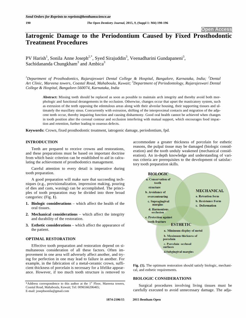
A good preparation will make sure that succeeding tech-niques (e.g., provisionalization, impression making, pouring of dies and casts, waxing) can be accomplished. The princi-ples of tooth preparation may be divided into three broad categories: (Fig. 1).
1. Biologic considerations – which affect the health of the oral tissues.
2. Mechanical considerations – which affect the integrity and durability of the restoration.
3. Esthetic considerations – which affect the appearance of the patient.
OPTIMAL RESTORATION
Effective tooth preparation and restoration depend on si-multaneous consideration of all these factors. Often im-provement in one area will adversely affect another, and try-ing for perfection in one may lead to failure in another. For example, in the fabrication of a metal-ceramic crown, suffi-cient thickness of porcelain is necessary for a lifelike appear-ance. However, if too much tooth structure is removed to
*Address correspondence to this author at the 1st Floor, Mareena towers,
Coastal Road, Mahaboula, Kuwait; Tel: 0096566206465;
E-mail: [email protected]
accommodate a greater thickness of porcelain for esthetic reasons, the pulpal tissue may be damaged (biologic consid-eration) and the tooth unduly weakened (mechanical consid-eration). An in-depth knowledge and understanding of vari-ous criteria are prerequisites to the development of satisfac-tory tooth preparation skills.
Fig. (1). The optimum restoration should satisfy biologic, mechani-
cal, and esthetic requirements.
BIOLOGIC CONSIDERATIONS
Surgical procedures involving living tissues must be
carefully executed to avoid unnecessary damage. The adja-
Iatrogenic Damage to the Periodontium by Fixed Prosthodontic Procedures The Open Dentistry Journal, 2015, Volume 9 191
cent teeth, soft tissues, and the pulp of the tooth being pre-
pared are easily damaged in tooth preparation. If poor prepa-
ration leads to inadequate marginal fit or deficient crown contour, plaque control around fixed restorations will be-
come more difficult. This will impede the long-term mainte-
nance of dental health.
Iatrogenic Damage During Tooth Preparation
Adjacent Teeth
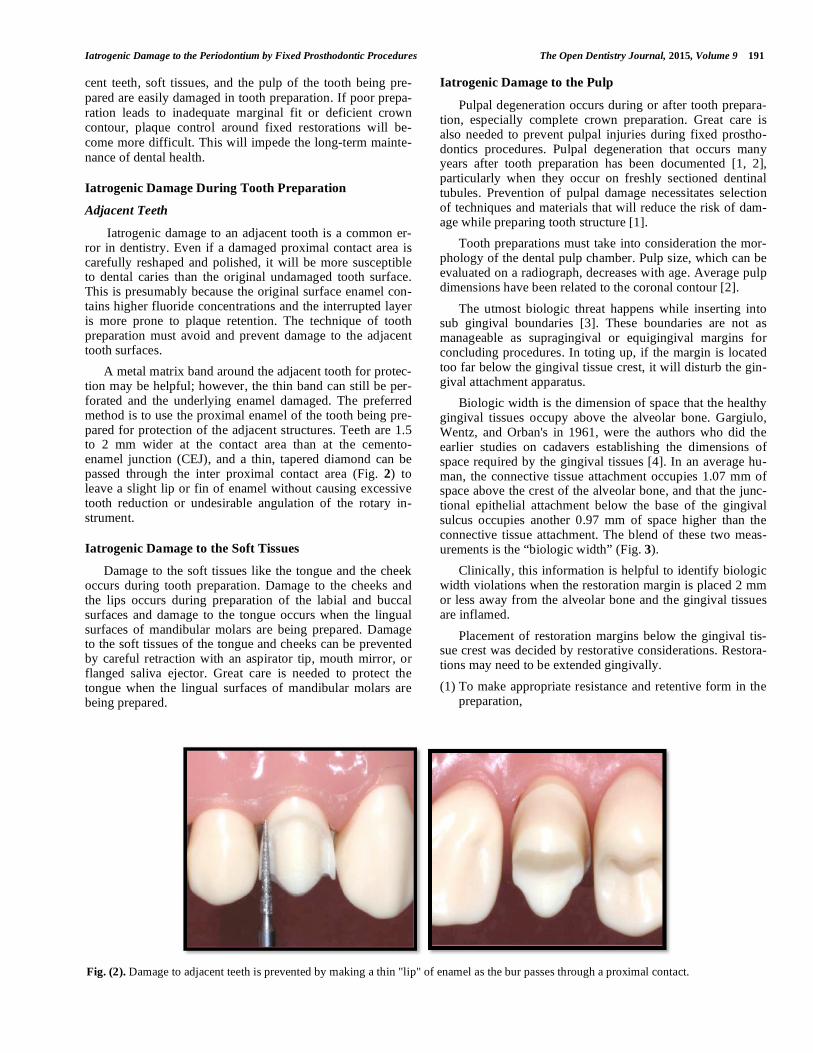
Iatrogenic damage to an adjacent tooth is a common er-ror in dentistry. Even if a damaged proximal contact area is carefully reshaped and polished, it will be more susceptible to dental caries than the original undamaged tooth surface. This is presumably because the original surface enamel con-tains higher fluoride concentrations and the interrupted layer is more prone to plaque retention. The technique of tooth preparation must avoid and prevent damage to the adjacent tooth surfaces.
A metal matrix band around the adjacent tooth for protec-tion may be helpful; however, the thin band can still be per-forated and the underlying enamel damaged. The preferred method is to use the proximal enamel of the tooth being pre-pared for protection of the adjacent structures. Teeth are 1.5 to 2 mm wider at the contact area than at the cemento-enamel junction (CEJ), and a thin, tapered diamond can be passed through the inter proximal contact area (Fig. 2) to leave a slight lip or fin of enamel without causing excessive tooth reduction or undesirable angulation of the rotary in-strument.
Iatrogenic Damage to the Soft Tissues
Damage to the soft tissues like the tongue and the cheek occurs during tooth preparation. Damage to the cheeks and the lips occurs during preparation of the labial and buccal surfaces and damage to the tongue occurs when the lingual surfaces of mandibular molars are being prepared. Damage to the soft tissues of the tongue and cheeks can be prevented by careful retraction with an aspirator tip, mouth mirror, or flanged saliva ejector. Great care is needed to protect the tongue when the lingual surfaces of mandibular molars are being prepared.
Iatrogenic Damage to the Pulp
Pulpal degeneration occurs during or after tooth prepara-tion, especially complete crown preparation. Great care is also needed to prevent pulpal injuries during fixed prostho-dontics procedures. Pulpal degeneration that occurs many years after tooth preparation has been documented [1, 2], particularly when they occur on freshly sectioned dentinal tubules. Prevention of pulpal damage necessitates selection of techniques and materials that will reduce the risk of dam-age while preparing tooth structure [1].
Tooth preparations must take into consideration the mor-phology of the dental pulp chamber. Pulp size, which can be evaluated on a radiograph, decreases with age. Average pulp dimensions have been related to the coronal contour [2].
The utmost biologic threat happens while inserting into sub gingival boundaries [3]. These boundaries are not as manageable as supragingival or equigingival margins for concluding procedures. In toting up, if the margin is located too far below the gingival tissue crest, it will disturb the gin-gival attachment apparatus.
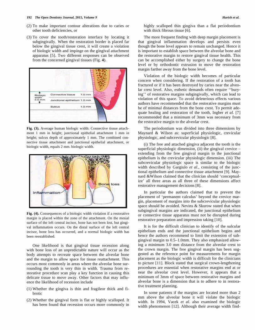
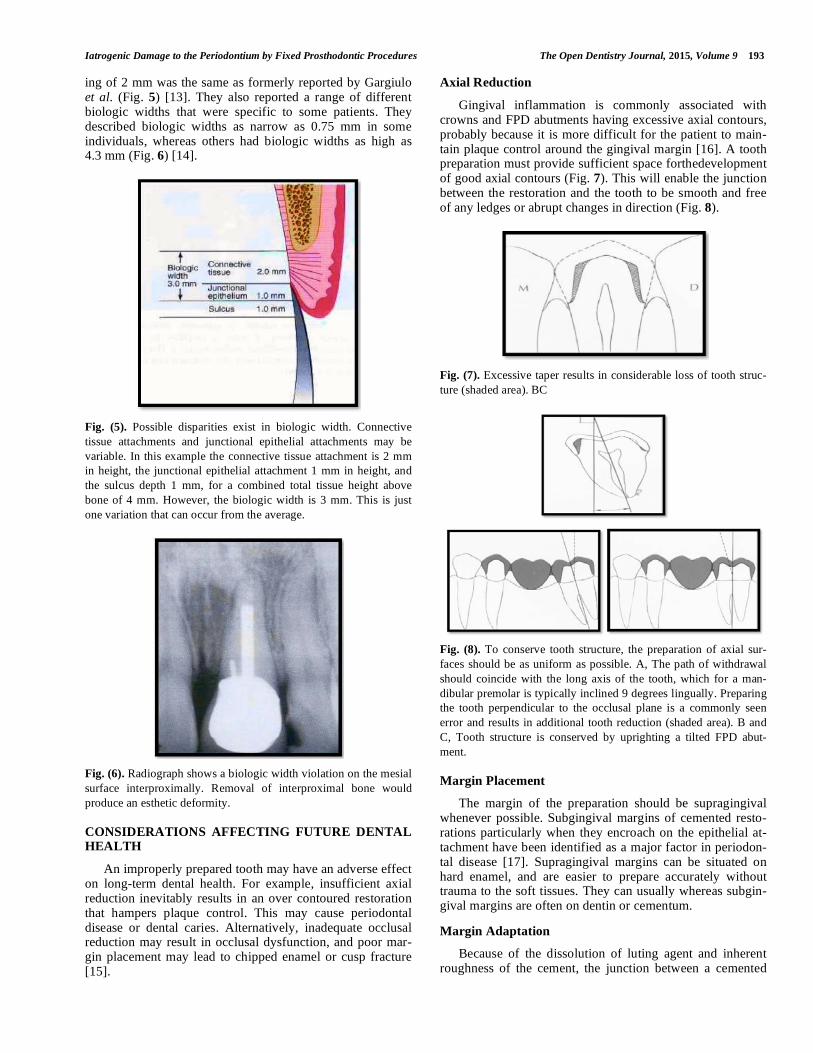
Biologic width is the dimension of space that the healthy gingival tissues occupy above the alveolar bone. Gargiulo, Wentz, and Orban's in 1961, were the authors who did the earlier studies on cadavers establishing the dimensions of space required by the gingival tissues [4]. In an average hu-man, the connective tissue attachment occupies 1.07 mm of space above the crest of the alveolar bone, and that the junc-tional epithelial attachment below the base of the gingival sulcus occupies another 0.97 mm of space higher than the connective tissue attachment. The blend of these two meas-urements is the “biologic width” (Fig. 3).
Clinically, this information is helpful to identify biologic width violations when the restoration margin is placed 2 mm or less away from the alveolar bone and the gingival tissues are inflamed.
Placement of restoration margins below the gingival tis-sue crest was decided by restorative considerations. Restora-tions may need to be extended gingivally.
(1) To make appropriate resistance and retentive form in the preparation,
Fig. (2). Damage to adjacent teeth is prevented by making a thin "lip" of enamel as the bur passes through a proximal contact.
192 The Open Dentistry Journal, 2015, Volume 9 Harish et al.
(2) To make important contour alterations due to caries or other tooth deficiencies, or
(3) To cover the tooth/restoration interface by locating it subgingivally. When the restoration border is placed far below the gingival tissue crest, it will create a violation of biologic width and impinge on the gingival attachment apparatus [5]. Two different responses can be observed from the concerned gingival tissues (Fig. 4).
Fig. (3). Average human biologic width: Connective tissue attach-
ment 1 mm in height; junctional epithelial attachment 1 mm in
height; sulcus depth of approximately 1 mm. The combined con-
nective tissue attachment and junctional epithelial attachment, or
biologic width, equals 2 mm. biologic width.
Fig. (4). Consequences of a biologic width violation if a restorative
margin is placed within the zone of the attachment. On the mesial
surface of the left central incisor, bone has not been lost, but gingi-
val inflammation occurs. On the distal surface of the left central
incisor, bone loss has occurred, and a normal biologic width has
been reestablished.
One likelihood is that gingival tissue recession along
with bone loss of an unpredictable nature will occur as the body attempts to recreate space between the alveolar bone and the margin to allow space for tissue reattachment. This occurs most commonly in areas where the alveolar bone sur-rounding the tooth is very thin in width. Trauma from re-storative procedure scan play a key function in causing this delicate tissue to move away. Other factors that may influ-ence the likelihood of recession include
(1) Whether the gingiva is thin and fragileor thick and fi-brotic
(2) Whether the gingival form is flat or highly scalloped. It has been found that recession occurs more commonly in
highly scalloped thin gingiva than a flat periodontium with thick fibrous tissue [6].
The more frequent finding with deep margin placement is that gingival inflammation develops and persists even though the bone level appears to remain unchanged. Hence it is important to establish space between the alveolar bone and the restorative margin to restore gingival tissue health. This can be accomplished either by surgery to change the bone level or by orthodontic extrusion to move the restoration margin farther away from the bone level.
Violation of the biologic width becomes of particular concern when considering. If the restoration of a tooth has fractured or if it has been destroyed by caries near the alveo-lar crest level. Also, esthetic demands often require ‘‘bury-ing’’ of restorative margins subgingivally, which can lead to violation of this space. To avoid deleterious effects various authors have recommended that the restorative margins must be of minimal distances from the bone crest. To permit ade-quate healing and restoration of the tooth, Ingber et al. [7] recommended that a minimum of 3mm was necessary from the restorative margin to the alveolar crest.
The periodontium was divided into three dimensions by Maynard & Wilson as: superficial physiologic, crevicular physiologic, and subcrevicular physiologic [8].
(i) The free and attached gingiva adjacent the tooth is the superficial physiologic dimension, (ii) the gingival crevice – extending from the free gingival margin to the junctional epithelium is the crevicular physiologic dimension. (iii) The subcrevicular physiologic space is similar to the biologic width described by Gargiulo et al., consisting of the junc-tional epithelium and connective tissue attachment [9]. May-nard &Wilson claimed that the clinician should ‘conceptual-ize’ all three areas as all three of these dimensions affect restorative management decisions [8].
In particular the authors claimed that to prevent the placement of ‘permanent calculus’ beyond the crevice mar-gin, placement of margins into the subcrevicular physiologic space should be avoided. Nevins & Skurow stated that when subgingival margins are indicated, the junctional epithelium or connective tissue apparatus must not be disrupted during restorative preparation and impression taking [10].
It is for the difficult clinician to identify of the sulcular epithelium ends and the junctional epithelium begins and hence the authors recommend to limit the extension of sub-gingival margin to 0.5–1.0mm. They also emphasized allow-ing a minimum 3.0 mm distance from the alveolar crest to the crown margin. The free gingival margin has been sug-gested as the reference point for measurements for margin placement as the biologic width is difficult for the clinicians to picture [11]. Block stated that surgical crown-lengthening procedures are essential when restorative margins end at or near the alveolar crest level. However, it appears that a minimum of 3mm of space between restorative margins and alveolar bone is a dimension that is to adhere to in restora-tive treatment planning.
In some patients if the margins are located more than 2 mm above the alveolar bone it will violate the biologic width. In 1994, Vacek et al. also examined the biologic width phenomenon [12]. Although their average width find-
Iatrogenic Damage to the Periodontium by Fixed Prosthodontic Procedures The Open Dentistry Journal, 2015, Volume 9 193
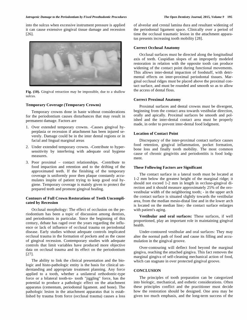
ing of 2 mm was the same as formerly reported by Gargiulo et al. (Fig. 5) [13]. They also reported a range of different biologic widths that were specific to some patients. They described biologic widths as narrow as 0.75 mm in some individuals, whereas others had biologic widths as high as 4.3 mm (Fig. 6) [14].
Fig. (5). Possible disparities exist in biologic width. Connective
tissue attachments and junctional epithelial attachments may be
variable. In this example the connective tissue attachment is 2 mm
in height, the junctional epithelial attachment 1 mm in height, and
the sulcus depth 1 mm, for a combined total tissue height above
bone of 4 mm. However, the biologic width is 3 mm. This is just
one variation that can occur from the average.
Fig. (6). Radiograph shows a biologic width violation on the mesial
surface interproximally. Removal of interproximal bone would
produce an esthetic deformity.
CONSIDERATIONS AFFECTING FUTURE DENTAL HEALTH
An improperly prepared tooth may have an adverse effect on long-term dental health. For example, insufficient axial reduction inevitably results in an over contoured restoration that hampers plaque control. This may cause periodontal disease or dental caries. Alternatively, inadequate occlusal reduction may result in occlusal dysfunction, and poor mar-gin placement may lead to chipped enamel or cusp fracture [15].
Axial Reduction
Gingival inflammation is commonly associated with crowns and FPD abutments having excessive axial contours, probably because it is more difficult for the patient to main-tain plaque control around the gingival margin [16]. A tooth preparation must provide sufficient space forthedevelopment of good axial contours (Fig. 7). This will enable the junction between the restoration and the tooth to be smooth and free of any ledges or abrupt changes in direction (Fig. 8).
Fig. (7). Excessive taper results in considerable loss of tooth struc-
ture (shaded area). BC
Fig. (8). To conserve tooth structure, the preparation of axial sur-
faces should be as uniform as possible. A, The path of withdrawal
should coincide with the long axis of the tooth, which for a man-
dibular premolar is typically inclined 9 degrees lingually. Preparing
the tooth perpendicular to the occlusal plane is a commonly seen
error and results in additional tooth reduction (shaded area). B and
C, Tooth structure is conserved by uprighting a tilted FPD abut-
ment.
Margin Placement
The margin of the preparation should be supragingival whenever possible. Subgingival margins of cemented resto-rations particularly when they encroach on the epithelial at-tachment have been identified as a major factor in periodon-tal disease [17]. Supragingival margins can be situated on hard enamel, and are easier to prepare accurately without trauma to the soft tissues. They can usually whereas subgin-gival margins are often on dentin or cementum.
Margin Adaptation
Because of the dissolution of luting agent and inherent roughness of the cement, the junction between a cemented

194 The Open Dentistry Journal, 2015, Volume 9 Harish et al.
restoration and the tooth is always a potential site for recur-rent caries The more accurately the restoration is adapted to the tooth, the lesser the chance of recurrent caries or perio-dontal disease [18]. Although a precise figure for acceptable margin adaptation is not available, a skilled technician can make a casting that fits to within 10 m [19], and a porcelein margin that fits to within 50 m [20], provided the tooth is properly prepared.
A well-designed preparation is characterized by the pres-ence of a smooth and even margin. Rough, irregular, or "stepped" junctions significantly increase the length of the margin and substantially reduce the adaptation of the restora-tion (Fig. 9).
Occlusal Considerations
A satisfactory tooth preparation should allow sufficient space for developing a functional occlusal scheme in the finished restoration.
IMPRESSIONS
An impression must provide thorough information about
the prepared teeth, surrounding teeth, and associated soft tissues. The impression must record the form of all prepared
surfaces and some of the unprepared tooth cervical to the
finish line. Impression making of tooth preparations that extend subgingivally with an elastic material is likely to
damage the soft tissues.
It is important to avoid damage to the gingival sulcus and junctional epithelium in the course of tooth preparation, gin-
gival retraction, and impression taking. When a complete-
coverage crown restoration is performed, the chances for damaging the periodontium is particularly more. The his-
tological components of the periodontium shows, that the
alveolar crest is covered by the supracrestal fiber complex (Sharpey’s fibers), the junctional epithelium, and the gingi-
val sulcus. The sulcus is very limited in depth - less than
1mm in a healthy periodontium. The sulcular epithelium is non-keratinized. The dento gingival junction (biologic
width) seals the underlying connective tissue of the perio-
dontium from the oral environment [21].
Iatrogenic disruption of the junctional epithelium may cause an inflammatory lesion in the gingival corium and fiber apparatus. This can lead to gingival recession, migra-tion of the junctional epithelium, and permanent bone loss [22]. The retraction cords may harm the attachment appara-tus if they are tightly packed for a prolonged period (i.e. more than five minutes). If the inflamed gingiva is retracted, it may result in permanent loss of attachment [23]. In other cases, the application of a retraction cord may be impossible when the gingival sulcus is damaged by perodontitis or a faulty restoration. (Fig. 10).
To summarize, any impression procedure must consider the delicacy of the junctional epithelium and attachment of the supra crestal fibers and take safety measures not to dis-turb them [24]. Adequate gingival retraction is necessary for impression making. The use of retraction cord has proved to be an effective method of soft tissue management. Injury to sulcular epithelium may be caused by placement of retrac-tion cord and cotton strings into the gingival sulcus. The trauma caused to the soft tissue depends upon the chemical agent with which the cord has been impregnated, the force used in packing the cord and the length of time the cord is left in place within the sulcus. To avoid forcing the cord into the sub-epithelial connective tissue the force used to place the cords should be minimal. Most importantly prior to its removal, the cord should be moistened to avoid tripping the sulcular epithelium [25].
For margins placed intra-crevicularly, impressions are more difficult to obtain as it requires displacing the free gin-gival tissues. Injudicious use of gingival retraction tech-niques causes injury to the biologic width and permanent alterations such as recession. Careful considerations for the tissues are required during crown preparation and impression making. If tube impressions are made, then the individual tubes should be carefully adapted and should relate accu-rately to the gingival line. Excessive digital force causes stripping of the gingiva. Gingival retraction cords used should not have excess diameter as any undue force during cord placement also causes damage to the attachment. Spe-cial precautions should be taken for thin and delicate gingi-val tissue and where attached gingiva is inadequate as undue insult to the tissues can cause recession. When placing cord
Fig. (9). A and B, Poor preparation design, leading to increased margin length. C, A rough, irregular margin will make the fabrication of an
accurately fitted restoration almost impossible. D, An accurately fitting margin is possible only if it is prepared smoothly.
Iatrogenic Damage to the Periodontium by Fixed Prosthodontic Procedures The Open Dentistry Journal, 2015, Volume 9 195
into the sulcus when excessive instrument pressure is applied it can cause extensive gingival tissue damage and recession [26].
Fig. (10). Gingival retraction may be impossible, due to a shallow
sulcus.
Temporary Coverage (Temporary Crowns)
Temporary crowns done in haste without considerations for the periodontium causes disturbances that may result in permanent damage. Factors are
1. Over extended temporary crowns. -Causes gingival hy-perplasia or recession if attachment has been injured se-verely. Damage could be in the inter dental regions or in facial and lingual marginal areas
2. Under extended temporary crowns. -Contribute to hyper-sensitivity by interfering with adequate oral hygiene measures.
3. Poor proximal – contact relationships. -Contribute to food impaction and retention and to the drifting of the approximated teeth. If the finishing of the temporary coverage is uniformly poor then plaque constantly accu-mulates inspite of patient trying to keep good oral hy-giene. Temporary coverage is mainly given to protect the prepared teeth and promote gingival healing.
Contours of Full Crown Restorations of Teeth Uncompli-cated by Recession
Occlusal morphology: The effect of occlusion on the pe-riodontium has been a topic of discussion among dentists, and periodontists in particular. Since the beginning of this century, debate has raged over the years regarding the influ-ence or lack of influence of occlusal trauma on periodontal disease. Early studies without adequate controls implicated occlusal trauma in the formation of pockets and as the cause of gingival recession. Contemporary studies with adequate controls that limit variables have produced more objective data on occlusal trauma and its effect on the periodontium [27].
The ability to link the clinical presentation and the bio-logic and histo-pathologic entity is the basis for clinical un-derstanding and appropriate treatment planning. Any force applied to a tooth, whether a unilateral orthodontic-type force or a bilateral tooth-to- tooth "jiggling" force, has the potential to produce a pathologic effect on the attachment apparatus (cementum, periodontal ligament. and bone). The pathologic lesion in the attachment apparatus that is estab-lished by trauma from force (occlusal trauma) causes a loss
of alveolar and crestal lamina dura and resultant widening of the periodontal ligament space. Clinically over a period of time the occlusal traumatic lesion in the attachment appara-tus presents increasing tooth mobility [28].
Correct Occlusal Anatomy
Occlusal surfaces must be directed along the longitudinal axis of teeth. Cuspidian slopes of an improperly modeled restoration in relation with the opposite tooth can produce widening of the contact point during functional movements. This allows inter-dental impaction of foodstuff, with detri-mental effects on inter-proximal periodontal tissues. Mar-ginal occlusal ridges must be placed above the proximal con-tact surface, and must be rounded and smooth so as to allow the access of dental floss.
Correct Proximal Anatomy
Proximal surfaces and dental crowns must be divergent, beginning from the contact area towards vestibular direction, orally and apically. Proximal surfaces be smooth and pol-ished and the inter-dental contact area must be properly made, in order to prevent inter-dental food lodgment.
Location of Contact Point
Discrepancy of the inter-proximal contact surface causes food retention, gingival inflammation, pocket formation, bone loss and finally tooth mobility. The most common cause of chronic gingivitis and periodontitis is food lodg-ment.
These Following Factors are Significant
The contact surface in a lateral tooth must be located at 1-2 mm below the greatest height of the marginal ridge; it should not exceed 1-2 mm in length in occluso-gingival di-rection and it should measure approximately 25% of the oro- vestibular width of the neighboring tooth; - in the upper arch the contact surface is situated slightly towards the vestibular area, from the median mesio-distal line and in the lower arch is located on the median line;- the contact surface enlarges with patient's aging.
Vestibular and oral surfaces: These surfaces, if well proportioned, play an important role in maintaining gingival health.
Under-contoured vestibular and oral surfaces: They may
alter the normal path of food and cause its filling and accu-mulation in the gingival groove.
Over-contouring will deflect food beyond the marginal
gingiva, reaching the attached gingiva. This fact removes the
marginal gingiva of self-cleaning mechanical action of food, which can stagnate in over protected gingival groove.
CONCLUSION
The principles of tooth preparation can be categorized
into biologic, mechanical, and esthetic considerations. Often
these principles conflict and the practitioner must decide
how the restoration should be designed. One area may be
given too much emphasis, and the long-term success of the
196 The Open Dentistry Journal, 2015, Volume 9 Harish et al.
procedure may be limited by a lack of consideration of other
factors. Experience will help in determining whether prepa-
rations are "complete." Each tooth preparation must be
measured by clearly defined criteria, which can be used to
identify and correct problems. Diagnostic tooth preparations
and evaluative impressions are often very helpful. Successful
preparation can be obtained most easily by systematically
following the steps. It is critical to refrain from "jumping
ahead" before the previous step has been evaluated and, if
necessary, corrected. If the clinician proceeds too rapidly,
precious chair time will be lost, and the quality of the prepa-ration will probably suffer.
CONFLICT OF INTEREST
The authors confirm that this article content has no con-flict of interest.
ACKNOWLEDGEMENTS
Declared none.
REFERENCES
[1] Baldissara P. Clinical and histological evaluation of thermal injury
thresholds in human teeth: a preliminary study. J Oral Rehabil 1997; 24: 791-801.
[2] Ohashi Y. Research related to anterior abutment teeth of fixed partial denture. Shikagakuho 1968; 68: 726.
[3] McKee JR. Comparing condylar position respectability for stan-dardized versus non standardized methods of achieving centric re-
lation. Prosthet Dent 1997; 77: 280. [4] Gargiulo AW, Wentz FM, Orban B. Dimension and relations of the
dentogingival junction in humans. J Periodontol 1961; 32: 262. [5] Parma-Benfenati S, Fugazzoto PA, Ruben MP. The effect of re-
storative margins on the postsurgical development and nature of the periodontium. Part I. Int J Periodont Rector Dent 1985; 6: 31.
[6] Olsson M, Lindhe S. Periodontol characteristics in individuals with varying forms of the upper central incisors. J Clin Periodontol
1991; 18: 78. [7] Ingber JS, Rose LF, Coslet JG. “The biologic width’’: a concept in
periodontics and restorative dentistry. Alpha Omega 1977; 70: 62-5.
[8] Maynard JG, Wilson RDK. Physiologic dimensions of the perio-dontium significant to the restorative dentist. J Periodontol 1979;
50: 170-4.
[9] Gargiulo AW, Wentz F, Orban B. Dimensions and relations of the
dento-gingival junction in humans. J Periodontol 1961; 32: 261-7. [10] Nevins M, Skurow HM. The intra-crevicular restorative margin,
the biologic width, and the maintenance of the gingival margin. Intl J Periodont Restor Dent 1984; 3: 31-49.
[11] Block PL. Restorative margins and periodontal health. A new look at an old perspective. J Prosthet Dent 1987; 57: 683-9.
[12] Vacek JS, Gehr ME, Asad DA, Richardson AC, Giambarresi LI. The dimensions of the human dento-gingival junction. Int J Perio-
dont Restorat Dent 1994; 14: 154-65. [13] Sorensen JA. A rationale for comparison of plaque-retaining prop-
erties of crown systems. J Prosthet Dent 1989; 62: 264. [14] Perel ML. Axial crown contours. J Prosthet Dent 1971; 25: 642.
[15] Baldissara P. Clinical and histological evaluation of thermal injury thresholds in human teeth: a preliminary study. J Oral Rehabil
1997; 24: 791. [16] Ohashi Y. Research related to anterior abutment teeth of fixed
partial denture. Shikagakuho 1968; 68: 726. [17] Silness J. Periodontal conditions in patients treated with dental
bridges. III. The relationship between the location of the crown margin and the periodontal condition. J Periodont Res 1970; 5: 225.
[18] Felton DA. Effect of in vivo crown margin discrepancies on perio-dontal health. J Prosthet Dent 1991; 65: 357.
[19] Byrne G. Casting accuracy of high palladium alloys. J Prosthet Dent 1986; 55: p. 297.
[20] Belser UC. Fit of three porcelain-fused to metal marginal designs in vivo: a scanning electron microscope study. J Prosthet Dent
1985; 53: 24. [21] Berkowitz BKB, Holland GR, Maxham BJ. Colour atlas and text-
book of oral anatomy. Wolfe Med Publ Ltd 1978; pp. 129-32. [22] Wilson RD. Restorative dentistry. In: Wilson TG, Kornman KS,
Newman MG, Eds. Advances in periodontics. Chicago: Quintes-sence Publ Co Inc 1992; pp. 226-30.
[23] Wise MD. Tooth preparation, gingival retraction, hydrocolloid technique, master cast systems, soldering. In: Failure in the restored
dentition: management and treatment. London: Quintessence Publ Co Ltd 1995; pp. 189-90.
[24] Nevins M, Cappetta EG. The biologic width: preventing postsurgi-cal recession. In: Nevins M, Mellonig, Eds. Periodontal therapy,
clinical approaches, and evidence of success. Chicago: Quintes-sence Publ Co Inc 1998; 1: pp. 305-7.
[25] Kois J, Vakay RT. Relationship of the periodontium to impression procedures. Compend Contin Educ Dent 2000; 21: 684-90.
[26] Benson BW, Bomberg TJ, Hatch RA. Tissue displacement methods in fixed prosthodontic. J Prosthet Dent 1986; 55: 175.
[27] Svanberg G. Lindhe J. Experimental tooth hyper mobility in the dog. Odontol Rev 1973; 24: 269.
[28] Meitner S. Co-destructive factors of marginal periodontal disease and repetitive mechanical injury. J Dent Res 1975; 54: 78.
Received: December 22, 2014 Revised: March 04, 2015 Accepted: March 10, 2015
© Harish et al.; Licensee Bentham Open.
This is an open access article licensed under the terms of the Creative Commons Attribution Non-Commercial License (http://creativecommons.org/-
licenses/by-nc/3.0/) which permits unrestricted, non-commercial use, distribution and reproduction in any medium, provided the work is properly cited.


![The Open Dentistry Journal · The Open Dentistry Journal, 2016, 10, ... the bleaching steps or during the bleaching period [9]. The literature review also does not show ... USA) cream](https://img.pdfslide.net/doc/110x75/5b50690f7f8b9a166e8e797a/the-open-dentistry-journal-the-open-dentistry-journal-2016-10-the-bleaching.jpg)


![The Open Dentistry Journal...632 The Open Dentistry Journal, 2020, Volume 14 Kochotwuttinont and Wayakanon modulus materials such as flowable resin composite [5 - 9] or low polymerization](https://img.pdfslide.net/doc/110x75/6133f17edfd10f4dd73b6a50/the-open-dentistry-journal-632-the-open-dentistry-journal-2020-volume-14-kochotwuttinont.jpg)