Embed Size (px)
DESCRIPTION
bacterial
Citation preview
Bacterial Classification, Anatomy, Nutrition, Growth, Metabolism and Genetics
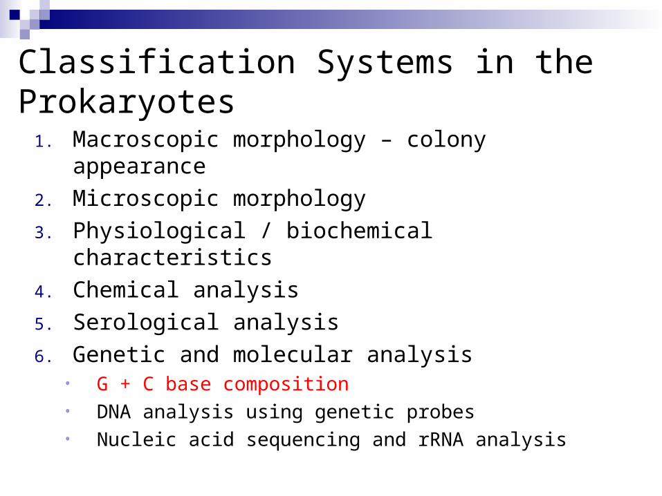
Classification Systems in the Prokaryotes
1. Macroscopic morphology – colony appearance
2. Microscopic morphology
3. Physiological / biochemical characteristics
4. Chemical analysis
5. Serological analysis
6. Genetic and molecular analysis• G + C base composition• DNA analysis using genetic probes• Nucleic acid sequencing and rRNA analysis
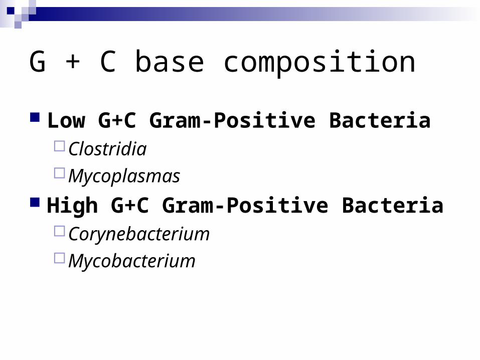
G + C base composition
Low G+C Gram-Positive BacteriaClostridiaMycoplasmas
High G+C Gram-Positive BacteriaCorynebacteriumMycobacterium
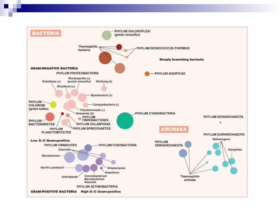
Bacterial Taxonomy Based on Bergey’s Manual Bergey’s Manual of Determinative
Bacteriology – five volume resource covering all known procaryotesclassification based on genetic information –
phylogenetic two domains: Archaea and Bacteria five major subgroups with 25 different phyla

Major Taxonomic Groups of Bacteria
Vol 1A: Domain Archaea primitive, adapted to extreme habitats and modes of
nutrition Vol 1B: Domain Bacteria Vol 2-5:
2 - Phylum Proteobacteria – Gram-negative cell walls
3 - Phylum Firmicutes – mainly Gram-positive with low G + C content
4 - Phylum Actinobacteria – Gram-positive with high G + C content
5 – Loose assemblage of phyla – All gram negative
Diagnostic Scheme for Medical Use
Uses phenotypic qualities in identificationrestricted to bacterial disease agentsdivides based on cell wall structure, shape,
arrangement, and physiological traits

Species and Subspecies
Species bacterial cells which share overall similar pattern of
traits Subspecies
Strain or variety culture derived from a single parent that differs in
structure or metabolism from other cultures of that species
Type subspecies that can show differences
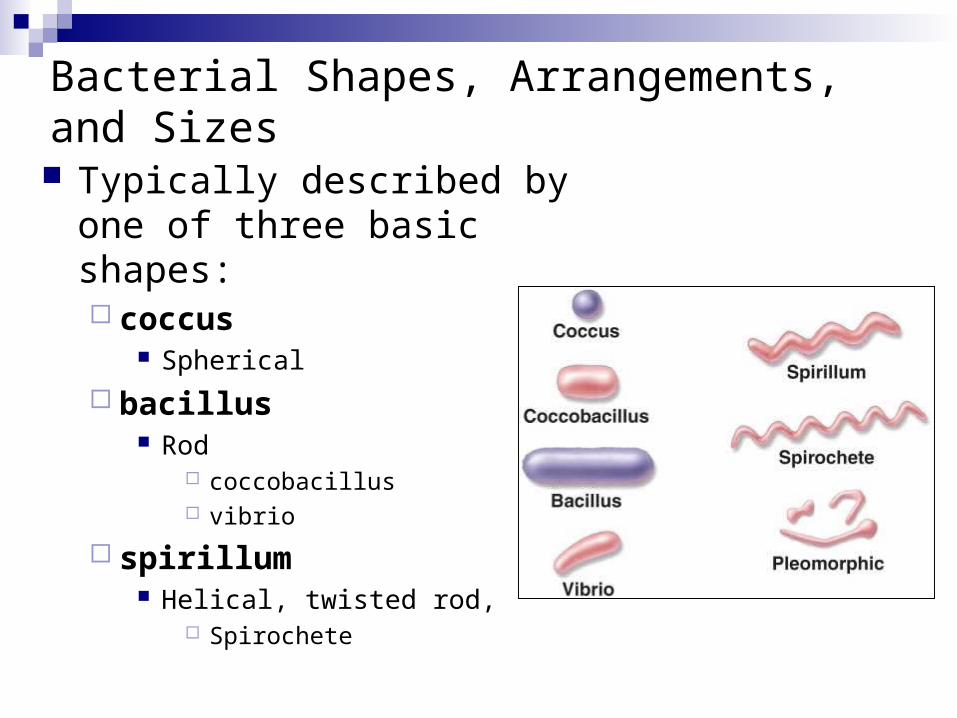
Bacterial Shapes, Arrangements, and Sizes
Typically described by one of three basic shapes: coccus
Spherical
bacillus Rod
coccobacillus vibrio
spirillum Helical, twisted rod,
Spirochete
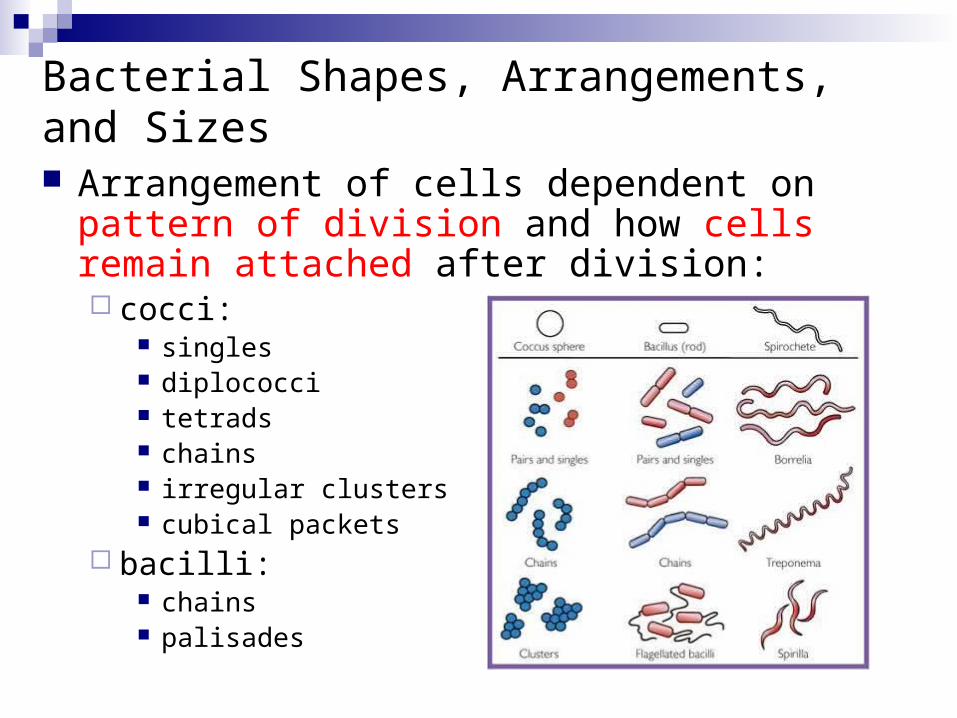
Bacterial Shapes, Arrangements, and Sizes
Arrangement of cells dependent on pattern of division and how cells remain attached after division: cocci:
singles diplococci tetrads chains irregular clusters cubical packets
bacilli: chains palisades
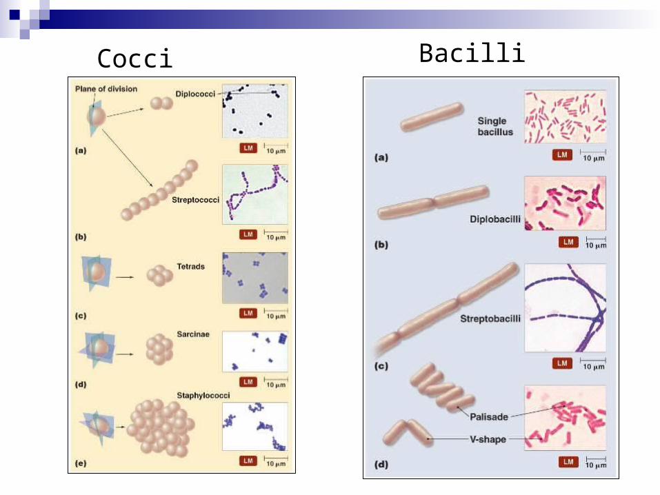
Cocci Bacilli
Bacterial anatomy

Generalized structure of a prokaryotic cell
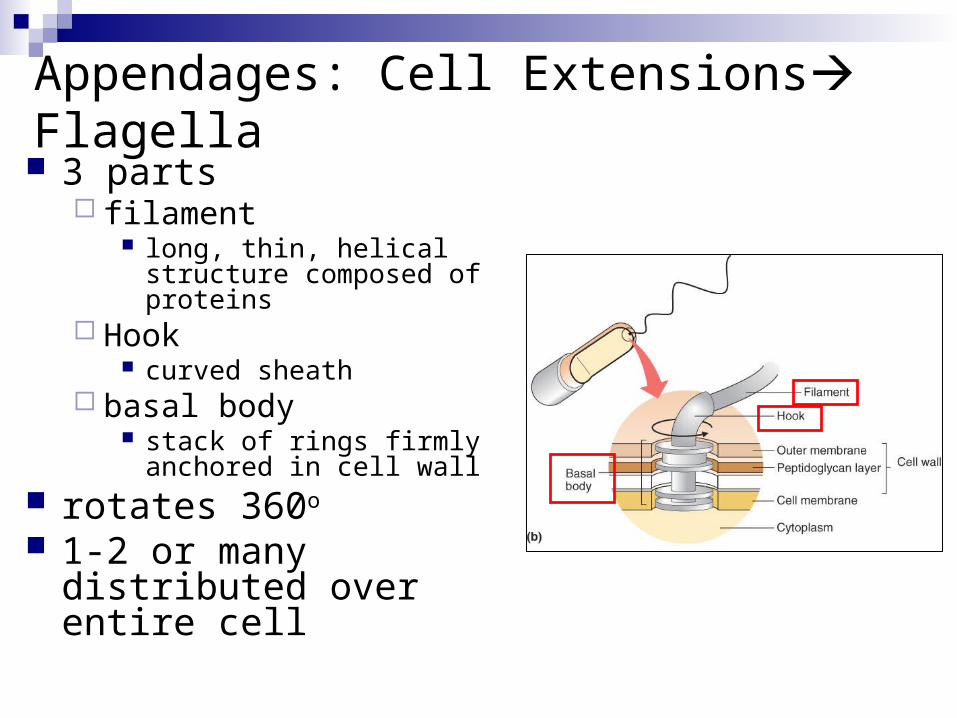
Appendages: Cell Extensions Flagella 3 parts
filament long, thin, helical structure
composed of proteins Hook
curved sheath basal body
stack of rings firmly anchored in cell wall
rotates 360o
1-2 or many distributed over entire cell
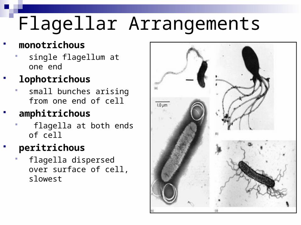
Flagellar Arrangements monotrichous
single flagellum at one end lophotrichous
small bunches arising from one end of cell
amphitrichous flagella at both ends of
cell peritrichous
flagella dispersed over surface of cell, slowest

Fig. 4.4
Movement by flagella
Polar Rotates counterclockwise Cell swims forward in
runs Reverse will stop it
Peritrichous All flagella sweep
towards one end
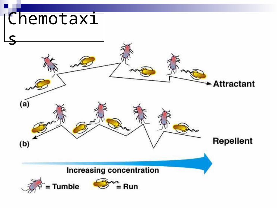
Chemotaxis
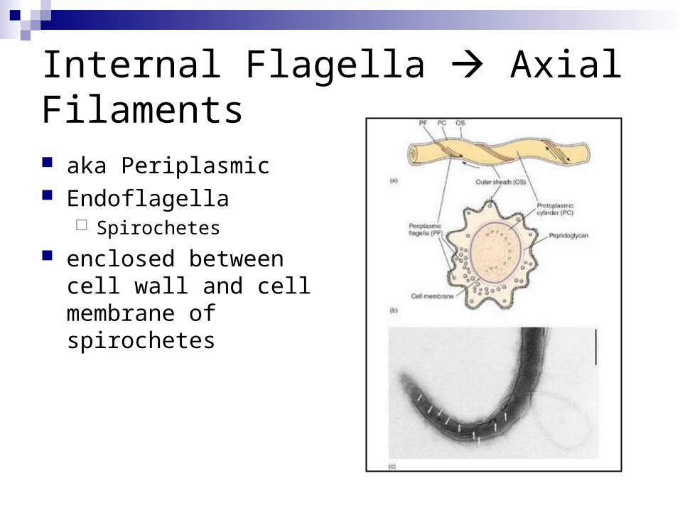
Internal Flagella Axial Filaments
aka Periplasmic Endoflagella
Spirochetes
enclosed between cell wall and cell membrane of spirochetes

Appendages for Attachment Fimbrae
fine hairlike bristles from the cell surface
function in adhesion to other cells and surfaces
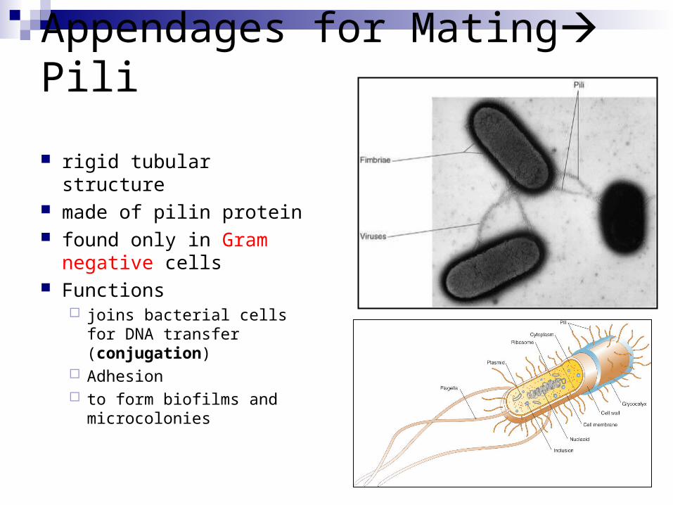
Appendages for Mating Pili
rigid tubular structure made of pilin protein found only in Gram
negative cells Functions
joins bacterial cells for DNA transfer (conjugation)
Adhesion to form biofilms and
microcolonies

The Cell Envelope External covering outside the cytoplasm Composed of few basic layers:
glycocalyx cell wall cell membrane
Maintains cell integrity
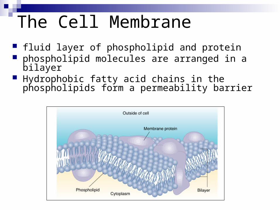
fluid layer of phospholipid and protein phospholipid molecules are arranged in a bilayer Hydrophobic fatty acid chains in the phospholipids form a
permeability barrier
The Cell Membrane
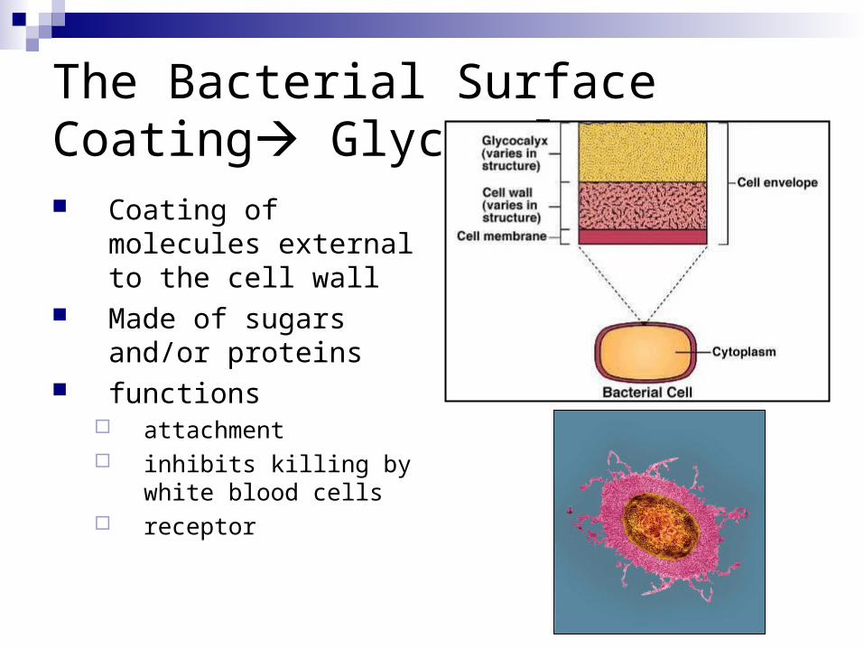
The Bacterial Surface Coating Glycocalyx Coating of molecules
external to the cell wall Made of sugars and/or
proteins functions
attachment inhibits killing by white
blood cells receptor

The Bacterial Surface Coating Glycocalyx 2 types:
1. slime layer - loosely organized and attached
2. capsule - highly organized, tightly attached
Cell Wall
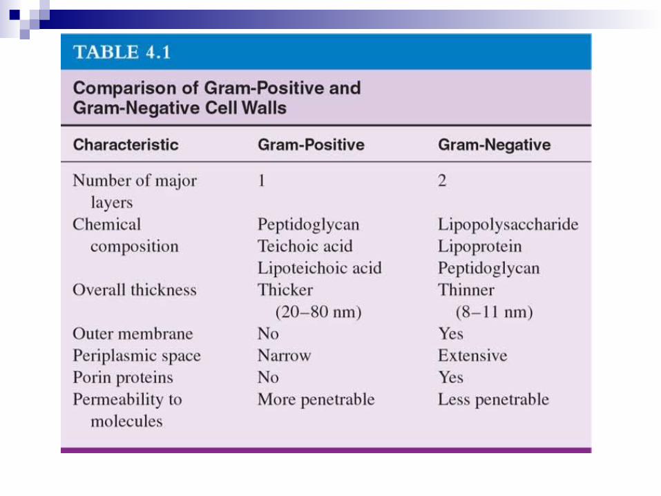
Four Groups Based on Cell Wall Composition:1. Gram positive cells
2. Gram negative cells
3. Bacteria without cell walls
4. Bacteria with chemically unique cell walls

Structure of the Cell Wall Peptidoglycan
macromolecule composed of a repeating framework of long glycan chains cross-linked by short
peptide fragments provides strong,
flexible support keep bacteria from
bursting or collapsing because of changes in osmotic pressure
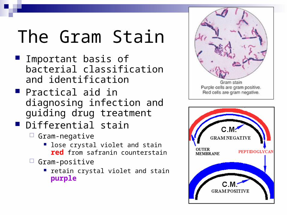
The Gram Stain Important basis of bacterial
classification and identification Practical aid in diagnosing infection
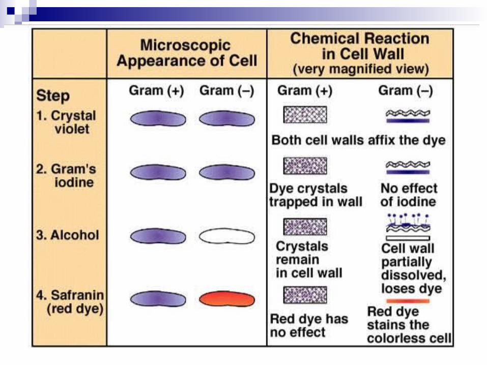
and guiding drug treatment Differential stain
Gram-negative lose crystal violet and stain red from
safranin counterstain Gram-positive
retain crystal violet and stain purple
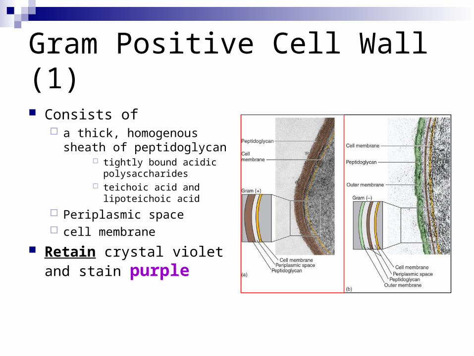
Gram Positive Cell Wall (1)
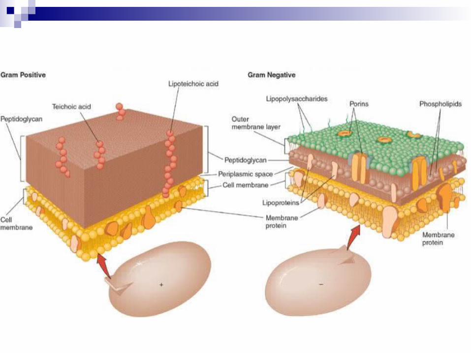
Consists of a thick, homogenous
sheath of peptidoglycan tightly bound acidic
polysaccharides teichoic acid and
lipoteichoic acid
Periplasmic space cell membrane
Retain crystal violet and stain purple
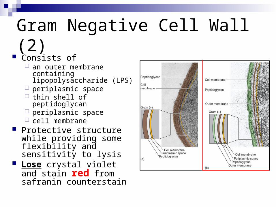
Gram Negative Cell Wall (2) Consists of
an outer membrane containing lipopolysaccharide (LPS)
periplasmic space thin shell of peptidoglycan periplasmic space cell membrane
Protective structure while providing some flexibility and sensitivity to lysis
Lose crystal violet and stain red from safranin counterstain

Gram Negative Cell Wall
LPS endotoxin that may
become toxic when released during infections
may function as receptors and blocking immune response
contains porin proteins in upper layer
Regulates molecules entering and leaving cell
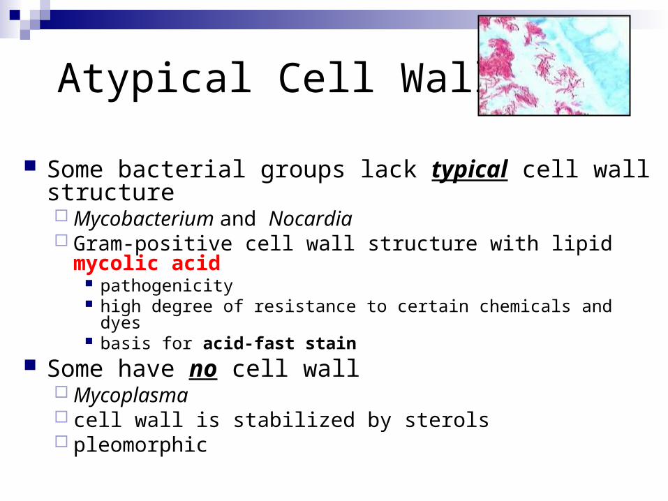
Atypical Cell Walls
Some bacterial groups lack typical cell wall structure Mycobacterium and Nocardia Gram-positive cell wall structure with lipid mycolic acid
pathogenicity high degree of resistance to certain chemicals and dyes basis for acid-fast stain
Some have no cell wall Mycoplasma cell wall is stabilized by sterols pleomorphic
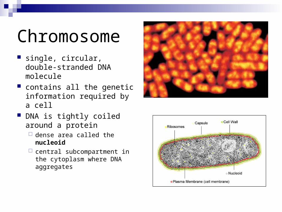
Chromosome single, circular, double-
stranded DNA molecule contains all the genetic
information required by a cell DNA is tightly coiled around
a protein dense area called the nucleoid central subcompartment in the
cytoplasm where DNA aggregates
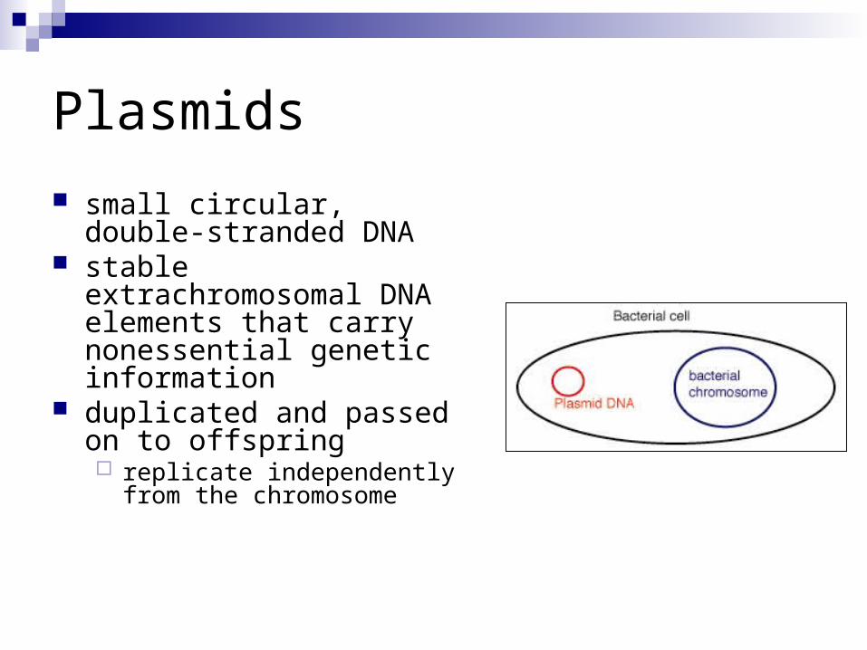
Plasmids
small circular, double-stranded DNA
stable extrachromosomal DNA elements that carry nonessential genetic information
duplicated and passed on to offspring replicate independently from the
chromosome
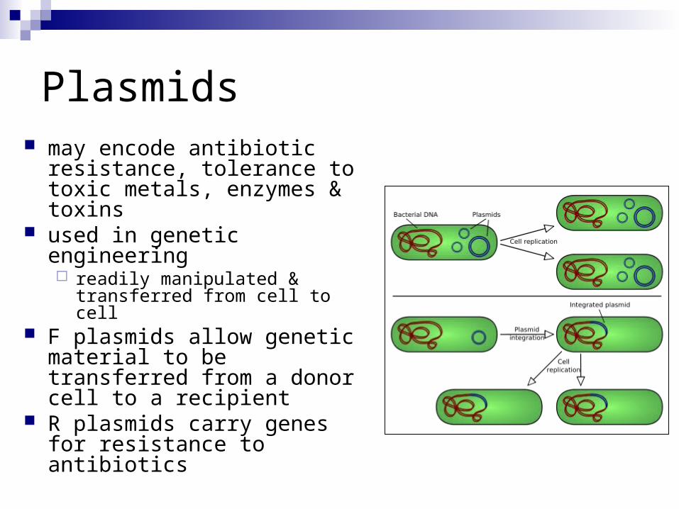
Plasmids may encode antibiotic
resistance, tolerance to toxic metals, enzymes & toxins
used in genetic engineering readily manipulated &
transferred from cell to cell F plasmids allow genetic
material to be transferred from a donor cell to a recipient
R plasmids carry genes for resistance to antibiotics
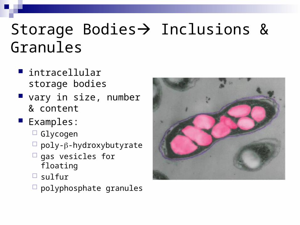
Storage Bodies Inclusions & Granules
intracellular storage bodies
vary in size, number & content
Examples: Glycogen poly--hydroxybutyrate gas vesicles for floating sulfur polyphosphate granules
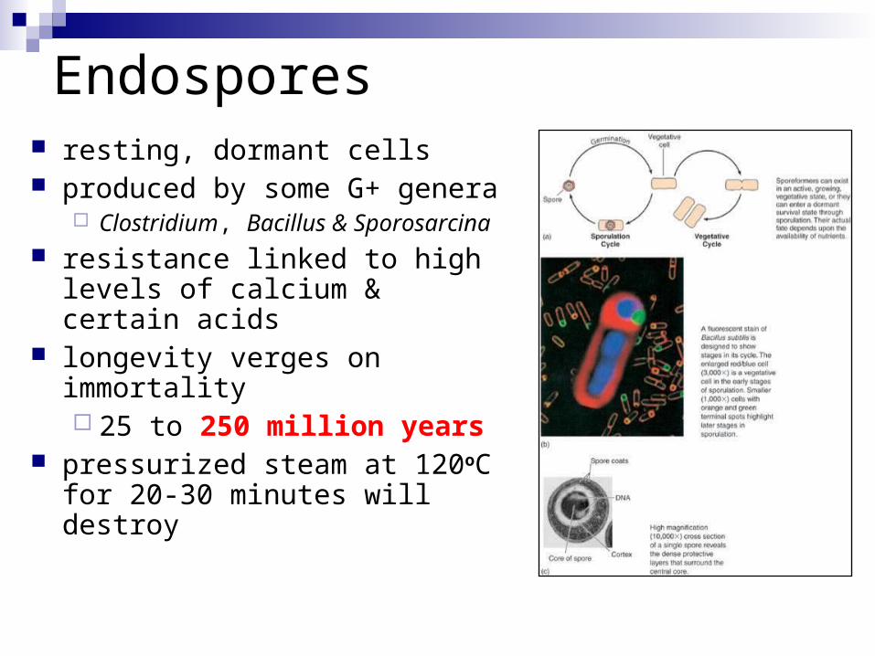
Endospores resting, dormant cells produced by some G+ genera
Clostridium, Bacillus & Sporosarcina resistance linked to high levels of
calcium & certain acids longevity verges on immortality
25 to 250 million years pressurized steam at 120oC for
20-30 minutes will destroy
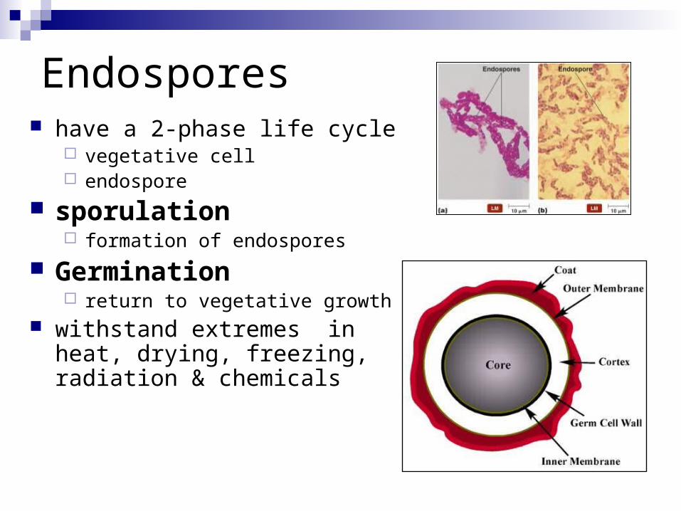
Endospores have a 2-phase life cycle
vegetative cell endospore
sporulation formation of endospores
Germination return to vegetative growth
withstand extremes in heat, drying, freezing, radiation & chemicals

Endospores• stressed cell
• undergoes asymmetrical cell division• creating small prespore and larger
mother cell• prespore contains:
Cytoplasm DNA dipicolinic acid
• mother cell matures the prespore into an endospore
• then disintegrates• environmental conditions are again
favorable• protective layers break down • spore germinates into a vegetative
cell

Microbial nutrition, growth, and metabolism
Microbial Nutrition
nutrition process by which chemical substances
(nutrients) are acquired from the environment and used for cellular activities
Two categories of essential nutrients:macronutrients micronutrients or trace elements
Inorganic nutrients Organic nutrients
Obtaining Carbon
Heterotroph organism that obtains carbon in an organic form
made by other living organisms proteins, carbohydrates, lipids and nucleic acids
Autotroph an organism that uses CO2 (an inorganic gas) as
its carbon sourcenot dependent on other living things

Growth Factors organic compounds
that cannot be synthesized by an organism & must be provided as a nutrient essential amino acids,
vitamins
Nutritional types Chemo-
Chemical compounds Photo-
light
Carbon source
Energy source
photoautotrophs CO2 sunlight
chemoautotrophs CO2 Simple inorganic chemicals
photoheterotrophs organic sunlight
chemoheterotrophs organic Metabolizing organic cmpds
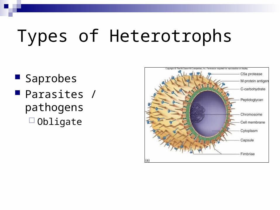
Types of Heterotrophs
Saprobes Parasites / pathogens
Obligate

Nutritional Movement
Osmosis Facilitated diffusion Active transport Endocytosis
PhagocytosisPinocytosis
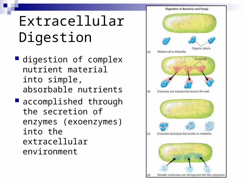
Extracellular Digestion
digestion of complex nutrient material into simple, absorbable nutrients
accomplished through the secretion of enzymes (exoenzymes) into the extracellular environment
Environmental Influences on Microbial Growth
1. temperature 2. oxygen requirements 3. pH 4. Osmotic pressure 5. UV light 6. pressure
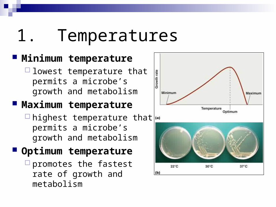
1. Temperatures Minimum temperature
lowest temperature that permits a microbe’s growth and metabolism
Maximum temperature highest temperature that
permits a microbe’s growth and metabolism
Optimum temperature promotes the fastest rate of
growth and metabolism

Temperature Adaptation Groups Psychrophiles
• optimum temperature 15oC• capable of growth at 0 - 20oC
Mesophiles • optimum temperature
10o - 40oC• most human pathogens
Thermophiles • optimum temperature 60oC• capable of growth at 40 - 70oC
Hyperthermophiles Archaea that grow optimally
above 80°C found in seafloor hot-water
vents
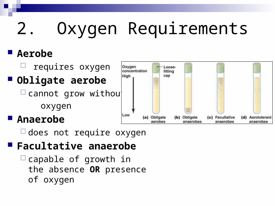
2. Oxygen Requirements Aerobe
requires oxygen
Obligate aerobe cannot grow without
oxygen
Anaerobe does not require oxygen
Facultative anaerobe capable of growth in the
absence OR presence of oxygen
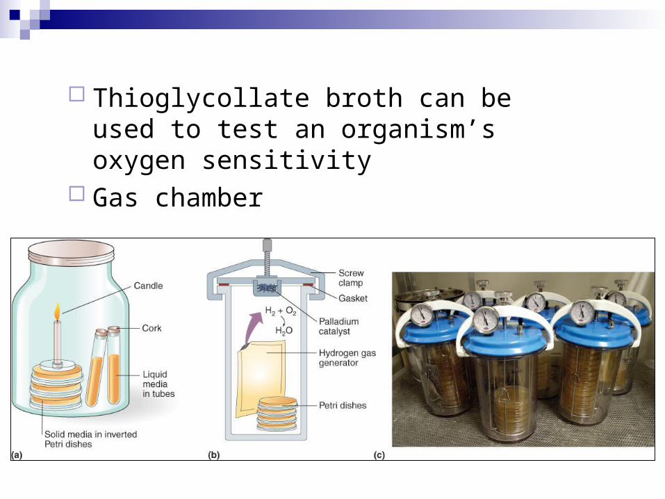
Thioglycollate broth can be used to test an organism’s oxygen sensitivity
Gas chamber
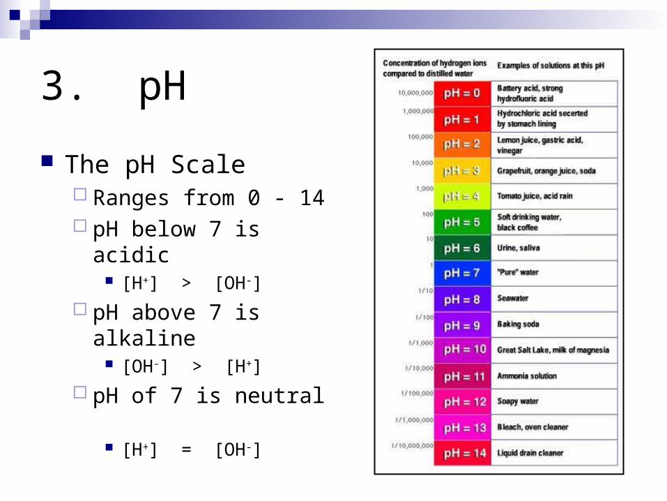
3. pH
The pH Scale Ranges from 0 - 14 pH below 7 is acidic
[H+] > [OH-]
pH above 7 is alkaline [OH-] > [H+]
pH of 7 is neutral [H+] = [OH-]
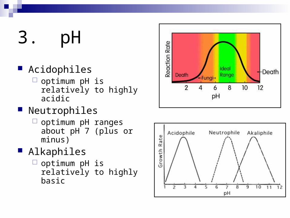
3. pH
Acidophiles optimum pH is relatively to
highly acidic Neutrophiles
optimum pH ranges about pH 7 (plus or minus)
Alkaphiles optimum pH is relatively to
highly basic
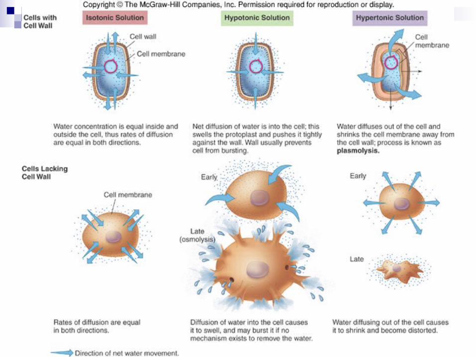
4. Osmotic Pressure Bacteria 80% water
Require water to grow Sufficiently hypertonic media at concentrations
greater than those inside the cell cause water loss from the cell Osmosis Fluid leaves the bacteria causing the cell to contract
Causes the cell membrane to separate Plasmolysis
Cell shrinkage extreme or obligate halophiles
Adapted to and require high salt concentrations
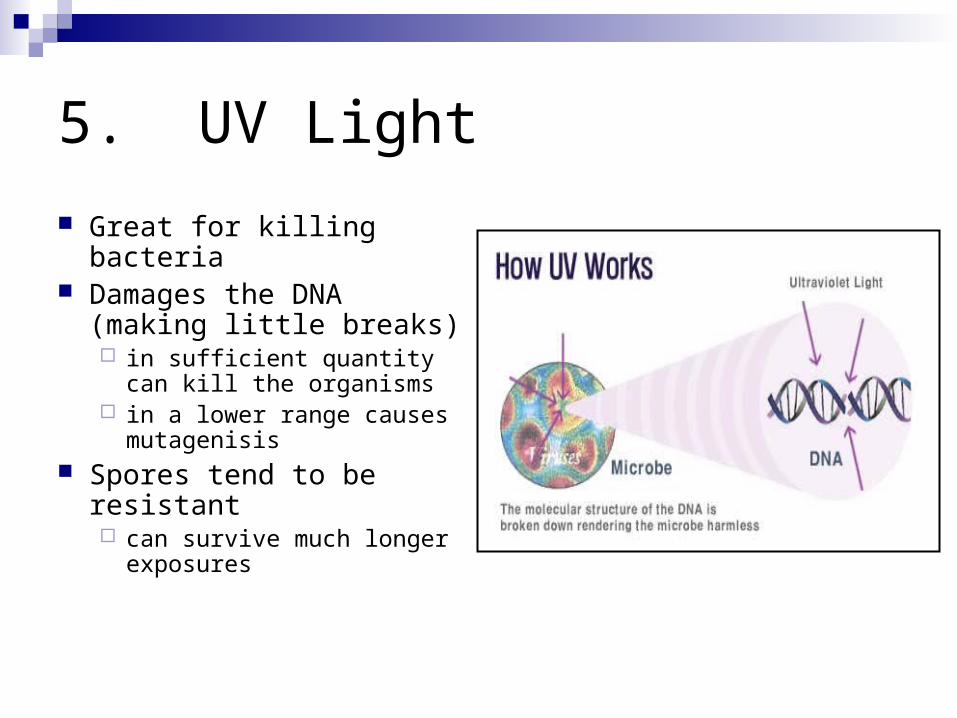
5. UV Light
Great for killing bacteria Damages the DNA
(making little breaks) in sufficient quantity can kill
the organisms in a lower range causes
mutagenisis Spores tend to be
resistant can survive much longer
exposures
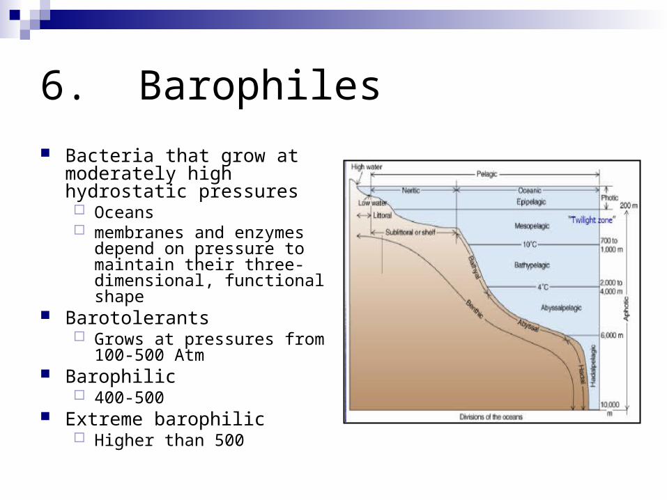
6. Barophiles
Bacteria that grow at moderately high hydrostatic pressures Oceans membranes and enzymes
depend on pressure to maintain their three-dimensional, functional shape
Barotolerants Grows at pressures from 100-
500 Atm Barophilic
400-500 Extreme barophilic
Higher than 500
Microbial Associations Symbiotic
organisms live in close nutritional relationships; Mutualism
Obligatory Dependent Both members benefit
Commensalism One member benefits Other member not harmed
Parasitism Parasite is dependent and benefits Host is harmed

Microbial Associations
Non-symbiotic organisms are free-livingrelationships not required for survival
Synergism members cooperate and share nutrients
Antagonism some member are inhibited or destroyed by others

Microbial Associations
Biofilms Complex relationships
among numerous microorganisms
Develop an extracellular matrix
Adheres cells to one another
Allows attachment to a substrate
Sequesters nutrients May protect individuals in
the biofilm
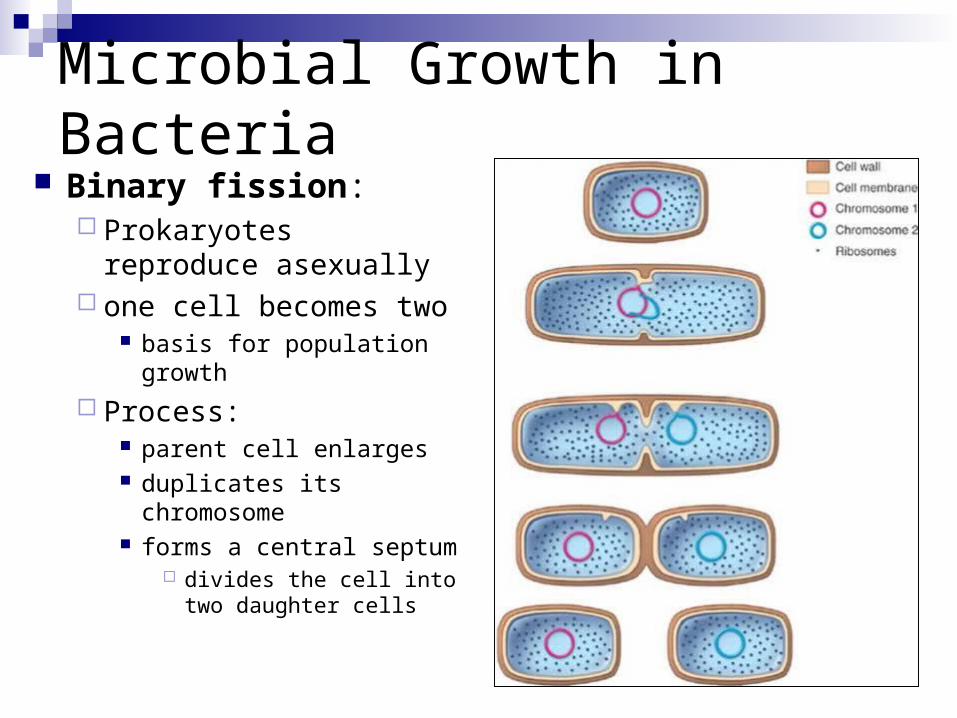
Microbial Growth in Bacteria Binary fission:
Prokaryotes reproduce asexually
one cell becomes two basis for population growth
Process: parent cell enlarges duplicates its chromosome forms a central septum
divides the cell into two daughter cells

Population Growth
Generation / doubling time time required for a complete
fission cycle Length of the generation time
is a measure of the growth rate of an organism
Some populations can grow from a small number of cells to several million in only a few hours!!
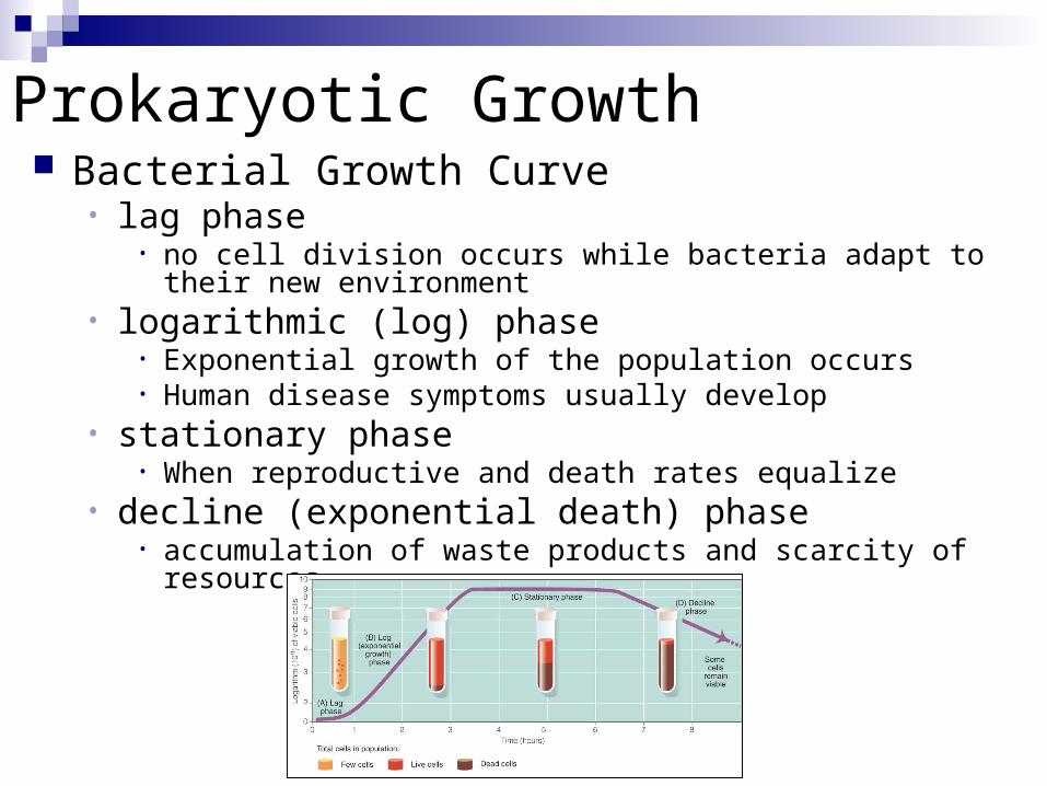
Prokaryotic Growth Bacterial Growth Curve
• lag phase• no cell division occurs while bacteria adapt to their new
environment• logarithmic (log) phase
• Exponential growth of the population occurs • Human disease symptoms usually develop
• stationary phase• When reproductive and death rates equalize
• decline (exponential death) phase• accumulation of waste products and scarcity of resources

Other Methods of Analyzing Population Growth
Turbidity Direct microscopic count Coulter counting
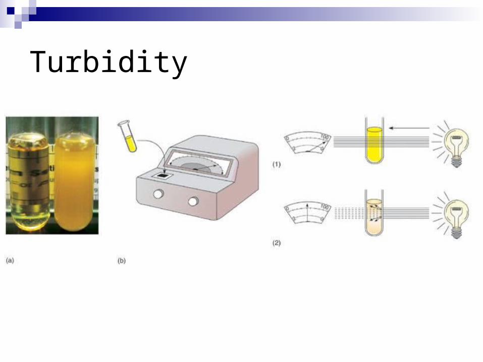
Turbidity
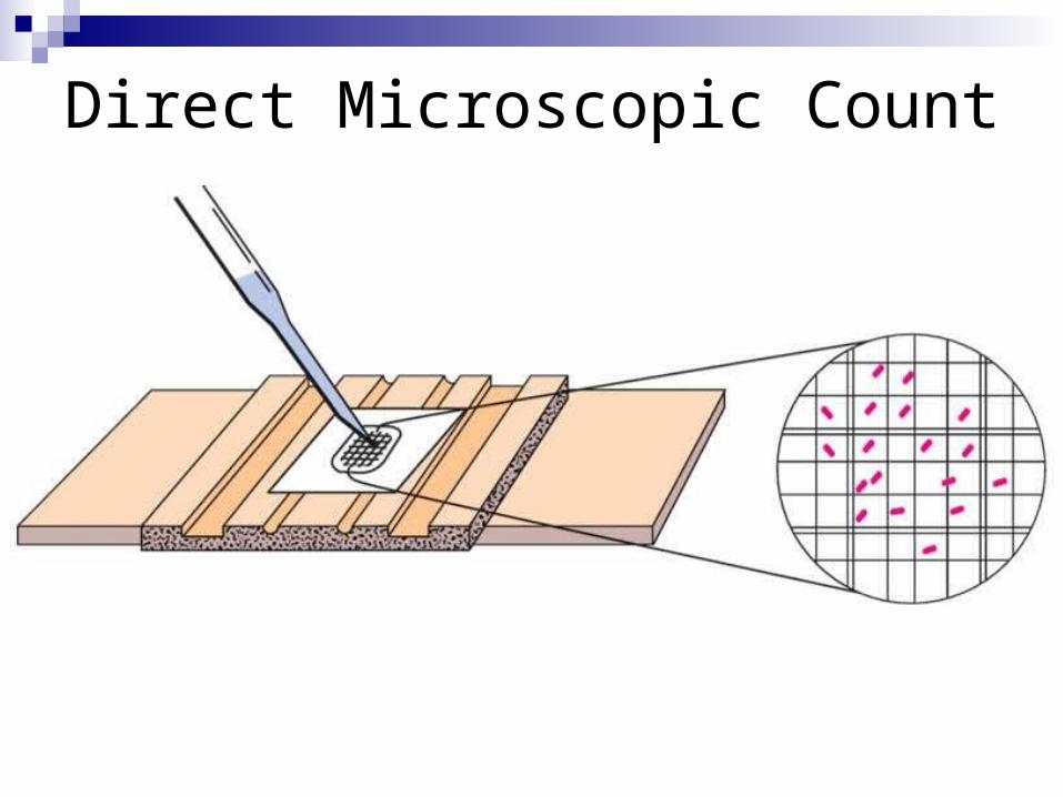
Direct Microscopic Count
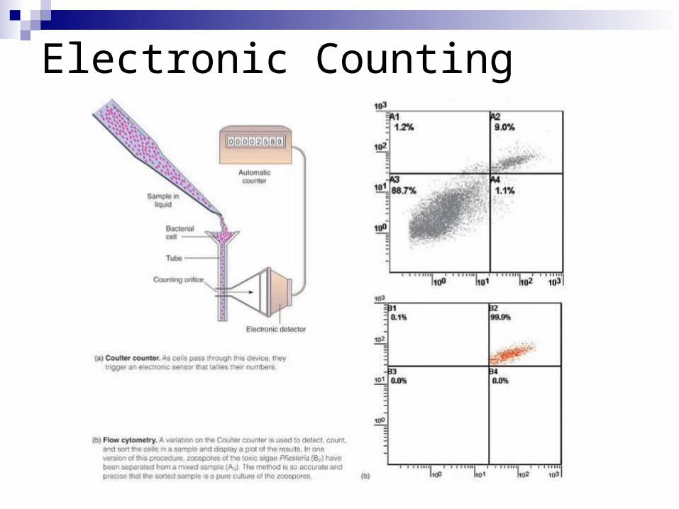
Electronic Counting
Microbial genetics
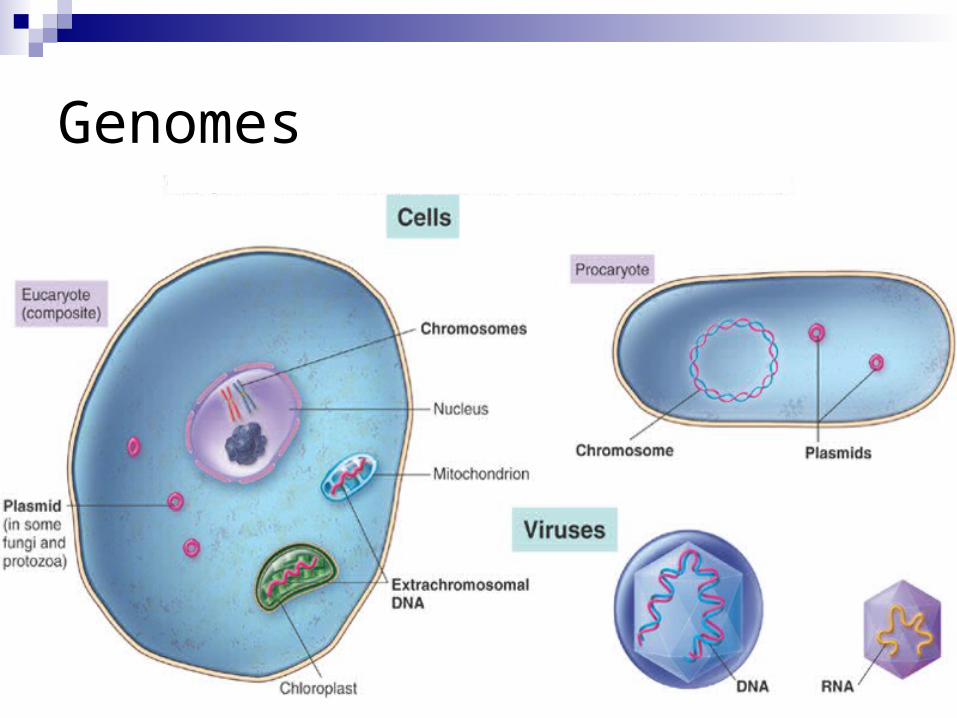
Genomes
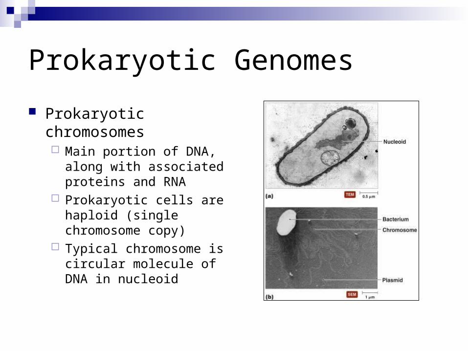
Prokaryotic Genomes
Prokaryotic chromosomes Main portion of DNA, along
with associated proteins and RNA
Prokaryotic cells are haploid (single chromosome copy)
Typical chromosome is circular molecule of DNA in nucleoid

DNA Replication in Prokaryotes
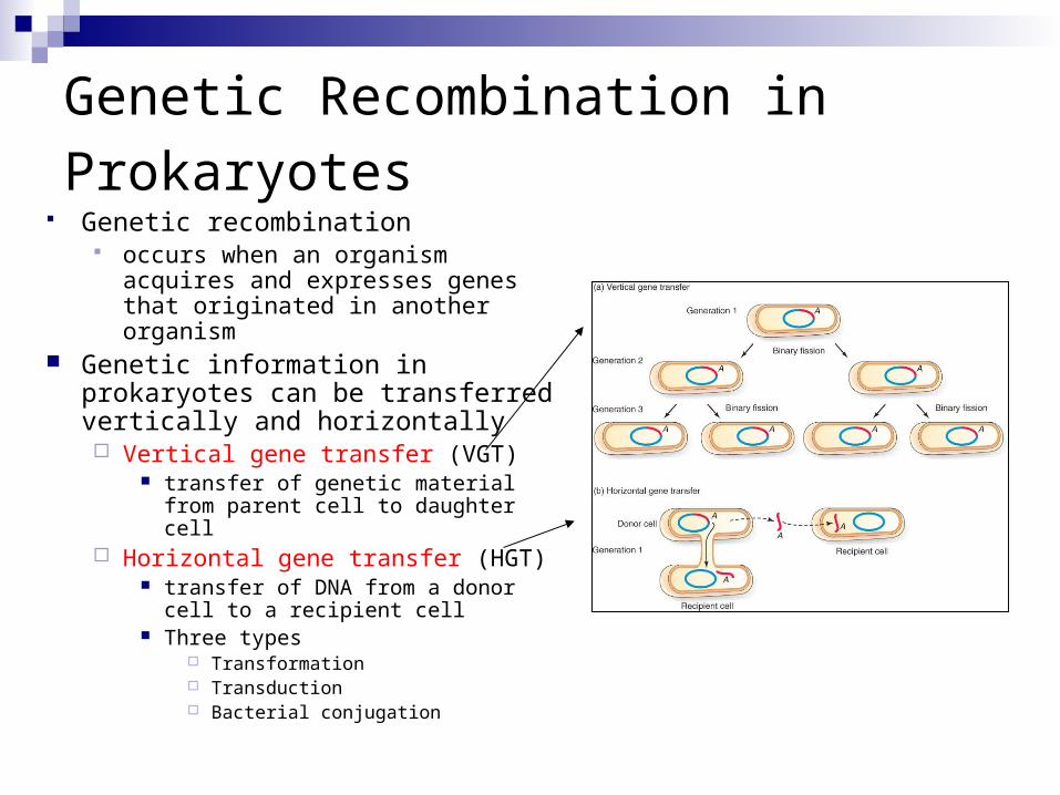
Genetic Recombination in Prokaryotes Genetic recombination
occurs when an organism acquires and expresses genes that originated in another organism
Genetic information in prokaryotes can be transferred vertically and horizontally Vertical gene transfer (VGT)
transfer of genetic material from parent cell to daughter cell
Horizontal gene transfer (HGT) transfer of DNA from a donor cell to
a recipient cell Three types
Transformation Transduction Bacterial conjugation

DNA Recombination Events 3 means for genetic recombination in
bacteria:1. Conjugation
2. Transformation
3. Transduction
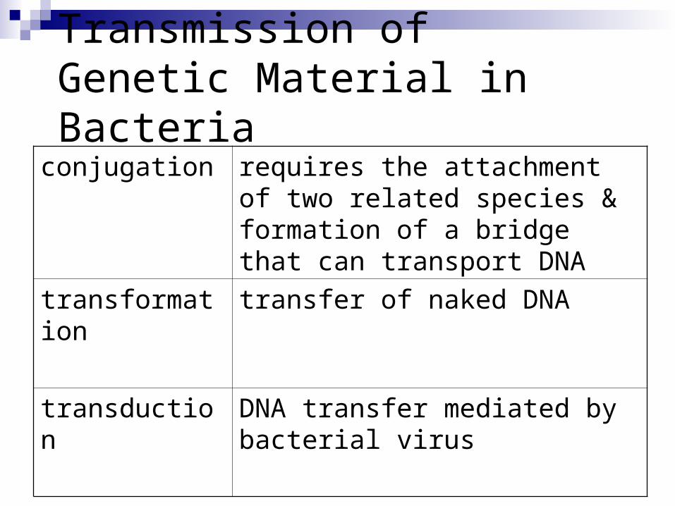
Transmission of Genetic Material in Bacteria
conjugation requires the attachment of two related species & formation of a bridge that can transport DNA
transformation transfer of naked DNA
transduction DNA transfer mediated by bacterial virus
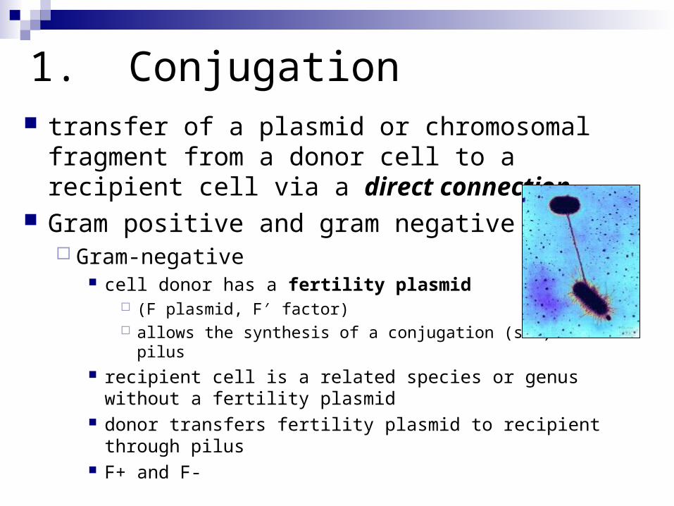
1. Conjugation transfer of a plasmid or chromosomal fragment
from a donor cell to a recipient cell via a direct connection
Gram positive and gram negative Gram-negative
cell donor has a fertility plasmid (F plasmid, F′ factor) allows the synthesis of a conjugation (sex) pilus
recipient cell is a related species or genus without a fertility plasmid
donor transfers fertility plasmid to recipient through pilus F+ and F-

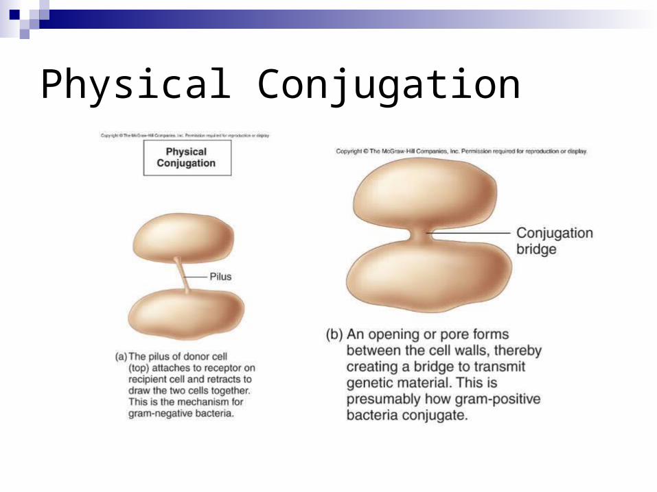
Physical Conjugation
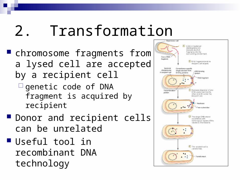
2. Transformation chromosome fragments from a
lysed cell are accepted by a recipient cell genetic code of DNA fragment is
acquired by recipient
Donor and recipient cells can be unrelated
Useful tool in recombinant DNA technology
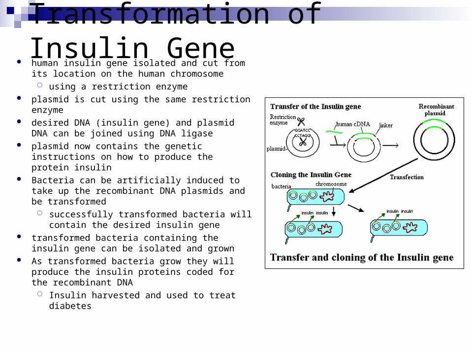
Transformation of Insulin Gene human insulin gene isolated and cut from its
location on the human chromosome using a restriction enzyme
plasmid is cut using the same restriction enzyme desired DNA (insulin gene) and plasmid DNA can
be joined using DNA ligase plasmid now contains the genetic instructions on
how to produce the protein insulin Bacteria can be artificially induced to take up the
recombinant DNA plasmids and be transformed successfully transformed bacteria will contain
the desired insulin gene transformed bacteria containing the insulin gene
can be isolated and grown As transformed bacteria grow they will produce
the insulin proteins coded for the recombinant DNA Insulin harvested and used to treat diabetes

3. Transduction DNA is transferred from one
bacterium to another by a virus
Bacteriophages Virus that infects bacteria consist of an outer protein
capsid enclosing genetic material
serves as a carrier of DNA from a donor cell to a recipient cell
3. Transduction
Two types: generalized transduction
random fragments of disintegrating host DNA are picked up by the phage during assembly
any gene can be transmitted this way
specialized transduction a highly specific part of the host genome is
regularly incorporated into the virus
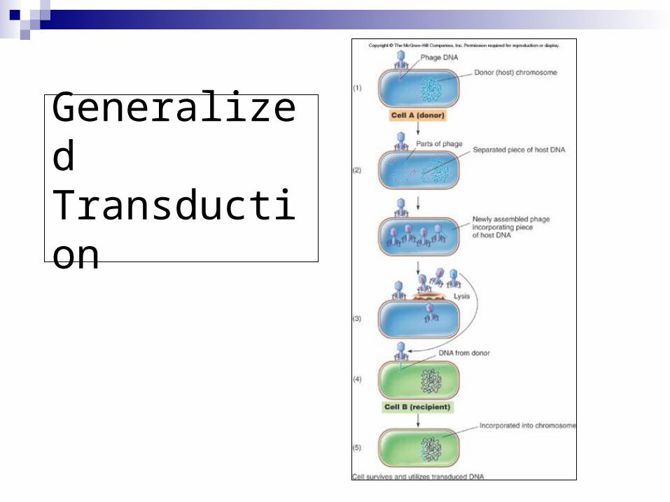
Generalized Transduction
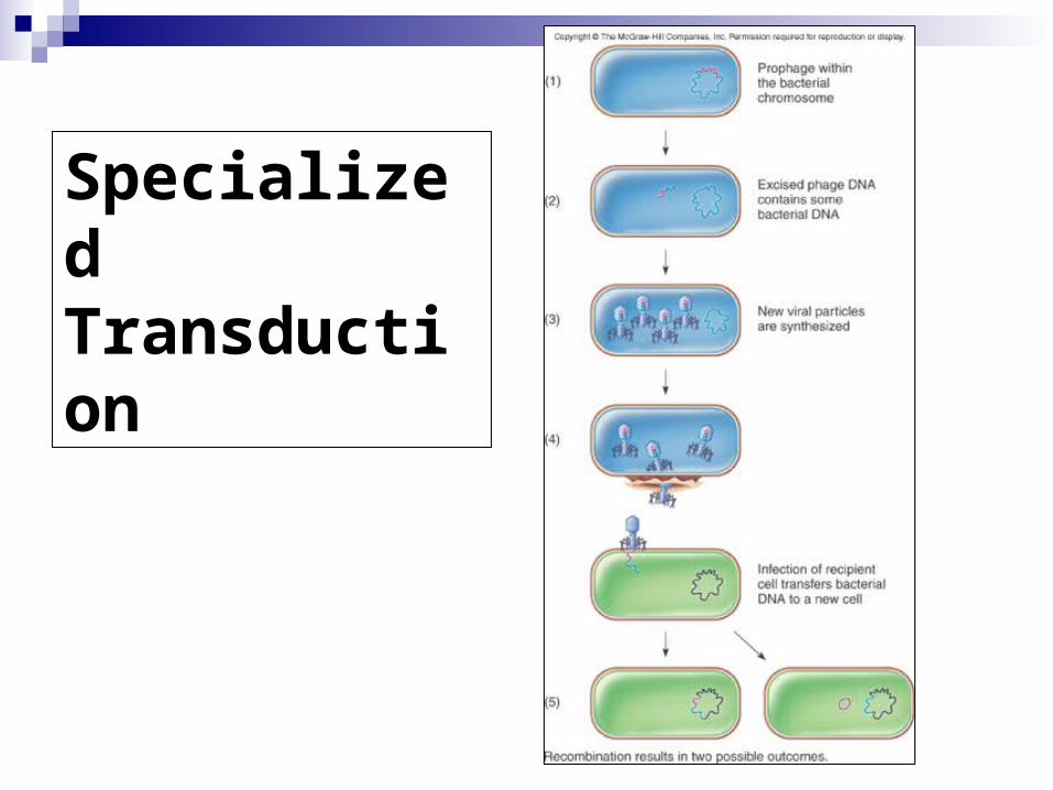
Specialized Transduction
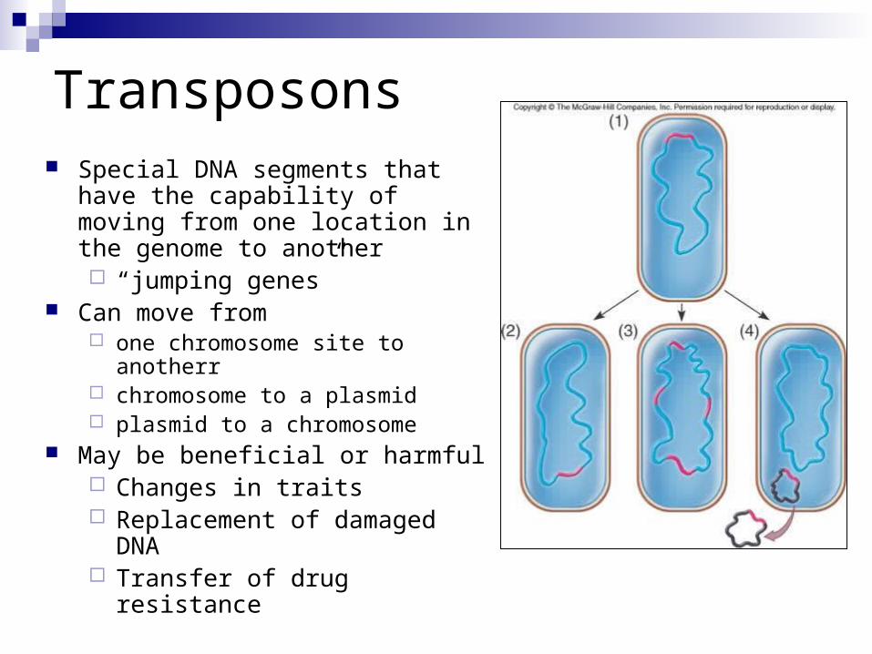
Transposons Special DNA segments that have the
capability of moving from one location in the genome to another “jumping genes”
Can move from one chromosome site to anotherr chromosome to a plasmid plasmid to a chromosome
May be beneficial or harmful Changes in traits Replacement of damaged DNA Transfer of drug resistance
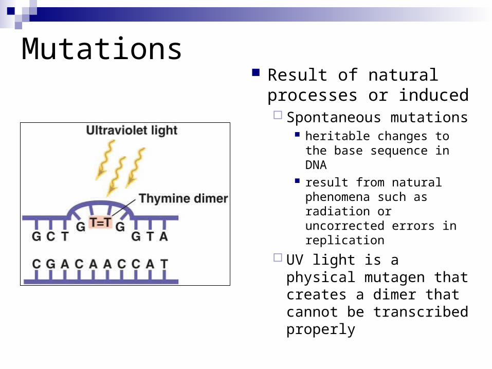
Mutations Result of natural
processes or induced Spontaneous mutations
heritable changes to the base sequence in DNA
result from natural phenomena such as radiation or uncorrected errors in replication
UV light is a physical mutagen that creates a dimer that cannot be transcribed properly
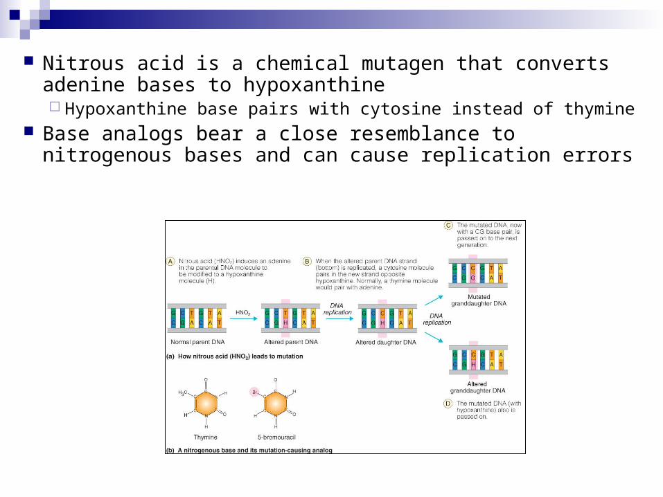
Nitrous acid is a chemical mutagen that converts adenine bases to hypoxanthine Hypoxanthine base pairs with cytosine instead of thymine
Base analogs bear a close resemblance to nitrogenous bases and can cause replication errors
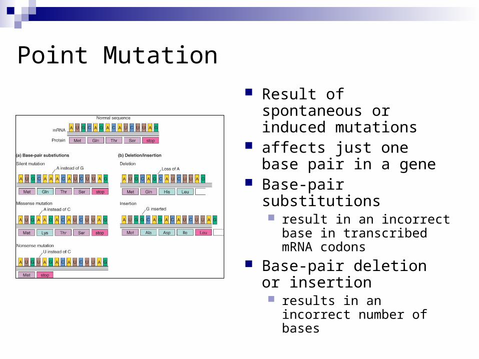
Result of spontaneous or induced mutations
affects just one base pair in a gene
Base-pair substitutions result in an incorrect base in
transcribed mRNA codons Base-pair deletion or
insertion results in an incorrect
number of bases
Point Mutation
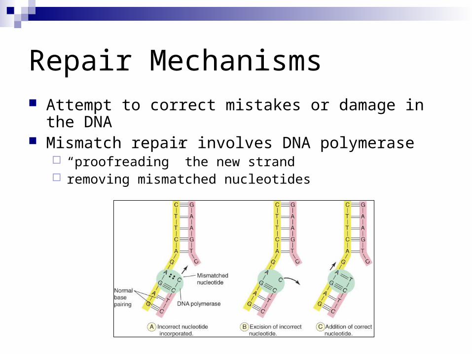
Attempt to correct mistakes or damage in the DNA Mismatch repair involves DNA polymerase
“proofreading” the new strand removing mismatched nucleotides
Repair Mechanisms
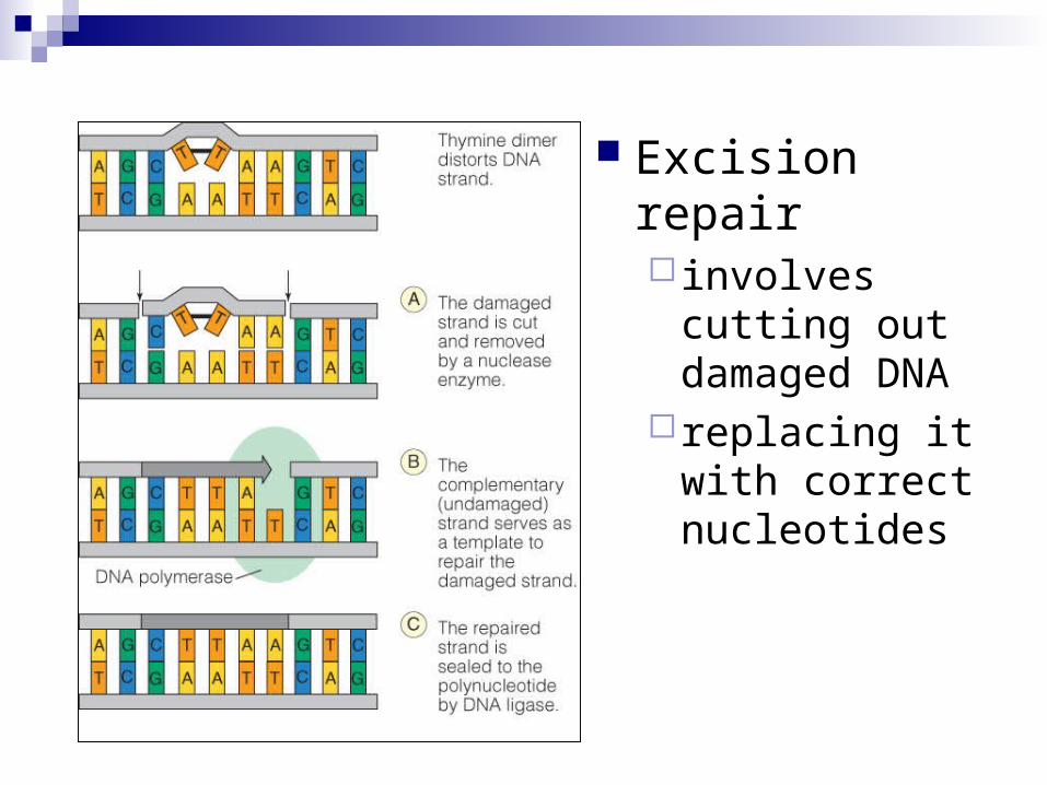
Excision repair involves cutting out
damaged DNA replacing it with
correct nucleotides