Embed Size (px)
Citation preview

1
PD-L1 expression in endometrial carcinoma cells and intratumoral immune cells:1
differences across histological and TCGA-based molecular subgroups2
Annukka Pasanen MD1,2, Terhi Ahvenainen MSc3,4, Teijo Pellinen PhD5, Pia Vahteristo PhD3,4, Mikko3
Loukovaara MD, PhD6, Ralf Bützow MD, PhD1,2,64
5
1Department of Pathology, University of Helsinki and Helsinki University Hospital, Helsinki, Finland.6
2Applied Tumor Genomics Research Program, University of Helsinki, Helsinki, Finland7
3Department of Medical and Clinical Genetics, Medicum, University of Helsinki, Helsinki, Finland.8
4Genome-Scale Biology Research Program, Research Programs Unit, University of Helsinki, Helsinki,9
Finland.10
5Institute for Molecular Medicine Finland (FIMM), Helsinki, Finland.11
6Department of Obstetrics and Gynecology, University of Helsinki and Helsinki University Hospital,12
Helsinki, Finland.13
14
Corresponding author:15
Annukka Pasanen, Department of Pathology, University of Helsinki and Helsinki University Hospital,16
Haartmaninkatu 3, 00290 Helsinki, Finland. Electronic address: [email protected]
18
19
20
21
22
23
Disclosures: The author(s) have no conflicts of interest or funding to disclose.24
25
26
27
2
Abstract28
PD-L1 is a biomarker that may predict the response to antiPD-1/PD-L1 immunotherapy. We29
evaluated the expression of PD-L1 in carcinoma cells and immune cells across30
histopathological and TCGA molecular subgroups of endometrial carcinoma.31
Our study included 842 patients with endometrial carcinoma. Direct sequencing of polymerase32
epsilon (POLE) exonuclease domain hot spots and conventional immunohistochemistry33
(MLH1, PMS2, MSH2, MSH6, p53) were conducted to identify TCGA classification-based34
molecular subgroups of endometrial carcinoma: POLE-mutated, mismatch repair (MMR)35
deficient, no specific molecular profile and p53-aberrant. Multiplex immunohistochemistry was36
performed to evaluate PD-L1 expression in carcinoma cells (Ca) and tumor-infiltrating immune37
cells (ICs). PD-L1 expression in carcinoma cells and in ICs was detected in 8.6% and 27.7%38
of the cases, respectively. Combined positive score (CPS) was ≥1% in 19.4% of the samples.39
PD-L1 positivity in carcinoma cells, ICs and CPS correlated with tumor T cell density (TILs,40
p<0.001). POLE-mutated and MMR-deficient tumors were more likely to present PD-L141
expressing ICs, CPS positivity and abundant TILs compared with other TCGA subgroups42
(p<0.001). No differences existed in Ca-PD-L1 expression (p=0.366). Within various43
histotypes, non-endometrioid carcinomas displayed the highest Ca-PD-L1, ICs and CPS44
(p<0.03). Advanced cancers showed more frequent Ca-PD-L1 positivity (p=0.016), CPS45
(p=0.029) and IC≥1% (p=0.037) positivity compared to early disease.46
In conclusion, PD-L1 expression profiles differ between molecular subclasses, histological47
subtypes and disease stage of endometrial carcinoma. Prospective studies are needed to explore48
the predictive value of various PD-L1 scoring systems within the subgroups of endometrial49
cancer. CPS presents methodological advantages over cell-type specific scoring systems.50
51
3
Key words: endometrial carcinoma, PD-L1, TCGA classification52
53
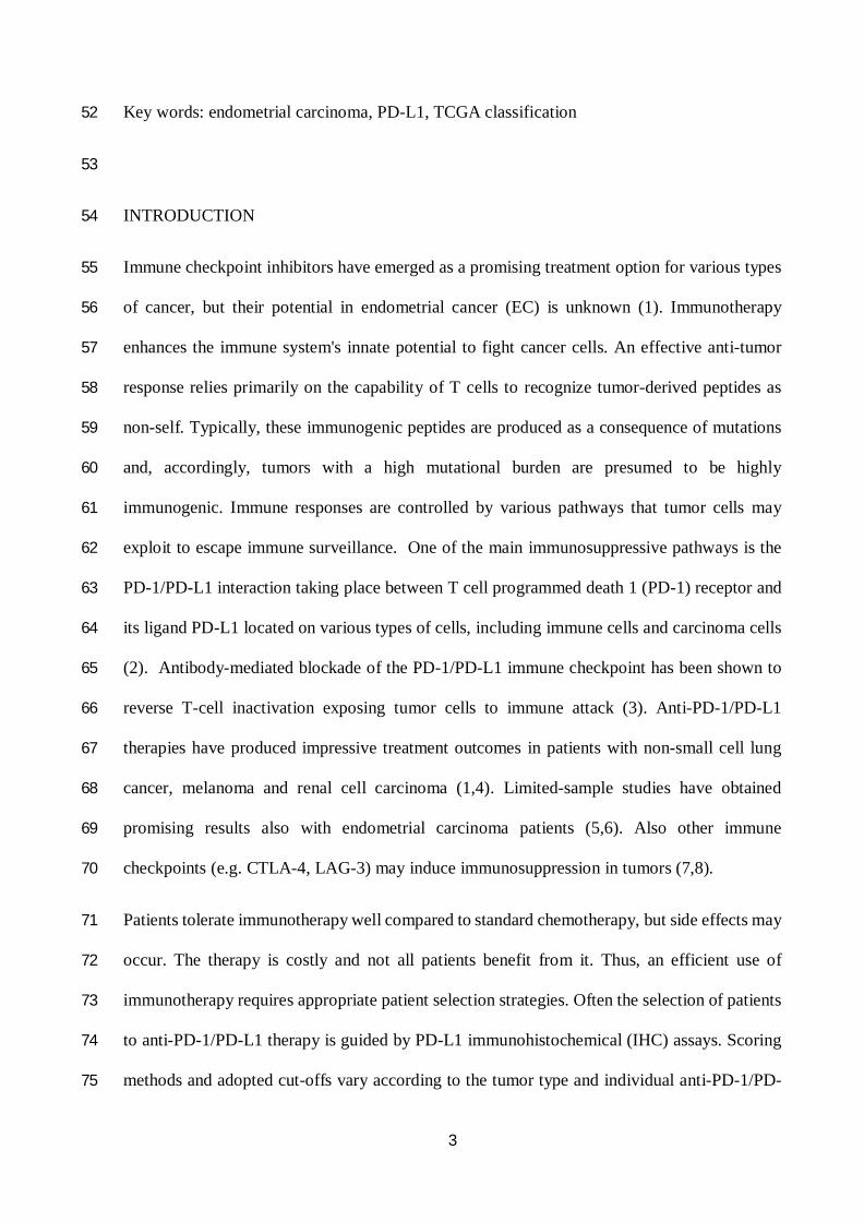
INTRODUCTION54
Immune checkpoint inhibitors have emerged as a promising treatment option for various types55
of cancer, but their potential in endometrial cancer (EC) is unknown (1). Immunotherapy56
enhances the immune system's innate potential to fight cancer cells. An effective anti-tumor57
response relies primarily on the capability of T cells to recognize tumor-derived peptides as58
non-self. Typically, these immunogenic peptides are produced as a consequence of mutations59
and, accordingly, tumors with a high mutational burden are presumed to be highly60
immunogenic. Immune responses are controlled by various pathways that tumor cells may61
exploit to escape immune surveillance. One of the main immunosuppressive pathways is the62
PD-1/PD-L1 interaction taking place between T cell programmed death 1 (PD-1) receptor and63
its ligand PD-L1 located on various types of cells, including immune cells and carcinoma cells64
(2). Antibody-mediated blockade of the PD-1/PD-L1 immune checkpoint has been shown to65
reverse T-cell inactivation exposing tumor cells to immune attack (3). Anti-PD-1/PD-L166
therapies have produced impressive treatment outcomes in patients with non-small cell lung67
cancer, melanoma and renal cell carcinoma (1,4). Limited-sample studies have obtained68
promising results also with endometrial carcinoma patients (5,6). Also other immune69
checkpoints (e.g. CTLA-4, LAG-3) may induce immunosuppression in tumors (7,8).70
Patients tolerate immunotherapy well compared to standard chemotherapy, but side effects may71
occur. The therapy is costly and not all patients benefit from it. Thus, an efficient use of72
immunotherapy requires appropriate patient selection strategies. Often the selection of patients73
to anti-PD-1/PD-L1 therapy is guided by PD-L1 immunohistochemical (IHC) assays. Scoring74
methods and adopted cut-offs vary according to the tumor type and individual anti-PD-1/PD-75
4
L1 agents. Proposed scoring algorithms evaluate PD-L1 positivity in carcinoma cells and/or76
immune cells separately or in combination (combined positive score, CPS) (9-11). Reported77
frequencies of PD-L1 positivity in endometrial carcinoma vary considerably (0.9-44.3%) even78
in unselected EC cohorts (Table 1) (12-24). Such variability may in part derive from different79
antibody clones and different cut-offs. In fact, notable interassay variation has been reported80
within commercially available PD-L1 immunohistochemical assays (25,26). Accuracy of IHC81
scorings may also suffer from problems related to traditional chromogenic PD-L182
immunohistochemistry. Staining of the tumor cells may be weak and unspecific cytoplasmic83
staining occurs. Moreover, intratumoral T cells and macrophages often present membranous84
staining and they may be misinterpreted as carcinoma cells (27). Multiplex IHC overcomes85
these limitations by simultaneous detection of a biomarker and numerous cell-specific markers86
on a single paraffin tissue section, allowing the identification and quantification of various cell87
types expressing the antigen of interest (28).88
Endometrial carcinoma is not a uniform disease entity, as it comprises various histological and89
molecular subgroups, each with their own clinicopathological characteristics. Given this90
heterogeneity, exhaustive biomarker studies rely on well-powered subclass analyses. The goal91
of our study was to explore PD-L1 expression and T cell inflammation within histological92
subtypes and TCGA-based molecular subgroups of endometrial cancer. Fluorescent multiplex93
immunohistochemistry was performed to overcome limitations related to traditional94
immunohistochemical evaluation.95
96
MATERIALS AND METHODS97
Patients who underwent primary surgical treatment for endometrial cancer at the Department98
of Obstetrics and Gynecology, Helsinki University Hospital, between January 1, 2007, and99
5
December 31, 2012, were identified (n = 965). Patients with adequate tumor samples for a tissue100
microarray (TMA) were included in the study (n = 842). Approvals of the Institutional Review101
Board and the National Authority for Medicolegal Affairs of Finland were obtained. Clinical102
data were collected from institutional medical records. Lacking follow-up data were obtained103
from Statistics Finland or completed by contacting primary physicians at the referring104
institutions.105
We performed immunohistochemistry on multicore tissue microarray (TMA) slides, prepared106
as described before (29). The following monoclonal antibodies were used for chromogenic107
immunohistochemistry: MLH1 (ES05, Dako), PMS2 (EPR3947, Epitomics), MSH-2 (G219-108
1129, BD Biosciences), MSH-6 (EPR3945, Abcam), p53 (DO-7, Dako) and PD-L1 (SP263,109
Ventana). TMA slides were scanned with 3-dimensional Histech Pannoramic 250 Flash II110
scanner by Fimmic Oy (Helsinki, Finland). Slide images were managed and analyzed with111
WebMicroscope Software (Fimmic Oy). Virtual slides were scored by a pathologist blinded to112
clinical data. A second investigator examined equivocal cases and a consensus was reached.113
Mismatch repair protein status was considered deficient (MMRd) when we observed a complete114
loss of nuclear expression in carcinoma cells of one or more MMR proteins (MLH1, PMS2,115
MSH2, MSH6) detected by immunohistochemistry. Aberrant p53 staining was defined as116
strong and diffuse nuclear staining or completely negative (‘null’) staining in carcinoma cells.117
Weak and heterogeneous staining was classified as wild type expression. Stromal cells and118
inflammatory cells served as internal control for MMR and p53 stainings. Samples with scarce119
carcinoma cells or with completely negative staining of the internal control (when applicable),120
were discarded. Representative images of MLH-1 and p53 staining patterns are shown in Figure121
1.122
The fluorescent multiplex immunohistochemistry was carried out as described by Blom et al.123
with following modifications (30). Primary antibodies were: PD-L1 (CST, E1L3N), CD3124

6
(Thermo, MA5-14482), CD163 (Abcam, ab188571), and PanEpi (cocktail of anti-PanCk, C-125
11, Abcam, Ab77531; anti-PanCk AE1/AE3, InVitrogen, 180132; E-cadherin, BD clone 36).126
Nuclei were stained using DAPI (Roche). Five-channel fluorescent images were acquired using127
Metafer 5 scanning and imaging platform (MetaSystems, Alltlussheim, Germany) equipped128
with AxioImager Z2 microscope with a 20x objective (Carl Zeiss, Goettingen, Germany) and a129
CoolCube 2m CCD camera (MetaSystems, Alltlussheim, Germany). The image analysis was130
carried out both visually by a pathologist and by a cell image analysis software (CellProfiler131
version 2.2.0). Scoring was primarily performed by a pathologist and in rare equivocal cases132
automated image analysis was used to support the decision-making. Necrotic areas and scarce133
samples (<100 cells) were excluded from scoring. PD-L1 expression was defined as partial or134
complete membranous staining in carcinoma cells and membranous and/or cytoplasmic135
staining in immune cells (CD3-positive T lymphocytes and CD163-positive macrophages136
within tumor nests and/or adjacent supporting stroma). We determined the percentage of137
positive carcinoma cells and immune cells separately and in combination. To calculate the138
combined positive score (CPS), we divided the total number of PD-L1-positive cells (carcinoma139
cells, lymphocytes, and macrophages) by the number of viable carcinoma cells, multiplied by140
100 (9). Semiquantitative scoring was adopted as follows: 0: <1% of the cells; 1: 1-4%; 2: 5-141
9%; 3: 10-49%; 4: ≥50%. The cut-off for positive PD-L1 staining was set at 1%. The cut off142
for strong positivity was set according to the results of a previous randomized trial (≥50% for143
carcinoma cells and ≥10% for immune cells) (31). Comparative images of conventional144
chromogenic immunohistochemistry and multiplex immunofluorescence of PD-L1 positive and145
negative cells are shown in Figure 2. Tumoral CD3+ lymphocytic infiltration (TILs) was semi-146
quantitatively scored as scarce, moderate or abundant.147
For DNA extraction, representative areas of formalin-fixed paraffin-embedded tumor tissue148
were macrodissected as identified by pathologist assessment. DNA was extracted by proteinase149
7
K/phenol-chloroform method. POLE exonuclease domain mutation screening of hot spots in150
exon 9 (c.857C>G, p.P286R; c.890C>T, p.S297F), exon 13 (c.1231G>C, p.V411L) and exon151
14 (c.1366G>C, p.A456P), was performed by direct sequencing. The following primers were152
used: Ex 9F (5’-3’): CCTAATGGGGAGTTTAGAGCTT; Ex 9R (5’-3’):153
CCCATCCCAGGAGCTTACTT; Ex 13F (5’-3’): TCTGTTCTCATTCTCCTTCCAG; Ex154
13R (5’-3’): CGGGATGTGGCTTACGTG; Ex 14F (5’-3’): TGACCCTGGGCTCTTGATTT;155
Ex 14R (5’-3’): ACAGGACAGATAATGCTCACC. PCR products were sequenced on an156
ABI3730xl Automatic DNA Sequencer at Institute for Molecular Medicine Finland (FIMM),157
Helsinki. Sequence graphs were analyzed both manually and with Mutation Surveyor158
(Softgenetics, State College, PA). Only cases with good-quality sequence for all the examined159
POLE hot spots were included in the analysis.160
Pearson χ² test and Fisher exact test were used for comparisons of categorical variables. Survival161
curves were calculated by the Kaplan-Meier method. A log-rank test was used to test for survival162
differences. Disease-specific survival was defined as the time from date of surgery to death from163
endometrial cancer. Statistical significance was set at p < 0.05. Cohen’s kappa statistics were164
calculated to measure the agreement between multiplex and chromogenic immunohistochemistry165
for PD-L1. Based on kappa references outlined by Landis and Koch, the strength of agreement166
was considered fair for kappa values between 0.21 and 0.40 and moderate for kappa values167
between 0.41 and 0.60 (32). Data were analyzed using IBM SPSS version 25 software (IBM168
Corp., Armonk, New York, USA).169
170
RESULTS171
Clinicopathological characteristics of the study cases are summarized in Table 2. Of the 842172
patients included in the study, 745 (88.5%) had endometrioid and 97 (11.5%) non-endometrioid173
carcinoma. Median follow-up of patients was 78 months (range 1 to 136 months). Sequencing174

8
of all the targeted genomic regions of POLE was successful for 553 cases. POLE mutation was175
detected in 7.4% of endometrioid carcinomas and 4.0% of non-endometrioid carcinomas (6.7%176
of all cases). MMR protein deficiency was found in 37.7% of endometrioid carcinomas and177
25.6% of non-endometrioid carcinomas (36.2% of all the cases). Aberrant p53 profile was178
detected in 10.8% of endometrioid tumors and 61.9% of non-endometrioid tumors (16.8% of179
all the samples). A minority of cases displayed multiple molecular features. Both POLE180
mutation and aberrant p53 expression were present in 0.4% of the cases and both POLE181
mutation and MMR protein deficiency in 0.2% of the patients. Only one sample (0.2%) had all182
three molecular alterations. These patients were allocated into the POLEmut molecular183
subgroup. Both MMR deficiency and aberrant p53 status were detected in 3.1% of the cases.184
These were classified as MMRd tumors.185
In the multiplex immunofluorescence staining, 8.6% of the cases presented PD-L1 expression186
on carcinoma cells (Ca≥1%) and 27.7% on intratumoral immune cells (ICs≥1%). CPS was187
positive (CPS≥1%) in 19.4% of the samples. High PD-L1 expression (Ca≥50% or ICs≥10%)188
was observed in 0.5% and 8.6% of the cases, respectively. Relative frequencies of189
semiquantitative staining scores are presented in Table 3. Tumors with moderate-abundant T190
cell density presented PD-L1 positivity in carcinoma cells (10.6%), ICs (36.6%) and CPS191
(26.8%) more frequently than tumors with scarce lymphocytic infiltration (Ca 5.6%, p=0.019;192
ICs 14.6%, p<0.001, CPS 8.4%, p<0.001). Concomitant presence of moderate-abundant T cell193
infiltrates and any PD-L1 positivity (“T cell inflamed PD-L1 positive” phenotype), was194
observed in 25.1% of all the tumors.195
Relative frequencies of PD-L1 positivity in carcinoma cells varied significantly between196
histotypes: endometrioid carcinoma 8.0%, clear cell carcinoma 14.7%, serous carcinoma 3.7%,197
undifferentiated carcinoma 14.7% and carcinosarcoma 20% (p=0.022, Figure 3). Observed198
relative frequencies of CPS≥1% were: endometrioid carcinoma 17.1%, clear cell carcinoma199

9
38.2%, serous carcinoma 37.0%, undifferentiated carcinoma 42.9% and carcinosarcoma 26.7%200
(p<0.001; Figure 3). Similar differences were noted in the immune cell expression of PD-L1201
with significantly higher relative frequencies of expression in the non-endometrioid carcinomas202
(p=0.028). By contrast, we found no statistically significant differences between histological203
subgroups and strong PD-L1 positivity in ICs (p=0.148, Figure 3). Our cohort included only204
one neuroendocrine carcinoma, which presented PD-L1 expression on both carcinoma cells and205
immune cells. We found no correlation between Ca-PD-L1 expression and grade of206
differentiation of endometrioid carcinomas (G1-2 vs G3, p=0.08), whereas CPS and IC≥10%207
PD-L1 expression were more frequent in G3 as compared to G1-2 endometrioid carcinomas208
(33.0% vs 14.3% and 20.8% vs 5.4%, respectively, p<0.001). The overall quantity of CD3+209
TILs (scarce-moderate vs abundant) did not differ significantly in histological subgroups210
(p=0.158) or between grade of differentiation of endometrioid carcinoma (p=0.722).211
PD-L1 expression profiles were also analyzed according to FIGO 2009 stage of disease (stage212
I-II vs III-IV, Figure 3). Samples from patients with advanced stage (III-IV) disease were more213
likely to present Ca-PD-L1 positivity (13.6% vs 7.5%, p=0.016), CPS (25.9% vs 18.0%,214
p=0.029) and IC≥1% (34.7% vs 26.2%, p=0.037) positivity as compared to early stage (I-II)215
disease. Differences in the IC≥10% (p=0.270) or the overall quantity of TILs (p=0.598) were216
not statistically significant. In advanced disease, strong Ca-PD-L1 positivity was found in 1.4%217
of the cases and strong IC positivity in 10.9% of the cases.218
Samples with successful POLE sequencing and immunohistochemical stainings of MMR219
proteins and p53 (512 cases), were stratified into TCGA-based molecular subclasses. POLEmut220
and MMRd tumors exhibited higher relative frequencies of immune cell PD-L1 positivity221
(55.9% and 40.9%) and CPS positivity (44.1% and 29.6%) compared to NSMP (IC: 13.9%,222
CPS: 9.1%) and p53ab cases (25.4%, 20.9%; p<0.001, Figure 4). Significant differences were223
observed also for strong positivity in ICs (p<0.001, Figure 4). POLEmut and MMRd cases were224
10
also more likely to present abundant intratumoral T cell infiltrates (26.5% and 27.8%225
respectively) compared to NSMP and p53ab cases (15.3% and 16.7% respectively; p=0.014).226
Similarly, we observed “T cell inflamed PD-L1 positive” phenotype more frequently in227
POLEmut and MMRd groups (50.0% and 34.9%, respectively) compared to other TCGA228
subclasses (16.3% and 17.9%; p<0.001). PD-L1 expression in carcinoma cells showed no229
correlation with TCGA classification (p=0.366).230
In Kaplan Meier analysis, disease specific survival segregated by histotype and TCGA231
subgroups as expected (p≤0.001, data not shown). POLEmut group had excellent outcomes (no232
disease related deaths in this group) and aberrant p53 status associated with poor disease233
specific survival. Scarce overall quantity of TILs predicted poor prognosis (p =0.001), whereas234
PD-L1 expression on carcinoma cells, ICs or CPS showed no correlation with outcome235
(p=0.298, p=0.592, p = 0.569, respectively).236
According to kappa statistics, multiplex and chromogenic immunohistochemistry scorings237
showed moderate agreement for CPS (kappa 0.540) and poor agreement for PD-L1 expression238
in carcinoma cells (kappa 0.279).239
240
DISCUSSION241
In the evolving era of personalized medicine, immunotherapy offers new treatment options for242
cancer patients. FDA has approved mismatch repair deficiency/microsatellite instability as243
selection criteria for anti PD-1/PD-L1 therapy (33). Treatment indications in mismatch repair244
stable EC and the role of biomarkers, including PD-L1, have remained unsettled. To facilitate245
prospective studies, we profiled PD-L1 expression across histopathological and TCGA246
molecular subgroups of endometrial carcinoma.247

11
In our study cohort, 8.6% of the cases presented PD-L1 expression in carcinoma cells and248
27.7% in the ICs. In line with previous studies, PD-L1 expression on carcinoma cells or on249
lymphocytes showed no correlation with survival (18,19). Non-endometrioid carcinomas were250
more likely to present PD-L1 positive carcinoma cells, CPS and ICs compared to endometrioid251
carcinomas. In the subgroup of endometrioid ECs, high grade of differentiation was associated252
with more frequent CPS and IC positivity compared to low grade disease.253
In a landmark study, The Cancer Genome Atlas (TCGA) identified 4 distinct molecular254
subgroups of endometrial carcinoma: POLE ultramutated, microsatellite instability255
hypermutated (MSI-H), copy-number-low microsatellite stable (MSS), and copy-number-high256
(34). Vast majority (90%) of the copy-number-high tumors presented TP53 mutations.257
Consequently, TP53 mutational analysis or immunohistochemical analysis of p53 expression258
have been proposed as a surrogate marker for this subgroup of tumors (35,36). POLE mutated259
tumors are characterized by defects in the proof-reading function of DNA polymerase epsilon260
and harbor the highest rate of somatic mutations, followed by MSI-H tumors characterized by261
defects in DNA mismatch repair activity. These highly mutated tumors have been reported to262
contain a large number of predicted neoantigens and activated cytotoxic tumor infiltrating T263
lymphocytes, often expressing PD-1 and PD-L1 (13,17,24,37,38). Corroborating these264
findings, we observed significantly higher relative frequencies of heavy T cell infiltrates and265
PD-L1 expressing ICs in the POLE mutated and MMR deficient groups compared to other266
TCGA subgroups. By contrast, we found no correlation between Ca-PD-L1 expression and the267
molecular subclasses.268
It has been speculated, that tumors most likely to respond to PD-1/PD-L1 blockade269
characteristically present an “adaptive resistance” phenotype (T cell inflamed PD-L1 positive270
phenotype, i.e. concomitant presence of intratumoral T cell infiltrates and PD-L1 positivity)271
(39-41). Consequently, based on previous studies and our results, POLEmut and MMRd tumors272

12
become natural candidates for immune checkpoint blockade therapy. Interestingly, in a phase273
II study of an anti-PD-1 agent in patients with various types of advanced cancer (including274
endometrial carcinoma), mismatch-repair status itself, and not PD-L1 expression, predicted275
clinical benefit (42).276
Clinical studies suggest a correlation between increasing levels of PD-L1 expression and drug277
efficacy, but definite scoring systems and cut-offs may be tumor-specific and still need to be278
determined (43,44). Most trials focus on PD-L1 expression on carcinoma cells. Nonetheless,279
various studies report associations between clinicopathological characteristics of EC and PD-280
L1 expression on immune cells rather than tumor cells (13-15,21). The significance of these281
correlations is unknown. In a trial including multiple cancer types, PD-L1 positivity on tumor-282
infiltrating immune cells, but not on tumor cells, predicted response to cancer treatment with283
an anti-PD-L1 agent, MPDL3280A (atezolizumab) (45). Accordingly, for atezolizumab284
treatment, expression in intratumoral immune cells (IC) is also used as an indicator for potential285
response (46). In a randomized lung cancer trial, patients with tumors expressing high levels of286
PD-L1 (defined as Ca≥50% or IC≥10%) derived the greatest benefit from atezolizumab287
treatment (31). We observed high tumoral Ca-PD-L1 expression in only 0.5% of the tumors.288
However, strong IC positivity (≥10%) was seen in 8.6% of the cases. The need for alternative289
treatment options is greatest in advanced stage (III-IV) endometrial carcinoma, which presented290
with stronger Ca-PD-L1, IC and CPS expression levels than early cancers.291
Intratumoral heterogeneity of protein expression may lead to decreased sensitivity in TMA292
studies. Clonal loss of MMR protein expression has been reported and it is not known whether293
focal MMR deficiency could invoke a PD-L1 response in a predominantly intact tumor.294
However, the rate of mismatch repair deficiency in our study was not lower than generally295
reported in the literature. In a study by Sloan et al., heterogeneous PD-L1 positivity in ECs296
typically consisted of individual cells or small clusters of cells, that were fairly evenly297

13
distributed throughout the tumor (18). Further, previous studies have shown that TMAs with298
three core biopsies per tumor adequately represent the tumor phenotype, even with antigens299
known to be heterogeneous (47,48). Since performing MMR or PD-L1 stainings on whole300
sections was not feasible for this vast cohort, to improve sensitivity, we included 4 tissue cores301
from each tumor in our TMA. We have previously demonstrated a high concordance between302
our TMA and the corresponding whole sections, as concerns expression of L1CAM, a highly303
heterogeneous antigen (29). As an advantage, TMA methodology allowed us to analyze a large304
number of cases by multiplex IHC and conventional standardized immunohistochemistry305
(Ventana clone SP263).306
In concordance analysis of multiplex IHC and conventional IHC, carcinoma cell proportion307
score showed only fair agreement, which in part reflects the difficulty of differentiating PD-L1308
positive carcinoma cells from macrophages in the chromogenic IHC. Accordingly, moderate309
agreement was found between CPS scorings. In some cases, chromogenic immunostainings310
presented equivocal staining in the stromal compartment, which may have led to false positivity311
in IC scoring and CPS. Multiplex immunohistochemistry aptly circumvented these limitations.312
Some of the differences between the staining results may be explained by the low cut off for313
PD-L1 positivity (1%) and the use of different PD-L1 antibody clones, i.e. E1L3N for multiplex314
and SP263 for chromogenic IHC. In our experience, multiplex immunohistochemistry clearly315
outperforms traditional IHC when analyzing PD-L1 expression in various cell types. However,316
at the moment it cannot be adopted in routine diagnostics and the problems related to cell-type317
specific scoring systems may be circumvented using a scoring method that combines positivity318
of both carcinoma and intratumoral immune cells. Based on our results, the correlation of such319
score (CPS) to clinicopathological characteristics of endometrial carcinoma is equal or better320
than score based on carcinoma cells only.321
14
In conclusion, we identified differences in PD-L1 expression between histological subtypes,322
disease stage and TCGA-based molecular subgroups of endometrial carcinoma. PD-L1323
positivity was more frequently observed in intratumoral immune cells compared to carcinoma324
cells. Based on our results, prospective trials should consider not only PD-L1 expression on325
carcinoma cells but also immune cells, when stratifying patients with endometrial carcinoma326
for immunotherapy. Combined scoring systems may present methodological advantages over327
cell-type specific scoring. Further studies are necessary to explore the predictive value of this328
differential expression of PD-L1, various scoring methods and the applicability of329
immunotherapy in different subgroups of endometrial cancer.330
331
332
333References334
335
1. Brahmer JR, Tykodi SS, Chow LQ, et al. Safety and activity of anti-PD-L1 antibody in336
patients with advanced cancer N Engl J Med 2012;366:2455-2465.337
2. Freeman GJ, Long AJ, Iwai Y, et al. Engagement of the PD-1 immunoinhibitory receptor338
by a novel B7 family member leads to negative regulation of lymphocyte activation J Exp339
Med 2000;192:1027-1034.340
3. Fife BT, Pauken KE, Eagar TN, et al. Interactions between PD-1 and PD-L1 promote341
tolerance by blocking the TCR-induced stop signal Nat Immunol 2009;10:1185-1192.342
4. Garon EB, Rizvi NA, Hui R, et al. Pembrolizumab for the treatment of non-small-cell lung343
cancer N Engl J Med 2015;372:2018-2028.344

15
5. Ott PA, Bang YJ, Berton-Rigaud D, et al. Safety and Antitumor Activity of Pembrolizumab345
in Advanced Programmed Death Ligand 1-Positive Endometrial Cancer: Results From the346
KEYNOTE-028 Study J Clin Oncol 2017;35:2535-2541.347
6. Le DT, Durham JN, Smith KN, et al. Mismatch repair deficiency predicts response of solid348
tumors to PD-1 blockade. Science 2017;357:409-413.349
7. Pardoll DM. The blockade of immune checkpoints in cancer immunotherapy Nat Rev350
Cancer 2012;12:252-264.351
8. Nirschl CJ, Drake CG. Molecular pathways: coexpression of immune checkpoint352
molecules: signaling pathways and implications for cancer immunotherapy Clin Cancer Res353
2013;19:4917-4924.354
9. www.agilent.com/cs/library/usermanuals/public/29219_pd-l1-ihc-22C3-pharmdx-gastric-355
interpretation-manual_us.pdf356
10. www.ventana.com/documents/PD-L1_SP142-NSCLC-Brochure.pdf357
11. www.ventana.com/documents/PD-L1_SP142-UC-Brochure.pdf358
12. Herzog TJ, Arguello D, Reddy SK, et al. PD-1, PD-L1 expression in 1599 gynecological359
cancers: Implications for immunotherapy. Gynecologic Oncology 2015;137:204-205.360
13. Howitt BE, Shukla SA, Sholl LM, et al. Association of Polymerase e-Mutated and361
Microsatellite-Instable Endometrial Cancers With Neoantigen Load, Number of Tumor-362
Infiltrating Lymphocytes, and Expression of PD-1 and PD-L1. JAMA Oncol 2015;1:1319-363
1323.364
14. Eggink FA, Van Gool IC, Leary A, et al. Immunological profiling of molecularly classified365
high-risk endometrial cancers identifies POLE-mutant and microsatellite unstable carcinomas366
as candidates for checkpoint inhibition. Oncoimmunology 2016;6:e1264565.367
15. Mo Z, Liu J, Zhang Q, et al. Expression of PD-1, PD-L1 and PD-L2 is associated with368
differentiation status and histological type of endometrial cancer Oncol Lett 2016;12:944-950.369

16
16. Bregar A, Deshpande A, Grange C, et al. Characterization of immune regulatory370
molecules B7-H4 and PD-L1 in low and high grade endometrial tumors Gynecol Oncol371
2017;145:446-452.372
17. Yamashita H, Nakayama K, Ishikawa M, et al. Microsatellite instability is a biomarker for373
immune checkpoint inhibitors in endometrial cancer Oncotarget 2017;9:5652-5664.374
18. Sloan EA, Ring KL, Willis BC, et al. PD-L1 Expression in Mismatch Repair-deficient375
Endometrial Carcinomas, Including Lynch Syndrome-associated and MLH1 Promoter376
Hypermethylated Tumors Am J Surg Pathol 2017;41:326-333.377
19. Li Z, Joehlin-Price AS, Rhoades J, et al. Programmed Death Ligand 1 Expression Among378
700 Consecutive Endometrial Cancers: Strong Association With Mismatch Repair Protein379
Deficiency. Int J Gynecol Cancer 2018;28:59-68.380
20. Kim J, Kim S, Lee HS, et al. Prognostic implication of programmed cell death 1 protein381
and its ligand expressions in endometrial cancer Gynecol Oncol 2018;149:381-387.382
21. Asaka S, Yen TT, Wang TL, et al. T cell-inflamed phenotype and increased Foxp3383
expression in infiltrating T-cells of mismatch-repair deficient endometrial cancers Mod Pathol384
2019;32:576-584.385
22. Crumley S, Kurnit K, Hudgens C, et al. Identification of a subset of microsatellite-stable386
endometrial carcinoma with high PD-L1 and CD8+ lymphocytes Mod Pathol 2019;32:396-387
404.388
23. Kucukgoz Gulec U, Kilic Bagir E, Paydas S, et al. Programmed death-1 (PD-1) and389
programmed death-ligand 1 (PD-L1) expressions in type 2 endometrial cancer Arch Gynecol390
Obstet 2019;.391
24. Talhouk A, Derocher H, Schmidt P, et al. Molecular Subtype Not Immune Response392
Drives Outcomes in Endometrial Carcinoma Clin Cancer Res 2018;.393
17
25. McLaughlin J, Han G, Schalper KA, et al. Quantitative Assessment of the Heterogeneity394
of PD-L1 Expression in Non-Small-Cell Lung Cancer. JAMA Oncol 2016;2:46-54.395
26. Hirsch FR, McElhinny A, Stanforth D, et al. PD-L1 Immunohistochemistry Assays for396
Lung Cancer: Results from Phase 1 of the Blueprint PD-L1 IHC Assay Comparison Project J397
Thorac Oncol 2017;12:208-222.398
27. Brunnstrom H, Johansson A, Westbom-Fremer S, et al. PD-L1 immunohistochemistry in399
clinical diagnostics of lung cancer: inter-pathologist variability is higher than assay variability.400
Mod Pathol 2017;30:1411-1421.401
28. Parra ER, Uraoka N, Jiang M, et al. Validation of multiplex immunofluorescence panels402
using multispectral microscopy for immune-profiling of formalin-fixed and paraffin-embedded403
human tumor tissues. Sci Rep 2017;7:13380-017-13942-8.404
29. Pasanen A, Tuomi T, Isola J, et al. L1 Cell Adhesion Molecule as a Predictor of Disease-405
Specific Survival and Patterns of Relapse in Endometrial Cancer Int J Gynecol Cancer406
2016;26:1465-1471.407
30. Blom S, Paavolainen L, Bychkov D, et al. Systems pathology by multiplexed408
immunohistochemistry and whole-slide digital image analysis Sci Rep 2017;7:15580-017-409
15798-4.410
31. Rittmeyer A, Barlesi F, Waterkamp D, et al. Atezolizumab versus docetaxel in patients411
with previously treated non-small-cell lung cancer (OAK): a phase 3, open-label, multicentre412
randomised controlled trial. The Lancet 2017;389:255-265.413
32. Landis JR, Koch GG. The measurement of observer agreement for categorical data.414
Biometrics 1977;33:159-174.415
33. www.fda.gov/newsevents/newsroom/pressannouncements/ucm560167.htm416
34. Cancer Genome Atlas Research Network, Kandoth C, Schultz N, et al. Integrated417
genomic characterization of endometrial carcinoma Nature 2013;497:67-73.418
18
35. Stelloo E, Bosse T, Nout RA, et al. Refining prognosis and identifying targetable419
pathways for high-risk endometrial cancer; a TransPORTEC initiative. Modern Pathology420
2015; 2015;28:836 <last_page> 844.421
36. Talhouk A, McConechy MK, Leung S, et al. A clinically applicable molecular-based422
classification for endometrial cancers. Br J Cancer 2015;113:299-310.423
37. Dolcetti R, Viel A, Doglioni C, et al. High Prevalence of Activated Intraepithelial Cytotoxic424
T Lymphocytes and Increased Neoplastic Cell Apoptosis in Colorectal Carcinomas with425
Microsatellite Instability. The American Journal of Pathology 1999;154:1805-1813.426
38. Pakish JB, Zhang Q, Chen Z, et al. Immune Microenvironment in Microsatellite-Instable427
Endometrial Cancers: Hereditary or Sporadic Origin Matters. Clin Cancer Res 2017;23:4473-428
4481.429
39. Tumeh PC, Harview CL, Yearley JH, et al. PD-1 blockade induces responses by430
inhibiting adaptive immune resistance Nature 2014;515:568-571.431
40. Teng MW, Ngiow SF, Ribas A, et al. Classifying Cancers Based on T-cell Infiltration and432
PD-L1 Cancer Res 2015;75:2139-2145.433
41. Althammer S, Tan TH, Spitzmuller A, et al. Automated image analysis of NSCLC434
biopsies to predict response to anti-PD-L1 therapy. J Immunother Cancer 2019;7:121-019-435
0589-x.436
42. Le DT, Uram JN, Wang H, et al. PD-1 Blockade in Tumors with Mismatch-Repair437
Deficiency N Engl J Med 2015;372:2509-2520.438
43. Borghaei H, Paz-Ares L, Horn L, et al. Nivolumab versus Docetaxel in Advanced439
Nonsquamous Non-Small-Cell Lung Cancer N Engl J Med 2015;373:1627-1639.440
44. Herbst RS, Baas P, Kim DW, et al. Pembrolizumab versus docetaxel for previously441
treated, PD-L1-positive, advanced non-small-cell lung cancer (KEYNOTE-010): a442
randomised controlled trial. Lancet 2016;387:1540-1550.443

19
45. Herbst RS, Soria JC, Kowanetz M, et al. Predictive correlates of response to the anti-PD-444
L1 antibody MPDL3280A in cancer patients. Nature 2014;515:563-567.445
46. www.gene.com/download/pdf/tecentriq_prescribing.pdf446
47. Fons G, Hasibuan SM, van der Velden J, et al. Validation of tissue microarray technology447
in endometrioid cancer of the endometrium. J Clin Pathol 2007;60:500-503.448
48. Camp RL, Charette LA, Rimm DL. Validation of tissue microarray technology in breast449
carcinoma. Lab Invest 2000;80:1943-1949.450
451
452
LEGENDS453454
Table 1. PD-L1 expression in endometrial carcinoma: overall frequency of PD-L1 positivity (tumoral455and immune cells, CPS), cut-offs for positive staining and significant correlations between PD-L1456expression and clinicopathological features.457
458END=endometrioid, NE=non-endometrioid, MSI=microsatellite instable, MSS=microsatellite stable,459MMRd/p=mismatch repair deficient/proficient, Chr=chromogenic immunostaining, CA=carcinoma cells, IC=immune460cells, CPS=combined positive score, mut=mutated, LVI=lymphovascular invasion, LN+=lymph nodal metastasis,461MI=myometrial invasion; NS=non-significant, PFS=progression-free survival, OS=overall survival; *values extracted462from graphs463
464465
Figure 1. MLH-1 and p53 immunohistochemistry: a) Endometrial carcinoma cells exhibiting466positive nuclear MLH-1 staining, b) Loss of MLH-1 expression in carcinoma cells with467tumoral lymphocytes as positive internal control, c) subclonal loss of MLH-1, d) wild type468p53, e) aberrant p53 (diffuse overexpression), f) aberrant p53 (null), stromal cells serving as469internal control.470
471Figure 2. PD-L1 positive (a,b) and negative (c,d) endometrial carcinoma: a,c) PD-L1,472chromogenic immunoassay; b, d) Fluorescent multiplexed immunoassay: PD-L1 (blue), EPI473(carcinoma cells, white), CD3 (T cells, green), CD163 (macrophages, red). Note the co-474localization of PD-L1 and epithelial or immune cell markers: PD-L1 positive carcinoma cells475(light blue), lymphocytes (turquoise) and macrophages (magenta)476
477
Figure 3. Frequency of PD-L1 positivity in carcinoma cells (≥1%), ICs (≥10%) and CPS478(≥1%) according to histological subgroups (p=0.022, p=0.148 and p<0.001, respectively) and479FIGO 2009 stage (p=0.037, p=0.270 and p=0.029, respectively). Ca=carcinoma cells,480ICs=immune cells, CPS=combined positive score481

20
482
Figure 4. Frequency of PD-L1 positivity in carcinoma cells (p=0.366), ICs (p<0.001), CPS483(p<0.001) and presence of heavy T cell infiltrates (p=0.014) according to molecular484subgroups. POLEmut = mutated POLE, MMRd= MMM deficient, NSMP = no specific485molecular type, p53ab = p53 aberrant. Ca=carcinoma cells, ICs=immune cells,486CPS=combined positive score487
488