Embed Size (px)
Citation preview
Effect of phosphodiesterase 7 (PDE7) inhibitors in experimental autoimmune
encephalomyelitis mice. Discovery of a new chemically diverse family of
compounds
Miriam Redondo,a José Brea,b Daniel I. Perez,a Ignacio Soteras,a Cristina Val,b
Concepción Perez,a Jose A. Morales-García,c,e Sandra Alonso-Gil,c,e Nuria Paul-
Fernandez,d,e Rocío Martin-Alvarez,d,e María Isabel Cadavid,b María Isabel Loza,b Ana
Perez-Castillo,c,e Guadalupe Mengod,d,e Nuria E. Campillo,a Ana Martinez,a
Carmen Gila,*
aInstituto de Química Médica (CSIC), Juan de la Cierva 3, 28006, Madrid (Spain)
bInstituto de Farmacia Industrial, Facultad de Farmacia, Universidad de Santiago de
Compostela, Campus Universitario Sur s/n, 15782 Santiago de Compostela (Spain)
cInstituto de Investigaciones Biomédicas (CSIC-UAM), Arturo Duperier 4, 28029,
Madrid (Spain)
dInstituto de Investigaciones Biomédicas de Barcelona (CSIC-IDIBAPS), Rosselló 161,
08036 Barcelona (Spain)
eCentro de Investigación Biomédica en Red sobre Enfermedades Neurodegenerativas
(CIBERNED)
∗ To whom correspondence should be addressed: C. Gil, Phone: +34 91 5622900. Fax:
+34 91 5644853. E-mail: [email protected]
Page 1 of 48
ACS Paragon Plus Environment
Journal of Medicinal Chemistry
123456789101112131415161718192021222324252627282930313233343536373839404142434445464748495051525354555657585960
ABSTRACT: Phosphodiesterase (PDE) 7 is involved in pro-inflammatory processes
being widely expressed both on lymphocytes and on certain brain regions. Specific
inhibitors of PDE7 have been recently reported as potential new drugs for the treatment
of neurological disorders due to their ability to increase intracellular levels of cAMP
and thus, modulating the inflammatory process, as a neuroprotective well-established
strategy. Multiple sclerosis is an unmet disease in which pathologies on inmunitary
system, T-cells, and specific neural cells are involved simultaneously. Therefore, PDE7
inhibitors able to interfere with all these targets may represent an innovative therapy for
this pathology.
Here, we report a new chemically diverse family of heterocyclic PDE7 inhibitors,
discovered and optimized by using molecular modeling studies, able to increase cAMP
levels in cells, decrease inflammatory activation on primary neural cultures and also
attenuate the clinical symptoms in the experimental autoimmune encephalomyelitis
(EAE) mouse model. These results led us to propose the use of PDE7 inhibitors as
innovative therapeutic agents for the treatment of multiple sclerosis.
Keywords: PDE7 inhibitors, multiple sclerosis, drug design, docking studies
Abbreviations: ADME, Absorption, Distribution, Metabolism and Excretion; BBB,
blood-brain barrier; cAMP, cyclic adenosine 3’,5’-monophosphate; CFA, complete
Freund’s adjuvant; cGMP, cyclic guanosine 3’,5’-monophosphate; CNS, central
nervous system; DCC, N,N'-dicyclohexylcarbodiimide; DMEM, Dulbecco's modified
Eagle's medium; DMSO, dimethyl sulfoxide; EAE, experimental autoimmune
encephalomyelitis; EDTA, ethylenediaminetetraacetic acid; ES, electrospray; FBS, fetal
Page 2 of 48
ACS Paragon Plus Environment
Journal of Medicinal Chemistry
123456789101112131415161718192021222324252627282930313233343536373839404142434445464748495051525354555657585960
bovine serum; FP, fluorescence polarisation; GFAP, glial fibrillary acidic protein; GM-
CSF, granulocyte-macrophage colony-stimulating factor; HAMS, Ham’s F-12 nutrient
mixture; HBSS, Hank's balanced salt solution; IC50, inhibitory concentration 50; IMAP,
Immobilized Metal Ion Affinity-Based Fluorescence Polarization, MOG, myelin
oligodendrocyte glycoprotein; mRNA, messenger ribonucleic acid; MS, mass
spectrometry; MTT, 3-[4, 5-dimethylthiazol-2-yl]-2, 5-diphenyltetrazolium bromide;
NMR, Nuclear magnetic resonance; LPS, lipopolysaccharide; PAMPA, parallel
artificial membrane permeability assay; PBL, porcine polar brain lipid; PBS, Phosphate
Buffer Saline; PDEs, phosphodiesterases; PDVF, polyvinylidene difluouride; PyBOP,
benzotriazol-1-yl-oxytripyrrolidinophosphonium hexafluorophosphate; SAR, structure
biological activity relationships; SD, standard deviation; SDS, sodium dodecyl sulfate;
SPA, Scintillation Proximity Assay; TEA, triethylamine; THF, tetrahydrofuran; UV,
ultra-violet light.
Page 3 of 48
ACS Paragon Plus Environment
Journal of Medicinal Chemistry
123456789101112131415161718192021222324252627282930313233343536373839404142434445464748495051525354555657585960
INTRODUCTION
The nucleotides cyclic adenosine 3’,5’-monophosphate (cAMP) and cyclic guanosine
3’,5’-monophosphate (cGMP) are two ubiquitous second messengers that mediate a
variety of cellular responses. Two enzymatic processes control the intracellular levels of
these nucleotides: the first one by regulation of their synthesis, achieved through the
action of adenylate or guanylate cyclase, and the second one through their hydrolysis
catalyzed by phosphodiesterases (PDEs).1 Up to now there are 11 known families of
PDEs that control the levels of cAMP and cGMP by catalyzing their hydrolysis to AMP
or GMP, respectively. The family members are classified according to their sequence
identity, cellular distribution, substrate specificity, and sensitivity to different PDE
inhibitors,2, 3 being good targets for pharmacological intervention. In fact, one of the
greatest advances in the PDE field in the last 15 years has been the increased
availability and more recently the clinical use of family-selective inhibitors.4 Therefore,
PDE inhibitors may have considerable therapeutic utility as anti-inflammatory agents,
antiasthmatics, vasodilators, smooth muscle relaxants, cardiotonic agents,
antidepressants, antithrombotics, and agents for improving memory and other cognitive
functions.5 The members of the PDE superfamily are well placed to be targets for
pharmacological intervention. As elevation of intracellular cAMP level shows
immunosuppressive and anti-inflammatory properties,1, 6 selective inhibitors of cAMP-
specific PDEs have been widely studied as therapeutic agents for the treatment of
human diseases.5 The PDEs responsible for controlling specifically the intracellular
levels of this nucleotide are PDE4, PDE7 and PDE8. More information is known about
PDE4 being involved in the hydrolysis of cAMP within immune and central nervous
system (CNS) cells. Thus, PDE4 inhibitors have been widely studied as efficient anti-
inflammatory agents for different diseases.7 However, a major drawback of these
Page 4 of 48
ACS Paragon Plus Environment
Journal of Medicinal Chemistry
123456789101112131415161718192021222324252627282930313233343536373839404142434445464748495051525354555657585960
compounds is the significant emetic effects. To overcome these adverse effects, several
strategies have been developed.8 An alternative approach is to target different cAMP
specific PDEs families as PDE79 expressed also in T-cells and CNS and being a good
target for the control of neuroinflammation.10
The PDE7 family is composed of two genes, PDE7A and PDE7B. High mRNA
concentrations of both PDE7A and PDE7B are expressed in rat brain and in numerous
peripheral tissues, although the distribution of these enzymes at the protein levels has
not been reported. Within the brain PDE7A mRNA is abundant in the olfactory bulb,
hippocampus, and several brain-stem nuclei.11 The highest concentrations of PDE7B
transcripts in the brain are found in the cerebellum, dentate gyrus of the hippocampus
and striatum.12, 13 There is very little information regarding the physiological functions
regulated by PDE7. It has been shown that PDE7 is involved in pro-inflammatory
processes and is necessary for the induction of T-cell proliferation.14 Specific inhibitors
of PDE7 have been recently reported as potential new drugs for the treatment of
neurological disorders15 by modulation of the inflammation process, a well-established
neuroprotective strategy. Moreover, multiple sclerosis involved simultaneously
pathologies on inmunitary system, T-cells, and specific neural cells, such as microglia
and oligodendrocytes. Current pharmacological treatments for multiple sclerosis have
severe drawbacks such as lack of efficacy and pharmacokinetics properties, being the
search for new treatments and attractive field of research. PDE7 inhibitors may
represent a well targeted and innovative therapy for this pathology.
Several years ago, our research group was the first one in reporting the first PDE7
selective inhibitors.16 Since then, a lot of efforts have been done to increase potency and
selectivity of this kind of compounds, conforming a great variety of diverse chemical
compounds with interesting pharmacological profiles.17
Page 5 of 48
ACS Paragon Plus Environment
Journal of Medicinal Chemistry
123456789101112131415161718192021222324252627282930313233343536373839404142434445464748495051525354555657585960
Following our on-going research on this field, we have recently demonstrated that
PDE7 inhibitors belonging to the quinazoline family enhance neuroprotection and
decrease neuroinflammation in well-characterized cellular and animal models of
Parkinson’s disease,18 spinal cord injury,19 and stroke.20
With the aim to design potent and specific PDE7 inhibitors, we developed a
pharmacophore model for PDE7A1 inhibitors using Catalyst/Hypogen program to
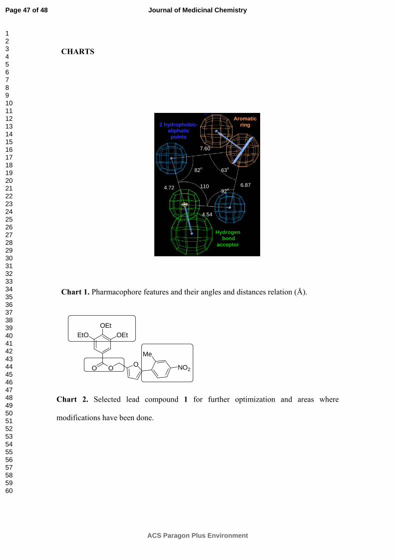
identify the chemical features that are responsible for the inhibitory activity.21 Four
pharmacophore features, such as one hydrogen bond acceptor, one aromatic ring and
two hydrophobic aliphatic points were identified to be involved in inhibitor-PDE7
interaction (Chart 1). This pharmacophore model was able to predict the activity of
external test set of PDE7 inhibitors with a correlation coefficient of 0.96. New leads
identification was carried out by performing virtual screening using validated
pharmacophoric queries and four chemical databases (Maybridge, Chemical Diversity,
Specs and National Cancer Institute). Further data reduction was done employing
virtual filters based on distances (T=1.5Å), angles (10º) and Lipinski’s rules of five.
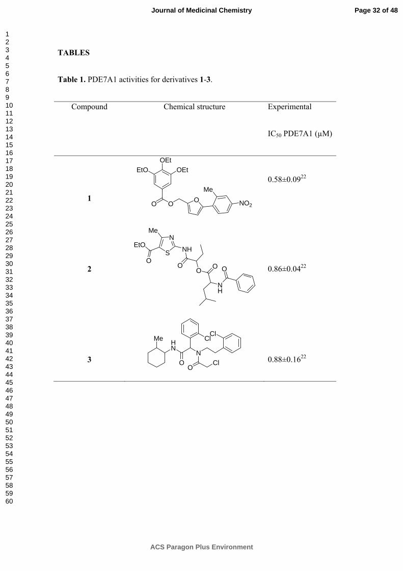
According to this procedure, three new hits (1-3) were found as potential PDE7A1
inhibitors structurally diverse from those previously known22 (Table 1). Then, the
biological activity of the hits was calculated using the catalytic domain of PDE7A1 and
a radiometric assay.
Here, Chart 1
Here, Table 1
Page 6 of 48
ACS Paragon Plus Environment
Journal of Medicinal Chemistry
123456789101112131415161718192021222324252627282930313233343536373839404142434445464748495051525354555657585960
These three hits were selected for further medicinal chemistry programs aimed to
increase not only efficacy but also the ADME profile to be considered for further drug
development in the neurodegenerative field. Here, the results obtained in the
development of the family derived from the furan 1 and its therapeutic potential for
multiple sclerosis are reported.
RESULTS AND DISCUSSION
Chemistry. The 5-(2-methyl-4-nitrophenyl)-2-furylmethyl 3,4,5-triethoxybenzoate
(1) has a furan heterocycle between two substituted phenyl rings moieties. In particular,
the structural modifications proposed are based on the modification of the number and
nature of substituents attached to both phenyl rings and the variation of the linker
between the furan and the alkyloxyphenyl ring (Chart 2).
Here, Chart 2
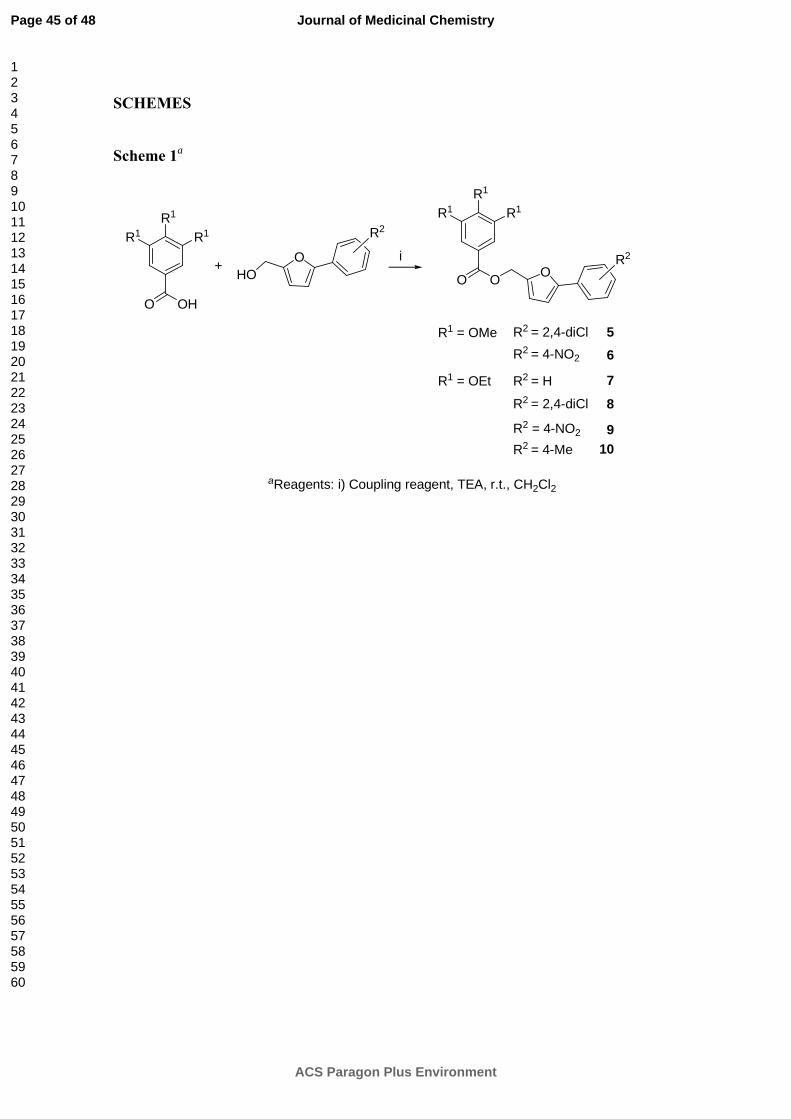
The synthetic routes for the preparation of the furan derivatives are summarized in
Schemes 1 and 2. The 5-phenyl-2-furylmethyl benzoate derivatives (Scheme 1) were
obtained in one synthetic step, after the coupling reaction of the alkyloxybenzoic acid
and the corresponding 5-phenyl-2-furylmethanol. These furan alcohols were
commercial available with the only exception of 5-(2,4-dichlorophenyl)-2-
furylmethanol (4) which was synthesized by reduction of the 5-(2,4-dichlorophenyl)-2-
furoic acid with lithium aluminum hydride as described in the supporting information.
Here, Scheme 1
Page 7 of 48
ACS Paragon Plus Environment
Journal of Medicinal Chemistry
123456789101112131415161718192021222324252627282930313233343536373839404142434445464748495051525354555657585960
The synthesis of benzyl 5-phenylfuroate and benzyl 5-phenylfuramide derivatives
(Scheme 2) involved the coupling of alkyloxybenzyl alcohol or alkyloxybenzyl amine
with the furoic acid derivative. Among different coupling reagents previously used,
benzotriazol-1-yl-oxytripyrrolidinophosphonium hexafluorophosphate (PyBOP) was
chosen as the most convenient one.
The structures of all the synthesized products were unequivocally confirmed by mass
spectrometry, elemental analysis, and 1H and 13C NMR spectroscopic data.
Here, Scheme 2
In vitro evaluation of PDE7 inhibition. The new derivatives here synthetized (5-32)
were tested for their inhibitory potencies against PDE7A1 using recombinant human
isoenzyme as described in the experimental section. All the compounds were tested at a
fixed concentration (10 µM) by duplicate. The percentage of inhibition obtained on
PDE7A1 for all the compounds at this concentration is showed in Table 2. From these
data we can conclude that several new compounds are inhibitors of PDE7A1.
Here, Table 2
As the biological assay conditions differs from that previously used,22 the lead
compound 1 was also here evaluated to compare the data with the new synthesized
derivatives. Noteworthy is the fact that when the lead compound 1 was evaluated in the
new experimental conditions using the complete enzyme instead of the catalytic domain
of PDE7A1, only a 29% of PDE7A1 inhibition at 10 µM was found. Regarding this
Page 8 of 48
ACS Paragon Plus Environment
Journal of Medicinal Chemistry
123456789101112131415161718192021222324252627282930313233343536373839404142434445464748495051525354555657585960

new data, modifications performed on the chemical structure of 1 increase in general the
inhibition of PDE7A1.
When the percentage of PDE7A1 inhibition was greater than 50%, the dose response
curve was determined and the IC50 values calculated (Table 3). Some of these new
compounds present IC50 values regarding PDE7A1 inhibition in the low micromolar
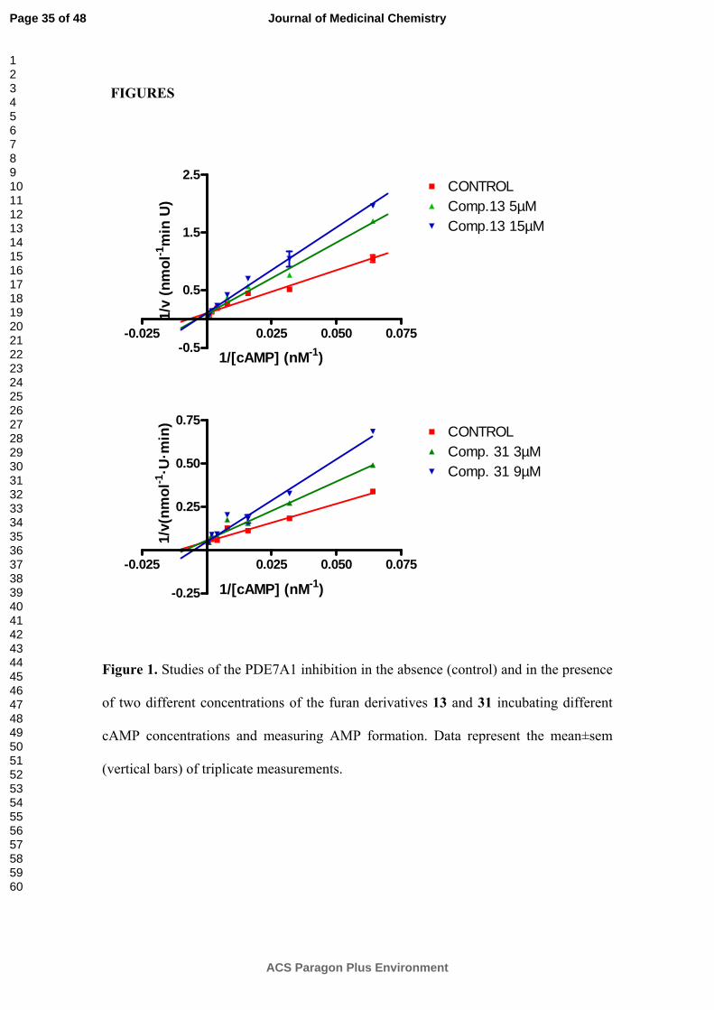
range being suitable candidates to be further explored. A kinetic study varying cAMP
concentration was performed with two of these compounds (13 and 31) to better
characterize the enzymatic inhibition. Double-reciprocal plotting of the data is depicted
in figure 1. The intercept of the plot in the horizontal axis (1/[cAMP]) changes when the
compound 13 and 31 concentrations increase, whereas the intercept in the vertical axis
(1/V) does not change. These results suggest that furan derivatives act as competitive
inhibitors of cAMP binding site and allow us to determine the Ki of these inhibitors,
(5.91 and 7.22) for compounds 13 and 31 respectively.
Here, Table 3
Here, Figure 1
Docking studies. In order to gain some insight in the binding mode of our family of
compounds, docking experiments were carried out with help of Surflex method
implemented in Sybyl according to the procedure described in the experimental section.
Considering the kinetic studies results, the docking studies were focused on the cAMP
binding site. After careful examination of docking solutions two binding modes that can
draw the chemical structure biological activity relationships (SAR) in this serie of
compounds were proposed. In fact, the different substituents and their location (ortho,
meta or para) when R3 is a phenyl ring, determine which binding mode is selected.
Page 9 of 48
ACS Paragon Plus Environment
Journal of Medicinal Chemistry
123456789101112131415161718192021222324252627282930313233343536373839404142434445464748495051525354555657585960
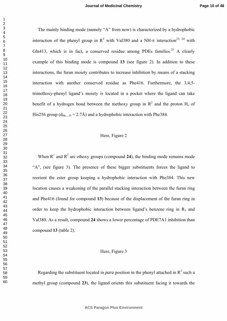
The mainly binding mode (namely “A” from now) is characterized by a hydrophobic
interaction of the phenyl group in R3 with Val380 and a NH-π interaction23, 24 with
Gln413, which is in fact, a conserved residue among PDEs families.25 A clearly
example of this binding mode is compound 13 (see figure 2). In addition to these
interactions, the furan moiety contributes to increase inhibition by means of a stacking
interaction with another conserved residue as Phe416. Furthermore, the 3,4,5-
trimethoxy-phenyl ligand’s moiety is located in a pocket where the ligand can take
benefit of a hydrogen bond between the methoxy group in R2 and the proton Hε of
His256 group (dHε…O = 2.7Å) and a hydrophobic interaction with Phe384.
Here, Figure 2
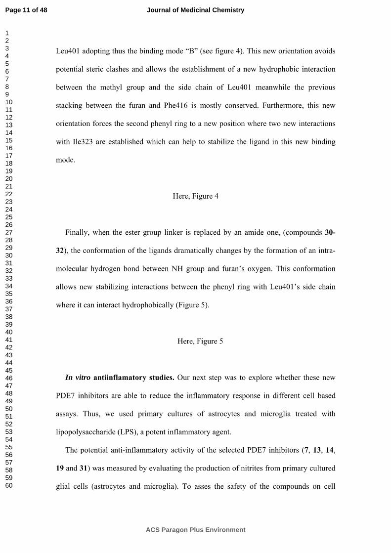
When R1 and R2 are ethoxy groups (compound 24), the binding mode remains mode
“A”, (see figure 3). The presence of these bigger substituents forces the ligand to
reorient the ester group keeping a hydrophobic interaction with Phe384. This new
location causes a weakening of the parallel stacking interaction between the furan ring
and Phe416 (found for compound 13) because of the displacement of the furan ring in
order to keep the hydrophobic interaction between ligand’s benzene ring in R3 and
Val380. As a result, compound 24 shows a lower percentage of PDE7A1 inhibition than
compound 13 (table 2).
Here, Figure 3
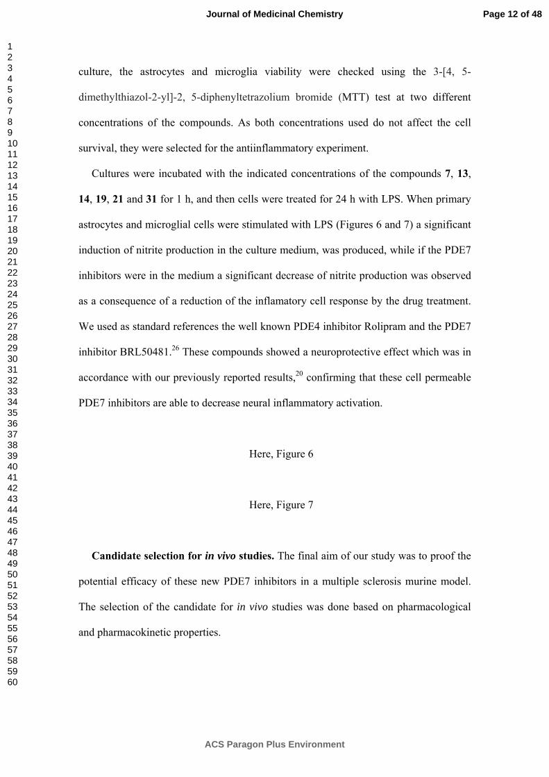
Regarding the substituent located in para position in the phenyl attached in R3 such a
methyl group (compound 23), the ligand orients this substituent facing it towards the
Page 10 of 48
ACS Paragon Plus Environment
Journal of Medicinal Chemistry
123456789101112131415161718192021222324252627282930313233343536373839404142434445464748495051525354555657585960
Leu401 adopting thus the binding mode “B” (see figure 4). This new orientation avoids
potential steric clashes and allows the establishment of a new hydrophobic interaction
between the methyl group and the side chain of Leu401 meanwhile the previous
stacking between the furan and Phe416 is mostly conserved. Furthermore, this new
orientation forces the second phenyl ring to a new position where two new interactions
with Ile323 are established which can help to stabilize the ligand in this new binding
mode.
Here, Figure 4
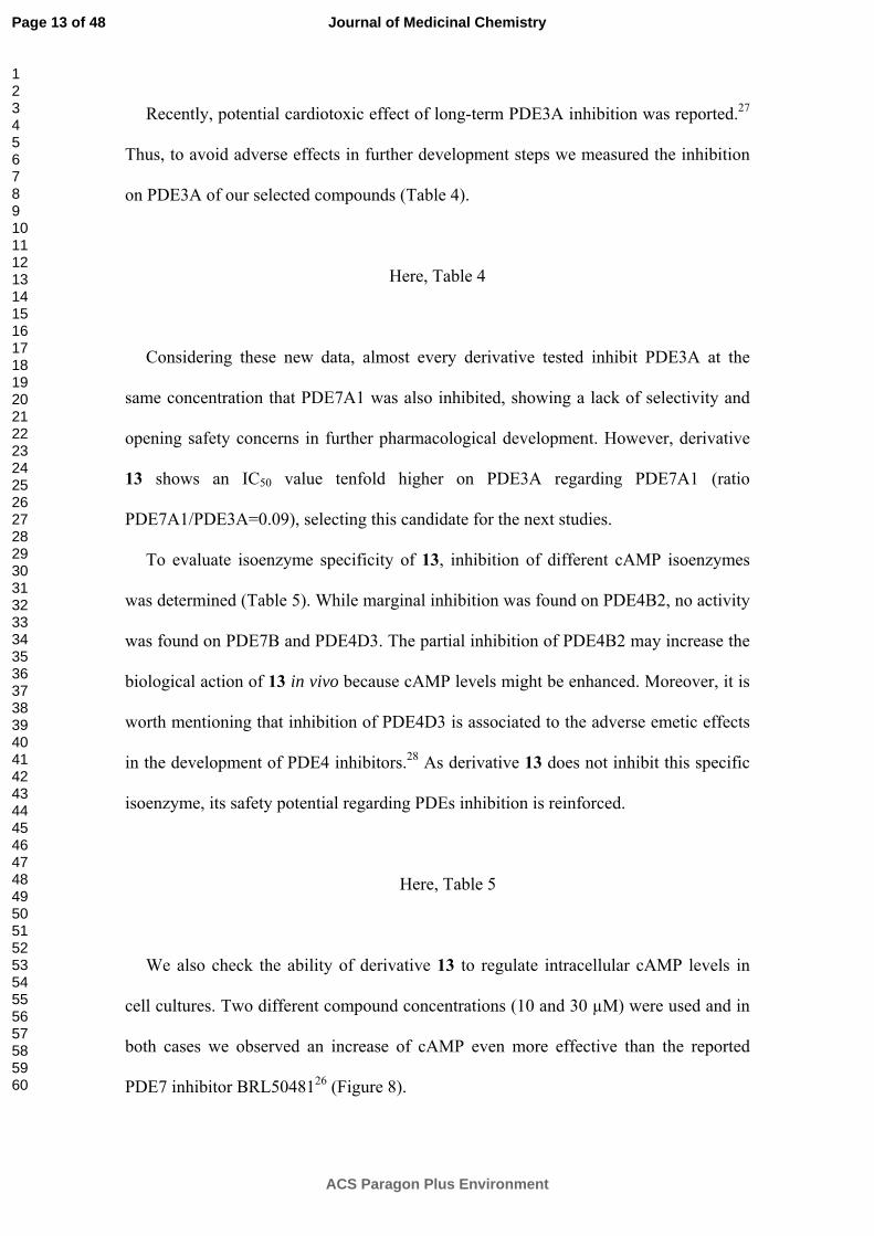
Finally, when the ester group linker is replaced by an amide one, (compounds 30-
32), the conformation of the ligands dramatically changes by the formation of an intra-
molecular hydrogen bond between NH group and furan’s oxygen. This conformation
allows new stabilizing interactions between the phenyl ring with Leu401’s side chain
where it can interact hydrophobically (Figure 5).
Here, Figure 5
In vitro antiinflamatory studies. Our next step was to explore whether these new
PDE7 inhibitors are able to reduce the inflammatory response in different cell based
assays. Thus, we used primary cultures of astrocytes and microglia treated with
lipopolysaccharide (LPS), a potent inflammatory agent.
The potential anti-inflammatory activity of the selected PDE7 inhibitors (7, 13, 14,
19 and 31) was measured by evaluating the production of nitrites from primary cultured
glial cells (astrocytes and microglia). To asses the safety of the compounds on cell
Page 11 of 48
ACS Paragon Plus Environment
Journal of Medicinal Chemistry
123456789101112131415161718192021222324252627282930313233343536373839404142434445464748495051525354555657585960
culture, the astrocytes and microglia viability were checked using the 3-[4, 5-
dimethylthiazol-2-yl]-2, 5-diphenyltetrazolium bromide (MTT) test at two different
concentrations of the compounds. As both concentrations used do not affect the cell
survival, they were selected for the antiinflammatory experiment.
Cultures were incubated with the indicated concentrations of the compounds 7, 13,
14, 19, 21 and 31 for 1 h, and then cells were treated for 24 h with LPS. When primary
astrocytes and microglial cells were stimulated with LPS (Figures 6 and 7) a significant
induction of nitrite production in the culture medium, was produced, while if the PDE7
inhibitors were in the medium a significant decrease of nitrite production was observed
as a consequence of a reduction of the inflamatory cell response by the drug treatment.
We used as standard references the well known PDE4 inhibitor Rolipram and the PDE7
inhibitor BRL50481.26 These compounds showed a neuroprotective effect which was in
accordance with our previously reported results,20 confirming that these cell permeable
PDE7 inhibitors are able to decrease neural inflammatory activation.
Here, Figure 6
Here, Figure 7
Candidate selection for in vivo studies. The final aim of our study was to proof the
potential efficacy of these new PDE7 inhibitors in a multiple sclerosis murine model.
The selection of the candidate for in vivo studies was done based on pharmacological
and pharmacokinetic properties.
Page 12 of 48
ACS Paragon Plus Environment
Journal of Medicinal Chemistry
123456789101112131415161718192021222324252627282930313233343536373839404142434445464748495051525354555657585960
Recently, potential cardiotoxic effect of long-term PDE3A inhibition was reported.27
Thus, to avoid adverse effects in further development steps we measured the inhibition
on PDE3A of our selected compounds (Table 4).
Here, Table 4
Considering these new data, almost every derivative tested inhibit PDE3A at the
same concentration that PDE7A1 was also inhibited, showing a lack of selectivity and
opening safety concerns in further pharmacological development. However, derivative
13 shows an IC50 value tenfold higher on PDE3A regarding PDE7A1 (ratio
PDE7A1/PDE3A=0.09), selecting this candidate for the next studies.
To evaluate isoenzyme specificity of 13, inhibition of different cAMP isoenzymes
was determined (Table 5). While marginal inhibition was found on PDE4B2, no activity
was found on PDE7B and PDE4D3. The partial inhibition of PDE4B2 may increase the
biological action of 13 in vivo because cAMP levels might be enhanced. Moreover, it is
worth mentioning that inhibition of PDE4D3 is associated to the adverse emetic effects
in the development of PDE4 inhibitors.28 As derivative 13 does not inhibit this specific
isoenzyme, its safety potential regarding PDEs inhibition is reinforced.
Here, Table 5
We also check the ability of derivative 13 to regulate intracellular cAMP levels in
cell cultures. Two different compound concentrations (10 and 30 µM) were used and in
both cases we observed an increase of cAMP even more effective than the reported
PDE7 inhibitor BRL5048126 (Figure 8).
Page 13 of 48
ACS Paragon Plus Environment
Journal of Medicinal Chemistry
123456789101112131415161718192021222324252627282930313233343536373839404142434445464748495051525354555657585960
Here, Figure 8
Finally, and to decipher if this compound has the drug profile to be administered in
vivo, we determined its ability to cross the blood-brain barrier (BBB), one of the major
obstacles for the treatment of diseases in the central nervous system. The majority of
compounds enter the brain by transcellular passive diffusion, which is driven by a
concentration gradient between the blood and the brain.29 Parallel Artificial Membrane
Permeability Assay (PAMPA) is a high throughput technique developed to predict
passive permeability through biological membranes. Here, we used the PAMPA-BBB
method described by Di et al.30 employing a brain lipid porcine membrane, to determine
the ability of compound 13 to penetrate into the brain. The in vitro permeabilities (Pe)
of commercial drugs through lipid membrane extract together with compound 13, were
determined and described. A good correlation between experimental and described
values Pe (exp)= 1.1512 (bibl) - 0.8973 (R2= 0.9779) was obtained (see supporting
information). From this equation, the Pe (exp) for 13 is (8.1 ± 0.1)10-6 cm s-1. Following
the pattern established in the literature for BBB permeation prediction31 that classify
compounds as CNS + when they present a permeability > 3.71 x 10⁻⁶ cm s-1, we can
consider that compound 13 is able to cross the BBB by passive permeation.
In vivo studies. Experimental autoimmune encephalomyelitis model. The results
found in cell cultures for the new PDE7 inhibitor 13 together with their ability to cross
the BBB, prompted us to evaluate it in chronic experimental autoimmune
encephalomyelitis (EAE) mice, a well establish murine model for multiple sclerosis.
EAE was induced in C57BL/6J mice by immunization with MOG35-55 in complete
Freund’s adjuvant on day 0. Clinical signs and score were monitored up to day 41. Mice
Page 14 of 48
ACS Paragon Plus Environment
Journal of Medicinal Chemistry
123456789101112131415161718192021222324252627282930313233343536373839404142434445464748495051525354555657585960
began to show neurological deficits on day 12, reaching a maximum score around day
16. A therapeutic regimen of administration was chosen to test the PDE7 inhibitor in the
EAE model. Thus a daily i.p. administration of compound 13 for 26 days started at day
5 after disease onset. The dose was selected considering the IC50 value on the target and
the cellular anti-inflammatory activity. As we can see in figure 9A, a clear and
significant attenuation of clinical symptoms during the first 16 days of treatment (day
32 after immunization figure 9A) was observed. However, this behavior is lost in the
last ten days of treatment. The body weight curve (Figure 9B), reflects these findings
and the compound 13-treated mice start to lose weight in the same frame-time than
clinical scores decrease (described above).
This experiment showed for the first time efficacy of PDE inhibitors on a well
established model of multiple sclerosis. Compound 13 administrated to the EAE when
the worst neurological score was measured, showed a recovery of the clinical symptoms
reaching a statistically significant plateau of maximum effect from day 21 to 31 p.i.
However, this attenuation of the symptoms was counterbalanced by the worsening of
the animals during the last 10 days of treatment until day 42, when the animals were
sacrificed. To decipher if this effect is due to the administrated compound (stability,
metabolism, etc) or to the animal model further studies are in progress.
Here, Figure 9
CONCLUSIONS
PDE7 inhibitors represent a new class of innovative drugs with great potential for
several neurological disorders. Their efficacy in animal models of Parkinson disease,
stroke, and spinal cord injury has been recently reported while their value for the
Page 15 of 48
ACS Paragon Plus Environment
Journal of Medicinal Chemistry
123456789101112131415161718192021222324252627282930313233343536373839404142434445464748495051525354555657585960
treatment of multiple sclerosis is here disclosed. We presented the medicinal chemistry
program around one hit found by using a pharmacophore model. The work lead to a
novel serie of PDE7 inhibitors, chemically diverse from those known and containing a
furan ring in their chemical structure.
The biological profile has been well characterized showing an increase of cAMP
level and a reduction of the inflammatory response in primary neural cell cultures.
Moreover, a clear strategy to select the best compound for in vivo testing, using
pharmacodynamic and pharmacokinetic criteria was followed. Finally, compound 13
was chosen due to its PDE7 selectivity and its ability to cross the BBB. This compound
is able to reverse clinical symptoms in an EAE mice model. All together these data
supported the potential of PDE7 inhibitors for the therapeutic pharmacological
treatment of multiple sclerosis.
Page 16 of 48
ACS Paragon Plus Environment
Journal of Medicinal Chemistry
123456789101112131415161718192021222324252627282930313233343536373839404142434445464748495051525354555657585960
EXPERIMENTAL
Chemical procedures. Substrates were purchased from commercial sources and
used without further purification. Melting points were determined with a Mettler Toledo
MP70 apparatus. Flash column chromatography was carried out at medium pressure
using silica gel (E. Merck, Grade 60, particle size 0.040-0.063 mm, 230-240 mesh
ASTM) with the indicated solvent as eluent. Compounds were detected with UV light
(254 nm). 1H NMR spectra were obtained on the Bruker AVANCE-300 spectrometer
working at 300 MHz or on a Varian INOVA 400 spectrometer working at 400 MHz.
Typical spectral parameters: spectral width 10 ppm, pulse width 9 μs (57°), data size 32
K. 13C NMR experiments were carried out on the Bruker AVANCE-300 spectrometer
operating at 75 MHz or on a Varian INOVA 400 spectrometer working at 100 MHz.
The acquisition parameters: spectral width 16 kHz, acquisition time 0.99 s, pulse width
9 μs (57°), data size 32 K. Chemical shifts are reported in values (ppm) relative to
internal Me4Si and J values are reported in Hz. Elemental analyses were performed by
the analytical department at CENQUIOR (CSIC), and the results obtained were within ±
0.4% of the theoretical values.
5-(4-Phenyl)-2-furylmethyl 3,4,5-triethoxybenzoate (7). PyBOP (358 mg, 0.68
mmol) was added as coupling reagent to a solution of 3,4,5-triethoxybenzoic acid (175
mg, 0.68 mmol) with TEA (158 µL, 1.15 mmol) in CH2Cl2 (10 mL) followed by 5-
phenyl-2-furylmethanol (100 mg, 0.57 mmol), and the resulting mixture was stirred at
25 ºC during 12 hours. The reaction mixture was evaporated in vacuo and the solid was
purified by column cromatography on silica using hexane/ethyl acetate (8:1) as eluent to
afford a yellow solid (246 mg, 87% yield). mp 71.1 ºC. 1H NMR (300 MHz, CDCl3): δ
7.70-7.29, 6.64, 6.56, 5.33, 4.10, 1.42, 1.35. 13C NMR (75 MHz, CDCl3): δ 166.5,
Page 17 of 48
ACS Paragon Plus Environment
Journal of Medicinal Chemistry
123456789101112131415161718192021222324252627282930313233343536373839404142434445464748495051525354555657585960
153.0, 149.5, 149.1, 129.2, 125.7, 128.1, 125.7, 124.3, 124.4, 113.7, 108.8, 106.3, 69.4,
65.2, 59.2, 16.0, 15.2. MS (ES, [M+K]+): m/ z = 449. Anal. C25H28O5 (C, H, O).
3,4,5-Trimethoxybenzyl 5-phenyl-2-furoate (13). PyBOP (639 mg, 1.23 mmol) was
added as coupling reagent to a solution of 5-phenyl-2-furoic acid (200 mg, 1.03 mmol)
with TEA (284 µL, 2.06 mmol) in CH2Cl2 (10 mL) followed by 3,4,5-trimethoxybenzyl
alcohol (210 µL, 1.27 mmol), and the resulting mixture was stirred at 25 ºC during 23
hours. The reaction mixture was evaporated in vacuo and the solid was purified by
column cromatography on silica using hexane/ethyl acetate (8:1) as eluent to afford a
white solid (90 mg, 24% yield). mp 98.4 ºC. 1H NMR (300 MHz, CDCl3): δ 7.79, 7.45-
7.26, 6.69, 5.29, 3.93-3.85. 13C NMR (75 MHz, CDCl3): δ 159.0, 158.2, 153.7, 143.9,
138.5, 131.7, 129.8, 129.4, 129.2, 125.3, 120.7, 107.3, 106.1, 67.1, 61.2, 56.6. MS (ES,
[M+Na]+): m/ z = 391. Anal. C21H20O6 (C, H, O).
3,4,5-Trimethoxybenzyl 5-(2-trifluoromethylphenyl)-2-furoate (14). PyBOP (393
mg, 0.76 mmol) was added as coupling reagent to a solution of 5-(2-
trifluoromethylphenyl)-2-furoic acid (200 mg, 0.76 mmol) with TEA (174 µL, 1.26
mmol) in CH2Cl2 (10 mL) followed by 3,4,5-trimethoxybenzyl alcohol (129 µL, 0.65
mmol), and the resulting mixture was stirred at 25 ºC during 12 hours. The reaction
mixture was evaporated in vacuo and the solid was purified by column cromatography
on silica using hexane/ethyl acetate (6:1) as eluent to afford a white solid (123 mg, 36%
yield). mp 89.8 ºC. 1H NMR (300 MHz, CDCl3): δ 7.80, 7.68, 7.53, 7.30, 6.79, 6.69,
5.30, 3.89, 3.86. 13C NMR (75 MHz, CDCl3): δ 158.8, 154.5, 153.7, 144.8, 138.5,
132.3, 131.6, 131.1, 129.5, 128.8, 127.3, 124.1, 120.2, 112.4, 106.1, 67.2, 61.3, 56.5.
MS (ES, [M+Na]+): m/ z = 459. Anal. C22H19F3O6 (C, H, O).
3,4,5-Trimethoxybenzyl 5-(4-chloro-2-nitrophenyl)-2-furoate (19). PyBOP (369
mg, 0.71 mmol) was added as coupling reagent to a solution of 5-(4-chloro-2-
Page 18 of 48
ACS Paragon Plus Environment
Journal of Medicinal Chemistry
123456789101112131415161718192021222324252627282930313233343536373839404142434445464748495051525354555657585960
nitrophenyl)-2-furoic acid (200 mg, 0.71 mmol) with TEA (164 µL, 1.19 mmol) in
CH2Cl2 (10 mL) followed by 3,4,5-trimethoxybenzyl alcohol (101 µL, 0.61 mmol), and
the resulting mixture was stirred at 25 ºC during 12 hours. The reaction mixture was
evaporated in vacuo and the solid was purified by column cromatography on silica
using hexane/ethyl acetate (6:1) as eluent to afford a white solid (130 mg, 38% yield).
mp 147.3 ºC. 1H NMR (300 MHz, CDCl3): δ 8.37-7.87, 7.86, 7.30, 5.87, 4.48, 4.44. 13C
NMR (75 MHz, CDCl3): δ 158.5, 153.8, 151.1, 148.4, 145.6, 138.5, 136.1, 132.8, 131.4,
131.2, 124.9, 122.0, 124.8, 120.2, 112.2, 106.0, 67.4, 61.3, 56.6. MS (ES, [M+Na]+): m/
z = 470. Anal.C25H28O5 (C, H, O).
3,4,5-Trimethoxybenzyl 5-(4-nitrophenyl)-2-furoate (21). PyBOP (509 mg, 0.98
mmol) was added as coupling reagent to a solution of 5-(4-nitrophenyl)-2-furoic acid
(200 mg, 0.82 mmol) with TEA (227 µL, 1.64 mmol) in CH2Cl2 (10 mL) followed by
3,4,5-trimethoxybenzyl alcohol (168 µL, 0.98 mmol), and the resulting mixture was
stirred at 25 ºC during 24 hours. The reaction mixture was evaporated in vacuo and the
solid was purified by column cromatography on silica using hexane/ethyl acetate (4:1)
as eluent to afford a white solid (34 mg, 10% yield). mp 161.2 ºC. 1H NMR (300 MHz,
CDCl3): δ 7.52, 7.93, 7.31, 6.95, 6.69, 5.30, 3.93-3.86. 13C NMR (75 MHz, CDCl3): δ
158.6, 155.4, 153.8, 147.9, 145.6, 143.2, 135.4, 131.4, 125.7, 124.8, 120.6, 110.5,
106.3, 67.5, 61.3, 56.6. MS (ES, [M+Na]+): m/ z = 436. Anal. C21H19NO8 (C, H, N, O).
3,4,5-Trimethoxybenzyl 5-(4-methyl-2-nitrophenyl)-2-furamide (31). PyBOP (618
mg, 1.19 mmol) was added as coupling reagent to a solution of 5-(4-methyl-2-
nitrophenyl)-2-furoic acid (300 mg, 1.19 mmol) with TEA (274 µL, 1.98 mmol) in
CH2Cl2 (10 mL) followed by 3,4,5-trimethoxybenzylamine (173 µL, 0.99 mmol), and
the resulting mixture was stirred at 25 ºC during 12 hours. The reaction mixture was
evaporated in vacuo and the solid was purified by column cromatography on silica
Page 19 of 48
ACS Paragon Plus Environment
Journal of Medicinal Chemistry
123456789101112131415161718192021222324252627282930313233343536373839404142434445464748495051525354555657585960
using hexane/ethyl acetate (3:1) as eluent to afford a yellow solid (56 mg, 14% yield).
mp 114.2 ºC. 1H NMR (300 MHz, CDCl3): δ 7.49, 7.34, 7.15, 6.65, 6.51, 4.48, 3.81,
3.75, 2.39. 13C NMR (75 MHz, CDCl3): δ 158.2, 153.9, 150.6, 148.4, 148.2, 141.1,
137.3, 133.9, 133.2, 125.2, 124.8, 120.7, 116.6, 111.5, 105.1, 61.3, 56.6, 43.8, 21.5. MS
(ES, [M+H]+): m/ z = 427. Anal. C22H22N2O7 (C, H, N, O).
Radiometric phosphodiesterase inhibition assay. The methodology used for
measuring human recombinant PDE7A1, PDE7B and PDE3A activity was based in a
Scintillation Proximity Assay (SPA) from Perkin Elmer (TRKQ7090). The activity of
the phosphodiesterase is measured by co-incubating the enzyme with [3H]cAMP and the
hydrolysis of the nucleotide is quantified by radioactivity measurement after binding of
[3H]AMP to scintillation binding bead.
Either 0.02 units of PDE7A1 (Calbiochem # 524751), 0.02 units of PDE3A
(Calbiochem # 524742) or 0.5 units of PDE7B (Abcam # ab79800) were incubated in a
96-well flexiplate with 5 nCi of [3H]cAMP and inhibitors in 100 μL of assay buffer
(contained in the kit) for 20 min at 30ºC. After the incubation time, 50µL of a solution
of SPA-beads (approximately 1 mg per well) were added to each well and plate was
shaken for 1 hour at room temperature. Finally, beads were settled for 30 min and
radioactivity was detected in a Microbeta Trilux reader.
IC50 values were calculated by non-linear regression fitting using GraphPad Prism.
Data (radioactivity vs log concentration) was fitted to a sigmoidal dose-response
equation: Y = Bottom + (Top-Bottom)/(1+10^((logIC50-X)*n)), where Bottom and Top
were the minimum and maximal inhibition for PDE, respectively, IC50 was the
concentration of compound that inhibited the PDE activity in a 50% and n was the slope
of the concentration-response curve.
Page 20 of 48
ACS Paragon Plus Environment
Journal of Medicinal Chemistry
123456789101112131415161718192021222324252627282930313233343536373839404142434445464748495051525354555657585960
The mode of inhibitory action for compound 13 and 31 was determined by varying
the concentration of unlabelled cAMP in the reaction cocktail within the range of 5nM
to 2μM in the presence of a fixed concentration of [3H]-cAMP tracer and inhibitor
concentrations. Enzyme activity data were analyzed using Lineweaver-Burk plots using
GraphPad Prism.
Fluorescence polarization phosphodiesterase inhibition assay. The ability of
compounds to inhibit PDE4B2 (human recombinant) and PDE4D3 (human
recombinant) was determined by IMAP Fluorescence Polarisation (FP) assay
(Molecular Devices R8175).
0.05U of PDE4B2 (Calbiochem # 524736), PDE4D3 (Calbiochem # 524733) were
incubated in a 96-well black half-area plate with 1nM of Fluorescein adenosine 3′,5′-
cyclic phosphate (contained in the kit) and inhibitors in 40µL of assay buffer. Plates
were mixed on a shaker for 10 seconds and incubated at ambient temperature for 60
minutes. IMAP binding reagent was added (60 μL of a 1 in 600 dilution in binding
buffer of the kit stock solution) to terminate the assay. Plates were allowed to stand at
ambient temperature for 1 hour. The FP ratio was measured with a Tecan Ultra
Evolution Reader.
IC50 values were calculated by non-linear regression fitting using GraphPad Prism.
Data (fluorescence polarization vs log concentration) was fitted to a sigmoidal dose-
response equation: Y = Bottom + (Top-Bottom)/(1+10^((logIC50-X)*n)), where Bottom
and Top were the minimum and maximal inhibition for PDE, respectively, IC50 was the
concentration of compound that inhibited the PDE activity in a 50% and n was the slope
of the concentration-response curve.
Molecular modeling. To carry out docking analysis, chain A of protein structures
were processed with help of Sybyl8.0 software32 adding hydrogens, and capping
Page 21 of 48
ACS Paragon Plus Environment
Journal of Medicinal Chemistry
123456789101112131415161718192021222324252627282930313233343536373839404142434445464748495051525354555657585960
terminal residues with neutral ends. Then, newly added hydrogen positions were
optimized with MMFF94 force field until a 0.01kcal/mol gradient was reached. Ligand
geometries were also minimized with MMFF94 force field until a 0.01kcal/mol gradient
was reached.
The docking studies were performed with Surflex (Sybyl). An “Automatic” protocol
was generated and dockings were carried out using 20 initial conformations for each
ligand allowing flexibility. Pre and post minimization was used considering 100
solutions in each case. Cscore scoring function was used with all the four options and
claiming for a relaxed structure.
Visual inspection of solutions of selected ligands was carried out in order to find
which ones allow the best agreement with SAR data. Finally the selected ligand-
complex geometries were minimized with MMFF94 force field until a 0.01kcal/mol
gradient was reached.
To test our docking protocol and validate the procedure, we carried out docking
experiments for some selected 3D structures The first one was for the complex of 3,5-
dimethyl-1-(3-nitrophenyl)-1H-pyrazole-4-carboxylic acid ethyl ester bound to PDE4D
(pdb code 1Y2K), as this ligand shares a high similarity with some of our inhibitors.
The high similarity between the docked solution (only one cluster within 2.0Å rmsd of
heavy atoms was found) and the experimental one gave us confidence in our protocol
(rmsd 0.2Å of heavy atoms vs exp., see figure S2 in the supporting information). In fact,
the main difference between both orientations is related with the benzene with supports
a nitro group. This difference can be explained due to the lack of water W1016 from the
pdb, which was not present in the model of PDE4D used for docking were only the
metal coordination waters (W1003-W1008) were retained. On the other hand, in order
to test our protocol specifically in PDE7, we carried out the docking experiment with
Page 22 of 48
ACS Paragon Plus Environment
Journal of Medicinal Chemistry
123456789101112131415161718192021222324252627282930313233343536373839404142434445464748495051525354555657585960
the 3D structures of pdb code 1ZKL and 3G3N. In the case of 1ZKL, the reproduction
of the experimental binding mode of IBMX inhibitor was only possible (rmsd 0.1Å of
heavy atoms vs exp., see figure S3 in the supporting information) when waters W573
and W542 were added aside with the metal coordination waters (W504-W509). This
fact is not surprising as those two additional water molecules are within 3.0Å from the
ligand. As only those two structures of PDE7A were available at the moment of this
study (pdb codes. 1ZKL, 3G3N), and given the redocking results obtained for 3G3N we
decided to use structure 1ZKL for docking purposes. Finally, as the best redocking
results were obtained for structure 1Y2K, which ligand is the most similar to our
inhibitors, we decided to retain only the metal coordination waters present in pdb code
1ZKL for docking.
Primary cell cultures. Glial cells were prepared from neonatal rat cerebral cortex, as
previously described by Luna-Medina et al.33 Briefly, after removal of the meninges the
cerebral cortex was dissected, dissociated, and incubated with 0.25% trypsin/EDTA at
37 ºC for 1 hour. After centrifugation, the pellet was washed 3 times with HBSS
(Gibco) and the cells were plated on non-coated flasks and maintained in
HAMS/DMEM (1:1) medium containing 10% FBS. After 15 days the flasks were
agitated on an orbital shaker for 4 hours at 240 rpm at 37 ºC, the supernatant was
collected, centrifuged, and the cellular pellet containing the microglial cells resuspended
in complete medium (HAMS/DMEM (1:1) containing 10% FBS) and seeded on
uncoated 96-well plates. Cells were allowed to adhere for 2 hours and the medium was
removed to eliminate non-adherent oligodendrocytes. New fresh medium containing 10
ng/mL of GM-CSF was added. The remaining astroglial cells adhered on the flasks
were then trypsinized, collected, centrifugated and plated onto 96-well plates with
complete medium. The purity of cultures obtained by this procedure was >98% as
Page 23 of 48
ACS Paragon Plus Environment
Journal of Medicinal Chemistry
123456789101112131415161718192021222324252627282930313233343536373839404142434445464748495051525354555657585960
determined by immunofluorescence with the OX42 (microglial marker) and the GFAP
(astroglial marker) antibodies. After 1 week in culture, cells were treated with Rolipram,
BRL50481 and the different compounds (7, 13, 14, 19, 21 and 31) at several
concentrations. Cell viability was then measured after 16h in culture. For nitrite release
quantification some cultures were also treated with lipopolysaccharide (LPS; 10µg/mL)
alone or in combination with compounds.
Cell viability assay. Cell viability was measured using the MTT assay from Roche,
based on the ability of viable cells to reduce yellow MTT to blue formazan. Briefly,
cells cultured in 96-well plates and treated with the indicated compounds for 16h were
incubated with MTT (0.5mg/mL, 4h) and subsequently solubilized in 10% SDS/0.01M
HCl for 12h in the dark. The extent of reduction of MTT was quantified by absorbance
measurement at 595nm according to the manufacturer´s protocol.
Nitrites measurement. Accumulation of nitrites in media was assayed by the
standard Griess reaction. After stimulation of cells with the different compounds,
supernatants were collected and mixed with an equal volume of Griess reagent (Sigma).
Samples were then incubated at room temperature for 15 minutes and absorbance read
using a plate reader at 492/540 nm.
cAMP measurements in Raw cells. Quantification of cAMP was carried out using
the EIA (enzyme immunoassay) kit from GE Healthcare. Briefly, Raw cells were
seeded at 3x10⁴/well in 96-well dishes and incubated overnight before the assay. After
60 min incubation with compound 13, cAMP intracellular levels were determined
following the manufacture’s instructions.
CNS penetration: In vitro Parallel artificial membrane permeability assay
(PAMPA)-Blood brain barrier (BBB). Prediction of the brain penetration was
evaluated using a parallel artificial membrane permeability assay (PAMPA).30 Ten
Page 24 of 48
ACS Paragon Plus Environment
Journal of Medicinal Chemistry
123456789101112131415161718192021222324252627282930313233343536373839404142434445464748495051525354555657585960
commercial drugs, phosphate buffer saline solution at pH 7.4 (PBS), Ethanol and
dodecane were purchased from Sigma, Acros organics, Merck, Aldrich and Fluka. The
porcine polar brain lipid (PBL) (catalog no. 141101) was from Avanti Polar Lipids. The
donor plate was a 96-well filtrate plate (Multiscreen® IP Sterile Plate PDVF membrane,
pore size is 0.45 µM, catalog no. MAIPS4510) and the acceptor plate was an indented
96-well plate (Multiscreen®, catalog no. MAMCS9610) both from Millipore. Filter
PDVF membrane units (diameter 30 mm, pore size 0.45 μm) from Symta were used to
filtered the samples. A 96-well plate UV reader (Thermoscientific, Multiskan spectrum)
was used for the UV measurements. Test compounds [(3-5 mg of Caffeine, Enoxacine,
Hydrocortisone, Desipramine, Ofloxacine, Piroxicam, Testosterone), (12 mg of
Promazine) and 25 mg of Verapamile and Atenolol] were dissolved in EtOH (1000 µL).
100 microlitres of this compound stock solution was taken and 1400 µL of EtOH and
3500 µL of PBS pH 7.4 buffer were added to reach 30% of EtOH concentration in the
experiment. These solutions were filtered. The acceptor 96-well microplate was filled
with 180 μL of PBS/EtOH (70/30). The donor 96-well plate was coated with 4 µL of
porcine brain lipid in dodecane (20 mg mL-1) and after 5 minutes, 180 μL of each
compound solution was added. 1-2 mg of every compound to be determined their ability
to pass the brain barrier were dissolved in 1500 µL of EtOH and 3500 µL of PBS pH 7.4
buffer, filtered and then added to the donor 96-well plate. Then the donor plate was
carefully put on the acceptor plate to form a “sandwich”, which was left undisturbed for
2h and 30 min at 25 °C. During this time the compounds diffused from the donor plate
through the brain lipid membrane into the acceptor plate. After incubation, the donor
plate was removed. UV plate reader determined the concentration of compounds and
commercial drugs in the acceptor and the donor wells. Every sample was analyzed at
three to five wavelengths, in 3 wells and in two independent runs. Results are given as
Page 25 of 48
ACS Paragon Plus Environment
Journal of Medicinal Chemistry
123456789101112131415161718192021222324252627282930313233343536373839404142434445464748495051525354555657585960
the mean [standard deviation (SD)] and the average of the two runs is reported. 10
quality control compounds (previously mentioned) of known BBB permeability were
included in each experiment to validate the analysis set.
EAE induction and treatment. Six-week-old female C57BL6 mice (15-20 g) were
purchased from Harlan (Spain). All experimental procedures followed the European
Communities Council Directive of November 24, 1986 (86/609/EEC). The protocol was
approved by the ethic committee of the University of Barcelona and of the Generalitat
de Catalunya. The mice were maintained on a 12h light/dark cycle at a constant
environmental temperature with free access to food and water for 1 week prior to
experimentation.
EAE was induced by subcutaneous immunization with 100μg MOG35-55 peptide
(EspiKem S.r.l., Italy) in 100μL complete Freund’s adjuvant (CFA) (Sigma-Aldrich)
enriched with Mycobacterium tuberculosis (H37Ra strain, Difco, Detroit, MI, USA).
Mice were immediately intraperitoneally injected with 200ng of Bordetella pertussis
toxin (Sigma-Aldrich) and again 48h after the immunization.
Animals (n=13) were weighed and examined for clinical signs on a daily basis.
Disease severity of EAE was graded according to a five-point scale: Grade 0 = no
disability; 1 = a flaccid tail; 2 = a mild but definite weakness of one or both hind legs; 3
= moderate paraparesis of one hind leg; 4 = no hind leg movement; 5 = a moribund state
with little or no spontaneous movement and impaired respiration.34
Stock solution of the compound (100mg/mL in DMSO) was diluted 1:50 in a solution
of 5% Tocrisolve (Tocris, UK) in distilled water. Mice were treated through daily
intraperitoneal (i.p.) injection starting on day 5 after the onset of the disease at a dose of
10 mg/kg of animal (n=7) or with only vehicle (n=7).
Page 26 of 48
ACS Paragon Plus Environment
Journal of Medicinal Chemistry
123456789101112131415161718192021222324252627282930313233343536373839404142434445464748495051525354555657585960
ACKNOWLEDGEMENTS
The authors gratefully acknowledge the financial support of Ministry of Science and
Innovation (MICINN), projects nos. SAF2009-13015-C02-01, SAF2009-13015-C02-
02, SAF2010-16365, SAF2009-1152, and PI10-01874; Instituto de Salud Carlos III
(ISCiii), project no. RD07/0060/0015 (RETICS program) and CIBERNED; Fundación
Española para la Ciencia y la Tecnología (FECYT), project no. FCT-09-INC-0367. M.
R. and D. I. P. acknowledge pre- and post-doctoral fellowship from the CSIC (JAE
program) respectively. BRAINco Biopharma is acknowledged.
SUPPORTING INFORMATION
Elemental analyses of compounds 5-32; experimental procedures for compounds 4-6,
8-12, 15-18, 20, 22-30 and 32; linear correlation between experimental and described
values in the PAMPA-BBB assay; figures S1 and S2. This material is available free of
charge via the Internet at http://pubs.acs.org.
REFERENCES
(1) Essayan, D. M. Cyclic nucleotide phosphodiesterase (PDE) inhibitors and
immunomodulation. Biochem. Pharmacol. 1999, 57, 965-973.
(2) Conti, M.; Jin, S. L. The molecular biology of cyclic nucleotide
phosphodiesterases. Prog. Nucleic Acid Res. Mol. Biol. 1999, 63, 1-38.
Page 27 of 48
ACS Paragon Plus Environment
Journal of Medicinal Chemistry
123456789101112131415161718192021222324252627282930313233343536373839404142434445464748495051525354555657585960
(3) Bender, A. T.; Beavo, J. A. Cyclic nucleotide phosphodiesterases: molecular
regulation to clinical use. Pharmacol. Rev. 2006, 58, 488-520.
(4) Francis, S. H.; Conti, M.; Houslay, M. D. Phosphodiesterases as drug targets.
Springer-Verlag Berlin Heidelberg: 2011.
(5) Lugnier, C. Cyclic nucleotide phosphodiesterase (PDE) superfamily: A new
target for the development of specific therapeutic agents. Pharmacol. Ther. 2006, 109,
366-398.
(6) Allison, A. C. Immunosuppressive drugs: the first 50 years and a glance
forward. Immunopharmacology 2000, 47, 63-83.
(7) Houslay, M. D.; Schafer, P.; Zhang, K. Y. Keynote review: phosphodiesterase-4
as a therapeutic target. Drug Discov. Today 2005, 10, 1503-1519.
(8) Giembycz, M. A. Life after PDE4: overcoming adverse events with dual-
specificity phosphodiesterase inhibitors. Curr. Opin. Pharmacol. 2005, 5, 238-244.
(9) Conti, M.; Beavo, J. Biochemistry and physiology of cyclic nucleotide
phosphodiesterases: essential components in cyclic nucleotide signaling. Annu. Rev.
Biochem. 2007, 76, 481-511.
(10) Giembycz, M. A.; Smith, S. J. Phosphodiesterase 7A: a new therapeutic target
for alleviating chronic inflammation? Curr. Pharm. Des. 2006, 12, 3207-3220.
(11) Miro, X.; Perez-Torres, S.; Palacios, J. M.; Puigdomenech, P.; Mengod, G.
Differential distribution of cAMP-specific phosphodiesterase 7A mRNA in rat brain
and peripheral organs. Synapse 2001, 40, 201-214.
(12) Sasaki, T.; Kotera, J.; Omori, K. Novel alternative splice variants of rat
phosphodiesterase 7B showing unique tissue-specific expression and phosphorylation.
Biochem. J. 2002, 361, 211-220.
Page 28 of 48
ACS Paragon Plus Environment
Journal of Medicinal Chemistry
123456789101112131415161718192021222324252627282930313233343536373839404142434445464748495051525354555657585960
(13) Reyes-Irisarri, E.; Perez-Torres, S.; Mengod, G. Neuronal expression of cAMP-
specific phosphodiesterase 7B mRNA in the rat brain. Neuroscience 2005, 132, 1173-
1185.
(14) Nakata, A.; Ogawa, K.; Sasaki, T.; Koyama, N.; Wada, K.; Kotera, J.; Kikkawa,
H.; Omori, K.; Kaminuma, O. Potential role of phosphodiesterase 7 in human T cell
function: comparative effects of two phosphodiesterase inhibitors. Clin. Exp. Immunol.
2002, 128, 460-466.
(15) Gil, C.; Campillo, N. E.; Perez, D. I.; Martinez, A. Phosphodiesterase 7 (PDE7)
inhibitors as new drugs for neurological and inflammatory disorders. Expert Opin. Ther.
Pat. 2008, 18, 1127-1139.
(16) Martinez, A.; Castro, A.; Gil, C.; Miralpeix, M.; Segarra, V.; Domenech, T.;
Beleta, J.; Palacios, J. M.; Ryder, H.; Miro, X.; Bonet, C.; Casacuberta, J. M.; Azorin,
F.; Piña, B.; Puigdomenech, P. Benzyl derivatives of 2,1,3-benzo- and benzothieno[3,2-
a]thiadiazine 2,2-dioxides: first phosphodiesterase 7 inhibitors. J. Med. Chem. 2000, 43,
683-689.
(17) Castro, A.; Jerez, M. J.; Gil, C.; Martinez, A. Cyclic nucleotide
phosphodiesterases and their role in immunomodulatory responses: advances in the
development of specific phosphodiesterase inhibitors. Med. Res. Rev. 2005, 25, 229-
244.
(18) Morales-Garcia, J.; Redondo, M.; Gil, C.; Alonso-Gil, S.; Martinez, A.; Santos,
A.; Perez-Castillo, A. Phosphodiesterase 7 inhibition preserves dopaminergic neurons in
cellular and rodent models of Parkinson disease. PLoS ONE 2011, 6, e17240.
(19) Paterniti, I.; Mazzon, E.; Gil, C.; Impllizzari, D.; Palomo, V.; Redondo, M.;
Perez, D. I.; Esposito, E.; Martinez, A.; Cuzzocrea, S. PDE 7 inhibitors: new potential
drugs for the therapy of spinal cord injury. PLoS ONE 2011, 6, e15937.
Page 29 of 48
ACS Paragon Plus Environment
Journal of Medicinal Chemistry
123456789101112131415161718192021222324252627282930313233343536373839404142434445464748495051525354555657585960

(20) Redondo, M.; Zarruk, J. G.; Ceballos, P.; Perez, D. I.; Perez, C.; Perez-Castillo,
A.; Moro, M. A.; Brea, J.; Val, C.; Cadavid, M. I.; Loza, M. I.; Campillo, N. E.;
Martinez, A.; Gil, C. Neuroprotective efficacy of quinazoline type phosphodiesterase 7
inhibitors in cellular cultures and experimental stroke model. Eur. J. Med. Chem. 2012,
47, 175-185.
(21) Jerez, M. J.; Castro, A.; Gil, C.; Martinez, A. Development of a pharmacophoric
model for specific PDE7 inhibitors. Drugs Fut. 2004, 29 (Supp. A), 129.
(22) Gil, C.; Castro, A.; Jerez, M. J.; Ke, H.; Wang, H.; Ballester, S.; González-
García, C.; Martínez, A. New PDE7 inhibitors leads for neurodegenerative diseases
discovered by using a pharmacophoric model. Drugs Fut. 2008, 33 (Supp. A), 228.
(23) Vaupel, S.; Brutschy, B.; Tarakeshwar, P.; Kim, K. S. Characterization of weak
NH-pi intermolecular interactions of ammonia with various substituted pi-systems. J.
Am. Chem. Soc. 2006, 128, 5416-5426.
(24) Mohan, N.; Vijayalakshmi, K. P.; Koga, N.; Suresh, C. H. Comparison of
aromatic NH...pi, OH...pi, and CH...pi interactions of alanine using MP2, CCSD, and
DFT methods. J. Comput. Chem. 2010, 31, 2874-2882.
(25) Ke, H.; Wang, H. Crystal structures of phosphodiesterases and implications on
substrate specificity and inhibitor selectivity. Curr. Top. Med. Chem. 2007, 7, 391-403.
(26) Smith, S. J.; Cieslinski, L. B.; Newton, R.; Donnelly, L. E.; Fenwick, P. S.;
Nicholson, A. G.; Barnes, P. J.; Barnette, M. S.; Giembycz, M. A. Discovery of BRL
50481 [3-(N,N-dimethylsulfonamido)-4-methyl-nitrobenzene], a selective inhibitor of
phosphodiesterase 7: in vitro studies in human monocytes, lung macrophages, and
CD8+ T-lymphocytes. Mol. Pharmacol. 2004, 66, 1679-1689.
(27) Movsesian, M. A.; Kukreja, R. C. Phosphodiesterase inhibition in heart failure.
Handb. Exp. Pharmacol. 2011, 237-249.
Page 30 of 48
ACS Paragon Plus Environment
Journal of Medicinal Chemistry
123456789101112131415161718192021222324252627282930313233343536373839404142434445464748495051525354555657585960
(28) Robichaud, A.; Stamatiou, P. B.; Jin, S. L.; Lachance, N.; MacDonald, D.;
Laliberte, F.; Liu, S.; Huang, Z.; Conti, M.; Chan, C. C. Deletion of phosphodiesterase
4D in mice shortens alpha(2)-adrenoceptor-mediated anesthesia, a behavioral correlate
of emesis. J. Clin. Invest. 2002, 110, 1045-1052.
(29) Di, L.; Kerns, E. H.; Carter, G. T. Strategies to assess blood-brain barrier
penetration. Expert Opin. Drug Discov. 2008, 3, 677-687.
(30) Di, L.; Kerns, E. H.; Fan, K.; McConnell, O. J.; Carter, G. T. High throughput
artificial membrane permeability assay for blood-brain barrier. Eur. J. Med. Chem.
2003, 38, 223-232.
(31) Crivori, P.; Cruciani, G.; Carrupt, P. A.; Testa, B. Predicting blood-brain barrier
permeation from three-dimensional molecular structure. J. Med. Chem. 2000, 43, 2204-
2216.
(32) SYBYL 8.0, Tripos International, 1699 South Hanley Rd., St. Louis, Missouri,
63144, USA.
(33) Luna-Medina, R.; Cortes-Canteli, M.; Alonso, M.; Santos, A.; Martinez, A.;
Perez-Castillo, A. Regulation of inflammatory response in neural cells in vitro by
thiadiazolidinones derivatives through peroxisome proliferator-activated receptor
gamma activation. J. Biol. Chem. 2005, 280, 21453-21462.
(34) McFarlin, D. E.; Blank, S. E.; Kibler, R. F. Recurrent experimental allergic
encephalomyelitis in the Lewis rat. J. Immunol. 1974, 113, 712-715.
Page 31 of 48
ACS Paragon Plus Environment
Journal of Medicinal Chemistry
123456789101112131415161718192021222324252627282930313233343536373839404142434445464748495051525354555657585960
TABLES
Table 1. PDE7A1 activities for derivatives 1-3.
Compound Chemical structure Experimental
IC50 PDE7A1 (µM)
1
EtOOEt
OEt
OO OMe
NO2
0.58±0.0922
2
N
S NH
OO
Me
EtO
OO
NH
O
0.86±0.0422
3
HN
N
Me
OO
Cl
ClCl
0.88±0.1622
Page 32 of 48
ACS Paragon Plus Environment
Journal of Medicinal Chemistry
123456789101112131415161718192021222324252627282930313233343536373839404142434445464748495051525354555657585960
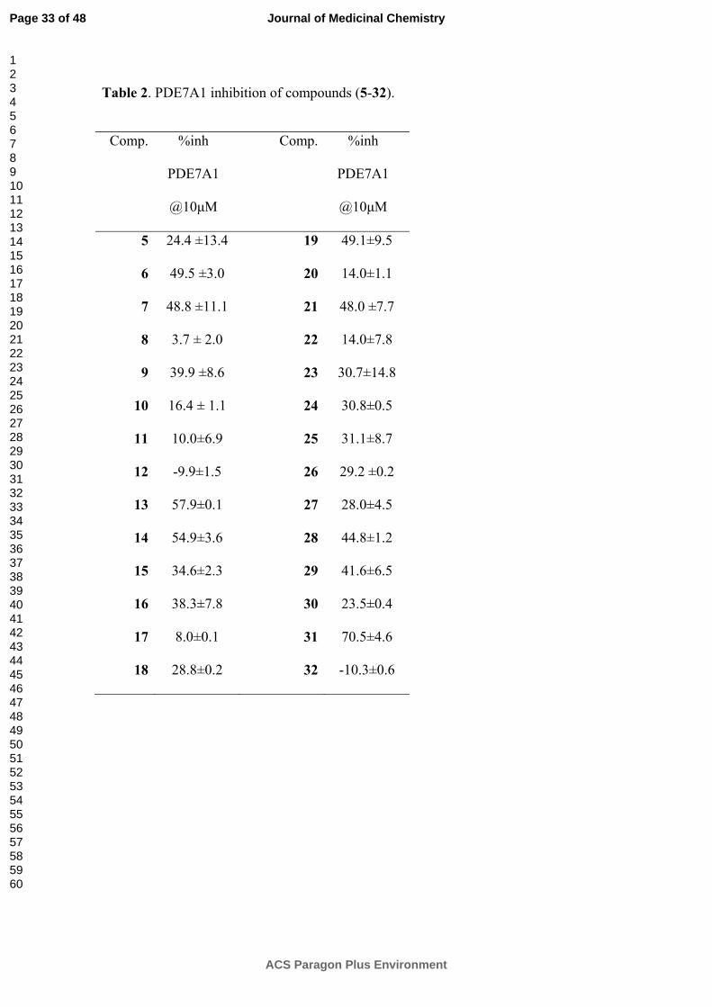
Table 2. PDE7A1 inhibition of compounds (5-32).
Comp. %inh
PDE7A1
@10μM
Comp. %inh
PDE7A1
@10μM
5 24.4 ±13.4 19 49.1±9.5
6 49.5 ±3.0 20 14.0±1.1
7 48.8 ±11.1 21 48.0 ±7.7
8 3.7 ± 2.0 22 14.0±7.8
9 39.9 ±8.6 23 30.7±14.8
10 16.4 ± 1.1 24 30.8±0.5
11 10.0±6.9 25 31.1±8.7
12 -9.9±1.5 26 29.2 ±0.2
13 57.9±0.1 27 28.0±4.5
14 54.9±3.6 28 44.8±1.2
15 34.6±2.3 29 41.6±6.5
16 38.3±7.8 30 23.5±0.4
17 8.0±0.1 31 70.5±4.6
18 28.8±0.2 32 -10.3±0.6
Page 33 of 48
ACS Paragon Plus Environment
Journal of Medicinal Chemistry
123456789101112131415161718192021222324252627282930313233343536373839404142434445464748495051525354555657585960
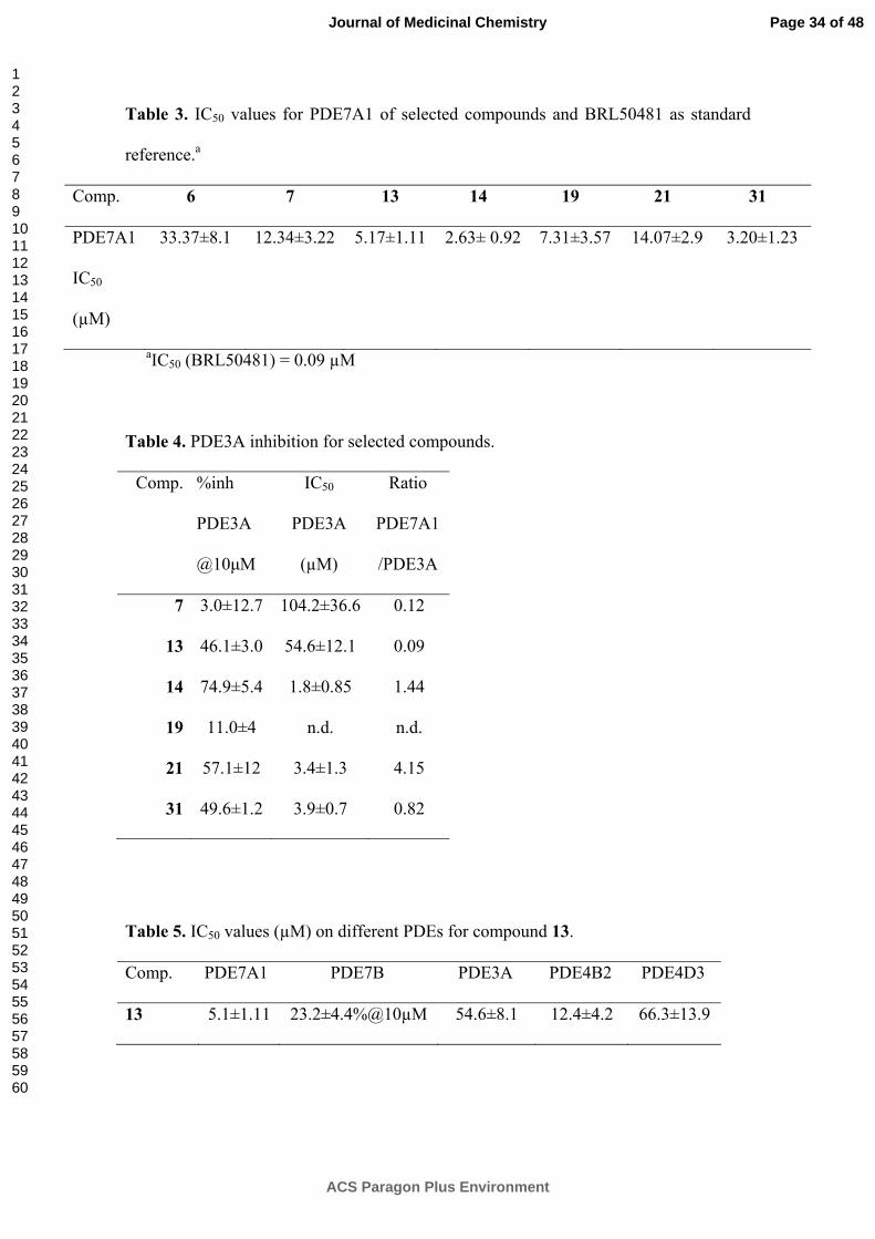
Table 3. IC50 values for PDE7A1 of selected compounds and BRL50481 as standard
reference.a
Comp. 6 7 13 14 19 21 31
PDE7A1
IC50
(µM)
33.37±8.1 12.34±3.22 5.17±1.11 2.63± 0.92 7.31±3.57 14.07±2.9 3.20±1.23
aIC50 (BRL50481) = 0.09 µM
Table 4. PDE3A inhibition for selected compounds.
Comp. %inh
PDE3A
@10μM
IC50
PDE3A
(µM)
Ratio
PDE7A1
/PDE3A
7 3.0±12.7 104.2±36.6 0.12
13 46.1±3.0 54.6±12.1 0.09
14 74.9±5.4 1.8±0.85 1.44
19 11.0±4 n.d. n.d.
21 57.1±12 3.4±1.3 4.15
31 49.6±1.2 3.9±0.7 0.82
Table 5. IC50 values (µM) on different PDEs for compound 13.
Comp. PDE7A1 PDE7B PDE3A PDE4B2 PDE4D3
13 5.1±1.11 23.2±4.4%@10µM 54.6±8.1 12.4±4.2 66.3±13.9
Page 34 of 48
ACS Paragon Plus Environment
Journal of Medicinal Chemistry
123456789101112131415161718192021222324252627282930313233343536373839404142434445464748495051525354555657585960
FIGURES
-0.025 0.025 0.050 0.075-0.5
0.5
1.5
2.5CONTROLComp.13 5µMComp.13 15µM
1/[cAMP] (nM-1)
1/v
(nm
ol-1
min
U)
-0.025 0.025 0.050 0.075
-0.25
0.25
0.50
0.75CONTROLComp. 31 3µMComp. 31 9µM
1/v(
nmol
-1·U
·min
)
1/[cAMP] (nM-1)
Figure 1. Studies of the PDE7A1 inhibition in the absence (control) and in the presence
of two different concentrations of the furan derivatives 13 and 31 incubating different
cAMP concentrations and measuring AMP formation. Data represent the mean±sem
(vertical bars) of triplicate measurements.
Page 35 of 48
ACS Paragon Plus Environment
Journal of Medicinal Chemistry
123456789101112131415161718192021222324252627282930313233343536373839404142434445464748495051525354555657585960
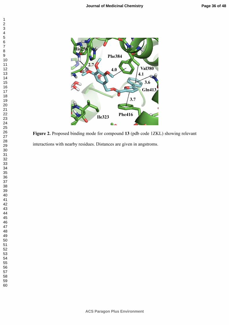
Figure 2. Proposed binding mode for compound 13 (pdb code 1ZKL) showing relevant
interactions with nearby residues. Distances are given in angstroms.
Page 36 of 48
ACS Paragon Plus Environment
Journal of Medicinal Chemistry
123456789101112131415161718192021222324252627282930313233343536373839404142434445464748495051525354555657585960
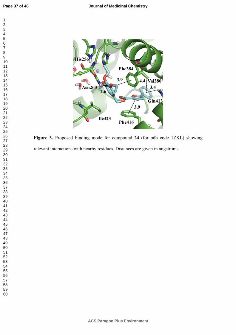
Figure 3. Proposed binding mode for compound 24 (for pdb code 1ZKL) showing
relevant interactions with nearby residues. Distances are given in angstroms.
Page 37 of 48
ACS Paragon Plus Environment
Journal of Medicinal Chemistry
123456789101112131415161718192021222324252627282930313233343536373839404142434445464748495051525354555657585960
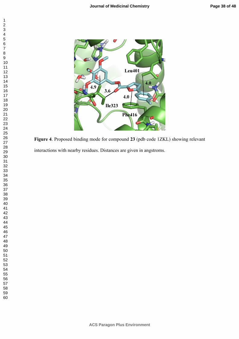
Figure 4. Proposed binding mode for compound 23 (pdb code 1ZKL) showing relevant
interactions with nearby residues. Distances are given in angstroms.
Page 38 of 48
ACS Paragon Plus Environment
Journal of Medicinal Chemistry
123456789101112131415161718192021222324252627282930313233343536373839404142434445464748495051525354555657585960
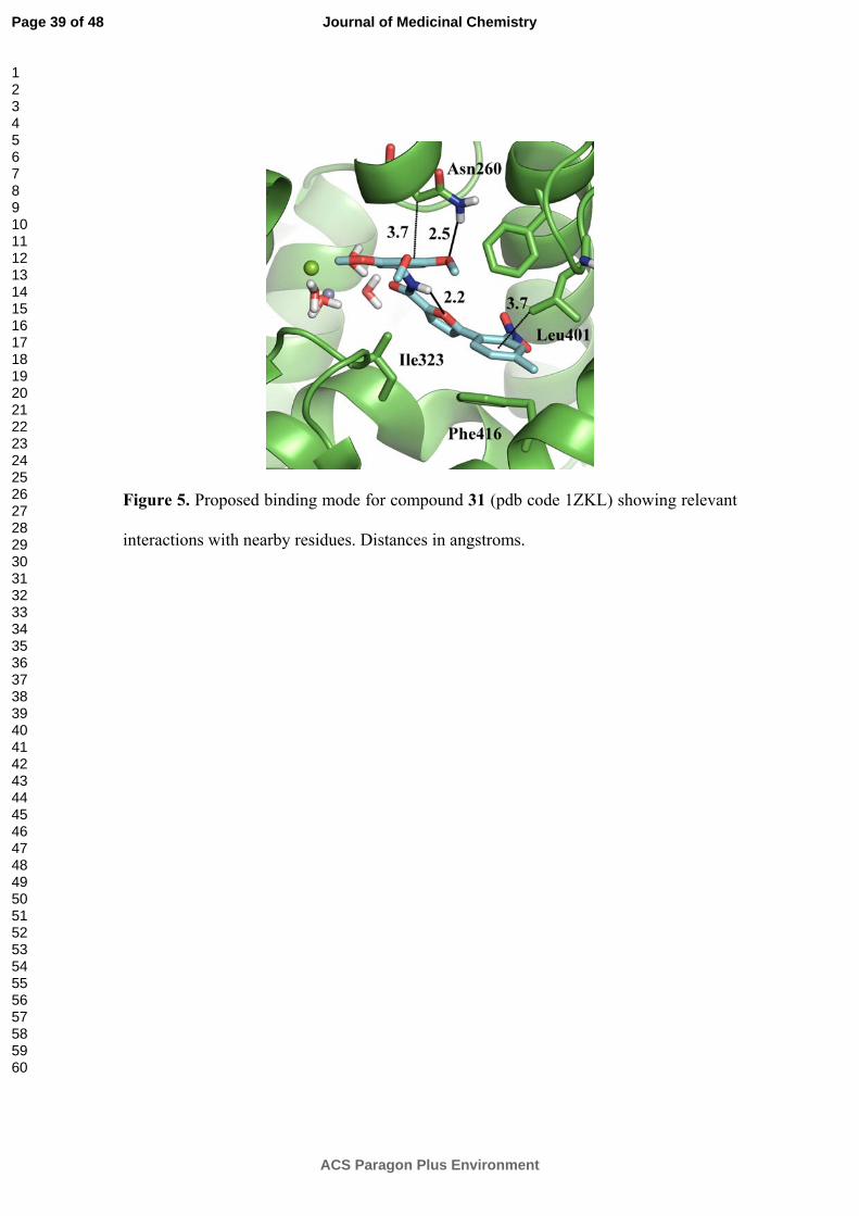
Figure 5. Proposed binding mode for compound 31 (pdb code 1ZKL) showing relevant
interactions with nearby residues. Distances in angstroms.
Page 39 of 48
ACS Paragon Plus Environment
Journal of Medicinal Chemistry
123456789101112131415161718192021222324252627282930313233343536373839404142434445464748495051525354555657585960
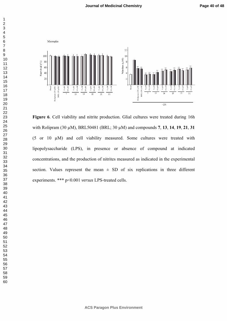
Figure 6. Cell viability and nitrite production. Glial cultures were treated during 16h
with Rolipram (30 µM), BRL50481 (BRL; 30 µM) and compounds 7, 13, 14, 19, 21, 31
(5 or 10 µM) and cell viability measured. Some cultures were treated with
lipopolysaccharide (LPS), in presence or absence of compound at indicated
concentrations, and the production of nitrites measured as indicated in the experimental
section. Values represent the mean ± SD of six replications in three different
experiments. *** p<0.001 versus LPS-treated cells.
Page 40 of 48
ACS Paragon Plus Environment
Journal of Medicinal Chemistry
123456789101112131415161718192021222324252627282930313233343536373839404142434445464748495051525354555657585960
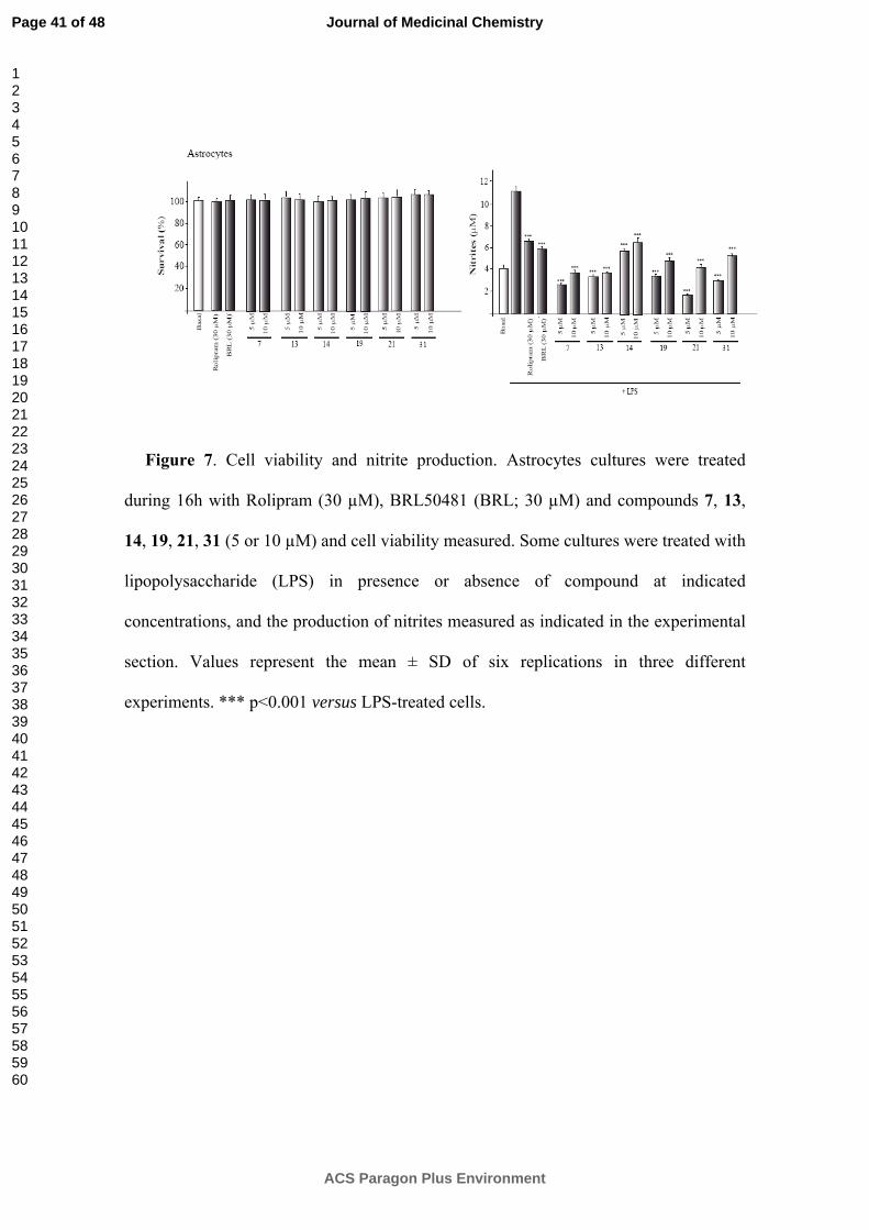
Figure 7. Cell viability and nitrite production. Astrocytes cultures were treated
during 16h with Rolipram (30 µM), BRL50481 (BRL; 30 µM) and compounds 7, 13,
14, 19, 21, 31 (5 or 10 µM) and cell viability measured. Some cultures were treated with
lipopolysaccharide (LPS) in presence or absence of compound at indicated
concentrations, and the production of nitrites measured as indicated in the experimental
section. Values represent the mean ± SD of six replications in three different
experiments. *** p<0.001 versus LPS-treated cells.
Page 41 of 48
ACS Paragon Plus Environment
Journal of Medicinal Chemistry
123456789101112131415161718192021222324252627282930313233343536373839404142434445464748495051525354555657585960
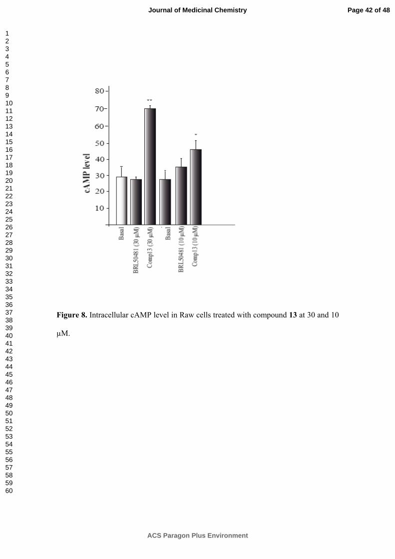
Figure 8. Intracellular cAMP level in Raw cells treated with compound 13 at 30 and 10
µM.
Page 42 of 48
ACS Paragon Plus Environment
Journal of Medicinal Chemistry
123456789101112131415161718192021222324252627282930313233343536373839404142434445464748495051525354555657585960
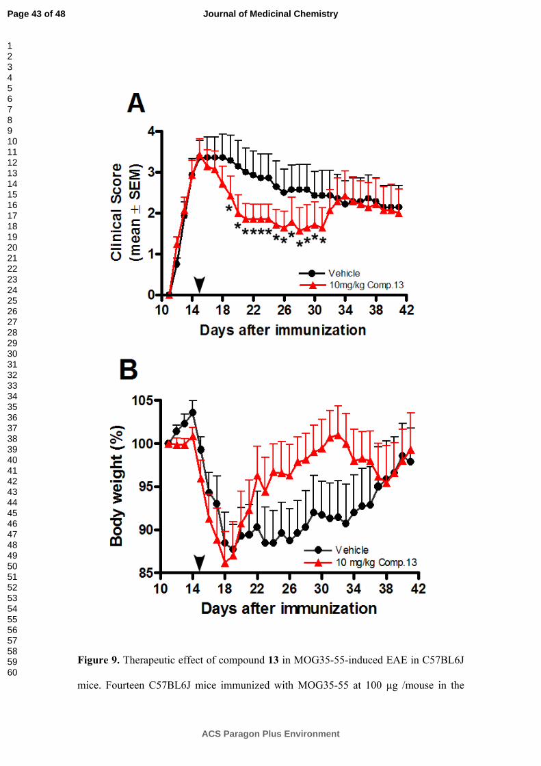
Figure 9. Therapeutic effect of compound 13 in MOG35-55-induced EAE in C57BL6J
mice. Fourteen C57BL6J mice immunized with MOG35-55 at 100 µg /mouse in the
Page 43 of 48
ACS Paragon Plus Environment
Journal of Medicinal Chemistry
123456789101112131415161718192021222324252627282930313233343536373839404142434445464748495051525354555657585960
presence of complete Freund’s adjuvant developed clinical signs on day 12. Animals
were divided into two groups. One group was treated with compound 13 starting from
day 5 after the onset of the disease (arrowhead) for 26 days. Mice in the control group
were administered vehicle only. A) EAE scores in drug treatment; B) percentage of
body weight variation. Results were expressed as the mean ± standard error of the mean
and statistical differences in EAE scores between the vehicle and the treatment group
was calculated by Mann-Whitney test (*p<0.0002). ●, Vehicle (n=7); ▲, 10 mg/kg
compound 13 (n=7) i.p. daily.
Page 44 of 48
ACS Paragon Plus Environment
Journal of Medicinal Chemistry
123456789101112131415161718192021222324252627282930313233343536373839404142434445464748495051525354555657585960
SCHEMES
Scheme 1a
R1R1
R1
O OH
O
R1R1
R1
OO O+HO
R2
R2
aReagents: i) Coupling reagent, TEA, r.t., CH2Cl2
i
R2 = 4-Me
R2 = 2,4-diClR1 = OMe
R1 = OEt
R2 = 4-NO2
56
R2 = 2,4-diCl
R2 = 4-NO2
R2 = H 7
8
910
Page 45 of 48
ACS Paragon Plus Environment
Journal of Medicinal Chemistry
123456789101112131415161718192021222324252627282930313233343536373839404142434445464748495051525354555657585960
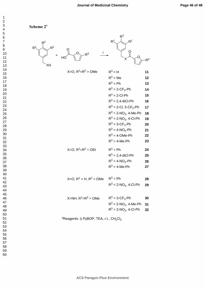
Scheme 2a
R1R2
R1
XH
O
R1R2
R1
X OR3
+ R3
HO
O Oi
aReagents: i) PyBOP, TEA, r.t., CH2Cl2
X=O, R1=R2 = OMe R3 = H
R3 = MeR3 = PhR3 = 2-CF3-Ph
R3 = 2-Cl-PhR3 = 2,4-diCl-Ph
R3 = 2-Cl, 5-CF3-Ph
R3 = 2-NO2, 4-Me-PhR3 = 2-NO2, 4-Cl-PhR3 = 3-CF3-PhR3 = 4-NO2-PhR3 = 4-OMe-PhR3 = 4-Me-Ph
11
13
17
161514
12
181920212223
X=O, R1=R2 = OEt R3 = PhR3 = 2,4-diCl-PhR3 = 4-NO2-PhR3 = 4-Me-Ph
24252627
X=NH, R1=R2 = OMe
X=O, R1 = H, R2 = OMe R3 = Ph
R3 = 2-NO2, 4-Cl-Ph2829
R3 = 3-CF3-Ph
R3 = 2-NO2, 4-Me-PhR3 = 2-NO2, 4-Cl-Ph
30
3132
Page 46 of 48
ACS Paragon Plus Environment
Journal of Medicinal Chemistry
123456789101112131415161718192021222324252627282930313233343536373839404142434445464748495051525354555657585960
CHARTS
Chart 1. Pharmacophore features and their angles and distances relation (Å).
OEtEtO OEt
OO OMe
NO2
Chart 2. Selected lead compound 1 for further optimization and areas where
modifications have been done.
63o
92o110
82o
4.72 6.87
4.54
7.60
Hydrogen bond
acceptor
2 hydrophobic-aliphatic points
Aromatic ring
Page 47 of 48
ACS Paragon Plus Environment
Journal of Medicinal Chemistry
123456789101112131415161718192021222324252627282930313233343536373839404142434445464748495051525354555657585960
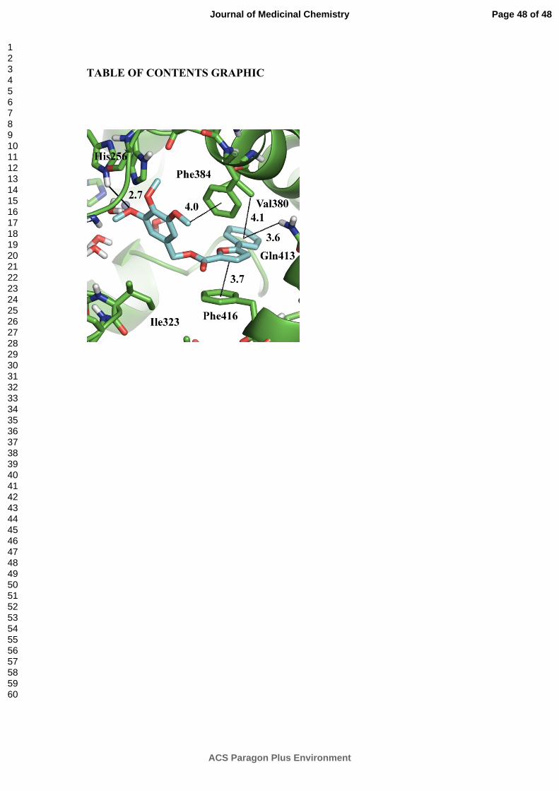
TABLE OF CONTENTS GRAPHIC
Page 48 of 48
ACS Paragon Plus Environment
Journal of Medicinal Chemistry
123456789101112131415161718192021222324252627282930313233343536373839404142434445464748495051525354555657585960