Embed Size (px)
Citation preview
Transworld Research Network 37/661 (2), Fort P.O.
Trivandrum-695 023 Kerala, India
Advances in Molecular Mechanisms and Pharmacology of Diabetic Complications, 2010:
17-38 ISBN: 978-81-7895-499-8 Editor: Milan Stefek
2. Glycation of proteins - their analysis and
physiological aspects
Kateřina Lacinová1, Adam Eckhardt1,2, Statis Pataridis1, Pavla Sedláková1 and Ivan Mikšík1,2
1Institute of Physiology, Academy of Sciences of the Czech Republic, Prague, Czech, Republic 2Cardiovascular Research Centre, Prague, Czech Republic
Abstract. Posttranslational non-enzymatic modifications of proteins (as well as of lipoproteins and glycoproteins) are important protein reactions, which are believed to play an important role in several physiological and pathophysiological processes, such as aging, diabetes mellitus, etc. Non-enzymatic chemical reactions that occur between an oxo-group typically stemming from reducing sugars (or arising from lipid oxidation/peroxidation) and the free amino groups of proteins/peptides lead to the formation of a plethora of still poorly characterized reaction products (the so-called Maillard reaction). A typical
example of these non-enzymatic changes is the formation of advanced glycation end products (AGEs). The accumulation of AGEs and the resulting structural alterations cause altered tissue properties (increased stiffness, reduced elasticity), which contribute to their reduced catabolism and to aging. The most susceptible proteins are those which are slowly metabolized, having a longer exposure time for chemical reactions with endo- and exogenous compounds. AGEs have been studied in connection with diabetes, aging and endothelial dysfunction. The AGEs of plasma and matrix proteins are candidate mediators
for various vascular complications, such as atherosclerosis.
1. Introduction Proteomics, analogous to the term genomics, describes the study of the complete set
of proteins present in a cell, organ, or organism at a given time. The genome tells us what
1111 Correspondence/Reprint request: Dr. Kateřina Lacinová, Institute of Physiology, Academy of Sciences of the Czech
Republic, Prague, Czech Republic. E-mail: [email protected]
Kateřina Lacinová et al. 18
could theoretically happen, whereas the proteome tells us what does happen. A genomic-
centered view of biologic processes is therefore incomplete and does not describe what
happens at the protein level [1]. The covalent assembly of proteins begins on the
ribosome with the formally sequential linear translation of RNA triplet codons, each
coding for one of the 20 naturally occurring amino acids, into the nascent polypeptide chain. The rest of a protein's life is a decidedly non-linear affair. Beginning with the
folding of the polypeptide, proteins can be modified in a number of ways, most notably
by interactions with other proteins and by covalent modification of the side chains of
selected amino acids or of the peptide groups. A simple interpretation of covalent
modifications is that they diversify the amino acid code, thus enabling the proteins to do
new things and behave differently. However, if simple and stoichiometric additions to
selected side chains or cleavage of the polypeptide was all that posttranslational
modifications entailed, the subject would not be remotely as interesting or as important as
it is to modern biology. Posttranslational modifications of proteins (PTMs) are highly
dynamic, often reversible, and not always stoichiometric changes. In most fields of
biochemistry today, discovering and understanding how, where, when, and under what
conditions specific PTMs occur is the goal of many researchers. For chemists, the study of PTMs represents an important opportunity both to contribute to an understanding of
these processes - and perhaps to discover new ones - and to overcome the technical
challenges of measuring these changes. On the other hand, some PTMs defy simple
chemical arguments and may have occurred as evolutionary accidents. Examples include
the PTM of proteins by the 76-amino-acid protein ubiquitin, a process that flags the
modified proteins to be degraded by an enzyme complex called the proteosome.
Reversible posttranslational modification (PTM) is a versatile way to regulate protein
activity. Historically, the best studied PTM is phosphorylation, largely because of the
relative ease of detecting protein phosphorylation in vivo and in vitro. However, many
other types of reversible PTMs exist, with acetylation, methylation, O-GlcNacylation,
and ubiquitination having become prominent more recently. Moreover, many proteins are multiply modified by various covalently attached groups.
Positive crosstalk is defined as a situation in which one PTM serves as a signal for
the addition or removal of a second PTM or for recognition by a binding protein that
carries out a second modification. In the case of negative crosstalk, there can be direct
competition for the modification of a single residue in a protein, or indirect effects,
wherein one modification masks the recognition site for a second PTM. For example, a
single Lys can be modified by ubiquitination or SUMOylation (e.g., proliferating cell
nuclear antigen); by acetylation and ubiquitination (e.g., protein P53); or by acetylation,
ubiquitination, and methylation (e.g., lysine 302 in estrogen receptor ). Both enzymatic and non-enzymatic posttranslational modifications exist. Proteins
undergo important non-enzymatic (chemical) posttranslational modifications with a
significant physiological/biological impact. The most susceptible proteins are those
which are slowly metabolized, having a longer exposure time for chemical reaction with endo- and exogenous compounds [2], [3].
2. Protein glycation
One of the typical non-enzymatic reactions is glycation, the reaction between the
reactive oxo-group of the sugar and the amino group of the protein [4], [5], [6], [7]. It is
now well known that the structure, biological function and turnover of proteins are altered when protein glycation takes place [8].
Protein glycation assays 19
Reducing sugars (as glucose or ribose) are known to form chemically early glycation
products with proteins at a rate proportional to the sugar concentration. In this process the
aldehyde group of the open chain form of glucose condenses with an amino group to
form a Shiff base. The amino acids involved may be the N-terminal (such as valine in
hemoglobin) and /or the -amino groups of lysine in numerous proteins. The first stage of the reaction, equilibrating within hours, is followed by an Amadori rearrangement in
which the double bond shifts to the C-2 of the sugar (glucose) to give a stable, covalently linked fructose derivative of the protein, designated Amadori-type glycation products. A
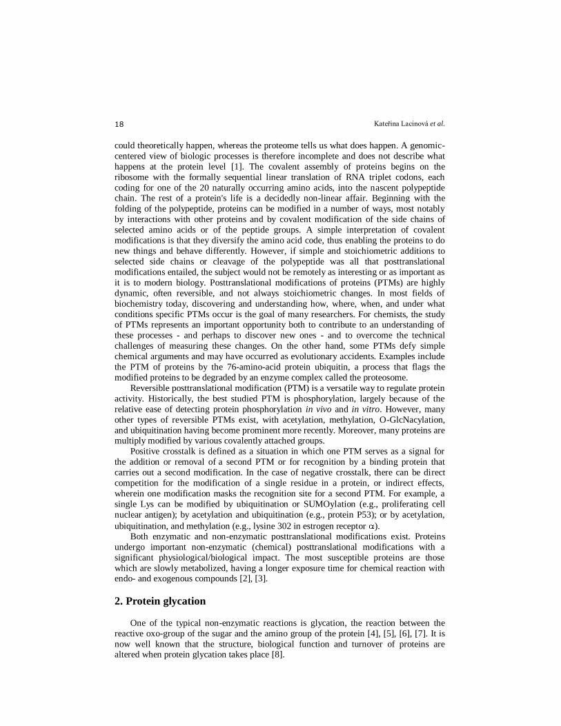
simplified scheme of the Maillard reaction is presented in Fig. 1. The reaction is initiated
by the nonenzymatic condensation reaction of a reducing sugar such as glucose with an
amine, either on a protein or on a low-molecular-weight compound. Following the
formation of a Schiff base, the Amadori rearrangement occurs and leads to a ketoamine
adduct. The ketoamine adduct can either undergo spontaneous slow reversion to
regenerate the free sugar and the amine, or the glyco portion can undergo dehydration
and rearrangement to form so-called deoxyglucosones. The following step is termination.
This is a reaction where a glucose-derived compound (pyrraline) and pentose-derived
protein cross-link (pentosidine) are formed, both of which are formed from glycated
proteins in the presence of oxygen. Formation of the Amadori product from the Schiff base, the first level of protein
glycation, is characterized by its slow kinetics and natural occurrence in the human body
[9]. However, these modifications are accelerated under pathophysiological conditions
strongly associated with a high concentration of glucose or hyperglycemia. For this
reason, glycation has often been related to chronic complications of diabetes mellitus,
renal failure, and degenerative changes occurring during aging [10], [11], [12]. Changes
in the structure and function of structural proteins, such as collagen, fibronectin, tubulin,
lens crystallin, myelin, laminin, and actin, have also been ascribed to AGE formation.
Moreover, AGE-protein adducts produced during glycation seem to inactivate metabolic
enzymes [8].
As for arginine residues, the guanidino group has an important role in glycation
processes and can be used as a chemical target for pharmacological approaches focused on preventing or slowing down glycation reactions. One of the two main pharmacological
approaches includes inhibition of the rearrangement of early products into AGEs and
thus the formation of crosslinked end products. The pharmacological agents studied are
mostly substituted guanidines, such as aminoguanidine, metformin, etc. The mechanism
of these inhibitors is based on trapping reactive dicarbonyl species, which are
intermediates in the formation of AGEs [13]. One way of achieving this goal is to inhibit
glycation by blocking the glycation sites without affecting the level of glucose. The
acetylation of lysine residues by acetylsalicylic acid (aspirin or ASA-acetylsalicylic acid)
inhibits the glycation of plasma proteins [14], [15], [16], hemoglobin [17], and lens
proteins [18], [19]. Acetylation has also been shown to prevent carbamylation, another post-
translational modification of lens crystallins [20]. Rao et al. [21] reported that acetylation of
the -amino groups of lens crystallins by ASA may protect against aggregation by blocking
glycation, carbamylation or isopeptide bond formation. The use of aspirin as a therapeutic agent to reduce diabetic complications was earlier reported by Reid et al. [22].
Due to the great complexity of biological samples (plasma, cellular and subcellular
fractions, etc.), direct identification of the glycated proteins in these substrates is certainly
a difficult task. Consequently, most glycation studies have been carried out by in vitro
experiments in order to extrapolate the information generated (results about glycation
sites, structure modification, etc.) to the in vivo situation. The in vitro glycation of
Kateřina Lacinová et al. 20
Figure 1. General scheme of the Maillard reaction [25].
specific proteins or their mixtures is usually carried out under pseudophysiological conditions [23]. Nonenzymatic molecular modifications induced by reactive carbonyl
species (RCS) generated by peroxidation of membrane phospholipid acyl chains play a
causal role in the aging process. Most of the biological effects of RCS, mainly of

Protein glycation assays 21
, -unsaturated aldehydes, di-aldehydes, and keto-aldehydes, are due to their capacity to react with cellular constituents, forming advanced lipoxidation end-products (ALEs) [24].
2.1. Products of posttranslational modification
Posttranslational modifications can be monotopic or polytopic (demonstrated as
cross-links). Cross-links are most important from a physiological point of view. It must be noted that cross-links involve two different mechanisms of their formation:
A) Enzymatically initiated cross-links
B) Non-enzymatically initiated cross-links
The first type forms physiological cross-links, the second type accompanies aging or
pathophysiological situations. The reaction between free amino groups of proteins (lysine, arginin, N-terminated amino acid) and oxo groups of sugars produces non-
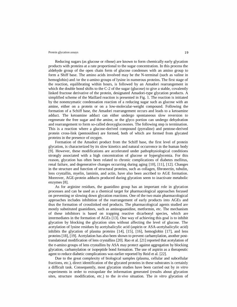
enzymatic glycation. A) Enzymatically initiated cross-links: Dehydroxylysino-norleucine (de-HLNL),
dehydrohydroxylysino-norleucine (deH-HLNL), dehydrodihydroxylysino-norleucine
(deH-DHLNL), hydroxylysino-5-ketonorleucine (HLKNL), dihydroxylysino-norleucine
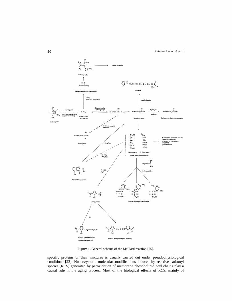
(DHLNL) (Fig.2), lysino-5-ketonorleucine (LKNL), histidinohydroxymerodesmosine
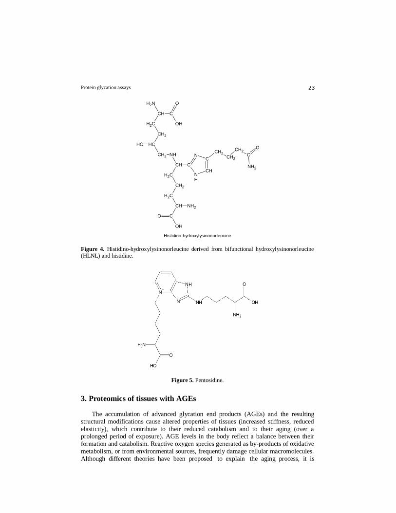
(HHMD) (Fig.3), aldol-histidine (Fig.3), histidino-hydroxylysinonorleucine (Fig.4),
hydroxyaldolhistidine, hydroxylysyl-pyridinolin (HL-Pyr), N-hexosyl-lysine and
N-hexosyl-hydroxylysine.
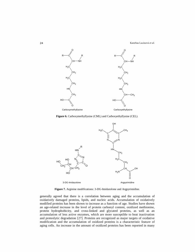
B) Non-enzymatically initiated cross-links: Pentosidine (Fig.5), crossline A and B,
vesperlysine, 1-alcyl-2-formyl-3,4-diglucosyl-pyrrole (AFGP), imidazolium compounds,
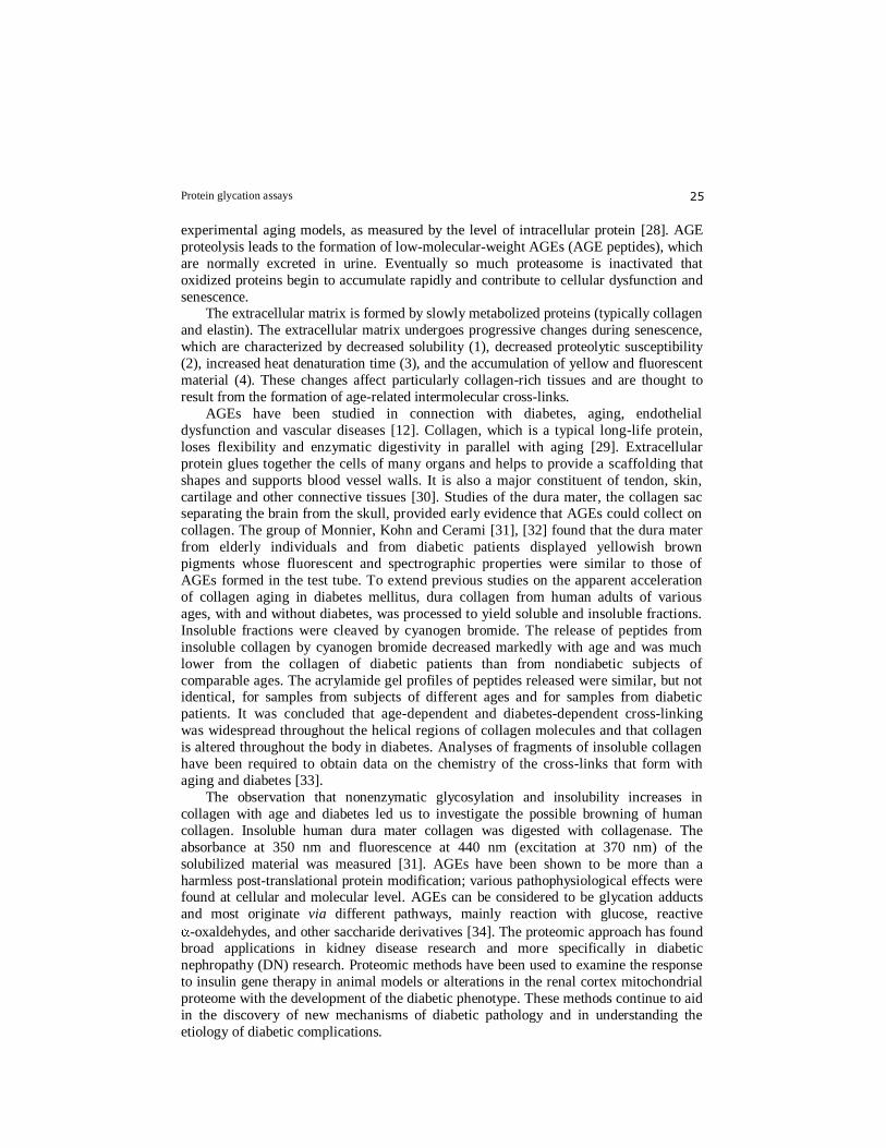
imidasolysine and furosine, pyrraline, carboxymethyllysine (CML), carboxyethyllysine(CEL)
(Fig.6), carboxymethylornithine (CMO), carboxymethylarginine (CMA), -Nω-(4-oxo-5-
dihydroimidazol-2-yl)-1-ornithine ( -NFC-1). Lysine-hydroxy-triosidine and arginine-hydroxy-triosidine.
Pentosidine is the best known cross-linking agent so far [26]. Pentosidine is a lysyl-
arginyl cross-link, which was first discovered in collagen from senescent human
extracellular matrix. Its structure was confirmed through synthesis from lysine and arginine residues with pentoses, in which ribose was the most reactive sugar. The
formation of pentosidine from the hexose glucose has been described. Both diabetes and
most predictably uremia are conditions which lead to increased pentosidine synthesis.
Another fact to be taken into account is the large number of compounds that arise
during post-translational modifications of proteins (typically glycation), most of which are
hydrolytically unstable, many of them monotopical (non-cross-linking) modifications.
Those arising from monotopical modifications include N -(carboxymethyl)lysine (CML),
N -(carboxyethyl)lysine (CEL), pyrraline, and glyoxal lysine amide (GOLA) [26]. Some of these adducts can be made resistant to acid hydrolysis, e.g. by sodium borohydride (sodium
borotritide) reduction, while others may be hydrolytically stable as they are. Some of these
adducts possess a characteristic luminescence, which can be used for selective detection [4].
However, polymerized molecules usually comprise only a fraction of the modifications
that have occurred in the protein; some authors even believe that monotopical (non-
polymerizing) modifications predominate. A number of the arising adducts (some of them are even hydrolytically stable) can be detected directly after protein fragmentation
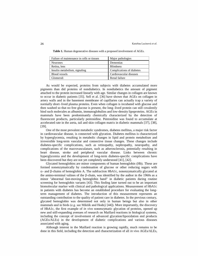
[5]. Arginine modifications: 3-DG-Imidazolone and Argpyrimidine (Fig. 7).
Kateřina Lacinová et al. 22
OH
O
OH
O
CNH2 CH
CH2
CH2
CH2
CH2
NH
CH2
CH2
C
NH2
CH2
CH2
CH
Lysino-norleucin
OH
OH
O
OH
O
CNH2 CH
CH2
CH2
CH
CH2
NH
CH2
CH2
C
NH2
CH2
CH2
CH
Hydroxylysino-norleucin
OH
OH
OH
O
OH
O
CNH2 CH
CH2
CH2
CH
CH2
NH
CH2
CH
C
NH2
CH2
CH2
CH
Dihydroxylysino-norleucin
Lysino-morleucine Hydroxylysino-norleucine Dihydroxylysino-norleucine
Figure 2. Lysino-morleucine, hydroxylysino-norleucine, dihydroxylysino-norleucine.
OH
O
C
NH2
CH
CH2CH2
CH2CH2
OH
O
C
NH2CH
CH2CH2
OH
OH
O
NH2
C
CH CH2
Deoxy py ridinoline
OH
OH
O
C
NH2
CH
CH2CH2
CHCH2
OH
O
C
NH2CH
CH2CH2
OH
OH
O
NH2
C
CH CH2
Py ridinoline
Deoxypyridinoline Pyridinoline
OH
ONH2
CCH
CH2
CH2
OH
O
NH2
CCH
CH2CH2OH
CH2 CH
CH2
CH
OH O
NH2
C
CHCH2
N
CH
CH
N
C
Aldol-histidine
Aldol-histidine
Figure 3. Pyridinolins : Deoxypyridinoline, Pyridinoline, Aldol-histidine.
Protein glycation assays 23
OH
O
C
NH2
CH
CH2
CH2
OH CH
CH2
OH
O C
NH2CH
CH2
CH2
CH2
NH
CH NH2
O
CCH2
CH2
CH2
C
N
NH
C
CH
Histidino-hydroxylysinonorleucine
Histidino-hydroxylysinonorleucine
Figure 4. Histidino-hydroxylysinonorleucine derived from bifunctional hydroxylysinonorleucine (HLNL) and histidine.
Figure 5. Pentosidine.
3. Proteomics of tissues with AGEs
The accumulation of advanced glycation end products (AGEs) and the resulting
structural modifications cause altered properties of tissues (increased stiffness, reduced
elasticity), which contribute to their reduced catabolism and to their aging (over a prolonged period of exposure). AGE levels in the body reflect a balance between their
formation and catabolism. Reactive oxygen species generated as by-products of oxidative
metabolism, or from environmental sources, frequently damage cellular macromolecules.
Although different theories have been proposed to explain the aging process, it is
Kateřina Lacinová et al. 24
R
CH2
CH2
R
O
C
NHCH
OH
O
C
CH2
NH
CH2
CH2
Carboxymethyllysine Carboxyethyllysine
CH3
R
CH2
CH2
R
O
C
NHCH
OH
O
C
CH
NH
CH2
CH2
Carboxymethyllysine Carboxyethyllysine
Figure 6. Carboxymethyllysine (CML) and Carboxyethyllysine (CEL).
OH
CH2
OH
CH
OH
CH CH2
R
NH
OC C
NN
C
3-DG-Imidazolone
NH2
OH
O
C
CH
CH2
CH2
CH2
NH
CH3 CH3
OH
C
N
C
C
C
N
Argpyrimidine
OH
CH2
OH
CH
OH
CH CH2
R
NH
OC C
NN
C
3-DG-Imidazolone
NH2
OH
O
C
CH
CH2
CH2
CH2
NH
CH3 CH3
OH
C
N
C
C
C
N
Argpyrimidine
3-DG-Imidazolone Argpyrimidine
Figure 7. Arginine modifications: 3-DG-Imidazolone and Argpyrimidine.
generally agreed that there is a correlation between aging and the accumulation of
oxidatively damaged proteins, lipids, and nucleic acids. Accumulation of oxidatively
modified proteins has been shown to increase as a function of age. Studies have shown
an age-related increase in the level of protein carbonyl content, oxidized methionine, protein hydrophobicity, and cross-linked and glycated proteins, as well as an
accumulation of less active enzymes, which are more susceptible to heat inactivation
and proteolytic degradation [27]. Proteins are recognized as major targets of oxidative
modification and the accumulation of oxidized proteins is a characteristic feature of
aging cells. An increase in the amount of oxidized proteins has been reported in many
Protein glycation assays 25
experimental aging models, as measured by the level of intracellular protein [28]. AGE
proteolysis leads to the formation of low-molecular-weight AGEs (AGE peptides), which
are normally excreted in urine. Eventually so much proteasome is inactivated that
oxidized proteins begin to accumulate rapidly and contribute to cellular dysfunction and
senescence. The extracellular matrix is formed by slowly metabolized proteins (typically collagen
and elastin). The extracellular matrix undergoes progressive changes during senescence,
which are characterized by decreased solubility (1), decreased proteolytic susceptibility
(2), increased heat denaturation time (3), and the accumulation of yellow and fluorescent
material (4). These changes affect particularly collagen-rich tissues and are thought to
result from the formation of age-related intermolecular cross-links.
AGEs have been studied in connection with diabetes, aging, endothelial
dysfunction and vascular diseases [12]. Collagen, which is a typical long-life protein,
loses flexibility and enzymatic digestivity in parallel with aging [29]. Extracellular
protein glues together the cells of many organs and helps to provide a scaffolding that
shapes and supports blood vessel walls. It is also a major constituent of tendon, skin,
cartilage and other connective tissues [30]. Studies of the dura mater, the collagen sac separating the brain from the skull, provided early evidence that AGEs could collect on
collagen. The group of Monnier, Kohn and Cerami [31], [32] found that the dura mater
from elderly individuals and from diabetic patients displayed yellowish brown
pigments whose fluorescent and spectrographic properties were similar to those of
AGEs formed in the test tube. To extend previous studies on the apparent acceleration
of collagen aging in diabetes mellitus, dura collagen from human adults of various
ages, with and without diabetes, was processed to yield soluble and insoluble fractions.
Insoluble fractions were cleaved by cyanogen bromide. The release of peptides from
insoluble collagen by cyanogen bromide decreased markedly with age and was much
lower from the collagen of diabetic patients than from nondiabetic subjects of
comparable ages. The acrylamide gel profiles of peptides released were similar, but not identical, for samples from subjects of different ages and for samples from diabetic
patients. It was concluded that age-dependent and diabetes-dependent cross-linking
was widespread throughout the helical regions of collagen molecules and that collagen
is altered throughout the body in diabetes. Analyses of fragments of insoluble collagen
have been required to obtain data on the chemistry of the cross-links that form with
aging and diabetes [33].
The observation that nonenzymatic glycosylation and insolubility increases in
collagen with age and diabetes led us to investigate the possible browning of human
collagen. Insoluble human dura mater collagen was digested with collagenase. The
absorbance at 350 nm and fluorescence at 440 nm (excitation at 370 nm) of the
solubilized material was measured [31]. AGEs have been shown to be more than a
harmless post-translational protein modification; various pathophysiological effects were found at cellular and molecular level. AGEs can be considered to be glycation adducts
and most originate via different pathways, mainly reaction with glucose, reactive
-oxaldehydes, and other saccharide derivatives [34]. The proteomic approach has found broad applications in kidney disease research and more specifically in diabetic
nephropathy (DN) research. Proteomic methods have been used to examine the response
to insulin gene therapy in animal models or alterations in the renal cortex mitochondrial
proteome with the development of the diabetic phenotype. These methods continue to aid
in the discovery of new mechanisms of diabetic pathology and in understanding the
etiology of diabetic complications.
Kateřina Lacinová et al. 26
Table 1. Human degenerative diseases with a proposed involvement of AGEs.
Failure of maintenance in cells or tissues Major pathologies
Neurones Dementias
Retina, lens Blindness
Insulin metabolism, signaling Complications of diabetes
Blood vessels Cardiovascular diseases
Glomeruli Renal failure
As would be expected, proteins from subjects with diabetes accumulated more
pigments than did proteins of nondiabetics. In nondiabetics the amount of pigment
attached to the protein increased linearly with age. Similar changes in collagen are known
to occur in diabetic patients [35]. Sell et al. [36] have shown that AGEs on collagen in
artery walls and in the basement membrane of capillaries can actually trap a variety of
normally short–lived plasma proteins. Even when collagen is incubated with glucose and
then washed so that no free glucose is present, the long–lived protein can still covalently
bind such molecules as albumin, immunoglobulins and low-density lipoproteins. AGEs in
mammals have been predominantly chemically characterized by the detection of
fluorescent products, particularly pentosidine. Pentosidine was found to accumulate at accelerated rate in the aorta, tail and skin collagen matrix in diabetic mammals [37], [38],
[39].
One of the most prevalent metabolic syndromes, diabetes mellitus, a major risk factor
in cardiovascular disease, is connected with glycation. Diabetes mellitus is characterized
by hyperglycemia, resulting in metabolic changes in lipid and protein metabolism and
irreversible long-term vascular and connective tissue changes. These changes include
diabetes-specific complications, such as retinopathy, nephropathy, neuropathy, and
complications of the macrovasculature, such as atherosclerosis, potentially resulting in
heart disease, stroke and peripheral vascular disease. Links between chronic
hyperglycemia and the development of long-term diabetes-specific complications have
been discovered but they are not yet completely understood [41], [42]. Glycated hemoglobins are minor components of human hemoglobin (Hb). These are
formed nonenzymatically by condensation of glucose or other reducing sugars with
- and -chains of hemoglobin A. The subfraction HbA1c, nonenzymatically glycated at
the amino-terminal valines of the -chain, was identified by the author in the 1960s as a minor "abnormal fast-moving hemoglobin band" in diabetic patients during routine
screening for hemoglobin variants [43]. This finding later turned out to be an important
biomolecular marker with clinical and pathological applications. Measurement of HbA1c
in patients with diabetes has become an established procedure for evaluating the long-
term management of diabetes. The introduction of this measurement represents an
outstanding contribution to the quality of patient care in diabetes. In the previous century,
glycated hemoglobin was determined not only in human beings but also in other
mammals and in birds (e.g. see Mikšík and Hodný [44]). More importantly, the discovery
of HbA1c, the first example of in vivo nonenzymatic glycation of proteins, opened up
new and still-expanding avenues of research on Maillard reactions in biological systems,
including the concept of involvement of advanced glycation/lipoxidation end products (AGEs/ALEs) in the development of diabetic complications and various diseases
associated with aging.
Although interest in the Maillard reaction is growing rapidly, much remains to be
done in this field, including the detection and characterization of all in vivo AGEs/ALEs,
Protein glycation assays 27
the development and clinical applications of AGE inhibitors and disruptors, as well as
investigations into the possible roles of the Maillard reaction in regulatory biology and
carcinogenesis [43]. The advanced lipoxidation end products (ALEs) are derived from
lipid peroxidation. They accumulate reactive carbonyl compounds (RCOs) derived from
carbohydrates and lipids, followed by carbonyl modifications of proteins ("carbonyl stress"). These soluble reactive intermediates, precursors of ALEs, are not only cytotoxic
per se but they also behave as mediators and propagators of oxidative stress and cellular
and tissue damage. The consequent loss of function and structural integrity of modified
biomolecules can have a wide range of downstream functional effects and may be the
cause of subsequent cellular dysfunctions and tissue damage. The causal role of ALEs in
aging and longevity is inferred from the following findings: a) their accumulation with
aging in several tissues and species; b) physiological interventions (dietary restrictions)
which increase longevity decrease ALE content; c) the longer the longevity of a species,
the lower is its lipoxidation-derived molecular damage; and finally, d) exacerbated levels
of ALEs are associated with pathological states [24].
The AGEs of plasma and matrix proteins are candidate mediators for various
vascular complications, such as atherosclerosis. A significantly larger accumulation of AGEs was detected in the aorta of stroke-prone spontaneously hypertensive rats [44].
Nε-(carboxymethyl)lysine (CML) is an advanced glycation end product formed in
proteins via combined non-enzymatic glycation and glycoxidation reactions [45]. CML
reacts with the receptor of advanced glycation end products, inducing impairment of
endothelium-dependent relaxation, and it is a marker of oxidative stress. During coronary
angiography, a peripheral venous blood sample was taken for CML measuring. Serum
CML levels were significantly increased in patients with acute myocardial infarction.
Elevation of the glycoxidation product N -(carboxymethyl)lysine is present in patients with acute myocardial infarction [45]. The passive biomechanical properties of vessels
change with age. An increase in thickness and stiffness, as well as a reduction in
elasticity of the aorta during aging were found both in rats and humans [46], [47], [48].
4. Separation, analysis and identification of non-enzymatically
posttranslationally modified proteins The question may arise why these post-translationally modified proteins are so
extensively studied today. Several reasons are involved. First, post-translational non-
enzymatic modifications obviously affect the biological functions of a number of
proteins: they may lead to increased protein insolubilization, protein exclusion from
normal metabolic pathways or alteration of its physiological function.
This appears to be the case e.g., of glycated crystallins, collagens, lens membrane
proteins, enzymes, etc.
Both in vitro and in vivo glycation lead either to polymerized protein species or to
monotopic (non-polymerized) protein molecules. In order to determine the presence of
protein polymers or modified (with a blocked free amino group) molecules, a number of
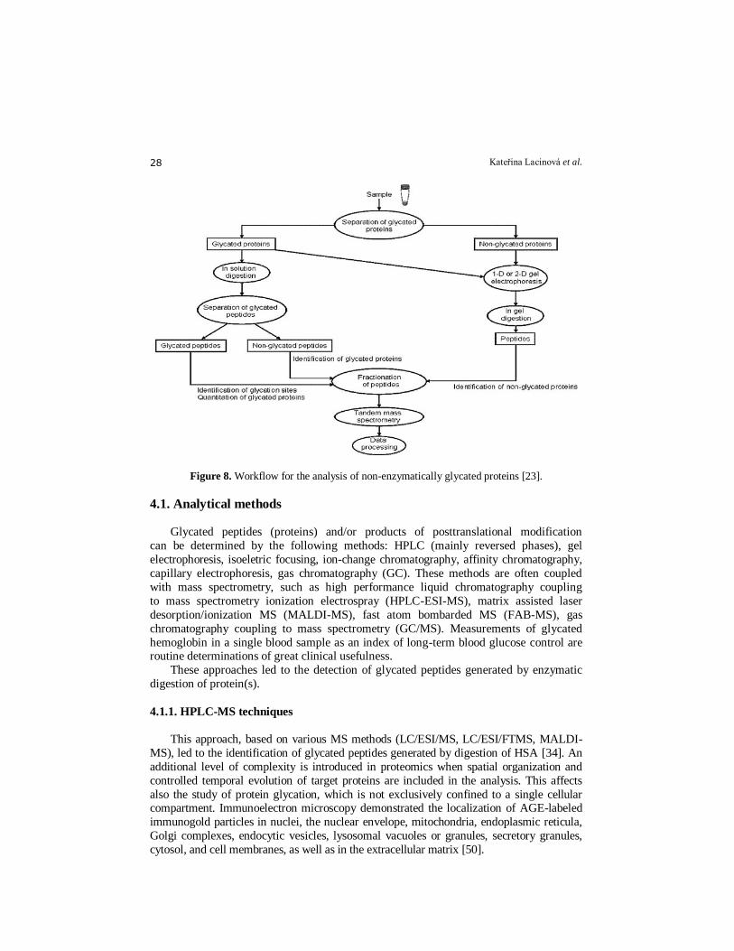
separation techniques are available. Proteomics is the systematic analysis of the proteins expressed by a cell or tissue, and
mass spectrometry is its essential analytical tool. New approaches have provided
landmark advances in determining functionally relevant properties of proteins, including
their quantity and involvement within protein complexes [49]. One of the possible path of
the analysis of non-enzymatically glycated proteins is represented in Fig. 8.
Kateřina Lacinová et al. 28
Figure 8. Workflow for the analysis of non-enzymatically glycated proteins [23].
4.1. Analytical methods
Glycated peptides (proteins) and/or products of posttranslational modification
can be determined by the following methods: HPLC (mainly reversed phases), gel
electrophoresis, isoeletric focusing, ion-change chromatography, affinity chromatography,
capillary electrophoresis, gas chromatography (GC). These methods are often coupled
with mass spectrometry, such as high performance liquid chromatography coupling
to mass spectrometry ionization electrospray (HPLC-ESI-MS), matrix assisted laser
desorption/ionization MS (MALDI-MS), fast atom bombarded MS (FAB-MS), gas
chromatography coupling to mass spectrometry (GC/MS). Measurements of glycated
hemoglobin in a single blood sample as an index of long-term blood glucose control are routine determinations of great clinical usefulness.
These approaches led to the detection of glycated peptides generated by enzymatic
digestion of protein(s).
4.1.1. HPLC-MS techniques
This approach, based on various MS methods (LC/ESI/MS, LC/ESI/FTMS, MALDI-
MS), led to the identification of glycated peptides generated by digestion of HSA [34]. An
additional level of complexity is introduced in proteomics when spatial organization and
controlled temporal evolution of target proteins are included in the analysis. This affects
also the study of protein glycation, which is not exclusively confined to a single cellular compartment. Immunoelectron microscopy demonstrated the localization of AGE-labeled
immunogold particles in nuclei, the nuclear envelope, mitochondria, endoplasmic reticula,
Golgi complexes, endocytic vesicles, lysosomal vacuoles or granules, secretory granules,
cytosol, and cell membranes, as well as in the extracellular matrix [50].
Protein glycation assays 29
Gontarev et al. [51] compared three different approaches to producing chips for
preparing affinity-sorbed glycated proteins for MALDI-MS analysis consisting of (a)
affinity sorption performed in microtest-tubes using Sepharose microparticles modified
with 3-aminophenylboronic acid followed by sorbent immersion into an agarose
gel on the chip surface; (b) fixation of Sepharose immobilized phenylboronic acid microparticles onto a chip surface using an agarose gel with subsequent affinity retention
of the target compounds directly on the chip spots; and (c) chemical activation of an
agarose gel directly on the chip surface with subsequent covalent immobilization of
3-aminophenylboronic acid. Due to the three-dimensional gel structure, the spot capacity
of affinity chips is very high, thus enabling an in situ preconcentration effect, which
provides an increase in sensitivity, even in complex biological fluids. On considering
these facts along with the above proposed workflow, enzymatic digestion is to be seen as
one of the most critical steps in understanding the information concerning glycation sites.
Protein modifications by the insertion of glucose molecules or derivatives modify the
digestion pattern of enzymatic reactions. Enzymatic digestion is usually carried out in
solution using conventional protocols involving trypsin after denaturation, reduction and
alkylation. The fractionation of peptides has proved to be very useful in proteomic analysis when coupled to MS and MS/MS detection. This coupling enables an increase in
resolution, which is of particular advantage on identifying proteins in complex samples.
Due to the complexity of the analysis of glycated proteins, a few methods reported for
this purpose include a fractionation step at the peptide level. The separation of peptides is
usually carried out by liquid chromatography, using conventional protocols in a C18
reversed phase. Liquid chromatography can be on-line connected to MS detectors in
electrospray (ESI) or atmospheric pressure chemical ionization (APCI) modes, and also
to MALDI systems with robotic sampling devices. The performance of peptide separation
on microfluidic chip-based devices is also being implemented in proteomic approaches to
take advantage of the benefits of using miniaturized devices. These microfluidic systems
enable peptide fractionation to be performed in shorter times than those carried out in chromatographic columns [52]. The application of mass spectrometry in its various
modes - ion trap, triple quadrupole (QqQ), quadrupole-ion trap (QIT), quadrupole-time of
flight (Q-TOF), tandem time of flight (TOF/ TOF), and Fourier transform (FT)-based
techniques - has been crucial in studying the relevance of the glycation process in
proteins. Continuous advances in this technique will be required to understand this
complex PTM as well as its role in several diseases. The main limitation is that this mass
increase can be observed also for enzymatic O- and N-glycosylations and thus
conclusions from this research should be evaluated cautiously.
The main problem in proteomic analysis of glycated peptides, which was reported by
Lapolla et al. [53], is the weak fragmentation of the peptide backbone with collision-
induced dissociation (CID) as the fragmentation mode. In addition, the CID-MS/MS
spectrum of glycated peptides is marked by a high abundance of signals corresponding to various degrees of neutral water loss from the sugar moiety. One other fragmentation
mode especially suited for the characterization of PTMs is electron-transfer dissociation
(ETD), which has recently been developed by Syka et al. [54]. This mode is similar to
electron capture dissociation (ECD), implemented on Fourier transform ion cyclotron
resonance mass spectrometers, but the electron source consists of aromatic anions such as
fluoranthene. ETD fragmentation consists of bond dissociation immediately after electron
transfer, thus avoiding the alteration of labile PTMs and providing complete sequence
information as a result. There are several studies that compare ETD and CID as
fragmentation modes in the analysis of glycated peptides [55], [52]. Their conclusions
Kateřina Lacinová et al. 30
can be summarized as follows: ETD-MS/MS enables an almost complete elucidation of
the sequence for glycated peptides, while signals corresponding to neutral water loss
dominate mass spectra provided by CID. A method based on neutral water loss scanning
by LC–MS/MS has recently been developed for the screening and sequencing of glycated
proteins [56]. One of the main reasons why so few methods have been reported for protein glycation is the lack of bioinformational tools for this PTM.
4.1.2. Gel electrophoresis
Gel electrophoresis is one of the crucial analytical methods used in proteomic
research. The field of proteomic analysis of non-enzymatically glycated proteins was
recently reviewed by Capote and Sanchez [23]. The process to be followed in proteomic
analysis is basically composed of three main steps: sample preparation, analysis by mass
spectrometry (MS) and data processing. In most cases, the experimental workflow
consists of a protein separation step usually one- or two-dimensional (1D or 2D) gel
electrophoresis, enzymatic hydrolysis of proteins in gel, chromatographic separation of
the resulting peptides and detection by tandem mass spectrometry (MS/MS) or alternatively MALDI/MS. Techniques such as 1D or 2D polyacrylamide gel electrophoresis
and, less frequently, chromatofocusing, size exclusion and anion-exchange liquid
chromatography have been reported in studying glycation in proteins, such as bovine
serum albumin [5]. However the equally important information on the preferred glycation
sites for each protein, the relevance of which has been discussed above, is lost during the
conventional enzymatic digestion of proteins.
4.1.3. Capillary electrophoresis
Capillary electromigration methods are more efficacious for analysis of peptides than
gel electrophoresis. For the separation of collagens and their glycated products, the following capillary electromigration methods are suitable: capillary zone electrophoresis
(CZE), capillary gel electrophoresis (CGE), micellar electrokinetic chromatography
(MEKC), capillary electrochromatography, and a combination of capillary electrophoresis
with HPLC and with MS. (For review see paper [57]).
Analyses of the structure and modifications of the collagen molecule/chain by
capillary electrophoresis require solubilization and fragmentation of the protein to smaller
peptides. In principle, two methods can be used: nonenzymatic (cleavage by bromcyan
CNBr) or enzymatic treatment (trypsin).
The chromatographic and electromigration behavior of individual peptides varies
considerably. An offline HPLC-CE coupling made it possible to discover minor changes
in the structure of collagen caused by posttranslational modifications. This HPLC-CE
technique for peptide analysis was used to identify posttranslational modifications in slowly metabolized test proteins [58].
Capillary zone electrophoresis (CZE) with UV spectrophotometric detection has been
employed for the investigation of chemical and structural changes of BSA caused by its
reaction with the above oxo-compounds exhibiting different degrees of reactivity [59].
4.1.4. Affinity chromatography
Affinity chromatography is a powerful protein separation method based on the
specific interaction between immobilized ligands and target proteins. Peptides can also be
Protein glycation assays 31
separated effectively by affinity chromatography through the use of peptide-specific
ligands. Both two-dimensional electrophoresis (2-DE)- and non-2-DE-based proteomic
approaches benefit from the application of affinity chromatography. Before protein
separation by 2-DE, affinity separation is used primarily for preconcentration and
pretreatment of samples. Prior to mass spectrometry (MS), preconcentration and capture of specific proteins or peptides to enhance sensitivity can be accomplished by using
affinity adsorption. With affinity chromatography using m-aminophenylboronate
columns, sufficient quantities of glycated proteins can be obtained, for e.g, fluorescence
measurements. Affinity chromatography of peptide mixtures coupled with mass
spectrometry provides a tool for the study of protein posttranslational modification
(PTM) sites and quantitative proteomics.
An emerging area of development of proteomics technology is miniaturization.
Affinity chromatography is becoming more widely used for exploring PTM and protein-
protein interactions, especially with a view toward developing new general tag systems
and strategies of chemical derivatization on peptides for affinity selection. More
applications of affinity-based purification can be expected, including increased resolution
in 2-DE, improved sensitivity of MS quantification, and incorporation of purification as part of multidimensional liquid chromatography experiments [60].
4.1.5. Immuno methods
The following immuno methods can be used: enzyme-linked immunosorbent assay
(ELISA) and radioimmunoassay (RIA).
For enhanced selectivity, RIA and ELISA have been proposed to evaluate the
glycation levels of intact proteins and their hydrolysis products. However, these
approaches provided misleading results when applied to complex physiological matrices.
4.2. Particular examples illustrating analysis of proteins
4.2.1. Analysis of albumin
Serum albumins represent in the context of glycation frequently studied model
proteins [61]. Basically, they are heterogeneous serum proteins; in human serum
albumin (HSA) four major reasons of this heterogeneity are considered: (a)
polymerization; (b) differences in the thiol content; (c) presence of bound compounds;
(d) occurrence of postsynthetic modifications of the protein [62], [63], [64]. In HSA the
principal glycation site is Lys 525, accounting for about 33% of the overall glycation. In the sequentially highly homological bovine serum albumin (BSA), glycation
was shown to occur in the peptide sequence covering the region 548–557. A number
of standard separation procedures have been used for this purpose, particularly
polyacrylamide gel electrophoresis (PAGE), gel permeation chromatography and borate
affinity chromatography.
Resistance of modified proteins to hydrolysis is a serious restriction for all methods
involving hydrolysis. Separation of intact proteins suffers mostly from very small
differences in physico-chemical characteristics between modified and unmodified
protein, making any separation a difficult task.
Mikulíková et al. [59] used HPLC with either UV or MS detection, capillary
electrophoresis and PAGE methods for analysis of tryptic digests of modified BSA. The
result depends on the nature of the modifiers used: glucose, being a mild modifier, offers
Kateřina Lacinová et al. 32
a rich profile of tryptic peptides, while ribose treatment led to a moderately modified
protein more resistant to enzymatic cleavage than native BSA. Reaction with dioxo-
compounds, i.e. glyoxal and glutaraldehyde, resulted in a modified protein highly
resistant to enzymatic cleavage. It is conceivable that this resistance is caused by cross-
linking reaction(s) of the dioxo-compounds with two free protein amino-groups; such cross-linking could be both inter- or intramolecular.
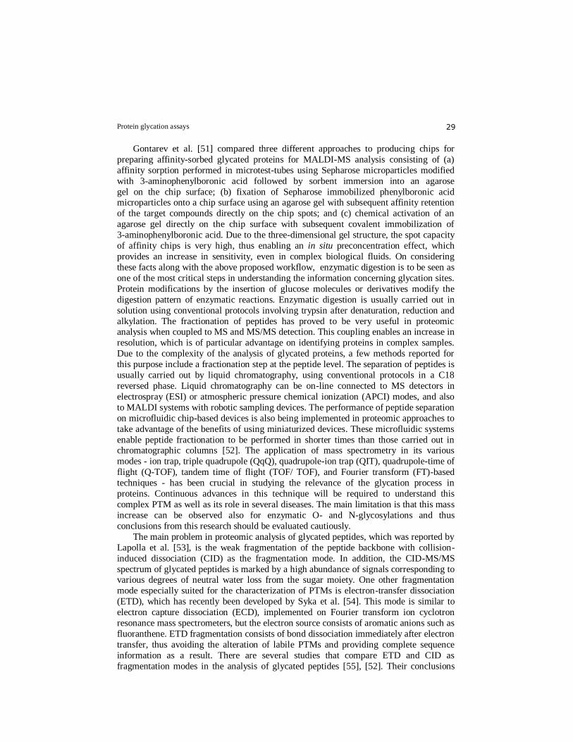
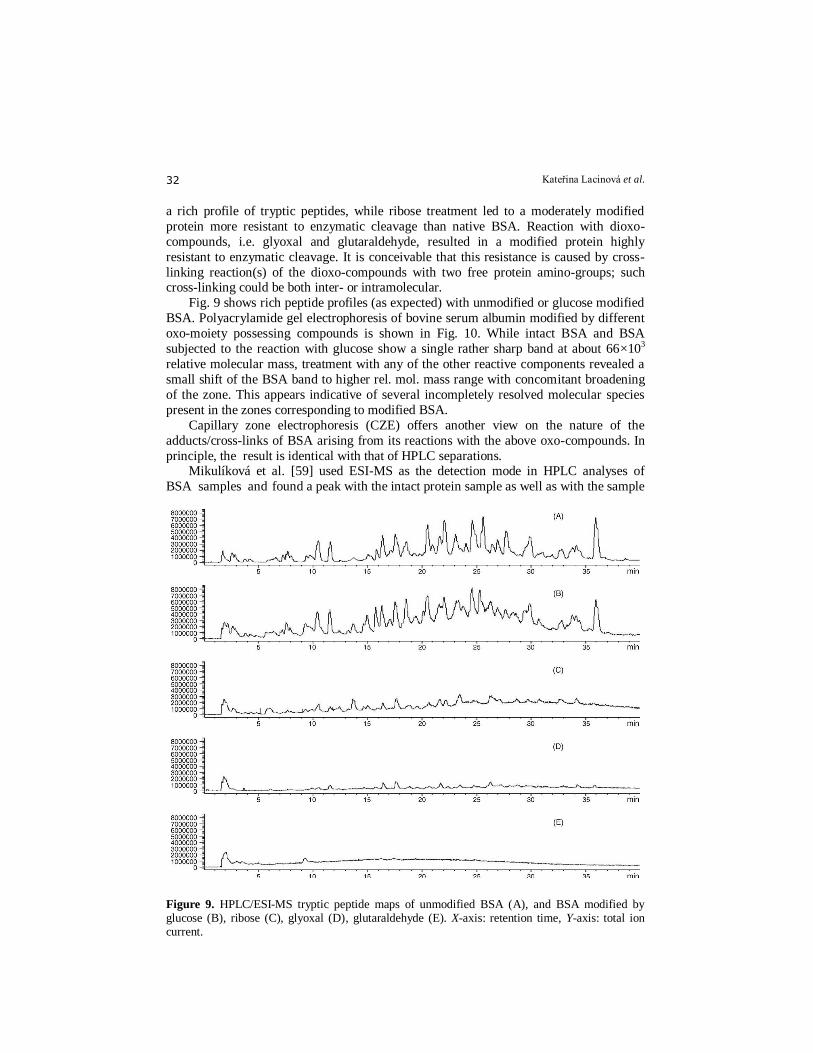
Fig. 9 shows rich peptide profiles (as expected) with unmodified or glucose modified
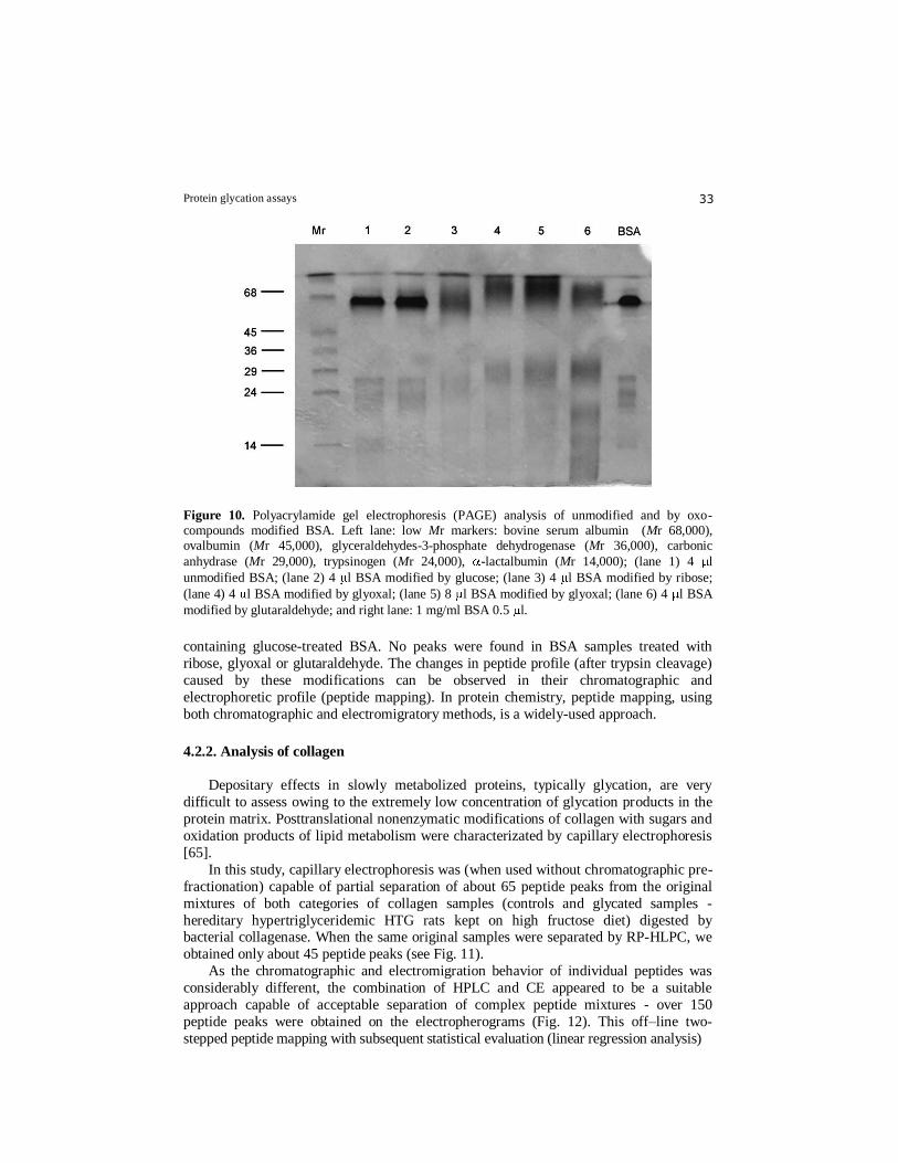
BSA. Polyacrylamide gel electrophoresis of bovine serum albumin modified by different
oxo-moiety possessing compounds is shown in Fig. 10. While intact BSA and BSA
subjected to the reaction with glucose show a single rather sharp band at about 66×103
relative molecular mass, treatment with any of the other reactive components revealed a
small shift of the BSA band to higher rel. mol. mass range with concomitant broadening
of the zone. This appears indicative of several incompletely resolved molecular species
present in the zones corresponding to modified BSA.
Capillary zone electrophoresis (CZE) offers another view on the nature of the
adducts/cross-links of BSA arising from its reactions with the above oxo-compounds. In
principle, the result is identical with that of HPLC separations. Mikulíková et al. [59] used ESI-MS as the detection mode in HPLC analyses of
BSA samples and found a peak with the intact protein sample as well as with the sample
11
Figure 9. HPLC/ESI-MS tryptic peptide maps of unmodified BSA (A), and BSA modified by glucose (B), ribose (C), glyoxal (D), glutaraldehyde (E). X-axis: retention time, Y-axis: total ion current.
Protein glycation assays 33
Figure 10. Polyacrylamide gel electrophoresis (PAGE) analysis of unmodified and by oxo-compounds modified BSA. Left lane: low Mr markers: bovine serum albumin (Mr 68,000), ovalbumin (Mr 45,000), glyceraldehydes-3-phosphate dehydrogenase (Mr 36,000), carbonic
anhydrase (Mr 29,000), trypsinogen (Mr 24,000), -lactalbumin (Mr 14,000); (lane 1) 4 l
unmodified BSA; (lane 2) 4 l BSA modified by glucose; (lane 3) 4 l BSA modified by ribose;
(lane 4) 4 l BSA modified by glyoxal; (lane 5) 8 l BSA modified by glyoxal; (lane 6) 4 l BSA
modified by glutaraldehyde; and right lane: 1 mg/ml BSA 0.5 l.
containing glucose-treated BSA. No peaks were found in BSA samples treated with
ribose, glyoxal or glutaraldehyde. The changes in peptide profile (after trypsin cleavage)
caused by these modifications can be observed in their chromatographic and
electrophoretic profile (peptide mapping). In protein chemistry, peptide mapping, using
both chromatographic and electromigratory methods, is a widely-used approach.
4.2.2. Analysis of collagen
Depositary effects in slowly metabolized proteins, typically glycation, are very
difficult to assess owing to the extremely low concentration of glycation products in the
protein matrix. Posttranslational nonenzymatic modifications of collagen with sugars and
oxidation products of lipid metabolism were characterizated by capillary electrophoresis
[65].
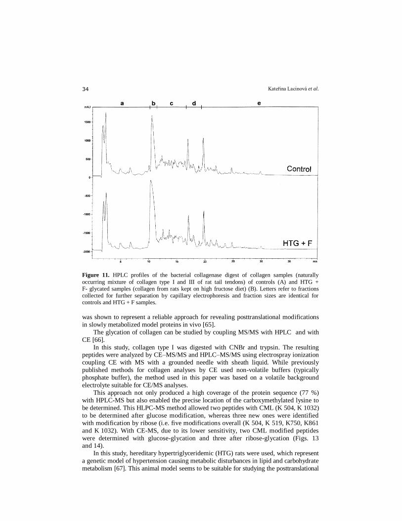
In this study, capillary electrophoresis was (when used without chromatographic pre-
fractionation) capable of partial separation of about 65 peptide peaks from the original
mixtures of both categories of collagen samples (controls and glycated samples -
hereditary hypertriglyceridemic HTG rats kept on high fructose diet) digested by bacterial collagenase. When the same original samples were separated by RP-HLPC, we
obtained only about 45 peptide peaks (see Fig. 11).
As the chromatographic and electromigration behavior of individual peptides was
considerably different, the combination of HPLC and CE appeared to be a suitable
approach capable of acceptable separation of complex peptide mixtures - over 150
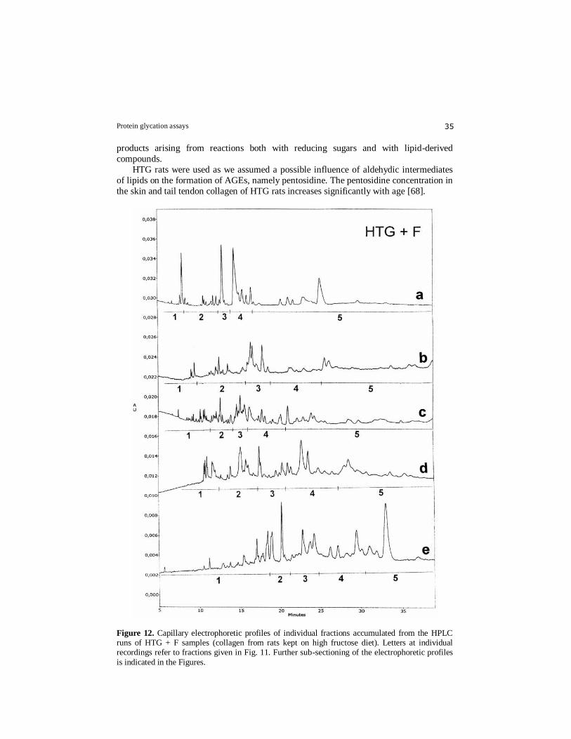
peptide peaks were obtained on the electropherograms (Fig. 12). This off–line two-
stepped peptide mapping with subsequent statistical evaluation (linear regression analysis)
Kateřina Lacinová et al. 34
Figure 11. HPLC profiles of the bacterial collagenase digest of collagen samples (naturally
occurring mixture of collagen type I and III of rat tail tendons) of controls (A) and HTG + F- glycated samples (collagen from rats kept on high fructose diet) (B). Letters refer to fractions collected for further separation by capillary electrophoresis and fraction sizes are identical for controls and HTG + F samples.
was shown to represent a reliable approach for revealing posttranslational modifications
in slowly metabolized model proteins in vivo [65].
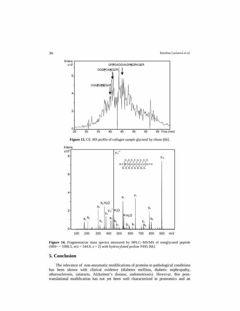
The glycation of collagen can be studied by coupling MS/MS with HPLC and with
CE [66].
In this study, collagen type I was digested with CNBr and trypsin. The resulting peptides were analyzed by CE–MS/MS and HPLC–MS/MS using electrospray ionization
coupling CE with MS with a grounded needle with sheath liquid. While previously
published methods for collagen analyses by CE used non-volatile buffers (typically
phosphate buffer), the method used in this paper was based on a volatile background
electrolyte suitable for CE/MS analyses.
This approach not only produced a high coverage of the protein sequence (77 %)
with HPLC-MS but also enabled the precise location of the carboxymethylated lysine to
be determined. This HLPC-MS method allowed two peptides with CML (K 504, K 1032)
to be determined after glucose modification, whereas three new ones were identified
with modification by ribose (i.e. five modifications overall (K 504, K 519, K750, K861
and K 1032). With CE-MS, due to its lower sensitivity, two CML modified peptides
were determined with glucose-glycation and three after ribose-glycation (Figs. 13 and 14).
In this study, hereditary hypertriglyceridemic (HTG) rats were used, which represent
a genetic model of hypertension causing metabolic disturbances in lipid and carbohydrate
metabolism [67]. This animal model seems to be suitable for studying the posttranslational
Protein glycation assays 35
products arising from reactions both with reducing sugars and with lipid-derived
compounds.
HTG rats were used as we assumed a possible influence of aldehydic intermediates
of lipids on the formation of AGEs, namely pentosidine. The pentosidine concentration in
the skin and tail tendon collagen of HTG rats increases significantly with age [68].
Figure 12. Capillary electrophoretic profiles of individual fractions accumulated from the HPLC runs of HTG + F samples (collagen from rats kept on high fructose diet). Letters at individual recordings refer to fractions given in Fig. 11. Further sub-sectioning of the electrophoretic profiles
is indicated in the Figures.
Kateřina Lacinová et al. 36
Figure 13. CE–MS profile of collagen sample glycated by ribose [66].
Figure 14. Fragmentation mass spectra measured by HPLC–MS/MS of nonglycated peptide
(MH+ = 1088.5, m/z = 544.8, z = 2) with hydroxylated proline P495 [66].
5. Conclusion
The relevance of non-enzymatic modifications of proteins to pathological conditions
has been shown with clinical evidence (diabetes mellitus, diabetic nephropathy,
atherosclerosis, cataracts, Alzheimer’s disease, endometriosis). However, this post-
translational modification has not yet been well characterized in proteomics and an
100 200 300 400 500 600 700 800 900 m/z
Protein glycation assays 37
extensive research into this issue is still required. The present Chapter provides an up-to-
date overview of strategies for proteomic analysis of non-enzymatically glycated
proteins. The modern methods (HPLC-MS/MS, MALDI) should ideally encompass
identification and quantification of glycated proteins, and characterization of glycation
sites [23]. Mass spectrometry (MS), especially when combined with HPLC or CE, is by far the most powerful technique when determination of both identity and site of
modification is required. It has been successfully employed on investigating non-
enzymatic protein glycation, a process relevant to diabetic disease. Due to the low
concentration of glycated proteins in the human organism, selective steps at protein and
peptide level are required to succeed in this complex task. Application of the set of
chromatographic and electrophoretic methods, HPLC/UV, HPLC/MS, PAGE, CZE/UV,
allowed an effective investigation of the nonenzymatic post-translational modifications of
proteins by oxo-compounds.
References 1. Matt, P., Carrel, T., White, M., Lefkovits, I., and Eyk, J.V. 2007, J. Thorac. Cardiovasc. Surg.,
133, 210-214. 2. Maillard, L.C. 1912, Seances Acad. Sci., 154, 66. 3. Trivelli, L.A., Ranney, H.M., and Lai, H.T. 1971, N. Engl. J. Med., 284(7), 353-7.
4. Deyl, Z., and Miksik, I. 1997, J. Chromatogr. B, Biomed. Sci. Appl., 699(1-2), 287-309. 5. Miksik, I. and Deyl, Z. 1997, J. Chromatogr. B, Biomed. Sci. Appl., 699(1-2), 311-45. 6. Monnier, V.M. 1989, Prog. Clin. Biol. Res., 304, 1-22. 7. Monnier, V.M., Sell, D.R., Nagaraj, R.H., Miyata, S., Grandhee, S., Odetti, P., and Ibrahim,
S.A. 1992, Diabetes, 41(2), 36-41. 8. Ulrich, P., and Cerami, A. 2001, Recent Progress Hormone Res., 56, 1-22. 9. Brock, J.W.C., Hinton, D.J.S., Cotham, W.E., Metz, T.O., Thorpe, S.R., Baynes, J.W., and
Ames, J.M. 2003, J. Proteome Res., 2, 506-513.
10. Baynes, J.W. 2001, Exp. Gerontol., 36, 1527-1537. 11. Brownlee, M. 2001, Nature, 14, 813-820. 12. Hipkiss, A.R. 2006, Exp. Gerontol., 41, 464-473. 13. Peyrou, J., and Sternberg, M. 2006, Pathol. Biol., (54), 405-409. 14. Day, J.F., Thorpe, S.R., and Baynes, J.W. 1979, J. Biol. Chem., 254, 595. 15. Day, J.F., Thornbug, R.W., Thorpe, S.R., and Baynes, J.W. 1979, J. Biol. Chem., 254, 9394. 16. Yue, D.K., McLennan, S., Handelsman, D.J., Delbridge, L., Reeve, T., and Turtle, J.R. 1984,
Diabetes, 33, 745.
17. Rendell, M., Nierenberg, J., Brannan, C., Valentine, J.L., Stephen, P.M., Dodds, S., Mercer, P.O., Smith, P.K., and Walder, J. 1986, J. Lab. Clin. Med., 108, 286.
18. Swamy, M.S., and Abraham, E.C. 1988, Invest Ophthalmol. Vis. Sci., 29, 30. 19. Huby, R., and Harding, J.J. 1988, Exp. Eye Res., 47, 53. 20. Crompton, M., Rixon, K.C., and Harding, J.J. 1985, Exp. Eye Res., 40, 297. 21. Rao, G.N., Lardis, M.P., and Cotlier, E. 1985, Biochem. Biophys. Res. Comm., 128, 1125. 22. Reid, J., MacDougall, A.J., and Andrews, M.M. 1957, Br. Med. J., 1071. 23. Capote, F. P., and Sanchez, J.-Ch. 2009, Mass Spectrometry Reviews, 28(1), 135-146. 24. Pamplona, R. 2008, Biochim. Biophys. Acta, 1777(10), 1249-62.
25. Rossomando, E. F. 1998, Nonenzymatic Modifications to Keratins by Ethanol, in HPLC in enzymatic analysis. John Wiley & Sons: New York.
26. Ahmed, N., et al. 2005, Mol. Nutr. Food. Res., 49, 691-699. 27. Earl, R. Stadtman. 2001, Annals of the New York Academy of Sciences, 928(Healthy), 22-38. 28. Grune, T., Shringarpure, R., Sitte, N., and Davies, K. 2001, J. Gerontol. A, Biol. Sci. Med. Sci.,
56(11), B 459-67. 29. Schnider, S. L., and Kohn, R. R. 1981, J. Clin. Invest., 67(6), 1630-5.
Kateřina Lacinová et al. 38
30. Brownlee, M., Vlassara, H., and Cerami, A. 1984, Ann. Intern. Med., 101(4), 527-37. 31. Monnier, V.M., Kohn, R.R., and Cerami, A. 1984, Proc. Natl. Acad. Sci. USA, 81(2), 583-7. 32. Kohn, R.R., Cerami, A., and Monnier, V. M. 1984, Diabetes, 33(1), 57-59. 33. Kohn, R.R. 1983, Connect Tissue Res., 11(2-3), 169-73. 34. Lapolla, A., Fedele, D., Seraglia, R., and Traldi, P. 2006, Mass Spectrometry Reviews, 25(5),
775-797. 35. Schnider, S.L., and Kohn, R.R. 1982, Exp. Gerontol., 17(3), 185-94. 36. Sell, D.R., and Monnier, V.M. 1989, J. Biol. Chem., 264(36), 21597-602. 37. Kern, T.S., and Engerman, R.L. 2001, Diabetes, 50(7), 1636-42. 38. McCance, D.R., Dyer, D.G., Dunn, J.A., Bailie, K.E., Thorpe, S.R., Baynes, J.W., and Lyons,
T.J. 1993, J. Clin. Invest., 91(6), 2470-8. 39. Nyengaard, J.R., Chang, K., Berhorst, S., Reiser, K.M., Williamson, J.R., and Tilton, R. G.
1997, Diabetes, 46(1), 94-106. 40. Reddy, G.K. 2004, Microvasc. Res., 68(2), 132-142.
41. Wautier, J.L., and Guillausseau, P.J. 2001, Diabetes Metab., 27(1), 535-542. 42. Rahbar, S. 2005, Ann. N.Y. Acad. Sci., 1043, 9-19. 43. Miksik, I., and Hodny, Z. 1992, Comp. Biochem. Physiol., 103B, 553-555. 44. Mizutani, K., Ikeda, K., Kawai, Y., and Yamori, Y. 1999, J. Hypertens., 17(4), 481-7. 45. Kralev, S., Zimmerer, E., Brueckmann, M., Lang, S., Kalsch, T., Rippert, A., Lin, J.,
Borggrefe, M., Hammes, H.P., and Suselbeck, T. 2009, Clin. Chem. Lab. Med., 47(4), 446-51. 46. Brüel, A., Ortoft, G., and Oxlund, H. 1998, Atherosclerosis, 140(1), 135-145. 47. Brüel, A., and Oxlund, H. 1996, Atherosclerosis, 127(2), 155-165.
48. Sonesson, B., Hansen, F., and Länne, T. 1997, Eur. J. Vasc. Endovasc. Surg., 14(4), 258-264. 49. Gygi, S.P., and Aebersold, R. 2000, Curr.Opin. Chem. Biol., 4(5), 489-494. 50. Ling, X., Sakashita, N., Takeya, M., Nagai, R., Horiuchi, S., and Takahashi, K. 1998, Lab.
Invest., 78, 1591-1606. 51. Gontarev, S., Shmanai, V., Frey, S.K., Kvach, M., and Schweigert, F.J. 2007, Rapid Commun.
Mass Spectrom., 21, 1-6. 52. Zhang, Q., Frolov, A., Tang, N., Hoffmann, R., van de Goor, T., Metz, T.Q., and Smith, R.D.
2007, Rapid Commun. Mass Spectrom., 21, 661-666.
53. Lapolla, A., Fedele, D., Reitano, R., and Aricó, N.C. 2004, J. Am. Soc. Mass Spectrom., 15, 496-509.
54. Syka, J.E., Coon, J.J., Schroeder, M.J., Shabanowitz, J., and Hunt, D.F. 2004, Proc. Natl. Acad. Sci. USA, 101, 9528-9533.
55. Zhang, Q., Tang, N., Brock, W.C., Mottaz, H.M., Ames, J.M., Baynes, J.W., Smith, R. D., and Metz, T.Q. 2007, J. Proteome Res., 6, 2323-2330.
56. Gadgil, H.S., Bondarenko, P.V., Treuheit, M.J., and Ren, D. 2007, Anal. Chem., 79, 5991-5999.
57. Miksik, I., Sedlakova, P., Mikulikova, K., and Eckhardt, A. 2006, Journal of Chromatography B, 841(1-2), 3-13.
58. Mikulikova, K., Eckhardt, A., and Miksik, I. 2006, J. Sep. Sci., 29, 1126-1131. 59. Mikulikova, K., Miksik, I., and Deyl, Z. 2005, Journal of Chromatography B, 815, 315-331. 60. Lee, W.C., and Lee, K.H. 2004, Anal. Biochem., 324(1), 1-10. 61. Cohen, M.P. 1996, Diabetes and Protein Glycation, Philadelphia, P.A., J.C. Press, 107. 62. Martin, B.K. 1965, Nature, 207(994), 274-6. 63. Gillette, J.R. 1973, Ann. N. Y. Acad. Sci., 6, 226. 64. Fehske, K.J., Muller, W.E. and, Wollert, U. 1981, Biochem. Pharmacol., 30(7), 687-92.
65. Eckhardt, A., Miksik, I., Charvatova, J., Deyl, Z., Forgacs, E., and Cserhati, T. 2005, Journal of Liquid Chromatography & Related Technologies, 28(10), 1437-1451.
66. Mikulikova, K., Eckhardt, A., Pataridis, S., and Miksik, I. 2007, Journal of Chromatography A, 1155, 125-133.
67. Stolba, P., Dobesova, Z., Husek, P., Opltova, H., Zicha, J., Vrana, A., Kunes, J. 1992, Life Sci., 51, 733-740.
68. Miksik, I, Zicha, J., Kunes, J., Deyl, Z. 1997, Life Sci., 60, 2119-2127.


![Elvevated Cortisol and or Simple Carbohydrates in … excess of fructose and glucose in the bloodstream causes extensive glycation and damage the proteins [14]. Advanced glycated products](https://img.pdfslide.net/doc/110x75/5adeb1587f8b9ae1408eac69/elvevated-cortisol-and-or-simple-carbohydrates-in-excess-of-fructose-and-glucose.jpg)