Embed Size (px)
Citation preview
*Professor and Head,**Professor,***P.G. Student, Dept of Oral Medicine and Radiology,Modern Dental College andResearch Center, Indore
NJDSR,Vol.1, January, 2012 7
MAXILLOFACIAL INJURIES- DIAGNOSIS AND IMAGING-WHEN AND WHICH??
P. V. Wanjari*, Arati Chaudhary**, Pratiksha Hada***,
INTRODUCTION
Maxillofacial injuries occur in a significantproportion of trauma patients. They are themost disruptive and distressingemergencies to the dental patients.Diagnostic imaging has traditionallyplayed a central role in providinginformation essential in the initialdiagnosis and treatment of facial injuries.In conventional radiography, anatomy isdepicted in only two dimensions1. With thedevelopment of three dimension techniqueeven smaller changes in the tissue densitydifferential can be identified. Although thefield of radiology has long played anexciting and critical role in dentistry,maxillofacial imaging has added a thirddimension to the effective diagnosis andmanagement of patients with facial bonefractures2.
CLINICAL EXAMINATION-
The affected area should be exposed andexamined for swelling, lacerations, bruisesand bony steps. Diplopia should bechecked following trauma to the orbitalfloor. Pooling of tears and leakage from
the eye indicate damage to thenasolacrimal apparatus, ecchymosis behindthe ear i.e ‘Battle’s sign’ is suggestive ofbasilar skull fracture involving middlecranial fossa where as circumorbitalecchymosis i.e ‘Panda eyes’(and dishdeformity are features of bilateralmaxillary fractures. Sublingual hematomais the most common sign indicative ofmandibular fracture and laceration of thechin indicates associated subcondylarfractures3.
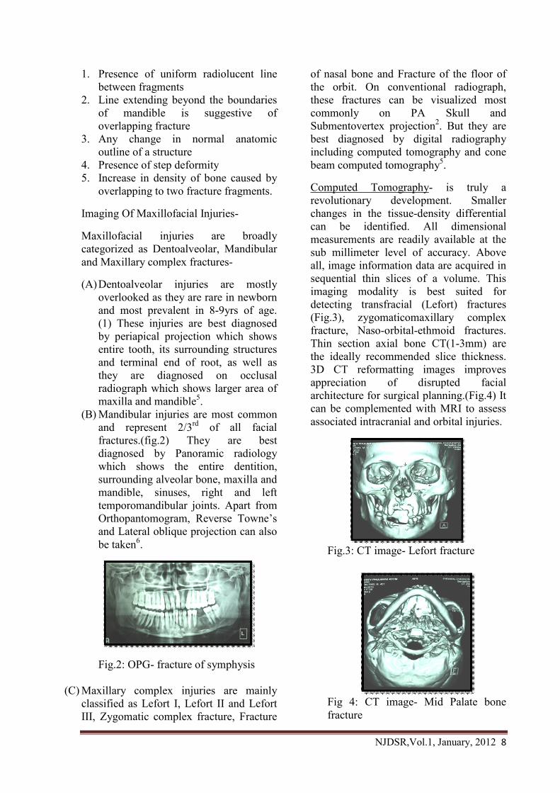
General Radiographic Features SuggestingFractures4 (Fig.1)-
Fig.1: Step Deformity (radiolucent line)-change in normal anatomic outline
ABSTARCT-
Traumatic dental injuries are emergent situations that need quick assessment andappropriate management. The proper diagnosis and treatment rendered determines theprognosis of the case. However, in dental practice, practitioners depend almost entirely ontwo dimension plain films. But, with the introduction of Three dimensional imaging offersthe diagnosis and treatment planning have become much easier. This article focuses on thediagnosis of maxillofacial injuries and recent advances in the field of dental radiology.
KEYWORDS-
Maxillofacial, Lacerations, Three dimensional imaging, Trauma, Computed tomography,Cone beam computed tomography
NJDSR,Vol.1, January, 2012 8
1. Presence of uniform radiolucent linebetween fragments
2. Line extending beyond the boundariesof mandible is suggestive ofoverlapping fracture
3. Any change in normal anatomicoutline of a structure
4. Presence of step deformity5. Increase in density of bone caused by
overlapping to two fracture fragments.
Imaging Of Maxillofacial Injuries-
Maxillofacial injuries are broadlycategorized as Dentoalveolar, Mandibularand Maxillary complex fractures-
(A)Dentoalveolar injuries are mostlyoverlooked as they are rare in newbornand most prevalent in 8-9yrs of age.(1) These injuries are best diagnosedby periapical projection which showsentire tooth, its surrounding structuresand terminal end of root, as well asthey are diagnosed on occlusalradiograph which shows larger area ofmaxilla and mandible5.
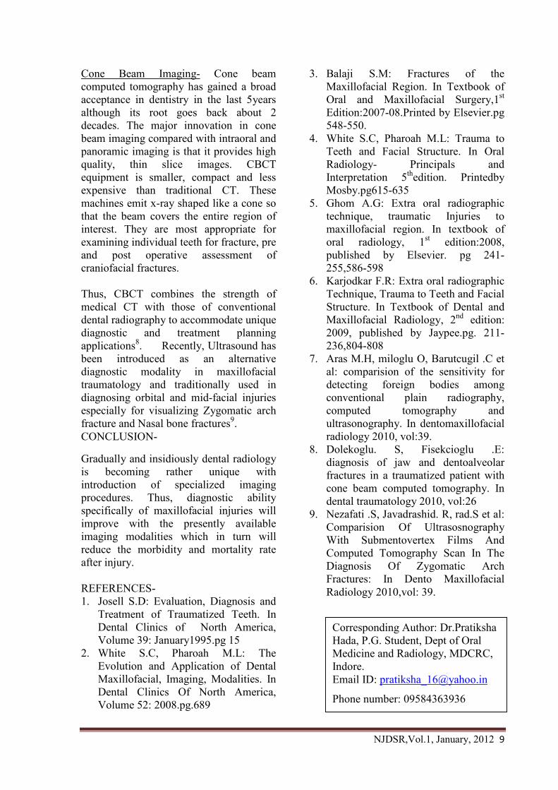
(B) Mandibular injuries are most commonand represent 2/3rd of all facialfractures.(fig.2) They are bestdiagnosed by Panoramic radiologywhich shows the entire dentition,surrounding alveolar bone, maxilla andmandible, sinuses, right and lefttemporomandibular joints. Apart fromOrthopantomogram, Reverse Towne’sand Lateral oblique projection can alsobe taken6.
Fig.2: OPG- fracture of symphysis
(C) Maxillary complex injuries are mainlyclassified as Lefort I, Lefort II and LefortIII, Zygomatic complex fracture, Fracture
of nasal bone and Fracture of the floor ofthe orbit. On conventional radiograph,these fractures can be visualized mostcommonly on PA Skull andSubmentovertex projection2. But they arebest diagnosed by digital radiographyincluding computed tomography and conebeam computed tomography5.
Computed Tomography- is truly arevolutionary development. Smallerchanges in the tissue-density differentialcan be identified. All dimensionalmeasurements are readily available at thesub millimeter level of accuracy. Aboveall, image information data are acquired insequential thin slices of a volume. Thisimaging modality is best suited fordetecting transfracial (Lefort) fractures(Fig.3), zygomaticomaxillary complexfracture, Naso-orbital-ethmoid fractures.Thin section axial bone CT(1-3mm) arethe ideally recommended slice thickness.3D CT reformatting images improvesappreciation of disrupted facialarchitecture for surgical planning.(Fig.4) Itcan be complemented with MRI to assessassociated intracranial and orbital injuries.
Fig.3: CT image- Lefort fracture
Fig 4: CT image- Mid Palate bonefracture
NJDSR,Vol.1, January, 2012 9
Cone Beam Imaging- Cone beamcomputed tomography has gained a broadacceptance in dentistry in the last 5yearsalthough its root goes back about 2decades. The major innovation in conebeam imaging compared with intraoral andpanoramic imaging is that it provides highquality, thin slice images. CBCTequipment is smaller, compact and lessexpensive than traditional CT. Thesemachines emit x-ray shaped like a cone sothat the beam covers the entire region ofinterest. They are most appropriate forexamining individual teeth for fracture, preand post operative assessment ofcraniofacial fractures.
Thus, CBCT combines the strength ofmedical CT with those of conventionaldental radiography to accommodate uniquediagnostic and treatment planningapplications8. Recently, Ultrasound hasbeen introduced as an alternativediagnostic modality in maxillofacialtraumatology and traditionally used indiagnosing orbital and mid-facial injuriesespecially for visualizing Zygomatic archfracture and Nasal bone fractures9.CONCLUSION-
Gradually and insidiously dental radiologyis becoming rather unique withintroduction of specialized imagingprocedures. Thus, diagnostic abilityspecifically of maxillofacial injuries willimprove with the presently availableimaging modalities which in turn willreduce the morbidity and mortality rateafter injury.
REFERENCES-1. Josell S.D: Evaluation, Diagnosis and
Treatment of Traumatized Teeth. InDental Clinics of North America,Volume 39: January1995.pg 15
2. White S.C, Pharoah M.L: TheEvolution and Application of DentalMaxillofacial, Imaging, Modalities. InDental Clinics Of North America,Volume 52: 2008.pg.689
3. Balaji S.M: Fractures of theMaxillofacial Region. In Textbook ofOral and Maxillofacial Surgery,1st
Edition:2007-08.Printed by Elsevier.pg548-550.
4. White S.C, Pharoah M.L: Trauma toTeeth and Facial Structure. In OralRadiology- Principals andInterpretation 5thedition. PrintedbyMosby.pg615-635
5. Ghom A.G: Extra oral radiographictechnique, traumatic Injuries tomaxillofacial region. In textbook oforal radiology, 1st edition:2008,published by Elsevier. pg 241-255,586-598
6. Karjodkar F.R: Extra oral radiographicTechnique, Trauma to Teeth and FacialStructure. In Textbook of Dental andMaxillofacial Radiology, 2nd edition:2009, published by Jaypee.pg. 211-236,804-808
7. Aras M.H, miloglu O, Barutcugil .C etal: comparision of the sensitivity fordetecting foreign bodies amongconventional plain radiography,computed tomography andultrasonography. In dentomaxillofacialradiology 2010, vol:39.
8. Dolekoglu. S, Fisekcioglu .E:diagnosis of jaw and dentoalveolarfractures in a traumatized patient withcone beam computed tomography. Indental traumatology 2010, vol:26
9. Nezafati .S, Javadrashid. R, rad.S et al:Comparision Of UltrasosnographyWith Submentovertex Films AndComputed Tomography Scan In TheDiagnosis Of Zygomatic ArchFractures: In Dento MaxillofacialRadiology 2010,vol: 39.
Corresponding Author: Dr.PratikshaHada, P.G. Student, Dept of OralMedicine and Radiology, MDCRC,Indore.Email ID: [email protected]
Phone number: 09584363936

![Large Unilocular Radiolucent Lesions of the Jaws: A ... conundrum for the Maxillofacial surgeon as to its most appropriate and ideal management modality [5]. Case Report A 23-year-old](https://img.pdfslide.net/doc/110x75/608379710bd44312f50c06db/large-unilocular-radiolucent-lesions-of-the-jaws-a-conundrum-for-the-maxillofacial.jpg)