Embed Size (px)
Citation preview

.Eukaryotes cell origin, structure
and function
Dr. Ganga Naik. S1st Year Ph.D
Department of Anatomy
Veterinary College
Hebbal, Bangalore -24

. Origin of life - Prokaryotes and Eukaryotes
Difference between prokaryotes and
eukaryotes
Different kind of eukaryotes - Cell structure
and function

almost 2 billionyears of strictlyunicellular life!
.
• .
Our understanding of the origin of life is
incomplete
Hypothesis that organic molecules formed
spontaneously and evolved into molecular
systems with the fundamental properties of life

.
• .
Many laboratory experiments lend support to
an abiotic origin of life through chemical
evolution
Spontaneous generation or abiogenesis.
Abiogenesis -
is the study of how biological life could arise
from inorganic matter through natural processes.
Inorganic compounds are of inanimate, not
biological origin.[1] Inorganic compounds lack
carbon and hydrogen atoms and are synthesized
by the agency of geological systems
Organized living structures have been found in
black shales of the Palaeoproterozoic
Francevillian B Formation in Gabon, dated at
2.1 billion years old. Eukaryotic life could have
evolved at that time.
Fossils that are clearly related to modern groups
start appearing an estimated 1.2 billion years
ago, in the form of a red alga, though recent
work suggests the existence of fossilized
filamentous algae in the Vindhya basin dating
back perhaps to 1.6 to 1.7 billion years ago.
Biomarkers suggest that at least stem eukaryotes
arose even earlier.
The presence of steranes in Australian shales
indicates that eukaryotes were present in these
rocks dated at 2.7 billion years old
Eukaryotes cell origin
The origin of the eukaryotic cell is considered
a milestone in the evolution of life,
since they include all complex cells and
almost all multicellular organisms.

.Miller–Urey experiment
• Demonstrated that most amino acids, were shown to be
racemically synthesized in conditions thought to be similar to
those of the early Earth.
• racemically - relating to, or constituting a compound or
mixture that is composed of equal amounts of dextrorotatory
and levorotatory forms of the same compound and is not
optically active
• In all living things, these amino acids are organized into
proteins, and the construction of these proteins is
mediated by nucleic acids, that are themselves
synthesized through biochemical pathways catalysed by
proteins.
• The experiment used water (H2O), methane (CH4),
ammonia (NH3), and hydrogen (H2). Miller and Urey
observed that as much as 10–15% of the carbon within
the system was now in the form of organic compounds.
Two percent of the carbon had formed amino acids that
are used to make proteins in living cells, with glycine as
the most abundant. Sugars were also formed. Nucleic
acids were not formed within the reaction.
• The Big Bang theory is the prevailing cosmological
model that describes the early development of the
Universe.[1] According to the Big Bang theory, the
Universe was once in an extremely hot and dense state
which expanded rapidly. This rapid expansion caused
the Universe to cool and resulted in its present
continuously expanding state. According to the most
recent measurements and observations, the Big Bang
occurred approximately 13.75 billion years ago,[2][3]
which is thus considered the age of the Universe.[4][5]
After its initial expansion from a singularity, the
Universe cooled sufficiently to allow energy to be
converted into various subatomic particles, including
protons,neutrons, and electrons. While protons and
neutrons combined to form the first atomic nuclei only
a few minutes after the Big Bang, it would take
thousands of years for electrons to combine with them
and create electrically neutral atoms. The first element
produced was hydrogen, along with traces of helium
and lithium. Giant clouds of these primordial elements
would coalesce through gravity to form stars and
galaxies, and theheavier elements would be synthesized
either within stars or during supernovae.

.Endosymbiotic theory
Konstantin Mereschkowski in 1905.
According to this theory, certain organelles
originated as free-living bacteria that were taken
inside another cell as endosymbionts.
The endosymbiotic theory argues that
mitochondria, plastids (e.g. chloroplasts), and
possibly other organelles of eukaryotic cells,
originate through symbiosis between multiple
microorganisms. According to this theory, certain
organelles originated as free-living bacteria that
were taken inside another cell as endosymbionts.
Mitochondria developed from proteobacteria (in
particular,Rickettsiales, the SAR11 clade,[1][2] or close
relatives) and chloroplasts from cyanobacteria.

.Endosymbiotic theory
Mitochondria developed from proteobacteria
(Rickettsiales)
chloroplasts from cyanobacteria.
cenancestor, is the most recent organism from
which all organisms now living on Earth descend.[1]
Thus it is the most recent common ancestor
(MRCA) of all current life on Earth. The LUA is
estimated to have lived some
3.5 to 3.8 billion years ago
The Proteobacteria are a major group (
phylum) of bacteria. They include a wide
variety of pathogens, such as Escherichia,
Salmonella, Vibrio, Helicobacter, and many
other notable genera. [2] Others are free-living,
and include many of the bacteria responsible
for nitrogen fixation.
• Cyanobacteria (/sa æno bæk t əriəɪˌ ʊ ˈ ɪ /), also
known as blue-green bacteria, blue-green
algae, and Cyanophyta, is a phylum of
bacteria that obtain their energy through
photosynthesis.[3] The name "cyanobacteria"
comes from the color of the bacteria (Greek
: κυανός (kyanós) = blue).
• The ability of cyanobacteria to perform
oxygenic photosynthesis
• the last universal ancestor (LUA), also called
the last universal common ancestor(LUCA),
or the cenancestor, is the most recent
organism from which all organisms now living
on Earth descend.[1] Thus it is the
most recent common ancestor (MRCA) of all
current life on Earth. The LUA is estimated to
have lived some 3.5 to 3.8 billion years ago
(sometime in the Paleoarchean era).[2][3]

.Endosymbiotic theory
Many separate organisms may have contributed
for development of cenancestor (
most recent common ancestor).
is the most recent organism from which all organisms
now living on Earth descend.[1] Thus it is the
most recent common ancestor (MRCA) of all current
life on Earth. The LUA is estimated to have
Mitochondria and chloroplasts
Surrounded by two membranes
Possess their own DNA
Possess ribosomes
70S (bacterial), 80S (eukaryotic)
Self replication
The Proteobacteria are a major group (phylum
) of bacteria. They include a wide variety of
pathogens, such as Escherichia, Salmonella,
Vibrio, Helicobacter, and many other notable
genera. [2] Others are free-living, and include
many of the bacteria responsible for
nitrogen fixation.
• Cyanobacteria (/sa æno bæk t əriəɪˌ ʊ ˈ ɪ /), also
known as blue-green bacteria, blue-green
algae, and Cyanophyta, is a phylum of bacteria
that obtain their energy through photosynthesis.
[3] The name "cyanobacteria" comes from the
color of the bacteria (Greek: κυανός (kyanós) =
blue).
• The ability of cyanobacteria to perform
oxygenic photosynthesis
• the last universal ancestor (LUA), also called
the last universal common ancestor(LUCA),
or the cenancestor, is the most recent organism
from which all organisms now living on Earth
descend.[1] Thus it is the
most recent common ancestor (MRCA) of all
current life on Earth. The LUA is estimated to
have lived some 3.5 to 3.8 billion years ago
(sometime in the Paleoarchean era).[2][3]

EARLIEST LIFE
Life arose - 3.8 billion years ago
The earliest cells were prokaryotic
• .
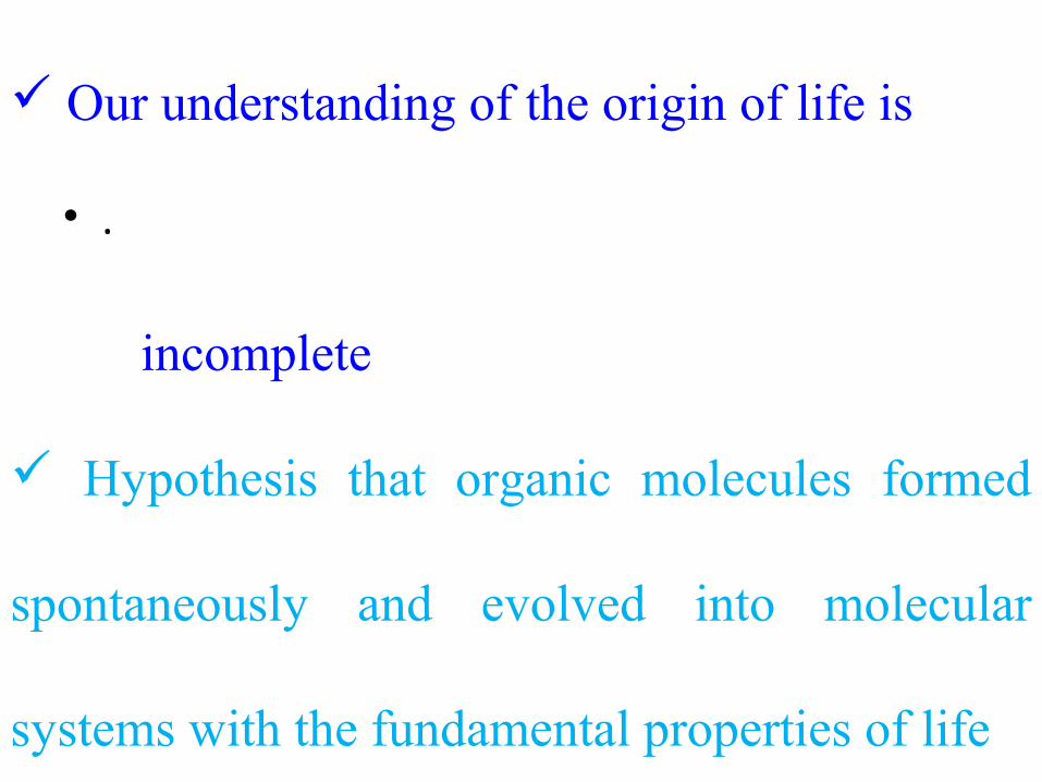
Prokaryotes
Contains
1. Nucleoid region – Contain
DNA
2. Cell membrane & Cell wall
3. Ribosomes
4. (no membrane) to make
proteins in their cytoplasm

Ancient prokaryotes fromWestern Australia.
Filamentous “Cyanobacteria”
3.5 BYA
Cyanobacteria are one of the earliest life forms known to have existed on Earth

Earliest filamentous microfossils 3.23 BYA
FROM: Rasmussen 2000 NATURE
Microfossils are fossils generally not larger than four millimeters, and commonly smaller than one millimeter, the study of which requires the use of light or electron microscopy. Fossils which can be studied with the naked eye or low-powered magnification, such as a hand lens, are referred to as macrofossils. Obviously, it can be hard to decide whether or not some organisms should be considered microfossils, and so there is no fixed size boundary

Microfossil Cyanobacteria

Evolution of Eukaryotes
• As early as 2.1 Bya eukaryotic cells appear as fossils
Figure 01A: Microfossils of probable eukaryotic cells
Reproduced from Schopf, J.W., Scientific American 239 (1978): 111-138. Courtesy of J. William Schopf, Professor of Paleobiology & Director of IGPP CSEOL
Figure 01B: Microfossils of probable eukaryotic cells
Figure 01C: Microfossils of probable eukaryotic cells

Origin of the Eukaryotes
Two theories to explain the origin of
membrane bound organelles
1. Endosymbiosum
2. Invagination
• a type of symbiosis in which one organism lives inside the
other, the two typically behaving as a single organism. It is
believed to be the means by which such organelles as
mitochondria and chloroplasts arose within eukaryotic
cellsendosymbiotic adj
• Invagination is the infolding of one part within another
part of a structure
• Both mitochondria and chloroplasts were once free living
organisms
– Engulfed by early eukaryotic cells
– Maintained rather than being digested
– “Endosymbionts”
• Most genes have been lost or transferred to the nucleus
– A few genes are retained
• ENDOSYMBIOSIS
Mitochondria and chloroplasts -
“Endosymbionts”
At present, host cells cannot live without
their endosymbionts,
these endosymbionts cannot live without
their host cell
Other organelles are likely
endosymbionts also
• .

• .

.
• .

Origin of the Eukaryotes

.• .

CYTOSKELETON
MITOCHONDRION
CENTRIOLES
LYSOSOME
GOLGI BODY
SMOOTH ER
ROUGH ER
RIBOSOMES
NUCLEUS
PLASMA MEMBRANE VESICLE
CYTOPLASM
Eukaryotes cells

Differences between Prokaryotic Cells and Eukaryotic cells
Prokaryotic Cells Eukaryotic cells
small cells (< 5 mm) larger cells (> 10 mm)
always unicellular often multicellular
no nucleus or any membrane-bound organelles
always have nucleus and other membrane-bound organelles
DNA is circular, without proteins DNA is linear and associated with proteins to form chromatin
ribosomes are small (70S) ribosomes are large (80S)
no cytoskeleton always has a cytoskeleton
cell division is by binary fission cell division is by mitosis or meiosis
reproduction is always asexual reproduction is asexual or sexual

Include most cells (other than bacteria)
1.Plants
2. Fungi
3. Animals
A fungus ( / f ŋ əsˈ ʌ ɡ /; plural: fungi[3] or funguses
[4]) is a member of a large group of eukaryotic
organisms that includes microorganisms such
as yeasts and molds(British English: moulds), as
well as the more familiar mushrooms.Fungal cells are most similar to animal cells, with the following exceptions:A cell wall that contains chitinLess definition between cells; the hyphae of higher fungi have porous partitions calledsepta, which allow the passage of cytoplasm, organelles, and, sometimes, nuclei. Primitive fungi have few or no septa, so each organism is essentially a giant multinucleate supercell; these fungi are described as coenocytic.Only the most primitive fungi, chytrids, have flagella.
Eukaryotes

Eukaryotic Cell
Contain 3 basic cell structures:
1.Nucleus
2.Cell Membrane
3.Cytoplasm with
organelles

Types of Eukaryotic Cells
Plant Cell Animal Cell

Plant CellPlant Cell
Made of cellulose which
forms very thin fibers
Gives shape to the cell
Strong and rigid
In plant cells only
– Protects and supports the
cell
– Gives shape to the cell
– A dead layer, ∴freely
permeable
– Resists entry of excess
water into the cell
• Cell wall

– Lies immediately against
the cell wall
– Made of protein and lipid
� ∴Selectively permeable
– Can control the movement
of materials into and out of
the cell
• Cell membrane
Plant CellPlant Cell

–Jelly-like substance
enclosed by cell membrane
–Contains organelles and granules
•e.g. chloroplast
•e.g. mitochondrion
–Contains the green pigment
chlorophyll
•To trap light energy, to
make food by
photosynthesis
•Contains starch grains
(products of
photosynthesis)
•Provide a medium for chemical reactions to take place
has specific functions
in cytoplasm–Non-living granules
–Starch granules
–Oil droplets
–Crystals of insoluble wastes
– large central vacuole– Contains cell sap
•a solution of chemicals (sugars, mineral salts, wastes, pigments)
• Cytoplasm
Plant CellPlant Cell

Nucleus
– Bounded by a nuclear membrane
– 1.Controls the normal activities of the cell
2. For heredity
– Contains thread-like chromosomes
– Each cell has fixed number of chromosomes
• Chromosomes carry genes
–genes control cell characteristics
Plant Cell

.
Animal cell

Animal cell
No cell wall and chloroplast
Small vacuoles
Stores glycogen granules and oil droplets
in the cytoplasm

mitochondrion
nucleus
glycogen granule
cell membrane
cytoplasmAnimal cellAnimal cell
vacuole

Different kinds of animal cells
white blood cell
red blood cell
cheek cellssperm
nerve cell
muscle cell
Amoeba
Paramecium

Structure Animal cells Plant cellscell membrane Yes yes
nucleus Yes yesnucleolus yes yesribosomes yes yes
ER yes yesGolgi yes yes
centrioles yes nocell wall no yes
mitochondria yes yescholorplasts no yes
One big vacuole no yescytoskeleton yes Yes

.Cytoplasm involved in
1. Energy formation and release
2. Protein synthesis

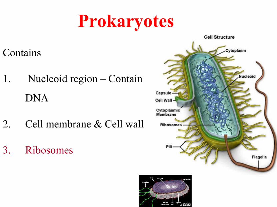
.Cytoplasm• Subdivided into
1.Cytocentrum
2. Endocentrum
3.Ectoplasm

.Cytoplasm
1.Cytocentrum• ER
• Pair of centriole
• Cytoskeletal
element

.Cytoplasm
2.Endocentrum• Largest
• Most of structural
components
.Cytoplasm
3. Ectoplasm• Narrow band

PLASMA MEMBRANE/Cell membrane
Surrounds the entire cytoplasm
Provides a selective barrier
regulates the transport of materials into
and out of the cell.

PLASMA MEMBRANE/Cell membrane
All membranes including the membranous
organelles are composed
1. Lipid and protein - mainly
2. Carbohydrate - small amount

.
Organelles
1. Rough Endoplasmic Reticulum
2. Smooth Endoplasmic Reticulum:
3. Golgi Complex:
4. Mitochondria:
5. Lysosomes:
6. Peroxisomes Or Microbodies:

.
Cytoskeletal
Elements And
Cytoplasmic
Matrix
a) Microtubules:
b) Centrioles Or
Diplosomes
c)Microfilaments:

.NUCLEUS
The nucleus is found in all cells except
mammalian red blood cells and platelets,

.NUCLEUS
It has DNA,
which is the storehouse of genetic information
controls protein synthesis in the cells.
,
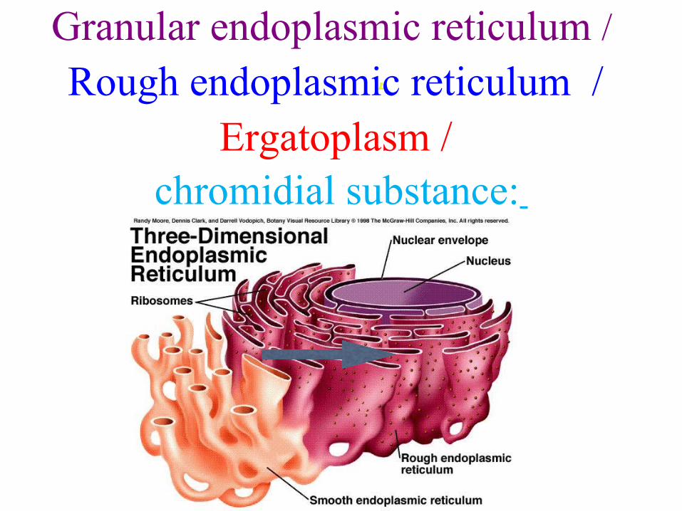
Granular endoplasmic reticulum /
Rough endoplasmic reticulum / Ergatoplasm /
chromidial substance:

E/M defined as a system of
interconnecting membrane bound
channels or cavities,

Granular endoplasmic reticulum
The membrane bound channels are either
a. Cistarnae (flattened sacs)
b. Tubular or
c. Vesicles
The membrane bound channels attached with
ribosomes on their outer surface.
May communicate with the nuclear envelope


.Granular endoplasmic reticulum
Ribosomes
are Rnp granules (Ribonuclear
protein),
the sites of synthesis of proteins from
amino acids.
Basophilia of the endoplasmic
reticulum is due to RNA of
ribosomes
and not due to membranes of EPR.
In nerve cells closely packed
flattened sacs of RER are called
Tigroid bodies or Nissl bodies.
Functions:
1. Contributed to the mechanical support of
cytoplasm
2. Synthesis of proteins for export

Ribosomes:
.

Ribosomes:
Occur in two forms.
1. Associated with endoplasmic reticulum
2. Occur free in cytoplasm

Ribosomes:
• Under E/M appear as small electron dense
particles 150Ǻ diameter formed by 2 subunits one
large and one small.
• have a characteristic sedimentation coefficient of
80s (sedgberg unit) with the larger heavier unit
having 60s and the smaller subunit having 40s
unit.
•

.
Several ribosomes associated with
endoplasmic reticulum synthesis proteins,
These proteins are meant for external
use.
Eg. Enzymes.

.
• Several ribosomes are attached to each other by a
strand of mRNA forming polysomes or
polyribosomes.

.
Polyribosomes
Synthesize proteins
intracellular use
Eg. Hemoglobin formed in red blood cells.

:
SMOOTH ENDOPLASMIC RETICULUM

SMOOTH ENDOPLASMIC RETICULUM:
Under electron microscope appear as three
dimensional network of membrane bound
tubules with no cisternae and lack ribosomes.
It may communicate with RER or with
nuclear envelope.

.
• .


Functions:
Production of steroid hormone, in
steroid secreting cells, adrenal cortex,
testes, ovary.
GOLGI COMPLEX:

GOLGI COMPLEX:
• Supranuclear position in secreting cells
• Multiple in number in hepatocyte (liver cells).
Forms a network around the nucleus in nerve cells,

.
• E/M appear as lamellae (3-12 in number) of
parallely arranged flattened curved membranous
sacs, vacuoles or vesicles.

Function:
Condensation and packing of secretory product by
loss of water.
Formation of lysosomes and peroxisomes in
leukocytes.
DNA directs RNA synthesis RNA exits nucleus
through a nuclear pore ribosome protein is
made proteins with proper code enter RER
proteins are modified in RER and lipids are made
in SER vesicles containing the proteins and
lipids bud off from the ER
ER vesicles merge with Golgi body proteins and
lipids enter Golgi each is fully modified as it
passes through layers of Golgi modified
products are tagged, sorted and bud off in Golgi
vesicles …
Golgi vesicles either merge with the plasma
membrane and release their contents OR remain
in the cell and serve a purpose
Completes the processing substances received from
the ER
Sorts, tags and packages fully processed proteins
and lipids in vesicles
.MITOCHONDRIA:

.
• Energy conversion system by
which chemical energy of food
stuffs is converted into high energy
phosphates (ATP)
MITOCHONDRIA:

.MITOCHONDRIA:

.
E/M –
Double membranous structure with
rounded ends or sausage shaped.
Both membranes have unit
membrane structure.

.Mitochondrial matrix
Electron dense granules
DNA and RNA strands

.
Mitochondrial matrix
DNA and RNA strands
• which directs the synthesis of enzymes of
mitochondria.
Because of the presence of DNA,
are semiautonomous organelles
capable of self replication.Enzyme localization:
1.Matrix: enzymes of Kreb’s cycle
2.Elementary particles: enzymes of oxidative
phosphorylation
3.Inner membrane:
Respiratory chain enzymes,
flavoproteins, dehydrogenases

.
• .
Function – synthesis of ATP3 major pathways involved in ATP production
1. Glycolysis2. Krebs Cycle3. Electron transport system .(ETS)

.
LYSOSOMES

LYSOSOMES
Membrane bound spherical uniformly
granular electron dense bodies.

LYSOSOMES
Present in all cells
but numerous in cells exhibiting phagocytic
activity.
E.g. Macrophages and WBC’s.

LYSOSOMES
• Associated with intracellular digestion and
phagocytosis.

LYSOSOMES• Contain more than 40 hydrolytic enzymes
• active at acid pH.
Eg. Acid phosphatase, Ribonuclease, sulphatase etc.
• If the material is quite large it may remain over in the form
of brownish pigments called lipoprotein pigments.
Seen in neurons,
heart muscles,
liver cells.
Increase in their number with age and are called
Senility pigment.
Residual bodies.
• The large undigested material is retained
• These bodies may be expelled from the cell by a process of
exocytosis as seen in macrophages.

PEROXISOMES OR MICROBODIES:
• .

PEROXISOMES OR MICROBODIES:
• Membrane bound bodies
•

PEROXISOMES OR MICROBODIES:
• Resemble lysosomes
• but do not contain lysosomal enzymes.

PEROXISOMES OR MICROBODIES:
Function :
Hydrogen peroxide is detoxified
Fatty acids are metabolized
They possess enzymes concerned with production
and destruction of hydrogen peroxide.

.
CYTOSKELETAL ELEMENTS
AND
CYTOPLASMIC MATRIX

.CYTOSKELETAL ELEMENTS
• Structure
Interconnected system of
– microtubules,
– microfilaments,
– intermediate filaments

.CYTOSKELETAL ELEMENTS
• Function
– gives cells internal organization, shape, and ability to
move

CYTOSKELETAL ELEMENTS AND CYTOPLASMIC MATRIX
MICROTUBULES:
Slender, hollow, cylindrical un
branched structures
Made of tubulin proteins (globular)
•

CYTOSKELETAL ELEMENTS AND CYTOPLASMIC MATRIX
MICROTUBULES:
Function
Separation of chromosomes during mitosis.
They function both to determine cell shape
and in a variety of cell movements, including
some forms of cell locomotion, the
intracellular transport of organelles, and
–Move chromosomes around during cell division
• Used to make cilia and flagella
•

CENTRIOLES (Diplosome):
• .

CENTRIOLES (Diplosome):
• A is a cylindrically-shaped cell structure
• Found in most eukaryotic cells,
• though it is absent in higher plants and most fungi
• Contained in an area of gelatinous material called
centrosphere.

CENTRIOLES (Diplosome):
• Wall is composed of 9 sets of microtubule triplets.

CENTRIOLES (Diplosome):
Functions:
involved in the organization of the mitotic spindle
in the completion of cytokinesis
Formation of mitotic spindles as asters in cell
division.

MICROFILAMENTS:
• .

MICROFILAMENTS:
1. Actin - 70A°
2. Myosin - 150 A°.
• In muscle cells well organized filamentous
components.

MICROFILAMENTS:
Functions:
Enable cells to change shape and move
(Participate in muscle contraction)
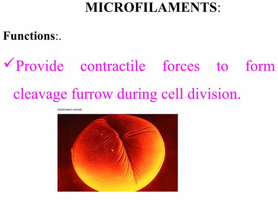
MICROFILAMENTS:
Functions:.
Provide contractile forces to form
cleavage furrow during cell division.

Cilia and flagella (structures for cell motility)
– Move whole cells or materials across the cell surface
– Microtubules wrapped in an extension of the plasma
membrane (9 + 2 arrangement of MT)

Thank you