Embed Size (px)
Citation preview

Ultrasound – Physics & Advances

Modes of Vibration
2 TYPES:
1. thickness mode
most common
Used in medical
crystals
2. radial mode

Natural frequency to which the transducer is sensitiveResonant frequency determined by thickness of crystal Thick crystal – low frequency sound Natural frequency – one that produces internal wavelengths that are
twice the thickness of crystalFrequency corresponding to half the wavelength is- fundamental
resonant frequency
Resonant Frequency
Transducers
Resonance Transducers Non Resonance Transducers

They are manufactured to operate in a “resonance” mode, whereby a voItage (commonly 150 V) of very short duration (a voltage spike of 1 msec) is applied, causing the piezoelectric material to initially contract, and subsequently vibrate at a natural resonance frequency.
The operating frequency is determined fromthe speed of sound inand the thickness of, the
piezoelectric material.
Resonance Transducers

Modern transducer design coupled with digital signal processing
enables “multifrequency or “multihertz” transducer operation,
whereby the center frequency can be adjusted in the transmit mode.
Excitation of the multifrequency transducer is accomplished with a
short square wave burst of 150 V with one to three cycles, unlike the
voltage spike used for resonance transducers.
Non-resonance (Broad-Bandwidth) “Multi-frequency” Transducers

Unlike the resonance transducer design, the piezoelectric element is intricately machined into a large number of small “rods,” and then filled with an epoxy resin to create a smooth surface.

A single vibrating point sets out waves in all directions
Waves move away as concentric circles
Characteristics of Ultrasound Beam

When two sound waves interact , they cancel each other or reinforce each other

The ultrasound beam propagates as a longitudinal wave from the
transducer surface into the propagation medium, and exhibits two distinct
beam patterns:
a slightly converging beam out to a distance specified by the geometry
and frequency of the transducer (the near field), and
a diverging beam beyond that point (the far field).
Beam Properties
The near field, also known as the Fresnel zone, is adjacent to the
transducer face and has a converging beam profile.
Beam convergence in the near field occurs because of multiple
constructive and destructive interference patterns of the ultrasound
waves from the transducer surface.
The Near Field
Near Field Length……….
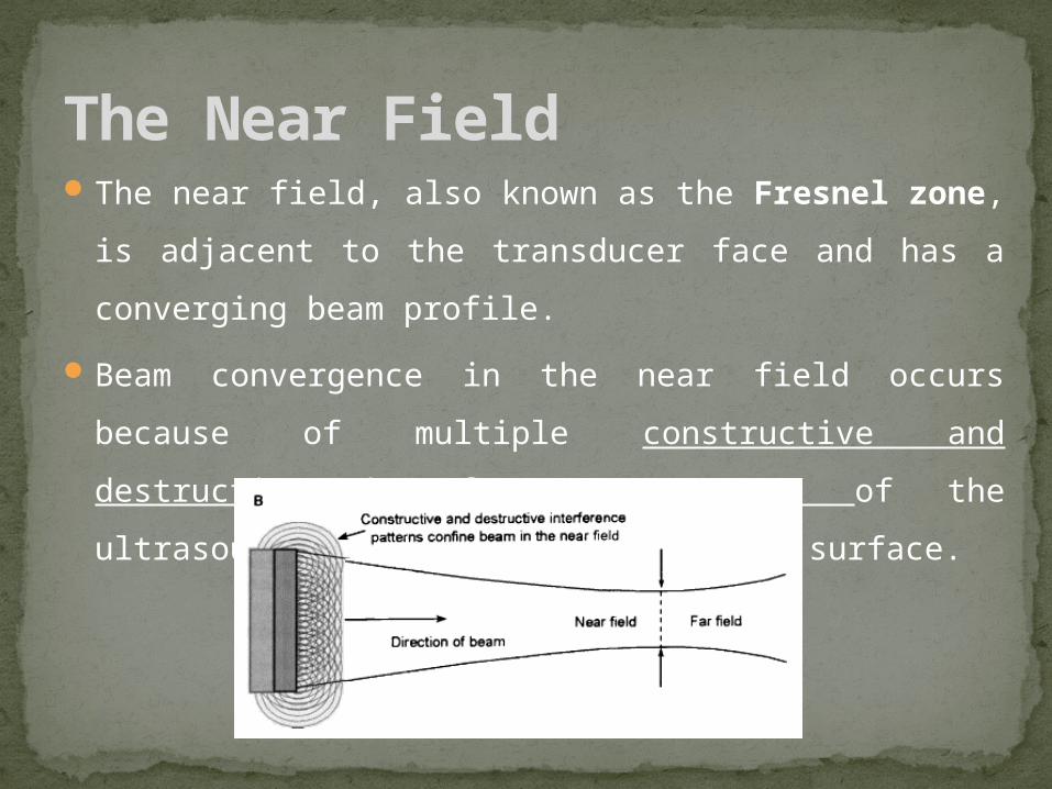
In single-element transducer The near field length for an
unfocused, single-element transducer is dependant on
transducer frequency and diameter
For multiple transducer element, an “effective” transducer diameter
is determined by the excitation of a group of’ transducer elements.
Because of the interactions of each of the individual beams and the
ability to focus and steer the overall beam, the formulas for a single-
element, unfocused transducer are not directly applicable.

Lateral resolution (the ability of the system to resolve objects in a
direction perpendicular to the beam direction) is dependent on the
beam diameter and is best at the end of the near field for a single-
element transducer.
Lateral resolution is worst in areas close to and far from the
transducer surface.

Pressure amplitude characteristics in the near field are very
complex, caused by the constructive and destructive interference
wave patterns of the ultrasound beam.
Peak ultrasound pressure occurs at the end of the near field,
corresponding to the minimum beam diameter for a single-element
transducer.

Pressures vary rapidly from peak compression to peak rarefaction
several times during transit through the near field.
Only when the far field is reached do the ultrasound pressure
variations decrease continuously.
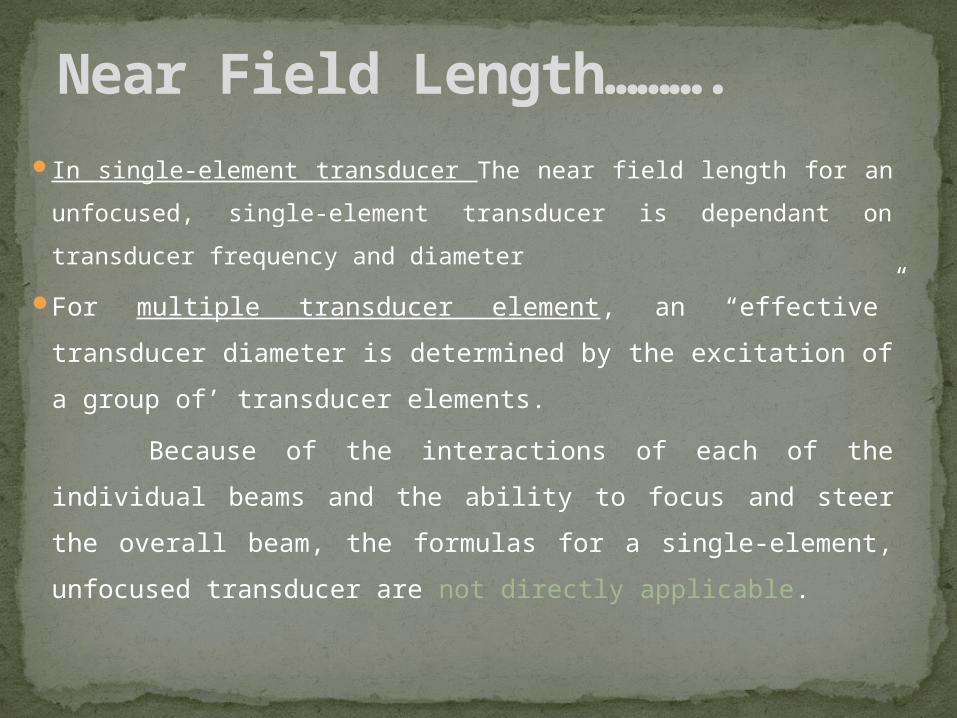
The far field is also known as the Fraunhofer zone, and is
where the beam diverges.
Less beam divergence occurs with:
High - frequency transducers
Large - diameter transducers
Far Field

High frequency beams –
fresnel zone is longer
depth resolution is superior
Disadvantage :
Tissue absorption is more, leading to deterioration of side to side
resolution
Solution: Focused Transducer

The shape of the ultrasound beam is affected by: the size and shape of the ultrasound source ,frequency & beam
focusing:
1. The size of the ultrasound: the effects of source size on beam shape are:
(i) a small source provides a narrow beam initially, is associated with a short Fresnel zone, and the beam diverges rapidly beyond the near field.
(ii) (ii) a large source provides a broader beam initially, gives a longer Fresnel zone, and the beam diverges more gradually, thus providing better resolution of deeper structures.
Factors influencing beam shape

2. Effect of beam frequency:
the length of the Fresnel zone increases as the beam frequency is increased. Also, the angle of divergence beyond the near field diminishes with increasing frequency leading to increase resolution . In practice, however, some of this advantage is taken away by increased beam attenuation at higher frequencies

Focused Transducers3. Focusing of the ultrasound beam:
The shape of the ultrasound beam can be influenced to varying extents by applying different focusing methods.
Shape of the crystal element: The crystal element can be suitably shaped by concave curvature to focus the ultrasound beam. This is an internal focusing method,
Acoustic lenses made from materials which propagate ultrasound at velocities different from that in soft tissue can be used to focus the beam by refraction.
Acoustic mirrors A concave mirror can be used to focus ultrasound by reflection Electronic focusing: Electronic focusing is employed in multicrystal transducers N.B: the degree of focusing will depend on the radius of curvature. Acoustic lenses
and mirrors provide external focusing.

Focal Distance
The focal distance, the length
from the transducer to the
narrowest beam width, is
shorter than the focal length of
a non-focused transducer and is
fixed.
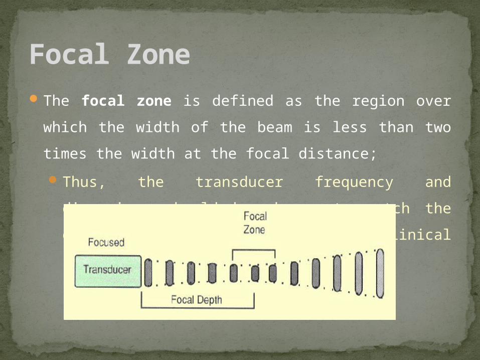
The focal zone is defined as the region over which the width of the
beam is less than two times the width at the focal distance;
Thus, the transducer frequency and dimensions should be chosen
to match the depth requirements of the clinical situation.
Focal Zone

The degree of focusing may be classified into three categories as follows:
1. strong focusing (or short focusing)
2. medium focusing
3. weak focusing (or long focusing) In all cases, fixed focusing gives a focal point which is nearer to the
transducer than the transition distance (length of the Fresnel zone). Strong focusing brings the focal point very close to the transducer,
typically 2 - 4 cm. It achieves a high degree of beam narrowing, but the beam diverges rapidly beyond the focal distance. It can only be applied to transducers for high resolution examinations of small parts.
Weak focusing gives a focal point further away from the transducer - typically more than 8 cm - and a gentle divergence of the beam beyond the focus. It is preferred in diagnostic applications because it provides an extended useful, narrow beam.
Classification of focusing


![CE5101 Lecture 8 - Radial Consolidation_ PVD and Surcharge (OCT 2013) [Compatibility Mode]](https://img.pdfslide.net/doc/110x75/55cf9adf550346d033a3d0f5/ce5101-lecture-8-radial-consolidation-pvd-and-surcharge-oct-2013-compatibility.jpg)