Embed Size (px)
Citation preview

Int J Cur Res Rev | Vol 10 • Issue 13 • July 2018 5
Validated RP-HPLC Method for Quantification of Paclitaxel in Human Plasma – Eliminates Negative Influence of Cremophor El
Hindu Kalluru1, Vinodhini C.2, Satish Srinivas K.3, Surulivel Rajan M.4, Chitra K.2, Mangathayaru K.1,*
1Department of Pharmacognosy, Faculty of Pharmacy, Sri Ramachandra Medical College and Research Institute, Porur, Chennai, Tamil Nadu, India; 2Department of Pharmaceutical Chemistry, Faculty of Pharmacy, Sri Ramachandra Medical College and Research Institute, Porur, Chen-nai, Tamil Nadu, India; 3Department of Radiation Oncology, Faculty of Medicine, Sri Ramachandra Medical College and Research Institute, Porur, Chennai, Tamil Nadu, India; 4Department of Pharmacy Practice, Manipal College of Pharmaceutical Sciences, Manipal University, Madhav Nagar, Saralebettu, Manipal, Karnataka, India.
ABSTRACTBackground: Literature reports innumerable methods for quantification of paclitaxel in biological matrices. Most of these involve complicated extraction procedures like solid phase extraction, separate procedure for elimination of interference of Cremophor El, advanced and expensive instruments.Objectives: The objective of the present research work is to develop and validate a simple, rapid, sensitive, economic and reproducible reverse phase – high performance liquid chromatography method for the estimation of paclitaxel concentration in human plasma that eliminates negative influence of Cremophor El on recovery of paclitaxel. Methods: Chromatographic separation of paclitaxel was carried out using C18 column (150 × 4.6 mm i.d., 4µm particle size, Wa-ters, Australia) with 60% acetonitrile, 40% of 10mM ammonium acetate buffer solution and 0.1% formic acid as a mobile phase at a flow rate of 1.0mL/min at ambient temperature. Validation was performed as per ICH Q2 guidelines.Results: In this system the retention time was 3min. The detection limit was 5ng/mL and limit of quantification with reproducibility was 15ng/mL. Plasma samples were extracted using single solvent (tertiary – Butyl Methyl Ether) liquid- liquid extraction with a recovery of 96-99%. Robustness of the method was established with variation in flow rate, detection wave length and mobile phase composition. Stability of paclitaxel during the study period was studied and found to be stable.Conclusion: The developed method is easy to perform, quick, reproducible with good recovery without negative influence of Cremophor El and applicable to quantify paclitaxel in regular clinical practice for individualization of the therapies.Key Words: Paclitaxel, Cremophor El, Single solvent extraction, Bioanalytical
Corresponding Author:Mangathayaru K., Department of Pharmacognosy, Faculty of Pharmacy, Sri Ramachandra Medical College and Research Institute, Porur, Chennai; Phone: 91 044 24768403, x 8927; Mob: 99400 14632; Fax: 091 44 24765995; Email: [email protected]
ISSN: 2231-2196 (Print) ISSN: 0975-5241 (Online)
Received: 07.06.2018 Revised: 15.06.2018 Accepted: 29.06.2018
INTRODUCTION
Paclitaxel is a very effective antineoplastic agent used in the treatment of ovarian, breast, cervix and non-small cell lung cancers (1). Paclitaxel was the first taxane derivative isolated from the bark of Taxusbrevifolia in the year 1971 (2) and being marketed from 1993 (3). It is a complex, hydrophobic molecule administered in a solution of Cremophor-EL (Cr El) and dehydrated alcohol in the ratio of 1:1.
Neutropenia, myelosuppression, gastrointestinal ailments, alopecia are the common adverse effects of paclitaxel treat-ment. Peripheral neuropathy is the major adverse effect of paclitaxel and increases with increasing cumulative dosage (2). Moreover, Cr El is widely known to cause severe hyper-sensitivity reactions requiring premedication (4).
It is well recognized that there is inter and intra individual variability in pharmacokinetic characteristics of paclitaxel.
Research ArticleInternational Journal of Current Research and ReviewDOI: 10.31782/IJCRR.2018.10132
IJCRRSection: HealthcareSci. Journal Impact
Factor: 5.385 (2017)ICV: 71.54 (2015)
Int J Cur Res Rev | Vol 10 • Issue 13 • July 20186
Kalluru et.al.: Validated RP-HPLC method for quantification of paclitaxel in human plasma – eliminates negative influence of cremophor el
Currently body surface area (BSA) is used in the dosing of paclitaxel, leading to significant inter individual differences in plasma concentrations and risk of severe and treatment limiting adverse effects (5, 6). Quantification of paclitaxel in the blood or plasma samples is necessary to establish pharmacokinetic parameters that can assess inter and intra individual variability, thereby making necessary dose adjust-ments to improve therapeutic efficacy and minimizing the adverse effects (7). It is also reported that Cr El has a nega-tive impact on the reproducibility of bioanalysis of paclitaxel due to entrapment of paclitaxel in the micelles of Cr El (8), and various methods are used to nullify the effect of Cr El on quantification of paclitaxel (9).
Therefore, it is necessary to develop a rapid, simple, sensi-tive, economic and reproducible method to quantify plasma concentrations of paclitaxel in regular clinical practice to promote individualization of treatments.
Till date several methods have been reported for the quan-tification of paclitaxel in biological matrices using various techniques like high performance liquid chromatography (HPLC), liquid chromatography-tandem mass spectroscopy (LC-MS), micellar electrokinetic chromatography (MEKC), capillary electrophoresis, immune assays etc (10-12). Most of these methods require tedious, complicated extraction procedures like solid phase extraction (SPE), advanced and expensive instruments like LC-MS which are not available in many hospitals, diagnostic and research labs.
We herewith report on our development of a rapid, simpli-fied, economic and sensitive method of paclitaxel quantifica-tion in human plasma by liquid-liquid extraction for applica-tion in clinical sample analysis.
METHODS
Instruments and ReagentsThe HPLC system - LC- 2010 (Shimadzu Corporation, Kyo-to, Japan) liquid chromatographic quaternary auto sampler pump model equipped with UV-Visible detector, Class VP software.C18 HPLC column with 150×4.6mm (i.d) with par-ticle size of 4µm (Waters India Pvt Ltd), Universal guard column with 20×3.9mm i.d and UV detector (Shimadzu), Ul-tra sonicator (PCI analytics, Vertex enterprises, India), Milli Q water system, Micro centrifuge and vortex shaker.
Paclitaxel (>99% purity) was purchased from Sigma-Al-drich, India. Paclitaxel formulation (Taxeleon 30mg/5mL) was kindly gifted by Neon Laboratories, Mumbai. Ace-tonitrile, t-butyl methyl ether (t-BME), methanol of HPLC grade, ammonium acetate and formic acid of analytical grade were used. Drug free plasma was obtained from healthy vol-unteers in our laboratory.
Preparation of stock solutionStock solution of paclitaxel was prepared by dissolving 10mg of paclitaxel in 10mL of acetonitrile. This stock solution was further diluted for the preparation of working standard solu-tions with diluent (Acetonitrile & water in 1:1 ratio) to get the concentration range of 6ng/mL to 6µg/mL.
Blood samplingThis method development is a part of pharmacokinetic drug interaction study in cancer patients and has been approved by Institutional ethics committee of our institution (IEC No: IEC-NI/16/Mar/51/16). Blood samples from six cancer pa-tients receiving 260mg of Paclitaxel were collected after pro-curing signed written informed consent. All the blood sam-ples were collected in heparinized vacutainers. The plasma samples were separated from the whole blood by centrifug-ing at 3000 rpm for 10min and were stored in refrigerator at -80°C until analysis.
Sample PretreatmentAbout 500µL of patient plasma was extracted with 1mL of t-BME twice and vortexed for 1 min. After centrifugation at 2000 rpm for 10 min, the organic layer was transferred into a 10 mL beaker. The t-BME extract was evaporated to dryness, residue was reconstituted with 1mL of diluent and filtered through 0.22 µ pore size nylon membrane filters, about 20 µL of the sample was applied to HPLC analysis as per the proposed chromatographic conditions (13).
HPLC ConditionsChromatographic separation of paclitaxel was performed using C18 column (150×4.6mm i.d., 4µm particle size) with 60% acetonitrile, 40% 10mM ammonium acetate buffer so-lution and 0.1% formic acid as mobile phase ata flow rate of 1.0 mL/min at ambient temperature. The detection wave-length was 230 nm. Detector output integration was carried using Class VP software to determine peak areas.
ValidationThe validation was performed as per ICH Q2(R1) guidelines (14).
Statistical methods The data was statistically treated using Microsoft excel 2010. Statistical methods like relative standard deviation, percent-age and coefficient of determination (r2) were used.
RESULTS
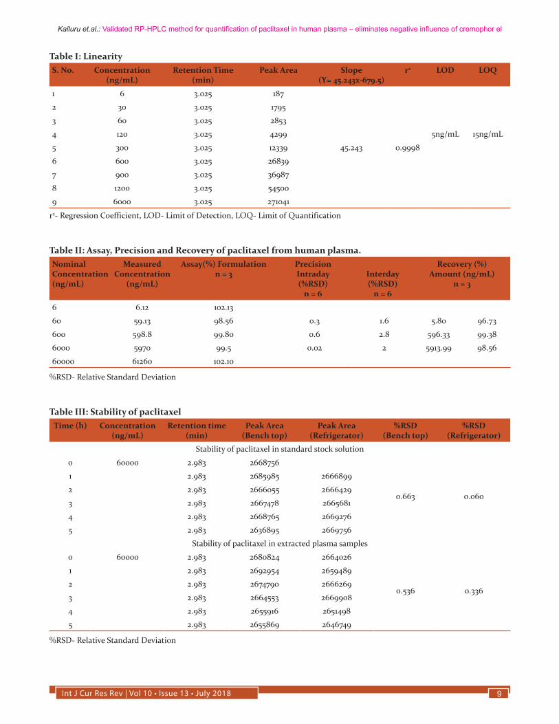
Linearity and detection limitsThe limit of detection was found to be 5 ng/mL and limit of quantification that can be reliably and reproducibly measured
Int J Cur Res Rev | Vol 10 • Issue 13 • July 2018 7
Kalluru et.al.: Validated RP-HPLC method for quantification of paclitaxel in human plasma – eliminates negative influence of cremophor el
is 15 ng/mL. Linearity of detector response was assessed for extracted plasma standards over the range of 6-6000 ng/mL with an r2 value of 0.9998 (Table I). For 0.5 mL aliquots of plasma samples, linearity was satisfactory between 6 and 6000 ng/mL.
PrecisionAssay precision was found to be satisfactory in the concen-tration range of 6-6000 ng/mL. The intraday and interday precision was assessed using six replicates of three concen-trations 60, 600, 6000 ng/mL and the relative standard devia-tion (%RSD) was found to be < 0.7% for intraday and < 3% for interday precision (Table II).
Formulation AssayAccuracy of the method was established by quantification of paclitaxel in the pharmaceutical dosage form (Taxeleon 30 mg/5mL, Neon laboratories) in the concentration range of 6-60000 ng/mL. The results shows that the peak shape is symmetrical with a good baseline and there was strong correlation between the standard solutions and pharmaceuti-cal dosage form solutions with respect to retention time and peak area. The assay performance data are presented in Table II.
RecoveryRecovery of paclitaxel was estimated by using spiked plas-ma samples of 500 µL with standard solutions of known con-centrations, extraction of plasma samples as per the sample pretreatment procedure, analyzing and comparison of peak areas of paclitaxel standard and extracted plasma samples. Recovery of paclitaxel measured in triplicates at three con-centrations (60 ng, 600 ng and 6 µg/mL) was in the range of 96-99% (Table II).
Selectivity and SpecificityChromatograms of blank plasma, paclitaxel standard in dilu-ent, paclitaxel extracted from patient plasma are illustrated in figure 1. The results of extraction from plasma and standard reveal a symmetrical peak shape and good baseline resolu-tion of paclitaxel. Interference due to plasma matrix com-ponents was not observed during the analysis. Using this system, the retention time for paclitaxel was 3 min and the runtime for total analysis of each run was efficiently main-tained for 5 min.
It is also observed that there is no interference of endogenous peaks, peak shape and retention time being the same for pa-clitaxel standard in diluent, paclitaxel formulation and ex-tracted plasma samples.
Stability of paclitaxelThe stability of paclitaxel standard solution and extracted plasma were assessed for 6h to determine the optimum re-
quirements for processing and storage during the analysis (Table III). We found that paclitaxel was quite stable in room temperature and at 4°C for the assay duration.
RobustnessThree chromatographic conditions (Detection wavelength, Flow rate and composition of mobile phase) were altered de-liberately at a range of ± 2% and under each condition sam-ples were analyzed to estimate the paclitaxel content. The results reveal that the values were in the acceptance limits of 98-102% with %RSD of less than 2% (Table IV).
Effect of Cremophor El on paclitaxel analysisIt is reported that the bioanalysis of paclitaxel reproducibility is negatively affected by Cr El (9). However, no such effects were observed in our study. This may be due to liquid-liquid extraction with t-BME, that nullifies the ion suppression ef-fects caused by Cr El (15).
DISCUSSION
The limit of detection and limit of quantification of the cur-rent developed method is sensitive enough to carry out the clinical studies/pharmacokinetic studies of paclitaxel for 24 h. These are equal to the methods using sophisticated solid phase extraction (10). The value of regression coeffi-cientr2=0.9998 confirmed the linear relationship between the concentration of the drug and area of the peak. This method is precise enough to perform continuous and regular quanti-fication of paclitaxel in human plasma. The minimal varia-tion in %RSD for interday and intraday precision indicates the complete harmony among repeated injection, repeated analysis, intraday and interday study.
The strong correlation between the standard solutions and pharmaceutical dosage form with respect to retention time and peak area establishes the accuracy of the method. The proposed method of analysis was highly selective and specif-ic as peaks of the analyte in spiked plasma and patient plas-ma were well resolved. Separation of peaks was confirmed by using blank plasma, plasma spiked with analyte and pa-tient plasma containing analyte along with other drugs. The recovery of paclitaxel was efficient and equal to the costly SPE techniques which were used in other methods (10).
The minor deliberate changes made in various experimental parameters did not significantly affect the peak area, recov-ery of the analyte indicating the robustness of the developed method. The stability of paclitaxel in various conditions has been well studied. In this method stability of paclitaxel in standard solutions and extracted from plasma was assessed for 5 h both at room temp (bench-top) and under refrigera-tion at 4°C to establish the stability of the analyte during the process of analysis. The %RSD values shows that there were
Int J Cur Res Rev | Vol 10 • Issue 13 • July 20188
Kalluru et.al.: Validated RP-HPLC method for quantification of paclitaxel in human plasma – eliminates negative influence of cremophor el
no considerable changes in their concentration after 5 h indi-cating that samples are stable during analysis.
The extraction solvent used in this method t-BME can elimi-nate the negative influences caused by the Cr El that are re-ported in other methods (9). It was successfully applied for quantification of paclitaxel in human plasma and pharma-ceutical dosage form.
CONCLUSION
The current developed RP-HPLC method for quantification of paclitaxel in human plasma is rapid, economic, sensitive and reproducible without negative influence of Cr El. Limit of quantification of this method was 15 ng/mL, making it applicable for clinical pharmacokinetic studies of paclitaxel. A number of methods have been described earlier for quan-tification of paclitaxel in various biological matrices using various techniques including HPLC. The later involve longer run times, usage of costly SPE procedures and methods to reduce the influence of CrEl. The method described here is equally sensitive yet simpler, using single solvent extraction procedure and having a short retention time of 3 min.
Mobile phase composition, sample extraction using t-BME and the use of C18 (15× 4.6 cm i.d, 4 µm particle size) column have attributed to the sensitivity, precision, reproducibility and shorter retention time of the method, which makes it a very practical method to use.
ACKNOWLEDGEMENTS
Our sincere thanks to Neon laboratories, Mumbai for gifting paclitaxel formulation (Taxeleon 30mg/5mL).
FUNDING
Our sincere thanks to the management, Sri Ramachandra University for supporting the research work in the form of GATE Young Faculty Research Grant for the year 2016-17 (Grant no – 54/Dean/2016)
REFERENCES1. Huizing MT, Misser VH, Pieters RC, ten Bokkel Huinink WW,
Veenhof CH, Vermorken JB, et al. Taxanes: A New Class of An-titumor Agents.Cancer Investigations1995; 13 (4): 381–404.
2. Jordan MA, Wilson L. Microtubules and Actin Filaments: Dy-namic Targets for Cancer Chemotherapy. Current Opinion in Cell Biology 1998; 123–130.
3. Jordan MA, Wilson L. Microtubules as a Target for Anticancer Drugs. Nature Reviews Cancer 2004; 4 (4): 253–265.
4. Gligorov J, Lotz JP. Preclinical Pharmacology of the Taxanes: Implications of the Differences. Oncologist 2004; 9 Suppl 2 (suppl 2): 3–8.
5. Steed H, Sawyer MB. Pharmacology, Pharmacokinetics and Pharmacogenomics of Paclitaxel. Pharmacogenomics2007; 8 (7): 803–815.
6. Gerritsen-van Schieveen P, Royer B. Level of Evidence for Therapeutic Drug Monitoring of Taxanes. Fundamentals & Clinical Pharmacology2011; 25 (4): 414–424.
7. Krens SD, McLeod HL, Hertz DL. Pharmacogenetics, Enzyme Probes and Therapeutic Drug Monitoring as Potential Tools for Individualizing Taxane Therapy. Pharmacogenomics 2013; 14 (5): 555–574.
8. Sparreboom A, Zuylen L Van, Brouwer E, Loos WJ, Bruijn P De, Gelderblom H et al. Cremophor EL-Mediated Alteration of Paclitaxel Distribution in Human Blood : Clinical Pharmacoki-netic Implications.Cancer Research 1999; 1454–1457.
9. Andersen A, Warren DJ, Brunsvig PF, Aamdal S, Kristensen GB, Olsen H. High Sensitivity Assays for Docetaxel and Pacli-taxel in Plasma Using Solid-Phase Extraction and High-Perfor-mance Liquid Chromatography with UV Detection. BMC Clini-cal Pharmacology 2006; 6 (1): 2.
10. HendrikxJJMA, Rosing H, SchinkelAH, Schellens JHM, Bei-jnen JH. Quantification of Taxanes in Biological Matrices: A Review of Bioanalytical Assays and Recommendations for De-velopment of New Assays. Bioanalysis 2014; 6 (7): 993–1010.
11. Palmas RA, Monteiro J, Fresco P. Analytical Methods for Taxa-nes Quantification in Diluted Formulations and Biological Sam-ples and Their Applications in Clinical Practice. International Journal of Pharmacy and Pharmaceutical Sciences 2014; 6 (6): 17–23.
12. Khan I, Iqbal Z, Khan A, Hassan M, Nasir F, Raza A et al. A sim-ple, rapid and sensitive RP-HPLC-UV method for the simultane-ous determination of sorafenib & paclitaxel in plasma and phar-maceutical dosage forms: Application to pharmacokinetic study. J Chromatogr B Analyt Technol Biomed Life Sci., 2016;DOI: 10.1016/j.jchromb.2016.08.029.
13. Yonemoto H, Ogino S, Nakashima MN, Wada M, Nakashima K. Determination of Paclitaxel in Human and Rat Blood Samples after Administration of Low Dose Paclitaxel by HPLC-UV De-tection. Biomedical Chromatography 2007; 21: 310-317.
14. https://www.gmp-compliance.org/guidelines/gmp-guideline/ich-q2r1-validation-of-analytical-procedures-text-and-meth-odology (Last accessed: October 28, 2017)
15. Bhaskar V. Identification and Reduction of Matrix Effects Caused by Cremophor EL in Bioanalysis Using Liquid Chroma-tography/Tandem Mass Spectrometry. Analytical & Bioanalyti-cal Techniques 2013; 4 (3): 167.
Int J Cur Res Rev | Vol 10 • Issue 13 • July 2018 9
Kalluru et.al.: Validated RP-HPLC method for quantification of paclitaxel in human plasma – eliminates negative influence of cremophor el
Table I: Linearity S. No. Concentration
(ng/mL)Retention Time
(min)Peak Area Slope
(Y= 45.243x-679.5)r2 LOD LOQ
1 6 3.025 187
45.243 0.9998
2 30 3.025 1795
3 60 3.025 2853
4 120 3.025 4299 5ng/mL 15ng/mL
5 300 3.025 12339
6 600 3.025 26839
7 900 3.025 36987
8 1200 3.025 54500
9 6000 3.025 271041
r2- Regression Coefficient, LOD- Limit of Detection, LOQ- Limit of Quantification
Table II: Assay, Precision and Recovery of paclitaxel from human plasma.Nominal Concentration (ng/mL)
Measured Concentration
(ng/mL)
Assay(%) Formulation n = 3
PrecisionIntraday (%RSD)
n = 6
Interday(%RSD)
n = 6
Recovery (%)Amount (ng/mL)
n = 3
6 6.12 102.13
60 59.13 98.56 0.3 1.6 5.80 96.73
600 598.8 99.80 0.6 2.8 596.33 99.38
6000 5970 99.5 0.02 2 5913.99 98.56
60000 61260 102.10
%RSD- Relative Standard Deviation
Table III: Stability of paclitaxel Time (h) Concentration
(ng/mL)Retention time
(min)Peak Area
(Bench top)Peak Area
(Refrigerator)%RSD
(Bench top)%RSD
(Refrigerator)
Stability of paclitaxel in standard stock solution
0 60000 2.983 2668756
0.663 0.060
1 2.983 2685985 2666899
2 2.983 2666055 2666429
3 2.983 2667478 2665681
4 2.983 2668765 2669276
5 2.983 2636895 2669756
Stability of paclitaxel in extracted plasma samples
0 60000 2.983 2680824 2664026
0.536 0.336
1 2.983 2692954 2659489
2 2.983 2674790 2666269
3 2.983 2664553 2669908
4 2.983 2655916 2651498
5 2.983 2655869 2646749
%RSD- Relative Standard Deviation

Int J Cur Res Rev | Vol 10 • Issue 13 • July 201810
Kalluru et.al.: Validated RP-HPLC method for quantification of paclitaxel in human plasma – eliminates negative influence of cremophor el
Table IV: Robustness of the methodParameter Concentration (ng/
ml)Retention Time
(min)Peak Area % Recovery
Detection wave length – 232nm 60 3.000 2814 98.63
600 3.000 26339 98.13
6000 3.000 265979 98.13
Detection wave length – 232nm 60 3.000 2832 99.26
600 3.000 27127 101.07
6000 3.000 272862 100.67
Flow Rate – 0.8ml/min 60 3.733 2885 100.41
600 3.733 272270 101.60
6000 3.733 267298 98.61
Flow rate – 1.2ml/min 60 2.500 2880 100.94
600 2.500 26390 98.32
6000 2.500 268775 99.16
Mobile Phase ratio – 70:30 60 2.133 2859 100.21
600 2.125 27716 102.24
6000 2.125 272932 100.67
Mobile Phase ration – 50:50 60 5.600 2796 98.00
600 5.600 26379 98.28
6000 5.600 269606 99.47
Figure 1: Chromatograms of paclitaxel; A. Chromatogram of drug free plasma, B. Chromatogram showing elution of peak with 60µg of standard paclitaxel spiked in plasma, C. Chromatogram showing elution of peak in patient plasma.


![Development and Validation of RP-HPLC Method for …...simultaneous estimation by HPLC [8,9]. The aim of this research work is the development of a simple, rapid and precise RP-HPLC](https://img.pdfslide.net/doc/110x75/5e403e331e099c466b433b95/development-and-validation-of-rp-hplc-method-for-simultaneous-estimation-by.jpg)