Embed Size (px)
Citation preview
BCOP BRD: Section 2 March 2006
2-1
2.0 BCOP TEST METHOD PROTOCOL COMPONENTS 2.1 Overview of How the BCOP Test Method is Conducted The basic procedures used to assess the effects of a test substance on an isolated bovine cornea were first reported by Gautheron et al. (1992). As described by Sina and Gautheron (1994, 1998), the BCOP assay uses isolated corneas from the eyes of freshly slaughtered cattle. Corneas free of defects are dissected with a 2 to 3 mm rim of sclera remaining to assist in subsequent handling, with care taken to avoid damage to the corneal epithelium and endothelium. Isolated corneas are mounted in specially designed corneal holders that consist of anterior and posterior compartments, which interface with the epithelial and endothelial sides of the cornea, respectively. Both chambers are filled with medium and the device is then incubated at 32 ± 1°C for one hour to allow the corneas to equilibrate with the medium and to resume normal metabolic activity. Following the equilibration period, fresh medium is added to both chambers, and a baseline opacity measurement is performed. Corneal opacity is measured quantitatively as the amount of light transmission through the cornea.
Two treatment protocols are used, one for liquids and surfactants, and one for solids. Test substances are applied to the epithelial surface of the cornea by addition to the anterior chamber of the corneal holder. Liquids are tested undiluted; surfactants are tested at a concentration of 10% in saline or deionized water. Corneas are incubated horizontally for 10 ± 1 minutes at 32 ± 1°C. The test substance is removed from the anterior compartment and the epithelial surface is washed at least three times. After refilling both chambers with fresh medium, a second opacity measurement is taken and the corneas are incubated again at 32 ± 1°C for two hours prior to taking a final opacity measurement. Solids are tested as solutions or suspensions at 20% concentration in saline or deionized water. Corneas are incubated horizontally for four hours at 32 ± 1°C. The test substance is removed from the compartment and the epithelial surface is washed at least three times with medium or until the corneal surface is free of visible particles. Fresh medium is added to both chambers and an opacity measurement is taken without further incubation. Immediately after completing the final opacity measurements, corneal permeability is determined quantitatively by evaluating changes in the barrier properties of the epithelium to sodium fluorescein. To the anterior compartment of the corneal holder, 1 mL of sodium fluorescein (0.4% for liquids and surfactants, 0.5% for solids) is added. The corneas are incubated horizontally for 90 minutes at 32 ± 1°C. The amount of dye that penetrates the cornea is determined by measuring the OD of the medium in the posterior chamber with a microplate reader or UV/VIS spectrophotometer set at 490 nm. A mean corrected opacity value (± standard deviation [SD]) and a mean corrected permeability value (OD units ± SD) are calculated for each treatment group. Most BCOP studies calculate an In Vitro Score for irritancy that combines both values using the following empirically derived formula (Sina et al. 1995): In Vitro Score = opacity value + 15 x OD490
BCOP BRD: Section 2 March 2006
2-2
value. A substance producing an In Vitro Score from 0 to 25 is considered a mild irritant, from 25.1 to 55 a moderate irritant, and from 55.1 and above a severe irritant. A few laboratories do not calculate an In Vitro Score, but evaluate the opacity and permeability values independently. Also, some companies, such as S.C. Johnson & Son, Inc., do not use the classification system described above to assign an ocular irritancy classification, but instead compare BCOP data for newly tested substances to benchmark materials, relying on a system of comparative toxicity instead of cutoff scores (Cuellar N and Swanson J, personal communication). These procedures were initially developed to assess the ocular irritation potential of pharmaceutical manufacturing intermediates and raw materials (Sina and Gautheron 1994; Sina 1994). However, as the BCOP test method gained more widespread use, the protocol has been modified by different investigators interested in using the assay to evaluate the ocular irritancy potential of other types of materials, including surfactant-based personal care cleaning formulations (Gettings et al. 1996), home care products (Casterton et al. 1996), alkaline liquid laundry detergents (Cater et al. 2002), oxidizing/reactive cleaning products (Swanson et al. 2003), and petrochemical products (Bailey et al. 2004). As a result of the different testing needs of different investigators, additional endpoints have been used, such as assessment of corneal hydration (Ubels et al. 1998; Cooper et al. 2001; Jones et al. 2001), and histological assessment of morphological alterations in the cornea (Curren et al. 2000; Swanson and Harbell 2000; Cater et al. 2001; Cooper et al. 2001; Jones et al. 2001; Burdick et al. 2002). If a histological evaluation of the cornea is performed, the cornea is fixed in an appropriate fixative (e.g., 10% neutral buffered formalin) after completing the corneal permeability steps of the assay. The cornea is fixed at room temperature for at least 24 hours before processing. After embedding the corneas, they are sectioned and stained with an appropriate stain such as hematoxylin and eosin. Corneal sections are examined for lesions in the epithelium, stroma, and endothelium. Sections from treated corneas are compared to those from concurrent negative and positive control corneas (Evans 1998; Curren et al. 2000). Other common modifications to the basic BCOP protocol include use of variable test substance exposure times and post-exposure periods that are specific to certain types of substances or products. For example, shorter exposure times are used for volatile organic solvents (Harbell J, personal communication), longer exposure times are used for diluted materials or for increased sensitivity in the mild range of irritancy (Gettings et al. 1996; Bruner et al. 1998; Cater et al. 2002, 2003), and longer post-exposure expression periods are used to test substances with a potentially delayed onset of irritancy (Rees et al. 2001; Cuellar et al. 2003, 2004; Gran et al. 2003; Swanson et al. 2003). 2.2 Description and Rationale for the Test Method Components The publicly available BCOP test method protocols reviewed for this section follow the basic methodology originally developed for the assay as outlined by Gautheron et al. (1994) and Sina and Gautheron (1994). The essential principles of the test method protocol include isolating and culturing the bovine cornea, treating the isolated cornea with a test substance,
BCOP BRD: Section 2 March 2006
2-3
collecting opacity and permeability data, and evaluating the data in relation to a prediction model (Curren and Harbell 1998). However, given the various uses and applications of the BCOP test method by different investigators and laboratories, and the evolution of the assay over time, a number of laboratory-specific differences have been noted regarding the conduct of the test method. Variations in the publicly available BCOP protocols include different instrumentation to evaluate opacity, different prediction models or in vitro classification systems, and differences in the use of positive controls, among other methodological variations. These test method protocol differences are described in detail in Section 2.2.1, where variations in specific test method components for the BCOP assay are discussed. The test method has been evaluated in several interlaboratory studies (Gautheron et al. 1994; Sina et al. 1995; Balls et al. 1995; Southee 1998) that have led to important refinements in the test method protocol. These refinements have been incorporated into two modified BCOP protocols: 1) the protocol used during Phase II of the European Community sponsored prevalidation study of the BCOP assay conducted from 1997 to 1998 (Southee 1998); and 2) the current protocol used by a contract testing laboratory for routine evaluation of the ocular irritancy potential of test substances and materials (Institute for In Vitro Sciences [IIVS], Gaithersburg, Maryland). The refinements in these protocols are based partly on experience gained with the assay, and partly on experiments designed to identify specific aspects of the protocol that might contribute to intra- and inter-laboratory variability. The following sections describe in detail the major components of the BCOP test method protocol. Similarities and differences in the test method components of available BCOP protocols are discussed. For many of these components, no rationale for inclusion in the BCOP was provided in the published literature; in such cases, historical use is considered the rationale. For each test method component, a summary is presented of information obtained from:
• IIVS, a nonprofit foundation that has performed the BCOP assay since 1997 in a GLP compliant testing facility.
• INVITTOX Protocol No.124 (1999). This protocol was used for the European Community sponsored prevalidation study of the BCOP assay conducted in 1997-1998.
• A literature search and review of publicly available BCOP protocols, which are based on the methodology first reported by Gautheron et al. (1992). These protocols are summarized in Appendix A.
• Discussion and personal communication with Dr. John Harbell (IIVS) and scientific experts who are members of the ICCVAM Ocular Toxicity Working Group (OTWG).
2.2.1 Materials, Equipment, and Supplies Needed 2.2.1.1 Bovine Eyes: Source, Collection/Handling and Quality Source: Several BCOP studies noted that bovine eyes were obtained from a local slaughterhouse that was close enough to the testing laboratory to allow for transport of the eyes to the laboratory within two to four hours after the animals were killed (Gautheron et al. 1994; Rachui et al. 1994; Sina et al. 1995; Casterton et al. 1996; INVITTOX 1996; INVITTOX 1999). Other BCOP studies noted that the bovine eyes were likewise obtained
BCOP BRD: Section 2 March 2006
2-4
from a local slaughterhouse, but reported different periods of time until use of the eyes. For example, Bruner et al. (1998) reported that eyes were used within 12 hours after receipt at the laboratory, and Cerven and Moreno (1998) reported that the eyes were examined within one hour after receipt at the laboratory without noting the amount of time that had passed postmortem. At IIVS, bovine eyes generally arrive in the testing laboratory within four to five hours of the first eyes being enucleated at the slaughterhouse, and eyes are processed immediately upon arrival at the laboratory (Harbell J, personal communication). Therefore, while a formal study to determine the maximal time not to be exceeded during the transport of eyes to the testing facility was not found in the published scientific literature, a maximum of five hours has been used in most BCOP protocols and appears to produce consistent results. No detail was provided in the study reports on the specific breed, age, or sex of the cattle used as the source of the bovine eyes. Based on information from other sources, it was found that the cattle sent to slaughterhouses are typically killed either for human consumption (e.g., calves for veal; steers 9 to 30 months old for prime, choice, select, or standard grades of beef), or for other commercial uses (e.g., cattle 42 to 96 months for commercial, utility, or cutter grades of beef). The cattle in the former category tend to be raised specifically for meat production and thus are of cattle breeds (e.g., Hereford) used to optimize the quality and quantity of beef for human consumption. The cattle in the latter category can include dairy cattle (e.g., Holstein) that are no longer useful for milk production (Doughty et al. 1995; North Dakota State University Extension Service 1999). Although bovine eyes are widely used in ocular irritancy evaluations, only a few studies were found that addressed potential sources of variability in bovine eyes obtained from slaughterhouse operations (Doughty et al. 1995; Doughty 1997, 2004). In one study, central corneal thickness (CCT) values ranged from 750 to 1450 µM (mean and SD of 1015 ± 104 µM) and horizontal corneal dimensions ranged from 27.5 to 34.5 mm (mean and SD of 29.8 ± 1.3 mm) in bovine eyes obtained from 315 Holstein and Hereford cattle killed at a local slaughterhouse over a one-year period (Doughty et al. 1995). These variations in corneal dimensions were proposed to be a result of obtaining the eyes from animals of different ages. Corneas with a horizontal dimension greater than 30.5 mm and CCT values equal to 1100 µM or greater were likely obtained from cattle older than eight years, while those with a horizontal diameter less than 28.5 mm and CCT less than 900 µM were likely from cattle less than five years old (Doughty et al. 1995). For this reason, eyes from mature cattle (i.e., greater than 60 months old) are not typically recommended. Additionally, eyes from cattle less than 12 months of age are believed to be inadequate since the eyes are still developing and the corneal thickness and corneal diameter are considerably smaller than that reported for eyes from adult cattle. However, as discussed below, a recent study suggests that eyes from younger animals may indeed be useful. It should be noted that these findings may be applicable only to the specific cattle breeds and slaughterhouse operation used in the study. However, they are suggestive of potential variability in corneas sizes and thicknesses of bovine eyes obtained from slaughterhouse operations. Limited information could be found on whether variable cornea sizes from animals of different ages might impact the performance of the BCOP test method. During
BCOP BRD: Section 2 March 2006
2-5
the European Community prevalidation study of BCOP, a small study was conducted to evaluate whether cornea size influenced BCOP test method results obtained for ethanol (Southee 1998). The investigators reported that the results suggested no apparent relationship between cornea size, basal opacity, or cornea response to ethanol. In addition, data provided by Johnson & Johnson Pharmaceutical Research and Development for 19 test substances suggests that the performance of the BCOP when using eyes from young (6-8 months) versus adult (> 24 months) animals is comparable (see Section 9.2.4). However, because there are limited data on this matter, further evaluation of potential variability among corneas from slaughterhouse animals may be necessary to investigate whether the size or age of the cornea influences the responsiveness of the cornea to irritating substances. Collection/Handling: Most BCOP studies noted that the bovine eyes were excised by a slaughterhouse employee with care taken to avoid damage to the cornea; however, details on the enucleation procedure and the specific steps taken to avoid corneal damage were not provided in any of the study reports. Depending on the slaughterhouse operation, it may take several hours for a slaughterhouse employee to collect the required number of eyes for use in a BCOP study at the testing facility. IIVS notes that they use bovine eyes that are collected by slaughterhouse employees at various times following exsanguination and decapitation of the cattle. To minimize mechanical and other types of damage to the eyes, this laboratory prefers the eyes be enucleated as soon as possible postmortem and requests that slaughterhouse employees not use detergent when rinsing the animal head to prevent exposure of the bovine eyes to potentially irritating substances (Harbell J, personal communication). To the extent possible, IIVS communicates their need for undamaged bovine eyes to the slaughterhouse, while recognizing the constraints of the slaughterhouse environment. Because the bovine eyes are collected during the process of slaughter, it is recognized that the bovine eyes may have been exposed to blood and other biological substances, including bacteria and other microorganisms (Doughty 1997). The BCOP studies varied in how the bovine eyes were handled after enucleation at the slaughterhouse and during transit prior to arrival at the testing facility. The two major variables in handling were differences in the solution used to store the eyes, and differences in the temperature of the eye storage container. Most studies noted that the eyes were immersed completely in Hanks’ Balanced Salt Solution (HBSS) in a suitably sized container. Of the 18 studies reviewed, four reported addition of the antibiotics penicillin and streptomycin to the HBSS (Bruner et al. 1998; INVITTOX 1999; Cooper et al. 2001; Jones et al. 2001), while the other studies appear not to have used antibiotics. With regard to the temperature of the collection vessel, some studies maintained the storage container at ambient temperature (Gautheron et al. 1994; Rachui et al. 1994; Casterton et al. 1996; INVITTOX 1996; INVITTOX 1999), while others maintained it on ice to keep the eyes cool and to minimize ambient temperature variation that would result due to seasonal changes (Cooper et al. 2001; Jones et al. 2001). The matter of temperature maintenance of the eye collection vessel was not addressed in the other reviewed studies.
BCOP BRD: Section 2 March 2006
2-6
Quality of Eyes: Currently, it appears that there are no standardized criteria for the selection of bovine eyes for the BCOP assay. Most BCOP studies reported that the eyes were carefully examined visually for defects, including opacity, scratches, and neovascularization, once they had arrived at the laboratory. A few studies also noted use of microscopes to assist in identifying damaged corneas. Rachui et al. (1994) commented that the eyes were carefully examined visually, or with the aid of a stereomicroscope. Swanson et al. (1995) stated that the corneas were examined microscopically after they were dissected, and only corneas free of defects were used in the BCOP assay. The quality of the corneas is evaluated at later steps in the assay, as well. For example, corneas that have a high baseline opacity reading (e.g., opacity greater than 10) after the initial one-hour equilibration period are discarded, a practice that is consistent among the reviewed BCOP protocols. Opacity that develops in the cornea prior to application of a test substance sometimes results from fine scratches not noticeable upon visual inspection (Harbell J, personal communication). 2.2.1.2 Instrument to Measure Light Transmission Through the Cornea Changes in light passage through the cornea have been most commonly assessed with a white light, dual-beam opacitometer (e.g., Spectro Designs OP-KIT, STAG BIO, Electro-Design). This type of opacitometer provides a center-weighted reading of light transmission through the cornea. There are two compartments, each with its own light source and photocell. One compartment is used for the treated cornea, while the other is used to calibrate and zero the instrument. The difference between photocell signals in the two compartments is measured electronically as a change in voltage, and is displayed digitally, generating numerical opacity values with arbitrary units. The BCOP assay was developed with the center-weighted opacitometer, and a majority of BCOP studies in the peer-reviewed literature report using this type of opacitometer. However, the center-weighted readings may underestimate opacity that develops as spots on the periphery of the isolated cornea (Southee 1998; van Goethem et al. 2002), and therefore some BCOP users have modified the method of reading opacity. Casterton et al. (1996) first reported the use of a UV/VIS spectrophotometer to evaluate corneal opacity. Corneal holders were modified to fit into the spectrophotometer and light absorbance (570 nm) readings performed through the center of the cornea. Absorbance values use a different scale than values obtained from the white light opacitometer; thus, BCOP data from the two instruments cannot be directly compared. This method of measuring opacity requires the use of a different classification procedure or prediction model to identify ocular irritants when compared to the traditional BCOP assay. Recognizing the limitations of the conventional opacitometer with its center-weighted readings, Janssen Pharmaceutica/Johnson & Johnson Pharmaceutical Research recently developed a new laser-based opacitometer that uses an adjustable laser beam in combination with a calibrated photocell (van Goethem et al. 2002). This opacitometer was designed to provide a more even distribution of light across the corneal surface and, thus, may provide an improved method of opacity assessment. However, the database of BCOP studies using this type of opacitometer is still relatively small, and thus additional studies are required to determine if such instruments provide a definitive advantage over the conventional opacitometer (i.e., center-weighted readings)
BCOP BRD: Section 2 March 2006
2-7
2.2.1.3 Instrument to Evaluate Permeability Over half of the BCOP studies used a UV/VIS spectrophotometer set at 490 nm to measure the amount of sodium fluorescein (based on optical density) that permeated through the cornea into the posterior chamber of the corneal holder. The remaining studies used a microtiter plate (microplate) reader (e.g., Dynatech MR 5000 and Molecular Devices Vmax kinetic microplate readers) to measure the amount of sodium fluorescein. The basic design of the two instruments is the same in that a selected wavelength of light passes through the samples and a photosensitive tube detects the amount of light transmitted through the sample. For this reason, either instrument would appear adequate. However, a standard spectrophotometer measures one sample at a time, while a microplate reader is capable of measuring the absorbance of 96 samples in about eight seconds. Thus, the microplate reader offers the advantage of processing large numbers of samples in a short amount of time. 2.2.1.4 Organ Culture Media A few variations in organ culture media were found in the publicly available BCOP study reports. All protocols used some form of complete Minimum Essential Medium (complete MEM), supplemented with 1% fetal bovine serum (FBS). One of the major differences, however, is that the earlier protocols used complete MEM containing phenol red (now considered an outdated practice), while the more recent protocols used complete MEM without phenol red. As part of the European Community prevalidation study of the BCOP assay, investigators evaluated the effect of phenol red in the BCOP incubation medium (Southee 1998). Results from a series of separate assays indicated that complete MEM without phenol red produced lower background opacity readings than phenol red containing MEM. The study report also noted that fluctuation in background values was less for medium without phenol red, attributed in part to the low background values. However, phenol red is useful in the medium during the rinsing procedure, when the test substance must be removed completely from the cornea; residual test substance can sometimes be identified by a shift in color of the phenol red (Harbell J, personal communication). A second notable variation is that some protocols prewarmed the complete MEM to 32°C, the temperature at which the corneal equilibration step and all incubations are performed. Prewarming the organ culture medium eliminates the time needed for the media temperature to equilibrate with the incubator system or the water bath. A few protocols also reported adjusting the pH of the complete MEM from 7.2 to 7.4 prior to use in the assay, although most did not. Adjustment of pH to a physiological level was likely performed in situations when sodium bicarbonate was added to the MEM by the testing facility to provide buffering capacity to the media. However, MEM with appropriate buffering capacity can be purchased, obviating the need for pH adjustment. Other slight differences appear to be related to the level of detail provided in the study reports. For example, some protocols reported use of the standard complete MEM supplements, such as L-glutamine, Ca++, Mg++, and sodium bicarbonate, while others did not, making it unclear whether the same supplements were used in different BCOP studies.
BCOP BRD: Section 2 March 2006
2-8
2.2.1.5 Solvents Differences in the use of solvents have been noted. Some reports noted that solid compounds were prepared as a 20% solution or suspension in 0.9% NaCl (Vanparys et al. 1993; INVITTOX 1996; INVITTOX 1999). In comparison, some solid and surfactant test substances were prepared in MEM (Gautheron et al. 1994; Rachui et al. 1994; Sina et al. 1995; Chamberlain et al. 1997; Cerven and Moreno 1998). IIVS uses sterile, deionized water or saline to dissolve or suspend solid test substances (Harbell J, personal communication). The European Community prevalidation study report noted that use of saline is preferred for dilutions, since it may prevent possible buffering effects and enhanced penetration of the test substance that could result from the use of organic solvents (Southee 1998). 2.2.1.6 Incubation Apparatus A majority of BCOP studies reported using a water bath for incubations (Rachui et al. 1994; INVITTOX 1996; Bruner et al. 1998; Cerven and Moreno 1998; INVITTOX 1999). A few studies reported carrying out incubations at room temperature (Sina et al. 1995; Casterton et al. 1996), while still others reported using a forced air incubator (Cassidy and Stanton 1997; Cooper et al. 2001); IIVS also currently uses a forced air incubator in its studies. An experiment was conducted during the European Community prevalidation study of the BCOP assay to evaluate whether similar results are obtained for the same test substance, when the assay is conducted using a water bath or a forced air incubator. This experiment evaluated one test substance identified as “CTAB”, which produces a severe response in the isolated cornea. Half of the exposed corneas were incubated for 30 minutes in a water bath, while half were incubated for 30 minutes in a forced air incubator; all other procedures were the same. The study authors concluded that there was a “distinct” difference in opacity and permeability values, and consequently, the mean in vitro score obtained for CTAB, depending on the incubation system used. The authors, however, did not state that the results were statistically significant. The study report notes “the water bath provides a more stable temperature than the air incubator which fluctuates when the door is opened. Water also provides greater heat conductivity, and hence the holders will reach 32º C quicker” (Southee 1998). Others have noted that the water bath allows for better heat transfer, but is technically more difficult to use. Sometimes there are cross-contamination problems, when water from the water bath seeps into the corneal holder or when the test substance seeps into water bath (Harbell J, personal communication). Both types of incubators have advantages and disadvantages. The water bath offers greater temperature control but greater opportunity for cross contamination. Until more information becomes available about the comparative advantages and disadvantages of the forced air incubator and the water bath, it would appear that both would be adequate for performing incubations. 2.2.1.7 Corneal Holder As described by Gautheron et al. (1992) and Sina and Gautheron (1998), the corneal holder for the BCOP assay consists of two chambers, each with a 5 mL volume. The main part of
BCOP BRD: Section 2 March 2006
2-9
the chamber is composed of either polypropylene (Sina and Gautheron 1998) or clear Plexiglas (Casterton 1998). The chamber design consists of a glass window on the outside of the chamber, and a 17 mm circular opening on the inner side on which the cornea rests (Gautheron et al. 1992; Ubels et al. 2002). The anterior chamber interfaces with the epithelial side of the cornea, while the posterior chamber interfaces with the endothelium. After the cornea is mounted over an O-ring that is positioned around the opening of the posterior chamber, the chambers are clamped together with three screws (Gautheron et al. 1992). Dosing holes located on the top of each chamber allow the epithelial and endothelial sides of the cornea to be treated independently. The distributors of the opacitometer (e.g., Spectro Designs OP-KIT, STAG BIO, Electro-Design) also supply the corneal holders. It appears that the laboratories that have used a UV/VIS spectrophotometer to measure opacity had the corneal holders specially made and designed for use with that instrument (Casterton et al. 1996; Casterton 1998; Ubels et al. 1998). More recently, studies by Ubels et al. (2000, 2002) have suggested potential limitations regarding the conventional corneal holder: 1) it has a circular opening 17 mm in diameter, yet the bovine cornea is oval shaped and has dimensions of about 24 mm vertically and 30 mm horizontally; 2) it has flat inner surfaces, whereas the bovine cornea is convex or curved. These elements of the corneal holder reportedly force the bovine cornea into an unnatural shape when mounted in the holder, causing the cornea to wrinkle. Ubels et al. (2002) also noted damage to all three corneal cell layers (epithelium, stroma, and endothelium) where the cornea comes in contact with the circular edge of the holder opening. Recognizing some of the potential limitations of the conventional corneal holder, Ubels et al. (2002) designed a new corneal holder with dimensions that better fit the bovine cornea and maintain its natural shape during the BCOP assay. The new holder was designed to contact the 2 to 3 mm rim of sclera left around the bovine cornea during dissection, rather than the corneal tissue. Studies showed that this refined corneal holder does not cause wrinkling of the mounted bovine cornea, nor does it damage the cell layers around the edge of the cornea (Ubels et al. 2002). However, the availability of this new corneal holder for purchase or use by other laboratories is not known. It would seem appropriate that consideration be given to the newly designed corneal holder as a potential refinement of the assay, once it does become commercially available, since it appears that this holder better fits the natural shape and curvature of the bovine cornea. 2.2.2 Dose-Selection Procedures, Including the Need for Any Dose Range-Finding
Studies or Acute Toxicity Data Prior to Conducting a Study As described below in Section 2.2.4.4, test substances are typically applied as neat chemicals (liquids), or diluted to prescribed concentrations (surfactants and solids) with preferred solvents. A few studies also described testing of personal care products, such as shampoos, at proposed end-user concentrations to mimic potential human exposure scenarios (Cooper et al. 2001; Jones et al. 2001).

BCOP BRD: Section 2 March 2006
2-10
2.2.3 Endpoints Measured In the BCOP assay, opacity is determined by the amount of light transmission through the cornea, and permeability is determined by the amount of sodium fluorescein dye that penetrates all corneal cell layers (i.e., the epithelium on the outer cornea surface through the endothelium on the inner cornea surface). In a majority of the BCOP studies reviewed, corneal opacity was measured quantitatively with the aid of a center-weighted opacitometer, resulting in opacity values measured on a continuous scale. The concentration of sodium fluorescein in the posterior corneal chamber, which interfaces with the endothelial side of the cornea, was quantitatively measured with the aid of UV/VIS spectrophotometry. Spectrophotometric measurements evaluated at 490 nm are recorded as optical density or absorbance values, which are measured on a continuous scale. The measurement of opacity is described in detail in Section 2.2.1.2. As previously noted, a few BCOP studies reported using a UV/VIS spectrophotometer instead of an opacitometer to evaluate corneal opacity (Casterton et al. 1996; Ubels et al. 2003). The measurement of permeability is standard across the reviewed BCOP studies. Typically, 1 mL of 4 mg/mL sodium fluorescein solution in MEM is used when testing liquid and surfactant substances, and a 5 mg/mL solution is used when testing solid substances. No rationale could be found for the use of different concentrations of sodium fluorescein for different types of substances. The sodium fluorescein solution is added to the anterior chamber, and the holder incubated horizontally for 90 minutes (Gautheron et al. 1992, 1994). The stock solutions of sodium fluorescein used for the BCOP assay are prepared to the specified concentrations, and then verified using a UV/VIS spectrophotometer to ensure the absorbances of the solutions fall within set limits. The UV/VIS spectrophotometer used for permeability measurements is calibrated with dilutions of sodium fluorescein solution to determine the linear portion of the absorbance curve and to define the limits outside of which the test substances require dilution (Southee 1998). More recent additions/endpoints to this assay include histological assessment of alterations in the cornea, and, less commonly, assessment of corneal hydration (Bruner et al. 1998; Ubels et al. 1998; Cooper et al. 2001; Jones et al. 2001). Based on the results of a major validation study of BCOP (Balls et al. 1995), it was found that certain severe ocular irritants are underpredicted using only the opacity and permeability endpoints. These findings prompted Curren et al. (2000) to investigate the usefulness of examining histological changes in the cornea in conjunction with the primary BCOP endpoints of opacity and permeability. Curren and colleagues found that three materials underpredicted using only the opacity and permeability endpoints -- parafluoroaniline, quinacrine, and sodium oxalate -- produced notable cellular damage throughout the epithelium and in other tissues that was indicative of severe ocular injury. For example, parafluoroaniline produced death of keratocytes, quinacrine produced microvacuolization throughout the epithelium as well as in keratocytes and the endothelium, and sodium oxalate produced refractile, crystal-like material throughout the epithelium into the basement membrane. Thus, assessment of histopathology in the BCOP assay may be considered
BCOP BRD: Section 2 March 2006
2-11
essential for ocular irritants where the mode of action does not result in significant opacity or permeability. It is widely recognized that histological evidence of corneal damage (or lack thereof) provides additional information for an assessment of ocular irritation. However, the additional expense and time required for such a detailed examination may not be warranted in all cases, such as when severe corneal effects are clearly indicated from the opacity and permeability assessments of the BCOP assay. Instead, histopathological effects could be useful for discriminating borderline moderate/severe cases, identifying alternate mechanisms of severe ocular damage that do not produce significant opacity or permeability, or for evaluating new chemistries where the mode of action is not readily predictable. Also, certain chemical classes, such as oxidizing agents that have a delayed onset of irritation in vivo, may require a histological assessment to fully evaluate the extent of injury. Therefore, the decision to perform a histological assessment of the treated cornea should likely be left to the discretion of the investigator. However, it would seem prudent for the corneas from all studies to be fixed in an appropriate fixative (e.g., 10% neutral buffered formalin), so that the tissues are available if histology is necessary or requested at a later time. At IIVS, the scoring of lesions in a histological evaluation of the isolated cornea is based primarily on the depth of injury, which is predictive of the degree and duration of the injury (Maurer et al. 2002). The three main tissue layers of the cornea (epithelium, stroma, endothelium) are evaluated, and the nature, degree and depth of lesion in each tissue layer are noted. Tissues from the treated corneas are always compared with tissues from the concurrent negative control cornea to distinguish between test substance induced injury and artifacts of handling or processing (Harbell J, personal communication). 2.2.4 Duration of Exposure 2.2.4.1 Pre-Exposure Preparations Pre-exposure preparations are consistent across BCOP protocols. Corneas free of defects are dissected with a 2 to 3 mm rim of sclera remaining to assist in subsequent handling, with care taken to avoid damage to the corneal epithelium and endothelium. Isolated corneas are mounted in specially designed corneal holders that consist of anterior and posterior compartments, which interface with the epithelial and endothelial sides of the cornea, respectively. Both chambers are filled with medium and the device is then equilibrated at 32°C for one hour to allow the corneas to equilibrate with the medium (the approximate temperature of the corneal surface in vivo is 32°C). This is intended to allow the corneas to resume normal metabolic activity. Following the equilibration period, fresh medium is added to both chambers and baseline opacity readings are taken for each cornea. Any corneas that show tissue damage or high opacity (e.g., > 10 opacity units) are discarded. The mean opacity of all equilibrated corneas is calculated. A minimum of three corneas with opacity values close to the average value for all corneas are selected as negative (or solvent) control corneas. The remaining corneas are then distributed into treatment groups and positive/other control groups.
BCOP BRD: Section 2 March 2006
2-12
2.2.4.2 Effects of Residual Equilibration Medium in the Test Substance Chamber As part of the European Community prevalidation study, the investigators evaluated whether residual medium left in the anterior chamber after the pre-exposure incubation had an effect on the opacity and permeability of the cornea to ethanol. Increasing volumes of complete MEM (ranging from 0 to 150 µL) were added to the anterior chamber with 0.75 mL of ethanol to simulate residual medium in the anterior chamber. After a 10-minute incubation at 32°C, opacity and permeability measurements were performed. The results showed that increasing amounts of residual medium produced a corresponding increase in the final in vitro score of ethanol. The in vitro score for ethanol with no residual media was 28.7, while the in vitro score for ethanol with 150 µL of media was 48.8 (Southee 1998). Based on these results, the prevalidation study report recommended that an aspiration method be used to remove as much medium as possible from the anterior chamber prior to addition of the test substance. The study report noted that one suitable method for removing all traces of incubation medium is to use a micropipette tip or blunt needle attached to a vacuum pump. 2.2.4.3 Test Substance Exposure Volume A majority of BCOP protocols consistently applied 0.75 mL of test substance to the cornea (Gautheron et al. 1994; Rachui et al. 1994; Balls et al. 1995; Swanson et al. 1995; INVITTOX 1996; Cassidy and Stanton 1997; Bruner et al. 1998; Cerven and Moreno 1998; INVITTOX 1999; Cooper et al. 2001; Jones et al. 2001). Liquids are typically tested neat, while surfactants and solids are solubilized or suspended at prescribed concentrations. A few protocols reported using 0.5 mL of test substance solution or suspension (Sina et al. 1995; Chamberlain et al. 1997). However, this volume is no longer used because in some cases it failed to cover the corneal surface completely (Harbell J, personal communication). In addition, one report noted a test substance volume of 1.0 mL (Casterton et al. 1996). However, this exception was likely used due to the fact that a unique corneal holder was used in this protocol, one customized for making opacity measurements with a UV/VIS spectrophotometer rather than an opacitometer, which required a larger volume than traditionally used (i.e., 0.75 mL). 2.2.4.4 Concentration Tested For the European Commission (EC) sponsored interlaboratory assessment of the BCOP assay, Gautheron et al. (1994) tested liquids neat (100%), surfactants at a concentration of 10%, and nonsurfactant solids at a concentration of 20% (w/v). The EC/British Home Office (HO) validation study of alternatives to the Draize eye test used the same concentrations in its evaluation of the BCOP assay (Balls et al. 1995), as did the European Community prevalidation study of the BCOP assay (Southee 1998). A majority of the other publicly available protocols used the same test substance concentrations, with a few exceptions. To address specific product development questions, Cooper et al. (2001) and Jones et al. (2001) tested surfactant-based hair-care formulations (shampoos and conditioners) at concentrations of 10% and 100%. Also, Gran et al. (2003) found that a test substance concentration of 50% (in addition to longer exposure/post-exposure times) produced a better correlation to in vivo results for certain reactive/oxidative solids, such as sodium percarbonate. Instead of testing
BCOP BRD: Section 2 March 2006
2-13
solids at a 20% concentration, Casterton et al. (1996) applied solid test substances undiluted (neat) to the cornea. Therefore, historical use generally supports testing liquid substances neat, surfactants at 10%, and nonsurfactant solids at 20%. However, it is recognized that these concentrations may require adjustment for certain chemical or product classes. 2.2.4.5 Application of Test Substance to Bovine Cornea A majority of the BCOP studies used two treatment protocols, one for liquids and surfactants, and one for nonsurfactant solids (Gautheron et al. 1992, 1994; Rachui et al. 1994; Balls et al. 1995; Sina et al. 1995; Chamberlain et al. 1997; Cerven and Moreno 1998; INVITTOX 1999). For both treatment protocols, the test substances were applied to the epithelial surface of the cornea using a micropipettor. The test substances were injected into the anterior chamber of the corneal holder through dosing holes on the top of the chamber (closed chamber method). IIVS uses the closed chamber method for nonviscous to slightly viscous liquids and solubilized solids. However, they have developed a refined procedure for application of semiviscous to viscous test substances, known as the “open chamber method.” In this method, the window-locking ring and glass window are removed from all appropriate anterior chambers and the holders are placed into a horizontal position (anterior chamber facing up). Approximately 0.75 mL of the viscous test substance (or enough test substance to completely cover the cornea) is applied directly to the epithelial surface of the cornea using a micropipettor or other appropriate device, such as a spatula. The corneal holder is reassembled prior to incubation of the test substance (Harbell J, personal communication). Casterton et al. (1996) reported a different procedure for application of solid substances. Solid substances were applied directly onto the cornea by removing the glass window of the corneal holder. Although, a specific weight or volume of solid was not reported, the authors stated that enough test substance was added to cover the cornea thoroughly. 2.2.4.6 Test Substance Exposure Duration Most BCOP protocols incubated liquids and surfactants for 10 minutes at 32 ± 1°C. The test substance was removed from the compartment and the epithelial surface washed at least three times. After replacing the medium, an opacity measurement was taken. The corneas were then returned to the incubator for an additional two hours and another opacity reading taken, which was used for the calculation of corneal opacity. Solutions or suspensions of solids were incubated horizontally for four hours at 32 ± 1°C. The test substance was removed from the compartment and the epithelial surface washed at least three times with medium or until the corneal surface was free of visible particles. Fresh medium was added to both chambers and an opacity measurement was taken without further incubation (Gautheron et al. 1992, 1994; Rachui et al. 1994; Balls et al. 1995; Sina et al. 1995; Chamberlain et al. 1997; Cerven and Moreno 1998; INVITTOX 1999). Shorter exposure times have been suggested for alcohols and volatile organic solvents, since the irritancy of these substances has been overpredicted with an exposure time of 10 minutes
BCOP BRD: Section 2 March 2006
2-14
(Harbell J, personal communication). Some protocol refinements may have to be made if the irritancy of alcohols and volatile organic solvents are consistently overestimated. Longer exposure times (e.g., 60 minutes and 24 hours) have been suggested for better discrimination of mild to moderate ocular irritants, and to differentiate subtle differences between similar formulations (Bruner et al. 1998; Cater et al. 2002, 2003; Harbell J, personal communication). IIVS reported that they use different exposure times to address certain chemicals/chemical classes (e.g., sodium percarbonate, volatile solvents), expected consumer exposure models (e.g., diluted shampoo), or to enhance comparisons across a chemical class (Gran et al. 2003; Harbell J, personal communication). For solid test substances, Casterton et al. (1996) used a shorter exposure time of one hour after applying the test substances undiluted (neat) to the cornea. Exposure was followed by a 1-hour post-rinse incubation period. This reduced exposure time has not been widely evaluated. Historical use generally supports an exposure time of 10 minutes for liquids and surfactants, and four hours for nonsurfactant solids. However, it is recognized that these generic exposure times may require adjustment for certain chemical classes, such as alcohols and volatile solvents. 2.2.4.7 Post-Exposure Incubation A majority of BCOP studies in the literature reported incubating the corneas that had been treated with liquids or surfactants for an additional two hours at 32 ± 1ºC after the 10-minute test substance exposure and the post-treatment rinse. Corneas treated with solid test substance were exposed to the test substance for four hours, and were not further incubated. However, Casterton et al. (1996) used a 1-hour post-exposure incubation when testing solids. Bruner et al. (1998) used longer post-exposure times to better discriminate the irritancy of formulations of a similar composition. IIVS sometimes uses longer post-exposure incubation times for better discrimination of mild to moderate ocular irritants and for substances with a delayed response (Harbell J, personal communication). IIVS also uses different post-exposure incubation times to address certain chemical (e.g., peroxides) and product classes and expected consumer exposure models (Gran et al. 2003). Historical use generally supports a post-exposure time of two hours for liquids and surfactants. Corneas treated with solids typically do not require further incubation beyond the 4-hour exposure period. However, it is recognized that these generic post-exposure times may require adjustment for certain chemical or product classes. 2.2.5 Known Limits of Use While a wide range of substances with various physicochemical characteristics can be tested in the BCOP assay, water insoluble solid substances that are less dense than water (i.e., float on top of the solvent) do not adequately contact the cornea during treatment (Sina and Gautheron 1998). Thus, the standard BCOP protocol for solid test substances (Gautheron et al. 1994) cannot be used for low density, water insoluble substances. In addition, Chamberlain et al. (1997) noted some false negative responses for substances tested with the
BCOP BRD: Section 2 March 2006
2-15
standard BCOP protocol (Gautheron et al. 1994) that had a delayed onset of irritation in vivo. However, test method users are addressing these limitations. For example, the method of applying solid test substances used by Casterton et al (1996), in which solids are sprinkled neat onto the cornea, may be useful to address the limitation of testing low density, insoluble solid substances. Protocols with longer exposure and post-exposure periods are under development to detect substances with a delayed onset of irritancy (Gran et al. 2003). However, the longest exposure/post-exposure period found is 24 hours (Bruner et al. 1998; Gran et al. 2003). Another potential limitation of the test method is that, although it takes into account some of the ocular effects evaluated in in vivo rabbit ocular irritancy tests and to some degree their severity, it does not consider all of the effects assessed in vivo. Reversibility of corneal lesions cannot be evaluated per se in the BCOP assay, but test method users propose that an assessment of the initial depth of corneal injury can be used to predict irreversible or reversible effects (Maurer et al. 2002). Furthermore, in Europe and Japan, there are concerns about the use of bovine tissue due to the risk of transmitting Bovine Spongiform Encephalopathy (BSE). 2.2.6 Nature of the Response Assessed 2.2.6.1 Corneal Opacity Corneal opacity is measured quantitatively with an opacitometer (e.g., ElectroDesign, Riom, France), which measures differences in light transmission between treated corneas and an air blank. Numerical opacity values with arbitrary units are obtained, with values typically ranging from 0 to 500, with higher opacity values occasionally reported. 2.2.6.2 Permeability The amount of dye that permeates the cornea is determined by measuring the OD/absorbance of the medium in the posterior chamber with a spectrophotometer set at 490 nm.
2.2.6.3 Histology Although a more recent addition to the BCOP assay, a histological evaluation of the type, degree and depth of injury at the tissue level, resulting from exposure of the cornea to a test substance appears to be a very useful addition to the assay (Curren et al. 2000; Cooper et al. 2001). 2.2.7 Appropriate Controls and the Basis for Their Selection 2.2.7.1 Negative Controls Some differences were found in the negative controls used in the BCOP assay. Seven BCOP studies used complete MEM as the negative control (Gautheron et al. 1994; Rachui et al. 1994; Rougier et al. 1994; Sina et al. 1995; Bruner et al. 1998; Cooper et al. 2001; Jones et al. 2001). Two studies used 0.9% saline (INVITTOX 1996; INVITTOX 1999). IIVS uses sterile, deionized water (Harbell J, personal communication). To test the possible differences in the use of complete MEM or saline as the negative control, the European Community prevalidation study compared the BCOP results obtained for saline and complete MEM (without phenol red). When incubated for 10 minutes, there was no apparent difference in the results in the opacity and permeability values of complete MEM and saline (Southee
BCOP BRD: Section 2 March 2006
2-16
1998). It appears that the three commonly used negative controls for the BCOP assay offer no distinct advantages or disadvantages. However, it is clear that a negative control is useful in the BCOP test, so that nonspecific changes in the test system can be detected. This type of control also provides a baseline for the assay endpoints, and ensures that the assay conditions do not inappropriately result in an irritant response. Any of the three commonly used negative controls (i.e., MEM without phenol red, 0.9% saline, or sterile, deionized water) is acceptable as long as the same negative control is used consistently within a laboratory. 2.2.7.2 Positive Controls As discussed by Harbell and Curren (2002), the function of the positive control is to ensure the test system is operating within normal limits and each experiment is properly executed, such that the toxic effects of interest can be properly detected. A concurrent positive control is included in each experiment to develop a historical database. Results from the positive control are compared to the historical control range and used to evaluate whether a particular study is acceptable. Because the positive control should allow for detection of an over- or under-response in the assay, the selected positive control should not produce responses at either the extreme low or the extreme high end of assay response. In the BCOP assay, different positive controls are used for the testing of liquid and solid test substances because of the different protocols for these two types of substances. Harbell and Curren (1998) recommend positive controls that produce both opacity and permeability (e.g., ethanol for liquid test substances and imidazole for solid test substances) in the BCOP assay. About half of the BCOP studies used one or more positive control substances. The most frequently used positive control for testing liquid test substances was 100% ethanol (Swanson et al. 1995; Cassidy and Stanton 1997; Bruner et al. 1998; Southee 1998; Cooper et al. 2001; Jones et al. 2001). Acetone (Gettings et al. 1996; Chamberlain et al. 1997; Harbell and Curren 1998) and N,N-dimethylformamide (Balls et al. 1995) were used less frequently. For solid test substances, only imidazole was used. Based on historical use in the BCOP assay, 100% ethanol or 100% acetone are the most commonly used positive controls for liquid test substances, while 20% (w/v) imidazole prepared in saline appears to be the only positive control used for solid test substances. Inclusion of a known severe ocular irritant substance in each experiment as a positive control demonstrates the functional adequacy of the test method and the consistency of laboratory operations in accurately identifying ocular corrosives and severe irritants. A positive control not only ensures the integrity of the test system and its proper execution, but also provides a measure of test method performance over time. 2.2.7.3 Solvent Control The protocol for testing solids requires that the test substance be dissolved or suspended in saline or water, which also are used for the negative control. However, other solvents are generally not used in the BCOP assay, following on the practice of not using solvents to dissolve test substances in the in vivo rabbit eye test. However, it would seem prudent that if a special solvent (other than sterile, deionized water or saline) is used to dissolve test
BCOP BRD: Section 2 March 2006
2-17
substances, a solvent control be added to the BCOP study. Such a control demonstrates that the solvent does not interfere with the test system. 2.2.7.4 Benchmark Substances Benchmark substances are often used during the testing of substances of unknown toxicity potential. The toxicity of the benchmark substance is generally well characterized (i.e., adequate human or animal toxicity data are available). A benchmark is selected to match the chemical or product type of the unknown substance, and is used to set an upper or a lower limit of response against which the unknown is compared (Harbell and Curren 2002). Benchmark substances are often selected from a list of reference chemicals for the assay and have the following properties:
• consistent and reliable source(s) • structural and functional similarity to the class of the substance being tested • known physical/chemical characteristics • supporting data on known effects in the in vivo rabbit eye test • known potency in the range of the desired response
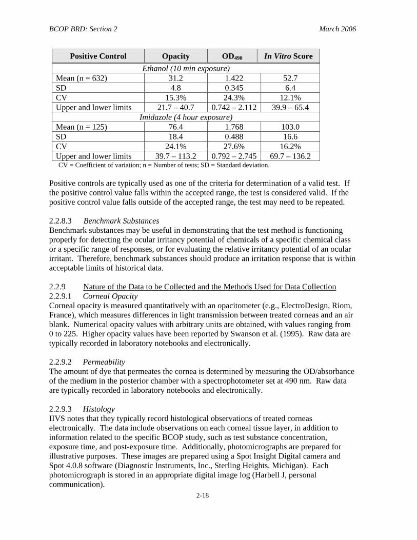
They are useful for evaluating the ocular irritancy potential of unknown chemicals of a specific chemical or product class, or for evaluating the relative irritancy potential of an ocular irritant within a specific range of irritant responses. 2.2.8 Acceptable Ranges of Control Responses and the Basis for the Acceptable Ranges 2.2.8.1 Negative/Solvent Controls A majority of BCOP studies reported using negative controls to correct the opacity and permeability values of the treated corneas. No range of acceptable/unacceptable values for the negative control was found. Historically, solvent controls have not been used in the BCOP assay. It would seem appropriate to establish an upper limit of both opacity and permeability for the negative or solvent control. Negative and solvent controls must produce the anticipated response to ensure the test system is functioning properly and that the specific test is valid. 2.2.8.2 Positive Controls In the BCOP studies that used positive controls, the accepted range were typically an In Vitro Irritancy Score that fell within two SDs of the historical mean value for the testing facility. The accepted range is updated every 3 months at IIVS. An example of historical data for positive controls was provided by IIVS (current as of July 22, 2004), as shown in the table below.
BCOP BRD: Section 2 March 2006
2-18
Positive Control Opacity OD490 In Vitro Score Ethanol (10 min exposure)
Mean (n = 632) 31.2 1.422 52.7 SD 4.8 0.345 6.4 CV 15.3% 24.3% 12.1% Upper and lower limits 21.7 – 40.7 0.742 – 2.112 39.9 – 65.4
Imidazole (4 hour exposure) Mean (n = 125) 76.4 1.768 103.0 SD 18.4 0.488 16.6 CV 24.1% 27.6% 16.2% Upper and lower limits 39.7 – 113.2 0.792 – 2.745 69.7 – 136.2 CV = Coefficient of variation; n = Number of tests; SD = Standard deviation.
Positive controls are typically used as one of the criteria for determination of a valid test. If the positive control value falls within the accepted range, the test is considered valid. If the positive control value falls outside of the accepted range, the test may need to be repeated. 2.2.8.3 Benchmark Substances Benchmark substances may be useful in demonstrating that the test method is functioning properly for detecting the ocular irritancy potential of chemicals of a specific chemical class or a specific range of responses, or for evaluating the relative irritancy potential of an ocular irritant. Therefore, benchmark substances should produce an irritation response that is within acceptable limits of historical data. 2.2.9 Nature of the Data to be Collected and the Methods Used for Data Collection 2.2.9.1 Corneal Opacity Corneal opacity is measured quantitatively with an opacitometer (e.g., ElectroDesign, Riom, France), which measures differences in light transmission between treated corneas and an air blank. Numerical opacity values with arbitrary units are obtained, with values ranging from 0 to 225. Higher opacity values have been reported by Swanson et al. (1995). Raw data are typically recorded in laboratory notebooks and electronically. 2.2.9.2 Permeability The amount of dye that permeates the cornea is determined by measuring the OD/absorbance of the medium in the posterior chamber with a spectrophotometer set at 490 nm. Raw data are typically recorded in laboratory notebooks and electronically. 2.2.9.3 Histology IIVS notes that they typically record histological observations of treated corneas electronically. The data include observations on each corneal tissue layer, in addition to information related to the specific BCOP study, such as test substance concentration, exposure time, and post-exposure time. Additionally, photomicrographs are prepared for illustrative purposes. These images are prepared using a Spot Insight Digital camera and Spot 4.0.8 software (Diagnostic Instruments, Inc., Sterling Heights, Michigan). Each photomicrograph is stored in an appropriate digital image log (Harbell J, personal communication).
BCOP BRD: Section 2 March 2006
2-19
Scoring of corneal lesions involves recording the nature, degree, and depth of the lesion observed in each tissue layer. The predominant lesions observed across the individual corneas within a treatment group are noted and serve the basis for the overall evaluation for a treatment group (Harbell J, personal communication).
2.2.10 Type of Media in Which Data Are Stored It can be inferred that studies performed in compliance with GLP guidelines (e.g., Balls et al. 1995; Swanson et al. 1995; Swanson and Harbell 2000; Southee 1998; Bailey et al. 2004) stored the data in a manner suitable for GLP compliant studies. It would seem appropriate that data from the BCOP be stored and archived in a manner consistent with international GLP guidelines (OECD 1998; EPA 2003a, 2003b; FDA 2003). GLP guidelines are nationally and internationally recognized rules designed to produce high-quality laboratory records. These guidelines provide a standardized approach to report and archive laboratory data and records, and information about the test protocol, to ensure the integrity, reliability, and accountability of a study (EPA 2003a,b; FDA 2003). 2.2.11 Measures of Variability Variability in the BCOP assay has been traditionally evaluated by calculating the mean (± SD) for the opacity values and the OD490 values for each treatment group and control group. Calculation of the mean score and SD provides the user with information on the performance of the test method. These values allow for an assessment of the performance of the test conducted and whether the observed variability between replicates is greater than would be considered acceptable. 2.2.12 Statistical or Nonstatistical Methods Used to Analyze the Resulting Data A majority of early BCOP studies used the mean opacity and mean permeability values (OD490) for each treatment group to calculate an in vitro score for each treatment group:
In Vitro Irritancy Score = mean opacity value + (15 x mean OD490 value)
Sina et al. (1995) reported that this formula was derived empirically during in-house and interlaboratory studies. The data generated for a series of 36 compounds in a multilaboratory study were subjected to a multivariate analysis to determine the equation of best fit between in vivo and in vitro data. This analysis was performed by scientists at two separate companies, who derived nearly identical equations. However, Casterton et al. (1996) reported evaluating the opacity and permeability values independently. As experience was gained with the assay and additional chemical and product classes were tested, it was found that some substances can induce significant permeability without an appreciable increase in opacity, and vice versa. For example, the anionic surfactant sodium lauryl sulfate (5%) can destroy the corneal epithelium and produce a high permeability value (OD490 = 2.538) without producing significant opacity (value of 7.7) (Cater et al. 2001). Other anionic and nonionic surfactants (Harbell J, personal communication), as well as some surfactant-based product formulations (Gettings et al. 1996), produce similar results in the BCOP assay. Therefore, while the In Vitro Irritancy Score has been used historically in the BCOP assay to provide a numerical value for comparison of the relative irritancy of test
BCOP BRD: Section 2 March 2006
2-20
substances, this scoring system is not applicable for substances that produce irritation through only one of the two assay endpoints. 2.2.13 Decision Criteria and the Basis for the Prediction Model Used to Classify a Test
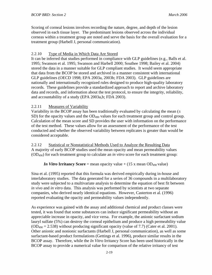
Chemical as a Severe Eye Irritant Once the opacity and OD490 values have been corrected for background opacity and the negative control values, they are entered into the formula for an In Vitro Irritancy Score. In vitro irritancy categories have been historically assigned based on predetermined ranges. The original prediction model was proposed by Gautheron et al. (1994) as follows:
In Vitro Score Range In Vitro Classification 0 - 25 mild irritant
25.1 - 55 moderate irritant 55.1 - 80 severe irritant
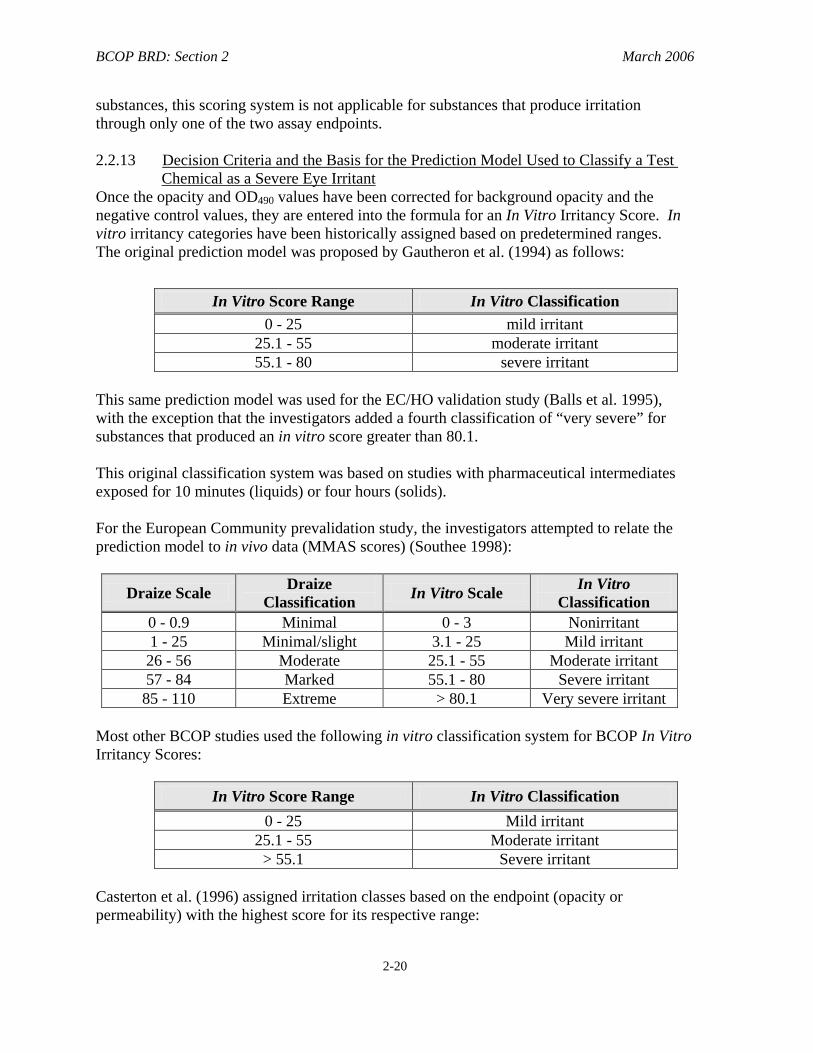
This same prediction model was used for the EC/HO validation study (Balls et al. 1995), with the exception that the investigators added a fourth classification of “very severe” for substances that produced an in vitro score greater than 80.1. This original classification system was based on studies with pharmaceutical intermediates exposed for 10 minutes (liquids) or four hours (solids). For the European Community prevalidation study, the investigators attempted to relate the prediction model to in vivo data (MMAS scores) (Southee 1998):
Draize Scale Draize Classification In Vitro Scale In Vitro
Classification 0 - 0.9 Minimal 0 - 3 Nonirritant 1 - 25 Minimal/slight 3.1 - 25 Mild irritant 26 - 56 Moderate 25.1 - 55 Moderate irritant 57 - 84 Marked 55.1 - 80 Severe irritant 85 - 110 Extreme > 80.1 Very severe irritant
Most other BCOP studies used the following in vitro classification system for BCOP In Vitro Irritancy Scores:
In Vitro Score Range In Vitro Classification 0 - 25 Mild irritant
25.1 - 55 Moderate irritant > 55.1 Severe irritant
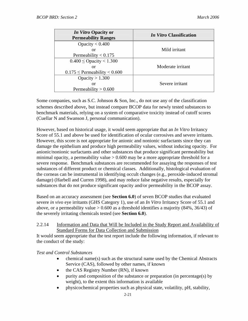
Casterton et al. (1996) assigned irritation classes based on the endpoint (opacity or permeability) with the highest score for its respective range:
BCOP BRD: Section 2 March 2006
2-21
In Vitro Opacity or Permeability Ranges In Vitro Classification
Opacity < 0.400 or
Permeability < 0.175 Mild irritant
0.400 ≤ Opacity < 1.300 or
0.175 ≤ Permeability < 0.600 Moderate irritant
Opacity > 1.300 or
Permeability > 0.600 Severe irritant
Some companies, such as S.C. Johnson & Son, Inc., do not use any of the classification schemes described above, but instead compare BCOP data for newly tested substances to benchmark materials, relying on a system of comparative toxicity instead of cutoff scores (Cuellar N and Swanson J, personal communication). However, based on historical usage, it would seem appropriate that an In Vitro Irritancy Score of 55.1 and above be used for identification of ocular corrosives and severe irritants. However, this score is not appropriate for anionic and nonionic surfactants since they can damage the epithelium and produce high permeability values, without inducing opacity. For anionic/nonionic surfactants and other substances that produce significant permeability but minimal opacity, a permeability value > 0.600 may be a more appropriate threshold for a severe response. Benchmark substances are recommended for assaying the responses of test substances of different product or chemical classes. Additionally, histological evaluation of the corneas can be instrumental in identifying occult changes (e.g., peroxide-induced stromal damage) (Harbell and Curren 1998), and may reduce false negative results, especially for substances that do not produce significant opacity and/or permeability in the BCOP assay. Based on an accuracy assessment (see Section 6.0) of seven BCOP studies that evaluated severe in vivo eye irritants (GHS Category 1), use of an In Vitro Irritancy Score of 55.1 and above, or a permeability value > 0.600 as a threshold identifies a majority (84%, 36/43) of the severely irritating chemicals tested (see Section 6.0). 2.2.14 Information and Data that Will be Included in the Study Report and Availability of
Standard Forms for Data Collection and Submission It would seem appropriate that the test report include the following information, if relevant to the conduct of the study: Test and Control Substances
• chemical name(s) such as the structural name used by the Chemical Abstracts Service (CAS), followed by other names, if known
• the CAS Registry Number (RN), if known • purity and composition of the substance or preparation (in percentage(s) by
weight), to the extent this information is available • physicochemical properties such as physical state, volatility, pH, stability,
BCOP BRD: Section 2 March 2006
2-22
chemical class, water solubility relevant to the conduct of the study • treatment of the test/control substances prior to testing, if applicable (e.g.,
warming, grinding) • stability, if known
Information Concerning the Sponsor and the Test Facility • name and address of the sponsor • name and address of the test facility • name and address of the Study Director
Justification of the Test Method and Protocol Used Test Method Integrity
• the procedure used to ensure the integrity (i.e., accuracy and reliability) of the test method over time (e.g., periodic testing of proficiency substances, use of historical negative and positive control data)
Criteria for an Acceptable Test
• acceptable concurrent negative control ranges based on historical data • acceptable concurrent positive control ranges based on historical data • if applicable, acceptable concurrent benchmark control ranges based on
historical data Test Conditions
• description of test system used • calibration information for measuring device used for measuring opacity and
permeability (e.g., opacitometer and spectrophotometer) • supporting information for the bovine corneas used including statements
regarding their quality • details of test procedure used • test concentration(s) used • description of any modifications of the test procedure • reference to historical data of the model (e.g., negative and positive controls,
proficiency substances, benchmark substances) • description of evaluation criteria used
Results
• tabulation of data from individual test samples (e.g., opacity and OD490 values and calculated in vitro irritancy score for the test substance and the positive, negative, and benchmark controls, reported in tabular form, including data from replicate repeat experiments as appropriate, and means ± SD for each experiment)
Description of Other Effects Observed
BCOP BRD: Section 2 March 2006
2-23
Discussion of the Results Conclusion A Quality Assurance Statement for Good Laboratory Practice (GLP)-Compliant Studies
• This statement indicates all inspections made during the study, and the dates any results were reported to the Study Director. This statement also serves to confirm that the final report reflects the raw data.
Additional reporting requirements for GLP-compliant studies are provided in the relevant guidelines (e.g., OECD 1998; EPA 2003a, 2003b; FDA 2003). 2.3 Basis for Selection of the Test Method System As discussed in Section 1.1.1, the assay developers wanted to develop a cornea-based assay, because the cornea is one of the main targets during accidental eye exposures. In addition, corneal effects are weighted heavily in the original in vivo ocular irritancy scoring systems (e.g., 80 out of a possible 110 points in the Draize eye test scoring system). Opacity in the isolated cornea was initially investigated since it is the only corneal endpoint graded in many in vivo ocular irritancy assays. Studies indicated, however, that some known irritant substances, such as sodium lauryl sulfate and certain medium-length chained alcohols, destroy the corneal epithelium without producing significant opacity. Damage to the epithelium was subsequently quantified for these substances by measuring penetration of the dye sodium fluorescein through the isolated cornea. Gautheron and colleagues refined the assay to measure opacity and permeability, two important components of ocular irritation, and found that the two endpoints predicted the ocular irritancy of a variety of substances (Gautheron et al. 1992, 1994; Sina and Gautheron 1998). Use of the BCOP test method offers some advantages over the traditional in vivo rabbit eye test. Bovine eyes are a relatively inexpensive, abundant by-product of the beef industry. Since the cornea is isolated from animals slaughtered for other purposes, the test method avoids the use of living animals bred specifically for the purpose of toxicity testing. The endpoints of opacity and permeability are measured quantitatively, minimizing the potential variability that could result from subjective evaluations used in the traditional in vivo rabbit eye test. The BCOP test method also allows precise control over the test substance volume, concentration and exposure time, as well as the post-exposure period during which irritation is expressed in the isolated cornea. Thus, different exposure and post-exposure conditions can be readily modeled in this system. Finally, when histology is added to the BCOP assay, a permanent record of the tissue is available. 2.4 Proprietary Components The BCOP assay does not employ any proprietary components.
BCOP BRD: Section 2 March 2006
2-24
2.5 Basis for the Number of Replicate and Repeat Experiments 2.5.1 Sample Replicates The numbers of corneas used to test a substance varied from study to study with three to six corneas used per compound. Early studies using the BCOP test method used six corneas (Gautheron et al. 1992, 1994). In the first interlaboratory study of the BCOP assay, Gautheron et al. (1994) observed that reducing the number of treated corneas to three did not adversely affect the assay results. There appeared to be a close correlation between scores obtained using three and six corneas. The authors concluded that three corneas were likely sufficient to obtain valid results. 2.5.2 Experimental Replicates None of the published reports indicated that repeating experiments is necessary. However, based on sound scientific judgment, it would seem reasonable to expect that equivocal responses or divergent results among test cornea would mandate repeating the experiment. 2.6 Compliance with Good Laboratory Practice Guidelines Southee (1998) reported that the BCOP studies were performed in compliance with GLP guidelines for nonclinical laboratory studies. IIVS also conducts GLP-compliant BCOP assays (e.g., Swanson et al. 1995; Gettings et al. 1996; Swanson and Harbell 2000; Bailey et al. 2004). However, other study reports did not note that the studies were conducted consistent with GLP guidelines. Conducting studies under GLP guidelines increases confidence in the quality and reliability of test data. Furthermore, if data using this test method is to be submitted to the EPA or another agency in response to Federal testing requirements, then compliance with appropriate GLP guidelines will be required. 2.7 Study Acceptance Criteria A test is acceptable if the positive control(s) gives an In Vitro Irritancy Score that falls within two SDs of the current historical mean, which is to be updated every three months. The negative/solvent control responses should result in opacity and permeability values that are less than the laboratory’s established upper limits of opacity and permeability values for bovine corneas treated with the respective negative or solvent control.