Embed Size (px)
Citation preview
Volume 118, Supplement 1, March 2011 BJOGAn International Journal of
Obstetrics and Gynaecology
Saving Mothers’ Lives
Reviewing maternal deaths to make motherhood safer: 2006–2008
March 2011
The Eighth Report of the Confidential Enquiries into Maternal Deaths in the United Kingdom
Centre for Maternal and Child EnquiriesImproving the health of mothers, babies and children
bjos_118_s1_title_sample4.qxp 2/4/2011 12:01 PM Page 1

Centre for Maternal and Child Enquiries MissionStatement
Abstract
In the triennium 2006–2008, 261 women in the UK died directly or indirectly related to pregnancy. The overall maternal
mortality rate was 11.39 per 100,000 maternities. Direct deaths decreased from 6.24 per 100,000 maternities in 2003–2005
to 4.67 per 100,000 maternities in 2006–2008 (p = 0.02). This decline is predominantly due to the reduction in deaths from
thromboembolism and, to a lesser extent, haemorrhage. For the first time there has been a reduction in the inequalities
gap, with a significant decrease in maternal mortality rates among those living in the most deprived areas and those in the
lowest socio-economic group. Despite a decline in the overall UK maternal mortality rate, there has been an increase in
deaths related to genital tract sepsis, particularly from community acquired Group A streptococcal disease. The mortality
rate related to sepsis increased from 0.85 deaths per 100,000 maternities in 2003–2005 to 1.13 deaths in 2006–2008, and
sepsis is now the most common cause of Direct maternal death. Cardiac disease is the most common cause of Indirect
death; the Indirect maternal mortality rate has not changed significantly since 2003–2005. This Confidential Enquiry identi-
fied substandard care in 70% of Direct deaths and 55% of Indirect deaths. Many of the identified avoidable factors remain
the same as those identified in previous Enquiries. Recommendations for improving care have been developed and are
highlighted in this report. Implementing the Top ten recommendations should be prioritised in order to ensure the overall
UK maternal mortality rate continues to decline.
Our aim is to improve the health of mothers, babies and children by carrying out confidential enquires and related
work on a nationwide basis and by widely disseminating our findings and recommendations.
Please cite this work as: Centre for Maternal and Child Enquiries (CMACE). Saving Mothers’ Lives: reviewing maternal
deaths to make motherhood safer: 2006–08. The Eighth Report on Confidential Enquiries into Maternal Deaths in the Uni-
ted Kingdom. BJOG 2011;118(Suppl. 1):1–203.
This work was undertaken by the Centre for Maternal and Child Enquries (CMACE). The work was funded by the
National Patient Safety Agency, the Scottish Programme for Clinical Effectiveness in Reproductive Health, by the Depart-
ment of Health, Social Services and Public Safety of Northern Ireland and the States of Jersey and Guernsey, and Isle of
Man. The views expressed in this publication are those of the Enquiry and not necessarily those of its funding bodies.
Ireland joined the Enquiry in January 2009, at the commencement of the 2009–11 triennium, and its contribution will be
included in the Saving Mothers’ Lives report for that triennium. The Irish office is located at the National Perinatal Epide-
miology Centre, Cork University Maternity Hospital, Cork.
All rights reserved. No part of this publication may be reproduced, stored or transmitted in any form or by any means,
without the prior written permission of CMACE, or in the case of reprographic reproduction, in accordance with the terms
of licences issued by the Copyright Licensing Agency in the UK (www.cla.co.uk). Enquiries concerning reproduction outside
the terms stated here should be sent to CMACE at the address printed on this page.
Making duplicate copies of this Report for legitimate clinical or other noncommercial purposes with the UK NHS is permit-
ted provided the CMACE is identified as the originator of the information. Making alterations to any of the information con-
tained within, or using the information in any other work or publication without prior permission, will be a direct breach of
copyright and may result in civil action.
The use of registered names, trademarks, etc., in this publication does not imply, even in the absence of a specific state-
ment, that such names are exempt from the relevant laws and regulation and therefore free for general use.
Product liability: CMACE can give no guarantee for information about drug dosage and application thereof contained in this
guideline. In every individual case the respective user must check its accuracy by consulting other pharmaceutical literature.
Published March 2011
CMACE, Chiltern Court, 188 Baker Street, London, NW1 5SD, UK
Tel.: + 44 207 486 1191 Fax: + 44 207 486 6226
Email: [email protected] Website: www.cmace.org.uk
Acknowledgements
ª 2011 Centre for Maternal and Child Enquiries (CMACE), BJOG 118 (Suppl. 1), 1–203 1

Saving Mothers’ Lives: Reviewing maternal deaths to make motherhoodsafer—2006–08
The Eighth Report of the Confidential Enquiries into Maternal Deaths in the United Kingdom
Director and Editor
Gwyneth Lewis OBE MSc MRCGP FFPH FRCOG FACOG DSc
Central Assessors and Authors
Roch Cantwell FRCPsych
Thomas Clutton-Brock FRCP FRCA
Griselda Cooper OBE FRCA FRCOG
Andrew Dawson MD FRCOG
James Drife MD FRCOG FRCP (Ed) FRCS (Ed) FCOG (SA) FFSRH
Debbie Garrod RM, DPSM, BA, MA, PGCE
Ann Harper OBE MD FRCOG FRCPI FFSRH
Diana Hulbert FRCS FCEM
Sebastian Lucas FRCP FRCPath
John McClure FRCA
Harry Millward-Sadler FRCPath, MHSM
James Neilson MD FRCOG
Catherine Nelson-Piercy FRCP FRCOG
Jane Norman MD FRCOG
Colm O’Herlihy MD FRCPI FRCOG FRANZCOG
Margaret Oates OBE FRCPsych FRCOG
Judy Shakespeare MRCP FRCGP
Michael de Swiet MD FRCP FRCOG
Catherine Williamson MD FRCP
Other authors and contributors
Valerie Beale RN RM Dip Man MSc
Marian Knight MPH DPhil FFPH
Christopher Lennox FRCOG
Alison Miller RN RM RDM
Dharmishta Parmar BA Hons
Jane Rogers BA PhD DPSM SRN RM
Anna Springett BSc MSc
Acknowledgements
2 ª 2011 Centre for Maternal and Child Enquiries (CMACE), BJOG 118 (Suppl. 1), 1–203
Contents
Acknowledgements
Foreword
Top ten recommendations
Back to Basics Margaret Oates, Ann Harper, Judy Shakespeare and Catherine Nelson-Piercy
Aims, objectives and definitions used in this report Gwyneth Lewis
Key Findings for 2006–2008
1. The women who died 2006–2008 Gwyneth Lewis
Maternal deaths Directly related to pregnancy
2. Thrombosis and thromboembolism James Drife
3. Pre-eclampsia and eclampsia James Neilson
4. Haemorrhage Jane Norman
5. Amniotic fluid embolism Andrew Dawson
6. Deaths in early pregnancy Colm O’Herlihy
7. Sepsis Ann Harper
Annex 7.1: A possible future approach to case definitions Sebastian Lucas
8. Anaesthesia John McClure and Griselda Cooper
Maternal deaths Indirectly related to pregnancy
9. Cardiac disease Catherine Nelson-Piercy
Annex 9.1. Pathological overview of cardiac deaths including sudden adult/arrhythmic death syndrome (SADS) Sebas-
tian Lucas
10. Other Indirect deaths Michael de Swiet, Catherine Williamson and Gwyneth Lewis
11. Deaths from psychiatric cause Margaret Oates and Roch Cantwell
Deaths apparently unrelated to pregnancy
12. Deaths apparently unrelated to pregnancy from Coincidental and Late causes including domestic abuse Gwyneth
Lewis
Annex 12.1: Domestic abuse
Key Issues and lessons for specific health service practice, organisation and/or health professionals
13. Midwifery Debbie Garrod, Valerie Beale and Jane Rogers
14. General Practice Judy Shakespeare
15. Emergency medicine Diane Hulbert
16. Critical Care Tom Clutton-Brock
17. Pathology overview Sebastian Lucas and Harry Millward Sadler
17.1 The main clinico-pathologies encountered at autopsy in maternal death and specific pathological scenarios
(Adapted from Royal College of Pathologists: Guidelines on Autopsy Practice. Scenario 5: Maternal Death. May
2010.)
Appendices
Appendix 1: The method of Enquiry
Appendix 2A: Summary of United Kingdom Obstetric Surveillance System (UKOSS) Report on near miss studies
Appendix 2B: Summary of Scottish Confidential Audit of Severe Maternal Morbidity Report 2008
Appendix 3: Contributors to the Maternal Death Enquiry for triennium 2006 08 and CMACE personnel
Appendix 4: CMACE Governance
Contents
ª 2011 Centre for Maternal and Child Enquiries (CMACE), BJOG 118 (Suppl. 1), 1–203 3
AcknowledgementsCMACE wishes to thank all the healthcare professionals and staff who assisted with the individual cases and who have con-
tributed their time and expertise and without whom this report would not have been possible. With their help this Enquiry
remains an outstanding example of professional self-audit, and will continue to improve the care provided to pregnant and
recently delivered women and their families.
In particular, thanks are due to:
• Professor Gwyneth Lewis, Director of the Maternal Death Enquiry and Editor of Saving Mothers’ Lives and her PA,
Charlene Bruneau.
• All Central Authors and Assessors, and other authors and contributors. Particular thanks go to several retiring
Central Authors and Assessors after many years of dedicated hard work and passion. These are Dr Griselda Coo-
per, Professor Michael de Swiet, Professor James Drife, Dr John McClure, Dr Harry Millward-Sadler and Dr Mar-
garet Oates.
• All the Regional Assessors (listed in Appendix 3).
• The Office of National Statistics.
• All CMACE regional staff for liaising with local clinicians and managing the data collection process and all staff at
Central Office involved in the work of the enquiry (listed in Appendix 3).
• Shona Golightly, Dr Kate Fitzsimons, Rachael Davey and James Hammond for help and assistance in the publication
of this report.
• Professor Oona Campbell, Department of Epidemiology and Reproductive Health, London School of Hygiene and
Tropical Medicine; Ms Mervi Jokinen, Practice and Standards Development Adviser, Royal College of Midwives; and
Miss Sara Paterson Brown, Consultant Obstetrician and Gynaecologist, Queen Charlotte’s Hospital, Imperial NHS
Trust, London, for providing external review to this Report.
Acknowledgements
4 ª 2011 Centre for Maternal and Child Enquiries (CMACE), BJOG 118 (Suppl. 1), 1–203
Foreword
The death of a mother, a young woman who had hopes
and dreams for a happy future but who dies before her
time, is one of the cruellest events imaginable. The short
and long-term impact of such a tragedy on her surviving
children, partner, wider family, the community and the
health workers who cared for her cannot be overestimated.
Yet despite considerable advances in maternity care, and
world-class care provided by highly trained and motivated
professionals, good maternal health is still not a universal
right, even in countries such as ours which have high-qual-
ity maternity services and very low maternal mortality and
morbidity rates.
This is one the most important reports published dur-
ing the unbroken, nearly 60-year history of the Confiden-
tial Enquiries into Maternal Deaths. It shows for the first
time in many years, a small but very welcome decline in
the overall maternal mortality as well as larger reductions
in deaths from some clinical causes. It is difficult to
ignore the apparent relationship between the significant
decline in deaths from pulmonary embolism, and to a les-
ser degree from other causes except from sepsis, and the
publication and implementation of clinical guidelines
which have been recommended in previous Enquiry
reports.
Perhaps more welcome, in terms of the overall public
health, are the first signs of a narrowing in the long-stand-
ing gap relating to pregnancy outcomes between the more
comfortable and most deprived women in our population.
This includes a significant reduction in the death rate
among Black African mothers. These improvements dem-
onstrate how our maternity services have changed to reach
out and care for a group of vulnerable mothers, many of
whom have sought refuge within our shores and who often
present with medical and social challenges.
The decline in the maternal mortality rate is all the more
impressive for having taken place against a background of
an increasing birth rate, which has sometimes stretched the
maternity services, and a generally older and less healthy
population of mothers. Moreover, the numbers of births to
women born outside the UK have risen, and these mothers
often have more complicated pregnancies, have more seri-
ous underlying medical conditions or may be in poorer
general health. It is also impressive that this reduction in
deaths has occurred at a time when some other developed
countries, such as the USA, are experiencing an increase in
maternal deaths.
These results have been hard won. The enthusiasm and
engagement of our maternity staff for embracing the work
of this Enquiry, and acting on its findings and recommen-
dations, is second to none. The reduction in deaths has
occurred at a time of considerable turbulence and reorgani-
sation in the way maternity services are provided in some
of the constituent countries of the UK. This Enquiry con-
tinues to be truly owned by health professionals who tell us
that they are proud to work in a healthcare system in
which they can participate in, and learn from, such honest
reviews of the worst possible outcomes. It is their commit-
ment that makes this review the envy of maternity workers
in other parts of the world, and why the Enquiry will be
proud to incorporate Ireland in the next Report for 2009–
11. Many other countries, rich and poor, are now starting
similar programmes and are benefitting from advice, prac-
tical help and mentoring by the assessors, particularly the
Director, Professor Gwyneth Lewis OBE, Professor James
Drife and the Centre for Maternal and Child Enquiries
(CMACE) team.
It is vital that this momentum is not lost and that low
mortality rates do not lead to inertia. Experience has taught
us that old messages need repeating, especially as new cad-
res of healthcare workers join the service, and there are
always new and unexpected challenges. These include the
rise in deaths from community-acquired Group A strepto-
coccal sepsis detailed in this report, which led to an earlier
public health alert. The emergence of H1N1 virus infection
will be covered in the next report covering the relevant
time period. In line with new ways of working, new ways
of disseminating the results and recommendations need to
be found. It is essential to include this report as part of the
Continuing Professional Development requirements for all
health professionals who may care for pregnant women,
and we expect the Colleges to develop innovative methods
to enable this to be taken forward.
All of those who contributed to the work of this
Enquiry, especially its assessors and authors, are to be con-
gratulated for developing such a readable and practical
book which, in the best traditions of maternity care, has
ª 2011 Centre for Maternal and Child Enquiries (CMACE), BJOG 118 (Suppl. 1), 1–203 5
Foreword
been written jointly by a multidisciplinary team of mater-
nity professionals. Such partnership is the bedrock of
maternity service provision. Several long-standing, hard-
working and eminent authors are retiring this triennium
and we owe them a huge debt of gratitude for the passion
and commitment they have given to the Enquiry over the
years. Our grateful thanks go to Dr Griselda Cooper OBE,
Professor Michael de Swiet, Professor James Drife, Dr John
McClure, Dr Harry Millward-Sadler and Dr Margaret Oates
OBE.
We commend this report to all health-service commis-
sioners and professionals as well as to those with a general
interest in pregnancy and birth. Learning and acting on the
important messages contained within each chapter will lead
to continuing improvements in the prevention and man-
agement of life-threatening complications of pregnancy. By
doing so we shall ensure that for every mother, pregnancy,
birth and the start of a new life are as healthy and happy
as possible.
Disclosure of interestProfessor Dame Sally C Davis, Dr Michael McBride, Dr
Tony Holohan, Dr Tony Jewell and Dr Harry Burns have
no competing interests to disclose.
Professor Dame Sally C Davies
Chief Medical Officer (Interim)—England
Dr Michael McBride
Chief Medical Officer—Northern Ireland
Dr Tony Holohan
Chief Medical Officer—Republic of Ireland
Dr Tony Jewell
Chief Medical Officer—Wales
Harry Burns
Dr Harry Burns
Chief Medical Officer—Scotland
Foreword
6 ª 2011 Centre for Maternal and Child Enquiries (CMACE), BJOG 118 (Suppl. 1), 1–203
‘Top ten’ recommendations
Keywords recommendations, Confidential Enquiry, maternal,
mortality.
The overwhelming strength of successive Enquiry Reports
has been the impact their findings have had on maternal
and newborn health in the UK and further afield. Over the
years there have been many impressive examples of how
the implementation of their recommendations and guide-
lines have improved policies, procedures and practice and
saved the lives of more mothers and babies. The encourag-
ing results given in this Report, in particular the reduction
in deaths from Direct causes, especially thromboembolism,
as well as among some minority ethnic groups, suggest that
previous recommendations have had a positive effect.
Another example is the increasing number of women
‘booking’ for maternity care by 12 completed weeks of ges-
tation, a key recommendation in earlier Reports and which
was chosen to be a cornerstone of maternity-care provision
in England. However, in other areas, improvements remain
to be seen, and therefore some recommendations from the
last Report are repeated here.
Arriving at the ‘Top ten’
Over time, as the evidence base for clinical interventions
has grown, and with the expansion of the Enquiry into
other professional areas and the wider social and public-
health determinants of maternal health, the number of rec-
ommendations made in this Report has increased.
Although these recommendations are important, the
increasing numbers make it difficult for commissioners and
service providers, in particular at hospital or Trust level, to
identify those areas that require action as a top priority.
Therefore, to ensure that the key overarching issues are not
lost, this Report, as with the last Report for 2003–05, con-
tains a list of the ‘Top ten’ recommendations which all
commissioners, providers, policy-makers, clinicians and
other stakeholders involved in providing maternity services
should plan to introduce, and audit, as soon as possible.
By their overarching or cross-cutting nature, most of these
recommendations are broad based and will require a multi-
disciplinary approach rather than having relevance for the
specific clinical practice of individual healthcare workers.
On an individual and team basis, therefore, all healthcare
professionals and teams providing maternity care should
also read the individual clinical recommendations relating
to specific clinical causes of death or their individual speci-
ality as well as these overarching ones.
These overarching recommendations were drawn up fol-
lowing detailed discussions between all of the assessors
involved in this Report. In some cases, they considered that
insufficient progress has been made since the last Report and
that a similar recommendation needs to be repeated here.
This list adds to, but does not replace, key recommenda-
tions made in earlier Reports.
Baseline data and audit of progress
All changes and interventions need to be monitored and
the outcome or impact must be audited to ensure that they
are resulting in beneficial changes to the quality of care or
services provided to pregnant or recently delivered women.
If not then remediable action to improve the outcomes can
be taken. It is recognised that the data needed to audit
these recommendations may not be currently available or
collected routinely in all units, but it could form part of a
future local audit or dataset. National data sets are cur-
rently being developed and it may be possible to incorpo-
rate these in future Reports.
Learning from specific individualChapter recommendations
Whereas the ‘Top ten’ recommendations are mainly of gen-
eral importance, the individual Chapters in this Report
contain more targeted recommendations for the identifica-
tion and management of particular conditions for specific
services or professional groups. These are no less important
and should be addressed by any relevant national bodies as
well as by local service commissioners, providers and indi-
vidual healthcare staff.
Top ten recommendations
These are not in any order of priority.
‘Top ten’ recommendations
ª 2011 Centre for Maternal and Child Enquiries (CMACE), BJOG 118 (Suppl. 1), 1–203 7
Service provision
Recommendation 1: Pre-pregnancycounselling
1.1 Women of childbearing age with pre-existing medi-
cal illness, including psychiatric conditions, whose con-
ditions may require a change of medication, worsen or
otherwise impact on a pregnancy, should be informed
of this at every opportunity. This is particularly impor-
tant since 50% of pregnancies are not planned. They
should be pro-actively offered advice about planning for
pregnancy and the need to seek pre-pregnancy counsel-
ling whenever possible. Prior to pregnancy, these
women should be offered specific counselling and have
a prospective plan for the management of their preg-
nancy developed by clinicians with knowledge of how
their condition and pregnancy interact.
1.2 Pre-pregnancy counselling services, starting for
women with pre-existing medical illnesses, but ideally
for all women planning a pregnancy, are a key part of
maternity services and should be routinely commis-
sioned as an integral part of the local maternity services
network. They could be provided by the GP practice,
specialist midwives or other specialist clinicians or
obstetricians, all of whom should be suitably trained
and informed. General practitioners should refer all
relevant women to the local services if they do not pro-
vide such counselling themselves.
Rationale
As in previous Reports, the findings of this triennium
show that many of the women who died from pre-exist-
ing diseases or conditions that may seriously affect the
outcome of their pregnancies, or that may require differ-
ent management or specialised services during pregnancy,
did not receive any pre-pregnancy counselling or advice.
As a result, their care was less than optimal because nei-
ther they nor their carers realised that closer surveillance
or changes to medications were appropriate. Furthermore,
unless women receive specific counselling that their drugs
are safe in pregnancy, some will stop taking essential
therapy because of their concerns about the risk to the
fetus.
The more common conditions that require pre-preg-
nancy counselling and advice include:
• epilepsy
• diabetes
• asthma
• congenital or known acquired cardiac disease
• autoimmune disorders
• renal or liver disease
• obesity: a body mass index of 30 or more
• severe pre-existing or past mental illness
• HIV infection.
Baselines and auditable standards
Maternity service commissioners and maternity services:
• Number and percentage of pregnant women with pre-
existing medical conditions for whom specialist pre-
conception counselling is offered at December 2011
and then by the end of 2013. A national maternity
record may enable such information to be included and
easier to identify.
Recommendation 2: Professionalinterpretation services
Professional interpretation services should be provided
for all pregnant women who do not speak English.
These women require access to independent interpreta-
tion services, as they continue to be ill-served by the
use of close family members or members of their own
local community as interpreters. The presence of rela-
tives, or others with whom they interact socially, inhi-
bits the free two-way passage of crucial but sensitive
information, particularly about their past medical or
reproductive health history, intimate concerns and
domestic abuse.
Rationale
Although it is known that where there is a concentration of
women from the same minority ethnic group their infor-
mation network concerning maternity care can be good,
this does not obviate the need for professional interpreting
services. A lack of availability of suitable interpreters is one
of the key findings running throughout this Report. The
use of family members, in some cases very young school-
age children of both sexes, or members of their own, usu-
ally tight-knit, community as translators causes concern
because:
• The woman may be too shy to seek help for intimate
concerns.
• It is not appropriate for a child to translate intimate
details about his or her mother and unfair on both the
woman and child.
• It is not clear how much correct information is con-
veyed to the woman, as the person who is interpreting
may not have a good grasp of the language, does not
understand the specific medical terminology or may
withhold information.
• Some women arrive in the UK late in their pregnancy,
and the absence of an interpreter means that a compre-
hensive booking history cannot be obtained.
‘Top ten’ recommendations
8 ª 2011 Centre for Maternal and Child Enquiries (CMACE), BJOG 118 (Suppl. 1), 1–203
• In some cases, the translator is a perpetrator of domes-
tic abuse against his partner, so the woman is unable to
ask for advice or help.
• Healthcare staff are unable to pass back their own clini-
cal concerns in an appropriate manner.
As a woman said in a recent Department of Health Task
Force Report against domestic and sexual abuse1 ‘even if
the perpetrator isn’t with you, he sends one of his family
members with you. And in the name of honour you can’t
ever talk about it. Especially if they say ‘‘I’m going to inter-
pret because she can’t speak English’’.’
Apart from the unsuitability of using family or commu-
nity members to undertake this role, those used in this
manner appeared to have had little knowledge of English
themselves. Commissioners and providers of maternity
services should therefore ensure that professional and
independent interpretation services are available in both
primary-care and secondary-care settings, to ensure that all
women can be confident that they can speak freely and in
confidence to their maternity-care providers. Telephone-
based services have proved very useful in similar situa-
tions.
Baselines and auditable standards
Maternity service commissioners and maternity services:
• The availability of a local service guideline on care for
women who do not speak English, including interpreta-
tion services.
• As part of a local maternity services needs assess-
ment, a local audit of the numbers and percentages
of pregnant women who require and are using pro-
fessional interpretation services per visit. Baseline
measurements by December 2011 and then by the
end of 2013.
Recommendation 3: Communicationsand referrals
3.1. Referrals to specialist services in pregnancy should
be prioritised as urgent. In some specialties, routine
referrals can take weeks or months, or even be rejected
because of local commissioning rules. This is unaccep-
table for pregnant women. The referral must clearly
state that the woman is pregnant, and its progress
must be followed up. Trainee doctors and midwives
should have a low threshold for referral ‘‘upwards’’
and must receive an immediate response. Referral
between specialties should be at a senior level. When
rapid referral is required, the senior doctor should use
the telephone.
3.2. Good communication among professionals is essen-
tial. This must be recognised by all members of the
team looking after a pregnant woman, whether she is
‘‘low risk’’ or ‘‘high risk’’. Her GP must be told that she
is pregnant. If information is required from another
member of the team, it is not enough to send a routine
request and hope for a reply. The recipient must
respond promptly, and if not, the sender must follow it
up. With a wide variety of communication methods
now available, including e-mail, texting and fax, teams
should be reminded that the telephone is not an obso-
lete instrument.
Rationale
There were a number of cases in this Report of women dying
before they had seen the specialist to whom they had been
referred because of medical problems. Some women received
appointments weeks after the original referral despite clearly
being very ill, but the progress of the referral was not fol-
lowed up. One or two women were also refused specialist
services because of local commissioning arrangements.
In many cases of substandard care assessed by this
Enquiry, there were major failures of communication
between healthcare workers that may have contributed to
the woman’s death in some cases. Notably, these included
GPs not being asked for information or not being con-
sulted about further referral and, in some cases, the GP not
being informed that the woman was pregnant. The con-
verse was also true, with the GP not passing on informa-
tion relevant to the woman’s health and wellbeing.
It is also evident from some of these cases that junior
trainees and midwives in the front line seeing women
attending as emergencies did not have proper support and
back up and need to have clear guidelines about when to
seek senior help. They should not be expected to manage
sick women alone, and if they ask for help and review, they
should be supported. Trainees need to communicate the
gravity and urgency of the situation clearly when discussing
women with consultants, who should ensure that they have
asked enough questions to enable themselves to assess the
situation fully and whether they need to attend in person.
They should also adhere to the recent Royal College of
Obstetricians and Gynaecologists (RCOG) guideline on the
responsibility of the consultant on call, which gives a clear
indication of the duties of a consultant obstetrician and
when they should attend.2
Baselines and auditable standards
Maternity service commissioners and maternity services:
• The number of maternity services with local guidelines
or protocols which have been developed to clarify their
‘Top ten’ recommendations
ª 2011 Centre for Maternal and Child Enquiries (CMACE), BJOG 118 (Suppl. 1), 1–203 9
communications and ‘escalation upwards’ referral
procedures. This includes the number of services that
have adopted the recent RCOG guideline on the
responsibility of the consultant on call.2
• The waiting times before being seen after a woman
has been referred for a specialist opinion and a sys-
tem for ensuring that women are seen with sufficient
urgency.
• As part of a local maternity services needs assessment, a
local audit of the numbers and percentages of pregnant
women who are refused referral to specialist services by
commissioners. Baseline measurements by December
2011 and then by the end of 2013.
Recommendation 4: Women withpotentially serious medicalconditions require immediate andappropriate multidisciplinaryspecialist care
Women with pre-existing disease at the start of preg-
nancy:
4.1 Women whose pregnancies are likely to be compli-
cated by potentially serious underlying pre-existing
medical or mental health conditions should be imme-
diately referred to appropriate specialist centres of
expertise where both care for their medical condition
and their obstetric care can be optimised. Providers
and commissioners should consider developing proto-
cols to specify which medical conditions mandate
at least a consultant review in early pregnancy. This
agreement should take place via local maternity
networks.
Pregnant women who develop potential complications:
4.2. Women whose pregnancies become complicated by
potentially serious medical or mental health conditions
should have an immediate referral to the appropriate
specialist centres of expertise as soon as their symptoms
develop.
4.3. In such urgent cases, referral can take place by
telephone contact with the consultant or their secretary
(to make sure they are available or identify an
alternative consultant if not), followed up by a fax if
necessary.
4.4. Midwives and GPs should be able to refer women
directly to both a obstetrician or a non-obstetric specia-
list - but must inform the obstetrician. The midwife
should, wherever possible, discuss this with, or alert, the
woman’s GP.
Rationale
Medical care is advancing rapidly, as are changes in the way
‘routine’ maternity care is provided in the UK, and women
must not be disadvantaged by this. It must be appreciated
that not all maternity centres are able or equipped to care
for pregnant women with major complications either pre-
ceding or developing in pregnancy. If women with underly-
ing medical conditions are to share in the advances in
medicine, more will require referral to tertiary or specialist
medical centres for their care in pregnancy.
This triennium, the assessors have been struck by the lack
of appropriate referral of potentially high-risk women, and
lack of consultant involvement remains a problem in the care
of women with serious medical problems. The reasons for
failure to refer are likely to be multiple. It may be that the
medical problem is beyond the resources of a secondary refer-
ral centre: for example complex liver disease in pregnancy.
This may require hepatobiliary surgeons, hepatologists and
haematologists skilled in the management of coagulopathy.
It may also be that, although the secondary referral cen-
tre has a ‘specialist’ centre, the clinicians there are insuffi-
ciently skilled in the management of pregnancy in women
with the disease that they specialise in, for example heart
disease. The local clinicians may be excellent at the man-
agement of ischaemic heart disease but not in caring for
congenital heart disease or cardiomyopathy.
It is also possible that the secondary centre may be too
small to develop sufficient expertise in the management of
the disease in question or to set up the combined medical/
obstetric clinics that have been recommended, for example
to care for insulin-dependent diabetes in pregnancy.
Baseline and auditable standards
Maternity service commissioners and maternity services:
Evidence of protocols in place in specialist centres which
specify which pregnant women with pre-existing or new
medical disorders should be referred for consultant obste-
trician assessment: measurement by December 2011 and
then by the end of 2013.
Quality of care
Recommendation 5: Clinical skills andtraining
5.1. Back to basics. All clinical staff must undertake reg-
ular, written, documented and audited training for the
identification and initial management of serious obste-
tric conditions or emerging potential emergencies, such
as sepsis, which need to be distinguished from com-
monplace symptoms in pregnancy.
5.2. All clinical staff must also undertake regular, writ-
ten, documented and audited training for:
‘Top ten’ recommendations
10 ª 2011 Centre for Maternal and Child Enquiries (CMACE), BJOG 118 (Suppl. 1), 1–203
The understanding, identification, initial management
and referral for serious commoner medical and mental
health conditions which, although unrelated to preg-
nancy, may affect pregnant women or recently delivered
mothers. These may include the conditions in recom-
mendation 1, although the list is not exclusive
The early recognition and management of severely ill
pregnant women and impending maternal collapse
The improvement of basic, immediate and advanced life
support skills. A number of courses provide additional
training for staff caring for pregnant women and new-
born babies.
Rationale
A lack of clinical knowledge and skills among some doc-
tors, midwives and other health professionals, senior or
junior, was one of the leading causes of potentially avoid-
able mortality this triennium. One of the commonest
findings in this Report was the initial failure by many
clinical staff, including GPs, Emergency Department staff,
midwives and hospital doctors, to immediately recognise
and act on the signs and symptoms of potentially life-
threatening conditions. To help with this, the assessors
have developed a short new section, Back to basics, which
is included in this Report for the first time. Although not
exhaustive, nor designed to replace more in-depth clinical
training, it does contain useful checklists to act as an
aide memoire. Its contents may appear simplistic or self-
evident to many readers, but it nevertheless reflects the
fact that these basic signs and symptoms were too often
overlooked and may have contributed to some maternal
deaths this triennium.
As with the previous Report, even sick women who were
admitted to specialist care were still failed by a lack of rec-
ognition of the severity of their illness or a failure to refer
for another opinion (see also Recommendation 6).
There is also a need for staff to recognise their
limitations and to know when, how and whom to call for
assistance.
Baseline and auditable standards
The provision of courses and a system for ensuring all staff
attend and complete the training as identified in the Clini-
cal Negligence Scheme for Trusts (CNST) Training Needs
Analysis. This is a level 1 requirement for CNST maternity
services in England. The record of attendees should be reg-
ularly audited to reinforce, familiarise and update all staff
with local procedures, equipment and drugs.
• Number and percentage of members of all cardiac arrest
teams who know where the maternity unit is and who
know the door codes for gaining immediate access to it.
Target 100%.
Recommendation 6: Specialist clinicalcare: identifying and managing verysick women
6.1. There remains an urgent need for the routine use
of a national modified early obstetric warning score
(MEOWS) chart in all pregnant or postpartum women
who become unwell and require either obstetric or
gynaecology services. This will help in the more timely
recognition, treatment and referral of women who have,
or are developing, a critical illness during or after preg-
nancy. It is equally important that these charts are also
used for pregnant or postpartum women who are
unwell and are being cared for outside obstetric and
gynaecology services e.g. Emergency Departments.
Abnormal scores should not just be recorded but should
also trigger an appropriate response.
6.2. The management of pregnant or postpartum women
who present with an acute severe illness, e.g. sepsis with
circulatory failure, pre-eclampsia/eclampsia with severe
arterial hypertension and major haemorrhage, requires a
team approach. Trainees in obstetrics and/or gynaecol-
ogy must request help early from senior medical staff,
including advice and help from anaesthetic and critical
care services. In very acute situations telephoning an
experienced colleague can be very helpful. The recent
RCOG guideline of the duties and responsibilities of
consultant on call should be followed.
6.3 Pregnant or recently delivered women with unex-
plained pain severe enough to require opiate analgesia
require urgent senior assessment/review.
Rationale
As mentioned in the Back to basics recommendation, a
lack of clinical knowledge and skills among some doctors,
midwives and other health professionals, senior or junior,
was one of the leading causes of potentially avoidable
mortality. This was not only the case when distinguishing
the signs and symptoms of potentially serious disease
from the commonplace symptoms of pregnancy in pri-
mary care or the Emergency Department but also once a
woman was admitted to hospital. There were a number
of healthcare professionals who either failed to identify
that a woman was becoming seriously ill or who failed to
manage emergency situations outside their immediate area
of expertise, and did not call for advice and help.
In many cases in this Report, and relevant to the issues
identified in the preceding paragraph, the early warning
signs of impending maternal collapse went unrecognised.
The early detection of severe illness in mothers remains a
challenge to all involved in their care. The relative rarity
‘Top ten’ recommendations
ª 2011 Centre for Maternal and Child Enquiries (CMACE), BJOG 118 (Suppl. 1), 1–203 11
of such events, combined with the normal changes in
physiology associated with pregnancy and childbirth,
compounds the problem. Modified early warning scoring
systems have been successfully introduced into other areas
of clinical practice, and the last Report gave an example
of a MEOWS chart. This is available on the CMACE
website at www.cmace.org.uk. These charts should be
introduced for all pregnant or postpartum women who
become unwell and require further treatment, including
following obstetric interventions and gynaecological sur-
gery.
A small but important point is that a recurrent theme
and recommendation throughout successive Reports, which
has made no impact, is that women who have unexplained
pain severe enough to require opiate analgesia have a
severe problem and must be referred for specialist investi-
gation and diagnosis. Women with cardiac disease,
impending aortic dissection and other causes of death were
missed in this way.
CNST and similar schemes in other UK countries may
wish to consider whether the use of MEOWS charts should
be part of the audit of notes carried out as part of the
assessment process.
Baseline and auditable standards
The number of maternity services who have adopted a ver-
sion of any existing MEOWS charts and trained all staff in
its use. Baseline measurement by December 2011 and then
by the end of 2013.
• The number of women in hospital following caesarean
section who had regular postoperative observations
taken and recorded on a MEOWS chart and had appro-
priate action taken when variances occurred. This could
be part of the suggested CNST, or similar, audit of notes.
Recommendation 7: Systolichypertension requires treatment
7.1 All pregnant women with pre-eclampsia and a systo-
lic blood pressure of 150–160 mmHg or more require
urgent and effective anti-hypertensive treatment in line
with the recent guidelines from the National Institute
for Health and Clinical Excellence (NICE)3. Considera-
tion should also be given to initiating treatment at
lower pressures if the overall clinical picture suggests
rapid deterioration and/or where the development of
severe hypertension can be anticipated. The target systo-
lic blood pressure after treatment is 150 mmHg.
Rationale
It is disappointing that in this triennium, as flagged up in the
last, the single most serious failing in the clinical care provided
for mothers with pre-eclampsia was the inadequate treatment
of their systolic hypertension. In several women, this resulted
in a fatal intracranial haemorrhage. Systolic hypertension was
also a key factor in most of the deaths from aortic dissection.
The last Report suggested that clinical guidelines should iden-
tify a systolic pressure above which urgent and effective anti-
hypertensive treatment is required. Since then, a recent NICE
guideline has identified that threshold as being 150–
160 mmHg.3 The guideline also recommends that pregnant
women with pre-eclampsia and a systolic blood pressure of
150 mmHg or more should be admitted to hospital for urgent
treatment. Clinically, it is also important to recognise
increases in, as well as the absolute values of, systolic blood
pressure. In severe and rapidly worsening pre-eclampsia, early
treatment at <150–160 mmHg is advisable if the trend sug-
gests that severe hypertension is likely.
Auditable standards
Specific local projects should be devised to audit manage-
ment and treatment of severe pre-eclampsia. For example,
one suggestion is to audit the number and proportion of
women with very severe pre-eclampsia (a systolic blood pres-
sure of 180 mmHg or more on two or more occasions) and
then who had a systolic pressure of 150 mmHg or less within
2 hours of starting antihypertensive treatment.
Recommendation 8: Genital tractinfection/sepsis
8.1 All pregnant and recently delivered women need to be
informed of the risks and signs and symptoms of genital
tract infection and how to prevent its transmission.
Advice to all women should include verbal and written
information about its prevention, signs and symptoms
and the need to seek advice early if concerned, as well as
the importance of good personal hygiene. This includes
avoiding contamination of the perineum by washing
hands before and after using the lavatory or changing
sanitary towels. It is especially necessary when the woman
or her family or close contacts have a sore throat or upper
respiratory tract infection.
8.2. All health care professionals who care for pregnant
and recently delivered women should adhere to local
infection control protocols and be aware of the signs and
symptoms of sepsis in the women they care for and the
need for urgent assessment and treatment. This is parti-
cularly the case for community midwives, who may be
the first to pick up any potentially abnormal signs during
their routine postnatal observations for all women, not
just those who have had a caesarean section. If puerperal
infection is suspected, the woman must be referred back
to the obstetric services as soon as possible.
‘Top ten’ recommendations
12 ª 2011 Centre for Maternal and Child Enquiries (CMACE), BJOG 118 (Suppl. 1), 1–203
8.3 High dose intravenous broad-spectrum antibiotic
therapy should be started as early as possible, as
immediate antibiotic treatment may be life saving. It
should be started within the first hour of recognition of
septic shock and severe sepsis without septic shock, as
each hour of delay in achieving administration of effec-
tive antibiotics is associated with a measurable increase
in mortality4,5.
8.4 There is an urgent need for a national clinical guide-
line to cover the identification and management of sep-
sis in pregnancy, labour and the postnatal period and
beyond. This should be available to all health profes-
sionals, maternity units, Emergency Departments, GPs
and Community Midwives. Until such time as a
national guideline is developed, the principles for the
management of acute sepsis as detailed in Chapter 16:
Critical Care of this Report should be adopted. These
are derived from those developed and updated by the
Surviving Sepsis Campaign4.
8.5 Consideration should be given to adopting a more
rational system for classifying maternal deaths from
sepsis, as suggested in Annex 7.1 in this Report.
Rationale
Unlike many other causes of direct maternal mortality,
deaths from genital tract sepsis have risen rather than
declined this triennium. Indeed, genital tract sepsis has
become the leading cause of Direct maternal death in the
UK for the first time since these Confidential Enquiries
into Maternal Deaths commenced in 1952. This is a real
cause for concern, particularly as it has occurred against a
background of an overall decrease in maternal mortality.
Many of these deaths were from community-acquired
Group A streptococcal disease, which mirrors the increased
incidence of Streptococcus A in the general population.
Although for some women, despite excellent care, the out-
come was unavoidable because of the rapid course and late
presentation of their illness, in others, possible opportuni-
ties to save lives were missed. The number of maternal
deaths from sepsis should be reduced still further.
Streptococcal sore throat is one of the most common bac-
terial infections of childhood. All of the mothers who died
from Group A streptococcal sepsis either worked with, or
had, young children. Several mothers had a history of recent
sore throat or respiratory infection, and some of these women
also had family members, especially children, with sore
throats, suggesting that spread from family members is a
further risk factor for developing life-threatening sepsis.
Therefore, all pregnant or recently delivered women need to
be advised of the signs and symptoms of infection and how
to take steps to prevent its transmisson. Women in these
circumstances should also be encouraged to seek urgent med-
ical advice from their GP or maternity services if they feel at
all ill.
As in previous Reports, delays in recognising sepsis, pre-
scribing antibiotics and seeking consultant help were com-
mon. Antibiotics were sometimes prescribed in inadequate
doses, were given orally rather than intravenously, were
given too late or were discontinued too soon. Immediate
aggressive treatment in the first ‘golden hour’ or so offers
the best hope of recovery, as each hour of delay in achiev-
ing administration of effective antibiotics is associated with
a measurable increase in mortality.5
Sepsis is complex, incompletely understood, often diffi-
cult to recognise and manage, and presents a continuing
challenge. Some deaths will always be unavoidable, but bet-
ter training, a structured approach, good care in the com-
munity, and, in hospital, prompt investigation and
treatment, particularly immediate intravenous antibiotic
treatment and early involvement of senior obstetricians, an-
aesthetists and critical-care consultants, may help in future
to save lives.
Auditable standards
The percentage of women who had an infection and who
had antibiotic therapy started within an hour of presumed
diagnosis. Target 100% within 1 hour.
• Until a national guideline is available, all maternity ser-
vices should use existing guidelines to develop their
own clinical guideline for the identification and man-
agement of pregnancy-related sepsis by the end of
December 2011. See Chapter 7.
• The number and percentage of women who receive
routine antibiotic prophylaxis in accordance with
recognised RCOG and NICE guidelines for induced
abortion, premature rupture of membranes, caesarean
section and following obstetric anal sphincter repair.
See Chapter 7.
• The frequency and completeness of routine postnatal
checks in the community compared with NICE Guide-
line on Postnatal Care.
Clinical governance
Recommendation 9: Serious IncidentReporting and Maternal Deaths
All maternal deaths must be subject to a high quality
local review. In England and Wales the framework for
such serious incidents (previously known as Serious
Untoward Incidents/SUIs) is set out in the NPSA’s
‘‘National Framework for Reporting and Learning from
Serious Incidents Requiring Investigation’’ issued in
‘Top ten’ recommendations
ª 2011 Centre for Maternal and Child Enquiries (CMACE), BJOG 118 (Suppl. 1), 1–203 13
March 2010. The results of such high quality reviews
must be disseminated and discussed with all maternity
staff and their recommendations implemented and
audited at regular intervals.
Rationale
The quality of the serious incident/SUI report forms relating
to maternal deaths assessed for this report was highly vari-
able, with many of dubious or poor quality. These findings
must be taken extremely seriously. For the unacceptable
reports, there was little or no evidence of critical thinking or
acceptance of shortcomings, little or no self-reflective discus-
sion and no evidence that obvious lessons had been identi-
fied, let alone learnt. In these cases, little or no action was
taken on any results, and, in many cases, staff were not
involved in the process or the follow up of any of the lessons
learnt. This was a common finding throughout all the chap-
ters in this Report and one which all assessors agree repre-
sents unacceptable practice that must be corrected as soon as
possible. The evaluation of such reports is recommended to
be a CNST, or similar, requirement in future.
Disappointingly, a similar recommendation was made in
the last Report, but little if any improvement has been seen
in the cases assessed this triennium; the impression is
rather the reverse.
The recommendation drew attention to the need to
highlight who was involved in such reviews—they needed
to include clinicians from relevant disciplines (including
anaesthetics) and must include clinicians who were not
involved with the death. Considering unbiased expert
review might assist real learning from individual deaths.
Auditable standards
Every maternal death should be critically reviewed as a seri-
ous incident and the lessons learnt should be actively dis-
seminated to all clinical staff, risk managers and
administrators. The precise educational and organisational
actions taken as a result must be recorded, audited and
regularly reported to the maternity services board by the
Chief Executive Officer.
Recommendation 10: Pathology
The standard of the maternal autopsy must be
improved. The numbers of locations where they are per-
formed should reduce, with specialist pathologists taking
them on as part of agreed job plans. More clinical dis-
cretion over reporting maternal deaths to coroners is
required, and there should be a complementary major
input by clinicians into obtaining more consented hos-
pital autopsies.
Rationale
Autopsy diagnoses are fundamental in the categorisation of
maternal deaths and their subsequent reviews, locally and
nationally, but, as evidenced by the findings in this Report,
this was often difficult to achieve. With the changing legis-
lation (Coroner and Justice Act 2009) and the introduction
of Medical Examiners to scrutinise all proffered death cer-
tificates, there will be a reduction in the number of coroni-
al autopsies in England and Wales. Also, when coroners do
authorise maternal death autopsies, there will be no legal
bar to transferring the autopsy away to another area where
pathological expertise is recognised.
Maternal death autopsies are often complex and chal-
lenging and require more expertise than the average
autopsy. They should also be performed according to an
all-embracing protocol, such as that adapted from the
Royal College of Pathologists recent guidance, which is
annexed to Chapter 17.6
Baseline and auditable standards
• A review of the quality of maternal autopsies performed
and their proportion of all maternal deaths. Baseline mea-
surement by December 2011 and then by end of 2013.
• The proportion of maternal death autopsies that are
consented versus coronial.
National guidelines and research
As discussed above, national guidelines are urgently
required for
• The identification and management of sepsis in preg-
nant and recently delivered women.
• How to undertake and act on the results of serious
incident reviews in maternity services.
Some key research questions also emerged during the
assessment of the women who died this triennium.
• What effect, if any, is the reduced number of routine
postnatal visits and clinical observations having on mater-
nal health?
What are the social, service and clinical factors that con-
tribute to maternal mortality rates remaining higher in
mothers from certain minority ethnic groups in the UK?
What are the barriers that hinder prompt and rapid
communications, referrals and urgent appointments
between health providers and maternity or specialist units
or health professionals that are required for sick pregnant
women or recently delivered mothers, and how can these
be overcome? How can maternity services, especially those
providing new models of care, ensure that working systems
for immediate referral are in place and implemented?
• To better determine how to reduce the number of Indi-
rect causes of maternal deaths, how can the better denom-
inator data required to undertake the necessary studies
‘Top ten’ recommendations
14 ª 2011 Centre for Maternal and Child Enquiries (CMACE), BJOG 118 (Suppl. 1), 1–203
regarding the incidence of medical problems in preg-
nancy, such as sepsis, asthma, epilepsy and stroke, be
obtained or collected?
• Why has the incidence of sepsis and Sudden Unex-
pected Adult/Arrhythmic deaths (SADS) in pregnancy
increased? Is it the result of chance, improved case ascer-
tainment or a real increase?
Disclosure of interestNone.
FundingThis work was undertaken by the Centre for Maternal and
Child Enquiries (CMACE) as part of the CEMACH pro-
gramme. The work was funded by the National Patient
Safety Agency; the Department of Health, Social Services
and Public Safety of Northern Ireland; NHS Quality
Improvement Scotland (NHS QIS); and the Channel
Islands and Isle of Man. j
References
1 The Task Force on the health aspects of violence against women and
children. Responding to Violence Against Women and Children-The
Role of The NHS. London: Department of Health, March 2010.
2 Royal College of Obstetricians and Gynaecologists. Responsibility of
Consultant on-Call. Good Practice No. 8. London: RCOG. March 2010
[www.rcog.org.uk]. Accessed 5 October 2010.
3 National Collaborating Centre for Women’s and Children’s Health.
Hypertension in pregnancy: the management of hypertensive disor-
ders during pregnancy. National Institute for Health and Clinical Excel-
lence Guideline 107. London: RCOG, August 2010 [http://
guidance.nice.org.uk/CG107/]. Accessed 5 October 2010.
4 Dellinger RP, Levy MM, Carlet JM, Bion J, Parker MM, Carlet JM,
et al. Surviving Sepsis Campaign: international guidelines for manage-
ment of severe sepsis and septic shock. Crit Care Med 2008;36:296–
327.
5 Royal College of Pathologists. Guidelines on Autopsy Practice. Sce-
nario 5: Maternal Death. London: Royal College of Pathologists;
2010.
ª 2011 Centre for Maternal and Child Enquiries (CMACE), BJOG 118 (Suppl. 1), 1–203 15
‘Top ten’ recommendations
Back to basicsM Oates1, A Harper2, J Shakespeare3, C Nelson-Piercy4
1 East Midlands Perintal Mental Health Clinical Network, Nottinghamshire Healthcare NHS Trust, Nottingham, UK; 2 Royal Jubilee Maternity
Services, Royal Maternity Hospital, Belfast, UK; 3 Summertown Health Centre, Oxford, UK; 4 Guy’s & St Thomas’ Foundation Trust and
Imperial College Healthcare Trust, Women’s Health Directorate, London, UK
Correspondence: Margaret R Oates, East Midlands Perintal Mental Health Clinical Network, Nottinghamshire Healthcare NHS Trust, Duncan
MacMillan House, Porchester Road, Nottingham NG3 6AA, UK. Email: [email protected]
Keywords recommendations, Confidential Enquiry, maternal,
mortality.
Introduction
Several common themes that need to be recognised by all
professionals providing maternity care have emerged from
all the Chapters of this Report. To aid learning and clinical
practice, some key overall good practice points have been
brought together in this new section of the Report. The les-
sons fall into the following main categories:
• improving basic medical and midwifery practice, such
as taking a history, undertaking basic observations and
understanding normality
• attributing signs and symptoms of emerging serious
illness to commonplace symptoms in pregnancy
• improving communication and referrals.
This aide memoire does not cover every eventuality and
should be taken as a signpost to help identify and exclude
the commoner disorders of pregnancy. It is not, however,
exclusive, nor does it replace the need for all health profes-
sionals to be up to date with their clinical practice and follow
the relevant clinical guidelines. An in-depth discussion of all
of these issues as they relate to specific causes of death can be
found in the individual chapters of Report, which should be
read in conjunction with this aide memoire.
Common symptoms
PyrexiaAlthough still very uncommon, deaths from sepsis, especially
community-acquired streptococcal Group A, have increased
over the last 10 years. In part, this mirrors the increased inci-
dence of strep A in the general population, but, whereas
some maternal deaths from sepsis are unavoidable, others
could still be avoided by earlier identification and treatment.
Becoming life-threateningly ill from sepsis, and streptococcal
sepsis in particular, shows the speed with which women can
become sick in pregnancy, sometimes dying within 12–
24 hours of first developing symptoms.
A raised temperature during pregnancy, labour or the
puerperium is usually caused by common minor ailments
such as a cold, ‘flu’ or other viral illness. But these are
diagnoses by exclusion. Pyrexia can be a sign of more seri-
ous infection, including puerperal sepsis, chorioamnionitis
or other genital tract sepsis, wound or breast infection,
pyelonephritis or pneumonia, which may lead to systemic
sepsis causing significant maternal morbidity and maternal
and fetal mortality. In some of the women with sepsis
described in this Report, earlier recognition of the severity
of the illness and recording of temperature and other vital
signs or earlier action on abnormal results might have
allowed earlier treatment and possibly a better outcome.
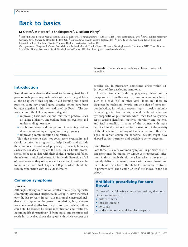
Sore throatSore throat is a very common symptom in primary care. It
can sometimes be caused by Group A streptococcal infec-
tion. A throat swab should be taken when a pregnant or
recently delivered woman presents with a sore throat, and
there should be a lower threshold for antibiotic treatment
in primary care. The Centor Criteria1 are shown in the box
below:
Antibiotic prescribing for sorethroats
If three of the following criteria are positive, then anti-
biotics are indicated1:
• history of fever
• tonsillar exudate
• no cough
• tender anterior cervical lymphadenopathy.
Oates et al.
16 ª 2011 Centre for Maternal and Child Enquiries (CMACE), BJOG 118 (Suppl. 1), 1–203
Some of the women who died had family members, espe-
cially children, with sore throats, suggesting that spread
from family members may be a further risk factor for
developing life-threatening sepsis. Therefore, all pregnant or
recently delivered women need to be advised of the risks
and signs and symptoms of infection and how to take steps
to prevent its transmisson. This includes the benefits of
good hygiene, such as avoiding contamination of the peri-
neum by the mother washing her own hands before, as well
as after, touching her perineum, especially when the woman
or her close family have a sore throat or upper respiratory
tract infection. Women in these circumstances should also
be encouraged to seek medical advice if they feel at all ill.
Pyrexia in the postnatal periodPostnatal observations and examinations are no longer as
routinely carried out in the community as in the past. It is
therefore very important that any symptoms, even if appar-
ently trivial, are noted and that appropriate clinical obser-
vations and examinations are performed to exclude or
detect developing infection as early as possible.
Back to basics: sepsis
Associated ‘red flag’ signs and symptoms that should
prompt urgent referral for hospital assessment, and, if the
women appears seriously unwell, by emergency ambulance:
• pyrexia > 38�C
• sustained tachycardia > 100 bpm
• breathlessness (RR > 20; a serious symptom)
• abdominal or chest pain
• diarrhoea and/or vomiting
• reduced or absent fetal movements, or absent fetal heart
• spontaneous rupture of membranes or significant
vaginal discharge
• uterine or renal angle pain and tenderness
• the woman is generally unwell or seems unduly
anxious, distressed or panicky.
A normal temperature does not exclude sepsis. Paraceta-
mol and other analgesics may mask pyrexia, and this
should be taken into account when assessing women
who are unwell.
Infection must also be suspected and actively ruled out
when a recently delivered woman has persistent vaginal
bleeding and abdominal pain. If there is any concern,
the woman must be referred back to the maternity unit
as soon as possible, certainly within 24 hours.
PainAll complaints of pain are potentially serious and must be
investigated thoroughly. However, the assessors have been
particularly concerned about neglected perineal and breast
pain in the puerperium. If a woman complains of perineal
pain after delivery, her perineum should be examined. If it
is known that there has been significant perineal trauma,
for example multiple vaginal lacerations or third-degree
tears, then the perineum should be inspected daily until
satisfactory healing has taken place.
Women complaining of breast pain should also be exam-
ined. Mothers with mastitis that does not respond to con-
servative measures or that becomes more severe within 12–
24 hours of onset should also be referred immediately for a
medical opinion. Breast abscesses are not obviously fluctu-
ant, and a surgical opinion may also be needed.
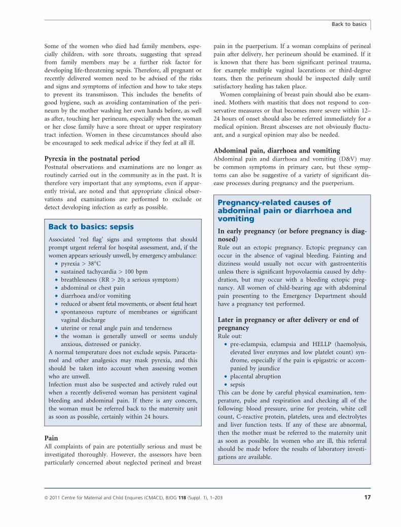
Abdominal pain, diarrhoea and vomitingAbdominal pain and diarrhoea and vomiting (D&V) may
be common symptoms in primary care, but these symp-
toms can also be suggestive of a variety of significant dis-
ease processes during pregnancy and the puerperium.
Pregnancy-related causes ofabdominal pain or diarrhoea andvomiting
In early pregnancy (or before pregnancy is diag-nosed)Rule out an ectopic pregnancy. Ectopic pregnancy can
occur in the absence of vaginal bleeding. Fainting and
dizziness would usually not occur with gastroenteritis
unless there is significant hypovolaemia caused by dehy-
dration, but may occur with a bleeding ectopic preg-
nancy. All women of child-bearing age with abdominal
pain presenting to the Emergency Department should
have a pregnancy test performed.
Later in pregnancy or after delivery or end ofpregnancyRule out:
• pre-eclampsia, eclampsia and HELLP (haemolysis,
elevated liver enzymes and low platelet count) syn-
drome, especially if the pain is epigastric or accom-
panied by jaundice
• placental abruption
• sepsis
This can be done by careful physical examination, tem-
perature, pulse and respiration and checking all of the
following: blood pressure, urine for protein, white cell
count, C-reactive protein, platelets, urea and electrolytes
and liver function tests. If any of these are abnormal,
then the mother must be referred to the maternity unit
as soon as possible. In women who are ill, this referral
should be made before the results of laboratory investi-
gations are available.
Back to basics
ª 2011 Centre for Maternal and Child Enquiries (CMACE), BJOG 118 (Suppl. 1), 1–203 17
BreathlessnessBreathlessness after delivery is very uncommon and needs a
full investigation to rule out serious underlying disease.
Although it is commoner in pregnancy, largely as the result
of physiological changes, it can also be the presenting
symptom of serious medical conditions, including cardiac
disease and other causes of pulmonary oedema, pulmonary
embolism and pneumonia. Anaemia has to be very severe
to cause breathlessness.
Back to basics: breathlessness2
Physiological breathlessness of pregnancyThis is experienced by up to 75% of pregnant women.
It can start in any trimester, and the onset is gradual. It
is often noticed by the woman when she is talking or at
rest, although it may get worse with exercise.
AsthmaIt is unusual for asthma to present for the first time in
pregnancy. Most women will have had the diagnosis
established before pregnancy. The breathlessness in
asthma is often associated with coughing, exhibits diur-
nal variation and may get worse with intercurrent respi-
ratory infections, hay fever and acid reflux. It improves
with bronchodilators.
Never assume that wheeze on auscultation represents
asthma, especially in a woman not known to have
asthma; it could be pulmonary oedema.
‘Red flag’ features suggesting more sinister underlying
pathology include:
• breathlessness of sudden onset
• breathlessness associated with chest pain
• orthopnoea or paroxysmal nocturnal dyspnoea.
Diagnoses to consider
Pulmonary embolusSudden onset breathlessness, may have associated pleu-
ritic pain, haemoptysis, dizziness.
PneumoniaMay have associated cough, fever, raised inflammatory
markers. It is important to remember that pregnant
women are particularly susceptible to viral (influenza
H1N1, varicella zoster) pneumonia.
Pulmonary oedemaMay have orthopnoea, paroxysmal nocturnal dyspnoea,
frothy/pink sputum. Auscultation may reveal inspiratory
fine crepitations and/or wheeze. Pulmonary oedema
may be due to:
• fluid overload, especially in the context of pre-
eclampsia
• mitral stenosis
• left ventricular failure.
Pulmonary hypertensionBreathlessness may be the only symptom and is worse
on exercise.
Investigations that should be considered are chest
X-ray, echocardiography and measurement of oxygen
saturations at rest and on exercise (in normal women,
the oxygen saturation ranges from 96 to 100% and
does not fall below 95% on exercise). There should be
a low threshold for referral of women with breathless-
ness in pregnancy from primary to secondary care,
particularly if they have any of the ‘red flag’ features
noted above.
HeadacheThis is common in pregnancy, but it can be a symptom of
serious underlying illness and should be taken seriously.
Back to basics: headache2
The commonest causes of headache in pregnancy are:
• tension headache—usually bilateral
• migraine—usually unilateral, may be preceded by
aura (often visual), associated with nausea, vomit-
ing and photophobia; may be new onset in preg-
nancy
• drug-related—most commonly caused by vasodila-
tors and in particular nifedipine.
‘Red flag’ features suggesting more sinister pathology
include:
• headache of sudden onset
• headache associated with neck stiffness
• headache described by the woman as the worst
headache she has ever had
• headache with any abnormal signs on neurological
examination.
Diagnoses to consider
Subarachnoid haemorrhageSudden, severe, often occipital so-called ‘thunderclap’
headache.
Cerebral venous thrombosisUnusually severe headache which may be associated
with focal signs.
Oates et al.
18 ª 2011 Centre for Maternal and Child Enquiries (CMACE), BJOG 118 (Suppl. 1), 1–203
Pre-eclamptic toxaemia/Impending eclampsiaMay be associated with seeing flashing lights and is usu-
ally associated with other clinical features of severe pre-
eclampsia, such as epigastric pain, hypertension, albu-
minuria and abnormal bloods.
Headache that is ‘the worst that the woman has ever
experienced’ is an indication for urgent brain imaging
in the absence of any other features because of concern
about cerebral venous thrombosis. However, less severe
headaches can be so non-specific that clinical judgement
should be the main guide to further referral to the neu-
rological services and investigation. The index of suspi-
cion should be high in pregnant women, and all serious
causes should be considered before dismissing headache
as benign.
Anxiety and distress in pregnancy and followingdeliveryThe ‘Blues’ is a period, lasting a few days, of tearfulness
and a feeling of being overwhelmed. It occurs in the
majority of mothers in the first 2 weeks following
delivery.
Episodes of tearfulness, worry, anxiety and depressive
symptoms are commonplace in pregnancy and the first few
weeks after delivery, particularly in first-time mothers.
Mostly these will be mild and self-limiting.
However, in some women these symptoms can be the
early signs of a more serious illness.
Back to basics: good mental healthpractice
• Review the woman in 2 weeks.
• Consider referral to psychiatric services if symptoms
persist.
• Refer urgently to psychiatric services in following cir-
cumstances:
o suicidal ideation
o uncharacteristic symptoms/marked change from
normal functioning
o mental health deteriorating
o persistent symptoms in late pregnancy and the first
6 weeks postpartum
o association with panic attacks and/or intrusive
obsessional thoughts
o morbid fears that are difficult to reassure
o profound low mood/ideas of guilt and worthless-
ness/insomnia and weight loss
o personal or family history of serious affective
disorder.
Unexplained physical symptomsIn a number of maternal deaths, symptoms of the underly-
ing physical condition were attributed to psychiatric disor-
der. In many women, this was because of non-specific
symptoms such as distress, agitation and loss of appetite.
In others, the symptoms of an acute confusional state
caused by the underlying physical condition were misinter-
preted as functional mental illness.
Clinicians should be aware of the clinical features and
causes of confusional states. It should be remembered that
physical illness can present as psychiatric disorder and can
co-exist with it.
Unexplained physical symptoms
Unexplained physical symptoms should not be attrib-
uted to psychiatric disorder:
• unless there is a clear pathway to symptom produc-
tion
• unless there is a known previous psychiatric history
• when they represent a marked change from normal
functioning
• when the only psychological symptoms are
behavioural and non-specific for example, distress
and agitation
• when the woman does not speak English or is from
an ethnic minority group.
Booking, history-taking and basicobservations
All maternity-care providers, and in particular midwives
and GPs, must recognise the crucial importance of:
• taking a comprehensive history and making a correct
risk assessment at booking
• referring the woman to the obstetrician or other spe-
cialist as necessary
• following up these referrals to ensure appropriate
action has been taken
• making, recording and acting upon basic observations
• re-assessing the woman’s risk status throughout her
pregnancy and in the postnatal period.
The antenatal booking historyOne of the most important events in the woman’s
maternity care is the antenatal booking history appoint-
ment. It is the first opportunity to assess her well-being
and determine her ‘risk status’. The majority of mater-
nity records currently use a ‘tick box’ format. In many
records of the women who died, the information docu-
mented was insufficient to enable risk assessment or an
Back to basics
ª 2011 Centre for Maternal and Child Enquiries (CMACE), BJOG 118 (Suppl. 1), 1–203 19
appropriate pathway of care to be planned. In other
women, a full history was taken and all information
appropriately recorded, but no appropriate follow up or
referral was made. This meant that some women who
had risk factors were booked for midwifery-led care. It is
vitally important that when a woman reports a ‘risk fac-
tor’, the midwife asks for additional relevant information,
records this in detail and acts upon it.
Communicating with the GPMidwives must ensure that history-taking is comprehen-
sive, includes two-way communication with the woman’s
GP and ensures timely referral to obstetrician and/or other
specialist where necessary. The importance of midwife–GP
communication cannot be over-emphasised, particularly as
midwifery services are now offered in a range of locations,
including Children’s Centres.
Communication between GPs andmidwives
• Midwives should notify GPs that a woman is preg-
nant.
• Midwives should seek additional information from
the GP if risk factors are identified.
• GPs should inform midwives about prior medical
and mental health problems.
• There should be auditable robust local systems in
place to enable two-way flow of information
throughout pregnancy and the postnatal period.
Using basic observations to assess and act uponrisk statusMidwives and GPs should be alert to changes in the
woman’s situation and that her risk status may change
several times during the course of the pregnancy and the
postnatal period. If a woman complains of any symptoms
that indicate a deviation from the norm, the midwife or
GP must take basic observations, which include tempera-
ture, pulse and respirations. If observations are found to
be abnormal, these must be followed up by appropriate
referral to GP or hospital. Following this referral, the
midwife has a duty to make sure appropriate action has
been taken.
In a number of women, it was clear that the women
who reported pain, temperature and feeling unwell
had appropriate midwifery care, with basic observations
being performed and appropriate referral and follow up
made. However, there were other situations where
midwives did not respond appropriately to these com-
plaints, failing to make basic observations or act on
abnormal results.
Improving communications and referralsIn many cases reported to the Enquiry, there were major
failures of communication between healthcare workers,
which undoubtedly contributed to the woman’s death in
some cases. Notably, these included GPs not being asked
for information or being consulted about further referral
and, in some cases, the GP not being informed that the
woman was pregnant. In addition, the assessors have
been struck by the lack of further referral by hospital
obstetricians of potentially high-risk women. The reasons
for failure to refer are likely to be multiple. However,
medical care is advancing rapidly, and patterns of the
delivery of care in the UK are changing. It must be
appreciated that not all maternity centres can care for
pregnant women with major complications either preced-
ing or developing in pregnancy. If women with underly-
ing medical or psychiatric conditions are to share in the
advances in medicine, all mothers who require it should
be referred to specialised centres for their care in preg-
nancy; this was not usually the case among the women
who died.
In many cases reported to the Enquiry, referral for spe-
cialist input (for example neurological or psychiatric con-
sultation) was made by a healthcare worker, including GPs,
midwives and obstetricians; however, there was a subse-
quent inappropriate delay in sending an appointment or
the woman was not seen.
Referrals to and from any healthprofessional, including initial GP/midwife referral letter
• Remember, referral is not treatment.
• Explain the importance of keeping the appoint-
ment.
• Always check that an appointment has been given
and that the woman has been seen.
• If you are concerned or think that an urgent
response is required, telephone a senior clinician.
• Always back up a fax, email, phone call with a
written letter (remember to copy in the midwife
and GP).
• Include in the referral letter details of:
o current problem and reason for referral
o details of any past medical history, including
mental health history, even if not directly rele-
vant to the presenting problem
o all medications she is currently on or has recently
stopped
o investigations so far.
Oates et al.
20 ª 2011 Centre for Maternal and Child Enquiries (CMACE), BJOG 118 (Suppl. 1), 1–203
Disclosure of interestNone.
FundingThis work was undertaken by the Centre for Maternal and
Child Enquiries (CMACE) as part of the CEMACH pro-
gramme. The work was funded by the National Patient
Safety Agency; the Department of Health, Social Services
and Public Safety of Northern Ireland; NHS Quality
Improvement Scotland (NHS QIS); and the Channel
Islands and Isle of Man. j
References
1 Cooper RJ, Hoffman JR, Bartlett JG, Besser RE, Gonzales R, Hickner
JM, et al. Principles of appropriate antibiotic use for acute pharyngitis
in adults: background. Ann Intern Med 2001;134:509–17.
2 Nelson-Piercy C. Handbook of Obstetric Medicine 4th Edition.
London: Informa Healthcare, 2010.
ª 2011 Centre for Maternal and Child Enquiries (CMACE), BJOG 118 (Suppl. 1), 1–203 21
Back to basics
Introduction: Aims, objectives and definitionsused in this ReportG Lewis
National Perinatal Epidemiology Unit (NPEU), University of Oxford, Oxford, UK
Correspondence: Professor Gwyneth Lewis, National Perinatal Epidemiology Unit (NPEU), University of Oxford, Old Road Campus, Oxford
OX3 7LF, UK. Email: [email protected]
Introduction
This, the eighth Report of the United Kingdom (UK) En-
quiries into Maternal Deaths, now known as Saving Moth-
ers’ Lives, continues the 56-year unbroken series of reviews
of maternal deaths undertaken to save more mothers’ lives
and, more generally, to improve maternity services overall.
Although the style and content of the Reports has changed
over this time, the essential aims and objectives remain the
same. It is because of the sustained commitment of all
health professionals who provide maternity and other ser-
vices for pregnant women in the UK that this Enquiry con-
tinues to be a highly respected and powerful force for
improvements in maternal health, both here in the UK and
internationally. As stated in previous Reports, and equally
valid today, reading the Report or preparing a statement
for an individual enquiry forms a crucial part of individual,
professional, self-reflective learning. As long ago as 1954, it
was recognised that participating in a confidential enquiry
had a ‘powerful secondary effect’ in that ‘each participant
in these enquiries, however experienced he or she may be,
and whether his or her work is undertaken in a teaching
hospital, a local hospital, in the community or the woman’s
home must have benefited from their educative effect’.1
Personal experience is therefore recognised as a valuable
tool for harnessing beneficial changes in individual prac-
tice.
Whereas many of the earlier Reports focused mainly on
clinical issues, more recent Reports, as with the very earliest
ones in the 1950s, have also focused on the wider public-
health issues that contribute to poorer health and social
outcomes. As a result, their findings and recommendations
have played a major part in helping in the development of
broader policies designed to help reduce health inequalities
for the poorest of our families and for the most vulnerable
and socially disadvantaged women. Particularly striking
have been successive Governments’ commitments to reduce
the wide variations in maternal mortality rates between the
most and least advantaged mothers as identified by these
Reports. By acting on similar findings in past Reports, this
Enquiry has also played a major part in defining the phi-
losophy of our maternity services that now expect each
individual woman and her family to be at the heart of
maternity services designed to meet her own particular
needs, rather than vice versa.
Telling the story
The methodology used by the Enquiry goes beyond count-
ing numbers. Its philosophy, and that of those who partici-
pate in its process, is to recognise and respect every
maternal death as a young woman who died before her
time, a mother, a member of a family and of her commu-
nity. It does not demote women to numbers in statistical
tables; it goes beyond counting numbers to listen and tell
the stories of the women who died so as to learn lessons
that may save the lives of other mothers and babies, as well
as aiming to improve the standard of maternal health over-
all. Consequently, its methodology and philosophy con-
tinue to form a major part of the strategies of the World
Health Organization (WHO) and its sister United Nations
organisations and other donor agencies to reduce maternal
deaths. The WHO maternal mortality review tool kit, and
programme, Beyond the Numbers,2 includes advice and
practical steps in choosing and implementing one or more
of five possible approaches to maternal death reviews
adaptable at any level and in any country. These
approaches are facility and community death reviews, Con-
fidential Enquiries into Maternal Deaths, near-miss reviews
and clinical audit.3 This work, in modified form, is now
undertaken in more than 54 countries, including many of
those with the poorest outcomes.
Learning lessons for continualimprovement
This Enquiry is the oldest example of the use of the mater-
nal mortality and morbidity surveillance cycle, now inter-
nationally adopted by the WHO programme Beyond the
Numbers, which promotes the use of maternal death or
morbidity reviews to make pregnancy safer.2 The cycle,
shown in Figure 1, is an ongoing process of deciding which
deaths to review and identifying the cases, collecting and
assessing the information, using it for recommendations,
Lewis
22 ª 2011 Centre for Maternal and Child Enquiries (CMACE), BJOG 118 (Suppl. 1), 1–203
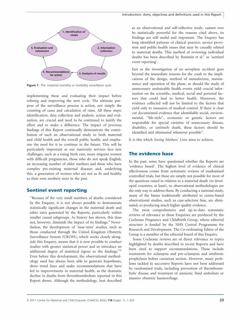
implementing these and evaluating their impact before
refining and improving the next cycle. The ultimate pur-
pose of the surveillance process is action, not simply the
counting of cases and calculation of rates. All these steps:
identification, data collection and analysis, action and eval-
uation, are crucial and need to be continued to justify the
effort and to make a difference. The impact of previous
findings of this Report continually demonstrate the contri-
bution of such an observational study to both maternal
and child health and the overall public health, and empha-
sise the need for it to continue in the future. This will be
particularly important as our maternity services face new
challenges, such as a rising birth rate, more migrant women
with difficult pregnancies, those who do not speak English,
an increasing number of older mothers and those who have
complex pre-existing maternal diseases and, underlying
this, a generation of women who are not as fit and healthy
as their own mothers were in the past.
Sentinel event reporting
‘‘Because of the very small numbers of deaths considered
by the Enquiry, it is not always possible to demonstrate
statistically significant changes in the maternal death and
other rates generated by the Reports, particularly within
smaller causal subgroups. As history has shown, this does
not, however, diminish the impact of its findings.4 Never-
theless, the development of ‘near-miss’ studies, such as
those conducted through the United Kingdom Obstetric
Surveillance System (UKOSS), which works closely along-
side this Enquiry, means that it is now possible to conduct
studies with greater statistical power and so introduce an
additional degree of statistical rigour to the findings.5,6
Even before this development, the observational method-
ology used has always been able to generate hypotheses,
show trend lines and make recommendations that have
led to improvements in maternal health, as the dramatic
decline in deaths from thromboembolism reported in this
Report shows. Although the methodology, best described
as an observational and self-reflective study, cannot ever
be statistically powerful for the reasons cited above, its
findings are still useful and important. The Enquiry has
long identified patterns of clinical practice, service provi-
sion and public-health issues that may be causally related
to maternal deaths. This method of reviewing individual
deaths has been described by Rutstein et al.7 as ‘sentinel
event reporting’.
Just as the investigation of an aeroplane accident goes
beyond the immediate reasons for the crash to the impli-
cations of the design, method of manufacture, mainte-
nance and operation of the plane, so should the study of
unnecessary undesirable health events yield crucial infor-
mation on the scientific, medical, social and personal fac-
tors that could lead to better health. Moreover, the
evidence collected will not be limited to the factors that
yield only to measures of medical control. If there is clear
cut documented evidence that identifiable social, environ-
mental, ‘‘life-style’’, economic or genetic factors are
responsible for special varieties of unnecessary disease,
disability, or untimely death, these factors should be
identified and eliminated whenever possible’’.
It is this which Saving Mothers’ Lives aims to achieve.
The evidence base
In the past, some have questioned whether the Reports are
‘evidence based’. The highest level of evidence of clinical
effectiveness comes from systematic reviews of randomised
controlled trials, but these are simply not possible for most of
the questions raised in relation to a maternal death (in devel-
oped countries, at least), so observational methodologies are
the only way to address them. By conducting a national study,
many of the biases traditionally attributed to centre-based
observational studies, such as case-selection bias, are elimi-
nated, so producing much higher quality evidence.
The most comprehensive and up-to-date systematic
reviews of relevance to these Enquiries are produced by the
Cochrane Pregnancy and Childbirth Group, whose editorial
structure is funded by the NHS Central Programme for
Research and Development. The Co-ordinating Editor of the
Group is a member of the editorial board of this Enquiry.
Some Cochrane reviews are of direct relevance to topics
highlighted by deaths described in recent Reports and have
been cited to support recommendations. These include
treatments for eclampsia and pre-eclampsia and antibiotic
prophylaxis before caesarean section. However, many prob-
lems tackled in successive Reports have not been addressed
by randomised trials, including prevention of thromboem-
bolic disease and treatment of amniotic fluid embolism or
massive obstetric haemorrhage.
1. Identification of cases
2. Informationcollection
5. Evaluation andrefinement
4. Recommendationsfor action
3. Analysis of results
Figure 1. The maternal mortality or morbidity surveillance cycle.
Introduction: Aims, objectives and definitions used in this Report
ª 2011 Centre for Maternal and Child Enquiries (CMACE), BJOG 118 (Suppl. 1), 1–203 23
An important limitation of randomised trials is that,
unless they are very large, they may provide little informa-
tion about rare, but important, complications of treatments.
Safety issues are, therefore, sometimes better illuminated by
observational studies than by controlled trials.
Many causes of maternal death are very rare, and treat-
ment options for these may never be subjected to formal
scientific study. Inevitably, recommendations for care to
avoid such deaths in the future rely on lesser levels of evi-
dence, and frequently on ‘expert opinion’. This does not
mean that the Report is not evidence-based, merely that,
necessarily, the evidence cannot be in the form of a rando-
mised controlled trial or case–control study because of the
relative rarity of the condition.
The use of vignettes andrecommendations
The Centre for Maternal and Child Enquiries (CMACE)
policy on the use of vignettes and the development of rec-
ommendations is available on the website at www.cma-
ce.org.uk.
In the past, some Reports have been characterised by the
use of a significant number of detailed vignettes, stories that
broadly describe the circumstances surrounding the deaths
of individual women and the lessons that may be drawn
from them. Recognising that ensuring that everyone, includ-
ing family members and professional staff, require complete
reassurance about the guiding principle of maintaining con-
fidentiality out of respect for those who have died, the
number of vignettes has been reduced, as have some of the
more identifiable details given in them. CMACE only uses
vignettes to help in the identification of lessons learnt for
the improvement of future professional practice or overall
service delivery. The vignettes used in this Report do not
include the full circumstances of any individual case. They
neither provide nor imply a complete overall assessment or
judgement of the totality of care provided in a case,
although they may point to where general lessons may be
learnt from particular aspects of care. Individual details in
vignettes may be changed to protect the anonymity of the
woman. CMACE cannot confirm or deny the identity of
any individual woman, aspects of whose care may be
included within a vignette, because all records used in the
Enquiry are destroyed before publication of the Report.
Further details on the method of enquiry are included in
Appendix 1 of this Report.
Severe maternal morbidity, ‘nearmisses’
It is increasingly recognised that the study of near-miss
morbidity can complement enquiries into maternal death
and provide valuable additional information to guide pre-
vention and treatment of potentially life-threatening condi-
tions. Maternal deaths represent the tip of the iceberg of
disease; a much larger number of women suffer from near-
miss morbidity, increasing the power of these studies to
investigate risk factors for both the occurrence of disease
and progression to death and other severe complications.
In this Report, the latest results of two systems for severe
morbidity or ‘near-miss’ surveillance are reported in
Appendix 2 and, where relevant, in the individual Chapters
describing the rates of clinical causes of death. These are
the UKOSS5,6 and the Scottish Confidential Audit of Severe
Maternal Morbidity (SCASMM).8
The UK Obstetric Surveillance System(UKOSS)
Surveillance of specific near-miss maternal morbidities and
other rare disorders of pregnancy has been conducted
through the UKOSS since 2005.1 UKOSS is an active, nega-
tive surveillance system. Cases are actively sought through
a routine monthly mailing to nominated reporting obstetri-
cians, midwives, risk management midwives and anaesthe-
tists in all consultant-led maternity units in the UK. All
consultant-led units participate in UKOSS reporting.
Clinicians are asked to complete the monthly report card
indicating whether there has been a woman with one of
the conditions under study delivered in the unit during the
previous month. They are also asked to complete a ‘nil
report’ indicating if there have not been any cases;
this allows participation to be monitored and confirms
the denominator population for calculation of disease
incidence.
In response to a report of a case, collaborating clinicians
are sent a data collection form asking for further demo-
graphic and pregnancy information, as well as details of diag-
nosis, management and outcomes for mother and infant. For
some conditions, clinicians are also asked to supply details
about a comparison woman delivered in the same unit.
This system allows for a rolling programme of parallel
studies to be conducted, and, because case–control and
cohort studies may be carried out as well as descriptive
studies, a range of research questions can be addressed. In
addition to estimating disease incidence or prevalence,
UKOSS studies can be used to quantify risk and prognostic
factors, audit national management and prevention guide-
lines and describe disease management, as well as describe
outcomes for both mothers and their infants. Descriptive,
case–control and cohort studies are conducted and peer-
reviewed papers are published. Further details of surveil-
lance of near-miss morbidities using UKOSS are included
in the relevant chapters of this report, and key points are
summarised in Appendix 2A.
Lewis
24 ª 2011 Centre for Maternal and Child Enquiries (CMACE), BJOG 118 (Suppl. 1), 1–203
Scottish Confidential Audit of SevereMaternal Morbidity
An audit of a range of defined severe maternal morbidities
has been carried out continuously in all consultant-led
maternity units in Scotland since 2003. The methodology is
similar to that of UKOSS, with a designated midwife coor-
dinator in each unit who identifies cases and sends com-
pleted data to the Reproductive Health Programme of NHS
Quality Improvement Scotland, which analyses the data
and produces an annual report.8 Particularly detailed infor-
mation is collected and analysed for all cases of major
obstetric haemorrhage and of eclampsia. For these two
conditions, each unit also provides a self-assessment of the
quality of care provided. The continuous nature of the
audit using identical criteria and case definitions over sev-
eral years has allowed the identification of changes in the
rates of some morbidities, as well as assessment of compli-
ance with guidelines and changes in clinical management.
Appendix 2B is a summary of the findings from SCASMM
for the triennium 2006–2008, and some information from
the audit is included in other relevant chapters.
The aims and objectives of theEnquiry
The overall aim is to save the lives of as many mothers and
newborns as possible through the expert anonymous review
of the circumstances surrounding and contributing to each
maternal death in the UK. Apart from the specific issues
and learning points that may emerge from certain cases or
causes of death, the findings from individual cases are also
aggregated together to learn wider lessons and to formulate
and disseminate more general recommendations.
Its objectives are:
• to improve the care that pregnant and recently deliv-
ered women receive and to reduce maternal mortality
and morbidity rates still further, as well as the propor-
tion of deaths caused by substandard care.
• to assess the main causes of and trends in maternal
deaths and, where possible, severe morbidity and to
identify any avoidable, remediable or substandard fac-
tors that could be changed to improve care; to promul-
gate these findings and subsequent recommendations to
all relevant healthcare professionals and to ensure that
their uptake is audited and monitored.
• to make recommendations concerning the improve-
ment of clinical care and service provision, including
local audit, to commissioners of obstetric services and
to providers and professionals involved in caring for
pregnant women.
• to suggest directions for future areas for research and
audit at a local and national level.
• to contribute to regular shorter reports on overall
trends in maternal mortality as well as producing a
more in-depth triennial Report.
The Enquiry’s role in the provision ofhigh-quality clinical care
Although the Enquiry has always had the support of pro-
fessionals involved in caring for pregnant or recently deliv-
ered women, it is also a requirement that all maternal
deaths should be subject to this confidential enquiry, and
all health professionals have a duty to provide the informa-
tion required.
In participating in this Enquiry, all health professionals
are asked for two things:
• if they have been caring for a woman who died, to
provide the Enquiry with a full, accurate and unbiased
account of the circumstances leading up to her death,
with supporting records, and
• irrespective of whether they have been caring for a
woman who died or not, to reflect on and take any
actions that may be required, either personally or as
part of their wider institution, as a result of the recom-
mendations and lessons contained within this Report.
At a local commissioning level, maternity healthcare
commissioners, such as Primary Care Trusts and Local
Health Boards, should commission services which meet the
recommendations set out in this and previous Reports and
ensure that all staff participate in the Enquiry if required,
as part of their contract.
At service provider level, the findings of the Enquiry
should be used:
• to ensure that all staff are regularly updated and
trained on the signs and symptoms of critical illness,
such as infection, and the early identification, manage-
ment and resuscitation of seriously ill women
• to develop and regularly update multidisciplinary
guidelines for the management of complications during
or after pregnancy
• to review and modify, where necessary, the existing
arrangements for the provision of maternity or obstet-
ric care
• to ensure that all Direct and unexpected Indirect mater-
nal deaths are subject to a local review and critical inci-
dent report, which is made available to the Enquiry as
part of its own process of review, as well as disseminat-
ing its key findings and recommendations to all local
maternity staff
• to introduce an obstetric early warning system chart as
recommended in this and previous Reports
• to promote local audit and clinical governance.
At a national level—in every country, the findings of
successive Reports have been used to develop national
Introduction: Aims, objectives and definitions used in this Report
ª 2011 Centre for Maternal and Child Enquiries (CMACE), BJOG 118 (Suppl. 1), 1–203 25
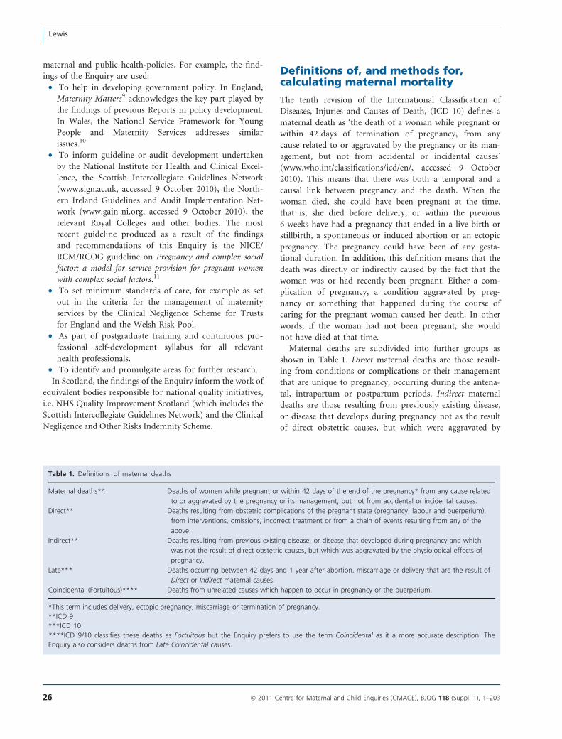
maternal and public health-policies. For example, the find-
ings of the Enquiry are used:
• To help in developing government policy. In England,
Maternity Matters9 acknowledges the key part played by
the findings of previous Reports in policy development.
In Wales, the National Service Framework for Young
People and Maternity Services addresses similar
issues.10
• To inform guideline or audit development undertaken
by the National Institute for Health and Clinical Excel-
lence, the Scottish Intercollegiate Guidelines Network
(www.sign.ac.uk, accessed 9 October 2010), the North-
ern Ireland Guidelines and Audit Implementation Net-
work (www.gain-ni.org, accessed 9 October 2010), the
relevant Royal Colleges and other bodies. The most
recent guideline produced as a result of the findings
and recommendations of this Enquiry is the NICE/
RCM/RCOG guideline on Pregnancy and complex social
factor: a model for service provision for pregnant women
with complex social factors.11
• To set minimum standards of care, for example as set
out in the criteria for the management of maternity
services by the Clinical Negligence Scheme for Trusts
for England and the Welsh Risk Pool.
• As part of postgraduate training and continuous pro-
fessional self-development syllabus for all relevant
health professionals.
• To identify and promulgate areas for further research.
In Scotland, the findings of the Enquiry inform the work of
equivalent bodies responsible for national quality initiatives,
i.e. NHS Quality Improvement Scotland (which includes the
Scottish Intercollegiate Guidelines Network) and the Clinical
Negligence and Other Risks Indemnity Scheme.
Definitions of, and methods for,calculating maternal mortality
The tenth revision of the International Classification of
Diseases, Injuries and Causes of Death, (ICD 10) defines a
maternal death as ‘the death of a woman while pregnant or
within 42 days of termination of pregnancy, from any
cause related to or aggravated by the pregnancy or its man-
agement, but not from accidental or incidental causes’
(www.who.int/classifications/icd/en/, accessed 9 October
2010). This means that there was both a temporal and a
causal link between pregnancy and the death. When the
woman died, she could have been pregnant at the time,
that is, she died before delivery, or within the previous
6 weeks have had a pregnancy that ended in a live birth or
stillbirth, a spontaneous or induced abortion or an ectopic
pregnancy. The pregnancy could have been of any gesta-
tional duration. In addition, this definition means that the
death was directly or indirectly caused by the fact that the
woman was or had recently been pregnant. Either a com-
plication of pregnancy, a condition aggravated by preg-
nancy or something that happened during the course of
caring for the pregnant woman caused her death. In other
words, if the woman had not been pregnant, she would
not have died at that time.
Maternal deaths are subdivided into further groups as
shown in Table 1. Direct maternal deaths are those result-
ing from conditions or complications or their management
that are unique to pregnancy, occurring during the antena-
tal, intrapartum or postpartum periods. Indirect maternal
deaths are those resulting from previously existing disease,
or disease that develops during pregnancy not as the result
of direct obstetric causes, but which were aggravated by
Table 1. Definitions of maternal deaths
Maternal deaths** Deaths of women while pregnant or within 42 days of the end of the pregnancy* from any cause related
to or aggravated by the pregnancy or its management, but not from accidental or incidental causes.
Direct** Deaths resulting from obstetric complications of the pregnant state (pregnancy, labour and puerperium),
from interventions, omissions, incorrect treatment or from a chain of events resulting from any of the
above.
Indirect** Deaths resulting from previous existing disease, or disease that developed during pregnancy and which
was not the result of direct obstetric causes, but which was aggravated by the physiological effects of
pregnancy.
Late*** Deaths occurring between 42 days and 1 year after abortion, miscarriage or delivery that are the result of
Direct or Indirect maternal causes.
Coincidental (Fortuitous)**** Deaths from unrelated causes which happen to occur in pregnancy or the puerperium.
*This term includes delivery, ectopic pregnancy, miscarriage or termination of pregnancy.
**ICD 9
***ICD 10
****ICD 9/10 classifies these deaths as Fortuitous but the Enquiry prefers to use the term Coincidental as it a more accurate description. The
Enquiry also considers deaths from Late Coincidental causes.
Lewis
26 ª 2011 Centre for Maternal and Child Enquiries (CMACE), BJOG 118 (Suppl. 1), 1–203
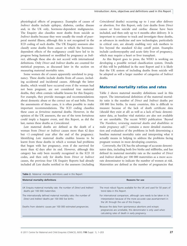
physiological effects of pregnancy. Examples of causes of
Indirect deaths include epilepsy, diabetes, cardiac disease
and, in the UK only, hormone-dependent malignancies.
The Enquiry also classifies most deaths from suicide as
Indirect deaths because they were usually the result of puer-
peral mental illness, although this is not recognised in the
ICD coding of such deaths. The UK Enquiry assessors also
classify some deaths from cancer in which the hormone-
dependent effects of the malignancy could have led to its
progress being hastened or modified by pregnancy as Indi-
rect, although these also do not accord with international
definitions. Only Direct and Indirect deaths are counted for
statistical purposes, as discussed later in the section on
measuring maternal mortality rates.
Some women die of causes apparently unrelated to preg-
nancy. These deaths include deaths from all causes, includ-
ing accidental and incidental causes. Although the latter
deaths, which would have occurred even if the woman had
not been pregnant, are not considered true maternal
deaths, they often contain valuable lessons for this Enquiry.
For example, they provide messages and recommendations
about domestic abuse or the correct use of seat belts. From
the assessments of these cases, it is often possible to make
important recommendations. The ICD coding classifies
these cases as fortuitous maternal deaths. However, in the
opinion of the UK assessors, the use of the term fortuitous
could imply a happier event, and this Report, as did the
last, names these deaths as Coincidental.
Late maternal deaths are defined as the death of a
woman from Direct or Indirect causes more than 42 days
but <1 completed year after the end of the pregnancy.
Identifying Late maternal deaths enables lessons to be
learnt from those deaths in which a woman had problems
that began with her pregnancy, even if she survived for
more than 42 days after its end. However, although this
category has only been recently recognised in the ICD 10
codes, and then only for deaths from Direct or Indirect
causes, the previous four UK Enquiry Reports had already
included all Late deaths notified to the assessors (including
Coincidental deaths) occurring up to 1 year after delivery
or abortion. For this Report, only Late deaths from Direct
causes or from suicide and cardiac disease have been
included, and then only up to 6 months after delivery. It is
important to continue to track and investigate these deaths,
as advances in medicine and new technologies, particularly
in critical care, are already enabling very sick mothers to
live beyond the standard 42-day cutoff point. Examples
include cardiomyopathy and acute fatty liver of pregnancy,
which may require a heart or liver transplant.
As this Report goes to press, the WHO is working on
developing a possible revised classification system. Details
of this will be published in the next Report and it is likely
that the UK system of including deaths from suicide will
be adopted as will a larger number of categories of Indirect
deaths.
Maternal mortality ratios and rates
Table 2 shows maternal morality definitions used in this
report. The international definition of the maternal mortal-
ity ratio is the number of Direct and Indirect deaths per
100 000 live births. In many countries, this is difficult to
measure because of the lack of death certificate data
(should they exist at all) as well as a lack of basic denomi-
nator data, as baseline vital statistics are also not available
or are unreliable. The recent WHO publication ‘Beyond
The Numbers; reviewing maternal deaths and disabilities to
make pregnancy safe’,2 contains a more detailed examina-
tion and evaluation of the problems in both determining a
baseline maternal mortality ratio and interpreting what it
actually means in helping to address the problems facing
pregnant women in most developing countries.
Conversely, the UK has the advantage of accurate denomi-
nator data, including both live births and stillbirths, and has
defined its maternal mortality rate as the number of Direct
and Indirect deaths per 100 000 maternities as a more accu-
rate denominator to indicate the number of women at risk.
Maternities are defined as the number of pregnancies that
Table 2. Maternal mortality definitions used in this Report
Maternal mortality definitions Reasons for use
UK Enquiry maternal mortality rate: the number of Direct and Indirect
deaths per 100 000 maternities
The most robust figures available for the UK and used for 50 years of
trend data in this Report.
The internationally defined maternal mortality ratio: the number of
Direct and Indirect deaths per 100 000 live births
For international comparison, although care needs to be taken in its
interpretation because of the more accurate case ascertainment in
the UK through the use of this Enquiry.
Deaths from obstetric causes per 100 000 estimated pregnancies. Because the data from spontaneous abortions and ectopic
pregnancies are unreliable, this denominator is only used when
calculating rates of death in early pregnancy.
Introduction: Aims, objectives and definitions used in this Report
ª 2011 Centre for Maternal and Child Enquiries (CMACE), BJOG 118 (Suppl. 1), 1–203 27

result in a live birth at any gestation or stillbirths occurring
at or after 24 completed weeks of gestation and are required
to be notified by law. This enables a more detailed picture of
maternal mortality rates to be established and is used for the
comparison of trends over time.
Furthermore, in the UK maternal mortality rate can be
calculated in two ways:
• through official death certification to the Registrars
General (the Office for National Statistics [ONS] and
its equivalents), or
• through deaths known to this Enquiry.
The overall maternal mortality rate is calculated from
the number of Direct and Indirect deaths. The ONS data
are based on death certificates where the cause of death is
directly or secondarily coded for a pregnancy-related condi-
tion, such as postpartum haemorrhage, eclampsia etc.
For the past 50 years the Enquiry has calculated its own
maternal mortality rate, because the overall number of
maternal deaths identified by the proactive case-finding
methodology used by this Enquiry has always exceeded
those officially reported. This is because not all maternal
deaths are recorded as such on death certificates. For exam-
ple, a large proportion of women known to the Enquiry
who died of pre-existing medical conditions influenced by
their pregnancy, for example cardiac disorders, epilepsy
and some malignancies, were excluded from the official sta-
tistics. Other women excluded from official data are those
who required long-term intensive care and whose final
cause of death was registered as a nonpregnancy condition,
such as multiple organ failure, even though the initiating
cause was an obstetric event. Conversely, the maternal
deaths known to the Registrars General may include Late
deaths because it is not possible to identify from the death
certificate when the delivery or termination occurred.
To aid the international comparison of the UK data with
those from other countries calculated by using the ICD-
defined Maternal Mortality Ratio, this Report has also cal-
culated the overall UK maternal mortality ratio as well as
the more complete Enquiry maternal mortality rate. These
are shown in Chapter 1. However, when making such com-
parisons, it is important to note two points:
• The criteria used by the UK assessors for Indirect deaths
are far more inclusive than those used in other countries.
For example, in this Enquiry all cases of cardiac disease,
asthma and epilepsy are coded as Indirect, as are cases of
suicide unless obviously occurring in women with a
longstanding previous psychiatric history.
• Case ascertainment is lower in the vast majority of
other countries because they do not undertake such
comprehensive enquiries.
These two facts alone increase the UK maternal mortality
rate when compared with other countries, and this is dis-
cussed in more detail in Chapter 1.
Case ascertainment
The role of the Office for National StatisticsSince the introduction of a new ONS computer program in
1993, all conditions given anywhere on the death certificate
are now coded, enabling a more extensive search of death
entry information to identify all conditions listed that sug-
gest a maternal death. In the past, this has helped in
improving case ascertainment, with a number of previously
unreported deaths being identified. Fortunately, for this
Report, the ONS record linkage study described below has
identified very few additional cases of Direct or Indirect
deaths. This is a reduction in the already small degree of
under-ascertainment calculated for previous Reports.
For the past 9 years, ONS has been able to match death
records of women of fertile age living in England and
Wales with birth registrations up to 1 year previously. The
aim is to identify deaths of all women in England and
Wales who died within 1 year of giving birth and to see
how many additional cases can be found. The methodol-
ogy, used in the past two triennia, was again applied for
this Report and again shows that the majority of these
deaths occurred Late, i.e. some months after delivery. The
vast majority of these Late deaths were the result of Coinci-
dental Late causes, and these are shown in Chapter14.
The ONS does not service Scotland, where maternal
death case ascertainment is achieved using information
from the General Register Office for Scotland, the Repro-
ductive Health Programme of NHS Quality Improvement
Scotland and the wider clinical community.
Denominator data used for calculatingmortality rates
Number of maternitiesIt is impossible to know the exact number of pregnancies
that occurred during this or any preceding triennium,
because not all pregnancies result in a registered live birth
or stillbirth. These data are unreliable because of the lack
of appropriate denominators, so the most common
denominator used throughout this and previous Reports is
the number of maternities rather than the total number of
pregnancies. Maternities are the number of pregnancies
that result in a live birth at any gestation or stillbirths
occurring at or after 24 completed weeks of gestation and
are required to be notified by law. The total number of
maternities for the UK for 2006–08 was 2 291 493.
Estimated pregnanciesThis denominator is used for calculating the rate of early
pregnancy deaths. It is a combination of the number of
maternities, together with legal terminations, hospital admis-
Lewis
28 ª 2011 Centre for Maternal and Child Enquiries (CMACE), BJOG 118 (Suppl. 1), 1–203
sions for spontaneous miscarriages (at <24 weeks of gesta-
tion) and ectopic pregnancies with an adjustment to allow
for the period of gestation and maternal ages at conception.
The estimate for the UK 2006–08 was 3 139 315. However,
the resulting total is still an underestimate of the actual num-
ber of pregnancies because these figures do not include other
pregnancies that miscarry early, those where the woman is
not admitted to hospital, or indeed those where the woman
herself may not even know she is pregnant.
Disclosure of interestsNone.
FundingThis work was undertaken by the Centre for Maternal and
Child Enquiries (CMACE) as part of the CEMACH pro-
gramme. The work was funded by the National Patient
Safety Agency; the Department of Health, Social Services
and Public Safety of Northern Ireland; NHS Quality
Improvement Scotland (NHS QIS); and the Channel
Islands and Isle of Man.
AcknowledgementsThis introduction has been discussed with Dr Marian
Knight, Head of UKOSS and Senior Clinical Research Fel-
low for the NPEU, Professor James Drife, Emeritus Profes-
sor of Obstetrics and Gynaecology for the University of
Leeds and Professor James Neilson, Professor of Obstetrics
and Gynaecology for the University of Leeds. j
References
1 Ministry of Health. Report of the Confidential Enquiry into Maternal
Deaths in England and Wales, 1952–1954. Reports on Public Health
and Medical Subjects No.97. London: HMSO, 1954.
2 World Health Organisation. Beyond the Numbers—Reviewing
Maternal Deaths and Complications to Make Pregnancy Safer. Gen-
eva: WHO, 2004. [www.who.int/reproductive-health]. Accessed 6
October 2010.
3 Lewis G. In: British Medical Bulletin. Pregnancy: Reducing Maternal
Death and Disability. Oxford: British Council, Oxford University Press,
2003.
4 Lewis G. Why mothers survive. In: Dob D, Cooper G, Holdcroft A,
editors. Crises in Childbirth—Why Mothers Survive. Lessons from the
Confidential Enquires into Maternal Deaths. Oxford: Radcliffe Press;
2007, pp 1–18. ISBN-10. 1 84619 0495.
5 Knight M, Kurinczuk JJ, Spark P, Brocklehurst P; on behalf of
UKOSS. United Kingdom Obstetric Surveillance System (UKOSS)
Annual Report 2008. Oxford: National Perinatal Epidemiology Unit,
2008. [www.npeu.ox.ac.uk]. Accessed 6 October 2010.
6 Knight M, Kurinczuk JJ, Tuffnell D, Brocklehurst P. The UK Obstetric
Surveillance System for rare disorders of pregnancy. BJOG
2005;112:263–5.
7 Rutstein DD, Berenberg W, Chalmers TC, Child CG, Fishman AP,
Perrin EB. Measuring the quality of care; a clinical method. N Engl J
Med 1976;294:582–8.
8 Scottish Confidential Audit of Severe Maternal Morbidity. 5th
Annual Report 2007. NHS Quality Improvement Scotland.
[www.nhshealthquality.org/nhsqis/6088.html]. Accessed 6 October
2010.
9 Department of Health. Maternity Matters: Choice, Access and Conti-
nuity of Care in a Safe Service. London: Department of Health. April
2007. [www.dh.gov.uk/publications]. Accessed 9 September 2010.
10 National Service Framework for Children, Young People and Mater-
nity Services in Wales. Cardiff. 2005. [www.wales.nhs.uk]. Accessed
9 October 2010.
11 National Collaborating Centre for Women’s and Children’s Health.
The Royal College of Midwives and the Royal College of Obstetri-
cians and Gynaecologists. Pregnancy and complex social factors: A
model for service provision for pregnant women with complex social
factors. London: RCOG, September 2010. [www.guideance.nice.
org.uk/CG10]. Accessed 9 October 2010.
ª 2011 Centre for Maternal and Child Enquiries (CMACE), BJOG 118 (Suppl. 1), 1–203 29
Introduction: Aims, objectives and definitions used in this Report
Chapter 1: The women who died 2006–2008G Lewis (on behalf of the Centre for Maternal and Child Enquiries)
National Perinatal Epidemiology Unit (NPEU), University of Oxford, Oxford, UK
Correspondence: Professor Gwyneth Lewis, National Perinatal Epidemiology Unit (NPEU), University of Oxford, Old Road Campus, Oxford
OX3 7LF, UK. Email: [email protected]
Keywords Confidential Enquiry, maternal, mortality.
Statistical analysis by Anna Springett
‘Whose are the faces behind the numbers? What were
their stories? What were their dreams? They left behind
children and families. They also left behind clues as to
why their lives ended so early.’1
Summary of key points
• The maternal mortality rate, calculated from all maternal
mortality Directly or Indirectly due to pregnancy identi-
fied by this Enquiry, for 2006–08 was 11.39 (95% CI
10.09–12.86) per 100 000 maternities compared with the
13.95 (95% CI 12.45–15.64) per 100 000 maternities
reported for the previous triennium, 2003–05. There has
therefore been a statistically significant decline in the
overall UK maternal mortality rate as calculated by this
Enquiry for the years 2006–08 compared with the 2003–
05 triennium (P = 0.02a).
• There has been a significant downward trend in the
maternal mortality rate among Black African women
(P = 0.003) and also for women from White ethnic back-
grounds (P = 0.04) over the last three triennia (9 years).
• Women with partners who were unemployed or whose
jobs were unclassified were nearly six times (P < 0.001)
more likely to die from maternal causes than women
with husbands or partners in employment. Although still
a yawning gap, this measure of inequality has also signif-
icantly reduced and is part of a downward trend from
2000 to 2002 when the difference was 12-fold
(P < 0.001), and 2003–05 when there was a seven-fold
difference (P < 0.001). Currently it is not possible to cal-
culate rates for single mothers because these data are not
routinely collected.
• There has also been a marked reduction in the difference
in maternal mortality in relation to the area of mothers’
residence. The death rate for women living in the most
deprived quintile, defined by postcode and area depriva-
tion scores, has also significantly reduced since the last
Report for 2003–05 (P = 0.01).
Clinical causes of death• The maternal mortality rate for mothers who died from
medical conditions that could only be the result of
pregnancy, such as obstetric haemorrhage or eclampsia,
and which are internationally classified as Direct deaths
significantly decreased (P = 0.02) to 4.67 (95% CI
3.86–5.64) this triennium compared with the rate of
6.24 (95% CI 5.27–7.40) per 100 000 maternities for
2003–05.
• The main reason for the decline in deaths from Direct
causes is a significant reduction in deaths from throm-
boembolism and, to a lesser extent, from other Direct
conditions such as haemorrhage. The prevention and
management of these two life-threatening conditions
has been subject to national clinical guidelines and
standards, arising in part from previous recommenda-
tions in earlier Reports of this Enquiry.
• The case fatality rate from ectopic pregnancy has
almost halved from an estimated 31.2 (95% CI 16.8–
57.9) per 100 000 estimated ectopic pregnancies for
2003–05 to 16.9 (95% CI 7.6–37.6) for this triennium
(P = 0.23). These findings, though not statistically sig-
nificant, suggest that previous messages concerning the
prompt diagnosis of this potentially fatal condition
may have been heeded at last. However, there remains
room for improvement.
aA P-value is a measure of probability that a difference between groups
happened by chance. For example, a P-value of 0.02 (P = 0.02) means that
there is a 1 in 50 chance the result occurred by chance. The lower the
P-value, the less likely that the difference seen is due to chance. The
statistical significance is usually set at P = 0.05, but this is open to
interpretation, as there is no hard and fast cutoff point.
Lewis
30 ª 2011 Centre for Maternal and Child Enquiries (CMACE), BJOG 118 (Suppl. 1), 1–203
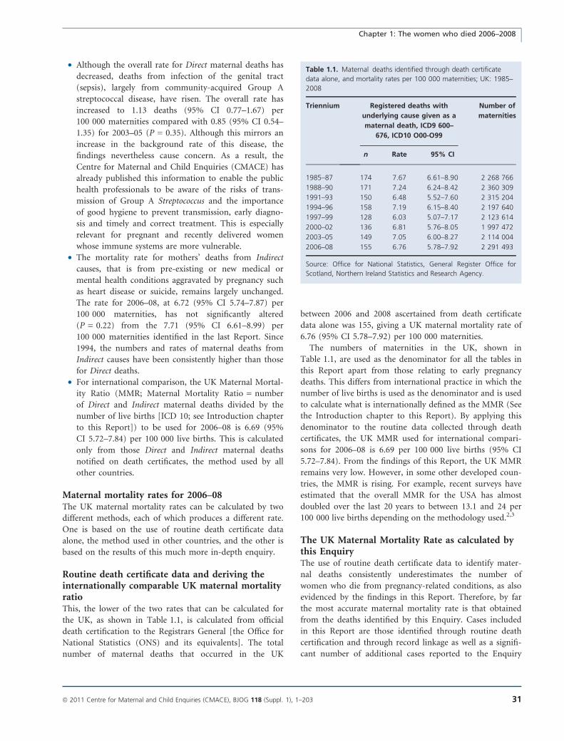
• Although the overall rate for Direct maternal deaths has
decreased, deaths from infection of the genital tract
(sepsis), largely from community-acquired Group A
streptococcal disease, have risen. The overall rate has
increased to 1.13 deaths (95% CI 0.77–1.67) per
100 000 maternities compared with 0.85 (95% CI 0.54–
1.35) for 2003–05 (P = 0.35). Although this mirrors an
increase in the background rate of this disease, the
findings nevertheless cause concern. As a result, the
Centre for Maternal and Child Enquiries (CMACE) has
already published this information to enable the public
health professionals to be aware of the risks of trans-
mission of Group A Streptococcus and the importance
of good hygiene to prevent transmission, early diagno-
sis and timely and correct treatment. This is especially
relevant for pregnant and recently delivered women
whose immune systems are more vulnerable.
• The mortality rate for mothers’ deaths from Indirect
causes, that is from pre-existing or new medical or
mental health conditions aggravated by pregnancy such
as heart disease or suicide, remains largely unchanged.
The rate for 2006–08, at 6.72 (95% CI 5.74–7.87) per
100 000 maternities, has not significantly altered
(P = 0.22) from the 7.71 (95% CI 6.61–8.99) per
100 000 maternities identified in the last Report. Since
1994, the numbers and rates of maternal deaths from
Indirect causes have been consistently higher than those
for Direct deaths.
• For international comparison, the UK Maternal Mortal-
ity Ratio (MMR; Maternal Mortality Ratio = number
of Direct and Indirect maternal deaths divided by the
number of live births [ICD 10; see Introduction chapter
to this Report]) to be used for 2006–08 is 6.69 (95%
CI 5.72–7.84) per 100 000 live births. This is calculated
only from those Direct and Indirect maternal deaths
notified on death certificates, the method used by all
other countries.
Maternal mortality rates for 2006–08The UK maternal mortality rates can be calculated by two
different methods, each of which produces a different rate.
One is based on the use of routine death certificate data
alone, the method used in other countries, and the other is
based on the results of this much more in-depth enquiry.
Routine death certificate data and deriving theinternationally comparable UK maternal mortalityratioThis, the lower of the two rates that can be calculated for
the UK, as shown in Table 1.1, is calculated from official
death certification to the Registrars General [the Office for
National Statistics (ONS) and its equivalents]. The total
number of maternal deaths that occurred in the UK
between 2006 and 2008 ascertained from death certificate
data alone was 155, giving a UK maternal mortality rate of
6.76 (95% CI 5.78–7.92) per 100 000 maternities.
The numbers of maternities in the UK, shown in
Table 1.1, are used as the denominator for all the tables in
this Report apart from those relating to early pregnancy
deaths. This differs from international practice in which the
number of live births is used as the denominator and is used
to calculate what is internationally defined as the MMR (See
the Introduction chapter to this Report). By applying this
denominator to the routine data collected through death
certificates, the UK MMR used for international compari-
sons for 2006–08 is 6.69 per 100 000 live births (95% CI
5.72–7.84). From the findings of this Report, the UK MMR
remains very low. However, in some other developed coun-
tries, the MMR is rising. For example, recent surveys have
estimated that the overall MMR for the USA has almost
doubled over the last 20 years to between 13.1 and 24 per
100 000 live births depending on the methodology used.2,3
The UK Maternal Mortality Rate as calculated bythis EnquiryThe use of routine death certificate data to identify mater-
nal deaths consistently underestimates the number of
women who die from pregnancy-related conditions, as also
evidenced by the findings in this Report. Therefore, by far
the most accurate maternal mortality rate is that obtained
from the deaths identified by this Enquiry. Cases included
in this Report are those identified through routine death
certification and through record linkage as well as a signifi-
cant number of additional cases reported to the Enquiry
Table 1.1. Maternal deaths identified through death certificate
data alone, and mortality rates per 100 000 maternities; UK: 1985–
2008
Triennium Registered deaths with
underlying cause given as a
maternal death, ICD9 600–
676, ICD10 O00-O99
Number of
maternities
n Rate 95% CI
1985–87 174 7.67 6.61–8.90 2 268 766
1988–90 171 7.24 6.24–8.42 2 360 309
1991–93 150 6.48 5.52–7.60 2 315 204
1994–96 158 7.19 6.15–8.40 2 197 640
1997–99 128 6.03 5.07–7.17 2 123 614
2000–02 136 6.81 5.76–8.05 1 997 472
2003–05 149 7.05 6.00–8.27 2 114 004
2006–08 155 6.76 5.78–7.92 2 291 493
Source: Office for National Statistics, General Register Office for
Scotland, Northern Ireland Statistics and Research Agency.
Chapter 1: The women who died 2006–2008
ª 2011 Centre for Maternal and Child Enquiries (CMACE), BJOG 118 (Suppl. 1), 1–203 31
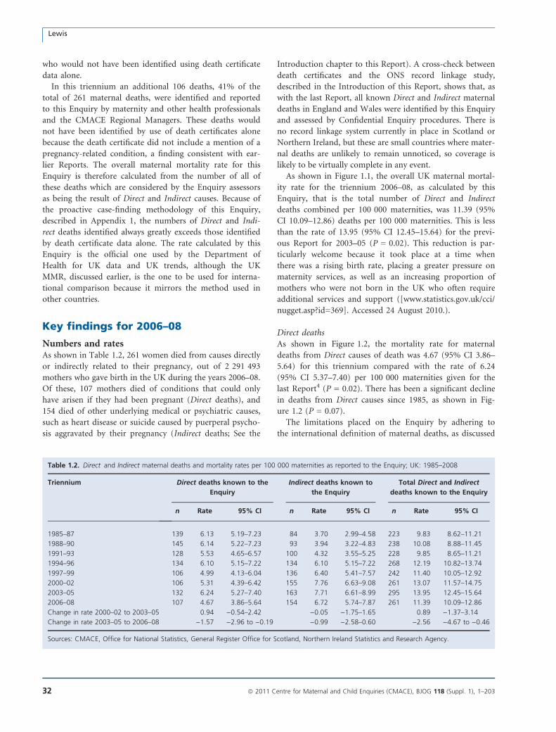
who would not have been identified using death certificate
data alone.
In this triennium an additional 106 deaths, 41% of the
total of 261 maternal deaths, were identified and reported
to this Enquiry by maternity and other health professionals
and the CMACE Regional Managers. These deaths would
not have been identified by use of death certificates alone
because the death certificate did not include a mention of a
pregnancy-related condition, a finding consistent with ear-
lier Reports. The overall maternal mortality rate for this
Enquiry is therefore calculated from the number of all of
these deaths which are considered by the Enquiry assessors
as being the result of Direct and Indirect causes. Because of
the proactive case-finding methodology of this Enquiry,
described in Appendix 1, the numbers of Direct and Indi-
rect deaths identified always greatly exceeds those identified
by death certificate data alone. The rate calculated by this
Enquiry is the official one used by the Department of
Health for UK data and UK trends, although the UK
MMR, discussed earlier, is the one to be used for interna-
tional comparison because it mirrors the method used in
other countries.
Key findings for 2006–08
Numbers and ratesAs shown in Table 1.2, 261 women died from causes directly
or indirectly related to their pregnancy, out of 2 291 493
mothers who gave birth in the UK during the years 2006–08.
Of these, 107 mothers died of conditions that could only
have arisen if they had been pregnant (Direct deaths), and
154 died of other underlying medical or psychiatric causes,
such as heart disease or suicide caused by puerperal psycho-
sis aggravated by their pregnancy (Indirect deaths; See the
Introduction chapter to this Report). A cross-check between
death certificates and the ONS record linkage study,
described in the Introduction of this Report, shows that, as
with the last Report, all known Direct and Indirect maternal
deaths in England and Wales were identified by this Enquiry
and assessed by Confidential Enquiry procedures. There is
no record linkage system currently in place in Scotland or
Northern Ireland, but these are small countries where mater-
nal deaths are unlikely to remain unnoticed, so coverage is
likely to be virtually complete in any event.
As shown in Figure 1.1, the overall UK maternal mortal-
ity rate for the triennium 2006–08, as calculated by this
Enquiry, that is the total number of Direct and Indirect
deaths combined per 100 000 maternities, was 11.39 (95%
CI 10.09–12.86) deaths per 100 000 maternities. This is less
than the rate of 13.95 (95% CI 12.45–15.64) for the previ-
ous Report for 2003–05 (P = 0.02). This reduction is par-
ticularly welcome because it took place at a time when
there was a rising birth rate, placing a greater pressure on
maternity services, as well as an increasing proportion of
mothers who were not born in the UK who often require
additional services and support ([www.statistics.gov.uk/cci/
nugget.asp?id=369]. Accessed 24 August 2010.).
Direct deathsAs shown in Figure 1.2, the mortality rate for maternal
deaths from Direct causes of death was 4.67 (95% CI 3.86–
5.64) for this triennium compared with the rate of 6.24
(95% CI 5.37–7.40) per 100 000 maternities given for the
last Report4 (P = 0.02). There has been a significant decline
in deaths from Direct causes since 1985, as shown in Fig-
ure 1.2 (P = 0.07).
The limitations placed on the Enquiry by adhering to
the international definition of maternal deaths, as discussed
Table 1.2. Direct and Indirect maternal deaths and mortality rates per 100 000 maternities as reported to the Enquiry; UK: 1985–2008
Triennium Direct deaths known to the
Enquiry
Indirect deaths known to
the Enquiry
Total Direct and Indirect
deaths known to the Enquiry
n Rate 95% CI n Rate 95% CI n Rate 95% CI
1985–87 139 6.13 5.19–7.23 84 3.70 2.99–4.58 223 9.83 8.62–11.21
1988–90 145 6.14 5.22–7.23 93 3.94 3.22–4.83 238 10.08 8.88–11.45
1991–93 128 5.53 4.65–6.57 100 4.32 3.55–5.25 228 9.85 8.65–11.21
1994–96 134 6.10 5.15–7.22 134 6.10 5.15–7.22 268 12.19 10.82–13.74
1997–99 106 4.99 4.13–6.04 136 6.40 5.41–7.57 242 11.40 10.05–12.92
2000–02 106 5.31 4.39–6.42 155 7.76 6.63–9.08 261 13.07 11.57–14.75
2003–05 132 6.24 5.27–7.40 163 7.71 6.61–8.99 295 13.95 12.45–15.64
2006–08 107 4.67 3.86–5.64 154 6.72 5.74–7.87 261 11.39 10.09–12.86
Change in rate 2000–02 to 2003–05 0.94 )0.54–2.42 )0.05 )1.75–1.65 0.89 )1.37–3.14
Change in rate 2003–05 to 2006–08 )1.57 )2.96 to )0.19 )0.99 )2.58–0.60 )2.56 )4.67 to )0.46
Sources: CMACE, Office for National Statistics, General Register Office for Scotland, Northern Ireland Statistics and Research Agency.
Lewis
32 ª 2011 Centre for Maternal and Child Enquiries (CMACE), BJOG 118 (Suppl. 1), 1–203
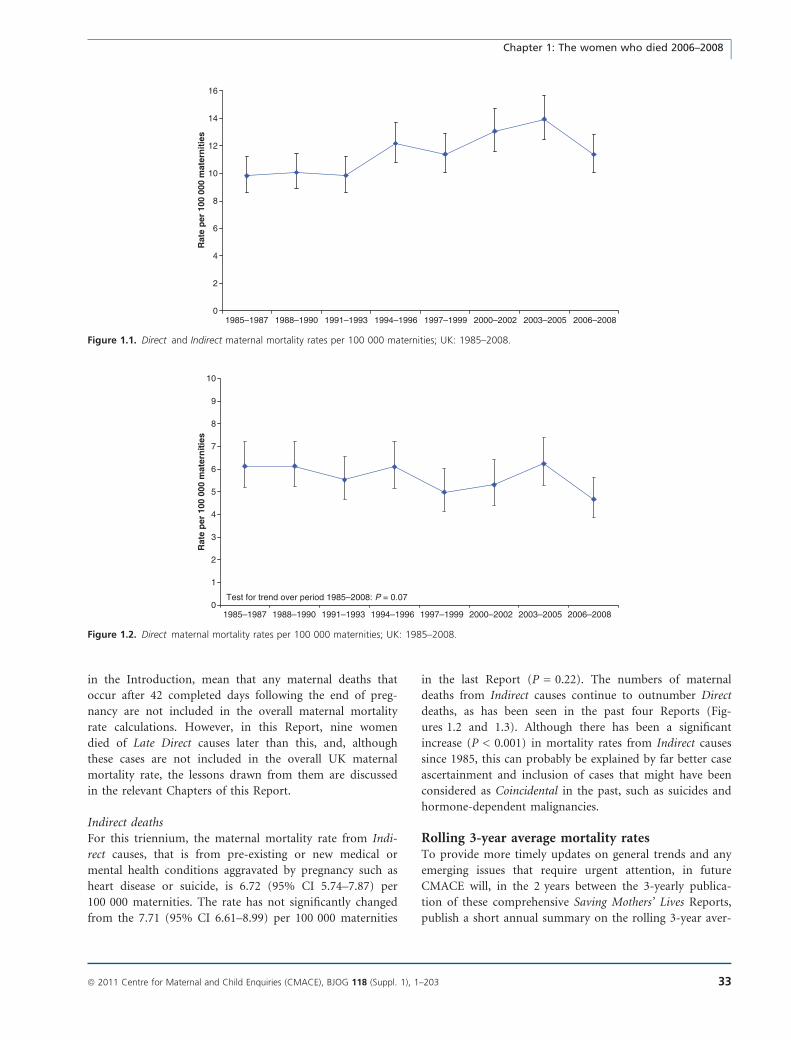
in the Introduction, mean that any maternal deaths that
occur after 42 completed days following the end of preg-
nancy are not included in the overall maternal mortality
rate calculations. However, in this Report, nine women
died of Late Direct causes later than this, and, although
these cases are not included in the overall UK maternal
mortality rate, the lessons drawn from them are discussed
in the relevant Chapters of this Report.
Indirect deathsFor this triennium, the maternal mortality rate from Indi-
rect causes, that is from pre-existing or new medical or
mental health conditions aggravated by pregnancy such as
heart disease or suicide, is 6.72 (95% CI 5.74–7.87) per
100 000 maternities. The rate has not significantly changed
from the 7.71 (95% CI 6.61–8.99) per 100 000 maternities
in the last Report (P = 0.22). The numbers of maternal
deaths from Indirect causes continue to outnumber Direct
deaths, as has been seen in the past four Reports (Fig-
ures 1.2 and 1.3). Although there has been a significant
increase (P < 0.001) in mortality rates from Indirect causes
since 1985, this can probably be explained by far better case
ascertainment and inclusion of cases that might have been
considered as Coincidental in the past, such as suicides and
hormone-dependent malignancies.
Rolling 3-year average mortality ratesTo provide more timely updates on general trends and any
emerging issues that require urgent attention, in future
CMACE will, in the 2 years between the 3-yearly publica-
tion of these comprehensive Saving Mothers’ Lives Reports,
publish a short annual summary on the rolling 3-year aver-
0
2
4
6
8
10
12
14
16
1985–1987 1988–1990 1991–1993 1994–1996 1997–1999 2000–2002 2003–2005 2006–2008
Rat
e p
er 1
00 0
00 m
ater
nit
ies
Figure 1.1. Direct and Indirect maternal mortality rates per 100 000 maternities; UK: 1985–2008.
0
1
2
3
4
5
6
7
8
9
10
1985–1987 1988–1990 1991–1993 1994–1996 1997–1999 2000–2002 2003–2005 2006–2008
Rat
e p
er 1
00 0
00 m
ater
nit
ies
Test for trend over period 1985–2008: P = 0.07
Figure 1.2. Direct maternal mortality rates per 100 000 maternities; UK: 1985–2008.
Chapter 1: The women who died 2006–2008
ª 2011 Centre for Maternal and Child Enquiries (CMACE), BJOG 118 (Suppl. 1), 1–203 33
age rates and trends. As a baseline for these annual
updates, Table 1.3 provides the first of these 3-year rolling
average UK maternal mortality rates since 2003. It shows
that there has been a continuing small decline in both
Direct and Indirect deaths (P = 0.01).
Deaths in pregnant women or new mothers up to6 months after delivery or the end of pregnancywhich were not apparently to the result of, oraffected by, pregnancy
Coincidental deathsTo ascertain which of the deaths in pregnant or recently
delivered women were directly or indirectly related to their
pregnancy, it is important to assess all the cases of mothers
who died while pregnant or in the 42 days following deliv-
ery. Those deaths then found to be unrelated to pregnancy
and which appear to be linked only by a temporal associa-
tion are classified as being Coincidental. By international
convention, these do not contribute to the statistics used to
calculate the UK maternal mortality rate determined by this
Enquiry (See the Introduction chapter to this Report). The
deaths of 50 women who died of such Coincidental causes
were assessed this triennium. This was a similar number to
the 55 identified in the last Report. However, although
apparently unrelated to pregnancy, these deaths often con-
tain important public-health messages, for example con-
cerning domestic abuse, substance misuse and the use of
seat belts, and are discussed in Chapter 12.
Late deathsLate maternal deaths are internationally classified as those
occurring between 6 weeks and 1 year (43–365 days) after
delivery. They can be from Direct, Indirect or Coincidental
causes, but none are included in the internationally defined
maternal mortality rate. For over 15 years these have been
regularly notified to the Enquiry through the record linkage
system developed by the ONS, described in detail in earlier
Reports, latterly also assisted by the CMACE Regional
Managers.
Experience has shown that although some of these
deaths continue to reveal important lessons for clinical care
0
1
2
3
4
5
6
7
8
9
10
1985–1987 1988–1990 1991–1993 1994–1996 1997–1999 2000–2002 2003–2005 2006–2008
Rat
e p
er 1
00 0
00 m
ater
nit
ies
Test for trend over period 1985–2008: P < 0.001
Figure 1.3. Indirect maternal mortality rates per 100 000 maternities; UK: 1985–2008.
Table 1.3. Rolling 3-year average Direct and Indirect maternal deaths and mortality rates per 100 000 maternities as reported to the Enquiry;
UK: 2003–08
3-year period Total UK maternities Direct deaths Indirect deaths Total Direct and Indirect
deaths
n Rate 95% CI n Rate 95% CI n Rate 95% CI
2003–05 2 114 004 132 6.24 5.26–7.41 163 7.71 6.61–8.99 295 13.95 12.45–15.64
2004–06 2 165 909 118 5.45 4.55–6.53 154 7.11 6.07–8.33 272 12.56 11.15–14.14
2005–07 2 220 979 113 5.09 4.23–6.12 146 6.57 5.59–7.73 259 11.66 10.32–13.17
2006–08 2 291 493 107 4.67 3.86–5.64 154 6.72 5.74–7.87 261 11.39 10.09–12.86
Lewis
34 ª 2011 Centre for Maternal and Child Enquiries (CMACE), BJOG 118 (Suppl. 1), 1–203
or maternity or community services, the majority, which
are from Coincidental causes, do not, and most of these
deaths have not been comprehensively assessed this trien-
nium. The exceptions are those mothers who died from
Late Direct and Late Indirect causes. Late Direct deaths are
important because, however long after delivery the terminal
event occurred and even though their lives may have been
prolonged through intensive care or support, there are
often lessons to be learnt from the presentation or manage-
ment of these mothers precipitating pregnancy-related or
childbirth-related events. There may also be crucial lessons
to be learnt from the deaths of mothers who died from
Late Indirect causes such as cardiomyopathy and suicide,
which generally tend to occur some months after delivery.
Thirty-three deaths, nine from Late Direct and 24 from
Late Indirect causes, which occurred within 6 months of
delivery, have been included and assessed and are listed in
Chapter 12 of this Report. CMACE did not collect infor-
mation on any other type of Late death this triennium.
The children left behindIn the period of this Report, it is estimated that at least
331 existing children and 147 live newborn babies lost their
mother to a reported Direct or Indirect death. Of the exist-
ing children whose mothers died a Direct or Indirect death,
26 were already in the care of social services, as were 23
children whose mothers died from Coincidental causes,
mainly related to substance misuse or violence. Any child
whose mother dies must face a far poorer start to family
life. The fact that so many of the children were already liv-
ing in complex circumstances with vulnerable families, or
were in care, continues to underscore the important pub-
lic-health dimension of this Enquiry.
The clinical reasons why mothers diedTable 1.4 gives the numbers and maternal mortality rates
per 100 000 maternities by specific cause of death by trien-
nium, since 1985, when the Enquiry expanded to cover the
whole of the UK. Table 1.5 shows the rates and 95% confi-
dence intervals for the main causes of maternal death in
the last three triennia for 2000–08 and Figure 1.4 shows
the leading causes of maternal deaths for this triennium in
ranked order.
Leading causes of death
Direct deathsThis triennium, there has been a change in the rankings of
Direct deaths by cause. Unlike in previous Reports, the
leading cause of Direct deaths for 2006–08 was genital tract
infection, described here as sepsis, followed by pre-eclamp-
sia/eclampsia, which keeps its second place ranking. Deaths
from thromboembolism, the leading cause of death in the
UK since 1985, have now dropped into third place, fol-
lowed by those from amniotic fluid embolism. Deaths from
haemorrhage have also dropped, to sixth place, following
those in early pregnancy. Mortality from anaesthesia
remains very low and is still the seventh Direct cause. Over-
all, the total numbers of Direct deaths have declined from
132 in the last Report to 107 in this. The decline in the
mortality rate from thromboembolism between the 2003–
05 and 2006–08 triennia is statistically significant
(P < 0.001), but the changes in other rates are small
enough to be chance findings. It will be important to see if
these changes are sustained in the Report for 2009–11.
Indirect deathsCardiac disease was again the leading cause of deaths from
Indirect causes as well as being the leading cause of death
overall. Mortality rates for deaths from Other Indirect
causes due to pre-existing medical conditions and those
from neurological conditions are also higher than for the
leading cause of Direct death, sepsis. Deaths from suicide,
the leading cause of maternal deaths in 2000–02, have
dropped a little more and now equal those from amniotic
fluid embolism.
The care the mothers received
Antenatal careTable 1.6 shows the type of antenatal care provided for the
mothers who died.
The women who did not come for careForty-one women who died from Direct or Indirect causes
had no antenatal care. For 27 it was because they died in
early pregnancy before their ‘booking’ appointment, so
only 14 (6%) women who died in later pregnancy or after
delivery received no antenatal care at all. Six of these con-
cealed their pregnancies. Another 21 (8%) booked for care
after 18 weeks of gestation. For another 11 women, these
data were not available. These 35 mothers (13%) who died
and who had not ‘booked’ by 18 weeks of gestation con-
trast with the 4% of mothers found not to have ‘booked’
with NHS maternity services by 18 weeks of gestation in a
study undertaken by the National Perinatal Epidemiology
Unit in 2006, covering the period of this Report.5 In 2010,
the latest NHS vital signs survey by the Department of
Health for England6 reported that 87% of mothers were
now booked by 13 weeks of gestation, compared with 58%
of the women who died from Direct and Indirect causes in
this Report. In addition to these late bookers, another 35
(14%) women were poor attenders for care, giving a total
of 26% of the mothers who died from Direct and Indirect
causes who were poor or nonattenders and, as a result, had
less than optimal antenatal care.
Chapter 1: The women who died 2006–2008
ª 2011 Centre for Maternal and Child Enquiries (CMACE), BJOG 118 (Suppl. 1), 1–203 35
Tab
le1.4
.N
um
ber
san
dra
tes
of
lead
ing
cause
sof
mat
ernal
dea
ths;
UK
:1985–2
008
Cau
seo
fd
eath
Nu
mb
ers
Rate
sp
er
100
000
mate
rnit
ies
1985–8
71988–9
01991–9
31994–9
61997–9
92000–0
22003–0
52006–0
81985–8
71988–9
01991–9
31994–9
61997–9
92000–0
22003–0
52006–0
8
Dir
ect
death
s
Sepsi
s9
17
15
16
18
13
18
26
0.4
00.7
20.6
50.7
30.8
50.6
50.8
51.1
3
Pre-
ecla
mpsi
aan
dec
lam
psi
a27
27
20
20
16
14
18
19
1.1
91.1
40.8
60.9
10.7
50.7
00.8
50.8
3
Thro
mbosi
san
d
thro
mboem
bolis
m
32
33
35
48
35
30
41
18
1.4
11.4
01.5
12.1
81.6
51.5
01.9
40.7
9
Am
nio
tic
fluid
embolis
m9
11
10
17
85
17
13
0.4
00.4
70.4
30.7
70.3
80.2
50.8
00.5
7
Early
pre
gnan
cydea
ths*
16
24
17
15
17
15
14
11
0.7
11.0
20.7
30.6
80.8
00.7
50.6
60.4
8
Ecto
pic
11
15
912
13
11
10
60.4
80.6
40.3
90.5
50.6
10.5
50.4
70.2
6
Sponta
neo
us
mis
carr
iage
46
32
21
15
0.1
80.2
50.1
30.0
90.0
90.0
50.0
50.2
2
Legal
term
inat
ion
13
51
23
20
0.0
40.1
30.2
20.0
50.0
90.1
50.0
90.0
0
Oth
er0
02
00
01
00.0
00.0
00.0
90.0
00.0
00.0
00.0
50.0
0
Hae
morr
hag
e10
22
15
12
717
14
90.4
40.9
30.6
50.5
50.3
30.8
50.6
60.3
9
Anae
sthes
ia6
48
13
66
70.2
60.1
70.3
50.0
50.1
40.3
00.2
80.3
1
Oth
erD
irec
t27
17
14
77
84
41.1
90.7
20.6
00.3
20.3
30.4
00.1
90.1
7
Gen
ital
trac
ttr
aum
a6
34
52
13
00.2
60.1
30.1
70.2
30.0
90.0
50.1
40.0
0
Fatt
yliv
er6
52
24
31
30.2
60.2
10.0
90.0
90.1
90.1
50.0
50.1
3
Oth
erca
use
s15
98
01
40
10.6
60.3
80.3
50.0
00.0
50.2
00.0
00.0
4
All
Direc
t139
145
128
134
106
106
132
107
6.1
36.1
45.5
36.1
04.9
95.3
16.2
44.6
7
Ind
irect
Car
dia
cdis
ease
23
18
37
39
35
44
48
53
1.0
10.7
61.6
01.7
71.6
52.2
02.2
72.3
1
Indirec
tneu
rolo
gic
al
conditio
ns
19
30
25
47
34
40
37
36
0.8
41.2
71.0
82.1
41.6
02.0
01.7
51.5
7
Psyc
hia
tric
cause
s–
––
915
16
18
13
––
–0.4
10.7
10.8
00.8
50.5
7
Indirec
tm
alig
nan
cies
––
––
11
510
3–
––
–0.5
20.2
50.4
70.1
3
Oth
erIn
direc
tca
use
s43
45
38
39
41
50
50
49
1.9
01.9
11.6
41.7
71.9
32.5
02.3
72.1
4
All
Indirec
t84
93
100
134
136
155
163
154
3.7
03.9
44.3
26.1
06.4
07.7
67.7
16.5
9
Co
inci
den
tal
26
39
46
36
29
36
55
50
1.1
51.6
51.9
91.6
41.3
71.8
02.6
02.1
8
Late
Direc
t–
13
10
47
411
9
Indirec
t–
10
23
32
39
45
71
24
*Th
eEa
rly
Preg
nan
cydea
ths
cate
gory
incl
udes
only
those
wom
enw
ho
die
dfr
om
the
follo
win
gD
irec
tca
use
s:ec
topic
pre
gnan
cy,
mis
carr
iage,
term
inat
ion
of
pre
gnan
cyor
oth
erra
reD
irec
t
conditio
ns
bef
ore
24
com
ple
ted
wee
ksof
pre
gnan
cynot
counte
del
sew
her
e.Th
ose
wom
enw
ho
die
dfr
om
oth
erca
use
sbef
ore
24
wee
ksof
ges
tation
are
counte
din
the
rele
vant
chap
ters
,
e.g.
Embolis
m,
Sepsi
s,In
direc
tet
c.
Lewis
36 ª 2011 Centre for Maternal and Child Enquiries (CMACE), BJOG 118 (Suppl. 1), 1–203
Maternity team and consultant led careFor 42% of the women who died, antenatal care was shared
between their GP, midwife and their obstetrician; so-called
traditional ‘shared care’. Many of these women saw a
member of the obstetric staff once or twice to check that
all was well, and in the main their care was managed by a
midwife. Forty-three (17%) of the mothers who died were
known to have been at higher risk of complications, and
their care was provided by the consultant-led maternity
team, including obstetricians and midwives. These propor-
tions have not changed since the last Report.
Midwifery careFifty-two (20%) of the women for whom information is
available had entirely midwifery-led care. For most this was
appropriate, but this is discussed further in Chapter 13.
This is double the number of women who received such
care in the last Report, but this would be expected because
it is in line with the policies for NHS maternity care
expressed in Maternity Matters7 and other documents.
These policies support the role of the midwife as being the
expert in the provision of routine antenatal and postnatal
maternity care and normal birth for women for whom
there are no known potential risk factors or complications.
As a reflection of the changing nature of maternity service
provision, only 4% of women had their care shared
between midwives and their GPs.
The women who died before deliveryAs shown in Table 1.7, 37% of women who died from
Direct or Indirect causes were undelivered at the time of
their death. For Direct deaths, the largest group were
women who died in early pregnancy of miscarriage, ectopic
pregnancy or sepsis. The causes of deaths among the Indi-
rect deaths groups were varied between medical conditions
including epilepsy and diabetes, as well as cardiac disease.
Eight of the 11 women who were murdered and whose
deaths were classified as Coincidental died while still preg-
Table 1.5. Numbers and rates per 100 000 maternities of maternal deaths reported to the Enquiry by cause; UK: 2000–08
Cause of death 2000–02 2003–05 2006–08
n Rate 95% CI n Rate 95% CI n Rate 95% CI
Direct deaths
Sepsis* 13 0.65 0.38–1.12 18 0.85 0.54–1.35 26 1.13 0.77–1.67
Pre-eclampsia and eclampsia 14 0.70 0.42–1.18 18 0.85 0.54–1.35 19 0.83 0.53–1.30
Thrombosis and thromboembolism 30 1.50 1.05–2.15 41 1.94 1.43–2.63 18 0.79 0.49–1.25
Amniotic fluid embolism 5 0.25 0.10–0.60 17 0.80 0.50–1.29 13 0.57 0.33–0.98
Early pregnancy deaths 15 0.75 0.45–1.25 14 0.66 0.39–1.12 11 0.48 0.27–0.87
Ectopic 11 0.55 0.30–0.99 10 0.47 0.25–0.88 6 0.26 0.12–0.58
Spontaneous miscarriage 1 0.05 0.01–0.36 1 0.05 0.01–0.34 5 0.22 0.09–0.52
Legal termination 3 0.15 0.05–0.47 2 0.09 0.02–0.38 0 0.00
Other 0 0.00 1 0.05 0.01–0.34 0 0.00
Haemorrhage 17 0.85 0.53–1.37 14 0.66 0.39–1.12 9 0.39 0.20–0.75
Anaesthesia 6 0.30 0.13–0.67 6 0.28 0.13–0.63 7 0.31 0.15–0.64
Other Direct 8 0.40 0.20–0.80 4 0.19 0.07–0.50 4 0.17 0.07–0.47
Genital tract trauma 1 0.05 0.01–0.36 3 0.14 0.05–0.44 0 0.00
Fatty liver 3 0.15 0.05–0.47 1 0.05 0.01–0.34 3 0.13 0.04–0.41
Other causes 4 0.20 0.08–0.53 0 0.00 1 0.04 0.01–0.31
All Direct 106 5.31 4.39–6.42 132 6.24 5.26–7.41 107 4.67 3.86–5.64
Indirect
Cardiac disease 44 2.20 1.64–2.96 48 2.27 1.71–3.01 53 2.31 1.77–3.03
Other Indirect causes 50 2.50 1.90–3.30 50 2.37 1.79–3.12 49 2.14 1.62–2.83
Indirect neurological conditions 40 2.00 1.47–2.73 37 1.75 1.27–2.42 36 1.57 1.13–2.18
Psychiatric causes 16 0.80 0.49–1.31 18 0.85 0.54–1.35 13 0.57 0.33–0.98
Indirect malignancies 5 0.25 0.10–0.60 10 0.47 0.25–0.88 3 0.13 0.04–0.41
All Indirect 155 7.76 6.63–9.08 163 7.71 6.61–8.99 154 6.72 5.74–7.87
Coincidental 36 1.80 1.30–2.50 55 2.60 2.00–3.39 50 2.18 1.65–2.88
Late deaths
Direct 4 11 9
Indirect 45 71 24
*Including early pregnancy deaths as the result of sepsis.
Sources: CMACE, Office for National Statistics, General Register Office for Scotland, Northern Ireland Statistics and Research Agency.
Chapter 1: The women who died 2006–2008
ª 2011 Centre for Maternal and Child Enquiries (CMACE), BJOG 118 (Suppl. 1), 1–203 37
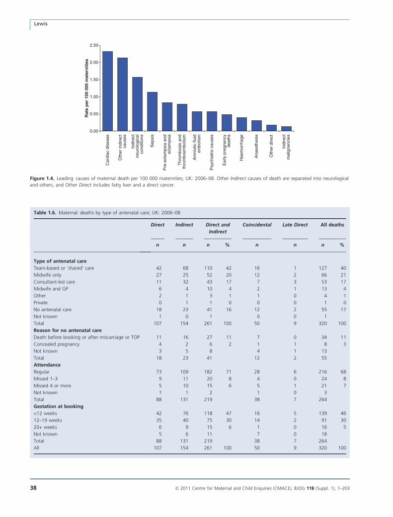
0.00
0.50
1.00
1.50
2.00
2.50
Car
diac
dis
ease
Oth
er in
dire
ctca
uses
Indi
rect
neur
olog
ical
cond
ition
s
Sep
sis
Pre
-ecl
amps
ia a
ndec
lam
psia
Thr
ombo
sis
and
thro
mbo
embo
lism
Am
niot
ic fl
uid
embo
lism
Psy
chia
tric
cau
ses
Ear
ly p
regn
ancy
deat
hs
Hae
mor
rhag
e
Ana
esth
esia
Oth
er d
irect
Indi
rect
mal
igna
ncie
s
Rat
e p
er 1
00 0
00 m
ater
nit
ies
Figure 1.4. Leading causes of maternal death per 100 000 maternities; UK: 2006–08. Other Indirect causes of death are separated into neurological
and others, and Other Direct includes fatty liver and a direct cancer.
Table 1.6. Maternal deaths by type of antenatal care; UK: 2006–08
Direct Indirect Direct and
Indirect
Coincidental Late Direct All deaths
n n n % n n n %
Type of antenatal care
Team-based or ‘shared’ care 42 68 110 42 16 1 127 40
Midwife only 27 25 52 20 12 2 66 21
Consultant-led care 11 32 43 17 7 3 53 17
Midwife and GP 6 4 10 4 2 1 13 4
Other 2 1 3 1 1 0 4 1
Private 0 1 1 0 0 0 1 0
No antenatal care 18 23 41 16 12 2 55 17
Not known 1 0 1 0 0 1
Total 107 154 261 100 50 9 320 100
Reason for no antenatal care
Death before booking or after miscarriage or TOP 11 16 27 11 7 0 34 11
Concealed pregnancy 4 2 6 2 1 1 8 3
Not known 3 5 8 4 1 13
Total 18 23 41 12 2 55
Attendance
Regular 73 109 182 71 28 6 216 68
Missed 1–3 9 11 20 8 4 0 24 8
Missed 4 or more 5 10 15 6 5 1 21 7
Not known 1 1 2 1 0 3
Total 88 131 219 38 7 264
Gestation at booking
<12 weeks 42 76 118 47 16 5 139 46
12–19 weeks 35 40 75 30 14 2 91 30
20+ weeks 6 9 15 6 1 0 16 5
Not known 5 6 11 7 0 18
Total 88 131 219 38 7 264
All 107 154 261 100 50 9 320 100
Lewis
38 ª 2011 Centre for Maternal and Child Enquiries (CMACE), BJOG 118 (Suppl. 1), 1–203
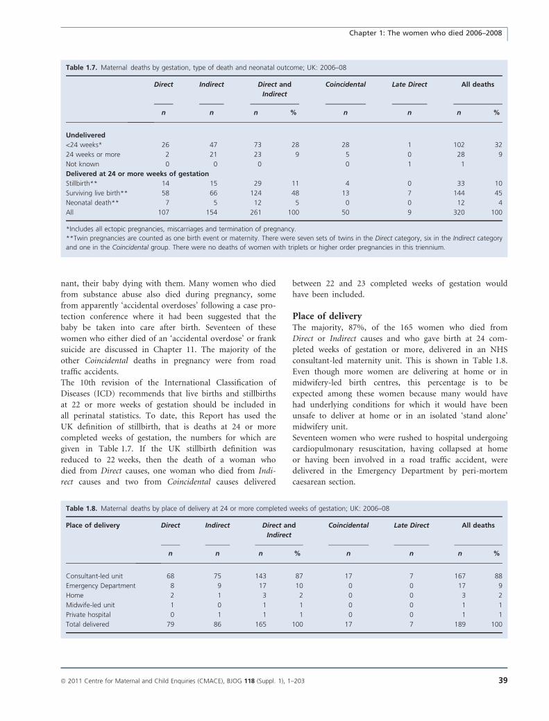
nant, their baby dying with them. Many women who died
from substance abuse also died during pregnancy, some
from apparently ‘accidental overdoses’ following a case pro-
tection conference where it had been suggested that the
baby be taken into care after birth. Seventeen of these
women who either died of an ‘accidental overdose’ or frank
suicide are discussed in Chapter 11. The majority of the
other Coincidental deaths in pregnancy were from road
traffic accidents.
The 10th revision of the International Classification of
Diseases (ICD) recommends that live births and stillbirths
at 22 or more weeks of gestation should be included in
all perinatal statistics. To date, this Report has used the
UK definition of stillbirth, that is deaths at 24 or more
completed weeks of gestation, the numbers for which are
given in Table 1.7. If the UK stillbirth definition was
reduced to 22 weeks, then the death of a woman who
died from Direct causes, one woman who died from Indi-
rect causes and two from Coincidental causes delivered
between 22 and 23 completed weeks of gestation would
have been included.
Place of deliveryThe majority, 87%, of the 165 women who died from
Direct or Indirect causes and who gave birth at 24 com-
pleted weeks of gestation or more, delivered in an NHS
consultant-led maternity unit. This is shown in Table 1.8.
Even though more women are delivering at home or in
midwifery-led birth centres, this percentage is to be
expected among these women because many would have
had underlying conditions for which it would have been
unsafe to deliver at home or in an isolated ‘stand alone’
midwifery unit.
Seventeen women who were rushed to hospital undergoing
cardiopulmonary resuscitation, having collapsed at home
or having been involved in a road traffic accident, were
delivered in the Emergency Department by peri-mortem
caesarean section.
Table 1.7. Maternal deaths by gestation, type of death and neonatal outcome; UK: 2006–08
Direct Indirect Direct and
Indirect
Coincidental Late Direct All deaths
n n n % n n n %
Undelivered
<24 weeks* 26 47 73 28 28 1 102 32
24 weeks or more 2 21 23 9 5 0 28 9
Not known 0 0 0 0 1 1
Delivered at 24 or more weeks of gestation
Stillbirth** 14 15 29 11 4 0 33 10
Surviving live birth** 58 66 124 48 13 7 144 45
Neonatal death** 7 5 12 5 0 0 12 4
All 107 154 261 100 50 9 320 100
*Includes all ectopic pregnancies, miscarriages and termination of pregnancy.
**Twin pregnancies are counted as one birth event or maternity. There were seven sets of twins in the Direct category, six in the Indirect category
and one in the Coincidental group. There were no deaths of women with triplets or higher order pregnancies in this triennium.
Table 1.8. Maternal deaths by place of delivery at 24 or more completed weeks of gestation; UK: 2006–08
Place of delivery Direct Indirect Direct and
Indirect
Coincidental Late Direct All deaths
n n n % n n n %
Consultant-led unit 68 75 143 87 17 7 167 88
Emergency Department 8 9 17 10 0 0 17 9
Home 2 1 3 2 0 0 3 2
Midwife-led unit 1 0 1 1 0 0 1 1
Private hospital 0 1 1 1 0 0 1 1
Total delivered 79 86 165 100 17 7 189 100
Chapter 1: The women who died 2006–2008
ª 2011 Centre for Maternal and Child Enquiries (CMACE), BJOG 118 (Suppl. 1), 1–203 39
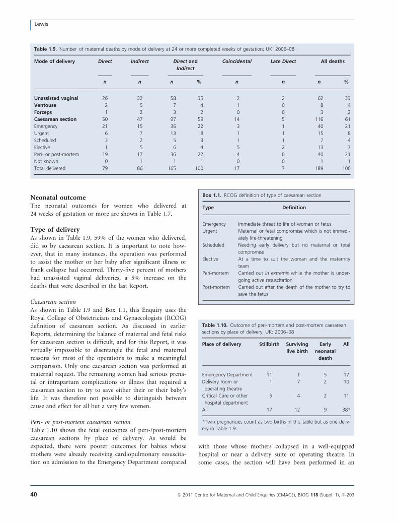
Neonatal outcomeThe neonatal outcomes for women who delivered at
24 weeks of gestation or more are shown in Table 1.7.
Type of deliveryAs shown in Table 1.9, 59% of the women who delivered,
did so by caesarean section. It is important to note how-
ever, that in many instances, the operation was performed
to assist the mother or her baby after significant illness or
frank collapse had occurred. Thirty-five percent of mothers
had unassisted vaginal deliveries, a 5% increase on the
deaths that were described in the last Report.
Caesarean sectionAs shown in Table 1.9 and Box 1.1, this Enquiry uses the
Royal College of Obstetricians and Gynaecologists (RCOG)
definition of caesarean section. As discussed in earlier
Reports, determining the balance of maternal and fetal risks
for caesarean section is difficult, and for this Report, it was
virtually impossible to disentangle the fetal and maternal
reasons for most of the operations to make a meaningful
comparison. Only one caesarean section was performed at
maternal request. The remaining women had serious prena-
tal or intrapartum complications or illness that required a
caesarean section to try to save either their or their baby’s
life. It was therefore not possible to distinguish between
cause and effect for all but a very few women.
Peri- or post-mortem caesarean sectionTable 1.10 shows the fetal outcomes of peri-/post-mortem
caesarean sections by place of delivery. As would be
expected, there were poorer outcomes for babies whose
mothers were already receiving cardiopulmonary resuscita-
tion on admission to the Emergency Department compared
with those whose mothers collapsed in a well-equipped
hospital or near a delivery suite or operating theatre. In
some cases, the section will have been performed in an
Table 1.9. Number of maternal deaths by mode of delivery at 24 or more completed weeks of gestation; UK: 2006–08
Mode of delivery Direct Indirect Direct and
Indirect
Coincidental Late Direct All deaths
n n n % n n n %
Unassisted vaginal 26 32 58 35 2 2 62 33
Ventouse 2 5 7 4 1 0 8 4
Forceps 1 2 3 2 0 0 3 2
Caesarean section 50 47 97 59 14 5 116 61
Emergency 21 15 36 22 3 1 40 21
Urgent 6 7 13 8 1 1 15 8
Scheduled 3 2 5 3 1 1 7 4
Elective 1 5 6 4 5 2 13 7
Peri- or post-mortem 19 17 36 22 4 0 40 21
Not known 0 1 1 1 0 0 1 1
Total delivered 79 86 165 100 17 7 189 100
Box 1.1. RCOG definition of type of caesarean section
Type Definition
Emergency Immediate threat to life of woman or fetus
Urgent Maternal or fetal compromise which is not immedi-
ately life-threatening
Scheduled Needing early delivery but no maternal or fetal
compromise
Elective At a time to suit the woman and the maternity
team
Peri-mortem Carried out in extremis while the mother is under-
going active resuscitation
Post-mortem Carried out after the death of the mother to try to
save the fetus
Table 1.10. Outcome of peri-mortem and post-mortem caesarean
sections by place of delivery; UK: 2006–08
Place of delivery Stillbirth Surviving
live birth
Early
neonatal
death
All
Emergency Department 11 1 5 17
Delivery room or
operating theatre
1 7 2 10
Critical Care or other
hospital department
5 4 2 11
All 17 12 9 38*
*Twin pregnancies count as two births in this table but as one deliv-
ery in Table 1.9.
Lewis
40 ª 2011 Centre for Maternal and Child Enquiries (CMACE), BJOG 118 (Suppl. 1), 1–203
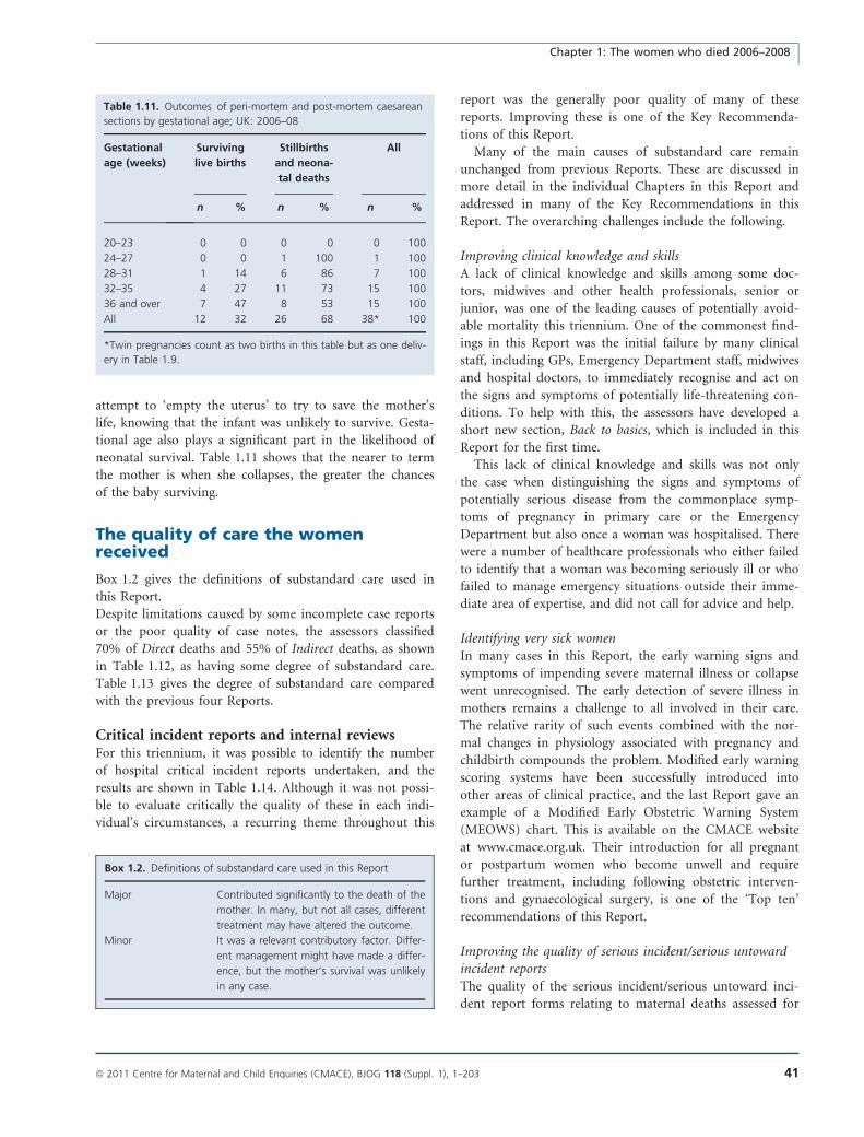
attempt to ‘empty the uterus’ to try to save the mother’s
life, knowing that the infant was unlikely to survive. Gesta-
tional age also plays a significant part in the likelihood of
neonatal survival. Table 1.11 shows that the nearer to term
the mother is when she collapses, the greater the chances
of the baby surviving.
The quality of care the womenreceived
Box 1.2 gives the definitions of substandard care used in
this Report.
Despite limitations caused by some incomplete case reports
or the poor quality of case notes, the assessors classified
70% of Direct deaths and 55% of Indirect deaths, as shown
in Table 1.12, as having some degree of substandard care.
Table 1.13 gives the degree of substandard care compared
with the previous four Reports.
Critical incident reports and internal reviewsFor this triennium, it was possible to identify the number
of hospital critical incident reports undertaken, and the
results are shown in Table 1.14. Although it was not possi-
ble to evaluate critically the quality of these in each indi-
vidual’s circumstances, a recurring theme throughout this
report was the generally poor quality of many of these
reports. Improving these is one of the Key Recommenda-
tions of this Report.
Many of the main causes of substandard care remain
unchanged from previous Reports. These are discussed in
more detail in the individual Chapters in this Report and
addressed in many of the Key Recommendations in this
Report. The overarching challenges include the following.
Improving clinical knowledge and skillsA lack of clinical knowledge and skills among some doc-
tors, midwives and other health professionals, senior or
junior, was one of the leading causes of potentially avoid-
able mortality this triennium. One of the commonest find-
ings in this Report was the initial failure by many clinical
staff, including GPs, Emergency Department staff, midwives
and hospital doctors, to immediately recognise and act on
the signs and symptoms of potentially life-threatening con-
ditions. To help with this, the assessors have developed a
short new section, Back to basics, which is included in this
Report for the first time.
This lack of clinical knowledge and skills was not only
the case when distinguishing the signs and symptoms of
potentially serious disease from the commonplace symp-
toms of pregnancy in primary care or the Emergency
Department but also once a woman was hospitalised. There
were a number of healthcare professionals who either failed
to identify that a woman was becoming seriously ill or who
failed to manage emergency situations outside their imme-
diate area of expertise, and did not call for advice and help.
Identifying very sick womenIn many cases in this Report, the early warning signs and
symptoms of impending severe maternal illness or collapse
went unrecognised. The early detection of severe illness in
mothers remains a challenge to all involved in their care.
The relative rarity of such events combined with the nor-
mal changes in physiology associated with pregnancy and
childbirth compounds the problem. Modified early warning
scoring systems have been successfully introduced into
other areas of clinical practice, and the last Report gave an
example of a Modified Early Obstetric Warning System
(MEOWS) chart. This is available on the CMACE website
at www.cmace.org.uk. Their introduction for all pregnant
or postpartum women who become unwell and require
further treatment, including following obstetric interven-
tions and gynaecological surgery, is one of the ‘Top ten’
recommendations of this Report.
Improving the quality of serious incident/serious untowardincident reportsThe quality of the serious incident/serious untoward inci-
dent report forms relating to maternal deaths assessed for
Table 1.11. Outcomes of peri-mortem and post-mortem caesarean
sections by gestational age; UK: 2006–08
Gestational
age (weeks)
Surviving
live births
Stillbirths
and neona-
tal deaths
All
n % n % n %
20–23 0 0 0 0 0 100
24–27 0 0 1 100 1 100
28–31 1 14 6 86 7 100
32–35 4 27 11 73 15 100
36 and over 7 47 8 53 15 100
All 12 32 26 68 38* 100
*Twin pregnancies count as two births in this table but as one deliv-
ery in Table 1.9.
Box 1.2. Definitions of substandard care used in this Report
Major Contributed significantly to the death of the
mother. In many, but not all cases, different
treatment may have altered the outcome.
Minor It was a relevant contributory factor. Differ-
ent management might have made a differ-
ence, but the mother’s survival was unlikely
in any case.
Chapter 1: The women who died 2006–2008
ª 2011 Centre for Maternal and Child Enquiries (CMACE), BJOG 118 (Suppl. 1), 1–203 41

this report was highly variable with many being of dubious
quality. The assessors considered some to be not worth the
paper they were written on and a few to be actually white-
washes or cover ups for unacceptable situations. For these
poor reports, there was little or no evidence of critical
thinking, an acceptance of shortcomings, little or no self-
reflective discussions and no evidence that obvious lessons
had been identified let alone learnt. In these cases, little or
no action was taken on any results, and in many cases, staff
were not involved in the process or the follow up of any of
the lessons learnt. This was a common finding throughout
all the Chapters in this Report and one which all assessors
agree represents unacceptable practice that must be cor-
rected as soon as possible.
Improving senior supportAs in the last Report, some women were not seen by an
appropriately trained senior or consultant doctor in time,
and a few never saw a consultant doctor at all, despite, in
Table 1.12. Numbers and percentages of cases of Direct and Indirect deaths by cause and degree of substandard care (SSC); UK: 2006–08
Cause Numbers of cases Percentages of cases Percentage of
cases with
no SSC
Total
number of casesMajor Minor Total Major Minor Total
Direct
Thrombosis and thromboembolism 6 4 10 33 22 56 44 18
Pre-eclampsia, eclampsia and acute
fatty liver of pregnancy
14 6 20 64 27 91 9 22
Haemorrhage 4 2 6 44 22 67 33 9
Amniotic fluid embolism 2 6 8 15 46 62 38 13
Early pregnancy deaths 6 – 6 55 – 55 45 11
Sepsis 12 6 18 46 23 69 31 26
Anaesthesia 3 3 6 43 43 86 14 7
Total Direct 47 28* 75* 44 26 70 30 107*
Indirect
Cardiac disease 13 14 27 25 26 51 49 53
Other Indirect causes 17 11 28 33 21 54 46 52
Indirect neurological causes 11 12 23 31 33 64 36 36
Psychiatric causes 6 1 7 46 8 54 46 13
Total Indirect 47 38 85 31 25 55 45 154
Total Direct and Indirect 94 66 160 36 25 61 39 261
*Includes one case from choriocarcinoma that is classified as Direct.
Table 1.13. Numbers and percentages of cases of Direct and
Indirect deaths by degree of substandard care. UK: 1997–2008
Numbers of cases Percentages of
cases
Number
of cases
Major Minor Total Major Minor Total
Direct
1997–99 53 11 64 50 10 60 106
2000–02 50 21 71 47 20 67 106
2003–05 72 12 84 55 9 64 132
2006–08 47 28* 75* 44 26 70 107*
Indirect
1997–99 26 20 46 13 10 22 205
2000–02 31 25 56 20 16 36 155
2003–05 45 20 65 28 12 40 163
2006–08 47 38 85 31 25 55 154
*Includes one case from choriocarcinoma that is classified as Direct.
Table 1.14. Number of critical incident reviews undertaken for
maternal death cases; UK: 2006–08
Type of hospital review Type of death Total
Direct and
Indirect
Direct Indirect n %
Review undertaken 96 100 196 80
Root cause analysis 37 26 63 26
Hospital/Trust review 28 22 50 20
Clinical governance review 15 25 40 16
Other 12 22 34 14
Type of review not known 4 5 9 4
No review undertaken 9 41 50 20
Not known 2 13 15
Total 107 154 261
Lewis
42 ª 2011 Centre for Maternal and Child Enquiries (CMACE), BJOG 118 (Suppl. 1), 1–203
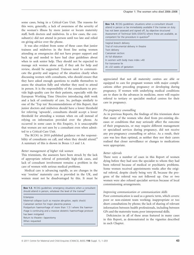
some cases, being in a Critical-Care Unit. The reasons for
this were, generally, a lack of awareness of the severity of
the woman’s illness by more junior or locum maternity
staff, both doctors and midwives. In a few cases, the con-
sultant(s) did not attend in person until too late and relied
on giving advice over the phone.
It was also evident from some of these cases that junior
trainees and midwives in the front line seeing women
attending as emergencies did not have proper support and
back up and needed to have had clear guidelines about
when to seek senior help. They should not be expected to
manage sick women alone and, if they ask for help and
review, should be supported. Trainees need to communi-
cate the gravity and urgency of the situation clearly when
discussing women with consultants, who should ensure that
they have asked enough questions to enable themselves to
assess the situation fully and whether they need to attend
in person. It is the responsibility of the consultants to pro-
vide high-quality care for their patients, especially with the
European Working Time Directive introducing shift work
and a lack of continuity of care. So, perhaps similarly to
one of the ‘Top ten’ Recommendations of this Report, that
junior doctors and midwives should have a lower threshold
for referring ‘upwards’, consultants should accept a lower
threshold for attending a woman when on call instead of
relying on information provided over the phone. As
occurred in some cases in this Enquiry, it is unacceptable
that women are not seen by a consultant even when admit-
ted to a Critical-Care Unit.
The RCOG in 2010 published guidance on the responsi-
bility of consultants on call, and when they should attend.8
A summary of this is shown in Boxes 1.3 and 1.4.
Better management of higher risk womenThis triennium, the assessors have been struck by the lack
of appropriate referral of potentially high-risk cases, and
lack of consultant involvement remains a problem in the
care of women with serious medical problems.
Medical care is advancing rapidly, as are changes in the
way ‘routine’ maternity care is provided in the UK, and
women must not be disadvantaged by this. It must be
appreciated that not all maternity centres are able or
equipped to care for pregnant women with major compli-
cations either preceding pregnancy or developing during
pregnancy. If women with underlying medical conditions
are to share in the advances in medicine, more will require
referral to tertiary or specialist medical centres for their
care in pregnancy.
Pre-pregnancy counsellingAs in previous Reports, the findings of this triennium show
that many of the women who died from pre-existing dis-
eases or conditions that may seriously affect the outcome
of their pregnancies, or may require different management
or specialised services during pregnancy, did not receive
any pre-pregnancy counselling or advice. As a result, their
care was less than optimal, as neither they nor their carers
realised that closer surveillance or changes to medications
were appropriate.
Better referralsThere were a number of cases in this Report of women
dying before they had seen the specialist to whom they had
been referred because of medical or psychiatric problems.
Some women received appointments weeks after the origi-
nal referral, despite clearly being very ill, because the pro-
gress of the referral was not followed up. One or two
women were also refused specialist services because of local
commissioning arrangements.
Improving communication or communication skillsPoor communication is used as a generic term, which covers:
poor or non-existent team working; inappropriate or too
short consultations by phone; the lack of sharing of relevant
information between health professionals, including between
GPs and the maternity team; poor interpersonal skills.
Deficiencies in all of these areas featured in many cases
in this Report, as demonstrated in the vignettes described
in each Chapter.
Box 1.3. RCOG guidelines: emergency situations when a consultant
should attend in person, whatever the level of the trainee8
Eclampsia
Maternal collapse (such as massive abruption, septic shock)
Caesarean section for major placenta praevia
Postpartum haemorrhage of more than 1.5 l where the haemor-
rhage is continuing and a massive obstetric haemorrhage protocol
has been instigated
Return to theatre—laparotomy
When requested
Box 1.4. RCOG guidelines: situations when a consultant should
attend in person or be immediately available if the trainee on duty
has not been assessed and signed off, by objective structured
Assessment of Technical Skills (OSATS) where these are available, as
competent for the procedure in question8
Vaginal breech delivery
Trial of instrumental delivery in theatre
Twin delivery
Caesarean section:
At full dilatation
In women with body mass index >40
For transverse lie
At <32 weeks of gestation
Chapter 1: The women who died 2006–2008
ª 2011 Centre for Maternal and Child Enquiries (CMACE), BJOG 118 (Suppl. 1), 1–203 43
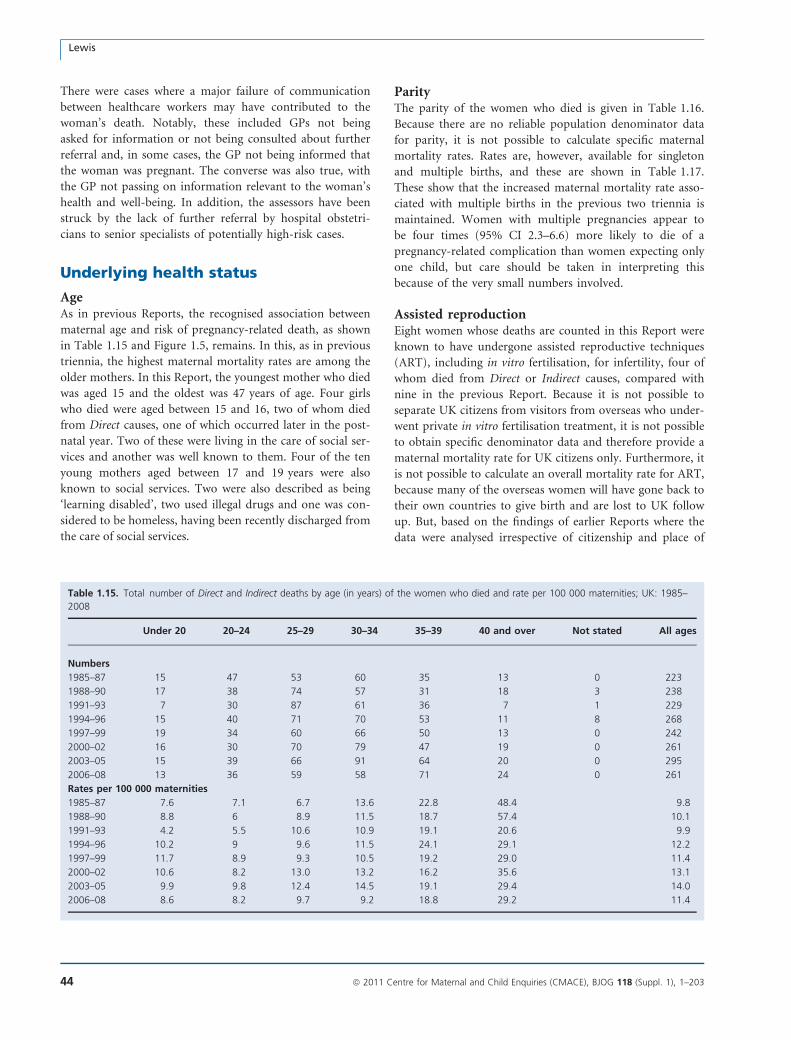
There were cases where a major failure of communication
between healthcare workers may have contributed to the
woman’s death. Notably, these included GPs not being
asked for information or not being consulted about further
referral and, in some cases, the GP not being informed that
the woman was pregnant. The converse was also true, with
the GP not passing on information relevant to the woman’s
health and well-being. In addition, the assessors have been
struck by the lack of further referral by hospital obstetri-
cians to senior specialists of potentially high-risk cases.
Underlying health status
AgeAs in previous Reports, the recognised association between
maternal age and risk of pregnancy-related death, as shown
in Table 1.15 and Figure 1.5, remains. In this, as in previous
triennia, the highest maternal mortality rates are among the
older mothers. In this Report, the youngest mother who died
was aged 15 and the oldest was 47 years of age. Four girls
who died were aged between 15 and 16, two of whom died
from Direct causes, one of which occurred later in the post-
natal year. Two of these were living in the care of social ser-
vices and another was well known to them. Four of the ten
young mothers aged between 17 and 19 years were also
known to social services. Two were also described as being
‘learning disabled’, two used illegal drugs and one was con-
sidered to be homeless, having been recently discharged from
the care of social services.
ParityThe parity of the women who died is given in Table 1.16.
Because there are no reliable population denominator data
for parity, it is not possible to calculate specific maternal
mortality rates. Rates are, however, available for singleton
and multiple births, and these are shown in Table 1.17.
These show that the increased maternal mortality rate asso-
ciated with multiple births in the previous two triennia is
maintained. Women with multiple pregnancies appear to
be four times (95% CI 2.3–6.6) more likely to die of a
pregnancy-related complication than women expecting only
one child, but care should be taken in interpreting this
because of the very small numbers involved.
Assisted reproductionEight women whose deaths are counted in this Report were
known to have undergone assisted reproductive techniques
(ART), including in vitro fertilisation, for infertility, four of
whom died from Direct or Indirect causes, compared with
nine in the previous Report. Because it is not possible to
separate UK citizens from visitors from overseas who under-
went private in vitro fertilisation treatment, it is not possible
to obtain specific denominator data and therefore provide a
maternal mortality rate for UK citizens only. Furthermore, it
is not possible to calculate an overall mortality rate for ART,
because many of the overseas women will have gone back to
their own countries to give birth and are lost to UK follow
up. But, based on the findings of earlier Reports where the
data were analysed irrespective of citizenship and place of
Table 1.15. Total number of Direct and Indirect deaths by age (in years) of the women who died and rate per 100 000 maternities; UK: 1985–
2008
Under 20 20–24 25–29 30–34 35–39 40 and over Not stated All ages
Numbers
1985–87 15 47 53 60 35 13 0 223
1988–90 17 38 74 57 31 18 3 238
1991–93 7 30 87 61 36 7 1 229
1994–96 15 40 71 70 53 11 8 268
1997–99 19 34 60 66 50 13 0 242
2000–02 16 30 70 79 47 19 0 261
2003–05 15 39 66 91 64 20 0 295
2006–08 13 36 59 58 71 24 0 261
Rates per 100 000 maternities
1985–87 7.6 7.1 6.7 13.6 22.8 48.4 9.8
1988–90 8.8 6 8.9 11.5 18.7 57.4 10.1
1991–93 4.2 5.5 10.6 10.9 19.1 20.6 9.9
1994–96 10.2 9 9.6 11.5 24.1 29.1 12.2
1997–99 11.7 8.9 9.3 10.5 19.2 29.0 11.4
2000–02 10.6 8.2 13.0 13.2 16.2 35.6 13.1
2003–05 9.9 9.8 12.4 14.5 19.1 29.4 14.0
2006–08 8.6 8.2 9.7 9.2 18.8 29.2 11.4
Lewis
44 ª 2011 Centre for Maternal and Child Enquiries (CMACE), BJOG 118 (Suppl. 1), 1–203

delivery, there appeared to be no appreciable increase in the
maternal mortality rate associated with ART. The small
numbers in this Report suggest that it does not seem to have
been the case in this triennium either.
ObesityObesity is defined by a woman’s Body Mass Index (BMI).
This is calculated by the person’s weight in kilograms
divided by the square of their height in metres (kg/m2).
Box 1.5 shows how the National Institute for Health and
Clinical Excellence (NICE)9 and the NHS Information Cen-
tre (NHSIC)10 classify BMI into the categories used in this
Report.
0
5
10
15
20
25
30
35
40
45
50
Under 20 20–24 25–29 30–34 35–39 40 and overMaternal age
Rat
e p
er 1
00 0
00 m
ater
nit
ies
Figure 1.5. Maternal mortality rates by age group; UK: 2006–08.
Table 1.16. Parity of the women who died; UK: 2006–08
Parity Direct Indirect Direct and
Indirect
n % n % n %
Nulliparous 46 43 49 32 95 36
Multiparous 61 57 105 68 166 64
1–4 58 54 99 64 157 60
5–10 3 3 6 4 9 3
All 107 100 154 100 261 100
Table 1.17. Direct (including Late Direct) and Indirect deaths and rates per 100 000 maternities for singleton and multiple births; UK: 1997–2008
Direct and Indirect deaths Relative risk 95% CI Maternities (n)
n Rate 95% CI
2006–08
Singleton 232 10.28 9.04–11.69 1.0 2 256 298
Multiple 14 39.74 23.54–67.11 3.9 2.3–6.6 35 226
Not known 24
Total 270
Singleton
1997–99 234 11.17 9.83–12.70 1.0 2 093 965
2000–02 255 12.96 11.46–14.65 1.0 1 967 834
2003–05 295 14.17 12.64–15.88 1.0 2 082 429
Multiple
1997–99 8 26.16 13.26–51.62 2.3 1.2–4.7 30 578
2000–02 6 20.24 9.28–44.17 1.6 0.7–3.5 29 638
2003–05 11 34.84 19.45–62.38 2.5 1.4–4.5 31 575
Sources: CMACE, Office for National Statistics, General Register Office for Scotland, Northern Ireland Statistics and Research Agency.
Chapter 1: The women who died 2006–2008
ª 2011 Centre for Maternal and Child Enquiries (CMACE), BJOG 118 (Suppl. 1), 1–203 45
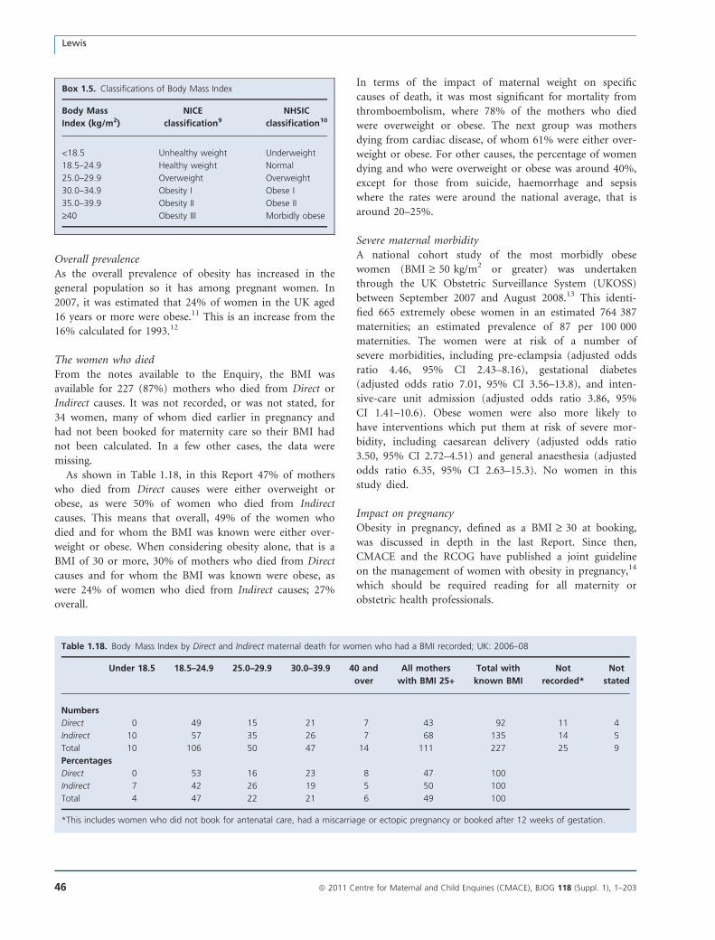
Overall prevalenceAs the overall prevalence of obesity has increased in the
general population so it has among pregnant women. In
2007, it was estimated that 24% of women in the UK aged
16 years or more were obese.11 This is an increase from the
16% calculated for 1993.12
The women who diedFrom the notes available to the Enquiry, the BMI was
available for 227 (87%) mothers who died from Direct or
Indirect causes. It was not recorded, or was not stated, for
34 women, many of whom died earlier in pregnancy and
had not been booked for maternity care so their BMI had
not been calculated. In a few other cases, the data were
missing.
As shown in Table 1.18, in this Report 47% of mothers
who died from Direct causes were either overweight or
obese, as were 50% of women who died from Indirect
causes. This means that overall, 49% of the women who
died and for whom the BMI was known were either over-
weight or obese. When considering obesity alone, that is a
BMI of 30 or more, 30% of mothers who died from Direct
causes and for whom the BMI was known were obese, as
were 24% of women who died from Indirect causes; 27%
overall.
In terms of the impact of maternal weight on specific
causes of death, it was most significant for mortality from
thromboembolism, where 78% of the mothers who died
were overweight or obese. The next group was mothers
dying from cardiac disease, of whom 61% were either over-
weight or obese. For other causes, the percentage of women
dying and who were overweight or obese was around 40%,
except for those from suicide, haemorrhage and sepsis
where the rates were around the national average, that is
around 20–25%.
Severe maternal morbidityA national cohort study of the most morbidly obese
women (BMI ‡ 50 kg/m2 or greater) was undertaken
through the UK Obstetric Surveillance System (UKOSS)
between September 2007 and August 2008.13 This identi-
fied 665 extremely obese women in an estimated 764 387
maternities; an estimated prevalence of 87 per 100 000
maternities. The women were at risk of a number of
severe morbidities, including pre-eclampsia (adjusted odds
ratio 4.46, 95% CI 2.43–8.16), gestational diabetes
(adjusted odds ratio 7.01, 95% CI 3.56–13.8), and inten-
sive-care unit admission (adjusted odds ratio 3.86, 95%
CI 1.41–10.6). Obese women were also more likely to
have interventions which put them at risk of severe mor-
bidity, including caesarean delivery (adjusted odds ratio
3.50, 95% CI 2.72–4.51) and general anaesthesia (adjusted
odds ratio 6.35, 95% CI 2.63–15.3). No women in this
study died.
Impact on pregnancyObesity in pregnancy, defined as a BMI ‡ 30 at booking,
was discussed in depth in the last Report. Since then,
CMACE and the RCOG have published a joint guideline
on the management of women with obesity in pregnancy,14
which should be required reading for all maternity or
obstetric health professionals.
Box 1.5. Classifications of Body Mass Index
Body Mass
Index (kg/m2)
NICE
classification9
NHSIC
classification10
<18.5 Unhealthy weight Underweight
18.5–24.9 Healthy weight Normal
25.0–29.9 Overweight Overweight
30.0–34.9 Obesity I Obese I
35.0–39.9 Obesity II Obese II
‡40 Obesity III Morbidly obese
Table 1.18. Body Mass Index by Direct and Indirect maternal death for women who had a BMI recorded; UK: 2006–08
Under 18.5 18.5–24.9 25.0–29.9 30.0–39.9 40 and
over
All mothers
with BMI 25+
Total with
known BMI
Not
recorded*
Not
stated
Numbers
Direct 0 49 15 21 7 43 92 11 4
Indirect 10 57 35 26 7 68 135 14 5
Total 10 106 50 47 14 111 227 25 9
Percentages
Direct 0 53 16 23 8 47 100
Indirect 7 42 26 19 5 50 100
Total 4 47 22 21 6 49 100
*This includes women who did not book for antenatal care, had a miscarriage or ectopic pregnancy or booked after 12 weeks of gestation.
Lewis
46 ª 2011 Centre for Maternal and Child Enquiries (CMACE), BJOG 118 (Suppl. 1), 1–203
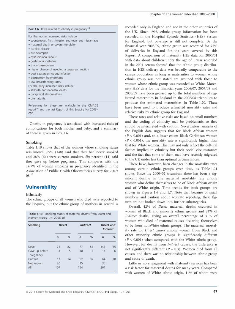
Obesity in pregnancy is associated with increased risks of
complications for both mother and baby, and a summary
of these is given in Box 1.6.
SmokingTable 1.19 shows that of the women whose smoking status
was known, 65% (148) said that they had never smoked
and 28% (64) were current smokers. Six percent (14) said
they gave up before pregnancy. This compares with the
14.7% of women smoking in pregnancy identified in the
Association of Public Health Observatories survey for 2007/
08.15
Vulnerability
EthnicityThe ethnic groups of all women who died were reported to
the Enquiry, but the ethnic group of mothers in general is
recorded only in England and not in the other countries of
the UK. Since 1995, ethnic group information has been
recorded in the Hospital Episode Statistics (HES) System
for England, but coverage is still not complete. By the
financial year 2008/09, ethnic group was recorded for 75%
of deliveries in England for the years covered by this
Report. A comparison of maternity HES data for 2000/01
with data about children under the age of 1 year recorded
in the 2001 census showed that the ethnic group distribu-
tion in HES delivery data was broadly comparable to the
census population as long as maternities to women whose
ethnic group was not stated are grouped with those to
women whose ethnic group was recorded as White. Mater-
nity HES data for the financial years 2006/07, 2007/08 and
2008/09 have been grossed up to the total numbers of reg-
istered maternities in England in the 2006/08 triennium to
produce the estimated maternities in Table 1.20. These
have been used to produce estimated mortality rates and
relative risks by ethnic group for England.
These rates and relative risks are based on small numbers
and the coding of ethnicity may be problematic so they
should be interpreted with caution. Nevertheless, analysis of
the English data suggests that for Black African women
(P < 0.001) and, to a lesser extent Black Caribbean women
(P < 0.001), the mortality rate is significantly higher than
that for White women. This may not only reflect the cultural
factors implied in ethnicity but their social circumstances
and the fact that some of them may have recently migrated
to the UK under less than optimal circumstances.
There have, however, been changes in the mortality rates
among certain ethnic groups over time, as Table 1.21
shows. Since the 2000–02 triennium there has been a sig-
nificant decline in the maternal mortality rate among
women who define themselves to be of Black African origin
and of White origin. Time trends for both groups are
shown in Figures 1.6 and 1.7. Note that because of small
numbers and caution about accurate reporting, these fig-
ures are not broken down into further subcategories.
Overall, 42% of Direct maternal deaths occurred in
women of Black and minority ethnic groups and 24% of
Indirect deaths, giving an overall percentage of 31% of
women who died of maternal causes declaring themselves
to be from nonWhite ethnic groups. The maternal mortal-
ity rate for Direct causes among women from Black and
other minority ethnic groups is significantly different
(P < 0.001) when compared with the White ethnic group.
However, for deaths from Indirect causes, the difference is
not significantly different (P = 0.3). Women died from all
causes, and there was no relationship between ethnic group
and cause of death.
Little or no engagement with maternity services has been
a risk factor for maternal deaths for many years. Compared
with women of White ethnic origin, 11% of whom were
Box 1.6. Risks related to obesity in pregnancy14
For the mother increased risks include:
• spontaneous first trimester and recurrent miscarriage
• maternal death or severe morbidity
• cardiac disease
• pre-eclampsia
• dysfunctional labour
• gestational diabetes
• thromboembolism
• higher chance of needing a caesarean section
• post-caesarean wound infection
• postpartum haemorrhage
• low breastfeeding rates.
For the baby increased risks include:
• stillbirth and neonatal death
• congenital abnormalities
• prematurity.
References for these are available in the CMACE
report14 and the last Report of this Enquiry for 2003–
053.
Table 1.19. Smoking status of maternal deaths from Direct and
Indirect causes; UK: 2006–08
Smoking Direct Indirect Direct and
Indirect
n % n % n %
Never 71 82 77 55 148 65
Gave up before
pregnancy
4 5 10 7 14 6
Current 12 14 52 37 64 28
Not known 20 15 35
All 107 154 261
Chapter 1: The women who died 2006–2008
ª 2011 Centre for Maternal and Child Enquiries (CMACE), BJOG 118 (Suppl. 1), 1–203 47
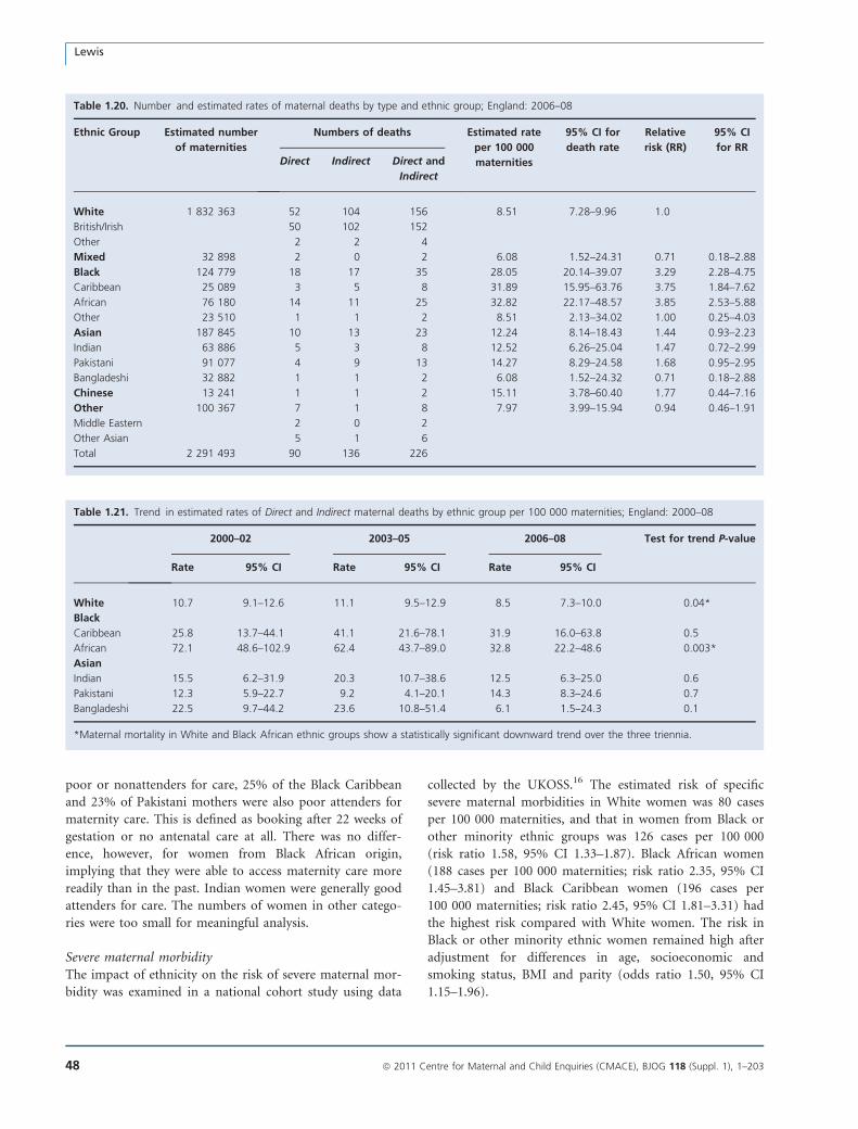
poor or nonattenders for care, 25% of the Black Caribbean
and 23% of Pakistani mothers were also poor attenders for
maternity care. This is defined as booking after 22 weeks of
gestation or no antenatal care at all. There was no differ-
ence, however, for women from Black African origin,
implying that they were able to access maternity care more
readily than in the past. Indian women were generally good
attenders for care. The numbers of women in other catego-
ries were too small for meaningful analysis.
Severe maternal morbidityThe impact of ethnicity on the risk of severe maternal mor-
bidity was examined in a national cohort study using data
collected by the UKOSS.16 The estimated risk of specific
severe maternal morbidities in White women was 80 cases
per 100 000 maternities, and that in women from Black or
other minority ethnic groups was 126 cases per 100 000
(risk ratio 1.58, 95% CI 1.33–1.87). Black African women
(188 cases per 100 000 maternities; risk ratio 2.35, 95% CI
1.45–3.81) and Black Caribbean women (196 cases per
100 000 maternities; risk ratio 2.45, 95% CI 1.81–3.31) had
the highest risk compared with White women. The risk in
Black or other minority ethnic women remained high after
adjustment for differences in age, socioeconomic and
smoking status, BMI and parity (odds ratio 1.50, 95% CI
1.15–1.96).
Table 1.20. Number and estimated rates of maternal deaths by type and ethnic group; England: 2006–08
Ethnic Group Estimated number
of maternities
Numbers of deaths Estimated rate
per 100 000
maternities
95% CI for
death rate
Relative
risk (RR)
95% CI
for RRDirect Indirect Direct and
Indirect
White 1 832 363 52 104 156 8.51 7.28–9.96 1.0
British/Irish 50 102 152
Other 2 2 4
Mixed 32 898 2 0 2 6.08 1.52–24.31 0.71 0.18–2.88
Black 124 779 18 17 35 28.05 20.14–39.07 3.29 2.28–4.75
Caribbean 25 089 3 5 8 31.89 15.95–63.76 3.75 1.84–7.62
African 76 180 14 11 25 32.82 22.17–48.57 3.85 2.53–5.88
Other 23 510 1 1 2 8.51 2.13–34.02 1.00 0.25–4.03
Asian 187 845 10 13 23 12.24 8.14–18.43 1.44 0.93–2.23
Indian 63 886 5 3 8 12.52 6.26–25.04 1.47 0.72–2.99
Pakistani 91 077 4 9 13 14.27 8.29–24.58 1.68 0.95–2.95
Bangladeshi 32 882 1 1 2 6.08 1.52–24.32 0.71 0.18–2.88
Chinese 13 241 1 1 2 15.11 3.78–60.40 1.77 0.44–7.16
Other 100 367 7 1 8 7.97 3.99–15.94 0.94 0.46–1.91
Middle Eastern 2 0 2
Other Asian 5 1 6
Total 2 291 493 90 136 226
Table 1.21. Trend in estimated rates of Direct and Indirect maternal deaths by ethnic group per 100 000 maternities; England: 2000–08
2000–02 2003–05 2006–08 Test for trend P-value
Rate 95% CI Rate 95% CI Rate 95% CI
White 10.7 9.1–12.6 11.1 9.5–12.9 8.5 7.3–10.0 0.04*
Black
Caribbean 25.8 13.7–44.1 41.1 21.6–78.1 31.9 16.0–63.8 0.5
African 72.1 48.6–102.9 62.4 43.7–89.0 32.8 22.2–48.6 0.003*
Asian
Indian 15.5 6.2–31.9 20.3 10.7–38.6 12.5 6.3–25.0 0.6
Pakistani 12.3 5.9–22.7 9.2 4.1–20.1 14.3 8.3–24.6 0.7
Bangladeshi 22.5 9.7–44.2 23.6 10.8–51.4 6.1 1.5–24.3 0.1
*Maternal mortality in White and Black African ethnic groups show a statistically significant downward trend over the three triennia.
Lewis
48 ª 2011 Centre for Maternal and Child Enquiries (CMACE), BJOG 118 (Suppl. 1), 1–203
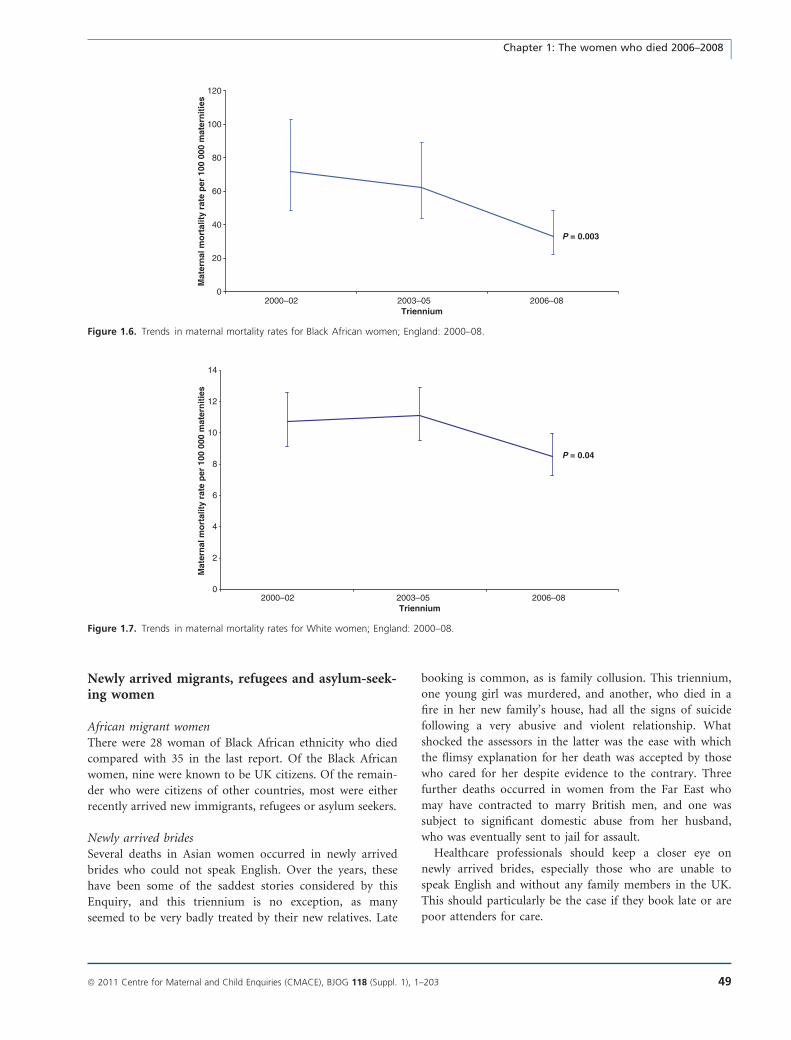
Newly arrived migrants, refugees and asylum-seek-ing women
African migrant womenThere were 28 woman of Black African ethnicity who died
compared with 35 in the last report. Of the Black African
women, nine were known to be UK citizens. Of the remain-
der who were citizens of other countries, most were either
recently arrived new immigrants, refugees or asylum seekers.
Newly arrived bridesSeveral deaths in Asian women occurred in newly arrived
brides who could not speak English. Over the years, these
have been some of the saddest stories considered by this
Enquiry, and this triennium is no exception, as many
seemed to be very badly treated by their new relatives. Late
booking is common, as is family collusion. This triennium,
one young girl was murdered, and another, who died in a
fire in her new family’s house, had all the signs of suicide
following a very abusive and violent relationship. What
shocked the assessors in the latter was the ease with which
the flimsy explanation for her death was accepted by those
who cared for her despite evidence to the contrary. Three
further deaths occurred in women from the Far East who
may have contracted to marry British men, and one was
subject to significant domestic abuse from her husband,
who was eventually sent to jail for assault.
Healthcare professionals should keep a closer eye on
newly arrived brides, especially those who are unable to
speak English and without any family members in the UK.
This should particularly be the case if they book late or are
poor attenders for care.
0
20
40
60
80
100
120
2000–02 2003–05 2006–08Triennium
Mat
ern
al m
ort
alit
y ra
te p
er 1
00 0
00 m
ater
nit
ies
P = 0.003
Figure 1.6. Trends in maternal mortality rates for Black African women; England: 2000–08.
0
2
4
6
8
10
12
14
Mat
ern
al m
ort
alit
y ra
te p
er 1
00 0
00 m
ater
nit
ies
P = 0.04
2000–02 2003–05 2006–08Triennium
Figure 1.7. Trends in maternal mortality rates for White women; England: 2000–08.
Chapter 1: The women who died 2006–2008
ª 2011 Centre for Maternal and Child Enquiries (CMACE), BJOG 118 (Suppl. 1), 1–203 49
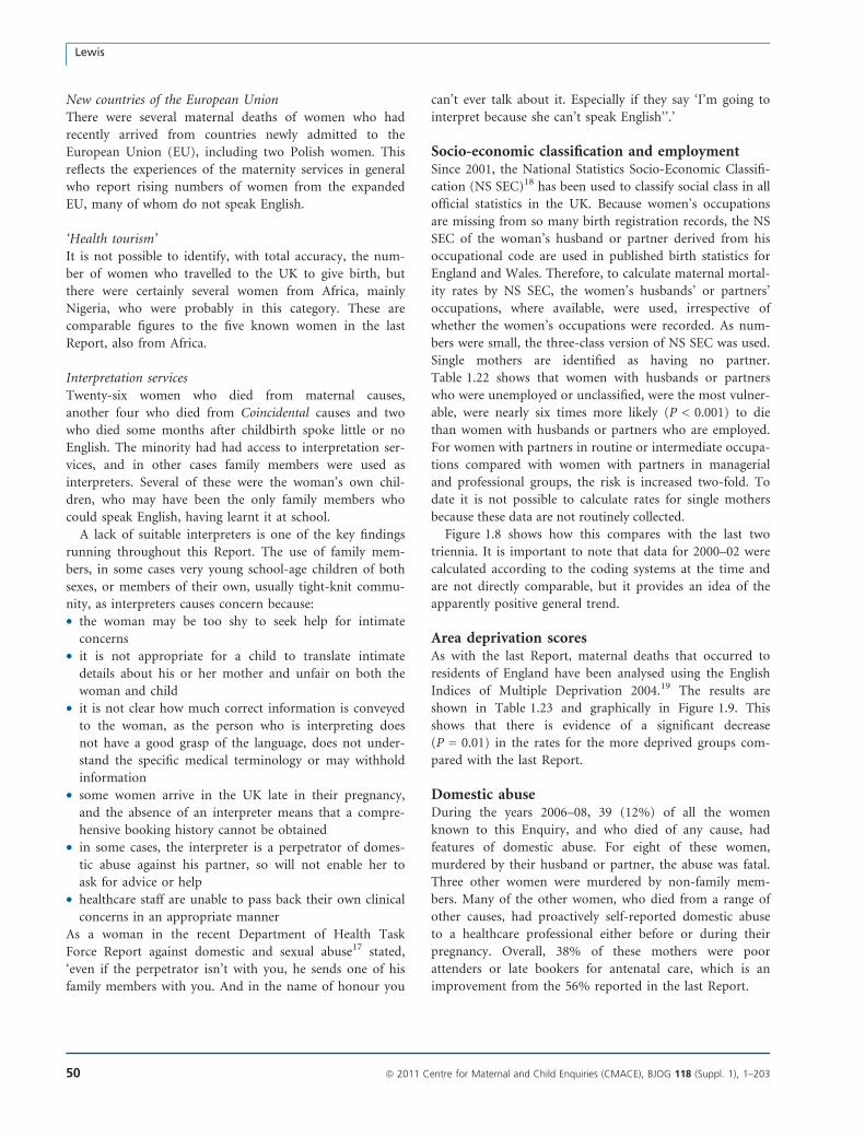
New countries of the European UnionThere were several maternal deaths of women who had
recently arrived from countries newly admitted to the
European Union (EU), including two Polish women. This
reflects the experiences of the maternity services in general
who report rising numbers of women from the expanded
EU, many of whom do not speak English.
‘Health tourism’It is not possible to identify, with total accuracy, the num-
ber of women who travelled to the UK to give birth, but
there were certainly several women from Africa, mainly
Nigeria, who were probably in this category. These are
comparable figures to the five known women in the last
Report, also from Africa.
Interpretation servicesTwenty-six women who died from maternal causes,
another four who died from Coincidental causes and two
who died some months after childbirth spoke little or no
English. The minority had had access to interpretation ser-
vices, and in other cases family members were used as
interpreters. Several of these were the woman’s own chil-
dren, who may have been the only family members who
could speak English, having learnt it at school.
A lack of suitable interpreters is one of the key findings
running throughout this Report. The use of family mem-
bers, in some cases very young school-age children of both
sexes, or members of their own, usually tight-knit commu-
nity, as interpreters causes concern because:
• the woman may be too shy to seek help for intimate
concerns
• it is not appropriate for a child to translate intimate
details about his or her mother and unfair on both the
woman and child
• it is not clear how much correct information is conveyed
to the woman, as the person who is interpreting does
not have a good grasp of the language, does not under-
stand the specific medical terminology or may withhold
information
• some women arrive in the UK late in their pregnancy,
and the absence of an interpreter means that a compre-
hensive booking history cannot be obtained
• in some cases, the interpreter is a perpetrator of domes-
tic abuse against his partner, so will not enable her to
ask for advice or help
• healthcare staff are unable to pass back their own clinical
concerns in an appropriate manner
As a woman in the recent Department of Health Task
Force Report against domestic and sexual abuse17 stated,
‘even if the perpetrator isn’t with you, he sends one of his
family members with you. And in the name of honour you
can’t ever talk about it. Especially if they say ‘I’m going to
interpret because she can’t speak English’’.’
Socio-economic classification and employmentSince 2001, the National Statistics Socio-Economic Classifi-
cation (NS SEC)18 has been used to classify social class in all
official statistics in the UK. Because women’s occupations
are missing from so many birth registration records, the NS
SEC of the woman’s husband or partner derived from his
occupational code are used in published birth statistics for
England and Wales. Therefore, to calculate maternal mortal-
ity rates by NS SEC, the women’s husbands’ or partners’
occupations, where available, were used, irrespective of
whether the women’s occupations were recorded. As num-
bers were small, the three-class version of NS SEC was used.
Single mothers are identified as having no partner.
Table 1.22 shows that women with husbands or partners
who were unemployed or unclassified, were the most vulner-
able, were nearly six times more likely (P < 0.001) to die
than women with husbands or partners who are employed.
For women with partners in routine or intermediate occupa-
tions compared with women with partners in managerial
and professional groups, the risk is increased two-fold. To
date it is not possible to calculate rates for single mothers
because these data are not routinely collected.
Figure 1.8 shows how this compares with the last two
triennia. It is important to note that data for 2000–02 were
calculated according to the coding systems at the time and
are not directly comparable, but it provides an idea of the
apparently positive general trend.
Area deprivation scoresAs with the last Report, maternal deaths that occurred to
residents of England have been analysed using the English
Indices of Multiple Deprivation 2004.19 The results are
shown in Table 1.23 and graphically in Figure 1.9. This
shows that there is evidence of a significant decrease
(P = 0.01) in the rates for the more deprived groups com-
pared with the last Report.
Domestic abuseDuring the years 2006–08, 39 (12%) of all the women
known to this Enquiry, and who died of any cause, had
features of domestic abuse. For eight of these women,
murdered by their husband or partner, the abuse was fatal.
Three other women were murdered by non-family mem-
bers. Many of the other women, who died from a range of
other causes, had proactively self-reported domestic abuse
to a healthcare professional either before or during their
pregnancy. Overall, 38% of these mothers were poor
attenders or late bookers for antenatal care, which is an
improvement from the 56% reported in the last Report.
Lewis
50 ª 2011 Centre for Maternal and Child Enquiries (CMACE), BJOG 118 (Suppl. 1), 1–203
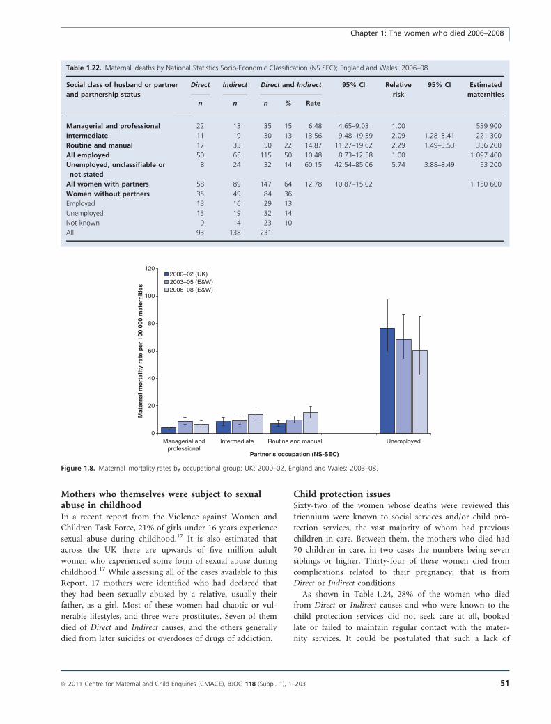
Mothers who themselves were subject to sexualabuse in childhoodIn a recent report from the Violence against Women and
Children Task Force, 21% of girls under 16 years experience
sexual abuse during childhood.17 It is also estimated that
across the UK there are upwards of five million adult
women who experienced some form of sexual abuse during
childhood.17 While assessing all of the cases available to this
Report, 17 mothers were identified who had declared that
they had been sexually abused by a relative, usually their
father, as a girl. Most of these women had chaotic or vul-
nerable lifestyles, and three were prostitutes. Seven of them
died of Direct and Indirect causes, and the others generally
died from later suicides or overdoses of drugs of addiction.
Child protection issuesSixty-two of the women whose deaths were reviewed this
triennium were known to social services and/or child pro-
tection services, the vast majority of whom had previous
children in care. Between them, the mothers who died had
70 children in care, in two cases the numbers being seven
siblings or higher. Thirty-four of these women died from
complications related to their pregnancy, that is from
Direct or Indirect conditions.
As shown in Table 1.24, 28% of the women who died
from Direct or Indirect causes and who were known to the
child protection services did not seek care at all, booked
late or failed to maintain regular contact with the mater-
nity services. It could be postulated that such a lack of
Table 1.22. Maternal deaths by National Statistics Socio-Economic Classification (NS SEC); England and Wales: 2006–08
Social class of husband or partner
and partnership status
Direct Indirect Direct and Indirect 95% CI Relative
risk
95% CI Estimated
maternitiesn n n % Rate
Managerial and professional 22 13 35 15 6.48 4.65–9.03 1.00 539 900
Intermediate 11 19 30 13 13.56 9.48–19.39 2.09 1.28–3.41 221 300
Routine and manual 17 33 50 22 14.87 11.27–19.62 2.29 1.49–3.53 336 200
All employed 50 65 115 50 10.48 8.73–12.58 1.00 1 097 400
Unemployed, unclassifiable or
not stated
8 24 32 14 60.15 42.54–85.06 5.74 3.88–8.49 53 200
All women with partners 58 89 147 64 12.78 10.87–15.02 1 150 600
Women without partners 35 49 84 36
Employed 13 16 29 13
Unemployed 13 19 32 14
Not known 9 14 23 10
All 93 138 231
0
20
40
60
80
100
120
Managerial andprofessional
UnemployedRoutine and manualIntermediate
Partner's occupation (NS-SEC)
Mat
ern
al m
ort
alit
y ra
te p
er 1
00 0
00 m
ater
nit
ies
2000–02 (UK)2003–05 (E&W)2006–08 (E&W)
Figure 1.8. Maternal mortality rates by occupational group; UK: 2000–02, England and Wales: 2003–08.
Chapter 1: The women who died 2006–2008
ª 2011 Centre for Maternal and Child Enquiries (CMACE), BJOG 118 (Suppl. 1), 1–203 51
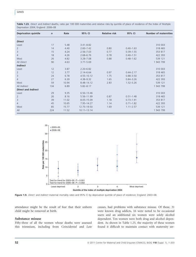
attendance might be the result of fear that their unborn
child might be removed at birth.
Substance misuseFifty-three of all the women whose deaths were assessed
this triennium, including from Coincidental and Late
causes, had problems with substance misuse. Of these, 31
were known drug addicts, 16 were noted to be occasional
users and an additional six women were solely alcohol
dependent. Ten women were both drug and alcohol depen-
dent. As shown in Table 1.25, the majority of these women
found it difficult to maintain contact with maternity ser-
Table 1.23. Direct and Indirect deaths, rates per 100 000 maternities and relative risks by quintile of place of residence of the Index of Multiple
Deprivation 2004; England: 2006–08
Deprivation quintile n Rate 95% CI Relative risk 95% CI Number of maternities
Direct
Least 17 5.48 3.41–8.82 310 003
2 14 4.40 2.60–7.42 0.80 0.40–1.63 318 465
3 15 4.24 2.56–7.03 0.77 0.39–1.55 353 817
4 18 4.26 2.68–6.76 0.78 0.40–1.51 422 393
Most 26 4.82 3.28–7.08 0.88 0.48–1.62 539 121
All Direct 90 4.63 3.77–5.69 1 943 799
Indirect
Least 12 3.87 2.20–6.82 310 003
2 12 3.77 2.14–6.64 0.97 0.44–2.17 318 465
3 24 6.78 4.55–10.12 1.75 0.88–3.50 353 817
4 27 6.39 4.38–9.32 1.65 0.84–3.26 422 393
Most 59 10.94 8.48–14.12 2.83 1.52–5.26 539 121
All Indirect 134 6.89 5.82–8.17 1 943 799
Direct and Indirect
Least 29 9.35 6.50–13.46 310 003
2 26 8.16 5.56–11.99 0.87 0.51–1.48 318 465
3 39 11.02 8.05–15.09 1.18 0.73–1.91 353 817
4 45 10.65 7.95–14.27 1.14 0.71–1.82 422 393
Most 85 15.77 12.75–19.50 1.69 1.11–2.57 539 121
All 224 11.52 10.11–13.14 1 943 799
0
5
10
15
20
25
30
35
Least deprived 2 3 4 Most deprived
Quintile of the index of multiple deprivation 2004
Rat
e p
er 1
00 0
00 m
ater
nit
ies
2003–052006–08
Test for trend for 2003–05: P < 0.001Test for trend for 2006–08: P = 0.002
Figure 1.9. Direct and Indirect maternal mortality rates and 95% CI by deprivation quintile of place of residence; England: 2003–08.
Lewis
52 ª 2011 Centre for Maternal and Child Enquiries (CMACE), BJOG 118 (Suppl. 1), 1–203
vices. However, for those who did, there was increasing evi-
dence of a greater emphasis on planned multidisciplinary
and multi-agency care, and in some cases the care they
received was outstanding. Issues relating to substance mis-
use and pregnancy are discussed in Chapter 11.
Risk factors and barriers to careMany of the women who died found it difficult to seek, or
to maintain contact with, maternity and/or other health
services. The many possible reasons for this have been dis-
cussed throughout this Chapter, and the main characteris-
tics of the women who found it difficult to attend are
summarised in Table 1.26. As many, if not all, of these risk
factors interplay with each other, it is hoped that a more
robust multivariate analysis will be undertaken in future
reports.
From the findings of this triennial Report there appears
to have been a considerable improvement in women
accessing and staying in touch with maternity services com-
pared with the last Report, where 81% of women who had
declared domestic abuse or who were known to the child
protection or social services were poor attenders, compared
with 32 and 39%, respectively for this Report. Similarly,
78% of known substance abusers did not have appropriate
care in the last Report, compared with 44% for this Report.
Improving access to care for the most disadvantaged or
vulnerable women has long been a recommendation of
these Reports. It is therefore heartening to see this issue
addressed in the latest guideline from the National Institute
for Health and Clinical Excellence on providing models of
maternity service provision for pregnant women with com-
plex social factors.20
Table 1.24. Characteristics of the antenatal care sought by pregnant or recently delivered women who were known to social and/or child
protection services and whose pregnancy exceeded 12 weeks of gestation; UK: 2006–08
Type of death Number of late or
non attenders for antenatal care
Number of deaths known
to social services and/or
child protection services
after 12 or more weeks
of gestation
Percentage
of deaths
Booked after
22 weeks or
missed more than
three visits
No ANC All
Direct 0 0 0 2 0
Indirect 6 3 9 30 30
All 6 3 9 32 28
Coincidental 5 1 6 11 55
Late deaths 5 1 6 15 40
Total 16 5 21 58 36
Table 1.25. Characteristics of the antenatal care sought by pregnant or recently delivered women who were known substance misusers and
whose pregnancy exceeded 12 weeks of gestation; UK: 2006–08
Type of death Number of late or non attenders for antenatal
care
Number of deaths
of substance misusers
after 12 or more weeks of gestation
Percentage
of deaths
Booked after 22 weeks
or missed more
than three visits
No ANC All
Direct 2 1 3 4 75
Indirect 4 3 7 20 35
All 6 4 10 24 42
Coincidental 5 2 7 11 64
Late deaths 5 0 5 13 38
Total 16 6 22 48 46
Chapter 1: The women who died 2006–2008
ª 2011 Centre for Maternal and Child Enquiries (CMACE), BJOG 118 (Suppl. 1), 1–203 53
What did you learn from this case and how has itchanged your practice?As part of completing the report forms for this Enquiry, all
healthcare workers are asked to reflect on what went well
and not so well and to describe what they learnt from the
case and if it has changed their practice. Although many
health professionals wrote in humbling terms, others
showed a complete lack of understanding of what had gone
wrong or were complacent in the extreme. This is an
underlying reason why one of the Key Recommendations,
to develop and undertake and act on the results of far
more robust serious incident/serious untoward incident
reviews, is so necessary.
A few healthcare workers blamed themselves when
clearly there was no reason for this, demonstrating the
effects that these generally unexpected and sudden deaths
in young women can have on those who care for them, as
well as on her baby, other children, family and wider com-
munity. It is important that all staff, as well as family
members, who are affected by a maternal death, be offered
support and counselling.
Discussion and conclusions
Compared with the recent Reports, the findings for this
Enquiry show a generally encouraging picture, which con-
tinues to demonstrate the value of this work. The results
show not only welcome reductions in deaths from some
clinical causes, especially thromboembolism, but also a
declining trend in the inequalities gap between well-off and
more deprived women. This public health aspect is one of
the key strengths of this Report. The findings suggest that
many of the previous lessons have been heeded and action
has been taken on their recommendations. Certainly, mid-
wives and other professionals from all around the country
have been actively engaged in many CMACE and other
local training meetings based on the findings and represen-
tative cases contained in the last Report. The Department
of Health in England also built the earlier recommenda-
tions relating to easier access to care for the most vulnera-
ble women, and early booking, into their policy for
maternity services, Maternity Matters.5 Finally, although we
are unable to test this association statistically, it is difficult
to ignore the apparent relationship between the significant
decline in deaths from pulmonary embolism, and to a les-
ser degree, haemorrhage, and the publication and imple-
mentation of guidelines for these which have been
recommended in previous Reports.
There were setbacks, however, particularly in the rise in
deaths from community-acquired infections, although this
has occurred at a time when the background population
mortality rate had also increased. This was before the
H1N1 pandemic. It is hoped that the strengthened recom-
mendations made in this Report for national and local
guidelines for the identification and management of sepsis,
and accompanying health information for mothers and
families, are implemented without delay. A recommenda-
tion for such a national guideline was made in the last
Table 1.26. Characteristics* of the women who were poor or nonattenders for antenatal care** and whose pregnancy was 12 weeks of
gestation or more; all causes; UK: 2006–08
Characteristic* Overall number of
women 12 weeks
gestation or more
Women who were poor or nonattenders at antenatal care
No
care
Poor
care
Total
Number
Overall
percentage
Substance misuse*** 48 6 15 21 44
Known to child protection services 45 4 16 20 44
Single unemployed 44 6 12 18 41
Known to social services 51 4 13 17 33
Domestic abuse 34 3 8 11 32
Partner unemployed 37 1 9 10 27
Most deprived quintile post code 91 9 11 20 22
Black Caribbean 9 2 0 2 22
No English 25 2 2 4 16
White 231 11 22 33 14
Black African 26 1 1 2 8
Partner employed 161 4 2 6 4
*Some women had more than one characteristic.
**Defined as no care at all or missed three or more antenatal visits.
***Including occasional drug use and alcoholism.
Lewis
54 ª 2011 Centre for Maternal and Child Enquiries (CMACE), BJOG 118 (Suppl. 1), 1–203
Report and is made again here, as are another two recom-
mendations that have been retained. These relate to the
management of systolic hypertension, the mismanagement
of which again features in this Report, and to improving
the quality of Critical Incident or Sudden Unexpected Inci-
dent reviews. One of the major themes identified by all of
the assessors, whatever the cause of death, was the highly
variable nature of these reports, and urgent action is
required to produce a national minimum standard for such
crucial opportunities for review, self-reflective learning and
personal and institutional change. Attention also needs to
be paid to other areas where there is room for developing
even better maternity services. These include pregnancy
planning, preconception care and improving the care of
vulnerable pregnant women and for those with complex
medical, social or mental health needs.
Overall, however, the findings of this Report are reassur-
ing, as the overall decline in the maternal mortality rate
has taken place against a background of an increasing birth
rate, sometimes stretched maternity services and a generally
older and less healthy population of mothers. Compared
with the general pregnant population, the women who died
tended to be older and more obese, had lifestyles which
put them at risk of poorer health and were more socially
disadvantaged. And, as also shown in the last Report, there
appeared to be a growing proportion of mothers with more
medically complex pregnancies. Other developed countries
are facing similar problems, and in some, for example the
USA, the maternal mortality rate appears to have doubled.2
Maternal deaths are extremely rare in the UK, unlike
many other parts of the world.21,22 Internationally, the
work of this Report continues to be held up as the ‘Gold
Standard’ for maternal death audits and reviews, and its
methodology is much in demand in developing and devel-
oped countries, ranging from the poorest African states to
the USA and Japan. This has been accelerated by a renewed
global emphasis by the United Nations and others on
reducing the 358 000 mainly avoidable maternal deaths,2
four million neonatal deaths23 and over three million still-
births24 each year. These over seven million deaths are lar-
gely the result of preventable or treatable maternal
conditions. This renewed effort is in response to the woeful
lack of progress in relation to the Millennium Development
Goal 5 relating to maternal mortality (MDG 5),25,26 where
countries have pledged to reduce their maternal mortality
rate by 75% by 2015. As a result, many of the resource-
poor countries with very high mortality rates are starting
to assess the numbers and causes of their maternal deaths,
and CMACE and the Saving Mothers’ Lives Director,
authors and assessors are providing help and support. The
results of these reviews are to identify remediable factors
and missed opportunities to form the basis for national or
local guidelines and recommendations for beneficial
changes to the health, maternity and neonatal services over-
all, as well as clinical practice.27,28
There is a danger that the comparatively small numbers of
maternal deaths in the UK will lead to complacency, but
there is always more to be done to improve maternal and
child health. As this Report has shown, new issues continue
to arise, and the next Report for 2009–11 will also cover the
H1N1 pandemic. The fact that a health alert concerning the
prevention and earlier detection of community-acquired
Group A streptococcal sepsis was issued before publication
of the main report shows the importance of this work to the
protection of pregnant or newly delivered mothers and the
wider public health. The significant decline in deaths from
thrombosis highlights the clinical benefits of this Enquiry.
The reduction in inequalities demonstrates that the earlier
recommendations for changes to the way services are deliv-
ered to the more vulnerable and excluded women may have
had an impact. Overall, these results continue to demon-
strate what a force this Enquiry is for the continuing
improvement of maternal and child health.
Disclosure of interestsNone.
FundingThis work was undertaken by the Centre for Maternal and
Child Enquiries (CMACE) as part of the CEMACH pro-
gramme. The work was funded by the National Patient
Safety Agency; the Department of Health, Social Services
and Public Safety of Northern Ireland; NHS Quality
Improvement Scotland (NHS QIS); and the Channel
Islands and Isle of Man.
AcknowledgementsThis Chapter has been discussed with Dr Marian Knight,
Head of UKOSS and Senior Clinical Research Fellow for
the NPEU, Professor James Drife, Emeritus Professor of
Obstetrics and Gynaecology for the University of Leeds and
Professor James Neilson, Professor of Obstetrics and
Gynaecology for the University of Leeds. j
References
1 Callagan W. Epilogue in Strategies to Reduce Pregnancy-Related
Deaths: From Identification and Review to Action. Atlanta, GA: Cen-
tres for Disease Control, American College of Obstetricians and
Gynaecologist, 2001.
2 Trends in Maternal Mortality 1990–2008. Estimates Developed by
WHO, UNICEF, UNFPA and the World Bank. Geneva: WHO, 2010.
[www.who.int/reproductivehealth]. Accessed 7 September 2010.
3 Amnesty International. Deadly Delivery. The Maternal Health Crisis in
the USA. New York: Amnesty International USA, 2010. [www.am-
nestyusa.org]. Accessed 9 September 2010.
Chapter 1: The women who died 2006–2008
ª 2011 Centre for Maternal and Child Enquiries (CMACE), BJOG 118 (Suppl. 1), 1–203 55
4 Lewis G (ed). The Confidential Enquiry into Maternal and Child
Health (CEMACH). Saving Mothers Lives; Reviewing Maternal Deaths
to Make Motherhood Safer 2003–05. The Seventh Report of the
United Kingdom Confidential Enquiries into Maternal Deaths in the
United Kingdom. London: CEMACH, 2007. [www.cmace.org.uk].
Accessed 9 September 2010.
5 Redshaw M, Rowe R, Hockley C, Brocklehurst P Recorded Delivery:
A National Survey of Women’s Experience of Maternity Care 2006.
National Perinatal Epidemiology Unit. Oxford: NPEU, 2007. ISBN 978
0 9735860 8 0. [www.npeu.ox.ac.uk]. Accessed 9 September 2010.
6 Department of Health. Statistical press notice: vital Signs Monitoring
Return—quarterly update return May 2010. 2010.
7 Department of Health. Maternity Matters: Choice, Access and Conti-
nuity of Care in a Safe Service. London: Department of Health,
2007. [www.dh.gov.uk/publications]. Accessed 9 September 2010.
8 Royal College of Obstetricians and Gynaecologists. Responsibility of
Consultant on-Call. Good Practice No. 8. London: RCOG, 2010.
9 National Institute for Health and Clinical Excellence. Obesity. Guid-
ance on the Prevention, Identification, Assessment and Management
of Overweight and Obesity in Adults and Children. London: National
Institute for Health and Clinical Excellence, 2006.
10 The Health and Information Centre. Statistics on Obesity, Physical
Activity and diet: England. London: NHS information centre, 2009.
[www.ic.nhs.uk/pubs]. Accessed 9 September 2010.
11 Office for National Statistics. Statistics on Obesity, Physical activity
and Diet: England. London: ONS, 2008.
12 Helsehurst N, Ells LJ, Simpson H, Batterham A, Wilkinson J, Summer-
bell CD. Trends in maternal obesity incidence rates, demographic
predictors and health inequalities in 38,821 women over a 15 year
period. BJOG 2007;114:187–94.
13 Knight M, Kurinczuk JJ, Spark P, Brocklehurst P. Extreme Obesity in
Pregnancy in the United Kingdom. Obstet Gynecol 2010;115:989–
97.
14 CMACE/RCOG. Management of Women with Obesity in Pregnancy.
London: Centre for Maternal and Child Enquiries and Royal College
of Obstetricians and Gynaecologists, 2010. [www.cmace.org.uk].
Accessed 9 September 2010.
15 Association of Public Health Observatories. Smoking in pregnancy
2007/08. In: Xxxx XX, editor. Health Profiles 2009. APHO.
16 Knight M, Kurinczuk JJ, Spark P, Brocklehurst P, on behalf of
UKOSS. Inequalities in maternal health: national cohort study of eth-
nic variation in severe maternal morbidities. BMJ 2009;338:b542.
17 Responding to violence against women and children-the role of the
NHS. The Report of the Sexual Violence Against Women Subgroup
of the Task Force on the Health Aspects of Violence Against Women
and Children. London: Department of Health, 2010. [www.dh.go-
v.uk/publications]. Accessed 9 September 2010.
18 The Office For National Statistics. The National Statistics Socio-eco-
nomic Classification (NS-SEC). SOC2010Vol3NS-SEC [www.ons.go-
v.uk/about-statistics/classifications/current/ns-sec/index.html].
Accessed 7 September 2010.
19 Office of the Deputy Prime Minister. The English Indices of Depriva-
tion 2004 (revised). London: HMSO, 2004. [www.communi-
ties.gov.uk/publications/communities/englishindices]. Accessed 7
September 2010.
20 National Collaborating Centre for Maternal and Child Health. Preg-
nancy and Complex Social Factors: A Model for Service Provision for
Pregnant Women with Complex Social Factors. National Institute for
Health and Clinical Excellence and Social Care Institute for Excel-
lence. London: RCOG, 2010. [www.guidance.nice.org.uk/CG110].
Accessed 10 September 2010.
21 Maternal Mortality in 2005. Estimates Developed by WHO, UNICEF,
UNFPA and the World Bank. Geneva: WHO, 2007. [www.who.int/re-
productivehealth]. Accessed 7 September 2010.
22 The World Health Organisation Annual Report for 2005. Make Every
Mother and Child Count. Geneva: WHO, 2005. [www.who.int].
Accessed 7 September 2010.
23 United Nations Children’s Fund. UNICEF The State of the Worlds
Children. Maternal and Newborn Health. New York: UNICEF,
2008.
24 Lawn J, Gravett M, Nunes T, Rubens C, Stanton C, the GAPPS
groups. Global report on preterm birth and stillbirth: definitions,
descriptions of the burden and opportunities to improve data. BMC
Pregnancy Childbirth 2010;10(Suppl1):S1.doi:10.1186/1471-2393-
10-S1-S1 [www.biomedcentral.com/1471-2393/10?issue=S1].
Accessed 7 September 2010.
25 The United Nations General Assembly. United Nations Millennium
Development Declaration. A/RES/55/2. New York: United Nations,
2000.
26 Ronsmans C, Graham WJ, on behalf of the The Lancet Maternal Sur-
vival Series steering group. Maternal mortality: who, when, where
and why? Lancet 2006;368:1189–200.
27 Lewis G. Reviewing maternal deaths to make pregnancy safer.
In: Moodley J, editor. Recent Advances in Obstetrics. Best
Pract Res Clin Obstet Gynaecol Amsterdam:Elsevier; 2008;22:
447–63.
28 Bacci A, Lewis G, Baltag V, Betran A. ‘‘Beyond The Numbers’’: Intro-
duction of Maternal Mortality and Severe Morbidity Audit in the
European Region. Reproductive Health Matters. Copenhagen: WHO,
2007.
Lewis
56 ª 2011 Centre for Maternal and Child Enquiries (CMACE), BJOG 118 (Suppl. 1), 1–203
Chapter 2: Thrombosis and thromboembolismJ Drife (on behalf of the Centre for Maternal and Child Enquiries)
University of Leeds, Leeds, UK
Correspondence: Professor James Drife, Emeritus Professor of Obstetrics and Gynaecology, University of Leeds, Leeds LS2 9JT, UK. Emails:
[email protected]; [email protected]
Keywords thrombosis, thromboembolism, Confidential Enquiry,
maternal, mortality.
Thromboembolism and thromboembolism: specific recommendation
• Obesity remains the most important risk factor for thromboembolism. New guidance on the management of women
with obesity in pregnancy has been published by Centre for Maternal and Child Enquries (CMACE) and the Royal
College of Obstetricians and Gynaecologists (RCOG),1 and weight-specific dosage advice on thromboprophylaxis is
now included in the revised RCOG guideline.2 These should be widely disseminated and implemented.
• Risk assessment in early pregnancy continues to be a key factor in reducing mortality. Obese women with a body
mass index (BMI) of 35 or more are unsuitable for midwife-only care, and should be seen in pregnancy by a consul-
tant obstetrician.
• Women are at risk of thromboembolism from the very beginning of pregnancy until the end of the puerperium, and all
health professionals must be aware of this. Early pregnancy units and gynaecology wards must carry out risk assessment
appropriate for pregnant women.
• Vulnerable women, such as those with mental illness or learning disability, may not be able to follow advice or self-
administer injections, and so require particular care.
• Chest symptoms appearing for the first time in pregnancy or the puerperium in at-risk women need careful assessment, and
there should be a low threshold for investigation.
Summary of key findings for 2006–08
The deaths of 18 women who died from thrombosis and/or
thromboembolism are counted in this chapter. This gives a
maternal mortality rate of 0.79 per 100 000 maternities
(95% CI 0.49–1.25). Sixteen of the 18 deaths were attrib-
uted to pulmonary embolism and two to cerebral vein
thrombosis. Additionally, four Late deaths attributed to pul-
monary embolism are counted in Chapter 12, but the les-
sons to be learnt from these cases are discussed here.
These 18 deaths represent a sharp, and statistically signif-
icant, fall from 41 deaths in 2003–05 and, as shown in
Table 2.1 and Figure 2.1, this total is by far the lowest since
the UK-wide Enquiry began in 1985. The fall in deaths
from pulmonary embolism, from 33 to 16, was mainly the
result of a reduction in antenatal deaths and deaths follow-
ing vaginal delivery. Deaths after caesarean section also fell
slightly. This is the first full triennium following publica-
tion of the 2004 RCOG guideline Thromboprophylaxis dur-
ing pregnancy, labour and after normal vaginal delivery.3 It
seems likely that the unprecedented fall in deaths is the
result of better recognition of at-risk women and more
widespread thromboprophylaxis.
Nevertheless, risk factors for thromboembolism were
present in 16 of the 18 women who died in 2006–08. Obes-
ity remains the most important of these. Fourteen women
were overweight (BMI ‡ 25 kg/m2), of whom 11 had a
BMI ‡ 30, including three who had a BMI ‡ 40. All the
deaths occurred before the publication in 2009 of the
updated guideline2, which now includes more extensive
weight-specific dosage advice on thromboprophylaxis.
Substandard care was present in 56% of women. This took
the form of inadequate risk assessment, inadequate throm-
boprophylaxis (by the standards at the time), failure to
investigate chest symptoms in at-risk women, and failure to
ensure multidisciplinary care involving a consultant obstetri-
cian or a psychiatrist for women who also had a pre-existing
medical or mental health illness that affected their treatment.
For the first time since the UK-wide Enquiry began in
1985, thromboembolism is no longer the leading cause of
Chapter 2: Thrombosis and thromboembolism
ª 2011 Centre for Maternal and Child Enquiries (CMACE), BJOG 118 (Suppl. 1), 1–203 57
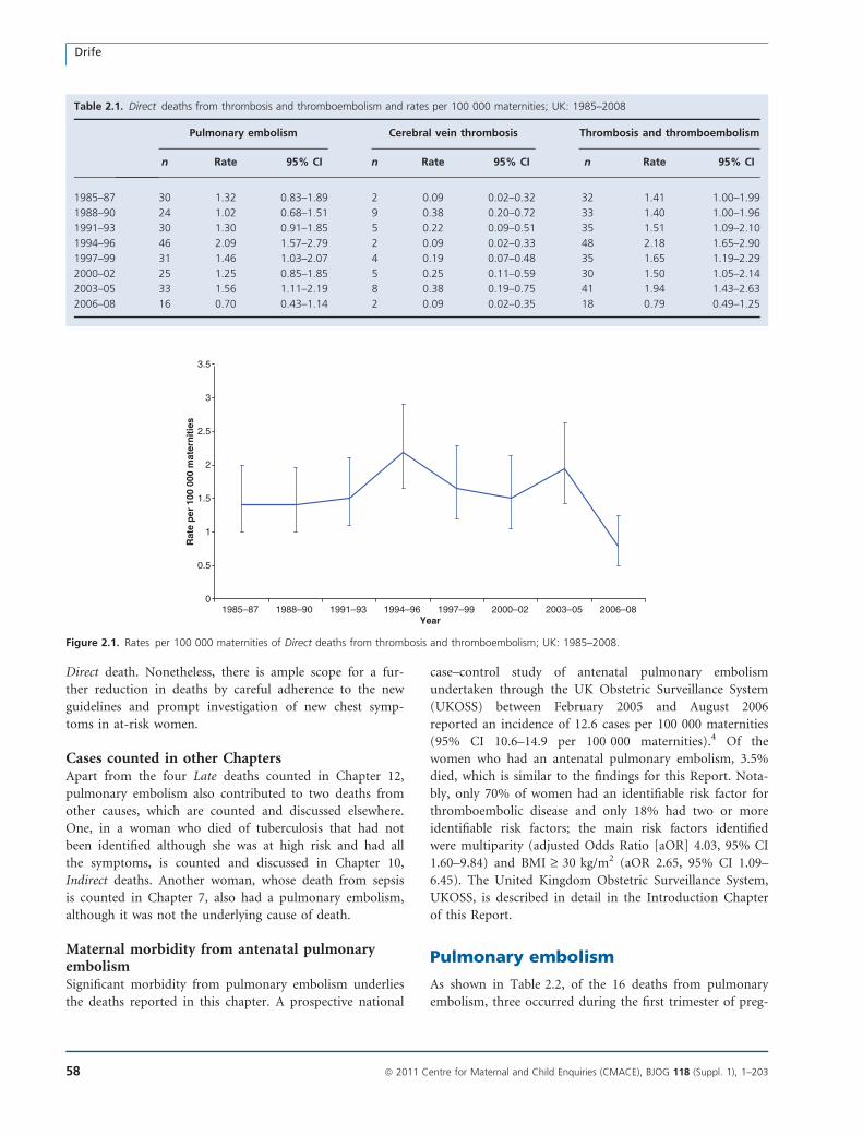
Direct death. Nonetheless, there is ample scope for a fur-
ther reduction in deaths by careful adherence to the new
guidelines and prompt investigation of new chest symp-
toms in at-risk women.
Cases counted in other ChaptersApart from the four Late deaths counted in Chapter 12,
pulmonary embolism also contributed to two deaths from
other causes, which are counted and discussed elsewhere.
One, in a woman who died of tuberculosis that had not
been identified although she was at high risk and had all
the symptoms, is counted and discussed in Chapter 10,
Indirect deaths. Another woman, whose death from sepsis
is counted in Chapter 7, also had a pulmonary embolism,
although it was not the underlying cause of death.
Maternal morbidity from antenatal pulmonaryembolismSignificant morbidity from pulmonary embolism underlies
the deaths reported in this chapter. A prospective national
case–control study of antenatal pulmonary embolism
undertaken through the UK Obstetric Surveillance System
(UKOSS) between February 2005 and August 2006
reported an incidence of 12.6 cases per 100 000 maternities
(95% CI 10.6–14.9 per 100 000 maternities).4 Of the
women who had an antenatal pulmonary embolism, 3.5%
died, which is similar to the findings for this Report. Nota-
bly, only 70% of women had an identifiable risk factor for
thromboembolic disease and only 18% had two or more
identifiable risk factors; the main risk factors identified
were multiparity (adjusted Odds Ratio [aOR] 4.03, 95% CI
1.60–9.84) and BMI ‡ 30 kg/m2 (aOR 2.65, 95% CI 1.09–
6.45). The United Kingdom Obstetric Surveillance System,
UKOSS, is described in detail in the Introduction Chapter
of this Report.
Pulmonary embolism
As shown in Table 2.2, of the 16 deaths from pulmonary
embolism, three occurred during the first trimester of preg-
Table 2.1. Direct deaths from thrombosis and thromboembolism and rates per 100 000 maternities; UK: 1985–2008
Pulmonary embolism Cerebral vein thrombosis Thrombosis and thromboembolism
n Rate 95% CI n Rate 95% CI n Rate 95% CI
1985–87 30 1.32 0.83–1.89 2 0.09 0.02–0.32 32 1.41 1.00–1.99
1988–90 24 1.02 0.68–1.51 9 0.38 0.20–0.72 33 1.40 1.00–1.96
1991–93 30 1.30 0.91–1.85 5 0.22 0.09–0.51 35 1.51 1.09–2.10
1994–96 46 2.09 1.57–2.79 2 0.09 0.02–0.33 48 2.18 1.65–2.90
1997–99 31 1.46 1.03–2.07 4 0.19 0.07–0.48 35 1.65 1.19–2.29
2000–02 25 1.25 0.85–1.85 5 0.25 0.11–0.59 30 1.50 1.05–2.14
2003–05 33 1.56 1.11–2.19 8 0.38 0.19–0.75 41 1.94 1.43–2.63
2006–08 16 0.70 0.43–1.14 2 0.09 0.02–0.35 18 0.79 0.49–1.25
0
0.5
1
1.5
2
2.5
3
3.5
1985–87 1988–90 1991–93 1994–96 1997–99 2000–02 2003–05 2006–08Year
Rat
e p
er 1
00 0
00 m
ater
nit
ies
Figure 2.1. Rates per 100 000 maternities of Direct deaths from thrombosis and thromboembolism; UK: 1985–2008.
Drife
58 ª 2011 Centre for Maternal and Child Enquiries (CMACE), BJOG 118 (Suppl. 1), 1–203
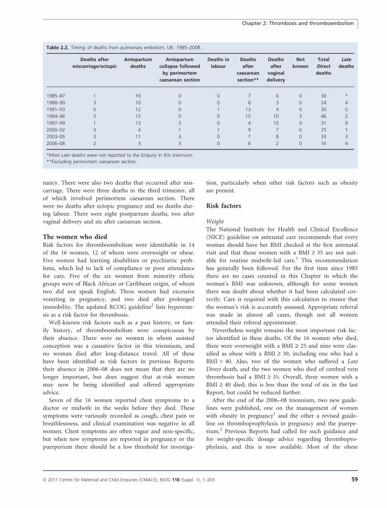
nancy. There were also two deaths that occurred after mis-
carriage. There were three deaths in the third trimester, all
of which involved perimortem caesarean section. There
were no deaths after ectopic pregnancy and no deaths dur-
ing labour. There were eight postpartum deaths, two after
vaginal delivery and six after caesarean section.
The women who diedRisk factors for thromboembolism were identifiable in 14
of the 16 women, 12 of whom were overweight or obese.
Five women had learning disabilities or psychiatric prob-
lems, which led to lack of compliance or poor attendance
for care. Five of the six women from minority ethnic
groups were of Black African or Caribbean origin, of whom
two did not speak English. Three women had excessive
vomiting in pregnancy, and two died after prolonged
immobility. The updated RCOG guideline2 lists hypereme-
sis as a risk factor for thrombosis.
Well-known risk factors such as a past history, or fam-
ily history, of thromboembolism were conspicuous by
their absence. There were no women in whom assisted
conception was a causative factor in this triennium, and
no woman died after long-distance travel. All of these
have been identified as risk factors in previous Reports:
their absence in 2006–08 does not mean that they are no
longer important, but does suggest that at-risk women
may now be being identified and offered appropriate
advice.
Seven of the 16 women reported chest symptoms to a
doctor or midwife in the weeks before they died. These
symptoms were variously recorded as cough, chest pain or
breathlessness, and clinical examination was negative in all
women. Chest symptoms are often vague and non-specific,
but when new symptoms are reported in pregnancy or the
puerperium there should be a low threshold for investiga-
tion, particularly when other risk factors such as obesity
are present.
Risk factors
WeightThe National Institute for Health and Clinical Excellence
(NICE) guideline on antenatal care recommends that every
woman should have her BMI checked at the first antenatal
visit and that those women with a BMI ‡ 35 are not suit-
able for routine midwife-led care.5 This recommendation
has generally been followed. For the first time since 1985
there are no cases counted in this Chapter in which the
woman’s BMI was unknown, although for some women
there was doubt about whether it had been calculated cor-
rectly. Care is required with this calculation to ensure that
the woman’s risk is accurately assessed. Appropriate referral
was made in almost all cases, though not all women
attended their referral appointment.
Nevertheless weight remains the most important risk fac-
tor identified in these deaths. Of the 16 women who died,
three were overweight with a BMI ‡ 25 and nine were clas-
sified as obese with a BMI ‡ 30, including one who had a
BMI > 40. Also, two of the women who suffered a Late
Direct death, and the two women who died of cerebral vein
thrombosis had a BMI ‡ 35. Overall, three women with a
BMI ‡ 40 died; this is less than the total of six in the last
Report, but could be reduced further.
After the end of the 2006–08 triennium, two new guide-
lines were published, one on the management of women
with obesity in pregnancy1 and the other a revised guide-
line on thromboprophylaxis in pregnancy and the puerpe-
rium.2 Previous Reports had called for such guidance and
for weight-specific dosage advice regarding thrombopro-
phylaxis, and this is now available. Most of the obese
Table 2.2. Timing of deaths from pulmonary embolism; UK: 1985–2008.
Deaths after
miscarriage/ectopic
Antepartum
deaths
Antepartum
collapse followed
by perimortem
caesarean section
Deaths in
labour
Deaths
after
caesarean
section**
Deaths
after
vaginal
delivery
Not
known
Total
Direct
deaths
Late
deaths
1985–87 1 16 0 0 7 6 0 30 *
1988–90 3 10 0 0 8 3 0 24 4
1991–93 0 12 0 1 13 4 0 30 5
1994–96 3 15 0 0 15 10 3 46 2
1997–99 1 13 3 0 4 10 0 31 9
2000–02 3 4 1 1 9 7 0 25 1
2003–05 3 11 4 0 7 8 0 33 3
2006–08 2 3 3 0 6 2 0 16 4
*Most Late deaths were not reported to the Enquiry in this triennium.
**Excluding perimortem caesarean section.
Chapter 2: Thrombosis and thromboembolism
ª 2011 Centre for Maternal and Child Enquiries (CMACE), BJOG 118 (Suppl. 1), 1–203 59
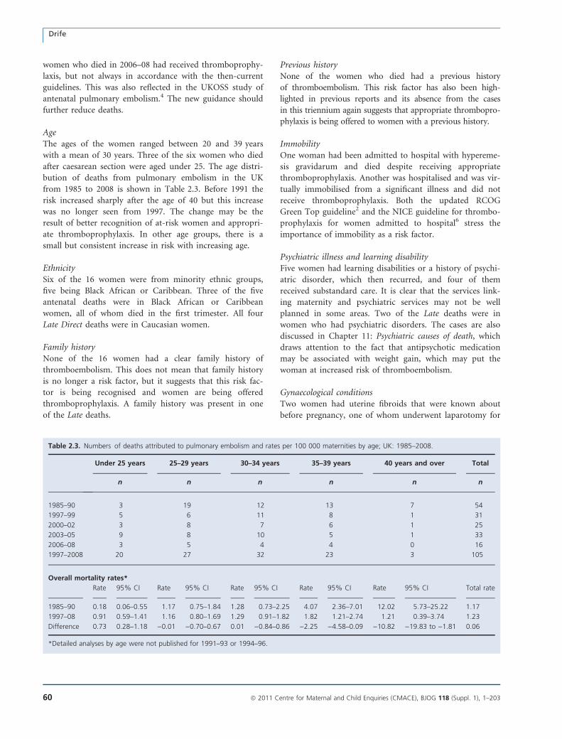
women who died in 2006–08 had received thromboprophy-
laxis, but not always in accordance with the then-current
guidelines. This was also reflected in the UKOSS study of
antenatal pulmonary embolism.4 The new guidance should
further reduce deaths.
AgeThe ages of the women ranged between 20 and 39 years
with a mean of 30 years. Three of the six women who died
after caesarean section were aged under 25. The age distri-
bution of deaths from pulmonary embolism in the UK
from 1985 to 2008 is shown in Table 2.3. Before 1991 the
risk increased sharply after the age of 40 but this increase
was no longer seen from 1997. The change may be the
result of better recognition of at-risk women and appropri-
ate thromboprophylaxis. In other age groups, there is a
small but consistent increase in risk with increasing age.
EthnicitySix of the 16 women were from minority ethnic groups,
five being Black African or Caribbean. Three of the five
antenatal deaths were in Black African or Caribbean
women, all of whom died in the first trimester. All four
Late Direct deaths were in Caucasian women.
Family historyNone of the 16 women had a clear family history of
thromboembolism. This does not mean that family history
is no longer a risk factor, but it suggests that this risk fac-
tor is being recognised and women are being offered
thromboprophylaxis. A family history was present in one
of the Late deaths.
Previous historyNone of the women who died had a previous history
of thromboembolism. This risk factor has also been high-
lighted in previous reports and its absence from the cases
in this triennium again suggests that appropriate thrombopro-
phylaxis is being offered to women with a previous history.
ImmobilityOne woman had been admitted to hospital with hypereme-
sis gravidarum and died despite receiving appropriate
thromboprophylaxis. Another was hospitalised and was vir-
tually immobilised from a significant illness and did not
receive thromboprophylaxis. Both the updated RCOG
Green Top guideline2 and the NICE guideline for thrombo-
prophylaxis for women admitted to hospital6 stress the
importance of immobility as a risk factor.
Psychiatric illness and learning disabilityFive women had learning disabilities or a history of psychi-
atric disorder, which then recurred, and four of them
received substandard care. It is clear that the services link-
ing maternity and psychiatric services may not be well
planned in some areas. Two of the Late deaths were in
women who had psychiatric disorders. The cases are also
discussed in Chapter 11: Psychiatric causes of death, which
draws attention to the fact that antipsychotic medication
may be associated with weight gain, which may put the
woman at increased risk of thromboembolism.
Gynaecological conditionsTwo women had uterine fibroids that were known about
before pregnancy, one of whom underwent laparotomy for
Table 2.3. Numbers of deaths attributed to pulmonary embolism and rates per 100 000 maternities by age; UK: 1985–2008.
Under 25 years 25–29 years 30–34 years 35–39 years 40 years and over Total
n n n n n n
1985–90 3 19 12 13 7 54
1997–99 5 6 11 8 1 31
2000–02 3 8 7 6 1 25
2003–05 9 8 10 5 1 33
2006–08 3 5 4 4 0 16
1997–2008 20 27 32 23 3 105
Overall mortality rates*
Rate 95% CI Rate 95% CI Rate 95% CI Rate 95% CI Rate 95% CI Total rate
1985–90 0.18 0.06–0.55 1.17 0.75–1.84 1.28 0.73–2.25 4.07 2.36–7.01 12.02 5.73–25.22 1.17
1997–08 0.91 0.59–1.41 1.16 0.80–1.69 1.29 0.91–1.82 1.82 1.21–2.74 1.21 0.39–3.74 1.23
Difference 0.73 0.28–1.18 )0.01 )0.70–0.67 0.01 )0.84–0.86 )2.25 )4.58–0.09 )10.82 )19.83 to )1.81 0.06
*Detailed analyses by age were not published for 1991–93 or 1994–96.
Drife
60 ª 2011 Centre for Maternal and Child Enquiries (CMACE), BJOG 118 (Suppl. 1), 1–203
malignant disease during pregnancy. Assisted conception
was not relevant to any of the deaths in this triennium,
though one woman who died during pregnancy had con-
ceived after intrauterine insemination.
Chest symptomsSeven of the 16 women reported chest symptoms to a doc-
tor or midwife in the weeks before they died. Two of the
three women who were Late deaths had also reported chest
symptoms.
Antepartum deathsOf the six women who had a pulmonary embolism in their
antenatal period, three died in the first trimester (up to
and including 12 weeks of gestation) and three died after
perimortem caesarean section in the third trimester. Only
one of these six women collapsed without prior warning;
the other five complained of respiratory symptoms, one to
the Emergency Department and four to their GPs. Two of
the three women who died in the first trimester were obese,
and all three were Black African or Caribbean.
The learning points from these deaths can be summar-
ised as:
1 Booking, with its formal risk assessment, often takes
place late in the first trimester. Before that time,
women are vulnerable.
2 Dehydration is associated with increased risk of embo-
lism, and hydration levels of women who are complain-
ing of vomiting should be assessed.
3 Chest symptoms arising for the first time in pregnancy
in an at-risk woman require investigation.
4 There is still insufficient awareness that women, partic-
ularly obese women, are at risk from thromboembolism
in early pregnancy.
5 The obstetric team should see pregnant women with
chest pain. In some cases women were referred for
assessment to the medical team, who were unaware of,
or underestimated, the risks of embolism in pregnancy.
6 Tachycardia is a critically important finding when a preg-
nant woman presents with a new symptom of breathless-
ness, though it is not diagnostic of pulmonary embolism
(and nor does its absence rule out the diagnosis).
7 Gynaecological wards should have a risk assessment
form specifically for women in early pregnancy, and it
must be completed.
The following is a typical example:
An obese woman with a history of chest pains before preg-
nancy saw her GP early in her pregnancy and thereafter
had contact on several occasions by phone and in person,
complaining about chest pain or shortness of breath. She
also developed hyperemesis and was eventually admitted to
hospital where tachycardia was noted. Investigation for
thromboembolism was commenced, but she collapsed and
died before treatment was initiated.
When reviewing these cases the assessors took into
account how difficult it is to decide whether common
symptoms are trivial or important. For example:
An obese mother consulted her GP in the third trimester
with shortness of breath. No abnormality was found on
clinical examination, and she was treated with an inhaler.
Shortly afterwards she collapsed and died.
The assessors felt that care was not substandard for this
woman, but the practice itself held a rigorous enquiry and
identified several areas for possible improvement. Top of
their list was the need to document the pulse rate. This
local enquiry is an example of good practice, which should
be more widely followed.
Deaths after miscarriage or termination of preg-nancyTwo women died after miscarriage, the underlying cause of
which, in one woman, should have meant that thrombo-
prophylaxis should have been considered.
Intrapartum deathsThere were no intrapartum deaths in this triennium.
Deaths after vaginal deliveryTwo women died after vaginal delivery, both after the third
week of their puerperium, and there were also two Late
deaths. Three of these four women had mental health
problems and found it difficult to stay in touch with both
the maternity and psychiatric services, although they were
available to them. In two women care was substandard. All
the cases demonstrate that there may be a long interval
between delivery and death. The major lessons to be learnt
from this group of deaths are as follows.
1 Action should be taken when an obese woman reports
the new onset of breathlessness.
2 Hospital guidelines on thromboprophylaxis should give
specific advice about obese women. The updated
RCOG guideline2 and the CMACE/RCOG obesity
guideline1 both recommend that women with a BMI ‡40 should be considered for postnatal thromboprophy-
laxis with low-molecular-weight heparin (LMWH)
regardless of the mode of delivery or the presence of
other risk factors.
3 Psychiatric involvement is required when a woman has
a history of previous postnatal mental health problems
and there is renewed concern about her mental health.
The following is a typical example:
A morbidly obese woman with a probable learning disabil-
ity was a poor attender for antenatal care. Some weeks after
Chapter 2: Thrombosis and thromboembolism
ª 2011 Centre for Maternal and Child Enquiries (CMACE), BJOG 118 (Suppl. 1), 1–203 61

delivery she became breathless and called her midwife, who
advised her to see her GP or go to the Emergency Depart-
ment if her breathlessness did not improve. She did not do
so and a few days later she collapsed and died.
In contrast to the general practice enquiry mentioned
above, the assessors noted that hospital internal enquiries
often focus only on acts or omissions by their own staff
and make no recommendations about how to help vulnera-
ble women. The assessors are concerned that hospital Seri-
ous Untoward Incident (SUI) reports they reviewed varied
greatly in quality. A national audit of such serious incident
reports is recommended.
Deaths after caesarean sectionSix women died after caesarean section; the three women
whose antepartum collapse was followed by perimortem
caesarean section have been considered separately. They all
died between 2 and 6 weeks after delivery. In addition, two
women died more than 42 days after caesarean section and
are classified as Late deaths.
Four of the six women were obese (BMI ‡ 30), including
one who had a BMI ‡ 40. Five of the six received LMWH;
the remaining woman refused LMWH because of needle
phobia. One woman had an underlying medical disease,
which was the indication for caesarean section, but she
died from pulmonary embolism. In one case it was unclear
whether an obese woman had symptoms before collapsing
in the late puerperium. Care was judged to be substandard
for four women.
The lessons to be learned from these cases are:
1 Women at risk of thromboembolism should be encour-
aged to report new chest symptoms after delivery.
2 Investigation is required when a woman with risk fac-
tors has new chest symptoms.
3 Miscalculation of the BMI at home booking can have
serious consequences. It can lead to an inappropriate
lack of obstetric input into antenatal care, misclassifica-
tion of the woman as being at moderate rather than
high risk of pulmonary embolism and inadequate
thromboprophylaxis.
The following is a typical example:
An obese woman was booked for midwife-led care and
referred to a consultant obstetrician after term. Labour was
induced, but caesarean section was required for fetal dis-
tress. Thromboprophylaxis consisted of Flowtron boots and
anti-embolism stockings: LMWH was started 7 hours after
delivery and continued for 4 days. Five weeks after delivery
she saw her GP with ‘chest infection and pain’ and was
given antibiotics. She died a few days later. According to
the autopsy, pulmonary embolism had been present for sev-
eral days before death.
The updated RCOG guidance2 recommends postnatal
thromboprophylaxis with LMWH to be continued for
1 week and longer if there are ongoing risk factors. The use
of properly applied graduated compression stockings of
appropriate strength is recommended in pregnancy and the
puerperium for those who are hospitalised after caesarean
section (combined with LMWH) and considered to be at
particularly high risk for venous thromboembolism [VTE]
(such as previous venous thromboembolism or more than
three risk factors).
Late deathsFour Late Direct deaths related directly to pulmonary
embolism are counted in Chapter 12. Three occurred
7 weeks after delivery and one much later in her postnatal
year. All the women were Caucasian; two had vaginal deliv-
eries and two were delivered by caesarean section. Two
women had normal body weight and no risk factors except
that one had a psychiatric disorder. Both these women
received good care including appropriate referral and
thromboprophylaxis. Both the other two women were
obese and received substandard care.
An older woman with a psychiatric disorder and social
problems had a family history of thromboembolism. After
delivery she was discharged home on ‘self-administered
Clexane’, but lacked the skills to give herself these injections.
Four weeks after delivery she attended the Emergency
Department with calf pain and breathlessness. She was
given an injection of LMWH and asked to come back next
day. Two weeks later she returned with a history of pleuritic
pain, developed severe breathlessness and died.
The treatment given took no account of her background,
risk factors and high likelihood of not returning when
asked. The RCOG guideline on acute management of
thromboembolic disease in pregnancy and the puerperium
recommends that any woman with signs and symptoms
suggestive of VTE should have objective testing performed
expeditiously and treatment with LMWH until the diagno-
sis is excluded. The therapeutic dose of LMWH should be
weight-related7 as shown in Table 2.4.
Cerebral vein thrombosis
There were two deaths from likely cerebral vein thrombo-
sis, though the diagnosis was not straightforward in either
woman. Both women were morbidly obese and both pre-
sented with headache and neurological symptoms after
delivery at term. They also both had previous medical com-
plications that could have caused dehydration.
A woman who may have had a co-existent encephalopathy
developed vomiting and dizziness near term, which persisted
Drife
62 ª 2011 Centre for Maternal and Child Enquiries (CMACE), BJOG 118 (Suppl. 1), 1–203
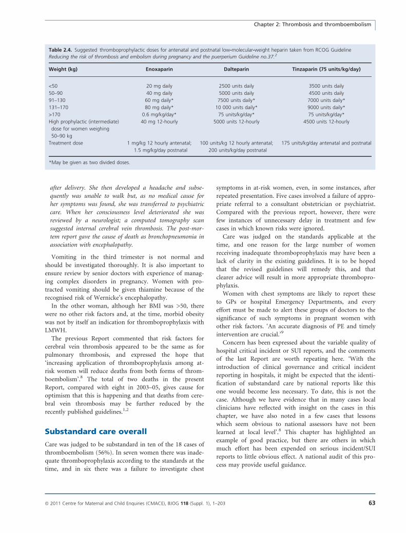
after delivery. She then developed a headache and subse-
quently was unable to walk but, as no medical cause for
her symptoms was found, she was transferred to psychiatric
care. When her consciousness level deteriorated she was
reviewed by a neurologist; a computed tomography scan
suggested internal cerebral vein thrombosis. The post-mor-
tem report gave the cause of death as bronchopneumonia in
association with encephalopathy.
Vomiting in the third trimester is not normal and
should be investigated thoroughly. It is also important to
ensure review by senior doctors with experience of manag-
ing complex disorders in pregnancy. Women with pro-
tracted vomiting should be given thiamine because of the
recognised risk of Wernicke’s encephalopathy.
In the other woman, although her BMI was >50, there
were no other risk factors and, at the time, morbid obesity
was not by itself an indication for thromboprophylaxis with
LMWH.
The previous Report commented that risk factors for
cerebral vein thrombosis appeared to be the same as for
pulmonary thrombosis, and expressed the hope that
‘increasing application of thromboprophylaxis among at-
risk women will reduce deaths from both forms of throm-
boembolism’.8 The total of two deaths in the present
Report, compared with eight in 2003–05, gives cause for
optimism that this is happening and that deaths from cere-
bral vein thrombosis may be further reduced by the
recently published guidelines.1,2
Substandard care overall
Care was judged to be substandard in ten of the 18 cases of
thromboembolism (56%). In seven women there was inade-
quate thromboprophylaxis according to the standards at the
time, and in six there was a failure to investigate chest
symptoms in at-risk women, even, in some instances, after
repeated presentation. Five cases involved a failure of appro-
priate referral to a consultant obstetrician or psychiatrist.
Compared with the previous report, however, there were
few instances of unnecessary delay in treatment and few
cases in which known risks were ignored.
Care was judged on the standards applicable at the
time, and one reason for the large number of women
receiving inadequate thromboprophylaxis may have been a
lack of clarity in the existing guidelines. It is to be hoped
that the revised guidelines will remedy this, and that
clearer advice will result in more appropriate thrombopro-
phylaxis.
Women with chest symptoms are likely to report these
to GPs or hospital Emergency Departments, and every
effort must be made to alert these groups of doctors to the
significance of such symptoms in pregnant women with
other risk factors. ‘An accurate diagnosis of PE and timely
intervention are crucial.’9
Concern has been expressed about the variable quality of
hospital critical incident or SUI reports, and the comments
of the last Report are worth repeating here. ‘With the
introduction of clinical governance and critical incident
reporting in hospitals, it might be expected that the identi-
fication of substandard care by national reports like this
one would become less necessary. To date, this is not the
case. Although we have evidence that in many cases local
clinicians have reflected with insight on the cases in this
chapter, we have also noted in a few cases that lessons
which seem obvious to national assessors have not been
learned at local level’.8 This chapter has highlighted an
example of good practice, but there are others in which
much effort has been expended on serious incident/SUI
reports to little obvious effect. A national audit of this pro-
cess may provide useful guidance.
Table 2.4. Suggested thromboprophylactic doses for antenatal and postnatal low-molecular-weight heparin taken from RCOG Guideline
Reducing the risk of thrombosis and embolism during pregnancy and the puerperium Guideline no.37.2
Weight (kg) Enoxaparin Dalteparin Tinzaparin (75 units/kg/day)
<50 20 mg daily 2500 units daily 3500 units daily
50–90 40 mg daily 5000 units daily 4500 units daily
91–130 60 mg daily* 7500 units daily* 7000 units daily*
131–170 80 mg daily* 10 000 units daily* 9000 units daily*
>170 0.6 mg/kg/day* 75 units/kg/day* 75 units/kg/day*
High prophylactic (intermediate)
dose for women weighing
50–90 kg
40 mg 12-hourly 5000 units 12-hourly 4500 units 12-hourly
Treatment dose 1 mg/kg 12 hourly antenatal;
1.5 mg/kg/day postnatal
100 units/kg 12 hourly antenatal;
200 units/kg/day postnatal
175 units/kg/day antenatal and postnatal
*May be given as two divided doses.
Chapter 2: Thrombosis and thromboembolism
ª 2011 Centre for Maternal and Child Enquiries (CMACE), BJOG 118 (Suppl. 1), 1–203 63
Pathological overview
Fourteen autopsy reports giving the cause of death as pul-
monary embolus were examined and considered in this sec-
tion: five were excellent, three were adequate and six were
poor. Four of the 14 are considered in other chapters,
because the death was primarily the result of another co-
morbidity; for example sepsis, tumour bulk and Late Coin-
cidental causes. Associated predisposing causes were also
often present in those classified as Direct deaths from
embolus. Significant obesity with a BMI of ‡35 was present
in five women, and one of these also had a family history
of thrombophilia from Protein S deficiency. In one woman
there was preceding significant, though not otherwise life-
threatening, inflammation of the genital tract. These raise
the question of where the dividing line between Direct,
Indirect and Coincidental should be drawn for this group
and whether all pulmonary embolic deaths should be
reclassified as Indirect.
The leg and pelvic veins were not examined/mentioned
in any of the six poor reports. One of these was a Late
Coincidental death of a woman on antipsychotic drugs.
These have been associated with deep vein thrombosis.
Given that there is a greater propensity for pregnancy to
be associated with pelvic vein than deep leg vein throm-
bosis, the site and location of the emboli is useful infor-
mation to be derived from the autopsy if not otherwise
known and in this case may have suggested reclassification
to being from Late Direct causes. In three women, there
was retrospectively a clear preceding clinical history of
dyspnoea, but in only one of these was there any attempt
to search for previous embolic episodes; in the other two
there was not even any histology taken. This is particu-
larly surprising in one woman, who had an enlarged and
dilated right heart, but the cause was not investigated.
Such examples contrast starkly with, for instance, some
deaths in the first trimester. In one, breathlessness had been
attributed to asthma. As well as identifying the pulmonary
emboli, histology confirmed that several were organised
and quite possibly preceded pregnancy and, furthermore,
that the features of asthma were not present. In another,
emboli complicated dehydration caused by hyperemesis:
the mammillary bodies were macroscopically and micro-
scopically normal.
Thromboembolism pathology: goodpractice points
• Examine and describe the pelvic and deep leg veins.
• Identify contributory co-morbid factors.
• Correlate the findings and histology with the clinical
history.
Conclusions
For many years these Reports have been suggesting that
most deaths from thromboembolism are preventable. There
is now evidence to support this. The 1995 RCOG guideline
on thromboprophylaxis after caesarean section10 was fol-
lowed by a sharp fall in deaths in this category, and now
the 2004 RCOG guideline3 has been followed by a sharp
fall in antenatal deaths and deaths following vaginal deliv-
ery, and, perhaps unexpectedly, in deaths from cerebral
vein thrombosis. The detailed advice in the 2009 revised
guideline2 should produce further improvement.
Guidelines, however, are effective only when there is a
widespread awareness of the risks and of the need to apply
the advice. The cases reported here demonstrate that most
deaths from thromboembolism occur in women with
known risk factors. This re-emphasises the need for careful
risk assessment as early as possible in pregnancy and
continuing reassessment of risk during and after pregnancy.
When an at-risk woman is identified, it is not enough
merely to give advice or make a referral. Some continuity
is needed to check that she has been able to follow the
advice or attend the appointment.
Symptoms must be interpreted in the light of the
woman’s background risk factors. This ought to be a state-
ment of the obvious, but several cases show that this does
not always happen. A distressing feature of this chapter is
the over-representation of vulnerable women. The many
pressures on the maternity services are no excuse for pro-
viding substandard service to women who require care in
the true sense of the word.
Disclosure of interestsNone.
FundingThis work was undertaken by the Centre for Maternal and
Child Enquiries (CMACE) as part of the CEMACH pro-
gramme. The work was funded by the National Patient
Safety Agency; the Department of Health, Social Services
and Public Safety of Northern Ireland; NHS Quality
Improvement Scotland (NHS QIS); and the Channel
Islands and Isle of Man.
AcknowledgementsThe pathology overview of this chapter was written by Dr
H Millward-Sadler, Consultant Histopathologist of South-
ampton University Hospitals NHS Trust. j
References
1 Centre for Maternal and Child Enquires and the Royal College of
Obstetricians and Gynaecologists. CMACE/RCOG Joint Guideline:
Drife
64 ª 2011 Centre for Maternal and Child Enquiries (CMACE), BJOG 118 (Suppl. 1), 1–203
Management of Women with Obesity in Pregnancy. London:
CMACE/RCOG, 2010.
2 Royal College of Obstetricians and Gynaecologists. Thrombosis and
Embolism during Pregnancy and the Puerperium. Reducing the Risk.
Guideline no.37. London: RCOG Press, 2009. [www.rcog.org.uk].
Accessed 5 October 2010.
3 Royal College of Obstetricians and Gynaecologists. Thromboprophy-
laxis During Pregnancy, Labour and After Normal Vaginal Delivery.
Guideline no.37. London: RCOG Press, 2004.
4 Knight M. Antenatal pulmonary embolism: risk factors, management
and outcomes. BJOG 2008;115:453–61.
5 National Collaborating Centre for Women’s and Children’s Health.
Antenatal Care: Routine Care for the Healthy Pregnant Woman. Lon-
don: RCOG Press, 2008.
6 National Clinical Guideline Centre—Acute and Chronic Condi-
tions. Venous Thromboembolism: Reducing the Risk of Venous
Thromboembolism (Deep Vein Thrombosis and Pulmonary
Embolism) in Patients Admitted to Hospital. London: NCGCACC
at the Royal College of Physicians, 2009. [www.nice.org.uk/
nicemedia/pdf/CG92FullGuideline.pdf]. Accessed 5 October 2010.
7 Royal College of Obstetricians and Gynaecologists. Thromboembolic
Disease in Pregnancy and the Puerperium: Acute Management.
Green-top Guideline no. 28. London: RCOG Press, 2007. [www.rco-
g.org.uk]. Accessed 5 October 2010.
8 Lewis G (ed). The Confidential Enquiry into Maternal and Child
Health (CEMACH). Saving Mothers’ Lives: Reviewing Maternal
Deaths to Make Motherhood Safer – 2003–2005. The Seventh
Report on Confidential Enquiries into Maternal Deaths in the United
Kingdom. London: CEMACH, 2007. [www.cmace.org.uk]. Accessed
5 October 2010.
9 Bourjeily G, Paidas M, Khalil H, Rosene-Montella K, Rodger M. Pul-
monary embolism in pregnancy. Lancet 2010;375:500–12.
10 Royal College of Obstetricians and Gynaecologists. Report of the
RCOG Working Party on Prophylaxis Against Thromboembolism in
Gynaecology and Obstetrics. London: RCOG Press, 1995.
ª 2011 Centre for Maternal and Child Enquiries (CMACE), BJOG 118 (Suppl. 1), 1–203 65
Chapter 2: Thrombosis and thromboembolism
Chapter 3: Pre-eclampsia and eclampsiaJ Neilson (on behalf of the Centre for Maternal and Child Enquiries)
Department of Women’s & Children’s Health; Institute of Translational Medicine, University of Liverpool, Liverpool Women’s Hospital,
Liverpool, UK
Correspondence: Professor James P Neilson, Department of Women’s & Children’s Health; Institute of Translational Medicine, University of
Liverpool, 1st Floor, Liverpool Women’s Hospital, Crown Street
Keywords eclampsia, pre-eclampsia, Confidential Enquiry, mater-
nal, mortality.
Pre-eclampsia and eclampsia: specific recommendations
• Pregnant women with a headache of sufficient severity to seek medical advice, or with new epigastric pain, should
have their blood pressure measured and urine tested for protein, as a minimum. Epigastric pain in the second half of
pregnancy should be considered to be the result of pre-eclampsia until proven otherwise.
• Any discussion between clinical staff about a woman with pre-eclampsia should include explicit mention of the systolic
pressure.
• Severe, life-threatening, hypertension must be treated effectively. Management protocols should recognise the need to
avoid very high systolic blood pressures which are associated with an increased risk of intracerebral haemorrhage.
• Systolic blood pressures of 150 mmHg, or above, require effective antihypertensive treatment. If the systolic pressure is
very high, >180 mmHg, this is a medical emergency that requires urgent as well as effective antihypertensive treatment.
• Intramuscular oxytocin, not Syntometrine, should be the routine drug for active management of the third stage of
labour.
• Women with severe pre-eclampsia need effective team care, based on clear communication and common under-
standing. There should be early engagement of intensive care specialists where appropriate. Efforts must be made to
re-engage and re-skill GPs who see women with complications during pregnancy. Women who elect to pay for pri-
vate obstetric care are entitled to as good a standard of care as they would receive in the NHS.
Pre-eclampsia/eclampsia: learningpoints
Eclampsia is important because it is a marker for severe
disease, but seizures also carry additional intrinsic risks.
Some automated blood pressure monitoring systems sys-
tematically underestimate systolic pressure in pre-eclampsia.
Summary of findings for 2006–08
Overall, the deaths of 22 women are counted and discussed
in this Chapter. Nineteen were the result of eclampsia or
pre-eclampsia and three were the result of acute fatty liver
of pregnancy (AFLP), the values are shown in Table 3.1.
In terms of deaths from eclampsia or pre-eclampsia the
mortality rate for this triennium was 0.83 per 100 000
maternities (95% CI 0.53–1.30) compared with 0.66 (0.39–
1.12) in the last Report.
The three deaths from AFLP are included in this Chapter
on the basis that this may be part of a spectrum of condi-
tions related to pre-eclampsia.1 In one of these AFLP was
an unexpected histopathological finding at autopsy in a
woman with the clinical syndrome of pre-eclampsia and
who died because of massive intra-abdominal haemorrhage.
There was only one such case in the last Report.
Fourteen of the 22 women died from cerebral causes;
nine from intracranial haemorrhage and five from anoxia
following cardiac arrest in association with eclamptic sei-
zures. Of the other causes of death, three women died from
liver complications (two from necrosis and one from sub-
capsular haemorrhage), two died from multi-organ failure
in intensive care units, two from complications of AFLP
and one from intra-abdominal haemorrhage of uncertain
Neilson
66 ª 2011 Centre for Maternal and Child Enquiries (CMACE), BJOG 118 (Suppl. 1), 1–203
source. There were no deaths from simple fluid overload.
The causes of death are compared with values from recent
triennia in Table 3.1.
Severe maternal morbidityA national study of eclampsia conducted through the UK
Obstetric Surveillance System (UKOSS) (The United King-
dom Obstetric Surveillance System (UKOSS) is described in
more detail in the Introduction Chapter of this Report.)
between February 2005 and February 2006 identified 214
cases,2 giving an estimated incidence of 27.5 cases per
100 000 maternities (95% CI 23.9–31.4 per 100 000 materni-
ties). This is almost a halving of the incidence of eclampsia
since 1992.3 The decrease in incidence has occurred almost
entirely in the group of women with diagnosed pre-eclamp-
sia. No women died in the UKOSS study, but eclampsia was
associated with significant additional maternal morbidity,
including cerebrovascular events, in five women (2.3%).
Combining the UKOSS data and the data from this Report,
the case fatality rate from eclampsia is estimated to be 3.1%.
Acute fatty liver of pregnancySurveillance of AFLP through UKOSS between February
2005 and August 2006 identified 57 women with AFLP in
an estimated 1 132 964 maternities;1 an estimated incidence
of 5.0 cases per 100 000 maternities (95% CI 3.8–6.5 per
100 000 maternities). Only one of these women died (case
fatality rate 1.8%, 95% CI 0–9.4%), but 60% were admitted
to a Critical Care unit. Eighteen percent of the women with
AFLP had twin pregnancies.
The women who died
Pre-eclampsia/eclampsiaThe ages of the women who died from pre-eclampsia/
eclampsia ranged between 19 and 39 years with a median
age of 31 years. Their gestations ranged from 21 to
41 weeks. It is sometimes said that pre-eclampsia at term is
a benign condition. This is not necessarily so. Early-onset
pre-eclampsia is, overall, a more aggressive condition than
late-onset disease, but fulminating, ultimately fatal, pre-
eclampsia also occurs at term. Four of the women were at,
or after, 40 weeks of gestation.
Parity ranged from 0 to 9; thirteen women were primigr-
avid. Four women had twin pregnancies. Seven women had
eclamptic fits. There was evidence of HELLP (haemolysis,
elevated liver enzymes and low platelet count) syndrome in
eight women. The body mass index (BMI) ranged between
20 and 43 with a median of 24. Most women were not
obese, but five had BMIs ‡30, including one BMI ‡40. Ten
of the women who died from pre-eclampsia/eclampsia were
white, and, of the remaining nine, six were Black Africans.
Black African women seem particularly susceptible to
aggressive forms of pre-eclampsia. To establish if this is
Table 3.1. Numbers and underlying cause of death due to eclampsia and pre-eclampsia, UK: 1991–2008
Cause of death 1997–99 2000–02 2003–05 2006–08
n Rate 95% CI n Rate 95% CI n Rate 95% CI n Rate 95% CI
Cerebral
Intracranial haemorrhage 7 0.33 0.16–0.69 9 0.45 0.23–0.87 9 0.43 0.22–0.82 9 0.39 0.20–0.75
Subarachnoid 0 0.00 0 0.00 0 0.00 0 0.00
Infarct 0 0.00 0 0.00 1 0.05 0.01–0.34 0 0.00
Oedema 0 0.00 0 0.00 0 0.00 0 0.00
Eclampsia 0 0.00 0 0.00 0 0.00 5 0.22 0.09–0.52
Subtotal 7 0.33 0.16–0.69 9 0.45 0.23–0.87 10 0.47 0.25–0.88 14 0.61 0.36–1.03
Pulmonary
Adult respiratory distress syndrome 2 0.09 0.02–0.38 1 0.05 0.01–0.36 0 0.00 0 0.00
Oedema 0 0.00 0 0.00 0 0.00 0 0.00
Subtotal 2 0.09 0.02–0.38 1 0.05 0.01–0.36 0 0.00 0 0.00
Hepatic
Rupture 2 0.09 0.02–0.38 0 0.00 0 0.00 1 0.04 0.01–0.31
Failure/necrosis 0 0.00 0 0.00 1 0.05 0.01–0.34 2 0.09 0.02–0.35
Other 5 0.24 0.10–0.57 4 0.20 0.08–0.53 3 0.14 0.05–0.44 2 0.09 0.02–0.35
Subtotal 7 0.33 0.16–0.69 4 0.20 0.08–0.53 4 0.19 0.07–0.50 5 0.22 0.09–0.52
Overall total 16 0.75 0.46–1.23 14 0.70 0.42–1.18 14 0.66 0.39–1.12 19 0.83 0.53–1.30
Acute fatty liver of pregnancy 4 0.19 0.07–0.50 3 0.15 0.05–0.47 1* 0.05 0.01–0.34 3* 0.13 0.04–0.41
*As a result of the very small numbers these cases are counted in this Chapter 3. Before 2003 they were counted in the now nonexistent chapter
‘Deaths from other Direct causes.’
Chapter 3: Pre-eclampsia and eclampsia
ª 2011 Centre for Maternal and Child Enquiries (CMACE), BJOG 118 (Suppl. 1), 1–203 67
true, and what might be the underlying genetic or
other pathophysiological mechanisms, further research is
required.
Acute fatty liver of pregnancyThe ages of the women who died from AFLP ranged
between 23 and 39 years with a median of 33 years. One
had a BMI >40. None were from minority ethnic groups.
Substandard careDisappointingly, 20 of the 22 cases demonstrated substan-
dard care; in 14 cases this was classed as ‘major’, and there
were, undoubtedly, avoidable deaths. Deaths from intracra-
nial haemorrhage, the single largest cause of death, indicate
a failure of effective antihypertensive therapy. Ensuring
effective antihypertensive therapy is the priority for
improving clinical care for these women. This has been em-
phasised in recent Reports and is yet again one of the ‘Top
ten’ recommendations in this Report.
Substandard care occurred both in hospitals and in the
community. There were four women in whom GPs made
errors. These were mainly around failure to refer appropri-
ately to specialist services, often because of a failure to
appreciate the significance of symptoms or signs of pre-
eclampsia. In one woman, a GP started outpatient antihy-
pertensive drug treatment in a woman with pre-eclampsia,
instead of referring her for specialist care. Another referred
a woman with heavy proteinuria for urological investiga-
tion. A woman with jaundice was referred by her GP to
the community midwife; it subsequently emerged that the
woman had HELLP syndrome. GPs have become increas-
ingly disengaged from antenatal care and consequently de-
skilled, and inappropriate decision-making may be an
increasing problem. The first port of call to a pregnant
woman with epigastric pain (an important symptom of
severe pre-eclampsia) may well be her GP. This problem
needs to be urgently addressed.
The significance of epigastric pain in pregnancy also
needs to be better understood in Emergency Departments:
A woman presented to an Emergency Department in early
third trimester with epigastric pain. Her blood pressure was
>150/90 mmHg and she had proteinuria +++. She was
diagnosed as having ‘gastritis’ and discharged home, where
she collapsed and died shortly afterwards. Autopsy showed
a cerebral haemorrhage and the typical histological features
of pre-eclampsia.
Treatment of systolic hypertensionThe single major failing in clinical care in the current trien-
nium was, again, inadequate treatment of hypertension,
with subsequent intracranial haemorrhage. For example:
A woman presented to an Emergency Department during
her second trimester with abdominal pain and vomiting.
Previously normotensive at the antenatal clinic, her blood
pressure rose from 191/110 to 210/130 mmHg while in the
Emergency Department. She had proteinuria +++. She
developed a severe headache and pain on breathing and
was transferred to the labour ward where she was given a
calcium channel blocker and opiate analgesia. Following an
eclamptic seizure, treatment with magnesium sulphate and
hydralazine infusion was started. First hands-on consultant
involvement occurred around 8–10 hours after admission,
at which time her blood pressure was still extremely high
(systolic pressure still >200 mmHg) and she was unrous-
able. She died shortly after from a large intracranial bleed.
This is illustrative of a number of cases in which there
was failure to appreciate the seriousness of the situation, a
failure to treat systolic hypertension effectively and a failure
of involvement of senior clinicians until it was too late. It
is systolic hypertension that poses the greatest risk of cere-
bral haemorrhage, and very high pressures need to be trea-
ted as medical emergencies. This woman, as others, had
life-threatening systolic hypertension for several hours
before her stroke.
The National Institute for Health and Clinical Excellence
(NICE) guidelines on hypertension in pregnancy recom-
mend that women with moderate pre-eclampsia should be
treated with oral labetalol if systolic pressure reaches 150–
160 mmHg. If there is severe pre-eclampsia (with very high
systolic pressures), treatment can be with either oral or intra-
venous labetalol, oral nifedipine, or intravenous hydral-
azine.4 A combination of drugs may be necessary. There is
little evidence of any benefit associated with any individual
drug regimen. The priority is to lower the dangerously high
systolic pressure. The NICE guideline recommends a target
systolic pressure of 150 mmHg.4 Ideally, both consultant
obstetricians and anaesthetists will be present for such emer-
gencies, and consideration should be given to invasive moni-
toring with an arterial line and high-dependency care.
As the vignette above illustrates, it is essential that the
response to therapy is monitored carefully and, if a response
is absent or inadequate, that suitable treatment is added.
It is also worth re-emphasising, as in the last Reports,
the observation that some automated blood pressure moni-
toring systems systematically under-estimate systolic pres-
sure in pre-eclampsia.
Third-stage managementThree women who died from cerebral haemorrhage devel-
oped very high blood pressures shortly after giving birth.
All had received intramuscular Syntometrine (oxytocin–erg-
ometrine) for active management of the third stage of
labour. One woman had been hypertensive during labour,
Neilson
68 ª 2011 Centre for Maternal and Child Enquiries (CMACE), BJOG 118 (Suppl. 1), 1–203
one had been normotensive, and one had not had her
blood pressure checked during what was a rapid labour.
Syntometrine is contraindicated in hypertensive women
because it may exacerbate hypertension, and a previous
Report has recommended that it be avoided in women
who do not have their blood pressures checked in fast
labours. It is now sensible to reach the conclusion that Syn-
tometrine should be avoided as a routine drug completely.
Intramuscular Syntocinon (oxytocin without ergometrine)
provides almost as effective prophylaxis against postpartum
haemorrhage as does Syntometrine, with fewer adverse
effects, including hypertension.5 Intramuscular oxytocin is
not licensed for this use simply because the manufacturers
have no commercial incentive to see it licensed. The use of
intramuscular oxytocin as the routine drug for active man-
agement of the third stage of labour is now recommended
by two NICE guideline development panels.4–6
EclampsiaIt is unprecedented to report so many deaths in apparently
direct association with eclamptic seizures. In three women,
cardiac arrests were witnessed by clinical staff shortly after
seizures: one woman, who had received no treatment, had
a seizure followed by cardiac arrest in her home in the
presence of a paramedic. Another woman was in hospital
and had received nifedipine but no magnesium sulphate,
because of concern about her oliguria. She too had a sei-
zure followed by cardiac arrest. The third woman, also in
hospital, had received both labetalol and magnesium sul-
phate before seizures and cardiac arrest. In a further two
women there was less certainty, as they were found col-
lapsed at home, but excellent autopsies showed unequivocal
histological evidence of pre-eclampsia and no other pathol-
ogy (e.g. cerebral haemorrhage), and it seems highly likely
that these deaths, too, were associated with eclampsia. It is
well recognised that cardiac arrest can occur in association
with seizures in people with epilepsy (Sudden Unexexpect-
ed Death in EPilepsy), and it seems plausible that cardiore-
spiratory arrest may have a similar mechanism in
association with eclamptic seizures. These findings remind
us that there are intrinsic risks to eclamptic seizures.
Five other women had pregnancies complicated with
pre-eclampsia but, because they died of other causes, are
counted elsewhere. Two women who died from cardiomy-
opathy are counted in Chapter 9, two are counted as Other
Indirect in Chapter 10 and one woman who died of amni-
otic fluid embolism is counted in Chapter 5.
Pathological overview
Autopsies were performed on 14 women for whom pre-
eclampsia was the clinical background to the cause of
death, but no report was available for two of these and in a
third the report was incomplete. Five of the reports were
excellent and six, including the incomplete report, were
poor or worse. Cerebral pathology was confirmed in three
and liver necrosis in one of the five excellent reports. In
one of these cases a woman died unexpectedly in preg-
nancy when normotensive but with ketonuria and oedema.
The autopsy report had minimal macroscopic findings: the
classical changes of pre-eclampsia were subsequently histo-
logically demonstrated in her uterus, liver and kidneys.
In the five poor autopsies, there was failure either to
address the clinical differential diagnoses or the concluding
cause of death bore no apparent relation to the autopsy find-
ings, and sometimes both. No histology was taken in four of
these cases. For two, the death was attributed to haemor-
rhage despite the clear clinical and pathological findings of
HELLP syndrome without a clinical history of bleeding in
the one, and a clear description of normal jugular veins (the
claimed source) in the other. The other cases have been clas-
sified and discussed with the Other Indirect deaths in Chap-
ter 10, though the possibility that pre-eclampsia was
involved cannot be excluded. In two of these there was
death from cerebral haemorrhage in women with recorded
preceding hypertension in pregnancy: in one this was mild
and in the other it was transient, though associated with
proteinuria. No adequate neuropathological examination
was performed, and there was no histological search for pre-
eclampsia in either case. The third is unascertained:
A diabetic woman was started on insulin in the second tri-
mester, and her last blood pressure at around 30 weeks had
risen to 140/95 mmHg. A few weeks later she collapsed at
home. A bitten tongue was the major autopsy finding: there
was no histology or toxicology.
The assessors cannot classify this as a death from pre-
eclampsia but feel that this possibility was not adequately
addressed either by direct exclusion or by the determina-
tion of a clear alternative cause of death. The autopsy
totally failed to address the possible clinical diagnoses and,
in particular, failed to search adequately for the histological
features of pre-eclampsia. The assessors have therefore clas-
sified it as unascertained.
Although most deaths associated with pre-eclampsia are
clear-cut, there are cases where the differential diagnoses
require a detailed autopsy assessment, if only for exclusion
of other possibilities.
Pre-eclampsia and eclampsia:pathological recommendation
Autopsies on women with a preceding history of pre-
eclampsia should have careful histological examination
of the kidneys, liver and uterine placental bed site.
Chapter 3: Pre-eclampsia and eclampsia
ª 2011 Centre for Maternal and Child Enquiries (CMACE), BJOG 118 (Suppl. 1), 1–203 69
Conclusion
The number of deaths from pre-eclampsia/eclampsia has not
fallen. The most pressing need, as before, is to treat hyperten-
sion (and especially systolic hypertension) quickly and effec-
tively to prevent haemorrhagic stroke. Iatrogenic
hypertension should be avoided by abandoning the use of
ergometrine in routine third-stage management. The inci-
dence of eclampsia has halved in the UK, presumably as a
result of the widespread use of magnesium sulphate, follow-
ing publication of the Magpie trial.7 The UKOSS study has
shown that the overall case fatality rate associated with
eclampsia is low in the UK, but serious morbidity can occur.2
However, this Enquiry, albeit over a different time-scale, has
identified an unprecedented number of deaths associated
with eclamptic seizures. This is a reminder that eclampsia is a
serious complication that, where possible, should be avoided.
Disclosure of interestsNone.
FundingThis work was undertaken by the Centre for Maternal and
Child Enquiries (CMACE) as part of the CEMACH pro-
gramme. The work was funded by the National Patient
Safety Agency; the Department of Health, Social Services
and Public Safety of Northern Ireland; NHS Quality
Improvement Scotland (NHS QIS); and the Channel
Islands and Isle of Man.
AcknowledgementsThis chapter has been seen and discussed with Mr Steven
Walkinshaw, Consultant Obstetrician of Liverpool
Women’s Hospital. The Pathology overview was written by
Dr H Millward-Sadler, Consultant Histopathologist of
Southampton University Hospitals NHS Trust. j
References
1 Knight M, Nelson-Piercy C, Kurinczuk JJ, Spark P, Brocklehurst P. A
prospective national study of acute fatty liver of pregnancy in the UK.
Gut 2008;57:951–6.
2 Knight M. Eclampsia in the United Kingdom 2005. BJOG
2007;114:1072–8.
3 Douglas KA, Redman CW. Eclampsia in the United Kingdom. BMJ
1994;309:1395–400.
4 National Collaborating Centre for Women’s and Children’s Health.
Hypertension in Pregnancy: The Management of Hypertensive Disor-
ders During Pregnancy. National Institute for Health and Clinical Excel-
lence Guideline 107. London: RCOG. August 2010. [www.guidance.
nice.org.uk/CG107/]. Accessed 5 October 2010.
5 McDonald SJ, Abbott JN, Higgins SP. Prophylactic ergometrine-oxyto-
cin versus oxytocin for the third stage of labour. Cochrane Database
Syst Rev 2004;Issue 1:CD000201.
6 National Collaborating Centre for Women’s and Children’s Health. In-
trapartum Care. Care of Healthy Women and their Babies During
Childbirth. London: RCOG. Revised Report 2008. [http://www.
ncc-wch.org.uk/guidelines/guidelilnes-programme/guidelines-programme-
published/intrapartum-care/] Accessed 5 October 2010.
7 Altman D, Carroli G, Duley L, Farrell B, Moodley J, Neilson J, et al. Do
women with pre-eclampsia, and their babies, benefit from magne-
sium sulphate? The Magpie Trial: a randomised placebo-controlled
trial Lancet 2002;359:1877–90.
Neilson
70 ª 2011 Centre for Maternal and Child Enquiries (CMACE), BJOG 118 (Suppl. 1), 1–203
Chapter 4: HaemorrhageJ Norman (on behalf of the Centre for Maternal and Child Enquiries)
University of Edinburgh Centre for Reproductive Biology, The Queens Medical Research Institute, Edinburgh, UK
Correspondence: Professor Jane Norman, Chair of Maternal and Fetal Health, University of Edinburgh Centre for Reproductive Biology, The
Queens Medical Research Institute, 47 Little France Crescent, Edinburgh EH16 4TJ, UK. Email: [email protected]
. Keywords obstetric haemorrhage, Confidential Enquiry, maternal,
mortality.
Obstetric haemorrhage: specific recommendation
• Despite the decline in numbers this triennium, obstetric haemorrhage remains an important cause of maternal death.
All units should have protocols in place for its identification and management, and all clinicians responsible for the
care of pregnant women, antenatally, postnatally and intrapartum, including those practicing in the community,
should carry out regular skills training for such scenarios.
• Early senior multidisciplinary team involvement is essential in the management of major obstetric haemorrhage.
• All clinicians should be aware of the guidelines for management of women who refuse blood transfusion.
• The welcome absence of deaths in relation to elective caesarean section for placenta praevia in this Report endorses
the recommendations in earlier Reports that senior staff should be involved in these deliveries.
• The recommendation in the previous Report that ‘All women who have had a previous caesarean section must have
their placental site determined. If there is any doubt, magnetic resonance imaging (MRI) can be used along with ultra-
sound scanning in determining if the placenta is accreta or percreta’ needs to be restated, as scans for placental locali-
sation site in women with previous caesarean sections are still sometimes not being performed.
• Women delivered by caesarean section should have regular observations of pulse and blood pressure for the first
24 hours after delivery recorded on a Modified Early Obstetric Warning score (MEOWS) chart. Abnormal scores on
MEOWS should be investigated and acted upon immediately.
• The Royal College of Obstetricians and Gynaecologists (RCOG) recommend that women with major placenta praevia
who have previously bled should be admitted and managed as inpatients from 34 weeks of gestation. Those with
major placenta praevia who remain asymptomatic, having never bled, require careful counselling before contemplating
outpatient care. Women with major placenta praevia who elect to remain at home should have the risks explained to
them and ideally require close proximity with the hospital.
Obstetric haemorrhage: learningpoints
Anaemia magnifies the effects of obstetric haemorrhage.
Antenatal anaemia should be diagnosed and treated
effectively: parenteral iron therapy should be considered
antenatally for women with iron deficiency anaemia
who do not respond to oral iron.
Moderate or excessive traction on the cord before pla-
cental separation is inappropriate. The appropriate ini-
tial management of uterine inversion is attempted
replacement.
Any decision to give women blood should be made
carefully, and all clinicians involved in blood transfusion
should be aware of the potential adverse effects of trans-
fusion and signs and symptoms of transfusion-related
complications.
Women known to be at risk of major haemorrhage, e.g.
those with placenta accreta and those who decline blood
and blood products, should be delivered in maternity
units with access to critical care, interventional radiol-
ogy and cell salvage.
Chapter 4: Haemorrhage
ª 2011 Centre for Maternal and Child Enquiries (CMACE), BJOG 118 (Suppl. 1), 1–203 71
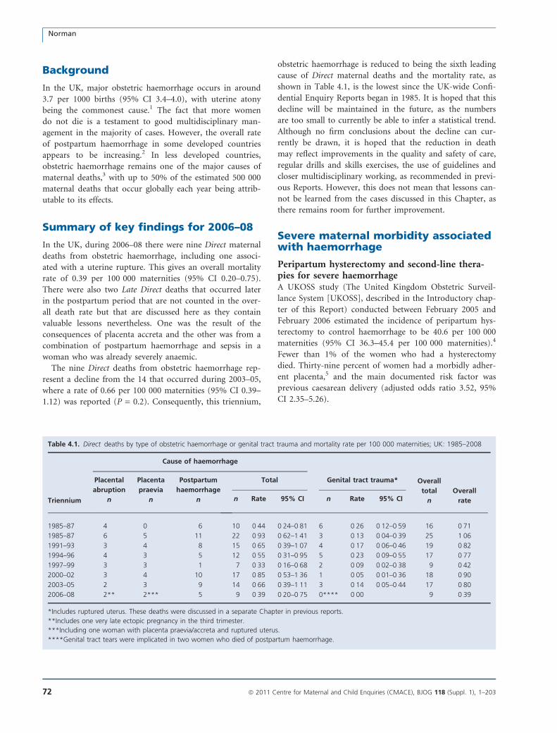
Background
In the UK, major obstetric haemorrhage occurs in around
3.7 per 1000 births (95% CI 3.4–4.0), with uterine atony
being the commonest cause.1 The fact that more women
do not die is a testament to good multidisciplinary man-
agement in the majority of cases. However, the overall rate
of postpartum haemorrhage in some developed countries
appears to be increasing.2 In less developed countries,
obstetric haemorrhage remains one of the major causes of
maternal deaths,3 with up to 50% of the estimated 500 000
maternal deaths that occur globally each year being attrib-
utable to its effects.
Summary of key findings for 2006–08
In the UK, during 2006–08 there were nine Direct maternal
deaths from obstetric haemorrhage, including one associ-
ated with a uterine rupture. This gives an overall mortality
rate of 0.39 per 100 000 maternities (95% CI 0.20–0.75).
There were also two Late Direct deaths that occurred later
in the postpartum period that are not counted in the over-
all death rate but that are discussed here as they contain
valuable lessons nevertheless. One was the result of the
consequences of placenta accreta and the other was from a
combination of postpartum haemorrhage and sepsis in a
woman who was already severely anaemic.
The nine Direct deaths from obstetric haemorrhage rep-
resent a decline from the 14 that occurred during 2003–05,
where a rate of 0.66 per 100 000 maternities (95% CI 0.39–
1.12) was reported (P = 0.2). Consequently, this triennium,
obstetric haemorrhage is reduced to being the sixth leading
cause of Direct maternal deaths and the mortality rate, as
shown in Table 4.1, is the lowest since the UK-wide Confi-
dential Enquiry Reports began in 1985. It is hoped that this
decline will be maintained in the future, as the numbers
are too small to currently be able to infer a statistical trend.
Although no firm conclusions about the decline can cur-
rently be drawn, it is hoped that the reduction in death
may reflect improvements in the quality and safety of care,
regular drills and skills exercises, the use of guidelines and
closer multidisciplinary working, as recommended in previ-
ous Reports. However, this does not mean that lessons can-
not be learned from the cases discussed in this Chapter, as
there remains room for further improvement.
Severe maternal morbidity associatedwith haemorrhage
Peripartum hysterectomy and second-line thera-pies for severe haemorrhageA UKOSS study (The United Kingdom Obstetric Surveil-
lance System [UKOSS], described in the Introductory chap-
ter of this Report) conducted between February 2005 and
February 2006 estimated the incidence of peripartum hys-
terectomy to control haemorrhage to be 40.6 per 100 000
maternities (95% CI 36.3–45.4 per 100 000 maternities).4
Fewer than 1% of the women who had a hysterectomy
died. Thirty-nine percent of women had a morbidly adher-
ent placenta,5 and the main documented risk factor was
previous caesarean delivery (adjusted odds ratio 3.52, 95%
CI 2.35–5.26).
Table 4.1. Direct deaths by type of obstetric haemorrhage or genital tract trauma and mortality rate per 100 000 maternities; UK: 1985–2008
Triennium
Cause of haemorrhage
Overall
total
n
Overall
rate
Placental
abruption
n
Placenta
praevia
n
Postpartum
haemorrhage
n
Total Genital tract trauma*
n Rate 95% CI n Rate 95% CI
1985–87 4 0 6 10 0Æ44 0Æ24–0Æ81 6 0Æ26 0Æ12–0Æ59 16 0Æ71
1985–87 6 5 11 22 0Æ93 0Æ62–1Æ41 3 0Æ13 0Æ04–0Æ39 25 1Æ06
1991–93 3 4 8 15 0Æ65 0Æ39–1Æ07 4 0Æ17 0Æ06–0Æ46 19 0Æ82
1994–96 4 3 5 12 0Æ55 0Æ31–0Æ95 5 0Æ23 0Æ09–0Æ55 17 0Æ77
1997–99 3 3 1 7 0Æ33 0Æ16–0Æ68 2 0Æ09 0Æ02–0Æ38 9 0Æ42
2000–02 3 4 10 17 0Æ85 0Æ53–1Æ36 1 0Æ05 0Æ01–0Æ36 18 0Æ90
2003–05 2 3 9 14 0Æ66 0Æ39–1Æ11 3 0Æ14 0Æ05–0Æ44 17 0Æ80
2006–08 2** 2*** 5 9 0Æ39 0Æ20–0Æ75 0**** 0Æ00 9 0Æ39
*Includes ruptured uterus. These deaths were discussed in a separate Chapter in previous reports.
**Includes one very late ectopic pregnancy in the third trimester.
***Including one woman with placenta praevia/accreta and ruptured uterus.
****Genital tract tears were implicated in two women who died of postpartum haemorrhage.
Norman
72 ª 2011 Centre for Maternal and Child Enquiries (CMACE), BJOG 118 (Suppl. 1), 1–203
On a UK-wide basis, surveillance of severe postpartum
haemorrhage requiring specific second-line therapies was
undertaken through UKOSS between October 2007 and
March 2009 (Knight M et al, unpublished UKOSS data –
personal communication). An estimated 24.4 women per
100 000 maternities were managed either with uterine com-
pression sutures, pelvic vessel ligation or embolisation, or
factor VII to treat severe haemorrhage (95% CI 21.7–27.3
per 100 000 maternities). An investigation in the group of
women managed with uterine compression sutures for fail-
ure to control haemorrhage leading to hysterectomy
showed that delay in placement of the suture (more than
2 hours between delivery and suture placement) was more
frequent in women who underwent hysterectomy (adjusted
odds ratio 3.86, 95% CI 1.65–8.99). This emphasises the
importance of early recognition and management of severe
haemorrhage to improve outcomes.
Morbidity from uterine ruptureThere were 111 cases of uterine rupture reported to
UKOSS between April 2009 and January 2010, (Knight M
et al, unpublished UKOSS data – personal communication)
representing an estimated incidence of 17.4 cases per
100 000 maternities (95% CI 14.3–21.0 per 100 000 mater-
nities). Eighty-six percent of cases occurred in women who
had had a previous caesarean delivery.
Trends in obstetric haemorrhageA fall in the rate of major obstetric haemorrhage, despite a
rise in rate of postpartum haemorrhage, is also apparent
from the Scottish audit of maternal morbidity published
for 2008,6 which is also described in Appendix 2B. These
results, in combination with a fall in the number of deaths
in Scotland and in the UK, also point to improved man-
agement of this condition.
The mothers who died
The age range of the mothers who died was from 22 to
36 years, with a median of 28 years. Five women were pri-
miparous, and three of the other four mothers had at least
one previous caesarean section. The Body Mass Index
(BMI) of eight of the nine mothers who died and whose
BMIs were known ranged between 20 and 38 with a med-
ian of 23. Six women were of normal weight, one was over-
weight and one was obese.
Five women described themselves as White British and
were regular attenders for antenatal care; a sixth concealed
her pregnancy. The three women from other ethnic groups
had had three or fewer antenatal visits, but two women
appeared to have arrived in the UK later in their pregnan-
cies in order to give birth here. One had little grasp of Eng-
lish, and one of the Late Direct deaths also occurred in a
woman from a minority ethnic background who was a
poor attender for care.
Substandard careAlthough both the number and proportion of deaths from
haemorrhage has fallen in this triennium, there remains
room for improvement. Substandard care was a factor in
six (66%) of the deaths. For four women it was considered
that different treatment may have altered the outcome, and
in two, although the outcome would probably not have
changed, there were lessons to be learnt. Obstetricians,
midwives and hospital management staff need to be vigi-
lant and ensure that care is optimised through use of regu-
lar drills and skills and adherence to national guidelines to
further reduce haemorrhage-related maternal death.
Antenatal haemorrhage
Placenta praevia and accretaTwo of the women whose deaths are counted in this Chap-
ter and a Late death of another woman followed a placenta
praevia. Two of these three women had placenta accreta as
well as placenta praevia, one of whom also had a uterine
rupture.
A woman who had undergone a number of previous caesar-
ean sections and who attended regularly for antenatal care
collapsed at home in the third trimester and was moribund
on arrival in the Emergency Department. At autopsy, a pla-
cental percreta and uterine rupture were found. Ultrasound
had not been performed antenatally to determine placental
site, despite her previous caesarean sections.
This illustrates the importance of following the guidelines
issued in the last Report, for 2003–05, that ‘all women who
have had a previous caesarean section must have their pla-
cental site determined. If there is any doubt, magnetic reso-
nance imaging (MRI) can be used along with ultrasound
scanning in determining if the placenta is accreta or percre-
ta’.7 These recommendations are endorsed by the recently
published ‘care bundle’ for the management of women with
placenta praevia after caesarean section produced by the
RCOG and the National Patient Safety Agency (NPSA).8
In one of the other women:
A woman whose placenta accreta had been diagnosed ante-
natally was correctly delivered in a tertiary referral centre
with a Critical Care Unit (CCU). At the time of delivery
the placenta was confirmed to be morbidly adherent and
was therefore left in situ. Although she made a good recovery
in the immediate postpartum period, she became unwell
with probable sepsis some weeks after delivery. She was
admitted to the initial booking hospital without a CCU and
underwent attempted manual removal of the placenta, at
Chapter 4: Haemorrhage
ª 2011 Centre for Maternal and Child Enquiries (CMACE), BJOG 118 (Suppl. 1), 1–203 73
which there was catastrophic bleeding, leading to dissemi-
nated intravascular coagulation and her eventual death
some days later.
Management of placenta accreta remains a challenge,
with no clear evidence on how it should be best conducted.
It was the principal indication for peripartum hysterectomy
in around 40% of women who underwent this procedure
in the UK in 2005.4 Good practice advice from the RCOG9
and the NPSA8 suggests that senior multidisciplinary plan-
ning is indicated, with predelivery discussion of the possi-
bility of the need for hysterectomy to control bleeding.
A woman who had a previous caesarean section and a
diagnosis of placenta praevia made by scan later in her preg-
nancy and who collapsed at home near term illustrates the
dangers of outpatient management of severe placenta prae-
via. Although outpatient management is now increasingly
undertaken, it is not without risk. There is only one small
(<60 women) randomised trial10 and no observational stud-
ies of sufficient size on which to base an estimate of the risks
of outpatient versus inpatient management.
In view of this, the RCOG recommends that ‘women
with major placenta praevia who have previously bled should
be admitted and managed as inpatients from 34 weeks of ges-
tation. Those with major placenta praevia who remain
asymptomatic, having never bled, require careful counselling
before contemplating outpatient care’.9
In a separate case, counted and discussed in Chapter 6, a
woman who had had several previous caesarean sections
developed a severe haemorrhage early in pregnancy fol-
lowed by chorioamnionitis.
Placental abruption and otherantepartum haemorrhage
A woman who had presented several times throughout her
pregnancy with abdominal pain and had been scanned on
at least four occasions by appropriately qualified staff had
an extrauterine pregnancy that was not diagnosed until the
third trimester. This rare incident illustrates the difficulty
in making this diagnosis in the second and third trimesters
and also highlights the need to consider ectopic pregnancy
in the differential diagnosis of abdominal pain throughout
pregnancy. The major learning points about scanning in
early pregnancy are discussed in Chapter 6.
In another woman:
A woman with significant systemic disease presented with an
intrauterine death and placental abruption in her early third
trimester. Despite her medical history, abdominal pain and
tense abdomen, delivery was delayed by many hours and
thereafter started initially with mifepristone. She became
increasingly unwell but was managed by junior staff without
multidisciplinary input. She eventually arrested and under-
went perimortem caesarean section over 2 days after the
intrauterine death was diagnosed. The cause of death at
autopsy was given as fluid overload and acute transfusion-
related lung injury.
Although NICE guidelines11 indicate that conservative
management and/or a delay in labour induction is some-
times appropriate in women with an intrauterine death,
this does not apply in the presence of major abruption or
infection. In these scenarios, delivery should be expedited.
Such women are at high risk of morbidity and mortality
and should be managed by an experienced multidisciplin-
ary team. This case also highlights the complications of
blood transfusion: acute transfusion-related lung injury is
now a leading cause of transfusion-related mortality and
morbidity. The decision to transfuse women should be
made carefully, and all clinicians involved in blood transfu-
sion should be aware of the potential adverse effects of
transfusion and signs and symptoms of transfusion-related
complications.12
Postpartum haemorrhage
Compared with the two previous Reports, the number of
deaths from postpartum haemorrhage (PPH) has halved to
five. The RCOG have recently published guidelines for the
management of postpartum haemorrhage.13 These empha-
sise the importance of active management of the third stage
with prophylactic oxytocic administration to prevent post-
partum haemorrhage and prompt multidisciplinary man-
agement in the treatment should it occur.
Although there are now a wide array of therapies for
postpartum haemorrhage, ranging from vessel embolisation
to recombinant factor VIIa, it is disappointing that a major
failure in three of the five women who died (60%) was a
lack of routine observation in the postpartum period, or a
failure to appreciate that bleeding was occurring. In all
three women there was a lack of optimal postoperative
measurement of pulse and blood pressure, or recognition
of abnormal vital signs such as oxygen saturation and
respiratory rate, even when it was known the mother had
sustained a large bleed. The use of MEOWS charts, as
advocated in the last Report,7 should help to alert caregiv-
ers to abnormal trends in haemodynamic measurements,
but they are only useful if observations are performed and
abnormal readings acted upon. Regular observations of
pulse and blood pressure should be made postdelivery.
MEOWS charts enable the identification of readings that
require further action.
Underlying, longstanding and untreated anaemia, which
is often linked with maternal deaths from haemorrhage in
developing countries, was also found in a few mothers who
died in this Report.
Norman
74 ª 2011 Centre for Maternal and Child Enquiries (CMACE), BJOG 118 (Suppl. 1), 1–203
A woman who died some months after delivery from multi-
organ failure secondary to bacterial pneumonia also sus-
tained a postpartum haemorrhage of 1–2 l during a
caesarean section. Despite the fact that she was unwell at
the start of the procedure, and also had a haemoglobin of
only 7.5 g/dl, senior staff were not alerted until she began
to bleed excessively.
Antenatal anaemia should be diagnosed and treated
effectively: parenteral iron therapy should be considered
antenatally for women with iron deficiency anaemia who
do not respond to oral iron.
The case of a woman for whom the stigma of being an
unmarried mother appeared too much and who therefore
concealed her pregnancy, subsequently dying at home of a
catastrophic postpartum haemorrhage, although counted
here, is discussed in Chapter 11.
Women who refuse blood productsThe management of women who refuse blood products
presents a continuing challenge in obstetric practice. In
each of the two previous Reports, there were two deaths
from haemorrhage in such women; although only one is
reported here, there was a further woman for whom this
was important but who died from an unrelated cause. Such
mothers are known to be at increased risk of death if they
suffer a major obstetric haemorrhage: a recent estimate
from the Netherlands puts this at around three- to four-
fold.14 Clear guidelines for the management of these
women were contained in an earlier Enquiry Report7 and
in guidance from the RCOG.15 Good practice advice is to
ensure that women discuss their plans to avoid blood
transfusion with both a consultant anaesthetist and an
obstetrician in their antenatal period, that women enter
labour with a haemoglobin >10.5 g/dl, that cell salvage is
available where appropriate and that operative abdominal
and vaginal deliveries are performed by senior anaesthetic
and obstetric staff. A further lesson is that erythropoietin is
not an effective alternative to red cell transfusion in major
acute haemorrhage because it takes 10–14 days to effect an
increase in haemoglobin levels.16 Women who refuse blood
products, their families and religious advisors need to be
aware of this.
Management of the third stage of labourUterine inversion occurred as a result of traction on the
placenta before separation in a woman who had a retained
placenta. This resulted in bleeding and vasovagal shock.
Uterine replacement was not attempted, but Syntocinon
was given. Clinicians should be aware that developments
in the treatment of major haemorrhage do not mean that
elementary clinical management can be neglected. Moder-
ate or excessive traction on the cord before placental
separation is inappropriate. Attempted replacement is the
appropriate initial management of uterine inversion.
Births in midwife-led units or at homeWith increasing emphasis on the availability of births at
home or in midwife-led units, caregivers should ensure that
there are appropriate guidelines for management where
labour and delivery are not straightforward. Obstetricians
and midwives should develop guidelines for the manage-
ment of obstetric emergencies that may occur in the com-
munity, including uterine inversion and antenatal and
postpartum haemorrhage, and all practitioners should keep
up to date with emergency drills for uncommon events.
Pathological overview
The autopsies of five deaths from haemorrhage were
reviewed this triennium together with two further deaths,
one associated with a Grade 4, major placenta praevia and
another with retained placenta. Both of these were classified
as anaesthetic deaths and are counted in Chapter 8. There
were two deaths from placenta praevia or accreta and three
from uterine rupture or genital tract tears during delivery.
The death of one woman from a placenta praevia did not
come to autopsy, but the report on the placenta and subto-
tal hysterectomy was available. Although the report con-
firmed that there was no abruption, there was no mention
of the site of the placental bed; this should have been com-
mented on. Three of the other reports were excellent. For
instance, in one woman, cervical tears extending into the
broad ligaments had been sutured, but she remained hypo-
tensive. When she developed chest pain, an embolus was
suspected. The autopsy very carefully excluded both pul-
monary and amniotic fluid embolism. This contrasts with a
very poor report which was in ‘tick box’ format with mini-
mal or no other description. The consultant obstetrician,
who attended the autopsy, commented in his report to the
coroner on discrepancies between his observations at the
autopsy and those by the pathologist. These discrepancies
did not alter the cause or mode of death but do suggest
failures of interpretation and of clinicopathological correla-
tion amplified by poor report writing. Review of these
deaths emphasises the need to address the clinical issues
and for good clinicopathological correlation.
Conclusion
The decline in haemorrhage-related deaths in this triennium,
although not statistically significant because of the very small
numbers, is gratifying, and it is hoped that this can be main-
tained in future. The absence of deaths in relation to elective
caesarean sections for placenta praevia endorses the previous
Chapter 4: Haemorrhage
ª 2011 Centre for Maternal and Child Enquiries (CMACE), BJOG 118 (Suppl. 1), 1–203 75
recommendations made in successive Reports that senior
staff should be involved in these deliveries.
Amongst the deaths, the lack of early senior multidisci-
plinary involvement, the lack of close postoperative moni-
toring and the failure to act on signs and symptoms that a
woman is seriously unwell, including readings from
MEOWS charts, remain important contributors to maternal
death from haemorrhage. All clinicians involved in the care
of pregnant women could further reduce the risk of haem-
orrhage-related maternal death by improvements in these
elementary aspects of clinical care.
Disclosure of interestsNone.
FundingThis work was undertaken by the Centre for Maternal and
Child Enquiries (CMACE) as part of the CEMACH pro-
gramme. The work was funded by the National Patient
Safety Agency; the Department of Health, Social Services
and Public Safety of Northern Ireland; NHS Quality
Improvement Scotland (NHS QIS); and the Channel
Islands and Isle of Man.
AcknowledgementsThe chapter has been seen by and commented on by
Dr Brian Magowan, Consultant Obstetrician of Borders
General Hospital and Dr Rhona Hughes, Consultant
Obstetrician for Royal Infirmary of Edinburgh.
References
1 Brace V. Learning from adverse outcomes—major haemorrhage in
Scotland in 2003–05. BJOG 2007;114:1388–96.
2 Knight M, Callaghan WM, Berg C, Alexander S, Bouvier-Colle M-H,
Ford JB, et al. Trends in postpartum hemorrhage in high resource
countries: a review and recommendations from the International
Postpartum Hemorrhage Collaborative Group. BMC Pregnancy Child-
birth 2009;9:55.
3 Khan KS, Wojdyla D, Say L, Gulmezoglu AM, Van Look PF. WHO
analysis of causes of maternal death: a systematic review. Lancet
2006;367:1066–74.
4 Knight M, Kurinczuk JJ, Spark P, Brocklehurst P. Cesarean delivery
and peripartum hysterectomy. Obstet Gynecol 2008;111:97–105.
5 Knight M. Peripartum hysterectomy in the UK: management
and outcomes of the associated haemorrhage. BJOG 2007;114:
1380–7.
6 Scottish Confidential Audit of Severe Maternal Morbidity. Annual
Report 2008. NHS Quality Improvement Scotland, 2010.
[www.nhshealthquality.org/maternityservices]. Accessed 6 Septem-
ber 2010.
7 Lewis G (editor). The Confidential Enquiry into Maternal and Child
Health (CEMACH). Saving Mothers Lives; reviewing maternal deaths
to make motherhood safer 2003–05. The Seventh Report of the Uni-
ted Kingdom Confidential Enquiries into Maternal Deaths in the Uni-
ted Kingdom. London: CEMACH, 2007. [www.cmace.org.uk].
Accessed 6 September 2010.
8 National Patient Safety Agency and Royal College of Obstetricians
and Gynaecologists. Placenta praevia after caesarean section care
bundle: background information for health professionals. London:
NPSA, 2010.
9 Royal College of Obstetricians and Gynaecologists. Placenta praevia
and placenta praevia accreta: diagnosis and management. Clinical
Guideline no 27. London: RCOG Press, 2005.
10 Wing DA, Paul RH, Millar LK. Management of the symptomatic pla-
centa praevia: a randomised, controlled trial of inpatient versus out-
patient expectant management. Am J Obstet Gynecol 1996;175:
806–11.
11 National Collaborating Centre for Women’s and Children’s Health
commissioned by the National Institute for Clinical Excellence. Induc-
tion of labour. Clinical Guideline. 2008. London: RCOG Press, 2008.
12 Triulzi DJ. Transfusion-related acute lung injury: current concepts for
the clinician. Anesth Analg 2009;108:770–6.
13 Royal College of Obstetricians and Gynaecologists. Prevention of
postpartum haemorrhage. Clinical Guideline no 52. London: RCOG
Press, 2009.
14 Van Wolfswinkel ME, Zwart J, Schutte J, Duvekot J, Pel M, Van
Roosmalen J. Maternal mortality and serious maternal morbidity in
Jehovah’s witnesses in The Netherlands. BJOG 2009;116:1103–8;
discussion 1108–10.
15 Royal College of Obstetricians and Gynaecologists. Blood transfusion
in obstetrics. Clinical guideline No. 47. London: RCOG Press, 2008.
16 Ng T et al. Recombinant erythropoietin in clinical practice. Postgrad-
uate Medical Journal 2003;79:367–70.
Norman
76 ª 2011 Centre for Maternal and Child Enquiries (CMACE), BJOG 118 (Suppl. 1), 1–203
Chapter 5: Amniotic fluid embolismAJ Dawson (on behalf of the Centre for Maternal and Child Enquiries)
Consultant Obstetrician, Nevill Hall Hospital, Abergavenny, UK
Correspondence: Amendment after online publication (11 March 2011): the correct address for this author is Nevill Hall Hospital, Abergavenny
NP7 7EG, UK. Email: [email protected]
Keywords amniotic fluid embolism, Confidential Enquiry, mater-
nal, mortality.
Amniotic fluid embolism: specific recommendations
• All maternal death autopsies should be performed as soon after death as possible, and not delayed by several days,
because the diagnosis of AFE then becomes difficult if not impossible.
At autopsy:
• confirm the diagnosis using immunochemistry
• in clinically classical cases where no squames can be found, search for mucins.
Amniotic fluid embolism: learningpoints
Amniotic fluid embolism (AFE) should no longer be
regarded as a condition with near universal maternal
mortality. High-quality supportive care can result in
good outcomes for both mother and baby depending
on the place of collapse.
There were several examples of excellent practice this
triennium, including prompt peri-mortem caesarean
section.
AFE may be confused with other presentations, includ-
ing eclampsia, septic or anaphylactic shock and pulmo-
nary embolism, but ultimately the immediate action
taken should be resuscitative and the initial treatment is
unlikely to differ.
Summary of key findings for 2006–08
In this triennium, the deaths of 13 mothers who died of
AFE were reported to the Enquiry, giving a mortality rate
of 0.57 per 100 000 maternities (95% CI 0.33–0.98).
Although AFE continues to rank as a major cause of Direct
maternal deaths, it has fallen from being the second to the
fourth leading cause of Direct deaths this triennium.
As shown in Table 5.1, the numbers and mortality rate for
this triennium are less than the 17 deaths and mortality rate
of 0.80 per 100 000 maternities (95% CI 0.50–1.29) attrib-
uted to AFE for the period 2003–05. This decline is not sta-
tistically significant. Neither does it appear that there is an
upwards trend, which is consistent with the findings of the
latest morbidity study from the United Kingdom Obstetric
Surveillance System (UKOSS) (A detailed description of UK
Obstetric Surveillance System (UKOSS) can be found in the
Introduction Chapter of this Report.) discussed below.
Maternal morbidity: the incidence of amnioticfluid embolismProspective national surveillance of AFE has been under-
taken through UKOSS since 2005. A recent analysis of cases
reported over 4 years between February 2005 and February
2009 documented an incidence of 2.0 cases per 100 000
Table 5.1. Direct deaths attributed to amniotic fluid embolism and
rates per 100 000 maternities; UK: 1985–2008
Triennium n Rate 95% CI
1985–87 9 0.40 0.21–0.75
1987–90 11 0.47 0.26–0.83
1991–93 10 0.43 0.23–0.80
1994–96 17 0.77 0.48–1.24
1997–99 8 0.38 0.19–0.74
2000–02 5 0.25 0.11–0.59
2003–05 17 0.80 0.50–1.29
2006–08 13 0.57 0.33–0.98
Chapter 5: Amniotic fluid embolism
ª 2011 Centre for Maternal and Child Enquiries (CMACE), BJOG 118 (Suppl. 1), 1–203 77
maternities (95% CI 1.5–2.5).1 There was no significant
change in incidence over the 4 years, but the authors note
that, because of small case numbers, surveillance over this
period has limited statistical power to detect any trends in
incidence. In this series, 20% of women with AFE died
(95% CI 11–32%); fatality was significantly associated with
Black or other minority ethnicity (adjusted odds ratio 11.8,
95% CI 1.40–99.5). Surveillance is ongoing, and all cases of
AFE, whether the woman survived or not, should be
reported to UKOSS, the details of which are given in
Box 5.1. Combining the results from this Enquiry and the
latest UKOSS report gives a case fatality rate of 16.5%. His-
torically, AFE has been seen as a universally fatal condition
with mortality approaching 100%, but with improved
approaches to resuscitation, it is possible that improve-
ments in mortality from AFE are now being achieved if the
mother collapses in a well-equipped facility.
The diagnosis of amniotic fluid embolismThe diagnosis of AFE has been accepted on clinical grounds
since the 1991–93 Report,2 and this definition has also been
used in the UKOSS methodology.1 The last Report3 con-
tained a discussion on how this compares with classifications
in other countries, including a chart highlighting the differ-
ence in clinical features in AFE compared with pulmonary
embolism, which is often where confusion may arise. In the
present triennium, five cases arose out of diagnostic uncer-
tainty in the immediate clinical situation: possible pulmo-
nary embolism, septic shock, anaphylaxis and eclampsia.
Disseminated intravascular coagulation (DIC) was a fea-
ture in ten of the cases (77%). In all three cases where DIC
was not identified, the women died very quickly after their
collapse, a maximum of 1½ hours. It seems likely that in
these cases the initial effect of the AFE was sufficient to
cause cardiopulmonary collapse before DIC set in.
The women who diedIn this triennium the ages of the mothers who died ranged
between 31 and 42 years with a median of 36 years, slightly
older than in the previous triennium where the median age
was 33 years. Eight of the women (62%) were parous
(maximum para two), and the remainder were nulliparous.
The median gestational age at death was 40 weeks, ranging
between 31 and 41 weeks. There were two twin pregnancies
(15%). Seven (54%) of the women had pre-existing medi-
cal or psychiatric conditions. These cases included treated
essential hypertension, diabetes and anorexia. In the
UKOSS morbidity study,1 an association with increased
maternal age was observed only in women from ethnic
minorities in the study population.
In ten women (77%), the critical event occurred at or
before delivery, and, of these, six (46%) were in established
labour or in the process of delivery. The remainder experi-
enced their critical event after delivery. The UKOSS study1
found 55% of the AFE events that they studied, including
severe morbidity, occurred at or before delivery, with the
rest occurring after birth.
Of the 12 women for whom BMI data were available,
seven had a BMI <30, five were obese with a BMI ‡30, one
of whom had a BMI >35.
Six (46%) of the women were from ethnic minority
groups including Black African and Asian. This compares
with 29% in the previous Report. The UKOSS study1
found that women who died were significantly more likely
to be from an ethnic minority group than those who sur-
vived (adjusted odds ratio 4.64, 95% CI 1.11–19.5).
The index of deprivation for the women who died was
evenly distributed through the groups. Only three of the
women were smokers, and none had any history of known
substance abuse. Ten of the women had booked for antena-
tal care by 19 weeks of gestation. In none of the remaining
three did the late booking influence the content or quality
of care relating to the death.
There was considerable homogeneity in the presentation
of the cases. They were all characterised by a sudden and
unexpected collapse at home or in hospital. For example:
An older mother had a generally uneventful pregnancy and
was admitted at term in early labour. She collapsed shortly
after admission and underwent immediate caesarean sec-
tion. She suffered a massive haemorrhage after delivery
and, despite excellent emergency care, died very quickly.
The clinical circumstancesAll of the women reported to have AFE in the current tri-
ennium were declared dead after delivery, nine of whom
(69%) underwent a peri-mortem caesarean section follow-
ing their collapse. Four women delivered vaginally before
collapse, two of whom had instrumental deliveries. Unlike
previous Reports, none of the women were given Syntoci-
non during labour or delivery. Four women had either arti-
ficial or spontaneously ruptured membranes at full cervical
dilatation and one at 7 cm. With the exclusion of two pre-
term babies, the babies’ weights ranged from 2870 to
Box 5.1. The UK amniotic fluid embolism register
All cases of AFE, whether the woman survived or not, should be
reported to the AFE Register at UKOSS. Cases may be reported
either through the local hospital UKOSS contact, via email to
[email protected], or to:
UKOSS
National Perinatal Epidemiology Unit
Old Road Campus
Oxford
OX5 3DH
Dawson
78 ª 2011 Centre for Maternal and Child Enquiries (CMACE), BJOG 118 (Suppl. 1), 1–203
4490 g with a median of 3570 g. Five of the women had
total fetal weights of 4000 g or more, including one set of
twins, and a woman who had gross polyhydramnios.
Of the women who died, six (46%) had been induced
using prostin analogues. This appears to be an over-rep-
resented proportion compared with births statistics for
England, Scotland and Wales,4–6 which were a little over
20% during the present triennium (birth statistics are
not collated for Northern Ireland). In the recent study
from UKOSS,1 the occurrence of amniotic fluid embo-
lism was significantly associated with induction of labour
(adjusted odds ratio 3.86, 95% CI 2.04–7.31), although
there was no association between induction of labour
and fatality.
The babies survived delivery in all but one case where a
delay in delivery was unavoidable as the mother collapsed
outside a maternity unit. The Enquiry’s documentation
relates to the immediate event only, and so it is possible
that some babies may not have survived the neonatal per-
iod or may have suffered birth injury.
Quality of careIn five cases there was no substandard care, and care was
considered to be exemplary in two of these. In the other
eight cases (62%) there was some degree of substandard
care, although the outcome may have been inevitable. The
learning points included very poor organisation of transfer
facilities in a unit; communication breakdowns, including
absence of a clear lead during resuscitation; delays in con-
tacting the consultant obstetrician; delay in communicating
to relatives that problems had occurred and two cases
where documentation of care leading up to the death was
judged to be unacceptably inadequate. Additionally, in two
of these cases there was avoidable delay in performing cae-
sarean section and achieving delivery within 5 minutes of
collapse, as recommended in the Managing Obstetric Emer-
gencies and Trauma manual,7 although it seems unlikely
that the outcome would have differed. It is important to
remember that in these circumstances the caesarean section
is being carried out to assist resuscitation of the mother
and a delay in transferring her to the operating theatre
may be unnecessary. While the role of peri-mortem caesar-
ean section may not always influence the outcome in AFE,
if a decision is made to carry out the procedure, it needs
to be timely, especially as AFE is now regarded as a poten-
tially treatable condition.
A multiparous woman was induced because of polyhy-
dramnios. Induction was followed by vaginal bleeding and
blood-stained liquor. After a while she collapsed with no
recordable blood pressure, a feeble pulse and a low Glasgow
Coma Score. Collapse was accompanied by fetal bradycar-
dia, and she was resuscitated in the hope this would assist
the fetal condition. She appeared to improve a little but
then collapsed again and developed DIC after an emergency
caesarean section.
Two cases were judged to be of sufficiently substandard
care that a different outcome might have resulted. In a case
of assisted delivery, the instrument was applied several
times before delivery was accomplished. The assessors were
unable to determine whether or not the multiple attempts
had led to access by amniotic fluid to the maternal circula-
tion. In another case the assessors considered that the delay
in providing resuscitative measures might have contributed
to the fatal outcome.
Four serious untoward incident reports and one root
cause analysis report were available to the assessors. Of
these, only one serious untoward incident report was
deemed to be of high quality. Six other deaths were
reported as having been subject to serious incident report-
ing, but as these reports were not submitted to the
Enquiry it was not possible to assess the quality of these
local reviews or the effectiveness of the learning opportu-
nities.
Pathological overview
All cases were subject to autopsy. AFE was confirmed in 11
cases and suspected in the remaining two. There appears to
be less uncertainty about the cause of death in the cases
classified as AFE in this Report for 2006–08 compared with
the 2003–05 Report.3 Five autopsy reports were considered
to have aspects that were substandard. A further problem
in five cases was a delay of at least 4 days between death
and the autopsy: this may have been the reason for nega-
tive histology in one of the cases, though squames were
found in the others. Autolysis could certainly have
impaired the demonstration of other pathology in this case.
In 11 cases, fetal squames were found on routine histology,
and these were confirmed by immunochemistry in five,
with a sixth confirmed by histochemistry.
There were still a few cases, as in previous Reports,
where the assessors had to make a decision about the cause
of death on clinical grounds. For example:
A mother was induced with vaginal prostin because of being
overdue and had a precipitate labour and birth with no
immediate problems. She was later found collapsed and,
after resuscitation had been commenced, started to bleed
very heavily, which was consistent with DIC. Resuscitation
proved impossible. AFE could not be confirmed at autopsy
but was not fully sought, as immunochemistry was not
used.
In another case there was a classical history of collapse
in labour, but no squames were found by immunochemis-
Chapter 5: Amniotic fluid embolism
ª 2011 Centre for Maternal and Child Enquiries (CMACE), BJOG 118 (Suppl. 1), 1–203 79
try despite an extensive search. This case was accepted
because the quality of the autopsy report suggested that
other possible causes had been reasonably excluded. If true,
it suggests that squames are a common surrogate marker
for the syndrome but not necessarily its precipitating cause.
Conclusions
Amniotic fluid embolism, particularly if the collapse happens
in a well-equipped unit, should now be considered a treat-
able and survivable event in the majority of cases. It has now
fallen to fourth place among causes of Direct maternal deaths
but nevertheless continues to be a significant factor in mater-
nal mortality rates in the UK. The clinical presentation of
AFE can be confused with other causes of collapse, but effec-
tive resuscitation remains the essential common response
irrespective of the underlying cause for collapse.
When a death does occur, a detailed prompt autopsy
should be performed that includes immunochemistry or
histochemistry. All cases, fatal or not, should be reported
to UKOSS.
Disclosure of interestsNone.
FundingThis work was undertaken by the Centre for Maternal and
Child Enquiries (CMACE) as part of the CEMACH pro-
gramme. The work was funded by the National Patient
Safety Agency; the Department of Health, Social Services
and Public Safety of Northern Ireland; NHS Quality
Improvement Scotland (NHS QIS); and the Channel
Islands and Isle of Man.
AcknowledgementsThis Chapter had been seen and discussed with Mr Derel
Tuffnell, Consultant in Obstetrics and Gynaecology of
Bradford Royal Infirmary and Dr Marian Knight, Head of
UKOSS and Senior Clinical Research Fellow for the NPEU.
The Pathology overview was written by Dr H Hillward-
Sadler, Consultant Histopathologist of Southampton Uni-
versity Hospitals NHS Trust. j
References
1 Knight M, Tuffnell D, Brocklehurst P, Spark P, Kurinczuk JJ. Incidence
and risk factors for amniotic-fluid embolism. Obstet Gynecol
2010;115:910–7.
2 Department of Health Report on Confidential Enquiries into Maternal
Deaths in the United Kingdom 1991–1993. London: HMSO, 1995.
3 Lewis G, editor; The Confidential Enquiry into Maternal and Child
Health (CEMACH). Saving Mothers’ Lives: Reviewing Maternal Deaths
to Make Motherhood Safer—2003–2005. The Seventh Report on
Confidential Enquiries into Maternal Deaths in the United Kingdom.
London: CEMACH, 2007. [www.cmace.org.uk]. Accessed 5 October
2010.
4 English Births. [www.hesonline.nhs.uk/Ease/servlet/ContentServer?site
ID=1937&categoryID=1022]. Accessed 7 April 2010.
5 Scottish Births. [www.isdscotland.org/isd/1612.html]. Accessed 7 April
2010.
6 Welsh births. [wales.gov.uk/topics/statistics/headlines/health2010/
0210/?lang=en] Accessed 7 April 2010.
7 Howell C, Grady K, Cox C, editors. Managing Obstetric Emergencies
and Trauma—the MOET Course Manual, 2nd edn. London: RCOG,
2007.
Dawson
80 ª 2011 Centre for Maternal and Child Enquiries (CMACE), BJOG 118 (Suppl. 1), 1–203
Chapter 6: Deaths in early pregnancyC O’Herlihy (on behalf of the Centre for Maternal and Child Enquiries)
University College Dublin School of Medicine and Medical Science, National Maternity Hospital, Dublin, Ireland
Correspondence: Professor Colm O’Herhily, University College Dublin School of Medicine and Medical Science, National Maternity Hospital,
Holles Street, Dublin 2, Ireland. Email: [email protected]
Keywords early pregnancy, Confidential Enquiry, maternal, mor-
tality.
Death in early pregnancy: specific recommendation
• All women of reproductive age presenting to Emergency Departments with gastrointestinal symptoms should have a
pregnancy test.
• Gastrointestinal symptoms, particularly diarrhoea and dizziness, in early gestation are important indicators of ectopic
pregnancy. These features need to be emphasised to all clinical staff.
• The term ‘pregnancy of unknown location’ based on early pregnancy ultrasound examination should be abandoned.
An early pregnancy ultrasound which fails to identify an intrauterine sac should stimulate active exclusion of tubal
pregnancy, and even in the presence of a small uterine sac, ectopic pregnancy cannot be excluded.
• As emphasised in the Royal College of Obstetricians and Gynaecologists (RCOG) Guideline, The Care of Women
Requesting Induced Abortion,1 abortion care should include a strategy for minimising the risk of infective morbidity,
at a minimum antibiotic prophylaxis. This should be applied whether the abortion is carried out medically or surgi-
cally.
Death in early pregnancy: learningpoints
Abnormal placentation at the site of a previous cae-
sarean scar can lead to haemorrhagic catastrophes
in the mid-trimester, as well as in later pregnancy.
The possibility of morbid adherence should be consid-
ered when evacuation of retained placenta is under-
taken following miscarriage in women who have had
a previous caesarean section, and this procedure
should be performed by staff of appropriate seniority.
Heavy bleeding or bleeding persisting for more than
2 weeks following a diagnosis of non-continuing
pregnancy needs to be recognised as an indication
for medical or gynaecological review and consideration
of surgical evacuation of retained products of concep-
tion.
Unless ‘handover’ communication between hospital doc-
tors is meticulous, truncated shift patterns may result in
failure to appreciate a woman’s deteriorating clinical
status.
Summary of key findings 2006–08
Although fewer women died during 2006–08 of causes
directly resulting from complications arising from early
pregnancy (before 24 completed weeks of gestation) than
in any previous triennium, substandard aspects of care
were identifiable in a majority of the 11 women discussed
in this chapter. Six deaths occurred because of ruptured
ectopic pregnancies and five followed haemorrhagic com-
plications of spontaneous miscarriages. Table 6.1 docu-
ments the trends in early pregnancy deaths since the UK
Report began in 1985.
In addition, seven other early Direct deaths which
occurred secondary to sepsis are counted and discussed in
Chapter 7. Of these, two were associated with spontaneous
miscarriages between 9 and 16 weeks of gestation, two fol-
lowed pregnancy terminations, one performed medically
and one surgically induced, and the rest were the result of
infection following spontaneous premature rupture of
membranes. Another death, attributed to cocaine interact-
ing with an anaesthetic for a procedure in early pregnancy,
is counted in Chapter 8. Five women died from thrombo-
embolism in early pregnancy and are counted and dis-
cussed in Chapter 2. In this triennium, no deaths were
Chapter 6: Deaths in early pregnancy
ª 2011 Centre for Maternal and Child Enquiries (CMACE), BJOG 118 (Suppl. 1), 1–203 81
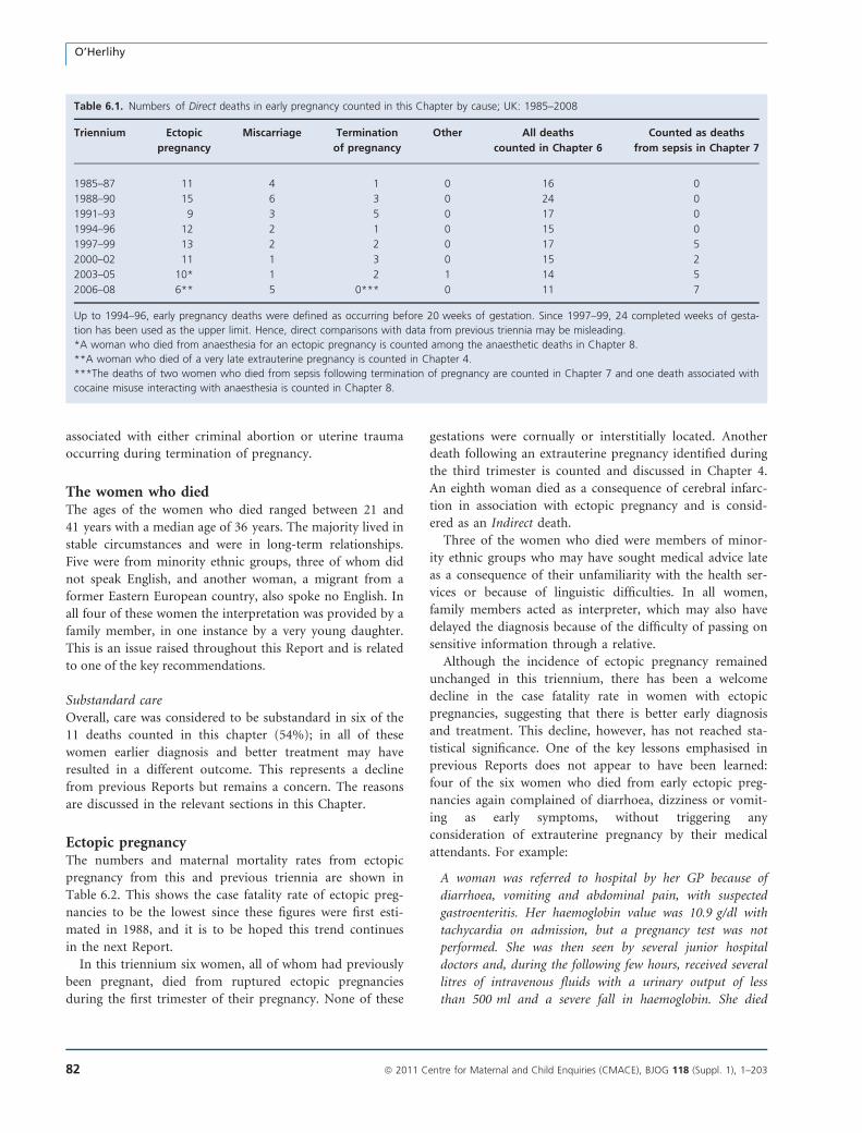
associated with either criminal abortion or uterine trauma
occurring during termination of pregnancy.
The women who diedThe ages of the women who died ranged between 21 and
41 years with a median age of 36 years. The majority lived in
stable circumstances and were in long-term relationships.
Five were from minority ethnic groups, three of whom did
not speak English, and another woman, a migrant from a
former Eastern European country, also spoke no English. In
all four of these women the interpretation was provided by a
family member, in one instance by a very young daughter.
This is an issue raised throughout this Report and is related
to one of the key recommendations.
Substandard careOverall, care was considered to be substandard in six of the
11 deaths counted in this chapter (54%); in all of these
women earlier diagnosis and better treatment may have
resulted in a different outcome. This represents a decline
from previous Reports but remains a concern. The reasons
are discussed in the relevant sections in this Chapter.
Ectopic pregnancyThe numbers and maternal mortality rates from ectopic
pregnancy from this and previous triennia are shown in
Table 6.2. This shows the case fatality rate of ectopic preg-
nancies to be the lowest since these figures were first esti-
mated in 1988, and it is to be hoped this trend continues
in the next Report.
In this triennium six women, all of whom had previously
been pregnant, died from ruptured ectopic pregnancies
during the first trimester of their pregnancy. None of these
gestations were cornually or interstitially located. Another
death following an extrauterine pregnancy identified during
the third trimester is counted and discussed in Chapter 4.
An eighth woman died as a consequence of cerebral infarc-
tion in association with ectopic pregnancy and is consid-
ered as an Indirect death.
Three of the women who died were members of minor-
ity ethnic groups who may have sought medical advice late
as a consequence of their unfamiliarity with the health ser-
vices or because of linguistic difficulties. In all women,
family members acted as interpreter, which may also have
delayed the diagnosis because of the difficulty of passing on
sensitive information through a relative.
Although the incidence of ectopic pregnancy remained
unchanged in this triennium, there has been a welcome
decline in the case fatality rate in women with ectopic
pregnancies, suggesting that there is better early diagnosis
and treatment. This decline, however, has not reached sta-
tistical significance. One of the key lessons emphasised in
previous Reports does not appear to have been learned:
four of the six women who died from early ectopic preg-
nancies again complained of diarrhoea, dizziness or vomit-
ing as early symptoms, without triggering any
consideration of extrauterine pregnancy by their medical
attendants. For example:
A woman was referred to hospital by her GP because of
diarrhoea, vomiting and abdominal pain, with suspected
gastroenteritis. Her haemoglobin value was 10.9 g/dl with
tachycardia on admission, but a pregnancy test was not
performed. She was then seen by several junior hospital
doctors and, during the following few hours, received several
litres of intravenous fluids with a urinary output of less
than 500 ml and a severe fall in haemoglobin. She died
Table 6.1. Numbers of Direct deaths in early pregnancy counted in this Chapter by cause; UK: 1985–2008
Triennium Ectopic
pregnancy
Miscarriage Termination
of pregnancy
Other All deaths
counted in Chapter 6
Counted as deaths
from sepsis in Chapter 7
1985–87 11 4 1 0 16 0
1988–90 15 6 3 0 24 0
1991–93 9 3 5 0 17 0
1994–96 12 2 1 0 15 0
1997–99 13 2 2 0 17 5
2000–02 11 1 3 0 15 2
2003–05 10* 1 2 1 14 5
2006–08 6** 5 0*** 0 11 7
Up to 1994–96, early pregnancy deaths were defined as occurring before 20 weeks of gestation. Since 1997–99, 24 completed weeks of gesta-
tion has been used as the upper limit. Hence, direct comparisons with data from previous triennia may be misleading.
*A woman who died from anaesthesia for an ectopic pregnancy is counted among the anaesthetic deaths in Chapter 8.
**A woman who died of a very late extrauterine pregnancy is counted in Chapter 4.
***The deaths of two women who died from sepsis following termination of pregnancy are counted in Chapter 7 and one death associated with
cocaine misuse interacting with anaesthesia is counted in Chapter 8.
O’Herlihy
82 ª 2011 Centre for Maternal and Child Enquiries (CMACE), BJOG 118 (Suppl. 1), 1–203
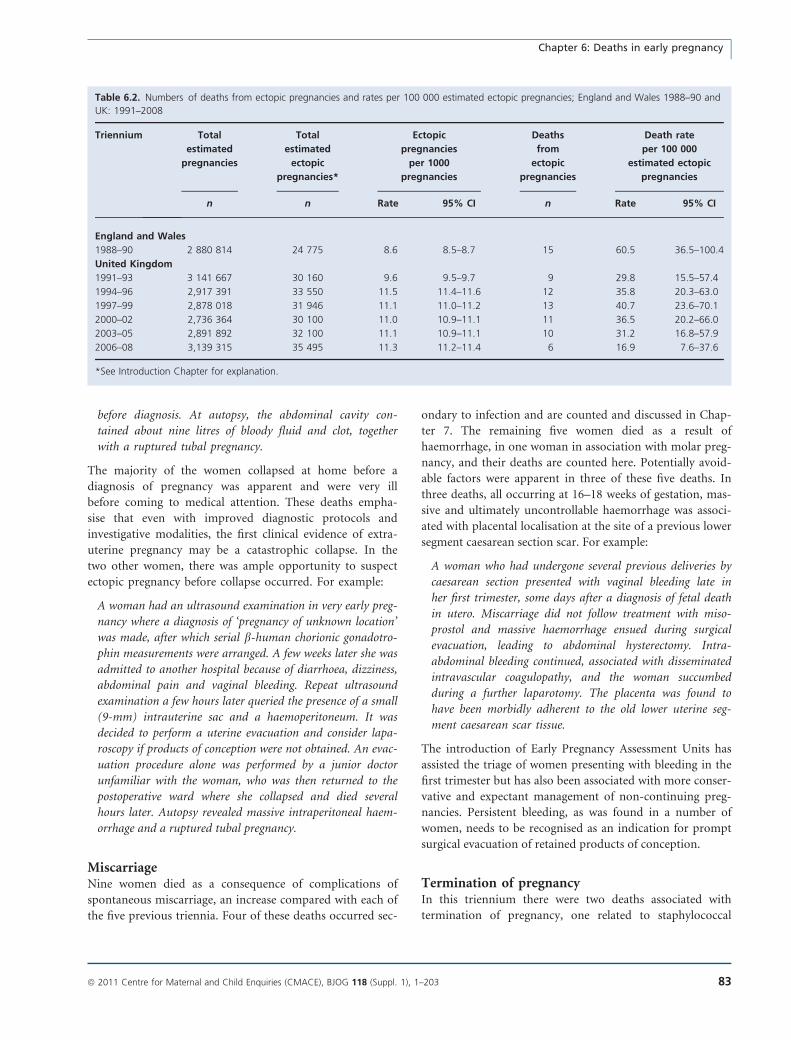
before diagnosis. At autopsy, the abdominal cavity con-
tained about nine litres of bloody fluid and clot, together
with a ruptured tubal pregnancy.
The majority of the women collapsed at home before a
diagnosis of pregnancy was apparent and were very ill
before coming to medical attention. These deaths empha-
sise that even with improved diagnostic protocols and
investigative modalities, the first clinical evidence of extra-
uterine pregnancy may be a catastrophic collapse. In the
two other women, there was ample opportunity to suspect
ectopic pregnancy before collapse occurred. For example:
A woman had an ultrasound examination in very early preg-
nancy where a diagnosis of ‘pregnancy of unknown location’
was made, after which serial b-human chorionic gonadotro-
phin measurements were arranged. A few weeks later she was
admitted to another hospital because of diarrhoea, dizziness,
abdominal pain and vaginal bleeding. Repeat ultrasound
examination a few hours later queried the presence of a small
(9-mm) intrauterine sac and a haemoperitoneum. It was
decided to perform a uterine evacuation and consider lapa-
roscopy if products of conception were not obtained. An evac-
uation procedure alone was performed by a junior doctor
unfamiliar with the woman, who was then returned to the
postoperative ward where she collapsed and died several
hours later. Autopsy revealed massive intraperitoneal haem-
orrhage and a ruptured tubal pregnancy.
MiscarriageNine women died as a consequence of complications of
spontaneous miscarriage, an increase compared with each of
the five previous triennia. Four of these deaths occurred sec-
ondary to infection and are counted and discussed in Chap-
ter 7. The remaining five women died as a result of
haemorrhage, in one woman in association with molar preg-
nancy, and their deaths are counted here. Potentially avoid-
able factors were apparent in three of these five deaths. In
three deaths, all occurring at 16–18 weeks of gestation, mas-
sive and ultimately uncontrollable haemorrhage was associ-
ated with placental localisation at the site of a previous lower
segment caesarean section scar. For example:
A woman who had undergone several previous deliveries by
caesarean section presented with vaginal bleeding late in
her first trimester, some days after a diagnosis of fetal death
in utero. Miscarriage did not follow treatment with miso-
prostol and massive haemorrhage ensued during surgical
evacuation, leading to abdominal hysterectomy. Intra-
abdominal bleeding continued, associated with disseminated
intravascular coagulopathy, and the woman succumbed
during a further laparotomy. The placenta was found to
have been morbidly adherent to the old lower uterine seg-
ment caesarean scar tissue.
The introduction of Early Pregnancy Assessment Units has
assisted the triage of women presenting with bleeding in the
first trimester but has also been associated with more conser-
vative and expectant management of non-continuing preg-
nancies. Persistent bleeding, as was found in a number of
women, needs to be recognised as an indication for prompt
surgical evacuation of retained products of conception.
Termination of pregnancyIn this triennium there were two deaths associated with
termination of pregnancy, one related to staphylococcal
Table 6.2. Numbers of deaths from ectopic pregnancies and rates per 100 000 estimated ectopic pregnancies; England and Wales 1988–90 and
UK: 1991–2008
Triennium Total
estimated
pregnancies
Total
estimated
ectopic
pregnancies*
Ectopic
pregnancies
per 1000
pregnancies
Deaths
from
ectopic
pregnancies
Death rate
per 100 000
estimated ectopic
pregnancies
n n Rate 95% CI n Rate 95% CI
England and Wales
1988–90 2 880 814 24 775 8.6 8.5–8.7 15 60.5 36.5–100.4
United Kingdom
1991–93 3 141 667 30 160 9.6 9.5–9.7 9 29.8 15.5–57.4
1994–96 2,917 391 33 550 11.5 11.4–11.6 12 35.8 20.3–63.0
1997–99 2,878 018 31 946 11.1 11.0–11.2 13 40.7 23.6–70.1
2000–02 2,736 364 30 100 11.0 10.9–11.1 11 36.5 20.2–66.0
2003–05 2,891 892 32 100 11.1 10.9–11.1 10 31.2 16.8–57.9
2006–08 3,139 315 35 495 11.3 11.2–11.4 6 16.9 7.6–37.6
*See Introduction Chapter for explanation.
Chapter 6: Deaths in early pregnancy
ª 2011 Centre for Maternal and Child Enquiries (CMACE), BJOG 118 (Suppl. 1), 1–203 83
toxic shock syndrome after surgical termination and one
secondary to Clostridium septicum infection following medi-
cal termination; prophylactic antibiotics were not adminis-
tered in either case. Both women are counted and discussed
in Chapter 7. One further death is cited in Chapter 11.
Other deaths before 24 completed weeks of gesta-tionA total of 26 women died of Direct causes of maternal
death at less than 24 weeks’ gestation, of whom 11 are
counted in this chapter. Of the other early Direct deaths,
five were from thromboembolism and are discussed in
Chapter 2, two were the result of intracranial haemorrhage
and fatty liver and are discussed in Chapter 3, seven were
related to sepsis and are discussed in Chapter 7, and one
was caused by staphylococcal toxic shock syndrome after
surgical termination and is discussed in Chapter 8.
There were also 46 Indirect deaths at <24 weeks of gesta-
tion. Twelve were from cardiac disease and are counted in
Chapter 9, 30 were from other Indirect causes and are
counted in Chapter 10 and four had psychiatric causes and
are counted and discussed in Chapter 11.
Gratifyingly, no maternal deaths appeared to have
occurred as a direct result of pregnant women dying of
ovarian hyperstimulation syndrome following assisted fer-
tility therapy this triennium, but one or two cases did
occur in nonpregnant women, which are not currently clas-
sified as maternal deaths.
Pathology commentAutopsy reports were available in all 11 early pregnancy
deaths; all but one of these reports were conducted to a
high standard. As a general rule, autopsies in such circum-
stances should carefully detail the pathological features in
the genital tract and other identifiable co-morbidity, and
the clinical diagnosis should be histologically confirmed
whenever possible.
Conclusion
The number of women dying before 24 completed weeks
of gestation was gratifyingly less than in any previous trien-
nium. Nevertheless, this total could have been lower still if
medical attendants were aware of the potential significance
of diarrhoea as an early symptom of ectopic pregnancy and
that evacuation of miscarriage during the mid-trimester is
a potentially dangerous procedure. An early pregnancy
ultrasound which fails to identify an intrauterine sac should
stimulate active exclusion of tubal pregnancy, and, even in
the presence of a small uterine sac, ectopic pregnancy can-
not be excluded.
Disclosure of interestsNone.
FundingThis work was undertaken by the Centre for Maternal and
Child Enquiries (CMACE) as part of the CEMACH pro-
gramme. The work was funded by the National Patient
Safety Agency; the Department of Health, Social Services
and Public Safety of Northern Ireland; NHS Quality
Improvement Scotland (NHS QIS); and the Channel
Islands and Isle of Man.
AcknowledgementsThis Chapter has been seen and discussed with Professor
Allan Templeton, Chair of Obstetrics and Gynaecology for
the University of Aberdeen.
Reference
1 Royal College of Obstetricians and Gynaecologists. The Care
of Women Requesting Induced Abortion. Evidence-based
Clinical Guideline Number 7. London: RCOG, September 2004. [http://
www.rcog.org.uk/womens-health/clinical-guidance/care-women-requesting-
induced-abortion]. Accessed 9 September 2010.
O’Herlihy
84 ª 2011 Centre for Maternal and Child Enquiries (CMACE), BJOG 118 (Suppl. 1), 1–203
Chapter 7: SepsisA Harper (on behalf of the Centre for Maternal and Child Enquiries)
Royal Jubilee Maternity Services, Royal Maternity Hospital, Belfast, UK
Correspondence: Dr Ann Harper, Royal Jubilee Maternity Services, Royal Maternity Hospital, Grosvenor Road, Belfast BT12 6BA, UK. Emails:
[email protected]; [email protected]
Keywords infection, sepsis, Confidential Enquiry, maternal,
mortality.
Genital tract sepsis: key message
Be aware of sepsis—beware of sepsis
Genital tract sepsis: specific recommendations
Education• All pregnant and recently delivered women need to be informed about the risks and signs and symptoms of genital
tract infection and how to prevent its transmisson. Advice should include verbal and written information about pre-
vention, signs and symptoms, and the need to seek advice early if concerned, as well as the importance of good per-
sonal hygiene. This includes avoiding contamination of the perineum by washing hands before and after using the
lavatory or changing sanitary towels, and is specially necessary when the woman or her family or close contacts have a
sore throat or upper respiratory tract infection.
• All healthcare professionals who care for pregnant and recently delivered women should have regular training in the
early recognition of abnormal vital signs and serious illness. They should be aware of the signs and symptoms of severe
sepsis and the need for urgent assessment and treatment to avoid the often rapid progress of this condition. This is par-
ticularly important for community midwives who may be the first to pick up any potentially abnormal signs during
their routine postnatal checks.
Identification and monitoring• Sepsis is often insidious in onset, and carers need to be alert to any changes that may indicate developing infection.In the commu-
nity, vital signs should always be checked in women who have any signs or symptoms of possible infection, and if infection is
likely, the woman must be referred to the obstetric services as soon as possible. In hospital, Modified Early Obstetric Warning
Scoring system (MEOWS) charts should be used to help in the more timely recognition, treatment and referral of women who
have, or are developing, a critical illness.
Immediate antibiotic treatment may be life saving• If sepsis is suspected in the community, urgent referral to hospital is indicated. In hospital, high-dose intravenous
broad-spectrum antibiotics should be started immediately without waiting for the results of investigations, as once
infection becomes systemic the woman’s condition can deteriorate extremely rapidly over a period of a few hours.
Guidelines• Guidelines for the detection, investigation and management of suspected sepsis should be available for all maternity
units, Emergency Departments, GPs and community midwives. A national guideline to cover the identification and
management of sepsis in pregnancy, labour, the postnatal period and beyond, which should include specific informa-
tion about Group A streptococcal infection, would raise awareness of sepsis, support investigations and management,
and help healthcare organisations respond in a timely and consistent way. This should be a priority.
Chapter 7: Sepsis
ª 2011 Centre for Maternal and Child Enquiries (CMACE), BJOG 118 (Suppl. 1), 1–203 85

Group A b-haemolytic streptococcus(Streptococcus pyogenes): learningpoints
The number of maternal deaths from Group A b-hae-
molytic streptococcus (Streptococcus pyogenes) infection
has been increasing over the past 10 years.
Group A streptococcus is typically community based
and 5–30% of the population are asymptomatic carriers
on skin or in throat.1 It is easily spread by person-to-
person contact or by droplet spread from a person with
infection.
Streptococcal sore throat is one of the most common
bacterial infections of childhood, and all of the mothers
who died from Group A streptococcal sepsis either
worked with, or had, young children. Several mothers
or family members had a history of recent sore throat
or respiratory infection.
Contamination of the perineum is more likely when a
woman or her family or close contacts have a sore
throat or upper respiratory infection as the organism
may be transferred from the throat or nose via her
hands to her perineum. Antenatal education should
raise awareness of this and the importance of good per-
sonal hygiene and washing hands before and after using
the lavatory or changing sanitary towels.
Introduction
Unlike many other causes of direct maternal mortality,
deaths from genital tract sepsis have risen rather than
declined this triennium. Indeed, genital tract sepsis has
become the leading cause of Direct maternal death in the
UK for the first time since these Confidential Enquiries
into Maternal Deaths commenced in 1952. This is a cause
for concern, particularly as it has occurred against a back-
ground of an overall decrease in maternal mortality. But,
as discussed in this Chapter, many of these deaths were
from community-aquired Group A streptococcal disease,
mirroring an overall background increase in mortality from
this disease in the general population.2 For many of these
women, the outcome was unavoidable despite excellent care
because of the rapid course and late presentation of the ill-
ness. However, in others, possible opportunities to save
lives may have been missed and lessons remain to be
learnt.
Sepsis should never be underestimated. Its course is
often insidious and staff need to be aware that women with
serious illness, especially sepsis, may appear deceptively well
before suddenly collapsing, often with little or no warning.
Once established, sepsis may be fulminating and irreversible
with rapid deterioration into septic shock, disseminated
intravascular coagulation and multi-organ failure. The clin-
ical course is often so short, especially in Group A strepto-
coccal infection, that by the time women present to
hospital, it is too late to save them. As a healthcare worker
said: ‘Even with modern medicine, an experienced team of
doctors and midwives could not save a young pregnant
woman. The rapid deterioration caused by the overwhelm-
ing sepsis, despite desperate attempts to resuscitate her, will
never be forgotten.’
Sepsis is complex, incompletely understood, often diffi-
cult to recognise and manage, and presents a continuing
challenge. Some deaths will always be unavoidable, but bet-
ter training, a structured approach, good care in the com-
munity, and, in hospital, prompt investigation and
treatment, particularly immediate intravenous antibiotic
treatment and early involvement of senior obstetricians, an-
aesthetists and critical care consultants, may help in future
to save some lives. Further information about the patho-
physiology, clinical features and management of sepsis is
given in Chapter 16 and should be read in conjunction
with this chapter.
Summary of key findings for 2006–08
The deaths of 29 women who died from genital tract sep-
sis, as traditionally defined by this Report, were reported
this triennium. Of these, 26 Direct deaths are counted in
this Chapter and the remaining three, which were Late
Direct deaths occurring more than 6 weeks after delivery,
outside the international classification for maternal deaths,
are counted in Chapter 12. One of these was also associated
with haemorrhage. These three deaths are discussed here
because the women concerned became ill before or soon
after delivery and they may contribute to the overall lessons
to be learnt from these cases.
The mortality rate from sepsis for this triennium, 2006–
08, is 1.13 (95% CI 0.77–1.67) per 100 000 maternities,
compared with 0.85 (95% CI 0.54–1.35) for the last report
and the rate of 0.65 (95% CI 0.38–1.11) for 2000–02,
although this increase has not reached statistical signifi-
cance (P = 0.1). These rates are shown in Table 7.1 and
Figure 7.1.
The main reason for the rise in maternal mortality
from sepsis in this triennium is the increased number of
deaths caused by community-acquired b-haemolytic strep-
tococcus Lancefield Group A (Streptococcus pyogenes).
Most women had signs and symptoms of severe sepsis by
the time they presented to hospital. Although there has
been much concern in recent years about hospital-
acquired infection with ‘superbugs’, there is no evidence
of this apart from one woman who was already known to
be a carrier.
Harper
86 ª 2011 Centre for Maternal and Child Enquiries (CMACE), BJOG 118 (Suppl. 1), 1–203
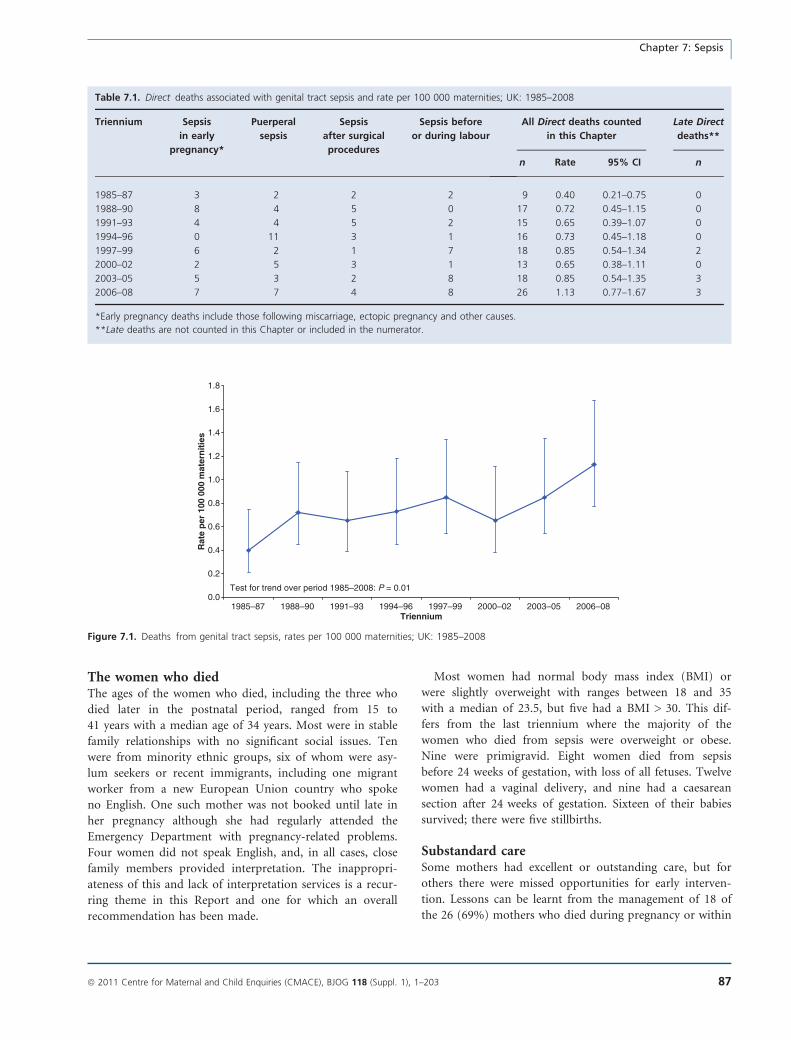
The women who diedThe ages of the women who died, including the three who
died later in the postnatal period, ranged from 15 to
41 years with a median age of 34 years. Most were in stable
family relationships with no significant social issues. Ten
were from minority ethnic groups, six of whom were asy-
lum seekers or recent immigrants, including one migrant
worker from a new European Union country who spoke
no English. One such mother was not booked until late in
her pregnancy although she had regularly attended the
Emergency Department with pregnancy-related problems.
Four women did not speak English, and, in all cases, close
family members provided interpretation. The inappropri-
ateness of this and lack of interpretation services is a recur-
ring theme in this Report and one for which an overall
recommendation has been made.
Most women had normal body mass index (BMI) or
were slightly overweight with ranges between 18 and 35
with a median of 23.5, but five had a BMI > 30. This dif-
fers from the last triennium where the majority of the
women who died from sepsis were overweight or obese.
Nine were primigravid. Eight women died from sepsis
before 24 weeks of gestation, with loss of all fetuses. Twelve
women had a vaginal delivery, and nine had a caesarean
section after 24 weeks of gestation. Sixteen of their babies
survived; there were five stillbirths.
Substandard careSome mothers had excellent or outstanding care, but for
others there were missed opportunities for early interven-
tion. Lessons can be learnt from the management of 18 of
the 26 (69%) mothers who died during pregnancy or within
Table 7.1. Direct deaths associated with genital tract sepsis and rate per 100 000 maternities; UK: 1985–2008
Triennium Sepsis
in early
pregnancy*
Puerperal
sepsis
Sepsis
after surgical
procedures
Sepsis before
or during labour
All Direct deaths counted
in this Chapter
Late Direct
deaths**
n Rate 95% CI n
1985–87 3 2 2 2 9 0.40 0.21–0.75 0
1988–90 8 4 5 0 17 0.72 0.45–1.15 0
1991–93 4 4 5 2 15 0.65 0.39–1.07 0
1994–96 0 11 3 1 16 0.73 0.45–1.18 0
1997–99 6 2 1 7 18 0.85 0.54–1.34 2
2000–02 2 5 3 1 13 0.65 0.38–1.11 0
2003–05 5 3 2 8 18 0.85 0.54–1.35 3
2006–08 7 7 4 8 26 1.13 0.77–1.67 3
*Early pregnancy deaths include those following miscarriage, ectopic pregnancy and other causes.
**Late deaths are not counted in this Chapter or included in the numerator.
0.0
0.2
0.4
0.6
0.8
1.0
1.2
1.4
1.6
1.8
1985–87 1988–90 1991–93 1994–96 1997–99 2000–02 2003–05 2006–08Triennium
Rat
e p
er 1
00 0
00 m
ater
nit
ies
Test for trend over period 1985–2008: P = 0.01
Figure 7.1. Deaths from genital tract sepsis, rates per 100 000 maternities; UK: 1985–2008
Chapter 7: Sepsis
ª 2011 Centre for Maternal and Child Enquiries (CMACE), BJOG 118 (Suppl. 1), 1–203 87

the 6-week postnatal period, although this does not mean
that all of these women’s lives would have been saved. In
most cases, the outcome was inevitable, but for 12 it might
have been different had the infection been diagnosed and
treated more promptly. In six others, although the outcome
would not have changed, lessons remain to be learnt. There
were also lessons to be learnt from the management of some
of the women who died later in their postnatal period.
The organisms involvedThe most common pathogen identified among the
women’s deaths was the b-haemolytic streptococcus Lance-
field Group A (Streptococcus pyogenes), of which there were
13 cases. There were five cases of Escherichia coli, one of
which also grew Enterococcus faecalis; three cases of Staphy-
lococcus aureus, one of which also grew mixed coliforms;
and one case each of Streptococcus pneumoniae, Morganella
morganii and Clostridium septicum. One healthcare worker,
known to be a methicillin-resistant Staphylococcus aureus
(MRSA) carrier, died some days after caesarean section
from septicaemia with Panton–Valentine leucocidin (PVL)
-positive MRSA, which is a particularly virulent subtype.
No pathogen was identified in four cases.
Group A b-haemolytic streptococcus (Streptococcuspyogenes)There were 13 deaths from b-haemolytic streptococcus
Lancefield Group A (Streptococcus pyogenes) this triennium
compared with eight in the last Report for 2003–05 and
three for maternal deaths from sepsis in 2000–02.
This organism should not be confused with the b-haemo-
lytic streptococcus Lancefield Group B (Streptococcus agalac-
tiae), which occasionally causes early neonatal mortality but
is a much less common cause of maternal mortality.
Group A streptococcus is typically community based,
and 5–30% of the population are asymptomatic carriers on
skin or in throat.1 It is easily spread by person-to-person
contact or by droplet spread from a person with infection.
Streptococcal sore throat is one of the most common bac-
terial infections of childhood, and all of the 13 women
who died from it either worked with, or had, young chil-
dren. Several mothers also had a history of recent sore
throat or respiratory infection. Contamination of the peri-
neum is more likely when a woman or her family or close
contacts have sore throats or other respiratory symptoms,
as the organism may be transferred from the throat or nose
via her hands to her perineum. Antenatal education should
raise awareness of this and the importance of good per-
sonal hygiene and avoiding contamination of the perineum
by washing hands before and after using the lavatory or
changing sanitary towels. All except one mother had intact
membranes until shortly before delivery, although several
had offensive, smelly or infected looking liquor when their
membranes ruptured. Studies have shown that bacteria
have the ability to cross intact membranes.3
Group A streptococcus can also cause serious illness such
as scarlet fever, bacteraemia, streptococcal shock syndrome
and necrotising fasciitis. Historically it is the classic organ-
ism associated with puerperal sepsis and was a major cause
of maternal mortality before antiseptic practice was intro-
duced and antibiotics became available.
Increased levels of Group A streptococcal infections tend
to occur between December and April, which was true for
most of the cases in this Report. Information from the
Health Protection Agency indicates that there were higher
than normal notifications of scarlet fever in some regions
of England from December 2007 to mid-March 2008,
which coincides with many of the cases reported here.4 For
most of the 13 women who died, there was no information
about subtype, but Types M, 12, T1 emm1 and emm11
were all identified.
The following vignette illustrates the typical symptoms
and the rapid and relentless course of Group A streptococ-
cal disease despite excellent care:
A woman in mid-pregnancy called an out-of-hours GP as she
was feverish, shivery and unwell and had a sore throat but
was diagnosed as having a probable viral infection. A few
hours later the GP visited again as she had developed con-
stant abdominal pain associated with vomiting, greenish
black diarrhoea, and reduced fetal movements but no vaginal
bleeding. The GP suspected placental abruption, and,
although she was rapidly transferred to hospital, on admis-
sion she was critically ill with marked tachycardia, breath-
lessness, cyanosis and confusion. The correct diagnosis of
septic shock was quickly recognised, fluid resuscitation was
started, senior consultants were called, advice was sought
from haematology and microbiology consultants and appro-
priate intravenous antibiotics were commenced immediately.
Despite intensive life support she died a few hours after
admission to hospital.
Sickle cell disease/traitSome of the women had underlying medical conditions
that may have increased their susceptibility to infection,
including three Black African women, two of whom died
from coliform and one from Staphylococcus aureus infection
following spontaneous preterm prelabour rupture of mem-
branes (PPROM) between 17 and 23 weeks of gestation.
One had sickle cell trait, and two had known sickle cell dis-
ease. Women with sickle cell disease are at increased risk of
infection because of poor splenic function as a result of
damage from sickle cell disease, and any anaemia may also
increase the risk of infection. Maternal mortality in sickle
cell disease is estimated to be around 1 in 220 (0.45%).5
Sickle cell disease in pregnancy is the subject of a UK
Harper
88 ª 2011 Centre for Maternal and Child Enquiries (CMACE), BJOG 118 (Suppl. 1), 1–203
Obstetric Surveillance System (UKOSS) surveillance study
from February 2010 until February 2011.
As highlighted in Chapter 10, pregnant women with
underlying disease, including sickle cell disease, should be
managed jointly under the care of a consultant obstetrician
and a specialist consultant in their underlying condition, in
this case a haematologist. All immunisations, including
against pneumococcus, should be up to date. Any infec-
tions should be treated promptly and prophylactic penicil-
lin is recommended. A clear plan of management should
be documented in the chart early in pregnancy. A national
guideline would also be helpful.
Sepsis in early pregnancyEight women, including seven counted in this Chapter and
one Late death, died from complications of infections arising
before 24 completed weeks of gestation. Two women died
from septic miscarriage and two after a termination of preg-
nancy. Of these, one did not receive post-procedure prophy-
lactic antibiotics and another died from Clostridium septicum
septicaemia and necrotising fasciitis. Clostridium infection is
a rare but previously reported cause of maternal death,
including after termination of pregnancy. Vaginal carriage
was the most likely source of infection and the inflammatory
focus in the uterus the most likely portal of entry. Necrotis-
ing fasciitis is characterised by an overwhelming fulminant
course with severe pain and muscle inflammation or necro-
sis; the majority of patients die within 24 hours of onset.
Sepsis following pregnancy loss:learning points
All units should have an effective and robust system in
place to ensure that peri-abortion antibiotic prophylaxis
(metronidazole 1 g rectally at the time of abortion plus,
commencing on the day of abortion, either doxycycline
100 mg orally twice daily for 7 days or azithromycin 1 g
orally) is offered routinely in accordance with RCOG
guidelines.6
Infection must be suspected and actively ruled out when
women who have had a recent termination of pregnancy
or spontaneous miscarriage have pyrexia, persistent
bleeding or abdominal pain, especially if the pain is
constant and severe. Vaginal swabs, ultrasound scan to
exclude retained products of conception and diagnostic
evacuation of uterus (evacuation of retained products of
conception) should be considered if there is still doubt;
haemoglobin, white cell count, C-reactive protein, and
blood cultures if pyrexia >38�C are minimum investiga-
tions; and high-dose broad-spectrum intravenous
antibiotics should be commenced immediately, without
waiting for microbiology results.
Four women, including a Late death, died from the conse-
quences of chorioamnionitis after spontaneous PPROM in
the second trimester. In one case the cause was Morganella
morganii. This is a Gram-negative rod bacterium often
found as part of the normal intestinal flora, but it can be a
rare cause of severe invasive disease and is naturally resis-
tant to many b-lactam antibiotics. Chorioamnionitis and
brain abscess due to Morganella morganii have both been
reported previously.7
Sepsis before deliveryNine women, including eight counted in this Chapter and
one Late death, developed sepsis before delivery after
24 weeks of gestation. Seven women had group A strepto-
coccal infection, one had Escherichia coli and for one
woman who was extremely unwell on admission there
was no information about whether any microbiological
investigations were performed before antibiotics were
given. Four had a caesarean section and five delivered
vaginally. All except two were extremely unwell on admis-
sion to hospital, and many received outstanding care once
admitted, even though nothing more could have been
done to save them.
Most of these women had similar symptoms. They
had a short history of feeling unwell; some had a recent
sore throat, cough or flu-like illness; several had severe
diarrhoea; a few had vomiting; some felt hot and cold
or shivery and had mild or severe pyrexia, although oth-
ers had no temperature. One woman was hypothermic
and several were tachycardic and hypotensive on admis-
sion. All had contractions and abdominal pain that in
some cases was constant, severe and not relieved by anal-
gesia.
Severe maternal infection also affects the fetus—five
babies died in utero, and those delivered by emergency
caesarean section for abnormal fetal cardiotocographs
needed resuscitation after delivery, as did a baby born
vaginally to a woman who had complained of pelvic pain
in late pregnancy. In her case, when the membranes rup-
tured at delivery, the liquor was heavily meconium-stained
and smelt offensive. The combination of severe abdominal
pain and abnormal or absent fetal heart is more usually
associated with placental abruption, but these cases dem-
onstrate that when a woman presents with these symp-
toms, genital tract sepsis must be considered in the
differential diagnosis.
Severe sepsis is often a cause of atonic uterine haemor-
rhage, which may be further exacerbated by disseminated
intravascular coagulation. For example, one woman had
uncontrollable bleeding after vaginal delivery and suffered a
cardiac arrest <10 hours after admission; another woman
who had an emergency caesarean section under general
anaesthesia because of abnormal fetal cardiotocograph had
Chapter 7: Sepsis
ª 2011 Centre for Maternal and Child Enquiries (CMACE), BJOG 118 (Suppl. 1), 1–203 89
a major intrapartum haemorrhage and cardiac arrest post-
operatively.
Of note, the membranes were intact almost until delivery
in eight of the nine women who developed sepsis before
delivery.
Sepsis after vaginal deliveryThere is a tendency to regard the puerperium as a low-risk
period compared with pregnancy and delivery, but signifi-
cant problems can develop during this time. Seven women
died from sepsis that developed after vaginal delivery and
they illustrate how fit, healthy women with an uncompli-
cated pregnancy and delivery can become critically ill and
die in a very short time. Sepsis is often insidious in onset
and may not reveal itself for several days postpartum, when
most women will be at home, especially with routine early
discharge now encouraged. Some women died from sepsis
resulting from perineal infection. For example:
A woman with a second-degree tear felt feverish a few days
after delivery and then developed severe lower abdominal
pain and diarrhoea. She was accurately and quickly
assessed as having sepsis by her community midwife and
GP and rapidly transferred to the Emergency Department,
whose staff as well as the maternity team had been alerted
in advance. She was extremely ill on admission to hospital,
and her condition deteriorated despite appropriate treat-
ment including triple antibiotic therapy. Despite maximum
support in intensive care, she died a few hours later. Blood
cultures and perineal swabs grew b-haemolytic streptococcus
Lancefield Group A.
Four women died from Group A streptococcal infection
shortly after postnatal admission. One had severe lower
abdominal pain and a very low temperature although her
pulse and blood pressure were normal. She developed
severe diarrhoea and mottled peripheries and then col-
lapsed. Despite immediate resuscitation and excellent inten-
sive care, she rapidly died from streptococcal toxic shock
syndrome.
MastitisA woman developed septic shock from severe breast infec-
tion. Women rarely die from breast infection. However,
postpartum mastitis is very common, and the symptoms
can mimic infection without it actually being present, mak-
ing diagnosis of infection all the more difficult. When a
woman complains of breast pain, the breasts should be
examined and vital signs should be recorded. If symptoms
do not settle or if they continue to deteriorate within 12–
24 hours of conservative management, the woman should
be referred urgently to her GP or maternity unit for antibi-
otic therapy (flucloxacillin 500 mg 6-hourly or erythromy-
cin 500 mg 6-hourly or equivalent oral antibiotic for 10–
14 days). Immediate referral to hospital is indicated if the
woman is clinically unwell, if there is no response to oral
antibiotics within 48 hours, if mastitis recurs, if there are
very severe or unusual symptoms or if there are any other
concerns. Breast abscesses are not obviously fluctuant, and
a surgical opinion may also be needed.
Sepsis after surgeryNine women, including eight counted in this Chapter and
one Late death, had a caesarean section, but in four
women it was performed as a result of their pre-existing
disease. Of the five women who developed obvious signs
and symptoms of infection after the section, four had
prolonged prelabour rupture of membranes and one
developed septic shock afterwards. One woman who had
had antibiotic prophylaxis after a caesarean section was
readmitted and died some weeks later from Staphylococcus
aureus wound infection and toxic shock syndrome. In
another case:
A woman had ruptured membranes for several days before
caesarean section for failure to progress in spontaneous
labour. She had been treated with oral amoxicillin and was
given a single dose of co-amoxyclav 1.2 g intravenously dur-
ing surgery. After delivery she had a cough and sore throat
and was discharged a few days later. She then felt cold, was
short of breath and was coughing up phlegm. Her commu-
nity midwife arranged for her to be reviewed at the hospi-
tal, but she collapsed and, even though an ambulance
arrived quickly, died in the Emergency Department. At
autopsy, b-haemolytic streptococcus Lancefield Group A was
isolated from her throat, lungs and uterus.
Overall learning pointsThe cases reported here demonstrate the huge challenges
in identifying and managing severe infection in pregnancy
or during or after the postnatal period. Fortunately, death
and serious illness from pregnancy-related sepsis are still
very rare. However, this means that many healthcare work-
ers will have never seen a case so awareness and the index
of suspicion can be low, and the fulminating nature of
many of the cases is surprising and shocking when it does
happen.
Raising public and professional awarenessSome women and their families did not realise how ill they
were or they did not disclose or trivialised significant
symptoms that might have allowed earlier intervention.
Some healthcare workers did not appreciate the signs and
symptoms or severity of the illness. There is therefore a
clear need to raise both maternal and professional aware-
ness about antenatal, intrapartum and puerperal sepsis so
that it can be prevented where possible, recognised quickly
Harper
90 ª 2011 Centre for Maternal and Child Enquiries (CMACE), BJOG 118 (Suppl. 1), 1–203
and managed effectively and immediately. This is one of
the major recommendations of this Report. In addition,
local guidelines or protocols must be available in all mater-
nity units, Emergency Departments and should be used by
GPs and community midwives. Until national guidelines
are produced, the Surviving Sepsis Campaign (www.surviv-
ingsepsis.org) has guidelines and care bundles that help to
guide the management of severely septic women.8 This is
also helpfully discussed in Chapter 16.
ProphylaxisAntibiotic prophylaxis is crucial in several clinical scenar-
ios, as shown in the Learning point box. In particular,
there is evidence demonstrating an association between
infection ascending from the lower genital tract and pro-
longed rupture of membranes. This increases the risk of
chorioamnionitis and should increase alertness for other
signs of sepsis. In women with PPROM, about one-third of
pregnancies have positive amniotic fluid cultures.3 Several
women who died had prolonged rupture of membranes,
and the lessons to be learnt, as in other cases, are largely
around the early recognition of sepsis.
Sepsis prophylaxis: learning points
All units should have an effective and robust system in
place to ensure that peri-abortion antibiotic prophylaxis
(metronidazole 1 g rectally at the time of abortion plus,
commencing on the day of abortion, either doxycycline
100 mg orally twice daily for 7 days or azithromycin 1 g
orally) is offered routinely in accordance with RCOG
guidelines.6
Routine antenatal antibiotic prophylaxis with erythro-
mycin (250 mg orally 6-hourly) for 10 days is recom-
mended in women who have PPROM before 37 weeks
of gestation.3
For prelabour rupture of the membranes at term (i.e.
after 37 weeks of gestation), if there is evidence of infec-
tion, a full course of broad-spectrum intravenous antibi-
otics should be prescribed.9
Women having a caesarean section should be offered
prophylactic antibiotics, such as a single parenteral dose
of first-generation cephalosporin or ampicillin, to
reduce the risk of postoperative infections (such as
endometritis and urinary tract and wound infections),
which occurs in about 8% of women who have had a
caesarean section.10
Broad-spectrum antibiotics are recommended following
obstetric anal sphincter repair (third- and fourth-degree
tears) to reduce the incidence of postoperative infections
and wound dehiscence.11
Early diagnosisWhen a woman becomes unwell, she is most likely to seek
help from her GP, community midwife, local maternity
hospital or Emergency Department, where she will proba-
bly be seen first by more junior members of the medical
team. It is therefore essential that such front-line staff are
fully aware of the signs and symptoms of sepsis and can
recognise critical illness. Telephone help lines have limita-
tions—in one case a woman sought advice on more than
one occasion because of persistent abdominal pain and
bleeding, but the urgency of the situation was not recogni-
sed and inappropriate advice was given.
There were also failures or delays in recognising the signs
and symptoms of sepsis and critical illness among hospital
staff. For example:
A woman who attended the maternity unit several times
with abdominal pain, vaginal discharge and suspected spon-
taneous rupture of membranes was never assessed by a senior
doctor. She was eventually admitted shivery and feverish,
with vomiting, diarrhoea, vaginal discharge, abdominal pain
and contractions. Although her temperature was normal, she
was hypotensive and tachycardic. She delivered soon after
admission, yet her observations were infrequent and no
action was taken despite her failure to improve. It was not
until she collapsed several hours later that antibiotics and
aggressive treatment were commenced but too late. A vaginal
swab taken on admission grew Group A streptococci.
A detailed history of symptoms is important to help diag-
nosis. Possible signs and symptoms are given in Box 7.1.
Unusual anxiety, panic and restlessness may be the result
of serious underlying illness:
A GP called to see a woman because of nausea, breathless-
ness and severe ‘after pains’ a few days after postnatal dis-
charge noted that she had a fast pulse and seemed ‘nervous
and worried’ and arranged hospital admission. A doctor
who saw her some time later observed that she was generally
unwell, seemed very anxious about being in hospital, and
looked ‘terrified’. No one recognised how ill she really was,
antibiotics were late and inadequate, and no consultant was
involved in her care until she arrested a few hours later.
Investigation and follow upThe cases discussed here illustrate various clinical scenarios
and also demonstrate that sepsis can mimic other condi-
tions such as gastroenteritis, ectopic pregnancy, placental
abruption, mastitis, pulmonary infection and pulmonary
embolism and must be included in the differential diagno-
sis of these conditions. The clinical picture in sepsis does
not always reflect the severity of the underlying illness. This
is a common problem in obstetric morbidity—the onset of
Chapter 7: Sepsis
ª 2011 Centre for Maternal and Child Enquiries (CMACE), BJOG 118 (Suppl. 1), 1–203 91
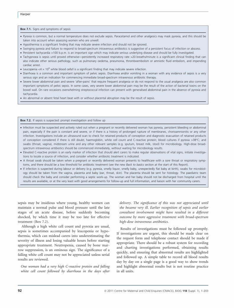
sepsis may be insidious where young, healthy women can
maintain a normal pulse and blood pressure until the late
stages of an acute disease, before suddenly becoming
shocked, by which time it may be too late for effective
treatment (Box 7.2).
Although a high white cell count and pyrexia are usual,
sepsis is sometimes accompanied by leucopenia or hypo-
thermia, which can mislead carers into underestimating the
severity of illness and losing valuable hours before starting
appropriate treatment. Neutropenia, caused by bone mar-
row suppression, is an ominous sign. The significance of a
falling white cell count may not be appreciated unless serial
results are reviewed.
One woman had a very high C-reactive protein and falling
white cell count followed by diarrhoea in the days after
delivery. The significance of this was not appreciated until
she became very ill. Earlier recognition of sepsis and earlier
consultant involvement might have resulted in a different
outcome by more aggressive treatment with broad-spectrum
high-dose intravenous antibiotics.
Results of investigations must be followed up promptly.
If investigations are urgent, this should be made clear on
the request form and telephone contact should be made if
appropriate. There should be a robust system for recording
and charting investigations performed, obtaining results
quickly, and ensuring that abnormal results are highlighted
and followed up. A simple table to record all blood results
day by day on a single page is a good way to show trends
and highlight abnormal results but is not routine practice
in all units.
Box 7.1. Signs and symptoms of sepsis
• Pyrexia is common, but a normal temperature does not exclude sepsis. Paracetamol and other analgesics may mask pyrexia, and this should be
taken into account when assessing women who are unwell.
• Hypothermia is a significant finding that may indicate severe infection and should not be ignored.
• Swinging pyrexia and failure to respond to broad-spectrum intravenous antibiotics is suggestive of a persistent focus of infection or abscess.
• Persistent tachycardia >100 b.p.m. is an important sign which may indicate serious underlying disease and should be fully investigated.
• Tachypnoea is sepsis until proved otherwise—persistently increased respiratory rate >20 breaths/minute is a significant clinical finding that can
also indicate other serious pathology, such as pulmonary oedema, pneumonia, thromboembolism or amniotic fluid embolism, and impending
cardiac arrest.
• Leucopenia <4 · 109 white blood cells/l is a significant finding that may indicate severe infection.
• Diarrhoea is a common and important symptom of pelvic sepsis. Diarrhoea and/or vomiting in a woman with any evidence of sepsis is a very
serious sign and an indication for commencing immediate broad-spectrum intravenous antibiotic therapy.
• Severe lower abdominal pain and severe ‘after-pains’ that require frequent analgesia or do not respond to the usual analgesia are also common
important symptoms of pelvic sepsis. In some cases, very severe lower abdominal pain may be the result of the action of bacterial toxins on the
bowel wall. On rare occasions overwhelming streptococcal infection can present with generalised abdominal pain in the absence of pyrexia and
tachycardia.
• An abnormal or absent fetal heart beat with or without placental abruption may be the result of sepsis.
Box 7.2. If sepsis is suspected: prompt investigation and follow up
• Infection must be suspected and actively ruled out when a pregnant or recently delivered woman has pyrexia, persistent bleeding or abdominal
pain, especially if the pain is constant and severe, or if there is a history of prolonged rupture of membranes, chorioamnionitis or any other
infection. Investigations include an ultrasound scan to check for retained products of conception and diagnostic evacuation of retained products
of conception considered if there is still doubt; haemoglobin, white cell count and C-reactive protein; blood cultures if pyrexia >38�C; and
swabs (throat, vagina), midstream urine and any other relevant samples (e.g. sputum, breast milk, stool) for microbiology. High-dose broad-
spectrum intravenous antibiotics should be commenced immediately, without waiting for microbiology results.
• Elevated C-reactive protein is an early marker of infection that should alert carers to make regular observations of vital signs, initiate investiga-
tions to locate a source of infection, and consider whether antibiotic treatment is indicated.
• A throat swab should be taken when a pregnant or recently delivered woman presents to healthcare with a sore throat or respiratory symp-
toms, and there should be a low threshold for antibiotic treatment (see the new Back to basics section at the start of this Report).
• If infection is suspected during labour or delivery (e.g. pyrexia, smelly liquor, smelly baby, unexpectedly flat baby at birth), swabs for microbiol-
ogy should be taken from the vagina, placenta and baby (ear, throat, skin). The placenta should be sent for histology. The paediatric team
should check the baby and consider performing a septic work-up. The woman and her baby should not be discharged from hospital until the
results are available, or at the very least with good arrangements for follow-up and full information, and liaison with her community carers.
Harper
92 ª 2011 Centre for Maternal and Child Enquiries (CMACE), BJOG 118 (Suppl. 1), 1–203
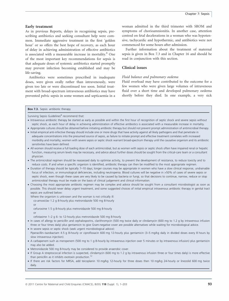
Early treatmentAs in previous Reports, delays in recognising sepsis, pre-
scribing antibiotics and seeking consultant help were com-
mon. Immediate aggressive treatment in the first ‘golden
hour’ or so offers the best hope of recovery, as each hour
of delay in achieving administration of effective antibiotics
is associated with a measurable increase in mortality.8 One
of the most important key recommendations for sepsis is
that adequate doses of systemic antibiotics started promptly
may prevent infection becoming established and may be
life-saving.
Antibiotics were sometimes prescribed in inadequate
doses, were given orally rather than intravenously, were
given too late or were discontinued too soon. Initial treat-
ment with broad-spectrum intravenous antibiotics may have
prevented pelvic sepsis in some women and septicaemia in a
woman admitted in the third trimester with SROM and
symptoms of chorioamnionitis. In another case, attention
centred on fetal decelerations in a woman who was hypoten-
sive, tachycardic and hypothermic, and antibiotics were not
commenced for some hours after admission.
Further information about the treatment of maternal
sepsis is given in Box 7.3 and in Chapter 16 and should be
read in conjunction with this section.
Clinical issues
Fluid balance and pulmonary oedemaFluid overload may have contributed to the outcome for a
few women who were given large volumes of intravenous
fluid over a short time and developed pulmonary oedema
shortly before they died. In one example, a very sick
Box 7.3. Sepsis: antibiotic therapy
Surviving Sepsis Guidelines8 recommend that:
• Intravenous antibiotic therapy be started as early as possible and within the first hour of recognition of septic shock and severe sepsis without
septic shock, as each hour of delay in achieving administration of effective antibiotics is associated with a measurable increase in mortality.
• Appropriate cultures should be obtained before initiating antibiotic therapy but should not prevent prompt administration of antimicrobial therapy.
• Initial empirical anti-infective therapy should include one or more drugs that have activity against all likely pathogens and that penetrate in
adequate concentrations into the presumed source of sepsis, as failure to initiate prompt and effective treatment correlates with increased
morbidity and mortality; women with severe sepsis or septic shock warrant broad-spectrum therapy until the causative organism and its antibiotic
sensitivities have been defined.
• All women should receive a full loading dose of each antimicrobial, but as women with sepsis or septic shock often have impaired renal or hepatic
function, measuring serum levels may be necessary, and advice about further doses should be sought from the critical-care team or a consultant
physician.
• The antimicrobial regimen should be reassessed daily to optimise activity, to prevent the development of resistance, to reduce toxicity and to
reduce costs. If and when a specific organism is identified, antibiotic therapy can then be modified to the most appropriate regimen.
• Duration of therapy should be typically 7–10 days; longer courses may be appropriate in women who have a slow clinical response, undrainable
focus of infection, or immunological deficiencies, including neutropenia. Blood cultures will be negative in >50% of cases of severe sepsis or
septic shock, even though these cases are very likely to be caused by bacteria or fungi, so that decisions to continue, narrow, reduce or stop
antimicrobial therapy must be made on the basis of clinical judgement and clinical information.
• Choosing the most appropriate antibiotic regimen may be complex and advice should be sought from a consultant microbiologist as soon as
possible. This should never delay urgent treatment, and some suggested choices of initial empirical intravenous antibiotic therapy in genital tract
sepsis are outlined below:
Where the organism is unknown and the woman is not critically ill:
co-amoxiclav 1.2 g 8-hourly plus metronidazole 500 mg 8-hourly
or
cefuroxime 1.5 g 8-hourly plus metronidazole 500 mg 8-hourly
or
cefotaxime 1–2 g 6- to 12-hourly plus metronidazole 500 mg 8-hourly
• In cases of allergy to penicillin and cephalosporins, clarithromycin (500 mg twice daily or clindamycin (600 mg to 1.2 g by intravenous infusion
three or four times daily) plus gentamicin to give Gram-negative cover are possible alternatives while waiting for microbiological advice.
• In severe sepsis or septic shock (seek urgent microbiological advice):
Piperacillin–tazobactam 4.5 g 8-hourly or ciprofloxacin 600 mg 12-hourly plus gentamicin (3–5 mg/kg daily in divided doses every 8 hours by
slow intravenous injection).
• A carbapenem such as meropenem (500 mg to 1 g 8-hourly by intravenous injection over 5 minutes or by intravenous infusion) plus gentamicin
may also be added.
• Metronidazole 500 mg 8-hourly may be considered to provide anaerobic cover.
• If Group A streptococcal infection is suspected, clindamycin (600 mg to 1.2 g by intravenous infusion three or four times daily) is more effective
than penicillin as it inhibits exotoxin production.12
• If there are risk factors for MRSA, add teicoplanin 10 mg/kg 12-hourly for three doses then 10 mg/kg 24-hourly or linezolid 600 mg twice
daily.
Chapter 7: Sepsis
ª 2011 Centre for Maternal and Child Enquiries (CMACE), BJOG 118 (Suppl. 1), 1–203 93
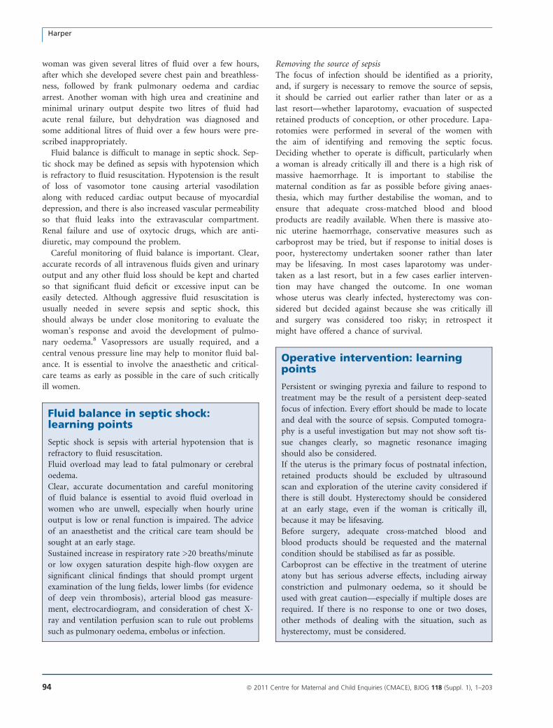
woman was given several litres of fluid over a few hours,
after which she developed severe chest pain and breathless-
ness, followed by frank pulmonary oedema and cardiac
arrest. Another woman with high urea and creatinine and
minimal urinary output despite two litres of fluid had
acute renal failure, but dehydration was diagnosed and
some additional litres of fluid over a few hours were pre-
scribed inappropriately.
Fluid balance is difficult to manage in septic shock. Sep-
tic shock may be defined as sepsis with hypotension which
is refractory to fluid resuscitation. Hypotension is the result
of loss of vasomotor tone causing arterial vasodilation
along with reduced cardiac output because of myocardial
depression, and there is also increased vascular permeability
so that fluid leaks into the extravascular compartment.
Renal failure and use of oxytocic drugs, which are anti-
diuretic, may compound the problem.
Careful monitoring of fluid balance is important. Clear,
accurate records of all intravenous fluids given and urinary
output and any other fluid loss should be kept and charted
so that significant fluid deficit or excessive input can be
easily detected. Although aggressive fluid resuscitation is
usually needed in severe sepsis and septic shock, this
should always be under close monitoring to evaluate the
woman’s response and avoid the development of pulmo-
nary oedema.8 Vasopressors are usually required, and a
central venous pressure line may help to monitor fluid bal-
ance. It is essential to involve the anaesthetic and critical-
care teams as early as possible in the care of such critically
ill women.
Fluid balance in septic shock:learning points
Septic shock is sepsis with arterial hypotension that is
refractory to fluid resuscitation.
Fluid overload may lead to fatal pulmonary or cerebral
oedema.
Clear, accurate documentation and careful monitoring
of fluid balance is essential to avoid fluid overload in
women who are unwell, especially when hourly urine
output is low or renal function is impaired. The advice
of an anaesthetist and the critical care team should be
sought at an early stage.
Sustained increase in respiratory rate >20 breaths/minute
or low oxygen saturation despite high-flow oxygen are
significant clinical findings that should prompt urgent
examination of the lung fields, lower limbs (for evidence
of deep vein thrombosis), arterial blood gas measure-
ment, electrocardiogram, and consideration of chest X-
ray and ventilation perfusion scan to rule out problems
such as pulmonary oedema, embolus or infection.
Removing the source of sepsisThe focus of infection should be identified as a priority,
and, if surgery is necessary to remove the source of sepsis,
it should be carried out earlier rather than later or as a
last resort—whether laparotomy, evacuation of suspected
retained products of conception, or other procedure. Lapa-
rotomies were performed in several of the women with
the aim of identifying and removing the septic focus.
Deciding whether to operate is difficult, particularly when
a woman is already critically ill and there is a high risk of
massive haemorrhage. It is important to stabilise the
maternal condition as far as possible before giving anaes-
thesia, which may further destabilise the woman, and to
ensure that adequate cross-matched blood and blood
products are readily available. When there is massive ato-
nic uterine haemorrhage, conservative measures such as
carboprost may be tried, but if response to initial doses is
poor, hysterectomy undertaken sooner rather than later
may be lifesaving. In most cases laparotomy was under-
taken as a last resort, but in a few cases earlier interven-
tion may have changed the outcome. In one woman
whose uterus was clearly infected, hysterectomy was con-
sidered but decided against because she was critically ill
and surgery was considered too risky; in retrospect it
might have offered a chance of survival.
Operative intervention: learningpoints
Persistent or swinging pyrexia and failure to respond to
treatment may be the result of a persistent deep-seated
focus of infection. Every effort should be made to locate
and deal with the source of sepsis. Computed tomogra-
phy is a useful investigation but may not show soft tis-
sue changes clearly, so magnetic resonance imaging
should also be considered.
If the uterus is the primary focus of postnatal infection,
retained products should be excluded by ultrasound
scan and exploration of the uterine cavity considered if
there is still doubt. Hysterectomy should be considered
at an early stage, even if the woman is critically ill,
because it may be lifesaving.
Before surgery, adequate cross-matched blood and
blood products should be requested and the maternal
condition should be stabilised as far as possible.
Carboprost can be effective in the treatment of uterine
atony but has serious adverse effects, including airway
constriction and pulmonary oedema, so it should be
used with great caution—especially if multiple doses are
required. If there is no response to one or two doses,
other methods of dealing with the situation, such as
hysterectomy, must be considered.
Harper
94 ª 2011 Centre for Maternal and Child Enquiries (CMACE), BJOG 118 (Suppl. 1), 1–203
Laparotomy for suspected intra-abdominal infection
should involve a general surgeon as well as an obstetri-
cian, as surgeons have a fundamentally different
approach to laparotomy and are more likely to make a
mid-line incision to allow full exploration of the
abdominal cavity.
Leadership and continuity of care
When managing complex cases, it must be clear who is in
charge of the woman’s care. This is particularly important
when an unwell pregnant woman presents to the Emergency
Department rather than the Maternity or Gynaecology
Departments, as there may be a tendency for everyone to
assume that someone else is looking after her. In one case,
although several doctors were involved in one woman’s early
care, no consultant took overall responsibility, nor were anti-
biotics given, until she collapsed over 24 hours after admis-
sion, by which time the outcome was inevitable.
Documentation and communicationMany different medical, nursing and midwifery staff may
be involved in the ongoing care of an unwell woman and
each may see her only once or twice. This makes the assess-
ment of any changes in the woman’s condition very diffi-
cult. In some of the cases already discussed in this Chapter,
the mother’s vital signs were either not recorded routinely
or their significance was not recognised. One of the ‘Top
ten’ recommendations in the last Report was for routine
use of a MEOWS chart to help in the more timely recogni-
tion, treatment and referral of women who have, or are
developing, a critical illness. A MEOWS chart is the easiest
way to see trends in a woman’s condition and to alert staff
to take appropriate action or call for help. For example:
A woman in late pregnancy with a short history of sore
throat, vomiting, diarrhoea and abdominal pain was tachy-
cardic and hypotensive on admission. Sepsis was not diag-
nosed for some hours.
The severity of her condition might have been recognised
sooner if a MEOWS chart had been used, and earlier antibi-
otic treatment and multidisciplinary care in a high-depen-
dency setting might possibly have changed the outcome.
Involvement of other staffAnaesthetic and critical-care staff play a vital part in the
effective management of sepsis and should be involved as
early as possible, particularly when there is circulatory or
respiratory failure. For six women, also discussed in Chap-
ter 8, there were significant delays in seeking help from an-
aesthetists and critical-care specialists.
In some cases, hospital departments seemed to be very
busy and staff were said to be overstretched, although it is
not clear if this was directly related to the outcome. For
example, it took several hours for two ill women to be seen
by a doctor or obstetric team because they were said to be
very busy elsewhere. The husband of another very sick
woman complained about the wait in a busy maternity unit
and was told that, as his wife was not seriously ill, the doctor
was seeing other more urgent patients. She died shortly after.
Apart from the women whose rapidly fulminating dis-
ease meant they died at home or shortly after admission in
the Emergency Department, most other women died in
critical care or operating theatres, reflecting the gravity of
their condition. However, a few died in hospital wards,
illustrating that seriously ill women were sometimes man-
aged in inappropriate settings with inexperienced staff
looking after them.
Sometimes there was a lack of co-ordinated care and lit-
tle or no support from senior staff when requested, but
there were also many examples of excellent teamwork. For
example, as the assessors noted:
This woman was extremely ill on admission and could not
have been saved. Her care could not have been better.
Everyone, including consultants, was called and attended
extremely promptly, even though it was the middle of the
night. The teamwork was excellent.
For most cases, there was a critical review by the Trust,
and some also invited external assessment. Some of these
reviews were excellent, but, as with other cases counted in
other chapters, many focused on irrelevant issues and failed
to identify major elements of substandard care or to learn
anything from the event. Recitfying this is an overall rec-
ommendation in this Report.
Disclosure of interestsNone.
FundingThis work was undertaken by the Centre for Maternal and
Child Enquiries (CMACE) as part of the CEMACH pro-
gramme. The work was funded by the National Patient
Safety Agency; the Department of Health, Social Services
and Public Safety of Northern Ireland; NHS Quality
Improvement Scotland (NHS QIS); and the Channel
Islands and Isle of Man.
References
1 Health Protection Agency. Interim UK guidelines for management of
close community contacts of invasive group A streptococcal disease.
Health Protection Agency, Group A Streptococcus Working Group.
Commun Dis Public Health 2004;7:354–61.
Chapter 7: Sepsis
ª 2011 Centre for Maternal and Child Enquiries (CMACE), BJOG 118 (Suppl. 1), 1–203 95
2 Lamagni TL, Efstratiou A, Dennis J, Nair P, Kearney J, George R,
et al. Increase in invasive group A streptococcal infections in Eng-
land, Wales and Northern Ireland, 2008–9. Euro Surveill 2009;14:
pii: 19110.
3 Royal College of Obstetricians and Gynaecologists. Preterm prela-
bour rupture of membranes. Green-top Guideline Number 44. Lon-
don: RCOG, 2006 [www.rcog.org.uk/womens-health/clinical-
guidance/preterm-prelabour-rupture-membranes-green-top-44].
Accessed 9 October 2010.
4 Health Protection Agency. Group A streptococcal infections: 2007/
08 seasonal update. Health Protection Report, Health Protection
Agency News Arch. Volume 2, No. 15; 11 April 2008.
5 The Management of Sickle Cell Disease. National Institutes of
Health. National Heart, Lung and Blood Institute, Division of Blood
Diseases and Resources. NIH Publication No. 02-2117, 4th edn.
Bethesda (MD): NIH, 2002. pp. 146.
6 Royal College of Obstetricians and Gynaecologists. The Care of
Women Requesting Induced Abortion. Evidence-based Clinical
Guideline Number 7. Guideline Summary. London: RCOG, 2004
[www.rcog.org.uk/womens-health/clinical-guidance/care-women-
requesting-induced-abortion]. Accessed 9 October 2010.
7 Abdalla J, Saad M, Samnani I, Lee P, Moorman J. Central nervous
system infection caused by Morganella morganii. Am J Med Sci
2006;331:44–7.
8 Dellinger RP, Levy MM, Carlet JM, Blon J, Parker MM, Jaeschke R,
et al. Surviving Sepsis Campaign: International guidelines for man-
agement of severe sepsis and septic shock: 2008: [published correc-
tion appears in Crit Care Med 2008;36:1394–6]. Crit Care Med
2008;36:296–327.
9 National Collaborating Centre for Women’s and Children’s Health.
Intrapartum Care. Care of Healthy Women and Their Babies Dur-
ing Childbirth. Clinical guideline 55. London: RCOG Press, 2007.
pp. 206 [www.guidance.nice.org.uk/CG55]. Accessed 9 October
2010.
10 National Collaborating Centre for Women’s and Children’s Health.
Caesarean Section. Clinical Guideline. National Institute for Health
and Clinical Excellence. London: RCOG Press, 2004 [www.guid-
ance.nice.org.uk/CG13]. Accessed 9 October 2010.
11 Royal College of Obstetricians and Gynaecologists. The Management
of Third- and Fourth-Degree Perineal Tears. Green-top Guideline
Number 29. London: RCOG, 2007 [www.rcog.org.uk/womens-
health/clinical-guidance/management-third-and-fourth-degree-peri-
neal-tears-green-top-29]. Accessed 9 October 2010.
12 Morgan MS. Diagnosis and management of necrotising fasciitis: a
multiparametric approach. J Hosp Infect 2010;75:249–57.
96 ª 2011 Centre for Maternal and Child Enquiries (CMACE), BJOG 118 (Suppl. 1), 1–203
Harper
Annex 7.1. A possible future approach to casedefinitionsSebastian Lucas
Background
In previous triennial maternal deaths reviews, as in this,
almost all deaths due to Group A beta-haemolytic Strepto-
coccus pyogenes (GAS), as well as other infections that can
be related to pregnancy or delivery, have been discussed
and counted in Chapter 7: Sepsis as Direct maternal
deaths from genital tract infection. Other infections unre-
lated to the genital tract but whose effects might have
been amplified by the altered immune state of pregnancy,
such as tuberculosis, pneumococcal meningitis and pneu-
monia, fungal infections and HIV disease are generally
considered as Indirect maternal deaths and counted in
Chapter 10. Similarly, although the 2009–10 H1N1 influ-
enza epidemic occurred after this triennium, such cases
will be discussed in the next Report and considered to be
Indirect deaths.
Under ‘genital tract sepsis’ in the last Report, for 2003-5,
GAS and E.coli were the commonest causes of infection,
and these were considered by subsets: sepsis in early preg-
nancy; sepsis before delivery; sepsis after vaginal delivery
(puerperal sepsis); sepsis after surgery; sepsis before or dur-
ing labour. In the current Report these divisions continue
to enable trend analysis, especially since the death rates
from GAS have increased, but this Annex explores the pos-
sibility of using a new system for classifying pregnancy-
related sepsis. This is based on practical experience of sep-
tic deaths in pregnant and non-pregnant women, reviews
of the 2006-08 cases and of the literature, as well as discus-
sions with microbiologists.
As a result, this Annex proposes that, under the sys-
tem outlined here, about one third of the deaths in this
triennium which would currently be considered to be
pregnancy-related sepsis, would be no longer classifiable
as due to Direct or Indirect causes. Instead, they may
actually be Coincidental to pregnancy. The new system
may provide a more useful way of considering remedi-
able factors and of determining the source of infection
and whether it is a hospital care associated infection
(HCAI) or community-acquired. Of these, the latter cate-
gory of community-acquired infections are likely to be
the majority.
The WHO classification of puerperalsepsis
The World Health Organisation (WHO)1 in 1995 stratified
and classified puerperal infections as follows:
Puerperal infections – general
• Puerperal sepsis and urinary tract infection
• Infections related to the birth process, but not of the
genito-urinary tract
• Incidental infections
Puerperal sepsis is then described as follows:
Infection of the genital tract occurring at any time
between rupture of membranes or labour, and the 42nd
day postpartum, in which two or more of the following are
present;
• Pelvic pain
• Fever
• Abnormal vaginal discharge
• Abnormal smell of discharge
• Delay in reduction of size of uterus.
A proposed new pathogeneticclassification of sepsis in pregnancy
The WHO system is a clinical case definition, not a patho-
logical classification, and mainly intended for application
in resource-poor (ie diagnostic pathology-poor) settings.
The proposed new classification system suggested here
builds on the above concepts. It is based on consideration
of the:
• timing of infection
• source of infection
• route of infection into mother
• role of operative interventions
• type of bacterial infection
• pathology – gross and histopathological
The proposal identifies five main categories. These are
shown in Table A7.1 together with an analysis of how the
maternal deaths from sepsis assessed by this Enquiry in the
United Kingdom for 2006-08 would satisfy these criteria.
It is important to note the classifications and numbers
discussed in this Annex are only suggestions, and for this
triennium the figures quoted in Chapter 7 must be used
as they help identify trends and rates as well as contain
Annex 7.1. A possible future approach to case definitions
ª 2011 Centre for Maternal and Child Enquiries (CMACE), BJOG 118 (Suppl. 1), 1–203 97
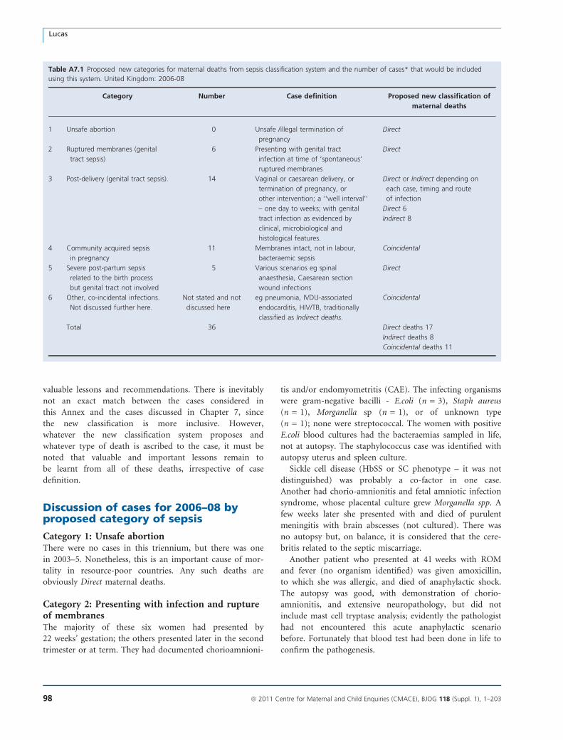
valuable lessons and recommendations. There is inevitably
not an exact match between the cases considered in
this Annex and the cases discussed in Chapter 7, since
the new classification is more inclusive. However,
whatever the new classification system proposes and
whatever type of death is ascribed to the case, it must be
noted that valuable and important lessons remain to
be learnt from all of these deaths, irrespective of case
definition.
Discussion of cases for 2006–08 byproposed category of sepsis
Category 1: Unsafe abortionThere were no cases in this triennium, but there was one
in 2003–5. Nonetheless, this is an important cause of mor-
tality in resource-poor countries. Any such deaths are
obviously Direct maternal deaths.
Category 2: Presenting with infection and ruptureof membranesThe majority of these six women had presented by
22 weeks’ gestation; the others presented later in the second
trimester or at term. They had documented chorioamnioni-
tis and/or endomyometritis (CAE). The infecting organisms
were gram-negative bacilli - E.coli (n = 3), Staph aureus
(n = 1), Morganella sp (n = 1), or of unknown type
(n = 1); none were streptococcal. The women with positive
E.coli blood cultures had the bacteraemias sampled in life,
not at autopsy. The staphylococcus case was identified with
autopsy uterus and spleen culture.
Sickle cell disease (HbSS or SC phenotype – it was not
distinguished) was probably a co-factor in one case.
Another had chorio-amnionitis and fetal amniotic infection
syndrome, whose placental culture grew Morganella spp. A
few weeks later she presented with and died of purulent
meningitis with brain abscesses (not cultured). There was
no autopsy but, on balance, it is considered that the cere-
britis related to the septic miscarriage.
Another patient who presented at 41 weeks with ROM
and fever (no organism identified) was given amoxicillin,
to which she was allergic, and died of anaphylactic shock.
The autopsy was good, with demonstration of chorio-
amnionitis, and extensive neuropathology, but did not
include mast cell tryptase analysis; evidently the pathologist
had not encountered this acute anaphylactic scenario
before. Fortunately that blood test had been done in life to
confirm the pathogenesis.
Table A7.1 Proposed new categories for maternal deaths from sepsis classification system and the number of cases* that would be included
using this system. United Kingdom: 2006-08
Category Number Case definition Proposed new classification of
maternal deaths
1 Unsafe abortion 0 Unsafe /illegal termination of
pregnancy
Direct
2 Ruptured membranes (genital
tract sepsis)
6 Presenting with genital tract
infection at time of ‘spontaneous’
ruptured membranes
Direct
3 Post-delivery (genital tract sepsis). 14 Vaginal or caesarean delivery, or
termination of pregnancy, or
other intervention; a ‘‘well interval’’
– one day to weeks; with genital
tract infection as evidenced by
clinical, microbiological and
histological features.
Direct or Indirect depending on
each case, timing and route
of infection
Direct 6
Indirect 8
4 Community acquired sepsis
in pregnancy
11 Membranes intact, not in labour,
bacteraemic sepsis
Coincidental
5 Severe post-partum sepsis
related to the birth process
but genital tract not involved
5 Various scenarios eg spinal
anaesthesia, Caesarean section
wound infections
Direct
6 Other, co-incidental infections.
Not discussed further here.
Not stated and not
discussed here
eg pneumonia, IVDU-associated
endocarditis, HIV/TB, traditionally
classified as Indirect deaths.
Coincidental
Total 36 Direct deaths 17
Indirect deaths 8
Coincidental deaths 11
Lucas
98 ª 2011 Centre for Maternal and Child Enquiries (CMACE), BJOG 118 (Suppl. 1), 1–203
All these deaths should be regarded as Direct, i.e. they
almost certainly would not have happened when they did if
the patient had not been pregnant (this logically includes
the anaphylaxis death also).
Category 3: Sepsis post-delivery (genital tractsepsis).Fourteen women fall into this category. Following vaginal
or caesarean delivery, termination of pregnancy (TOP) or
miscarriage, there is a ‘‘well interval’’ ranging from one day
to weeks; with genital tract infection as evidenced by clini-
cal, microbiological and histological features.
This category includes the ‘Semmelweis scenario’ of
post-partum overwhelming GAS infection via the genital
tract, where the agent might have come from the patient’s
midwife or doctors2. However, as will be seen, this was
potentially possible in only 5 of the cases. From review of
the circumstances, the majority of the women died of
infections that were present in their lower gut flora (eg
E.coli or staphylococci) or were acquired post-partum from
the community.
This group is sub-divided into those women who devel-
oped sepsis whilst still in hospital (n = 2) and those who
were re-admitted septic from home (n = 12). They can also
be sub-divided into those who had caesarean section
(n = 3), vaginal delivery (n = 8), miscarriage (n = 1), or
termination of pregnancy (n = 2).
The infecting agents comprise faecal flora (n = 4), which
is subcategorised as E. coli (n = 2, one of which was ESBL),
Staph aureus (n = 1) and Clostridium septicum (n = 1);
GAS (n = 6); pneumococcus (n = 2); MRSA (n = 1) and
one unknown agent.
The two women who died following a termination of
pregnancy had accidental perineum-derived infections.
Staph aureus caused a disseminated necrotising infection in
one woman who presented a few days later; at autopsy, the
uterus was not perforated. The other woman developed a
disseminated necrotising infection due to Clostridium septi-
cum several weeks after TOP. A third woman had a trau-
matic vaginal delivery where a tear developed into a recto-
vaginal fistula and necrotising fasciitis due to the virulent
ESBL E. coli.
Three women died after caesarean section (CS). All had
ascending endomyometritis, presenting within a week of
operation. The organisms were respectively GAS, MRSA
and E.coli; in the latter case, a serious co-morbidity was
chronic fibrosing eosinophilic myocarditis, which doubtless
exacerbated the infection.
The timing of the post-vaginal delivery GAS (n = 5) and
pneumococcal (n = 2), and the post-CS GAS (n = 1),
infections is important. Clinical sepsis developed from 2 to
35 days post-delivery, with a median of five days, and all
had gone home. Where an autopsy had been done, all had
endomyometritis, i.e. ascending genital tract sepsis. From
the known rapidity of progression of GAS infection, it is
thus likely that most, if not all, of these infections were
acquired in the community and not from any hospital
staff.
Two vignettes illustrate some of the problems in evaluat-
ing deaths in this category:
A woman had a normal vaginal delivery in hospital but
collapsed at home some days later. After rapid transfer to
the Emergency Department, the initial clinical evaluation
was pulmonary embolism (PE), and she was given strepto-
kinase but died shortly after. The autopsy found no embo-
lism but grew Streptococcus pneumoniae in the blood.
The genital tract was also inflamed, indicating ingress of
the pneumococcus via the vagina. The clinicians disputed
the absence of PE, querying whether thrombolysis could
actually remove all the evidence of PE at autopsy. Consul-
tation with many pathologists and reviewing the limited lit-
erature strongly support the contention that up to two
hours of resuscitation with thrombolysis cannot dissolve
large pulmonary emboli.
Another woman was treated for Group B Streptococcus
colonisation of the genital tract in her second trimester. She
seemed well and delivered vaginally at term. Some weeks
later, she re-presented septic; her blood culture identified
GAS, but, despite antibiotics, she died in multi-organ fail-
ure a few days later. There was no autopsy.
It is not certain that pregnancy or delivery was the cause
of this death, as it seems more probable that she suffered
Table A7.2 The causes of the infections listed in Table A7.1. This
includes all Direct deaths from Sepsis in Chapter 7 and Indirect
deaths in Chapter 10
Organisms Number and percent
of cases
Group A Streptococcus pyogenes (GAS) 14 (39%)
E.coli 5 (14%)
Streptococcus pneumoniae 4 (11%)
Staphylococcus aureus
(Methicillin-resistant
Staphylococcus aureus MRSA 1)
4 (11%)
Clostridium septicum 1 (3%)
Morganella sp Morganella morganii 1 (3%)
Gram-negative rods not otherwise
specified
1 (3%)
Not known 6 (17%)
Total 36 (100%)
Annex 7.1. A possible future approach to case definitions
ª 2011 Centre for Maternal and Child Enquiries (CMACE), BJOG 118 (Suppl. 1), 1–203 99
the same infection scenario as described in the next section
where such deaths are considered Coincidental.
Are these deaths Direct or Indirect? For the GAS and
pneumococcal infections that occurred some days after
vaginal or CS delivery, eight in all, it is arguable that
these are community-acquired infections and that the
genital tract, traumatised by delivery, was a more
susceptible substrate for entry of infection, i.e. they are
Indirect.
Category 4: Admitted with severe sepsis from thecommunity, membranes intact, not in labour atonset of illness.These eleven women form a most interesting group of sep-
sis patients, traditionally classified as Direct deaths from
genital tract sepsis but, under the proposed new classifica-
tion system, are now considered to be Coincidental. Where
the organism was known, it was GAS (n = 6) and the
pneumococcus (n = 2). Eight of the women were in the
third trimester, one in the first, and two in the second.
Only one was obese.
Nine women presented in septic shock (GAS n = 6,
pneumococcus n = 1, not known n = 2) and two with
meningitis (pneumococcus n = 1, CSF mixed growth
n = 1). The clinical presentation was generally rapid: six of
the eleven died either at home or within 24 hours’ admis-
sion to hospital. Eight of eleven were delivered (two
through peri-mortem caesarean section). A typical case is
described below:
A woman with small children nearing the end of pregnancy
had a sore throat then presented very ill to hospital. DIC
and low platelets were identified, and pre-mortem blood
culture grew GAS. She died a few short hours after delivery.
The autopsy found DIC, excluded AFE, and noted the
absence of chorio-amnionitis.
Sepsis was not considered in several of those admitted or
there was delay in undertaking blood cultures to test for it.
A woman with pneumococcus had the sickle HbSC pheno-
type as a risk factor, which had not been recorded properly
in her medical notes as she was thought only to have sickle
trait, HbAS.
The autopsies (n = 9) were mostly done adequately, and
in two cases the positive microbiology came from autopsy
samples. DIC was noted in four cases. In two others there
was lack of examination of the genital tract, including the
placenta, cord and membranes.
Thus, in only five of these eleven cases were there histo-
pathological information on the presence or absence of
chorio-amnionitis, funisitis and endomyometritis (CAE)
and, intriguingly, none indicated inflammation of the geni-
tal tract. That placental cultures in some cases grew GAS
indicates infection in the blood but does not specify actual
tissue inflammation. (In other cases outside this series, in
the same taxonomic category and known to the author,
there is also no CAE). This raises the question as to how
the infection gets into the mother. This scenario of preg-
nancy-related sepsis, though dominated by GAS infection,
is pathogenetically nothing to do with a category 3 type,
classical ‘Semmelweis syndrome’ of direct peri-partum
infection of the genital tract.
Five of the six women with GAS had a history of a ‘sore
throat’, indicating the source of infection was from the
community (possibly from their other children at school).
Whether the bacterium passes directly from the nasophar-
ynx into the blood stream, or goes from nasopharynx to
hand to vagina, ascending the genital tract into blood
stream, is not clear.
Both have been documented3. If it goes via the latter
route, it appears to happen so quickly that there is no
induction of inflammation in the genital tract. GAS is car-
ried transiently in the vagina in a small proportion of third
trimester women (about 0.03% on cross-sectional studies)4;
the pneumococcus is also carried in some women5.
The other important pathogenetic issue is whether
women are susceptible to these overwhelming bacterial
infections by the nature of being pregnant, in any of the
trimesters. Whilst there is good evidence for a temporary
cell-mediated immunosuppresssion due to pregnancy (and
influencing the course of mycobacterial infections, some
viruses, and listeriosis), there is none such for pyogenic
bacterial infections. Central UK surveillance of GAS infec-
tions indicate that in the 15-44 year age group, the rate of
GAS bacteraemia is low at 1.5/100,000 women per year,
and it is only in this age range that slightly more women
than men acquire this infection. But there is currently no
collection of data on co-morbidities or status such as preg-
nancy to provide more useful epidemiological informa-
tion6.
In conclusion, it may be that in this significant group of
pregnant women who die of overwhelming sepsis, the preg-
nancy per se has nothing to do with the infection, i.e. the
deaths are Coincidental. It is important that further epide-
miological studies, in conjunction with better clinico-patho-
logical investigations, be done to understand more about this
particular, very concerning scenario of sepsis in pregnancy.
Category 5: Post-partum sepsis directly related todelivery, but not involving the genital tract.Five women died of overwhelming infection that, in all but
one, was acquired whilst in hospital care. Two of them
were neurological in origin:
A woman delivered twins before term. Her blood pressure
rose, but the diagnosis of pre-eclampsia was not immedi-
ately recognised. Despite MgSO4, her hypertension contin-
Lucas
100 ª 2011 Centre for Maternal and Child Enquiries (CMACE), BJOG 118 (Suppl. 1), 1–203
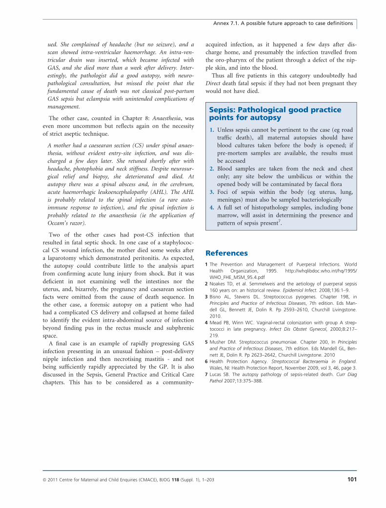
ued. She complained of headache (but no seizure), and a
scan showed intra-ventricular haemorrhage. An intra-ven-
tricular drain was inserted, which became infected with
GAS, and she died more than a week after delivery. Inter-
estingly, the pathologist did a good autopsy, with neuro-
pathological consultation, but missed the point that the
fundamental cause of death was not classical post-partum
GAS sepsis but eclampsia with unintended complications of
management.
The other case, counted in Chapter 8: Anaesthesia, was
even more uncommon but reflects again on the necessity
of strict aseptic technique.
A mother had a caesearan section (CS) under spinal anaes-
thesia, without evident entry-site infection, and was dis-
charged a few days later. She retuned shortly after with
headache, photophobia and neck stiffness. Despite neurosur-
gical relief and biopsy, she deteriorated and died. At
autopsy there was a spinal abscess and, in the cerebrum,
acute haemorrhagic leukoencephalopathy (AHL). The AHL
is probably related to the spinal infection (a rare auto-
immune response to infection), and the spinal infection is
probably related to the anaesthesia (ie the application of
Occam’s razor).
Two of the other cases had post-CS infection that
resulted in fatal septic shock. In one case of a staphylococ-
cal CS wound infection, the mother died some weeks after
a laparotomy which demonstrated peritonitis. As expected,
the autopsy could contribute little to the analysis apart
from confirming acute lung injury from shock. But it was
deficient in not examining well the intestines nor the
uterus, and, bizarrely, the pregnancy and caesarean section
facts were omitted from the cause of death sequence. In
the other case, a forensic autopsy on a patient who had
had a complicated CS delivery and collapsed at home failed
to identify the evident intra-abdominal source of infection
beyond finding pus in the rectus muscle and subphrenic
space.
A final case is an example of rapidly progressing GAS
infection presenting in an unusual fashion – post-delivery
nipple infection and then necrotising mastitis - and not
being sufficiently rapidly appreciated by the GP. It is also
discussed in the Sepsis, General Practice and Critical Care
chapters. This has to be considered as a community-
acquired infection, as it happened a few days after dis-
charge home, and presumably the infection travelled from
the oro-pharynx of the patient through a defect of the nip-
ple skin, and into the blood.
Thus all five patients in this category undoubtedly had
Direct death fatal sepsis: if they had not been pregnant they
would not have died.
Sepsis: Pathological good practicepoints for autopsy
1. Unless sepsis cannot be pertinent to the case (eg road
traffic death), all maternal autopsies should have
blood cultures taken before the body is opened; if
pre-mortem samples are available, the results must
be accessed
2. Blood samples are taken from the neck and chest
only; any site below the umbilicus or within the
opened body will be contaminated by faecal flora
3. Foci of sepsis within the body (eg uterus, lung,
meninges) must also be sampled bacteriologically
4. A full set of histopathology samples, including bone
marrow, will assist in determining the presence and
pattern of sepsis present7.
References
1 The Prevention and Management of Puerperal Infections. World
Health Organization, 1995. http://whqlibdoc.who.int/hq/1995/
WHO_FHE_MSM_95.4.pdf
2 Noakes TD, et al. Semmelweis and the aetiology of puerperal sepsis
160 years on: an historical review. Epidemiol Infect. 2008;136:1–9.
3 Bisno AL, Stevens DL. Streptococcus pyogenes. Chapter 198, in
Principles and Practice of Infectious Diseases, 7th edition. Eds Man-
dell GL, Bennett JE, Dolin R. Pp 2593–2610, Churchill Livingstone.
2010.
4 Mead PB, Winn WC. Vaginal-rectal colonization with group A strep-
tococci in late pregnancy. Infect Dis Obstet Gynecol, 2000;8:217–
219.
5 Musher DM. Streptococcus pneumoniae. Chapter 200, In Principles
and Practice of Infectious Diseases, 7th edition. Eds Mandell GL, Ben-
nett JE, Dolin R. Pp 2623–2642, Churchill Livingstone. 2010
6 Health Protection Agency. Streptococcal Bacteraemia in England.
Wales, NI: Health Protection Report, November 2009, vol 3, 46, page 3.
7 Lucas SB. The autopsy pathology of sepsis-related death. Curr Diag
Pathol 2007;13:375–388.
ª 2011 Centre for Maternal and Child Enquiries (CMACE), BJOG 118 (Suppl. 1), 1–203 101
Annex 7.1. A possible future approach to case definitions
Chapter 8: AnaesthesiaJ McClure1, G Cooper2 (on behalf of the Centre for Maternal and Child Enquiries)1 Simpson Centre for Reproductive Health, Department of Anaesthesia and Pain Medicine, Royal Infirmary, Edinburgh, UK; 2 6 Lord Austin
Drive, Marlbrook, Bromsgrove, Worcestershire, UK
Correspondence: Dr John H McClure, Simpson Centre for Reproductive Health, Department of Anaesthesia and Pain Medicine, Royal
Infirmary, Edinburgh EH16 5SA, UK. Email: [email protected]
Keywords anaesthesia, Confidential Enquiry, maternal, mortality.
Anaesthesia: specific recommendations
• The effective management of failed tracheal intubation is a core anaesthetic skill that should be rehearsed and assessed
regularly.
• The recognition and management of severe, acute illness in a pregnant woman requires multidisciplinary teamwork.
An anaesthetist and/or critical-care specialist should be involved early.
• Obstetric and gynaecology services, particularly those without an on-site critical-care unit, must have a defined local
guideline to obtain rapid access to, and help from, critical-care specialists.
Summary of cases for 2006–08
The central assessors in anaesthesia reviewed 127 cases in
which anaesthetic services were involved in the care of a
woman who died from either a Direct or Indirect cause of
maternal death and for whom notes were available. This
comprised 49% (127 of 261) of all the maternal deaths in
this triennium. From these deaths the assessors identified
seven (3%) women who died from problems directly asso-
ciated with anaesthesia and whose deaths are counted in
this Chapter. The overall mortality rates from Direct deaths
from anaesthesia over the past eight triennia are shown in
Table 8.1.
In addition to the seven women who died from Direct
anaesthetic causes, the assessors considered that in a further
18 deaths anaesthetic management contributed to the out-
come or there were lessons to be learned. There were also 12
women with severe pregnancy-induced hypertension or sep-
sis for whom obstetricians or gynaecologists failed to consult
with anaesthetic or critical-care services sufficiently early,
which the assessors considered may have contributed to the
deaths. These deaths are counted in the relevant chapters of
this Report, but the lessons to be learned are discussed below.
Women with severe acute illness or significant co-mor-
bidity present a major challenge to obstetric, anaesthetic
and midwifery services, particularly when the maternity ser-
vice is already stretched with peak activity in its normal
workload. Many case reports highlighted the fact that pro-
viding good high-dependency care was difficult when the
maternity service was already very busy.
Table 8.1. Direct deaths attributed to anaesthesia and rate per
100 000 maternities; UK: 1985–2008
Triennium
Direct deaths
attributable
to anaesthesia
Percentage
of Direct deaths Rate 95% CI
1985–87 6 4.3 0.26 0.12-0.58
1988–90 4 2.8 0.17 0.07-0.44
1991–93 8 6.3 0.35 0.18-0.68
1994–96 1 0.7 0.05 0.01-0.26
1997–99 3 2.8 0.14 0.05-0.42
2000–02 6 5.7 0.30 0.14-0.66
2003–05 6 4.5 0.28 0.13-0.62
2006–08 7 6.5 0.31 0.15-0.64
McClure, Cooper
102 ª 2011 Centre for Maternal and Child Enquiries (CMACE), BJOG 118 (Suppl. 1), 1–203

Deaths directly due to anaesthesia
The women who diedIn this triennium, two women died from failure to ventilate
the lungs, four from postoperative complications and one
from leucoencephalitis. Two of the seven women were obese.
Failure to ventilate the lungsThere were two deaths as a result of a failure to ventilate the
lungs, one after induction of general anaesthesia and one
when a tracheostomy tube came out in a critical-care ward.
In the first woman, the anaesthetist failed to stop trying to
intubate the trachea even though oxygenation was achieved
through an intubating laryngeal mask airway (ILMA).
Failed intubation guidelines1 were followed, to an extent, in
this very stressful situation, but oesophageal intubation
through the ILMA was not recognised and further hypoxia
occurred. The woman was coughing but was not allowed to
wake up, and a second dose of thiopental and a long-acting
neuromuscular-blocking drug were given even though the
end-expiratory CO2 monitor indicated that the woman’s
lungs were not being ventilated. Cricothyrotomy was not
attempted.
Patients do not die from a ‘failure to intubate’. They die
either from failure to stop trying to intubate or from undiag-
nosed oesophageal intubation.
Bruce Scott 19862
Much of the success that these Confidential Enquiries into
Maternal Deaths have had in reducing Direct deaths from
anaesthesia has been from tackling airway-related deaths.
In the 1967–69 triennial report for England and Wales3
there were 32 such deaths. Striving for absence of such
deaths is achievable, as in 1994–96, but requires continual
efforts. Management of failed intubation/ventilation is a
core anaesthetic skill that should be rehearsed and assessed
regularly.4 The infrequent use of general anaesthesia in
obstetrics, and hence the lack of experience, makes this
extremely important.5 In one region of the UK, during a
5-year period from 1999 to 2003, there was an incidence of
failed tracheal intubation of one in 238 general anaethetics.
In half of these women, there was a failure to follow an
accepted protocol for failed tracheal intubation.6 Recent
surveillance of failed tracheal intubation for obstetric gen-
eral anaesthesia conducted through the United Kingdom
Obstetric Surveillance System (UKOSS which is discussed
in detail in the Introductory Chapter of this Report)
between April 2008 and January 2010 identified 51 women
in an estimated 1.4 million maternities, giving an estimated
rate of failed intubation of 36 cases per million maternities
(95% CI 27–48 per million maternities [personal commu-
nication with UKOSS]).
Human errors and failures in clinical practice are inevita-
ble. Strategies and guidelines need to be in place when errors
or failures occur.7 Assessment of nontechnical skills, such as
situation awareness, is given high priority in the aviation
industry but is also very important in medical practice. In
anaesthesia, one must always consider the worst-case sce-
nario, implement drills and be aware of what may happen
under stress. An anaesthetist may perceive failed intubation
as failed performance, which creates pressure to persist.
There is the added fear of acid aspiration, but the impor-
tance of maternal oxygenation cannot be overemphasised.
In managing a rare critical situation, it is important to be
prepared to implement an accepted drill fully and to main-
tain priorities. Drills should not be complex and must be
practiced regularly. The anaesthetist must avoid blind alleys
and the natural behaviour patterns of fixation and denial.
It is also pertinent to note that the woman discussed
above had a working epidural in labour when it was decided
to perform a Category 2 caesarean section. Subsequently
there was a sustained fetal bradycardia, which escalated the
urgency of caesarean section to Category 1. The epidural
had not been topped up to provide surgical anaesthesia for
caesarean section because the anaesthetist planned to top
the epidural up in theatre. If the epidural had been topped
up when it was decided she was to have a caesarean section,
general anaesthesia may not have been required.
Epidural anaesthesia for operativedelivery: learning points
Epidural analgesia that has been working well in labour
should be topped up to provide full surgical anaesthesia
without delay once the decision for operational delivery
has been made. If the woman cannot be immediately
transferred to an operating theatre and full epidural
anaesthesia is established on the labour ward, the anaes-
thetist and full resuscitation equipment should be imme-
diately available and full ‘epidural monitoring’ provided.
A second woman died in a critical-care unit from loss of
the airway when her tracheostomy tube came out while
being turned. She had a known airway problem and known
difficulty with her tracheostomy.
A clear strategy of management for this scenario was
required in advance, including the use of small tracheal
tubes and quick referral for senior help out of hours.
Autopsy revealed a very small larynx and lobular brown tis-
sue obscured the tracheostomy orifice.
In these two cases, non-consultant anaesthetists were
exposed to rare emergencies without immediate senior
backup. Inexperience of rare situations is difficult to
Chapter 8: Anaesthesia
ª 2011 Centre for Maternal and Child Enquiries (CMACE), BJOG 118 (Suppl. 1), 1–203 103
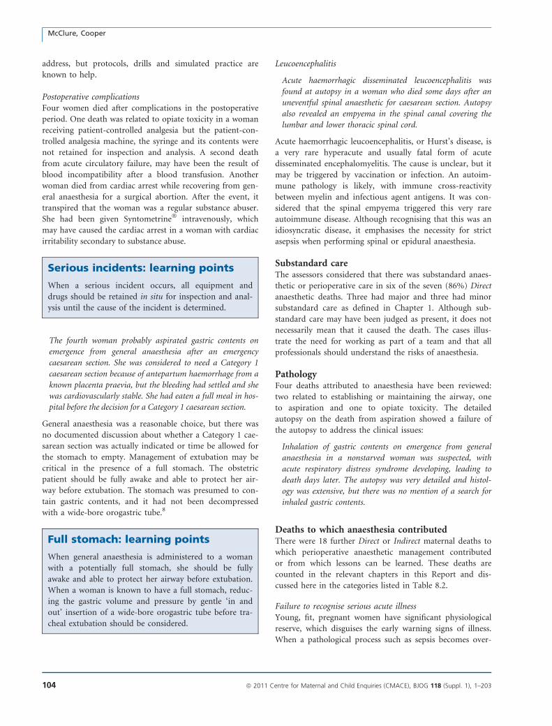
address, but protocols, drills and simulated practice are
known to help.
Postoperative complicationsFour women died after complications in the postoperative
period. One death was related to opiate toxicity in a woman
receiving patient-controlled analgesia but the patient-con-
trolled analgesia machine, the syringe and its contents were
not retained for inspection and analysis. A second death
from acute circulatory failure, may have been the result of
blood incompatibility after a blood transfusion. Another
woman died from cardiac arrest while recovering from gen-
eral anaesthesia for a surgical abortion. After the event, it
transpired that the woman was a regular substance abuser.
She had been given Syntometrine� intravenously, which
may have caused the cardiac arrest in a woman with cardiac
irritability secondary to substance abuse.
Serious incidents: learning points
When a serious incident occurs, all equipment and
drugs should be retained in situ for inspection and anal-
ysis until the cause of the incident is determined.
The fourth woman probably aspirated gastric contents on
emergence from general anaesthesia after an emergency
caesarean section. She was considered to need a Category 1
caesarean section because of antepartum haemorrhage from a
known placenta praevia, but the bleeding had settled and she
was cardiovascularly stable. She had eaten a full meal in hos-
pital before the decision for a Category 1 caesarean section.
General anaesthesia was a reasonable choice, but there was
no documented discussion about whether a Category 1 cae-
sarean section was actually indicated or time be allowed for
the stomach to empty. Management of extubation may be
critical in the presence of a full stomach. The obstetric
patient should be fully awake and able to protect her air-
way before extubation. The stomach was presumed to con-
tain gastric contents, and it had not been decompressed
with a wide-bore orogastric tube.8
Full stomach: learning points
When general anaesthesia is administered to a woman
with a potentially full stomach, she should be fully
awake and able to protect her airway before extubation.
When a woman is known to have a full stomach, reduc-
ing the gastric volume and pressure by gentle ‘in and
out’ insertion of a wide-bore orogastric tube before tra-
cheal extubation should be considered.
Leucoencephalitis
Acute haemorrhagic disseminated leucoencephalitis was
found at autopsy in a woman who died some days after an
uneventful spinal anaesthetic for caesarean section. Autopsy
also revealed an empyema in the spinal canal covering the
lumbar and lower thoracic spinal cord.
Acute haemorrhagic leucoencephalitis, or Hurst’s disease, is
a very rare hyperacute and usually fatal form of acute
disseminated encephalomyelitis. The cause is unclear, but it
may be triggered by vaccination or infection. An autoim-
mune pathology is likely, with immune cross-reactivity
between myelin and infectious agent antigens. It was con-
sidered that the spinal empyema triggered this very rare
autoimmune disease. Although recognising that this was an
idiosyncratic disease, it emphasises the necessity for strict
asepsis when performing spinal or epidural anaesthesia.
Substandard careThe assessors considered that there was substandard anaes-
thetic or perioperative care in six of the seven (86%) Direct
anaesthetic deaths. Three had major and three had minor
substandard care as defined in Chapter 1. Although sub-
standard care may have been judged as present, it does not
necessarily mean that it caused the death. The cases illus-
trate the need for working as part of a team and that all
professionals should understand the risks of anaesthesia.
PathologyFour deaths attributed to anaesthesia have been reviewed:
two related to establishing or maintaining the airway, one
to aspiration and one to opiate toxicity. The detailed
autopsy on the death from aspiration showed a failure of
the autopsy to address the clinical issues:
Inhalation of gastric contents on emergence from general
anaesthesia in a nonstarved woman was suspected, with
acute respiratory distress syndrome developing, leading to
death days later. The autopsy was very detailed and histol-
ogy was extensive, but there was no mention of a search for
inhaled gastric contents.
Deaths to which anaesthesia contributedThere were 18 further Direct or Indirect maternal deaths to
which perioperative anaesthetic management contributed
or from which lessons can be learned. These deaths are
counted in the relevant chapters in this Report and dis-
cussed here in the categories listed in Table 8.2.
Failure to recognise serious acute illnessYoung, fit, pregnant women have significant physiological
reserve, which disguises the early warning signs of illness.
When a pathological process such as sepsis becomes over-
McClure, Cooper
104 ª 2011 Centre for Maternal and Child Enquiries (CMACE), BJOG 118 (Suppl. 1), 1–203
whelming, decompensation is acute and profound. It is
emphasised that death in pregnancy or the puerperium can
ensue within hours of sepsis becoming apparent.
There were ten deaths as the result of a failure to recog-
nise and manage major, acute, serious illness.
Early recognition of serious acute illness in an otherwise
fit pregnant woman is a skill that only comes with experi-
ence. Multidisciplinary assessment by senior medical staff is
invaluable if carried out before the situation becomes irre-
trievable. There is evidence that early warning scoring sys-
tems or more specific Modified Early Obstetric Warnings
Scoring systems (MEOWS) are being used more frequently
in maternity units and gynaecology wards, but they are not
always acted upon.
Pre-eclampsia/eclampsiaIn eight of the 19 women who died of pre-eclampsia/
eclampsia, including HELLP syndrome, the anaesthetic
management, or rather its lack, may have contributed to
the death. In six of these deaths, the assessors considered
that senior advice or assistance from an anaesthetic or criti-
cal-care specialist was requested too late.
One woman was admitted with severe hypertension and +++
proteinuria. She was referred to a urologist in spite of being
very unwell on admission. She required immediate control
and regular, frequent monitoring of her blood pressure. Help
from an anaesthetist and high-dependency care could have
helped this woman from the outset but was not requested.
The immediate postoperative management of women
with severe pre-eclampsia/eclampsia is the responsibility of
both the consultant obstetrician and the consultant anaes-
thetist. It should be a joint decision whether care, treat-
ment and monitoring are provided in a labour ward high-
dependency unit or a critical-care ward.
Another woman had eclamptic fits at home, in the ambu-
lance and on the labour ward. She was treated with magne-
sium sulphate and an intravenous bolus of labetalol. Her
blood pressure dropped quickly, and she had a cardiac arrest.
This woman warranted early involvement of the anaesthe-
tist, high-dependency care and regular, frequent monitoring
of her blood pressure. Frequent noninvasive measurements
of systolic arterial pressure are useful in monitoring a trend.
Direct arterial pressure monitoring should be considered if a
more accurate measurement of systolic and diastolic arterial
pressures is required, particularly in the morbidly obese
woman, but should not delay treatment.
Severe pre-eclampsia: learning point
Severe pre-eclampsia requires immediate control and
regular, frequent monitoring of blood pressure with
high-dependency care. Anaesthetic services and, on
occasions, critical-care services should be involved in
management at an early stage.
SepsisIn six deaths from sepsis there were identifiable delays in
seeking senior help or advice and help from anaesthetists
and critical-care specialists.
One woman went to see her GP a week postpartum feeling very
unwell. The GP sent her directly to the Emergency Department
where she was found to be tachycardic and acidotic. There was
then a significant delay in asking for help with resuscitation
from an anaesthetist or critical-care specialist. She died in a
critical-care unit a few weeks after delivery.
Puerperal sepsis can quickly become a life-threatening
obstetric emergency.
Sepsis: learning point
Circulatory collapse can happen suddenly in sepsis, and
management should be multidisciplinary.
The management of circulatory collapse caused by sepsis
includes early administration of high-dose broad-spec-
trum intravenous antibiotics, careful fluid resuscitation,
inotropic support and cardiovascular monitoring in a
critical-care environment.
Early blood analysis, including blood culture, arterial
gas and plasma lactate measurement, is also required
(see Chapter 16).
Surgery may be necessary to remove the source of the
sepsis.
Table 8.2. Categories under which Direct and Indirect deaths
attributed to anaesthesia are discussed
Number*
Anaesthesia was a contributory factor or lesson to be
learned
Failure to recognise serious illness 10
Poor management of pre-eclampsia/eclampsia 8
Poor management of sepsis 6
Poor management of postpartum haemorrhage 5
Poor management of haemorrhage in early pregnancy 5
Others
Failure to consult with an anaesthetist or critical-care
specialist early
12
Obesity BMI >30 kg/m2 9
Anaphylaxis 1
Thromboprophylaxis 1
*Some deaths had more than one contributory factor.
Chapter 8: Anaesthesia
ª 2011 Centre for Maternal and Child Enquiries (CMACE), BJOG 118 (Suppl. 1), 1–203 105
Postpartum haemorrhageThe nine deaths from obstetric haemorrhage represent a
substantial decline from the 14 deaths reported in 2003–05.
The management of both expected major haemorrhage in
women with known placenta accreta and unexpected exces-
sive intraoperative blood loss has also improved.9 However,
there is evidence of poor postpartum care in which contin-
ued bleeding went unrecognised in the immediate postpar-
tum period, and perioperative care by anaesthetists
contributed. Skin colour and morbid obesity present major
challenges in both diagnosis and management of hypovola-
emia.
The deaths of five women who died from postpartum
haemorrhage were assessed. Of these:
• two women had emergency caesarean sections
• one woman suffered an inverted uterus
• one woman had a secondary postpartum haemorrhage
with sepsis a number of weeks after a difficult caesarean
section. She was admitted to a hospital without a
critical-care unit for evacuation of the products of con-
ception
• one woman who refused blood on religious grounds died
from perineal bleeding after an instrumental delivery.
In these cases there was evidence of a lack of apprecia-
tion of the risk of postpartum haemorrhage and failure to
use high-dependency care.
One woman underwent a difficult caesarean section with a
blood loss of more than one litre. The epidural anaesthetic
was converted to a general anaesthetic because of pain
when the uterus was exteriorised. She was receiving a Synt-
ocinon� infusion. A blood transfusion was prescribed and
hourly urine measurements were requested, and she
appeared comfortable because of epidural morphine. How-
ever, she did not have regular postoperative observations
and died of a postpartum haemorrhage.
There was a lack of appreciation of the risk of postpar-
tum haemorrhage, which was not diagnosed because of a
lack of monitoring in the early postpartum period.
An obese woman with pre-eclampsia treated with labetalol
and methyl dopa had a caesarean section under epidural
anaesthesia. She died of a postpartum haemorrhage. There
was failure to recognise hypovolaemia secondary to blood
loss because of pre-eclampsia and b-blockade. The signs of
hypovolaemia were further modified by the woman’s skin
colour and epidural analgesia.
Continuing concealed intra-abdominal haemorrhage may
go unnoticed in postnatal wards, but signs of ongoing
haemorrhage were also missed in labour ward high-depen-
dency units. This may reflect a manpower or training issue,
but MEOWS systems can only help if they trigger a
response.
Haemorrhage in early pregnancyPoor anaesthetic care contributed to the deaths of five
women who died from haemorrhage in early pregnancy.
These cases are counted as early pregnancy deaths in Chap-
ter 6.
Two women died of postoperative haemorrhage after
surgical evacuation of the uterus in early pregnancy, both of
whom had at least two previous caesarean sections; one was
known to have a low-lying placenta. Three further women
died from haemorrhage in early pregnancy and their anaes-
thetic management was considered to be less than optimal.
All of these women were managed by gynaecological ser-
vices where there was failure to recognise the severity of
haemorrhage or anaemia, failure to quickly control the
haemorrhage surgically and/or resuscitate under full moni-
toring in an operating theatre. In one case:
A woman who underwent surgical evacuation of the uterus
but actually had an ectopic pregnancy died several hours
postoperatively from intra-abdominal bleeding. No routine
observations were made overnight.
This woman had been anaesthetised before the operating
surgeon had met her, and the opportunity to establish that
abdominal pain was a relevant presenting complaint was
missed.
Haemorrhage: learning points
• Women known to be at risk of major haemorrhage,
e.g. women with placenta accreta or who decline
blood and blood products, should be delivered in
major maternity units with access to critical care, in-
terventional radiology and cell salvage.
• Circulatory collapse can happen suddenly in haem-
orrhage, and management should be multidisciplin-
ary. The management of circulatory collapse due to
haemorrhage includes surgery, intravenous fluid,
blood and blood products and cardiovascular moni-
toring. Fluid resuscitation and inotropic support
should be used cautiously before surgical control of
the haemorrhage.
• The symptoms and signs of hypovolaemia are difficult
to recognise if there is any of the following:
• language difficulty
• obesity
• pre-eclampsia
• brown/black skin
• b-blockade.
McClure, Cooper
106 ª 2011 Centre for Maternal and Child Enquiries (CMACE), BJOG 118 (Suppl. 1), 1–203
Chronic illness or co-morbidityPregnancy and childbirth are promoted as normal events,
but maternity services are increasingly seeing older women
with significant co-morbidity becoming pregnant. The
assessors for this triennium saw many examples of good
care of such women where they had been referred to high-
risk multidisciplinary and anaesthetic clinics early, for
example:
A woman who had a heart transplant was successfully
delivered by caesarean section under general anaesthesia in
a cardiac theatre by a multidisciplinary team with full
monitoring and inotropic support. Unfortunately, she died
of vascular rejection some weeks after delivery.
However, there were also examples of lack of forward
planning:
A woman with severe liver disease had an emergency cae-
sarean section in her local maternity unit under general
anaesthesia early in her third trimester because of fetal
distress. She was admitted to a critical-care bed postpar-
tum but collapsed a few days after delivery because of
intra-abdominal bleeding. She continued to bleed and was
eventually transferred to a specialist liver unit but died
there.
This high-risk woman required full multidisciplinary
care from the outset and should have been delivered in a
maternity unit able to provide specialist liver advice and
management. This is a lesson that again needs to be high-
lighted.
Failure to consult with an anaesthetist or critical-care spe-cialist earlyThere were 12 deaths where referral to an anaesthetist or a
critical-care specialist happened too late and supportive
organ therapy was delayed. Anaesthetists were often not
informed of women admitted with significant pre-eclamp-
sia or sepsis because trainees in obstetrics and gynaecology
did not appreciate how ill they were.
Formal consultations from critical-care specialists should
be encouraged before critical care is actually required.
There was evidence that in consultant-led obstetric and
gynaecology services without on-site critical-care beds,
there was delay in receiving advice and on-site help from
critical-care specialists.
ObesityVenous cannulation of the morbidly obese may present a
challenge and should not be undertaken by those with little
experience because of the risk of failure and making subse-
quent attempts much more difficult. Familiarity with spe-
cial beds for the morbidly obese is necessary in case a
woman needs to be laid flat for resuscitation. The Centre
for Maternal and Child Enquiries and the Royal College of
Obstetricians and Gynaecologists have recently published
clinical guidelines for the management of obese women in
pregnancy and childbirth.10
Anaphylaxis
A woman died after suffering an unexpected acute anaphy-
lactic reaction to an antibiotic given during labour. She was
not known to be allergic before this.
The failure to initiate perimortem caesarean section on the
labour ward within 4 minutes of cardiac arrest to deliver
the fetus, may have contributed to the unsuccessful mater-
nal resuscitation.11 Acute anaphylaxis requires an immedi-
ate medical response including treatment with adrenaline.
A formal anaphylaxis protocol,12 similar to a cardiopulmo-
nary resuscitation chart, should be immediately available
for all clinical staff to follow. These charts are already avail-
able in every operating theatre.13
Anaphylaxis: learning point
Acute anaphylaxis management charts should be imme-
diately available in all clinical areas.
Thromboprophylaxis
A woman had an uneventful emergency caesarean section
under epidural anaesthesia but died from a pulmonary
embolism a few weeks later. Thromboprophylaxis was
given in too low a dose and was started too late. The
removal of the epidural catheter was inexplicably delayed,
resulting in a subsequent delay in initiating thrombo-
prophylaxis. This may have been contributory in this
high-risk woman.
Thromboprophylaxis guidelines after caesarean section14
recommend the first dose of low-molecular-weight heparin
(LMWH) be given 4 hours after spinal or epidural anaes-
thesia is established or after removal of an epidural catheter
to reduce the risk of a spinal canal haematoma. Epidural
catheter removal should be timed to avoid delay in giving
LMWH. If the epidural catheter is to remain in situ for fur-
ther potential surgery or postpartum analgesia, it should be
removed 12 hours after a dose of LMWH and 4 hours
before the next dose.
The first dose of LMWH should not be delayed without
good reason, e.g. coagulopathy, high risk of postpartum
haemorrhage or a traumatic, bloody epidural/spinal proce-
dure.
Chapter 8: Anaesthesia
ª 2011 Centre for Maternal and Child Enquiries (CMACE), BJOG 118 (Suppl. 1), 1–203 107
WorkloadA number of case reports highlight that peak labour ward
activity coincided with the emergency admission of a preg-
nant woman with an acute, severe illness. Many notes sug-
gest that the midwifery, obstetric and anaesthetic workforce
was already fully committed at times of peak activity in
normal workload. This produces difficulties if a pregnant
woman with an acute, severe illness is admitted and
requires high-dependency care in addition. When staffing
levels are calculated on average activity, there needs to be a
clear contingency plan for all disciplines to obtain further
skilled assistance.
Workload: learning point
A clear contingency plan to provide additional skilled
assistance should be in place if maternity staff are
already fully committed at times of peak activity, if a
woman with an acute, severe illness requires high-
dependency care.
Serious incidents and hospital enquiriesMost maternal deaths within hospital now initiate a full
serious incident or internal hospital enquiry, the reports of
which were available to the Regional and Central Assessors.
These reports are still of variable quality and clearly com-
pleted in-house and therefore open to bias. Hospital man-
agers are again asked to consider whether unbiased external
input would assist in this process and ensure greater objec-
tivity.
Disclosure of interestsJ McClure was a Clinical Research Fellow for one year
(1979) sponsored by Astra Pharmaceuticals (now AstaZe-
neca) and has undertaken consultancy work and one spon-
sored lecture tour since – the association ended over
10 years ago.
G Cooper has no interests to disclose.
FundingThis work was undertaken by the Centre for Maternal and
Child Enquiries (CMACE) as part of the CEMACH pro-
gramme. The work was funded by the National Patient
Safety Agency; the Department of Health, Social Services
and Public Safety of Northern Ireland; NHS Quality
Improvement Scotland (NHS QIS); and the Channel
Islands and Isle of Man.
AcknowledgementsThis Chapter has been peer reviewed by the new Central
Anaesthetic Assessors, Dr Steve Yentis, Consultant Anaes-
thetist for Chelsea and Westminster Hospital and Dr Paul
Clyburn, Consultant Anaesthetist for University Hospital of
Wales and the UK Regional Assessors in Anaesthesia. j
References
1 Difficult Airway Society Guidelines. [www.das.uk.com/guidelines/
downloads.html]. Accessed 9 September 2010.
2 Scott DB. Endotracheal intubation: friend or foe. BMJ
1986;292:157–8.
3 Department of Health and Social Security. Report on confidential en-
quiries into maternal deaths in England and Wales 1967–1969. Lon-
don, UK: HMSO, 1972.
4 CCT in Anaesthetics. Curriculum, annex B—basic level training.
[www.rcoa.ac.uk/docs/CCTAnnexB.pdf]. Accessed 9 September
2010.
5 Searle RD, Lyons G. Vanishing experience in training for obstetric
general anaesthesia: an observational study. Int J Obstet Anesth
2008;17:233–7.
6 Rahman K, Jenkins JG. Failed tracheal intubation in obstetrics: no
more frequent but still managed badly. Anaesthesia 2005;60:168–
71.
7 Bromiley M. Have you ever made a mistake? A Patient Liaison Group
Debate. R Coll Anaesth Bull 2008;48:2442–5.
8 Rhodes A, Hughes KR, Cohen DG. An argument for orogastric tubes
during caesarean section. Int J Obstet Anesth 1996;5:156–9.
9 Knight M. Peripartum hysterectomy in the UK: management and
outcomes of the associated haemorrhage. BJOG 2007;114:1380–7.
10 CMACE/RCOG. Management of women with obesity in pregnancy.
London, UK: Centre for Maternal and Child Enquiries and Royal Col-
lege of Obstetricians and Gynaecologists. March 2010. [www.cma-
ce.org.uk]. Accessed 9 September 2010.
11 Howell C, Grady K, Cox C. Managing Obstetric Emergencies and
Trauma—the MOET Course Manual, 2nd edn. London, UK: RCOG
Press; 2007.
12 Resuscitation Council (UK). Anaphylaxis algorithm. London: Resusci-
tation Council (UK), 2008.
13 The Association of Anaesthetists of Great Britain & Ireland Anaphy-
laxis Guideline. Management of a Patient with Suspected Anaphy-
laxis During Anaesthesia: Safety drill. London: the Association of
Anaesthetists of Great Britain & Ireland, 2009.
14 Royal College of Obstetricians and Gynaecologists. Green-Top
Guideline No. 37. Reducing the risk of thrombosis and embolism
during pregnancy and the puerperium. London: Royal College of
Obstetricians and Gynaecologists, 2009.
McClure, Cooper
108 ª 2011 Centre for Maternal and Child Enquiries (CMACE), BJOG 118 (Suppl. 1), 1–203
Chapter 9: Cardiac diseaseC Nelson-Piercy
Guy’s & St Thomas’ Foundation Trust and Imperial College Healthcare Trust, Women’s Health Directorate, London, UK
Correspondence: Professor Catherine Nelson-Piercy, Guy’s & St Thomas’ Foundation Trust and Imperial College Healthcare Trust, Women’s
Health Directorate, 10th Floor, North Wing, Westminster Bridge Road, London SE1 7EH, UK. Email: [email protected]
Keywords cardiac disease, Confidential Enquiry, maternal,
mortality.
Cardiac disease: specific recommendation
• Women with a known history of cardiac disease must be referred for consultant-led obstetric care in a maternity unit
where there is a joint obstetric/cardiology clinic or a cardiologist with expertise in the care of women with heart dis-
ease in pregnancy.
Cardiac disease: learning points
There must be a low threshold for further investigation
of pregnant or recently delivered women who complain
of chest pain that is severe, or radiates to the neck, jaw
or back, or is associated with other features such as agi-
tation, vomiting or breathlessness, tachycardia, tachyp-
noea, orthopnea or acidosis. This is especially important
for women who smoke, are obese or who have hyper-
tension. Appropriate investigations to rule out, or
confirm, cardiac disease or aortic dissection include
an electrocardiogram (ECG), a chest X-ray, cardiac
enzymes (Troponin), an echocardiogram and computed
tomography pulmonary angiography.
Women with chest, back or epigastric pain severe
enough to require opiate analgesia must be fully investi-
gated for all possible causes, including cardiac disease.
Wheezing can be a feature of pulmonary oedema as well
as asthma. Pulmonary oedema requires investigation
with a chest X-ray and an echocardiogram and oxygen
saturation.
Arterial blood gases are frequently measured when
investigating suspected pulmonary embolus and may
also provide important information about underlying
cardiac disease. Hypoxaemia is a feature of pulmonary
oedema, and a metabolic acidosis (increased base excess,
reduced bicarbonate), with or without an elevated serum
lactate, is a feature of a reduced cardiac output second-
ary to cardiac disease.
The curriculum and training of obstetricians following
the advanced training skills module in maternal medi-
cine and maternal and fetal medicine subspecialisation
should reflect the importance of heart disease as a cause
of maternal death. Such training should equip the
obstetrician with knowledge of when and which women
with pre-existing or new onset heart disease to refer to
specialists.
Introduction
The deaths of 53 women who died from heart disease associ-
ated with, or aggravated by, pregnancy were reported to the
Enquiry in 2006–08. These are classified as Indirect maternal
deaths. This gives a maternal mortality rate for cardiac dis-
ease for 2006–08 of 2.31 per 100 000 maternities (95% CI
1.77–3.03) compared with 2.27 and 2.20 per 100 000 mater-
nities (95% CIs 1.67–2.96 and 1.64–2.96, respectively, for the
previous two triennia as shown in Table 9.1. It therefore
remains not only the commonest cause of Indirect maternal
death but the commonest cause overall.
In addition to these women, lessons arising from eight
other women known to the Enquiry who died from cardiac
disease later after delivery are discussed and considered
here, although they are counted as Late deaths in Chapter
12. Of these, there were six Late Indirect cardiac deaths
Chapter 9: Cardiac disease
ª 2011 Centre for Maternal and Child Enquiries (CMACE), BJOG 118 (Suppl. 1), 1–203 109
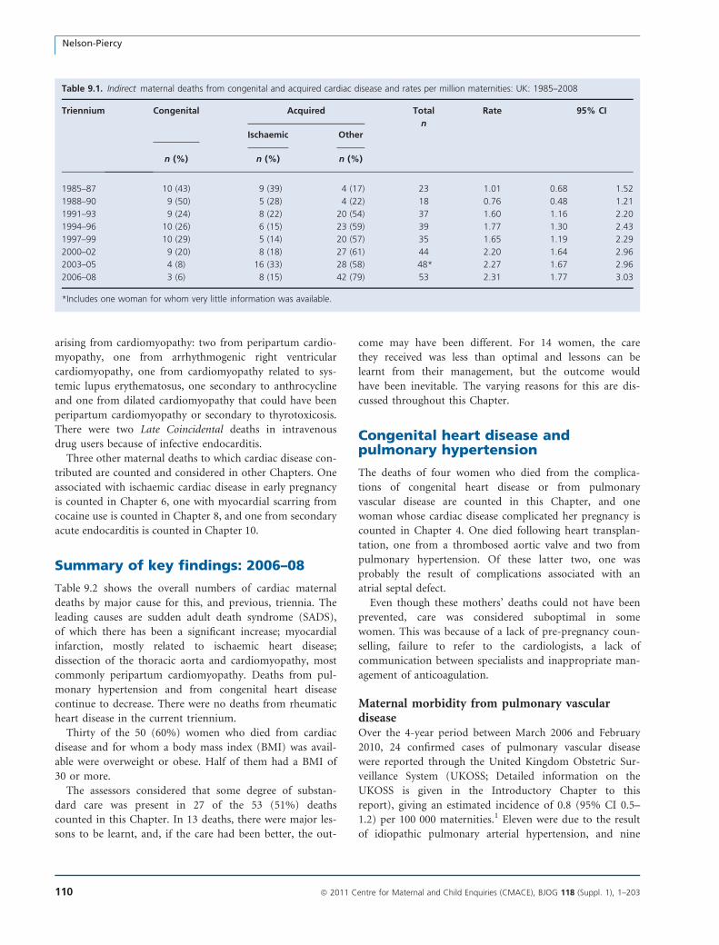
arising from cardiomyopathy: two from peripartum cardio-
myopathy, one from arrhythmogenic right ventricular
cardiomyopathy, one from cardiomyopathy related to sys-
temic lupus erythematosus, one secondary to anthrocycline
and one from dilated cardiomyopathy that could have been
peripartum cardiomyopathy or secondary to thyrotoxicosis.
There were two Late Coincidental deaths in intravenous
drug users because of infective endocarditis.
Three other maternal deaths to which cardiac disease con-
tributed are counted and considered in other Chapters. One
associated with ischaemic cardiac disease in early pregnancy
is counted in Chapter 6, one with myocardial scarring from
cocaine use is counted in Chapter 8, and one from secondary
acute endocarditis is counted in Chapter 10.
Summary of key findings: 2006–08
Table 9.2 shows the overall numbers of cardiac maternal
deaths by major cause for this, and previous, triennia. The
leading causes are sudden adult death syndrome (SADS),
of which there has been a significant increase; myocardial
infarction, mostly related to ischaemic heart disease;
dissection of the thoracic aorta and cardiomyopathy, most
commonly peripartum cardiomyopathy. Deaths from pul-
monary hypertension and from congenital heart disease
continue to decrease. There were no deaths from rheumatic
heart disease in the current triennium.
Thirty of the 50 (60%) women who died from cardiac
disease and for whom a body mass index (BMI) was avail-
able were overweight or obese. Half of them had a BMI of
30 or more.
The assessors considered that some degree of substan-
dard care was present in 27 of the 53 (51%) deaths
counted in this Chapter. In 13 deaths, there were major les-
sons to be learnt, and, if the care had been better, the out-
come may have been different. For 14 women, the care
they received was less than optimal and lessons can be
learnt from their management, but the outcome would
have been inevitable. The varying reasons for this are dis-
cussed throughout this Chapter.
Congenital heart disease andpulmonary hypertension
The deaths of four women who died from the complica-
tions of congenital heart disease or from pulmonary
vascular disease are counted in this Chapter, and one
woman whose cardiac disease complicated her pregnancy is
counted in Chapter 4. One died following heart transplan-
tation, one from a thrombosed aortic valve and two from
pulmonary hypertension. Of these latter two, one was
probably the result of complications associated with an
atrial septal defect.
Even though these mothers’ deaths could not have been
prevented, care was considered suboptimal in some
women. This was because of a lack of pre-pregnancy coun-
selling, failure to refer to the cardiologists, a lack of
communication between specialists and inappropriate man-
agement of anticoagulation.
Maternal morbidity from pulmonary vasculardiseaseOver the 4-year period between March 2006 and February
2010, 24 confirmed cases of pulmonary vascular disease
were reported through the United Kingdom Obstetric Sur-
veillance System (UKOSS; Detailed information on the
UKOSS is given in the Introductory Chapter to this
report), giving an estimated incidence of 0.8 (95% CI 0.5–
1.2) per 100 000 maternities.1 Eleven were due to the result
of idiopathic pulmonary arterial hypertension, and nine
Table 9.1. Indirect maternal deaths from congenital and acquired cardiac disease and rates per million maternities: UK: 1985–2008
Triennium Congenital Acquired Total
n
Rate 95% CI
Ischaemic Other
n (%) n (%) n (%)
1985–87 10 (43) 9 (39) 4 (17) 23 1.01 0.68 1.52
1988–90 9 (50) 5 (28) 4 (22) 18 0.76 0.48 1.21
1991–93 9 (24) 8 (22) 20 (54) 37 1.60 1.16 2.20
1994–96 10 (26) 6 (15) 23 (59) 39 1.77 1.30 2.43
1997–99 10 (29) 5 (14) 20 (57) 35 1.65 1.19 2.29
2000–02 9 (20) 8 (18) 27 (61) 44 2.20 1.64 2.96
2003–05 4 (8) 16 (33) 28 (58) 48* 2.27 1.67 2.96
2006–08 3 (6) 8 (15) 42 (79) 53 2.31 1.77 3.03
*Includes one woman for whom very little information was available.
Nelson-Piercy
110 ª 2011 Centre for Maternal and Child Enquiries (CMACE), BJOG 118 (Suppl. 1), 1–203
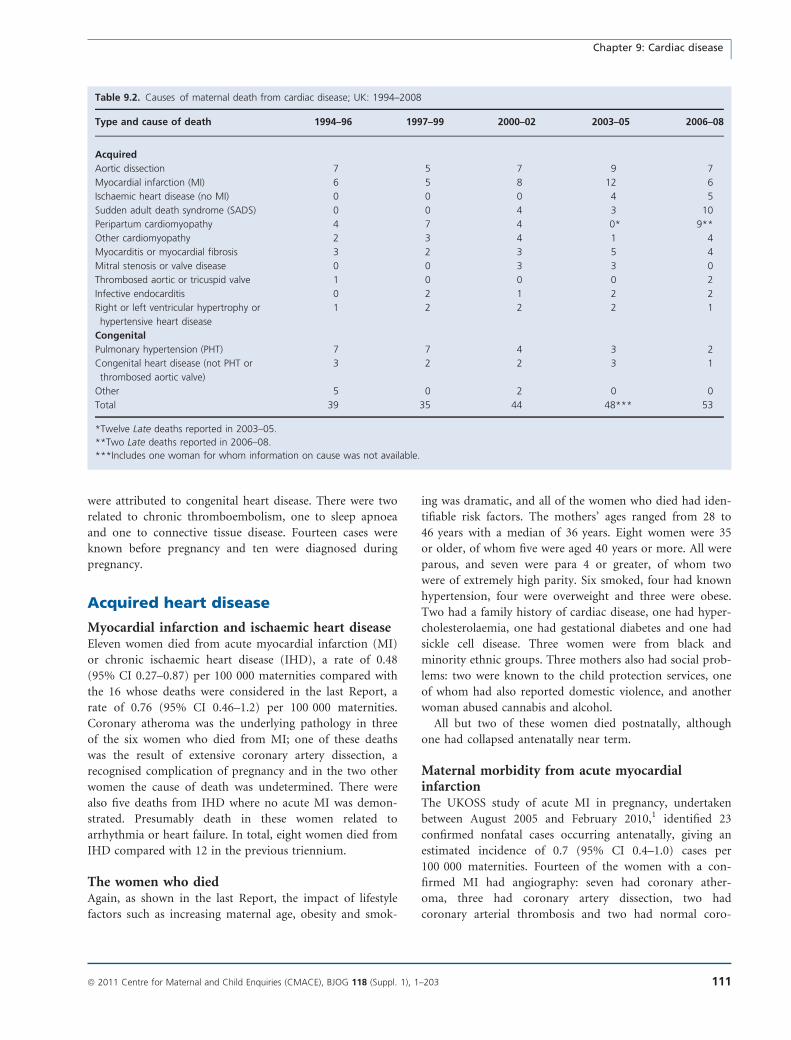
were attributed to congenital heart disease. There were two
related to chronic thromboembolism, one to sleep apnoea
and one to connective tissue disease. Fourteen cases were
known before pregnancy and ten were diagnosed during
pregnancy.
Acquired heart disease
Myocardial infarction and ischaemic heart diseaseEleven women died from acute myocardial infarction (MI)
or chronic ischaemic heart disease (IHD), a rate of 0.48
(95% CI 0.27–0.87) per 100 000 maternities compared with
the 16 whose deaths were considered in the last Report, a
rate of 0.76 (95% CI 0.46–1.2) per 100 000 maternities.
Coronary atheroma was the underlying pathology in three
of the six women who died from MI; one of these deaths
was the result of extensive coronary artery dissection, a
recognised complication of pregnancy and in the two other
women the cause of death was undetermined. There were
also five deaths from IHD where no acute MI was demon-
strated. Presumably death in these women related to
arrhythmia or heart failure. In total, eight women died from
IHD compared with 12 in the previous triennium.
The women who diedAgain, as shown in the last Report, the impact of lifestyle
factors such as increasing maternal age, obesity and smok-
ing was dramatic, and all of the women who died had iden-
tifiable risk factors. The mothers’ ages ranged from 28 to
46 years with a median of 36 years. Eight women were 35
or older, of whom five were aged 40 years or more. All were
parous, and seven were para 4 or greater, of whom two
were of extremely high parity. Six smoked, four had known
hypertension, four were overweight and three were obese.
Two had a family history of cardiac disease, one had hyper-
cholesterolaemia, one had gestational diabetes and one had
sickle cell disease. Three women were from black and
minority ethnic groups. Three mothers also had social prob-
lems: two were known to the child protection services, one
of whom had also reported domestic violence, and another
woman abused cannabis and alcohol.
All but two of these women died postnatally, although
one had collapsed antenatally near term.
Maternal morbidity from acute myocardialinfarctionThe UKOSS study of acute MI in pregnancy, undertaken
between August 2005 and February 2010,1 identified 23
confirmed nonfatal cases occurring antenatally, giving an
estimated incidence of 0.7 (95% CI 0.4–1.0) cases per
100 000 maternities. Fourteen of the women with a con-
firmed MI had angiography: seven had coronary ather-
oma, three had coronary artery dissection, two had
coronary arterial thrombosis and two had normal coro-
Table 9.2. Causes of maternal death from cardiac disease; UK: 1994–2008
Type and cause of death 1994–96 1997–99 2000–02 2003–05 2006–08
Acquired
Aortic dissection 7 5 7 9 7
Myocardial infarction (MI) 6 5 8 12 6
Ischaemic heart disease (no MI) 0 0 0 4 5
Sudden adult death syndrome (SADS) 0 0 4 3 10
Peripartum cardiomyopathy 4 7 4 0* 9**
Other cardiomyopathy 2 3 4 1 4
Myocarditis or myocardial fibrosis 3 2 3 5 4
Mitral stenosis or valve disease 0 0 3 3 0
Thrombosed aortic or tricuspid valve 1 0 0 0 2
Infective endocarditis 0 2 1 2 2
Right or left ventricular hypertrophy or
hypertensive heart disease
1 2 2 2 1
Congenital
Pulmonary hypertension (PHT) 7 7 4 3 2
Congenital heart disease (not PHT or
thrombosed aortic valve)
3 2 2 3 1
Other 5 0 2 0 0
Total 39 35 44 48*** 53
*Twelve Late deaths reported in 2003–05.
**Two Late deaths reported in 2006–08.
***Includes one woman for whom information on cause was not available.
Chapter 9: Cardiac disease
ª 2011 Centre for Maternal and Child Enquiries (CMACE), BJOG 118 (Suppl. 1), 1–203 111
nary arteries. In view of the increasing prevalence of risk
factors for IHD among women delivering in the UK and
the mortality noted in this report, this is likely to repre-
sent an underestimate of the true underlying burden of
nonfatal disease. An additional 17 cases were reported that
did not meet the criteria for MI during pregnancy,
including four women with a postnatal MI, one woman
whose MI occurred pre-pregnancy and two women with
severe angina.
Quality of careCare was considered substandard in five of the 11 deaths
(46%). As in the last Report, failure to consider acute coro-
nary syndrome (MI, which is subdivided into ST elevation
myocardial infarction [STEMI] and non-STEMI) as a cause
of chest pain—i.e. neck pain, jaw pain, left arm pain, nau-
sea or dizziness in women with risk factors—was a
repeated finding, and this led to a failure to perform
appropriate investigations. For example:
A woman who smoked presented to the Emergency Depart-
ment with chest pain and breathlessness some weeks after
delivery. Acute coronary syndrome was not considered in
the differential diagnosis, possibly in view of a normal
ECG, her age and the quality of the pain. Serial ECGs and
Troponin were not requested, and, even though the working
diagnosis was pulmonary embolus, a V/Q scan was not per-
formed. She was discharged from the Clinical Decision Unit
and died shortly afterwards.
Another woman with risk factors for IHD presented during
pregnancy with chest pain. An ECG showed clear changes
of ischaemia, and the Troponin level was raised. However,
a normal echocardiogram falsely reassured the cardiologists,
who did not investigate her symptoms further. Some days
later she complained of heartburn, vomiting and dizziness
and died of myocardial ischaemia.
ECGs can be normal between bouts of angina or chest
pain. Therefore, a single normal ECG does not exclude
ischaemia, and serial ECGs and Troponin should form part
of the investigations for chest pain. Although the absence
of regional wall abnormality on an echocardiogram
excludes a full thickness STEMI, a normal echocardiogram
does not exclude non-STEMI. It is possible that the cardi-
ologists were reluctant to perform a coronary angiogram
because of the pregnancy.
Myocardial infarction and ischaemicheart disease: learning points
Ischaemic heart disease has now become a common car-
diac cause of death in pregnancy.
Current trends in lifestyle factors, including smoking,
and increasing age at childbirth, are likely to be contrib-
uting to this increase in incidence. All the women who
died had identifiable risk factors including:
• obesity
• age >35 years
• parity >3
• smoking
• diabetes
• pre-existing hypertension
• family history.
Myocardial infarction and acute coronary syndrome can
present with atypical features in pregnancy such as
abdominal or epigastric pain and vomiting or dizziness.
A single normal ECG does not exclude ischaemia, espe-
cially if performed when the woman is pain-free. There
should be a low threshold for further investigation (such
as serial ECGs and Troponin) of ischaemic-sounding
chest pain, especially in women with known risk factors.
There should also be a low threshold for emergency coro-
nary intervention (coronary angiogram with angioplasty
with or without stenting), which will allow treatment of
both acute atheromatous coronary occlusion and the less
common coronary artery dissection with stenting.
Aortic dissectionAortic dissection was, again, another major cause of cardiac
death. Seven women died from aortic dissection compared
with nine and seven in the two previous triennia, respec-
tively. The ages of the women ranged between 26 and
38 years with a median of 34 years. The majority of moth-
ers died within a few days after childbirth, although one
had suffered the dissection in late pregnancy, and the oth-
ers during their third trimester. Two were overweight, and
another two were obese. The dissection was type A
(ascending aorta) in five women and type B (descending
aorta) in two. One woman had Ehlers–Danlos syndrome
type IV with some features of Marfan syndrome, one had a
bicuspid aortic valve and another had a family history of
dissection. Other risk factors included pre-eclampsia, twins
and hypertension.
Care was substandard in five of the seven women (71%).
In four there was failure to investigate chest pain. For
example:
A woman presented during pregnancy with what was
thought to be gastritis because of vomiting and diarrhoea.
She also described constant sternal pain for which she
required opiate analgesia, but, despite the severity of the
pain, no further investigations were undertaken. A few days
later, after delivery, she complained of dizziness, was found
to be anaemic and was transfused, again without an
Nelson-Piercy
112 ª 2011 Centre for Maternal and Child Enquiries (CMACE), BJOG 118 (Suppl. 1), 1–203
underlying diagnosis being made. She then complained of
chest tightness and collapsed with an asystolic arrest, pre-
sumably when the dissection she had suffered late in preg-
nancy extended.
Another woman who complained of sudden onset chest
pain was not investigated further or referred to a physician.
After delivery, she was nursed in a low-dependency area
despite severe ongoing symptoms, and the midwife noted a
delay of more than 2 hours before medical staff attended to
reassess her after her condition had deteriorated. Her cardio-
megaly was missed on the chest X-ray, and, despite the rec-
ommendations in the last Report, a woman with severe
chest pain caused by dissection was given repeated doses of
opiates without further urgent investigation.
As recommended in several earlier Reports, and reinforced
here, chest pain severe enough to require opiate analgesia
mandates further investigation for an underlying cause.
Aortic dissection: learning points
Not all chest pain in pregnancy is the result of pulmo-
nary emboli.
Aortic dissection is a rare but potentially fatal cause of
chest or interscapular pain in pregnancy, particularly in
the presence of systolic hypertension, and must be con-
sidered as part of the differential diagnosis of chest
pain.
Women with Marfan syndrome are at high risk of aortic
dissection, but previously apparently normal pregnant
women may also suffer this complication, most com-
monly at or near term, or postnatally.
Women with severe chest or abdominal pain requiring
opiate analgesia must be investigated. Appropriate
imaging includes a computed tomography chest scan
or magnetic resonance imaging, or a transthoracic or
transoesophageal echocardiogram.
Other acquired cardiac disease
CardiomyopathyThirteen women died from cardiomyopathy. In six of them
this was attributed to peripartum cardiomyopathy, and a
further three deaths were stated to have been the result of
dilated cardiomyopathy where the diagnosis could have
been peripartum cardiomyopathy. In addition to these nine
probable deaths from peripartum cardiomyopathy, four
other women died from cardiomyopathy from other causes.
Two were from dilated cardiomyopathy, one from right
ventricular myocardial fibrosis secondary to myotonic dys-
trophy and another from arrhythmogenic right ventricular
cardiomyopathy (ARVC). Risk factors included pre-
eclampsia, obesity and hypertension.
An additional six women are classified as having died of
Late Indirect peripartum cardiomyopathy some weeks after
delivery. These deaths are counted in Chapter 12. Although
these women are classified as Late deaths, two presented
antenatally with a known diagnosis of cardiomyopathy, one
shortly after delivery and the other three several weeks
postpartum. Two died from cardiomyopathy related to
acute immune diseases, and another suffered a dilated car-
diomyopathy secondary to chemotherapy. There was one
further Late death from ARVC.
It is important to remember that peripartum cardiomy-
opathy may present up to 5–6 months postpartum but is
still directly related to the pregnancy. Similarly, it may
present antenatally or in the puerperium, but death may
occur many months later.
A recent US study2 of over 14 million pregnancy hospi-
talisations in 2004–06, found a prevalence of hospitalisa-
tions with cardiomyopathy of 0.46 per 1000 deliveries (0.18
for apparent peripartum cardiomyopathy and 0.28 for
other cardiomyopathies).
The women who diedThe age range of the women who died was between 17 and
38 years with a median of 26 years. Ten of the 11 women,
nine Indirect and two Late, who died of definite or proba-
ble peripartum cardiomyopathy were parous. Five women
were overweight, of whom one was obese. Of these 11
women, four presented antenatally, three in the third tri-
mester and one who died in early pregnancy and who had
had another child only a few months earlier.
Most women had risk factors, including two with previ-
ous peripartum cardiomyopathy. Three were over the age
of 35, five had hypertension (pre-existing, pregnancy-
induced hypertension or pre-eclampsia), six were smokers
and three were from Black ethnic backgrounds.
In total, the assessors regarded the care of seven of the
13 (54%) women who died from cardiomyopathy as sub-
standard. For example:
A woman with a previous history of peripartum cardiomy-
opathy was not counselled by her cardiologists about the
risk of recurrence. In her next pregnancy, despite numerous
mentions of palpitations, she was not referred back to a car-
diologist. When she was eventually referred as a matter of
urgency, near term, the consultant cardiologist refused to
see her and said in the maternal death reporting form that
the obstetric registrar did not mention the previous left ven-
tricular dysfunction. She then self-referred to an Emergency
Department, where she was eventually seen by a cardiology
registrar. The registrar misinterpreted the echocardiogram
as showing no significant dysfunction, not realising that a
Chapter 9: Cardiac disease
ª 2011 Centre for Maternal and Child Enquiries (CMACE), BJOG 118 (Suppl. 1), 1–203 113
normal left ventricle should be hyperdynamic in late preg-
nancy. A consultant cardiologist who reviewed the echocar-
diogram after her death said it showed ‘at least moderate’
left ventricular dysfunction.
A young woman who booked very late, and was a smoker,
complained of breathlessness in the third trimester, which
her obstetrician assumed to be the result of anaemia. She
was also very breathless, tachypnoeic and tachycardic when
she presented a few weeks later in preterm labour. When she
was examined, her wheeze was not recognised as a possible
sign of pulmonary oedema and no investigations were
undertaken nor any thought given to alternative diagnoses
other than chest infection. All attention was focused on the
management of her labour, despite the fact that she
remained tachycardic and tachypnoeic. Following a period
of loss of consciousness, arterial blood gases were measured,
but the very abnormal base excess was not recognised or
noticed or appreciated as abnormal or significant. She deliv-
ered normally and then collapsed, but this was a gradual
demise as the result of worsening acidosis and unrecognised
heart failure. Autopsy showed a large heart.
Women with known cardiomyopathy need pre-preg-
nancy counselling about the risk of recurrence/deteriora-
tion. It is crucial to refer women with previous
cardiomyopathy to specialist units with joint clinics where
both the obstetricians and cardiologists are familiar with
the expected changes of pregnancy. These mothers need
serial echocardiograms in pregnancy, especially if their
baseline echocardiograms in early pregnancy are abnormal.
In some women who died from cardiomyopathy, there
was a failure to diagnose pulmonary oedema, failure to
monitor women in a high-dependency area, failure to
repeat echocardiograms and failure to provide a cardiac
review to women who should have had one. There were
delays recognising worsening heart failure postpartum. In
several women, symptoms and signs were wrongly attrib-
uted to asthma (because of wheeze), anaemia, panic attacks
or chest infection.
Missing pulmonary oedema and missing acidosis because
of ignoring tachypnoea and a large base excess are common
mistakes in maternal cardiac deaths.
Peripartum cardiomyopathy: learningpoints
Women in late pregnancy or within 6 months of delivery
with symptoms of breathlessness, oedema or orthopnoea
and the signs of tachypnoea and tachycardia may have
peripartum cardiomyopathy, and investigation with a
chest X-ray and an echocardiogram are indicated.
Women with peripartum cardiomyopathy should be
managed by cardiologists with expertise in this condi-
tion and their care should be discussed, if appropriate,
with the regional cardiac transplant centre.
Women with pre-existing cardiomyopathy of whatever
origin must, like all women with heart disease, have
expert and informed pre-pregnancy counselling.
Deaths from other causesFour women died from myocarditis. In two women the
myocarditis was thought to be viral and in one it was
related to an autoimmune disease. In a case of essential
hypertension:
A morbidly obese woman who lived between several coun-
tries booked late despite a very poor obstetric history and
known hypertension. She was poorly compliant with her
medications as well as attending for care. She only had two
antenatal visits, neither of which was in a hospital antena-
tal clinic. Her antihypertensive medications were changed
by doctors in different countries. She collapsed and died in
her third trimester after coughing up blood and foaming at
the mouth. The autopsy was substandard, but her heart
was dilated. In this context of poorly controlled severe
hypertension, the likely diagnosis was of ‘flash’ pulmonary
oedema and hypertensive heart failure, perhaps related to
diastolic dysfunction.
This difficult case represents many of the challenges fac-
ing those who work in maternity services. This mother was
extremely high risk, highly mobile and difficult to engage
in care. It highlights the need for every effort to be made
to ensure that women at risk are assessed urgently by an
obstetrician providing care for high-risk mothers and that
their hypertension is controlled, if necessary by admission
to hospital.
Infective endocarditisTwo women died from infective endocarditis, and a further
two died Late Coincidental deaths much later after delivery.
All were known to abuse drugs, three of the four intrave-
nously. In one woman the organism was a staphylococcus,
in one it was Streptococcus mutans, in one it was a Gram-
positive coccus and in the fourth no organism was identi-
fied. The following is a representative case:
A known intravenous drug user presented to the Emergency
Department with a possible deep vein thrombosis (DVT)
during pregnancy. She was allowed home after promising to
return but did not do so until a week later with worsening
symptoms. She was not admitted but referred to an outpa-
tient DVT clinic, and the results of her arterial blood gases,
showing acidosis and increased lactate, were missed. She
Nelson-Piercy
114 ª 2011 Centre for Maternal and Child Enquiries (CMACE), BJOG 118 (Suppl. 1), 1–203
returned later the same day with chest pain and eventually
was diagnosed as having sepsis and infective endocarditis,
from which she died. Her autopsy showed tricuspid valve
vegetations caused by a Gram-positive coccus, but no men-
tion was made of a DVT.
A pregnant intravenous drug user in these circumstances
should have been admitted to establish the presumed diag-
nosis of DVT and to commence treatment. Arterial blood
gases are often performed when looking for hypoxia if pul-
monary embolism is suspected, but a metabolic acidosis
signifies underlying pathology and should raise the suspi-
cion of underlying sepsis.
Thrombosed mechanical valvesTwo women died from thrombosed mechanical valves, a
rare cause of cardiac death. One was a known intrave-
nous drug user and the other had a congenital cardiac
condition. Both women were converted to low-molecu-
lar-weight heparin (LMWH) in their pregnancies, and
there were issues with compliance. Recent data highlight
the importance of adequate doses of LMWH and the
need for good compliance and anti-Xa monitoring when
managing women with mechanical heart valves in
pregnancy.3
Sudden adult/arrhythmic death syndromeThe deaths of ten women who died from SADS are dis-
cussed from a pathological point of view in the Annex to
this Chapter on the pathological overview of maternal
deaths. This is because SADS is a pathological diagnosis
and extra attention needs to be paid to these women given
the rise in the number of deaths from this cause in this tri-
ennium.
Conclusion
Deaths from heart disease remain the leading cause of
maternal death. The commonest causes are cardiomyopa-
thy, myocardial infarction/ischaemic heart disease and aor-
tic dissection. Regrettably, clinicians are still failing to
consider heart disease as a possible cause for symptoms
and signs in pregnancy and therefore fail to perform the
appropriate investigations. Some women with known heart
disease before pregnancy are not offered or referred to
appropriate multidisciplinary care in specialist units.
Disclosure of interestsCatherine Nelson-Piercy received fees for speaking and
consultancy from Sanofi Aventis and Leo-Pharma and she
is the Editor-in-Chief of the journal Obstetric Medicine.
FundingThis work was undertaken by the Centre for Maternal and
Child Enquiries (CMACE) as part of the CEMACH pro-
gramme. The work was funded by the National Patient
Safety Agency; the Department of Health, Social Services
and Public Safety of Northern Ireland; NHS Quality
Improvement Scotland (NHS QIS); and the Channel
Islands and Isle of Man.
AcknowledgementsThis Chapter has been seen and discussed with Dr Sara
Thorne, Consultant Cardiologist, Birmingham, and Dr
Dawn Adamson, Consultant Cardiologist University Hospi-
tal of Coventry and Warwickshire. j
References
1 Knight M, Spark P, Pierce M, Kayem G, Kurinczuk JJ, Brocklehurst P,
et al. United Kingdom Obstetric Surveillance System (UKOSS) Annual
Report 2010. Oxford: National Perinatal Epidemiology Unit, 2010.
2 Kuklina EV, Callaghan WM. Cardiomyopathy and other myocardial
disorders among hospitalizations for pregnancy in the United States:
2004–2006. Obstet Gynecol 2010;155:93–100.
3 McClintock C. Use of therapeutic dose low molecular weight heparin
during pregnancy in women with mechanical heart valves. Obstet
Med 2010;3:40–2.
ª 2011 Centre for Maternal and Child Enquiries (CMACE), BJOG 118 (Suppl. 1), 1–203 115
Chapter 9: Cardiac disease
Annex 9.1. Pathological overview of cardiacdeaths including sudden adult/arrhythmic deathsyndrome (SADS)S Lucas (on behalf of the Centre for Maternal and Child Enquiries)
St Thomas’ Hospital, London, UK
Correspondence: Dr Sebastian Lucas, St Thomas’ Hospital, 2nd Floor, North Wing, London SE1 7EH, UK. Email: [email protected]
Cardiac deaths: pathology goodpractice points
• Do the heart examination well, with full dissection of
the coronary arteries, accurate weight of the heart,
measurement of the ventricle thicknesses and careful
evaluation of the valves.
• Correlate these data with the woman’s body weight
and body mass index (BMI).
• Do histology on a standard mid-horizontal slice of
heart muscle, to examine the right and left ventricles.
• Look at the lung histopathology for clues (e.g. pul-
monary hypertension).
• Retain a blood sample in case cocaine and other
stimulant drug analysis is necessary.
• Consider consulting a cardiac pathologist to gain
advice and support for the diagnosis.
• If the diagnosis is going to be SADS, make sure all
other reasonable cardiac diagnoses, as well as preg-
nancy-related diagnoses (such as amniotic fluid
embolism), are excluded.
• If SADS or a standard cardiomyopathy seems to be
the diagnosis, retain a sample of frozen spleen for
possible later genetic evaluation; the coroner will not
be expected to resource such investigations, but the
NHS cardiac genetic clinics will.
The main trends in cardiovascular pathology for this trien-
nium are the reduction in deaths associated with congenital
heart disease and the apparent rise in incidence of
SADS. This is better described as sudden arrhythmic death
syndrome where unexpected and unpredicted cardiac arrest
occurs and it is discussed later.
Cardiac pathology is what pathologists should do well, as
it is the commonest scenario of death they encounter in
routine practice in the UK. Broadly, for coronary ischaemic
heart disease, infective endocarditis, pulmonary hyperten-
sion and congenital heart disease and for dissection of
aorta and other vessels, this was true in the maternal deaths
reviewed this triennium. But it was not the case for diffi-
cult, non-obvious cases where acute cardiac failure was the
scenario.
Ischaemic heart disease
In the deaths examined, the usual patterns were seen of
coronary artery atheroma, with or without acute thrombo-
sis, and cardiac muscle damage, as either acute infarction
or chronic fibrotic ischaemic damage.
Two women had different specific causes of coronary
artery occlusion. One had postpartum dissection of the left
anterior coronary artery causing myocardial infarction. It is
remarkable that pregnancy is the major risk factor for such
dissection, albeit uncommon. It is presumed that the
hormonal effect on arterial muscle tone interacts on an
in-built medial damage to cause it. From the practical per-
spective, pathologists should note that, without very close
gross inspection, a dissection of a coronary artery can look
very like an acute thrombosis; histology is useful here to
confirm the process.
The other woman was an adolescent who presented sev-
eral months after pregnancy with myocardial infarction.
The aetiology was a rare autoimmune disease (Kawasaki
disease) with vasculitis of the coronary arteries and second-
ary stenosis and occlusion.
Dissection of the aorta
The seven women who died from dissection of the aorta had
autopsies. Not surprisingly, most were well done, although
three reports had no height or weight (therefore no autopsy
BMI was calculable). Although the gross pathology is self-
evident, it is arguable whether histopathology is required to
Lucas
116 ª 2011 Centre for Maternal and Child Enquiries (CMACE), BJOG 118 (Suppl. 1), 1–203
demonstrate the universal underlying aortic medial degener-
ation, but best practice is that it should be done. More
important are co-pathologies relevant to the final dissection:
two of the seven women had clinical pre-eclampsia, but the
renal histopathology (glomerular endotheliosis) was not
documented to confirm this.
None of the autopsy reports indicated whether genetic
studies of the fibrillin connective tissue gene were per-
formed to demonstrate Marfan and Ehlers–Danlos syn-
dromes if they had not already been evaluated. Essentially,
in the age-range of pregnant women with dissection of
the aorta, the pathogeneses are three-fold: inherited con-
nective tissue disease (e.g. Marfan); bicuspid aortic valve
association (no fibrillin abnormality); and idiopathic. Best
practice is to reserve spleen material in the freezer for
later DNA studies in all women with dissection of the
aorta who have normal aortic valves. Importantly, it is
not for coroners’ and fiscals’ local authorities to fund
such studies of potentially inheritable disease, it is for the
NHS.
Myocarditis and systemic lupuserythematosus
In the women for whom an autopsy took place, the
actual aetiology of the myocarditis was not established.
The causes include infection (usually enteroviral), drug
allergy (e.g. antibiotics), named immunopathological con-
ditions such as eosinophilic or giant cell myocarditis and
sarcoidosis, and idiopathic. Inevitably, there is potential
for overlap with peripartum cardiomyopathy, because in
some of those cases there is a myocarditis, and the clini-
cal and pathological case definitions need to be closely
observed. For myocarditis pathology, the Dallas criteria
apply: chronic inflammation (T-cell) in the myocardium
with active destruction of myofibres, i.e. not just an
interstitial inflammation which can happen in many sys-
temic inflammatory response states. A further difficulty
for pathologists is that the diagnosis of myocarditis is
not usually entertained until the fixed tissue histopathol-
ogy is available, by which time the opportunity for tak-
ing unfixed heart and blood samples for virology and
serodiagnosis is usually lost. Perhaps polymerase chain
reaction technology will become more available for the
retrospective diagnosis of viral myocarditis on fixed tis-
sue.
Peripartum and othercardiomyopathies
This is a confusing area of cardiac pathology, where only a
full cardiac examination protocol, in conjunction with clin-
ical data and family history, will produce the correct diag-
noses. Of the non-peripartum cardiomyopathies (PPCM),
two were arrhythmogenic right ventricular cardiomyopathy
(ARVCM). There is no known aetiological association of
ARVCM, which is partly inherited, with pregnancy. One
woman with myotonic dystrophy died of heart failure
immediately postpartum; again only an experienced cardiac
pathologist can really interpret the positive and negative
findings.
Only six of the nine women who died from PPCM had
an autopsy. The case definition of PPCM is the onset of
left ventricular systolic dysfunction and symptoms of heart
failure during the last month of pregnancy or the first
5 months postpartum. Sudden unexpected cardiac death
without previous heart failure is usually excluded. There
are characteristic features pathologically—enlarged heart,
dilated cardiomyopathy, irregular myofibres and fibrosis,
variable T-cell infiltration, on histopathology—and, criti-
cally, the absence of an alternative diagnosis. This is a
high-stringency definition and for autopsy pathologists pre-
sents the problem of how far to investigate cases of acute
or chronic heart failure during and after pregnancy, to sup-
port or exclude a clinical diagnosis of PPCM.
In two autopsies, the pathologists referred the case to a
cardiac pathologist for opinion, and drug screens (mainly
for stimulant drugs such as cocaine) were performed in
another two. One pathologist evidently had never heard of
the entity and, following a good autopsy that clearly sup-
ported PPCM, declared the ‘cause of the cardiac enlarge-
ment and failure’ to be ‘obscure’.
The pathogenesis of PPCM is unknown, although a non-
infective pregnancy-associated immune activation myocar-
ditis with destruction of myofibres is the current leading
hypothesis.1 The role of cardiac biopsy in diagnosing and
managing possible PPCM is underdeveloped; none of the
nine women in this triennium had one, although in most
cases there was the opportunity in hospitalised women to
attempt it.
Sudden adult/arrhythmic deathsyndrome
Ten women died of SADS during this triennium compared
with three in the last Report. This is greater than in any
previous Report. Six of them were obese, with booking
BMI ranging from 30 to 45.
The case definition for SADS is a sudden unexpected
cardiac death (i.e. presumed fatal arrhythmia) where all
other causes of sudden collapse are excluded, including a
drug screen for stimulant drugs such as cocaine. These
are exacting criteria. The UK Cardiac Pathology Network
(www.cpn.org.uk) is encouraging fuller autopsy examina-
tions (and reporting to a central database) and provides a
comprehensive list of possible diagnoses for sudden car-
Annex 9.1: Pathological overview of cardiac deaths
ª 2011 Centre for Maternal and Child Enquiries (CMACE), BJOG 118 (Suppl. 1), 1–203 117
diac death that is non-ischaemic (atheroma) and non-val-
vular (with the exception of mitral valve mucoid degener-
ation) in causation. The classification is divided into
those where the death is explained on gross and histologi-
cal criteria and those where it remains unexplained at our
current state of knowledge. PPCM, ARVCM, myocarditis,
left ventricular hypertrophy and fibrosis, and hypertensive
heart disease are ‘explained’; unexplained cardiac death in
epilepsy, alcohol abuse, obesity and diabetes are ‘unex-
plained’.
Under the SADS label come those unexplained cases
with a morphologically normal heart by gross examination
and histology. A proportion can be demonstrated by elect-
rographic examination of relatives (and sometimes of the
woman before death) to have a conduction defect, a chan-
nelopathy, e.g. long QT syndrome. In this triennium, no
deaths were ascribed to a specific arrhythmia, but that
probably reflects the imperfect evaluation of them.
SADS, cardiac hypertrophy andobesity in pregnancy
This is the area of cardiac death evaluated least well histori-
cally. In Chapters 10 and 17, the sections on unascertained
causes of death indicate that some of these might be SADS
if better clinicopathological information had been forth-
coming. Further, in Chapter 9, the cases described as being
the result of ‘other causes, hypertrophy and fibrosis’ are
probably SADS cases, of which there are up to six. In these
cases, the autopsy was not comprehensive enough to enable
the assessors to allocate the case to a particular cardiac cat-
egory. Four of the women had a BMI ‡30.
Here we group together the women who died of sudden
unexpected cardiac death who were not hypertensive during
pregnancy (or who were well treated) and did not have one
of the ‘explained’ cardiac pathologies. It includes those who
were obese and those with otherwise unexplained left ven-
tricular hypertrophy. The reason for this grouping is to high-
light:
• the likelihood that pregnancy presents this scenario more
frequently than is seen in the general population due to
the physiological stresses of pregnancy and delivery bring-
ing out an underlying potential cardiac arrhythmia
• the crucial need for more comprehensive data collection
and better categorisation to improve knowledge of the
epidemiology of SADS.
It is established that obesity and cardiac hypertrophy are
associated in the absence of concurrent hypertension, and
that obesity and cardiac hypertrophy are both risk factors
for arrhythmia and sudden cardiac death. Recently, the
notion of ‘obesity cardiomyopathy’ is gaining ground,2 and
it applies particularly to those with a central versus a
peripheral pattern of obesity. Such women have large
hearts, and in the ten women with SADS, the median heart
weight was 390 g (range 270–585 g). Of the heart to body
weight ratios, seven of the ten were >0.42% of body
weight, compared with the normal mean of 0.40% (range
0.38–0.42).3
In future, pathologists reviewing such cases must docu-
ment optimally the morphology of the body (height,
weight, BMI, pattern of obesity) as well as that of the heart
so that closer analysis can be undertaken. The heart mor-
phology required is listed in Best Practice points (above).
The published European guidelines on examination of the
heart provide the necessary information.4
The fact that pregnancy makes the obesity–heart issue
complicated, because of the greatly altered vascular physiol-
ogy and the introduction of novel pathologies such as pre-
eclampsia, is shown by the following case study.
An obese woman, previously normotensive, was admitted
during her third trimester with a blood pressure of 170/
108 mmHg and proteinuria. Despite antihypertensive treat-
ment, her blood pressure rose further, and she developed
flash pulmonary oedema with breathlessness, followed by
cardiac arrest. A peri-mortem caesarean section resulted in
a live birth. At autopsy, her heart weighed 585 g, with left
ventricular hypertrophy of 2.5 cm lateral wall thickness.
Histopathology showed cardiac myofibre hypertrophy and
mild interstitial fibrosis; the renal glomeruli had the endo-
theliosis lesion of pre-eclampsia. The conclusion on causa-
tion was a combination of obesity-related heart hypertrophy
plus pre-eclampsia.
Ultimately, these deaths are individually unpredictable,
and obesity needs to be addressed as a public health issue
if the fatalities are not to rise further in the next report. j
References
1 Silwa K, Fett J, Elkayam U. Peripartum cardiomyopathy. Lancet
2006;368:687–93.
2 Duflou J, Virmani R, Rabin I, Burke A, Farb A, Smialek J. Sudden
death as a result of heart disease in morbid obesity. Am Heart J
1995;130:306–13.
3 Smith HL. The relation of the weight of the heart to the weight of
the body and of the weight of the heart to age. Am Heart J
1928;4:79–93.
4 Basso C, Burke M, Fornes P, Gallagher PJ, de Gouveia RH, Sheppard
M, et al. Guidelines for autopsy investigation of sudden cardiac death.
Virchows Arch 2008;452:11–8.
118 ª 2011 Centre for Maternal and Child Enquiries (CMACE), BJOG 118 (Suppl. 1), 1–203
Lucas
Chapter 10: Other Indirect deathsM de Swiet1, C Williamson2, G Lewis3 (on behalf of the Centre for Maternal and Child Enquiries)1 Emeritus Professor of Obstetric Medicine, Imperial College London, London, UK; 2 Institute of Reproductive Developmental Biology,
Imperial College London, London, UK; 3 National Perinatal Epidemiology Unit (NPEU), University of Oxford, Oxford, UK
Correspondence: Professor Michael de Swiet, Flat 15, Wren View, 75 Hornsey Lane, London N6 5LH, UK. Email: [email protected]
Keywords risk, Confidential Enquiry, maternal, mortality.
Other Indirect deaths: specific recommendations
• All women who are planning pregnancies that are likely to be complicated by potentially serious medical conditions
should have pre-pregnancy counselling.
• Women whose pregnancies are complicated by potentially serious medical conditions should be referred to appropri-
ate specialist centres of expertise where both care for their medical condition and their obstetric care can be opti-
mised.
• Lack of consultant involvement remains a problem in the care of women with serious medical problems. Maternity
units should consider developing protocols to specify which medical conditions mandate consultant review.
• Health workers who are caring for women in pregnancy with conditions with which they are unfamiliar should con-
sult experts. If necessary this consultation can be by telephone and most experts are only too pleased to help.
• Medical conditions that may be the cause of symptoms that are more commonly seen by obstetricians should not be
ignored, e.g. seizures can be caused by epilepsy as well as eclampsia.
• Multiple admissions or attendances for emergency care demand further investigation and are often an indication for
referral to specialists in other disciplines.
• Undiagnosed pain requiring opiates demands immediate consultant involvement and investigation.
• The need remains for physicians who do not work directly with pregnant women to know more about the interaction
between the conditions that they are treating and pregnancy.
• Appropriate and professional interpretation services must be made available to women who do not speak English.
Other Indirect deaths: learning points
Most women with epilepsy require an increased lamotri-
gine dose in pregnancy to maintain good seizure con-
trol. Clinicians should adjust their management
protocols accordingly.
Women with epilepsy or undiagnosed syncope are still
unaware of the very rare but real risk of drowning while
bathing unattended. A shower is preferable and the
bathroom door should remain unlocked.
There should be no hesitation in arranging a chest X-
ray for women with significant chest symptoms. Simi-
larly, magnetic resonance imaging and computed
tomography of the brain can be used to exclude cerebral
pathology.
Clinicians should be aware that haemoglobin SC disease
can cause sickle cell crisis and is as dangerous as haemo-
globin SS disease.
Women should be given advice about sexual intercourse
in the postnatal period as fatal air embolism has been
reported as a result of this. The medical assessors to this
Enquiry recommend abstinence for 6 weeks, or gentle
intercourse and avoidance of positions where excess air
could be forced into the vagina.
Introduction
Indirect maternal deaths are defined as deaths resulting
from previously existing disease or diseases that develop
during pregnancy and which do not have direct obstetric
Chapter 10: Other Indirect deaths
ª 2011 Centre for Maternal and Child Enquiries (CMACE), BJOG 118 (Suppl. 1), 1–203 119
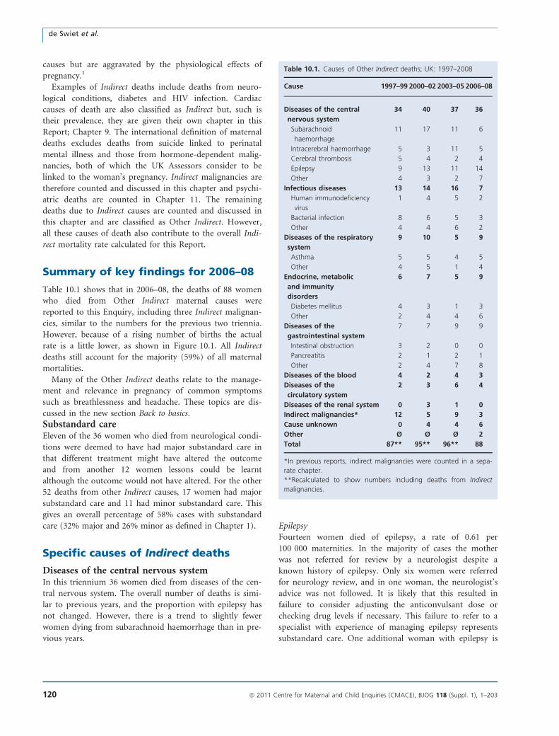
causes but are aggravated by the physiological effects of
pregnancy.1
Examples of Indirect deaths include deaths from neuro-
logical conditions, diabetes and HIV infection. Cardiac
causes of death are also classified as Indirect but, such is
their prevalence, they are given their own chapter in this
Report; Chapter 9. The international definition of maternal
deaths excludes deaths from suicide linked to perinatal
mental illness and those from hormone-dependent malig-
nancies, both of which the UK Assessors consider to be
linked to the woman’s pregnancy. Indirect malignancies are
therefore counted and discussed in this chapter and psychi-
atric deaths are counted in Chapter 11. The remaining
deaths due to Indirect causes are counted and discussed in
this chapter and are classified as Other Indirect. However,
all these causes of death also contribute to the overall Indi-
rect mortality rate calculated for this Report.
Summary of key findings for 2006–08
Table 10.1 shows that in 2006–08, the deaths of 88 women
who died from Other Indirect maternal causes were
reported to this Enquiry, including three Indirect malignan-
cies, similar to the numbers for the previous two triennia.
However, because of a rising number of births the actual
rate is a little lower, as shown in Figure 10.1. All Indirect
deaths still account for the majority (59%) of all maternal
mortalities.
Many of the Other Indirect deaths relate to the manage-
ment and relevance in pregnancy of common symptoms
such as breathlessness and headache. These topics are dis-
cussed in the new section Back to basics.
Substandard careEleven of the 36 women who died from neurological condi-
tions were deemed to have had major substandard care in
that different treatment might have altered the outcome
and from another 12 women lessons could be learnt
although the outcome would not have altered. For the other
52 deaths from other Indirect causes, 17 women had major
substandard care and 11 had minor substandard care. This
gives an overall percentage of 58% cases with substandard
care (32% major and 26% minor as defined in Chapter 1).
Specific causes of Indirect deaths
Diseases of the central nervous systemIn this triennium 36 women died from diseases of the cen-
tral nervous system. The overall number of deaths is simi-
lar to previous years, and the proportion with epilepsy has
not changed. However, there is a trend to slightly fewer
women dying from subarachnoid haemorrhage than in pre-
vious years.
EpilepsyFourteen women died of epilepsy, a rate of 0.61 per
100 000 maternities. In the majority of cases the mother
was not referred for review by a neurologist despite a
known history of epilepsy. Only six women were referred
for neurology review, and in one woman, the neurologist’s
advice was not followed. It is likely that this resulted in
failure to consider adjusting the anticonvulsant dose or
checking drug levels if necessary. This failure to refer to a
specialist with experience of managing epilepsy represents
substandard care. One additional woman with epilepsy is
Table 10.1. Causes of Other Indirect deaths; UK: 1997–2008
Cause 1997–99 2000–02 2003–05 2006–08
Diseases of the central
nervous system
34 40 37 36
Subarachnoid
haemorrhage
11 17 11 6
Intracerebral haemorrhage 5 3 11 5
Cerebral thrombosis 5 4 2 4
Epilepsy 9 13 11 14
Other 4 3 2 7
Infectious diseases 13 14 16 7
Human immunodeficiency
virus
1 4 5 2
Bacterial infection 8 6 5 3
Other 4 4 6 2
Diseases of the respiratory
system
9 10 5 9
Asthma 5 5 4 5
Other 4 5 1 4
Endocrine, metabolic
and immunity
disorders
6 7 5 9
Diabetes mellitus 4 3 1 3
Other 2 4 4 6
Diseases of the
gastrointestinal system
7 7 9 9
Intestinal obstruction 3 2 0 0
Pancreatitis 2 1 2 1
Other 2 4 7 8
Diseases of the blood 4 2 4 3
Diseases of the
circulatory system
2 3 6 4
Diseases of the renal system 0 3 1 0
Indirect malignancies* 12 5 9 3
Cause unknown 0 4 4 6
Other Ø Ø Ø 2
Total 87** 95** 96** 88
*In previous reports, indirect malignancies were counted in a sepa-
rate chapter.
**Recalculated to show numbers including deaths from Indirect
malignancies.
de Swiet et al.
120 ª 2011 Centre for Maternal and Child Enquiries (CMACE), BJOG 118 (Suppl. 1), 1–203
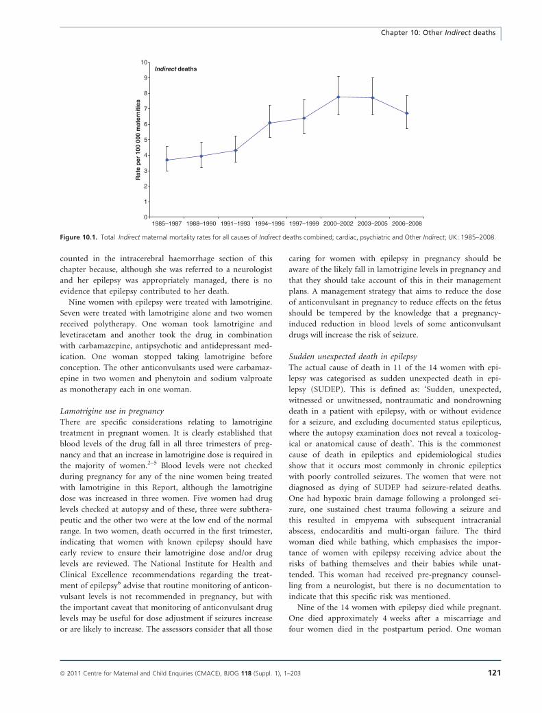
counted in the intracerebral haemorrhage section of this
chapter because, although she was referred to a neurologist
and her epilepsy was appropriately managed, there is no
evidence that epilepsy contributed to her death.
Nine women with epilepsy were treated with lamotrigine.
Seven were treated with lamotrigine alone and two women
received polytherapy. One woman took lamotrigine and
levetiracetam and another took the drug in combination
with carbamazepine, antipsychotic and antidepressant med-
ication. One woman stopped taking lamotrigine before
conception. The other anticonvulsants used were carbamaz-
epine in two women and phenytoin and sodium valproate
as monotherapy each in one woman.
Lamotrigine use in pregnancyThere are specific considerations relating to lamotrigine
treatment in pregnant women. It is clearly established that
blood levels of the drug fall in all three trimesters of preg-
nancy and that an increase in lamotrigine dose is required in
the majority of women.2–5 Blood levels were not checked
during pregnancy for any of the nine women being treated
with lamotrigine in this Report, although the lamotrigine
dose was increased in three women. Five women had drug
levels checked at autopsy and of these, three were subthera-
peutic and the other two were at the low end of the normal
range. In two women, death occurred in the first trimester,
indicating that women with known epilepsy should have
early review to ensure their lamotrigine dose and/or drug
levels are reviewed. The National Institute for Health and
Clinical Excellence recommendations regarding the treat-
ment of epilepsy6 advise that routine monitoring of anticon-
vulsant levels is not recommended in pregnancy, but with
the important caveat that monitoring of anticonvulsant drug
levels may be useful for dose adjustment if seizures increase
or are likely to increase. The assessors consider that all those
caring for women with epilepsy in pregnancy should be
aware of the likely fall in lamotrigine levels in pregnancy and
that they should take account of this in their management
plans. A management strategy that aims to reduce the dose
of anticonvulsant in pregnancy to reduce effects on the fetus
should be tempered by the knowledge that a pregnancy-
induced reduction in blood levels of some anticonvulsant
drugs will increase the risk of seizure.
Sudden unexpected death in epilepsyThe actual cause of death in 11 of the 14 women with epi-
lepsy was categorised as sudden unexpected death in epi-
lepsy (SUDEP). This is defined as: ‘Sudden, unexpected,
witnessed or unwitnessed, nontraumatic and nondrowning
death in a patient with epilepsy, with or without evidence
for a seizure, and excluding documented status epilepticus,
where the autopsy examination does not reveal a toxicolog-
ical or anatomical cause of death’. This is the commonest
cause of death in epileptics and epidemiological studies
show that it occurs most commonly in chronic epileptics
with poorly controlled seizures. The women that were not
diagnosed as dying of SUDEP had seizure-related deaths.
One had hypoxic brain damage following a prolonged sei-
zure, one sustained chest trauma following a seizure and
this resulted in empyema with subsequent intracranial
abscess, endocarditis and multi-organ failure. The third
woman died while bathing, which emphasises the impor-
tance of women with epilepsy receiving advice about the
risks of bathing themselves and their babies while unat-
tended. This woman had received pre-pregnancy counsel-
ling from a neurologist, but there is no documentation to
indicate that this specific risk was mentioned.
Nine of the 14 women with epilepsy died while pregnant.
One died approximately 4 weeks after a miscarriage and
four women died in the postpartum period. One woman
Indirect deaths
0
1
2
3
4
5
6
7
8
9
10
1985–1987 1988–1990 1991–1993 1994–1996 1997–1999 2000–2002 2003–2005 2006–2008
Rat
e p
er 1
00 0
00 m
ater
nit
ies
Figure 10.1. Total Indirect maternal mortality rates for all causes of Indirect deaths combined; cardiac, psychiatric and Other Indirect; UK: 1985–2008.
Chapter 10: Other Indirect deaths
ª 2011 Centre for Maternal and Child Enquiries (CMACE), BJOG 118 (Suppl. 1), 1–203 121
died of SUDEP before she was aware that she was preg-
nant. Of the 11 women who had sought antenatal care,
only six were referred to a healthcare provider with an
interest in epilepsy. It is of paramount importance that
women with epilepsy are seen by healthcare providers with
expertise in the management of the condition. The reasons
for not referring varied. In some cases the obstetric and
midwifery team do not appear to have perceived maternal
epilepsy as a high-risk condition. This attitude is inappro-
priate given the large number of deaths from epilepsy in
pregnancy. For example:
One woman with a known history of epilepsy was taking
lamotrigine and had a seizure a few days after delivery. She
was treated as having eclampsia despite having no hyper-
tension, nor proteinuria, and the only biochemical abnor-
mality was raised serum urate. She was not referred to a
neurologist at any stage during pregnancy or the postnatal
period and lamotrigine levels were never checked. Even at
the incident review after her death there was no mention of
the need for referral to a neurologist or physician with expe-
rience in managing epilepsy.
In another case the neurology service did not make an
appointment until nearly 5 months after receiving an initial
referral. Failure of the neurology service to review this
woman despite an urgent referral represents substandard
care.
Only six of the 14 women with epilepsy had received
pre-pregnancy counselling. Several women might not have
died if they had been advised about the maternal risks in
pregnancies complicated by epilepsy, hence the key recom-
mendation at the beginning of this chapter.
One-third of the epileptic women who died in this
report had difficult social circumstances that were likely to
cause them to be excluded from mainstream healthcare
provision. Two had a history of domestic abuse necessitat-
ing involvement of the police or social services, another
had suffered physical abuse from her stepmother before
marriage at an early age and there was subsequent evidence
of bullying from her mother-in-law. Another woman had
paranoid schizophrenia. Healthcare providers should be
aware that women with epilepsy who also live in difficult
social circumstances are at particular risk of poor seizure
control and require additional effort to ensure their disease
is well controlled.
Cerebral artery thrombosisFour women died of cerebral thrombosis, a rate of 0.17 per
100 000 maternities. Two deaths occurred in the first tri-
mester. In the first, magnetic resonance imaging (MRI) was
inappropriately withheld because of unwarranted fears
about its use during pregnancy and in the other woman
ectopic pregnancy was not considered:
An older woman with learning difficulties who had a his-
tory of cerebrovascular accident (CVA) was admitted with
new symptoms of hemiparesis on the opposite side. She
developed right iliac fossa tenderness as an inpatient and
was subsequently found to have a ruptured ectopic preg-
nancy. There was a delay in gynaecological assessment and
it is likely that the resultant blood loss and cerebral hypop-
erfusion exacerbated her CVA.
There are also two women with likely cerebral vein throm-
bosis in this Report, counted in Chapter 2, but the diagno-
sis was not straightforward in either of them. Both
presented with headache and neurological symptoms after
delivery at term. They also both had previous medical com-
plications that could have caused dehydration. In addition
they were obese, which has become recognised as a clear
risk factor for venous thromboembolism. One may have
had Wernicke’s encephalopathy following protracted vom-
iting.
Intracranial haemorrhageEleven women died of intracranial haemorrhage, 0.48 per
100 000 maternities. Six had subarachnoid haemorrhage
and five had intracerebral bleeds. None of the bleeds were
associated with labour and only two women died undeliv-
ered. Six of the women presented with sudden collapse or
severe headache with rapid deterioration and subsequent
death. Four women had no previous symptoms to alert the
healthcare providers that they were at risk of an intracra-
nial bleed.
Of those that presented with symptoms, one woman
complained of headache several times while pregnant and
another had eye pain with blurred vision. The two women
who did not present with rapid deterioration of their men-
tal state both complained of headache at the time of pre-
sentation. Two women had antenatal hypertension and in
one woman this was not adequately monitored. Five
women had substandard care. The reasons ranged from
obstruction to obtaining MRI scans and failure of the clini-
cal team to think of the diagnosis, to inappropriate contin-
uation of medication that would exacerbate pre-existing
hypertension. The case described below illustrates the
importance of referral to senior clinicians with experience
in managing neurological disease in pregnancy. If this
woman had been investigated thoroughly the diagnosis
might have been made at a time when intervention was
possible:
An asylum seeker was treated with labetalol for hyperten-
sion at booking which subsequently settled and treatment
was stopped. She complained of episodic headache through-
out pregnancy and presented at term with frontal headache
and hypertension but no investigations were performed and
she was discharged home after delivery. A few days later
de Swiet et al.
122 ª 2011 Centre for Maternal and Child Enquiries (CMACE), BJOG 118 (Suppl. 1), 1–203
she complained of severe headaches, pain and visual distur-
bance and altered facial sensation and although she was
reviewed by junior medical, neurological and obstetric staff,
there is no evidence of senior review. An MRI was per-
formed to exclude cerebral vein thrombosis and arterial dis-
section; however, the possibility of cerebral haemorrhage is
not mentioned in the notes and computed tomography (CT;
the imaging modality most likely to demonstrate an acute
cerebral bleed) was not requested at this time. She was dis-
charged and re-admitted 2 weeks after delivery having col-
lapsed at home. A CT scan showed subarachnoid
haemorrhage.
Four of the women who died from subarachnoid haemor-
rhage had difficult social circumstances. One was an asy-
lum seeker and another was a refugee. Two were known to
social services through drug use, violence or both.
Although this is unlikely to have been a cause of the sub-
arachnoid haemorrhages, it is possible that these women
had more barriers to obtaining rapid antenatal/medical
assessment than women who do not have adverse social
circumstances. This in turn may have led to delayed diag-
nosis and therefore to worse clinical outcomes. Smoking is
a recognised risk factor for aneurysmal subarachnoid
haemorrhage and two of the women in this group were
smokers.
There were five women with intracerebral haemorrhage.
In one woman the symptoms occurred almost immediately
after delivery and another two presented several days post-
natally. For example:
A woman saw her GP in early pregnancy complaining of
several episodes of numbness, tingling and inability to lift
her arm over the past year or so. She was referred to a neu-
rologist and she received an appointment for 3 months
later. She then mentioned these episodes in the antenatal
clinic and was also referred for a neurology opinion. The
neurologist associate specialist who saw her shortly after-
wards recommended an MRI after delivery. She was admit-
ted near term with slurred speech, headache, visual
symptoms and numbness on one side and referred to the
medical team. The medical specialist registrar did not feel
that the woman needed an urgent review and suggested a
neurology review the following week but the obstetric team
re-requested a review to exclude remediable neurological
disease, clearly stating that they were happy for her to have
an MRI in pregnancy and recommended expediting the
scan but this was not performed. A few days after an
uneventful delivery she was found dead at home and an
autopsy revealed a large intracerebral haemorrhage.
In another case, a woman had a rare hereditary condition
that predisposed to intracranial haemorrhage. Neither her
GP nor her obstetric team appeared to realise that this
woman was at high-risk, further underlining the impor-
tance of communication and the need to refer to specialists
if the healthcare providers are not experienced at managing
complex medical disorders.
Other lessons to be learnt from these cases are that
women with new and potentially serious neurological
symptoms in pregnancy must be seen promptly by a spe-
cialist who also understands that all imaging modalities can
be used in pregnancy if necessary, and the implications of
headache and unilateral weakness or numbness. Further-
more, neurological symptoms late in pregnancy mandate
an urgent review and cerebral imaging if indicated.
Meningitis/infectionThree women died of central nervous system infection of
whom two had pneumococcal meningitis. Both of these
women had complained of headache the day before death,
but there were no specific clinical features to suggest men-
ingitis or sepsis. Both women deteriorated rapidly and died
the next day. In the third woman meningitis was part of
the differential diagnosis but no senior staff were involved
in her care and no antibiotics were given. She had a respi-
ratory arrest while being investigated and died soon after.
Other neurological deathsOther women died from a spectrum of rare or difficult to
explain neurological diseases. For example, one had a
recurrent carotid artery dissection despite excellent man-
agement by a multidisciplinary team that included neurolo-
gists, obstetricians and neurosurgeons. Another pregnant
mother, who had an unremarkable medical history and a
previous uncomplicated pregnancy, collapsed and died at
home after a 2-week history of nausea and vomiting. Her
autopsy showed a colloid cyst adjacent to the foramen of
Monro and other features consistent with raised intracra-
nial pressure that was thought to be the cause of death. A
further woman was found dead in the bath close to the
time of delivery. She had a history of undiagnosed absence
seizures and had been given an opiate for pain relief in
labour. Women with seizures should not bathe unsuper-
vised. Showering is preferable. While the opiate treatment
is unlikely to have caused her death alone, it would have
increased her risk of death should she have had a seizure
or syncopal attack.
Infectious diseasesThe deaths of seven women are discussed here.
Bacterial infectionThere has been concern about the increasing number of
deaths from acute streptococcal infections, which has also
been raised in Chapter 7. For this report the majority of
the streptococcal infections presenting in pregnancy and
Chapter 10: Other Indirect deaths
ª 2011 Centre for Maternal and Child Enquiries (CMACE), BJOG 118 (Suppl. 1), 1–203 123
for 6 weeks after delivery continue to be counted as Direct
deaths even if there was no evidence of genital tract infec-
tion. In future, these deaths may be classified differently
and considered elsewhere as discussed in the Annex to
Chapter 7. However, one woman who died from strepto-
coccal disease also had HIV infection and is counted here.
In another case:
A pregnant woman died from staphylococcal pneumonia
and septicaemia in association with Influenza group B. She
also had mild bronchial asthma for which she had attended
the Emergency Department a few days previously. Her care
in the Emergency Department was good. Her asthma was
not severe at the time and the advice to increase her salbu-
tamol inhalation and take a short course of oral steroids
was appropriate. Data from previous pandemics suggest
that women are more likely to die from influenza when
pregnant.
A further woman died in the puerperium from a Gram-
negative infection and liver abscesses having been admit-
ted in extremis with an acute abdomen. She had attended
her GP with back pain several times after delivery and
was reassured each time without investigation. Although
backache is common in the puerperium, multiple atten-
dances must be taken seriously and demand further inves-
tigation.
Viral infection: human immunodeficiency virusOne woman with known HIV infection was not responding
to treatment, perhaps because of failures of compliance.
She was admitted with overwhelming streptococcal pneu-
monia, had an inevitable miscarriage and died a few hours
later. In another case:
A woman with a complex social history refused admission
for a chest infection in pregnancy because she was con-
cerned her children would be taken into care and she only
saw a midwife a few weeks later. She was not fully booked
for several more weeks because she missed a scan appoint-
ment which was not followed up. She was eventually admit-
ted with pneumonia caused by Pneumocystis carinii and a
diagnosis of HIV infection was made. She was discharged
home and her midwives were not informed. Her HIV ther-
apy was delayed for several more weeks and none was given
when she went into preterm labour because there was no
HIV protocol on the delivery suite and no advice could be
obtained after hours. She was delivered by caesarean section
for fetal distress and died later from overwhelming sepsis
and colitis.
There is no evidence that the HIV team was involved in
her care. In newly diagnosed cases of HIV infection a con-
sultant obstetrician should be involved as soon as the diag-
nosis is made. HIV/AIDS is now a treatable disease.
TuberculosisTwo women from the Indian subcontinent died of tuber-
culosis (TB) but were diagnosed late. In one this was
because her chest symptoms were first ascribed to pulmo-
nary embolus. The other woman was initially thought to
be suffering from anorexia nervosa, an erroneous diagno-
sis confirmed by a consultant psychiatrist because of
weight loss in the first half of pregnancy. When TB was
suspected it was impossible to perform an immunological
TB skin test because the test was only available on certain
days of the week. She died shortly afterwards and her
autopsy showed miliary TB with nodules over lungs and
peritoneum. Anorexia is exceedingly uncommon in preg-
nancy, if only because such women do not ovulate, and
all underlying causes for weight loss should be excluded
before considering a psychiatric referral. Immunological
tests for TB should be readily available for high-risk
women, especially as TB is becoming more prevalent.
A UK Obstetric Surveillance System (UKOSS) prospective,
national study of TB in pregnancy between August 2005 and
August 2006 reported an estimated annual incidence of 42
cases per million maternities (95% confidence interval 29–59
per million maternities7). The disease was limited to women
from ethnic minorities as in the two women described here.
Extrapulmonary disease was as common as pulmonary in
the UKOSS study but only women with pulmonary disease
died. From these figures the estimated total number of cases
of TB in the triennium was 266 (95% CI 184–374). As two
pregnant women died from TB in this triennium, the esti-
mated case fatality rate of TB in pregnancy in 2006–08 is
1:133 known cases (95% CI 1:92 to 1:187).
Respiratory diseases
Bronchial asthmaFive women died from asthma this triennium, 0.22 per
100 000 maternities, a similar number to those in the two
previous Reports. None had specialist care in pregnancy
even though three had had many hospital admissions or
had brittle asthma that was particularly difficult to treat.
For example:
A woman who had asthma since childhood had at least 20
referrals for hospital care within the previous few years,
including some episodes where she had a cardiac arrest.
During her pregnancy she was cared for entirely within the
community and had no specialist referral. Asthma was not
mentioned in her GP’s referral letter.
Although asthma does not of itself deteriorate in preg-
nancy, women often withhold or decrease asthma medica-
tion because of the unfounded fear that it will harm the
fetus. They need specialist care in pregnancy if only to reas-
de Swiet et al.
124 ª 2011 Centre for Maternal and Child Enquiries (CMACE), BJOG 118 (Suppl. 1), 1–203
sure them that this is not so. Fears that medication for
women with asthma will harm the fetus extends to their
medical attendants, as in the case of another young woman
with brittle asthma who was incorrectly told by her GP to
stop prednisolone in early pregnancy.
In the case of a woman who died from acute severe asthma
after a medical termination of pregnancy:
A woman was known to be atopic and to suffer from
asthma. The nature of the drugs used for the termination
was not available to the assessors but they are likely to have
been Mifepristone, a progesterone and glucocorticoid antag-
onist, followed by misoprostol a prostaglandin of the E ser-
ies. The procedure took 2 days, as usual, but on discharge
she became breathless and she was reluctant to use her
asthma medications because she thought they might interact
with the drugs given for her termination. She died a few
hours after discharge.
There has been concern about the use of Mifepristone in
women with asthma because progesterone is a bronchodila-
tor and a progesterone antagonist might cause bronchocon-
striction. Also a glucocorticoid antagonist has the potential
to antagonise endogenous cortisol and steroids used for the
treatment of asthma. But the British National Formulary
(BNF) advises against the use of Mifepristone only in
severe and uncontrolled asthma, which this woman did not
have. With regard to misoprostol there has also been con-
cern about the use of prostaglandins in women with
asthma and the BNF also has a specific warning about this.
However, the evidence for prostaglandin-induced broncho-
constriction is only clear for prostaglandins of the F2a ser-
ies such as Carboprost, not those of the E series such as
Misoprostol. Many women with known asthma have been
give prostaglandin E for induction of labour without
adverse outcome. It seems unlikely that this was a case of
Mifepristone-induced or misoprostol-induced bronchocon-
striction. However, this incident should be noted in case of
further occurrences. Bronchial asthma is not a contraindi-
cation to medical termination of pregnancy.
Respiratory infectious diseaseFour women died of pneumonia this triennium, most of
whom had underlying social factors such as severe depriva-
tion. For example:
A pregnant woman who spoke no English died after admis-
sion from pneumonia without any causative organism being
found. According to the relative who acted as her inter-
preter, she had been breathless for a week but had not
reported this because she thought it normal for pregnancy.
Her condition deteriorated and she required ventilation on
the Critical-Care Unit until she was delivered. She died a
few days later.
This woman’s medical care was good once she was admit-
ted. The problem was the delay. It will never be known if
her breathlessness was trivial; but maternity services must
make efforts to make their services accessible to deprived
immigrants who do not speak English.
Endocrine, metabolic and immune diseases
EndocrineThree women died from diabetes mellitus, 0.13 per 100 000
maternities, all probably because of hypoglycaemia. In a
typical example:
A woman with known insulin-dependant diabetes went to
see her GP very early in pregnancy but was asked to come
back some weeks later for booking bloods and to be referred
to the antenatal clinic. A month later and a wasted 6 weeks
after she first saw her GP, the booking bloods showed a high
glycosylated haemoglobin indicating poor diabetic control.
The booking midwife consulted the diabetic specialist nurse
and arranged for her to be seen in the obstetrician’s and
diabetologist’s separate clinics. She saw a diabetic specialist
nurse who planned 2-weekly review. The diabetic nurse
noted several episodes of dangerously severe hypoglycaemia.
She first saw the consultant diabetologist a few months fol-
lowing her first visit to the GP and died of a hypoglycaemic
attack soon after.
All women with pre-existing diabetes should have pre-preg-
nancy counselling and this woman’s complicated diabetes
should have been under consultant care whether she was
pregnant or not. This woman should have been seen in a
combined diabetic/obstetric clinic much earlier in preg-
nancy, which could have been organised by telephone
immediately after she saw her GP given the importance of
good diabetic control in early pregnancy. Her own obstetric
centre itself was too small for a combined diabetic/obstetric
clinic and there was a lack of communication between
the ‘diabetic centre’ and the obstetric–midwifery team; the
woman was not advised to take her hand-held notes to the
diabetic centre. Her partner did not seem to have been
instructed on how to give glucagon.
This was a case of hypoglycaemic death in a woman trying
desperately to control diabetes in early pregnancy, as
occurred in the other two deaths from diabetes. This is a
problem noted in previous Reports. A fine balance has to be
drawn between correctly encouraging women to optimise
their diabetic control and frightening them so much that
they risk serious and sometimes fatal problems from hypo-
glycaemia. In addition, as noted in the Key recommenda-
tions, women whose pregnancies are complicated by
potentially serious medical conditions should be referred to
appropriate specialist centres of expertise where both care for
their medical condition and their obstetric care can be opti-
Chapter 10: Other Indirect deaths
ª 2011 Centre for Maternal and Child Enquiries (CMACE), BJOG 118 (Suppl. 1), 1–203 125
mised. The obstetric centre where this woman was managed
was too small to have a proper combined diabetic and
obstetric clinic. She should have been referred elsewhere.
Immunological disordersFour women, 0.17 per 100 000 maternities, died either
from systemic lupus erythematosus (SLE) or from other
causes where lupus was a factor. Another case associated
with SLE is counted in the Haematology section because
thrombotic thrombocytopoenic purpura was her primary
cause of death. For some of these women there were,
again, failures of communication. For example one recent
immigrant spoke little English and her relative, who tried
to translate, spoke even less. No history was recorded by
the GP at booking in early pregnancy and no examination
was performed. Three months later she complained of
multiple joint pains and soon after she was admitted des-
perately ill with polyarthralgia due to SLE. She was trans-
ferred to a tertiary centre but died 3 weeks later of multi-
organ failure. The need for interpreters in the community
is still not being met. Under these circumstances, consid-
eration should be given to referral to hospital where inter-
preters are usually more easily available, if only for a
single consultation.
In another case, also discussed in Chapter 11:
A woman was incorrectly told to stop hydroxychloroquine
when she became pregnant and when the equally impor-
tant problem of positive extractable nuclear antigens
(ENA) antibodies, which may cause congenital heart
block, was ignored. She was admitted several times in
pregnancy feeling unwell and was eventually seen by the
liaison psychiatrist. She became very disorientated after
delivery when she was found to have lupus nephritis. She
died soon after delivery from multiple organ failure.
The need remains for physicians who do not work directly
with pregnant women to know more about the interaction
between the conditions that they are treating and preg-
nancy.
One further woman died from anaphylaxis following
intravenous Augmentin given for pyrexia in labour. She
had been asked about drug allergy in pregnancy and before
the Augmentin was given. There was no substandard care.
Women still die from anaphylaxis.
Metabolic diseaseFailure of communication and unavailability of hospital
notes were the principal reasons why a woman died with, a
rare inborn error of metabolism, which predisposes to hyp-
oglycaemia and lactic acidosis.
She attended a physician in an obstetric clinic where her
previous pregnancy had been managed successfully. She
too spoke little English. Her hospital notes were unavail-
able and there was no booking letter but fortunately the
physician remembered her. She complained of nausea and
vomiting but was reluctant to be admitted. The physician
advised on outpatient treatment and told the woman how
she/he could be contacted. Her vomiting got much worse
and she was admitted to a gynaecology ward via the
Emergency Department. Her notes still could not be
found. The physician was not called despite a request
from her relatives who said that she had hypoglycaemia
and needed a central line. There were difficulties obtain-
ing intravenous access. She was eventually found to be
grossly acidotic and was admitted to the intensive-care
unit where she died some weeks later from septicaemia
and multi-organ failure.
The failures were that the junior staff did not listen to the
woman or her relatives and that her notes should also have
been available.
Another woman died from an extremely rare autosomal
recessive condition associated with other physical problems
leading to mild or fatal disease. Her consultant obstetrician
appears to have made no attempt to read or consult about
this condition, which has never been described in preg-
nancy before. Health workers who are caring for women in
pregnancy with conditions that they are not familiar with
should take the trouble to consult experts, if only by tele-
phone.
Diseases of the gastrointestinal tractThere were nine deaths from disease of the gastrointestinal
tract, of which two were from complications of duodenal
ulcers. In both women, the diagnosis was made too late.
One woman was admitted on several occasions in late
pregnancy with increasingly severe epigastric pain eventu-
ally requiring opiates. She delivered and went home with
no clinical observations recorded. She was readmitted a few
days later with a fatal haematemesis. Another woman
developed abdominal pain in mid-pregnancy which was
thought to be the result of her intercurrent pre-eclampsia.
After delivery the pain worsened with a tense abdomen and
guarding. After more than 24 hours she eventually saw a
consultant obstetrician and she died in surgery from a per-
forated duodenal ulcer. In both of these women the clini-
cians did not think beyond the possibility of pregnancy-
related complications soon enough. Consultants were
involved too late in the second woman, where steroids
given for fetal lung maturation may also have contributed
to the perforation.
Several women died from complications of cirrhosis
or liver disease and one from hyperlipidaemia. In these
and several other women the responsible clinicians
seemed to be unaware of the high-risk nature of the
de Swiet et al.
126 ª 2011 Centre for Maternal and Child Enquiries (CMACE), BJOG 118 (Suppl. 1), 1–203
women’s pregnancies. They were managed at different
hospitals for their medical conditions and there was a
lack of communication between services. They were
often treated inappropriately by junior staff. Women
with rare conditions and high-risk pregnancies such as
these need pre-pregnancy counselling and then to be
managed jointly at tertiary centres by the relevant spe-
cialist and obstetric services. They are likely to require
an anaesthetic assessment before delivery and any opera-
tive interventions must be undertaken by fully trained
consultants.
There were no deaths this triennium from Ogilvie syn-
drome. There was, however, one death from pseudomem-
branous colitis and two from Crohn’s disease.
One of these two women was admitted many times in preg-
nancy before she was found to be obstructed. She was dis-
charged severely anaemic following surgery and readmitted
moribund 3 weeks later. She could not be resuscitated and
died from complications of the original surgery. There had
been no communication between the surgeons and the
obstetric team. She had never been seen by a gastroenterolo-
gist despite her multiple admissions.
As indicated in the Key recommendations above: multiple
admissions or attendances for emergency care demand fur-
ther investigation and are often a reason for referral to spe-
cialists in other disciplines.
Diseases of the bloodTwo women died from thrombotic thrombocytopoenic
purpura (TTP) (one per million maternities), a rare hae-
mangiopathic haemolytic anaemia which is relatively more
common in pregnancy. One of these women died from a
myocardial infarct. Infarction is common in TTP because
of small-vessel coronary thrombosis. One woman with
known connective tissue disease went to an Emergency
Department with chest pain and was triaged by nursing
staff to the Obstetric Day Unit where she only ever saw a
junior doctor. Although her blood tests showed florid red
cell fragmentation, a main diagnostic feature of TTP, this
was not reported or transmitted to the specialist unit to
which she was referred the next day. She died with multiple
thrombi in the heart. Here again there was lack of referral
in early pregnancy to a specialist unit of a highly complex
case at very high risk.
A woman with haemoglobin SC disease developed a
urinary tract infection that was ignored and led to Strep-
tococcus pneumoniae sepsis and death from septicaemia.
She had been labelled as having sickle cell trait despite
having had previous sickle cell crisis and documented
haemoglobin SC disease on the hospital computer. She
had no pre-pregnancy counselling and no referral to a
specialist haematologist until the day that she died. Previ-
ous reports have emphasised the risk of haemoglobin SC
disease. Clinicians are unaware that it is a sickling condi-
tion and are misled by the relatively high haemoglobin
concentration.
Circulatory conditionsOne woman died of a spontaneous ruptured splenic artery.
Splenic artery rupture is relatively more common in preg-
nancy, whether an aneurysm is present or not. Another
woman died of a ruptured spleen, which is very uncom-
mon in pregnancy. In a further case of ruptured spleen it
is possible that this woman’s spleen ruptured during the
paramedics’ attempts at resuscitation, particularly as she
was morbidly obese.
Air embolusOne or two deaths from air embolus usually occur in each
triennium. In this case:
A woman who died from air embolus collapsed with
breathlessness during sexual intercourse about 2 weeks after
vaginal delivery. At autopsy there was frothy blood in the
pulmonary trunk and exuding from the myometrium and
there were gaping vessels in the uterus.
In this form of air embolus, air is forced from the vagina
into the uterus and enters the venous circulation through
the vessels of the raw vascular bed. The resultant froth in
the right ventricle effectively causes a cardiac arrest because
of lack of cardiac output. This is a recognised but rare
cause of maternal death. Couples are advised to preferably
abstain from sexual intercourse for 6 weeks postnatally or
have only gentle intercourse and to avoid positions/prac-
tices where excessive air could be forced into the vagina.
Rare conditionsA previously fit woman had a cyanotic attack in the night.
She was dead by the time she arrived in hospital. At
autopsy the larynx and epiglottis were oedematous. Because
of high immunoglobulin E tryptase levels and following
discussion with a clinical immunologist the cause of death
was given as idiopathic angio-oedema.
One woman died from benign metastasising leiomyoma-
tosis and another from congenital muscular dystrophy of
unknown type and severe kyphoscoliosis. They are both
counted in the other respiratory conditions section of
respiratory disease. Both had been advised against preg-
nancy but they elected to continue, despite medical advice.
Women with very serious medical complications, particu-
larly respiratory and cardiac, may die in pregnancy even
with the very good care that these women received. Once
fully informed of the situation, if they choose to continue
with pregnancy they should be allowed to do so with
dignity.
Chapter 10: Other Indirect deaths
ª 2011 Centre for Maternal and Child Enquiries (CMACE), BJOG 118 (Suppl. 1), 1–203 127
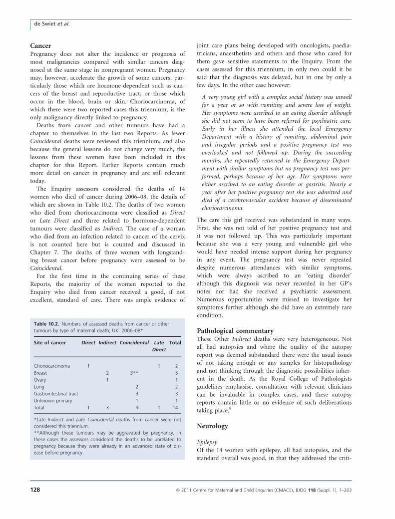
CancerPregnancy does not alter the incidence or prognosis of
most malignancies compared with similar cancers diag-
nosed at the same stage in nonpregnant women. Pregnancy
may, however, accelerate the growth of some cancers, par-
ticularly those which are hormone-dependent such as can-
cers of the breast and reproductive tract, or those which
occur in the blood, brain or skin. Choriocarcinoma, of
which there were two reported cases this triennium, is the
only malignancy directly linked to pregnancy.
Deaths from cancer and other tumours have had a
chapter to themselves in the last two Reports. As fewer
Coincidental deaths were reviewed this triennium, and also
because the general lessons do not change very much, the
lessons from these women have been included in this
chapter for this Report. Earlier Reports contain much
more detail on cancer in pregnancy and are still relevant
today.
The Enquiry assessors considered the deaths of 14
women who died of cancer during 2006–08, the details of
which are shown in Table 10.2. The deaths of two women
who died from choriocarcinoma were classified as Direct
or Late Direct and three related to hormone-dependent
tumours were classified as Indirect. The case of a woman
who died from an infection related to cancer of the cervix
is not counted here but is counted and discussed in
Chapter 7. The deaths of three women with longstand-
ing breast cancer before pregnancy were assessed to be
Coincidental.
For the first time in the continuing series of these
Reports, the majority of the women reported to the
Enquiry who died from cancer received a good, if not
excellent, standard of care. There was ample evidence of
joint care plans being developed with oncologists, paedia-
tricians, anaesthetists and others and those who cared for
them gave sensitive statements to the Enquiry. From the
cases assessed for this triennium, in only two could it be
said that the diagnosis was delayed, but in one by only a
few days. In the other case however:
A very young girl with a complex social history was unwell
for a year or so with vomiting and severe loss of weight.
Her symptoms were ascribed to an eating disorder although
she did not seem to have been referred for psychiatric care.
Early in her illness she attended the local Emergency
Department with a history of vomiting, abdominal pain
and irregular periods and a positive pregnancy test was
overlooked and not followed up. During the succeeding
months, she repeatedly returned to the Emergency Depart-
ment with similar symptoms but no pregnancy test was per-
formed, perhaps because of her age. Her symptoms were
either ascribed to an eating disorder or gastritis. Nearly a
year after her positive pregnancy test she was admitted and
died of a cerebrovascular accident because of disseminated
choriocarcinoma.
The care this girl received was substandard in many ways.
First, she was not told of her positive pregnancy test and
it was not followed up. This was particularly important
because she was a very young and vulnerable girl who
would have needed intense support during her pregnancy
in any event. The pregnancy test was never repeated
despite numerous attendances with similar symptoms,
which were always ascribed to an ‘eating disorder’
although this diagnosis was never recorded in her GP’s
notes nor had she received a psychiatric assessment.
Numerous opportunities were missed to investigate her
symptoms further although she did have an extremely rare
condition.
Pathological commentaryThese Other Indirect deaths were very heterogeneous. Not
all had autopsies and where the quality of the autopsy
report was deemed substandard there were the usual issues
of not taking enough or any samples for histopathology
and not thinking through the diagnostic possibilities inher-
ent in the death. As the Royal College of Pathologists
guidelines emphasise, consultation with relevant clinicians
can be invaluable in complex cases, and these autopsy
reports contain little or no evidence of such deliberations
taking place.8
Neurology
EpilepsyOf the 14 women with epilepsy, all had autopsies, and the
standard overall was good, in that they addressed the criti-
Table 10.2. Numbers of assessed deaths from cancer or other
tumours by type of maternal death; UK: 2006–08*
Site of cancer Direct Indirect Coincidental Late
Direct
Total
Choriocarcinoma 1 1 2
Breast 2 3** 5
Ovary 1 1
Lung 2 2
Gastrointestinal tract 3 3
Unknown primary 1 1
Total 1 3 9 1 14
*Late Indirect and Late Coincidental deaths from cancer were not
considered this triennium.
**Although these tumours may be aggravated by pregnancy, in
these cases the assessors considered the deaths to be unrelated to
pregnancy because they were already in an advanced state of dis-
ease before pregnancy.
de Swiet et al.
128 ª 2011 Centre for Maternal and Child Enquiries (CMACE), BJOG 118 (Suppl. 1), 1–203
cal questions of alternative diagnoses to epilepsy, which is
important in the case of SUDEP, and included ancillary
blood tests to identify anticonvulsant and other drugs. The
basic mechanism of SUDEP is thought to be an arrhythmia
occurring during an epileptic seizure, stopping the heart
beat.
Cerebral thrombosisAutopsies were performed in the minority of women with
ischaemic stroke because imaging pre-mortem usually
identifies the pathology. Thrombophilia was a factor
already known in one woman. The pathologist cannot
identify an inherited thrombophilic state post-mortem
because functional blood clotting tests cannot be
performed on autopsy blood. Genetic studies of DNA for
known prothrombotic conditions have not been validated
on autopsy material.
Not all of the cases of stroke were aetiologically resolved.
In one woman, who died of a thrombotic stroke in the
puerperium and did not have an autopsy, the diagnosis
could have been arterial occlusion, possibly from paradoxi-
cal thromboembolism, sagittal sinus or cerebral vein
thrombosis, or tuberculous meningitis. Obviously, knowing
which was the case, would have informed clinical audit, as
well as categorising the case better as a Direct, Indirect or
Coincidental death.
Cerebral haemorrhageSubarachnoid haemorrhage
Of the six women who died with a subarachnoid haemor-
rhage, only one had an autopsy. The others were diagnosed
at CT scan and/or craniotomy, with the aneurysm identi-
fied along with the subarachnoid and intracerebral haemor-
rhage. With a confident clinical cause of death there was
no requirement for a medico-legal autopsy. The single
autopsy exemplifies some of the problems in specifying the
cause of death when women present as ‘death in the com-
munity’:
An obese woman collapsed in a car park toward the end of
pregnancy and was dead on arrival at the Emergency
Department. Her booking blood pressure was 110/
80 mmHg. The neuropathological autopsy found a ruptured
berry aneurysm and subarachnoid haemorrhage; but the
heart weighed 411 g, definitely enlarged with left ventricular
hypertrophy. The kidney was not scrutinised for glomerular
endotheliosis (the histological marker for pre-eclampsia), so
the questions regarding the underlying predisposition for
aneurysm rupture remain open: was this essential hyperten-
sion despite the booking blood pressure, acute pre-eclampsia
with rise in blood pressure, obesity cardiomyopathy and
coincidental ruptured aneurysm or idiopathic left ventricu-
lar hypertrophy with ruptured aneurysm?
InfectionThe women with apparently primary bacterial pneumonias
were not well evaluated at autopsy apart from a women
who died in her second trimester from staphylococcal
pneumonia complicating type B influenza. For the other
women, no attempt at lung microbiology was made,
which is disappointing given the importance of the epide-
miology of community-acquired pneumonia in this age
group. In neither of the two HIV-positive women was a
coronial autopsy necessary, but in one woman it was per-
formed. However, the pathologist showed little insight
into the complex pathologies encountered in late-stage
HIV disease, and the report would have helped neither
the HIV doctors nor the obstetricians in their clinical
audit.
DiabetesAll the women who died of diabetic hypoglycaemia had
autopsies, which were not all well done. In two of the
three women the characteristic red neurone change in
the brain was not documented histologically and nor were
the glucose and insulin levels measured in blood and
vitreous fluids.
The associated dead-in-bed syndrome is worth mention-
ing as a relatively new entity. The basic mechanism of SU-
DEP is thought to be an arrhythmia occurring during an
epileptic seizure, stopping the heart beat. Similarly with
unexplained deaths in diabetes, the so-called diabetic
‘dead-in-bed syndrome’. This was documented twice in this
triennium in women who had been insulin-dependent dia-
betics for more than 10 years. For example:
A woman with longstanding type 1 diabetes for many years
and who had had many hypoglycaemic episodes, was found
dead in bed in mid-pregnancy. The post-mortem biochemis-
try for glucose and insulin was ambiguous, as is often the
case but the brain histopathology showed hypoglycaemic-
type neuronal necrosis. The diabetologist consulted on the
case proposed the sudden nocturnal death in diabetes sce-
nario, which comprises a diabetic autonomic neuropathy,
and a long QT interval—leading to fatal arrhythmia.9 Sig-
nificantly this is not just hypoglycaemia causing death; hyp-
oglycaemia is critically associated with cardiac electrical
instability.
Liver diseaseTwo women died as a result of bleeding from cirrhotic
portal hypertension. The deaths of two other women
highlight how complicated medicine can be, because what
actually happened was not finally resolved despite good
autopsies. In a woman who had liver abscesses and peri-
tonitis, the route of infection was not ascertained, despite
Chapter 10: Other Indirect deaths
ª 2011 Centre for Maternal and Child Enquiries (CMACE), BJOG 118 (Suppl. 1), 1–203 129
a thorough autopsy with many significant ‘negatives’. Pos-
sibilities include the throat, though this should have been
clinically obvious from severe local pain, the vagina and
the faecal flora. In the second difficult case, with focal
nodular hyperplasia of the liver and then fatal intra-
abdominal bleeding, the combination of the laparotomy
and autopsy findings concluded that there was probably
spontaneous tearing of liver–spleen adhesions. Focal nodu-
lar hyperplasia is not known to be associated with preg-
nancy.
Thrombotic thrombocytopoenic purpuraTTP is rare, and in both women it was diagnosed or sug-
gested before death. Rapid death, as in these women,
results from cardiac thrombotic microangiopathy with
ischaemic cardiac failure. The laboratory data of low plate-
lets with normal clotting factors is distinctive. The autopsy
histopathology is similarly characteristic, with thrombotic
microangiopathy in the renal glomeruli as well. One can
distinguish the two aetiological patterns of thrombotic
microangiopathy with immunohistochemistry: in TTP, the
thrombi are nearly pure platelets (stained well with anti-
bodies to CD61), whereas in disseminated intravascular
coagulation, the thrombi contain more fibrin (demonstra-
ble with anti-fibrin antibodies) than platelets.
Sickle cell diseaseThe pathology of the sickle cell deaths from sepsis is dis-
cussed in Chapter 7. In another death from sickling, a
woman with haemoglobin SS disease was being investigated
for chest pain in hospital when she collapsed and died. The
autopsy reportedly showed ‘myocardial ischaemia’. But cor-
onary artery disease at this age is rare, venous thromboem-
bolism is unlikely in haemoglobin SS disease, and the acute
sickle chest syndrome is a more likely pathology. Sickle
autopsies, like HIV and maternal autopsies, can be compli-
cated, and need consideration and a full protocol procedure
as described in the Royal College of Pathologists Autopsy
Guidelines on Sickle cell disease, 2005.10
Air embolismHere the autopsy is critical. Ideally, imaging with CT scan
will identify the gas in the heart before the body is opened,
but routine CT scanning of cadavers has yet to become the
norm. In this woman, the heart was correctly opened under
water in situ in the pericardium and gas bubbles burst from
the right ventricle; there was also frothy blood in the myome-
trium. It is important to note that the presence of gas bub-
bles in the meningeal vessels, once thought to indicate air
embolism, is an artefact that arises from the vacuum tempo-
rarily created within the cranium when the skull bone is
removed. In all peripartum death autopsies, the heart should,
as a matter of protocol, be opened under water.
Unascertained causes of deathThere were six women for whom the cause of death was
uncertain, often because the autopsy was inadequate. These
deaths are distinguished from sudden adult death syn-
drome (SADS), where an adequate autopsy has shown no
other cause for death as discussed in Chapter 9. For one
woman a phaeochromocytoma may have been the cause of
death. Another woman had been ill for some weeks before
death with breathlessness and vomiting. She clearly had a
major metabolic problem with grossly elevated blood urea
and serum creatinine and low serum sodium but no other
cause of death could be identified at autopsy. A third
woman with type 2 diabetes taking insulin had a cardiac
arrest. Her death was recorded as being the result of
eclampsia although there was no evidence to support this
other than modest hypertension and a bitten tongue. Her
autopsy was deficient and lacking many details. This death
could have been diabetes related; it could have been cardiac
relating to diabetes or hypertension; it could have been a
sudden adult cardiac death; but because of the other possi-
bilities, it has been classified as unascertained rather than
sudden adult death syndrome.
Conclusion
This triennium the assessors have been struck by the lack
of referral of potentially high-risk women. The reasons for
failure to refer are likely to be multiple. However, medical
care is advancing rapidly and patterns of the delivery of
care in the UK are changing. It must be appreciated that
not all maternity centres can care for pregnant women
with major complications either preceding or developing
in pregnancy. If women with underlying medical condi-
tions are to share in the advances in medicine, many more
must be referred to tertiary medical centres for their care
in pregnancy.
Disclosure of interestsNone.
FundingThis work was undertaken by the Centre for Maternal and
Child Enquiries (CMACE) as part of the CEMACH pro-
gramme. The work was funded by the National Patient
Safety Agency; the Department of Health, Social Services
and Public Safety of Northern Ireland; NHS Quality
Improvement Scotland (NHS QIS); and the Channel
Islands and Isle of Man.
AcknowledgementsThe neurology section was discussed with Dr Richard
Perry, Consultant Neurologist, Imperial College Healthcare
de Swiet et al.
130 ª 2011 Centre for Maternal and Child Enquiries (CMACE), BJOG 118 (Suppl. 1), 1–203
NHS Trust. The pathology section was written by Professor
Sebastian Lucas of Guys and St Thomas’ NHS Foundation
Healthcare Trust. j
References
1 World Health Organization International Classification of Diseases
ICD 9 and 10. Geneva: World Health Organization.
2 Tran TA, Leppik IE, Blesi K, Sathanandan ST, Remmel R. Lamotrigine
clearance during pregnancy. Neurology 2002;59:251–5.
3 Ohman I, Beck O, Vitols S, Tomson T. Plasma concentrations of
lamotrigine and its 2-N-glucuronide metabolite during pregnancy in
women with epilepsy. Epilepsia 2008;49:1075–80.
4 Pennell PB, Peng L, Newport DJ, Ritchie JC, Koganti A, Holley DK,
et al. Lamotrigine in pregnancy: clearance, therapeutic drug monitor-
ing, and seizure frequency. Neurology 2008;70:2130–6.
5 Fotopoulou C, Kretz R, Bauer S, Schefold JC, Schmitz B, Dudenhau-
sen JW, et al. Prospectively assessed changes in lamotrigine-concen-
tration in women with epilepsy during pregnancy, lactation and the
neonatal period. Epilepsy Res 2009;85:60–4.
6 National Institute for Health and Clinical Excellence. The Epilepsies:
The Diagnosis and Management of the Epilepsies in Adults and Chil-
dren in Primary and Secondary Care. London: NICE, 2004.
7 Knight M, Kurinczuk J, Nelson-Piercy C, Spark P, Brocklehurst P, on
behalf of UKOSS. Tuberculosis in pregnancy in the UK. BJOG
2009;116:584–8.
8 RCPath guidelines Royal College of Pathologists: Guidelines on
Autopsy Practice. Scenario 5: Maternal Death. May 2010. London:
RCPath 2010.
9 Tu E, Bagnall R, Duflou J, Lynch M, Twigg S, Semsarian C.
Post-mortem pathological and genetic studies in ‘‘dead in bed
syndrome’’ cases in type 1 diabetes mellitus. Hum Pathol 2010;41:
392–400.
10 Royal College of Pathologists. Guidelines for Autopsy Practice: sickle
cell disease. London: RCPath 2005.
ª 2011 Centre for Maternal and Child Enquiries (CMACE), BJOG 118 (Suppl. 1), 1–203 131
Chapter 10: Other Indirect deaths
Chapter 11: Deaths from psychiatric causesM Oates1, R Cantwell2 (on behalf of the Centre for Maternal and Child Enquiries)1 East Midlands Perintal Mental Health Clinical Network, Nottinghamshire Healthcare NHS Trust, Nottingham, UK; 2 Perinatal Mental Health
Service, Southern General Hospital, Glasgow, UK
Correspondence: Dr Margaret R Oates, East Midlands Perintal Mental Health Clinical Network, Nottinghamshire Healthcare NHS Trust,
Duncan MacMillan House, Porchester Road, Nottingham NG3 6AA, UK. Email: [email protected]
Keywords psychiatric, psychosis, suicide, Confidential Enquiry,
maternal, mortality.
Psychiatric deaths: specific recommendations
• As has been recommended before, but re-emphasised here, all women should be asked at their antenatal booking visit
about a previous history of psychiatric disorder as well as their current mental health. Women with a previous history
of serious affective disorder or other psychoses should be referred in pregnancy for psychiatric assessment and man-
agement even if they are well. A minimum requirement for management should be regular monitoring and support
for at least 3 months following delivery.
• Psychiatric services should have priority care pathways for pregnant and postpartum women. These will include a low-
ered threshold for referral and intervention, including admission and a rapid response time, for women in late pregnancy
and the first 6 weeks following delivery. Care by multiple psychiatric teams should be avoided. Risk assessments of preg-
nant or postpartum women should be modified to take account of risk associated with previous history, the distinctive
clinical picture of perinatal disorders and the violent method of suicide.
• All mental health trusts should have specialised community perinatal mental teams to care for pregnant and postpartum
women. These should be closely integrated with regional mother and baby units so that all women requiring psychiatric
admission in late pregnancy and the postpartum period can be admitted together with their infants.
• Caution needs to be exercised when diagnosing psychiatric disorder if the only symptoms are either unexplained phys-
ical symptoms or distress and agitation. This is particularly so when the woman has no prior psychiatric history or
when she does not speak English or comes from an ethnic minority.
Introduction and background
As in previous Reports, this chapter describes the key
features of, and derives lessons from, those maternal
deaths arising directly from a psychiatric condition, sui-
cide or accidental overdose of drugs of abuse, as well as
deaths from medical or other causes closely related to a
psychiatric disorder. These latter deaths include those
from the physical consequences of substance misuse and
delays in diagnosis and treatment because of the presence
or assumption of a psychiatric disorder, accidents and
violence.
Perinatal psychiatric disorder
Psychiatric disorder during pregnancy and following deliv-
ery is common, both new episodes and recurrences of pre-
existing conditions. Ten percent of new mothers are likely
to develop a depressive illness,1 of whom between a third
and a half will be suffering from a severe depressive illness.2
At least 2% of new mothers will be referred to a psychiatric
team during this time, and two per thousand will suffer
from a puerperal psychosis.3
The majority of women who develop mental health
problems during pregnancy or following delivery suffer
from mild depressive illness, often with accompanying anx-
iety. Such conditions are probably no more common than
at other times. In contrast, the risk of developing a serious
mental illness (bipolar disorder, other affective psychoses
and severe depressive illness) is reduced during pregnancy
but markedly elevated following childbirth, particularly
during the first 3 months.4
The prevalence of all psychiatric disorders, including
substance misuse, schizophrenia and obsessive compulsive
Oates, Cantwell
132 ª 2011 Centre for Maternal and Child Enquiries (CMACE), BJOG 118 (Suppl. 1), 1–203

disorder, is the same at conception as in the nonpregnant
female population. Pregnancy is not protective against
relapses of pre-existing serious mental illness, particularly if
the woman has stopped her usual medication at the begin-
ning of pregnancy.
Women who have had a previous episode of a serious
mental illness, either following childbirth or at other times,
are at an increased risk of developing a postpartum onset
illness, even if they have been well during pregnancy and
for many years previously. This risk is estimated as at least
one in two, that is these women have a 50% chance of it
recurring in a subsequent pergnancy.5,6 The last three
Reports7–9 also found that over half of the women who
died from suicide had a previous history of serious mental
illness. It is also known that a family history of bipolar dis-
order increases the risk of a woman developing puerperal
psychosis following childbirth.6
Specialist perinatal psychiatricservices
Serious psychiatric illnesses in the last few weeks of pregnancy
and the first few weeks following childbirth have a number of
distinctive clinical features including, importantly, the ten-
dency for sudden onset and rapid deterioration. Half of all
women with puerperal psychoses will have presented by day
seven postnatally, and 90% by 3 months postpartum,4 as
highlighted in the last three Reports. This, together with
other distinctive symptoms10 and the special needs of women
and their infants at this time, has led to national and interna-
tional acceptance of the need for special services for perinatal
psychiatric disorder.11,12 This includes the recommendation
that new mothers who require admission to a psychiatric unit
following birth should be admitted together with their infant
to a specialised mother and baby unit and that specialised
community teams are available for those managed at home.
The findings of the last three Enquiries underpin the impor-
tance of this strategy, as, with few exceptions, the women
who died had been cared for by non-specialised psychiatric
teams unfamiliar with these conditions.
Maternal suicide
Until recently, it had been thought that the maternal sui-
cide rate in pregnancy was lower than would be expected,13
with pregnancy exerting a so-called ‘protective effect’. The
last three Enquiry Reports found that maternal suicide was
more common than previously thought and was a leading
overall cause of maternal death. However, suicide during
pregnancy remains relatively uncommon, and the majority
of suicides associated with pregnancy occur following child-
birth. Overall, the suicide rate following delivery is little dif-
ferent to that among women in the general population, but
it may be that for a subgroup of women, those suffering
from serious mental illness, the suicide rate is substantially
elevated.14 One of the reasons for the misunderstanding
about maternal suicide is that research over the last
40 years has consistently shown that rates based upon coro-
ners’ verdicts alone are underestimates. For the last four tri-
ennia, the Office for National Statistics (ONS) has been
able to link mothers’ deaths up to 1 year after delivery with
recorded births. This revealed that until 2002, around half
of all maternal suicides had not been reported directly to
this Enquiry. Once this under-reporting was corrected, a
more realistic estimate of maternal suicide was possible.
Impact of previous enquiries
The findings and recommendations of the last three Enqui-
ries7–9 have influenced national policy and guidelines,
including the New Horizons Vision for Mental Health,11 The
National Service Framework for Children, Young People and
Maternity Services,15 and the National Institute for Health
and Clinical Excellence (NICE) Guidelines for antenatal and
postnatal mental health.12 All recommend that women
should be asked at their early pregnancy assessment about
their current mental health and a previous history of psychi-
atric disorder. Those at risk of developing a serious mental
illness following delivery should be proactively managed.
They also recommend that sufficient mother and baby units
and specialised community perinatal teams should be estab-
lished to manage women whose pregnancy or postpartum
year is complicated by serious mental illness. The recom-
mendations for screening at booking have been widely
implemented but by no means universally. Specialised
mother and baby units and perinatal psychiatric services are
still not available to women in many parts of the UK.
Key findings 2006–08
Psychiatric disorder is common in pregnancy and after
delivery, and this is reflected in this Report. Not all of the
deaths described in this Chapter are counted as Indirect
deaths due to psychiatric illness. As internationally defined
and discussed in Chapter 1, the only deaths which fit into
this category are those women who committed suicide dur-
ing pregnancy or within 42 days (6 weeks) of the end of
their pregnancy. There were 13 such women this trien-
nium. Deaths from suicide after this time are classified as
Late Indirect. This triennial Report includes only those
deaths that occurred after 6 weeks and within 6 months
(43–182 days) of delivery, of which there were 16. In previ-
ous Reports these deaths were followed up for a calendar
year after delivery, which makes a direct comparison with
earlier Reports difficult. However, it has been possible to
deduce some trends, and these are discussed later.
Chapter 11: Deaths from psychiatric causes
ª 2011 Centre for Maternal and Child Enquiries (CMACE), BJOG 118 (Suppl. 1), 1–203 133
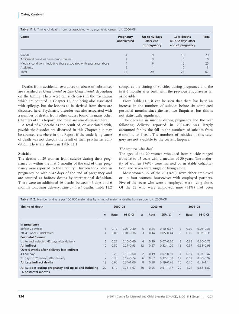
Deaths from accidental overdoses or abuse of substances
are classified as Coincidental or Late Coincidental, depending
on the timing. There were ten such cases in the triennium
which are counted in Chapter 12, one being also associated
with epilepsy, but the lessons to be derived from them are
discussed here. Psychiatric disorder was also associated with
a number of deaths from other causes found in many other
Chapters of this Report, and these are also discussed here.
A total of 67 deaths as the result of, or associated with,
psychiatric disorder are discussed in this Chapter but may
be counted elsewhere in this Report if the underlying cause
of death was not directly the result of their psychiatric con-
dition. These are shown in Table 11.1.
SuicideThe deaths of 29 women from suicide during their preg-
nancy or within the first 6 months of the end of their preg-
nancy were reported to the Enquiry. Thirteen took place in
pregnancy or within 42 days of the end of pregnancy and
are counted as Indirect deaths by international definition.
There were an additional 16 deaths between 43 days and 6
months following delivery, Late Indirect deaths. Table 11.2
compares the timing of suicides during pregnancy and the
first 6 months after birth with the previous Enquiries as far
as possible.
From Table 11.2 it can be seen that there has been an
increase in the numbers of suicides before six completed
postnatal months since the last two Enquiries, but this is
not statistically significant.
The decrease in suicides during pregnancy and the year
following delivery reported in 2003–05 was largely
accounted for by the fall in the numbers of suicides from
6 months to 1 year. The numbers of suicides in this cate-
gory are not available to the current Enquiry.
The women who diedThe ages of the 29 women who died from suicide ranged
from 16 to 43 years with a median of 30 years. The major-
ity of women (76%) were married or in stable cohabita-
tion, and seven were single or living alone.
Most women, 22 of the 29 (76%), were either employed
or, in four women, housewives with employed partners.
Five of the seven who were unemployed were living alone.
Of the 22 who were employed, nine (41%) had been
Table 11.1. Timing of deaths from, or associated with, psychiatric causes. UK: 2006–08
Cause Pregnancy
undelivered
Up to 42 days
after end
of pregnancy
Late deaths
43–182 days after
end of pregnancy
Total
Suicide 4 9 16 29
Accidental overdose from drugs misuse 2 3 5 10
Medical conditions, including those associated with substance abuse 4 16 5 25
Accidents 2 1 0 3
Total 12 29 26 67
Table 11.2. Number and rate per 100 000 maternities by timing of maternal deaths from suicide; UK: 2000–08
Timing of death 2000–02 2003–05 2006–08
n Rate 95% CI n Rate 95% CI n Rate 95% CI
In pregnancy
Before 28 weeks 1 0.10 0.03–0.40 5 0.24 0.10–0.57 2 0.09 0.02–0.35
28–41 weeks undelivered 4 0.05 0.01–0.36 3 0.14 0.05–0.44 2 0.09 0.02–0.35
Postnatal Indirect
Up to and including 42 days after delivery 5 0.25 0.10–0.60 4 0.19 0.07–0.50 9 0.39 0.20–0.75
All Indirect 10 0.50 0.27–0.93 12 0.57 0.32–1.00 13 0.57 0.33–0.98
Over 6 weeks after delivery late Indirect
43–90 days 5 0.25 0.10–0.60 2 0.19 0.07–0.50 4 0.17 0.07–0.47
91 days to 26 weeks after delivery 7 0.35 0.17–0.74 6 0.57 0.32–1.00 12 0.52 0.30–0.92
All Late Indirect deaths 12 0.60 0.34–1.06 8 0.38 0.19–0.76 16 0.70 0.43–1.14
All suicides during pregnancy and up to and including
6 postnatal months
22 1.10 0.73–1.67 20 0.95 0.61–1.47 29 1.27 0.88–1.82
Oates, Cantwell
134 ª 2011 Centre for Maternal and Child Enquiries (CMACE), BJOG 118 (Suppl. 1), 1–203
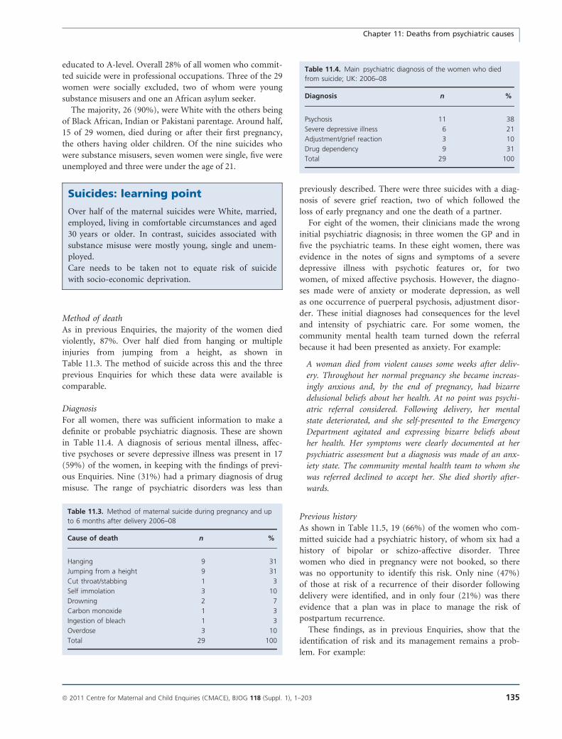
educated to A-level. Overall 28% of all women who commit-
ted suicide were in professional occupations. Three of the 29
women were socially excluded, two of whom were young
substance misusers and one an African asylum seeker.
The majority, 26 (90%), were White with the others being
of Black African, Indian or Pakistani parentage. Around half,
15 of 29 women, died during or after their first pregnancy,
the others having older children. Of the nine suicides who
were substance misusers, seven women were single, five were
unemployed and three were under the age of 21.
Suicides: learning point
Over half of the maternal suicides were White, married,
employed, living in comfortable circumstances and aged
30 years or older. In contrast, suicides associated with
substance misuse were mostly young, single and unem-
ployed.
Care needs to be taken not to equate risk of suicide
with socio-economic deprivation.
Method of deathAs in previous Enquiries, the majority of the women died
violently, 87%. Over half died from hanging or multiple
injuries from jumping from a height, as shown in
Table 11.3. The method of suicide across this and the three
previous Enquiries for which these data were available is
comparable.
DiagnosisFor all women, there was sufficient information to make a
definite or probable psychiatric diagnosis. These are shown
in Table 11.4. A diagnosis of serious mental illness, affec-
tive psychoses or severe depressive illness was present in 17
(59%) of the women, in keeping with the findings of previ-
ous Enquiries. Nine (31%) had a primary diagnosis of drug
misuse. The range of psychiatric disorders was less than
previously described. There were three suicides with a diag-
nosis of severe grief reaction, two of which followed the
loss of early pregnancy and one the death of a partner.
For eight of the women, their clinicians made the wrong
initial psychiatric diagnosis; in three women the GP and in
five the psychiatric teams. In these eight women, there was
evidence in the notes of signs and symptoms of a severe
depressive illness with psychotic features or, for two
women, of mixed affective psychosis. However, the diagno-
ses made were of anxiety or moderate depression, as well
as one occurrence of puerperal psychosis, adjustment disor-
der. These initial diagnoses had consequences for the level
and intensity of psychiatric care. For some women, the
community mental health team turned down the referral
because it had been presented as anxiety. For example:
A woman died from violent causes some weeks after deliv-
ery. Throughout her normal pregnancy she became increas-
ingly anxious and, by the end of pregnancy, had bizarre
delusional beliefs about her health. At no point was psychi-
atric referral considered. Following delivery, her mental
state deteriorated, and she self-presented to the Emergency
Department agitated and expressing bizarre beliefs about
her health. Her symptoms were clearly documented at her
psychiatric assessment but a diagnosis was made of an anx-
iety state. The community mental health team to whom she
was referred declined to accept her. She died shortly after-
wards.
Previous historyAs shown in Table 11.5, 19 (66%) of the women who com-
mitted suicide had a psychiatric history, of whom six had a
history of bipolar or schizo-affective disorder. Three
women who died in pregnancy were not booked, so there
was no opportunity to identify this risk. Only nine (47%)
of those at risk of a recurrence of their disorder following
delivery were identified, and in only four (21%) was there
evidence that a plan was in place to manage the risk of
postpartum recurrence.
These findings, as in previous Enquiries, show that the
identification of risk and its management remains a prob-
lem. For example:
Table 11.3. Method of maternal suicide during pregnancy and up
to 6 months after delivery 2006–08
Cause of death n %
Hanging 9 31
Jumping from a height 9 31
Cut throat/stabbing 1 3
Self immolation 3 10
Drowning 2 7
Carbon monoxide 1 3
Ingestion of bleach 1 3
Overdose 3 10
Total 29 100
Table 11.4. Main psychiatric diagnosis of the women who died
from suicide; UK: 2006–08
Diagnosis n %
Psychosis 11 38
Severe depressive illness 6 21
Adjustment/grief reaction 3 10
Drug dependency 9 31
Total 29 100
Chapter 11: Deaths from psychiatric causes
ª 2011 Centre for Maternal and Child Enquiries (CMACE), BJOG 118 (Suppl. 1), 1–203 135
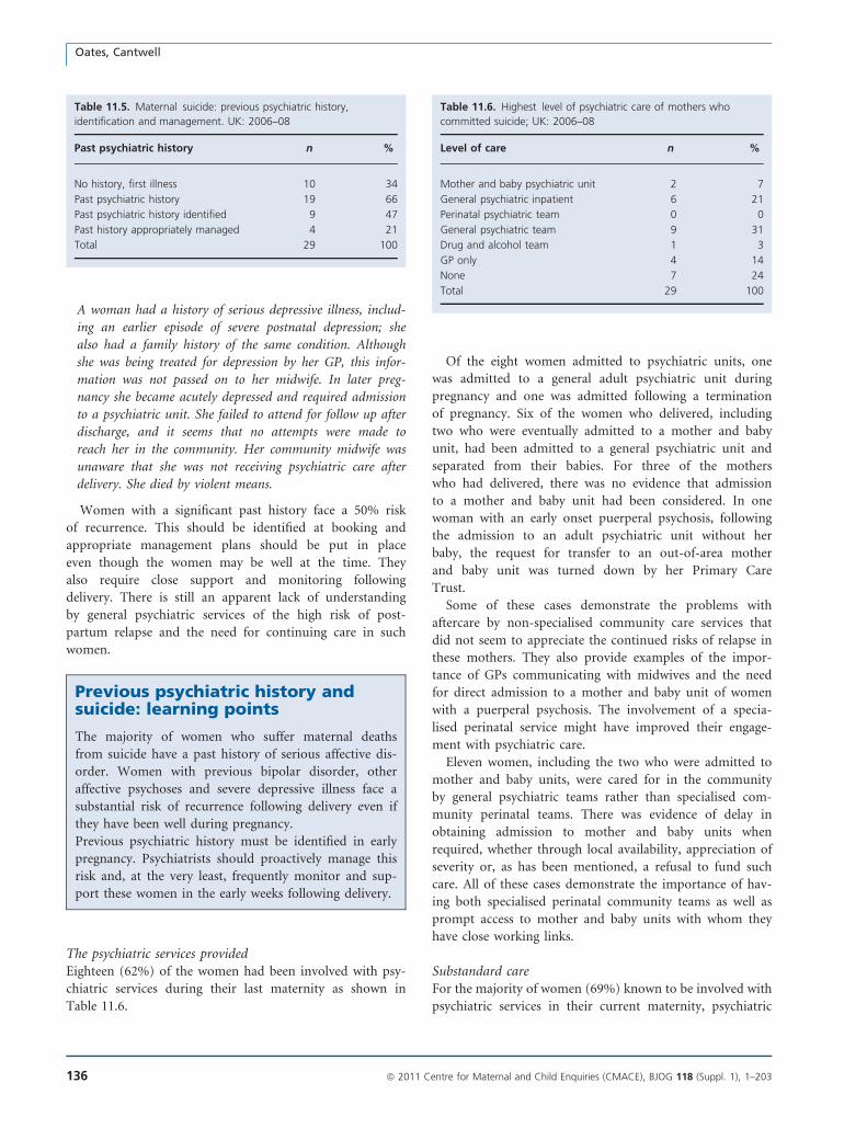
A woman had a history of serious depressive illness, includ-
ing an earlier episode of severe postnatal depression; she
also had a family history of the same condition. Although
she was being treated for depression by her GP, this infor-
mation was not passed on to her midwife. In later preg-
nancy she became acutely depressed and required admission
to a psychiatric unit. She failed to attend for follow up after
discharge, and it seems that no attempts were made to
reach her in the community. Her community midwife was
unaware that she was not receiving psychiatric care after
delivery. She died by violent means.
Women with a significant past history face a 50% risk
of recurrence. This should be identified at booking and
appropriate management plans should be put in place
even though the women may be well at the time. They
also require close support and monitoring following
delivery. There is still an apparent lack of understanding
by general psychiatric services of the high risk of post-
partum relapse and the need for continuing care in such
women.
Previous psychiatric history andsuicide: learning points
The majority of women who suffer maternal deaths
from suicide have a past history of serious affective dis-
order. Women with previous bipolar disorder, other
affective psychoses and severe depressive illness face a
substantial risk of recurrence following delivery even if
they have been well during pregnancy.
Previous psychiatric history must be identified in early
pregnancy. Psychiatrists should proactively manage this
risk and, at the very least, frequently monitor and sup-
port these women in the early weeks following delivery.
The psychiatric services providedEighteen (62%) of the women had been involved with psy-
chiatric services during their last maternity as shown in
Table 11.6.
Of the eight women admitted to psychiatric units, one
was admitted to a general adult psychiatric unit during
pregnancy and one was admitted following a termination
of pregnancy. Six of the women who delivered, including
two who were eventually admitted to a mother and baby
unit, had been admitted to a general psychiatric unit and
separated from their babies. For three of the mothers
who had delivered, there was no evidence that admission
to a mother and baby unit had been considered. In one
woman with an early onset puerperal psychosis, following
the admission to an adult psychiatric unit without her
baby, the request for transfer to an out-of-area mother
and baby unit was turned down by her Primary Care
Trust.
Some of these cases demonstrate the problems with
aftercare by non-specialised community care services that
did not seem to appreciate the continued risks of relapse in
these mothers. They also provide examples of the impor-
tance of GPs communicating with midwives and the need
for direct admission to a mother and baby unit of women
with a puerperal psychosis. The involvement of a specia-
lised perinatal service might have improved their engage-
ment with psychiatric care.
Eleven women, including the two who were admitted to
mother and baby units, were cared for in the community
by general psychiatric teams rather than specialised com-
munity perinatal teams. There was evidence of delay in
obtaining admission to mother and baby units when
required, whether through local availability, appreciation of
severity or, as has been mentioned, a refusal to fund such
care. All of these cases demonstrate the importance of hav-
ing both specialised perinatal community teams as well as
prompt access to mother and baby units with whom they
have close working links.
Substandard careFor the majority of women (69%) known to be involved with
psychiatric services in their current maternity, psychiatric
Table 11.5. Maternal suicide: previous psychiatric history,
identification and management. UK: 2006–08
Past psychiatric history n %
No history, first illness 10 34
Past psychiatric history 19 66
Past psychiatric history identified 9 47
Past history appropriately managed 4 21
Total 29 100
Table 11.6. Highest level of psychiatric care of mothers who
committed suicide; UK: 2006–08
Level of care n %
Mother and baby psychiatric unit 2 7
General psychiatric inpatient 6 21
Perinatal psychiatric team 0 0
General psychiatric team 9 31
Drug and alcohol team 1 3
GP only 4 14
None 7 24
Total 29 100
Oates, Cantwell
136 ª 2011 Centre for Maternal and Child Enquiries (CMACE), BJOG 118 (Suppl. 1), 1–203
care was less than optimal, although in some women it may
not have affected the final outcome. Eleven women were
managed by general adult psychiatric services and had been
treated by multiple psychiatric teams and/or had an inade-
quate risk assessment. In four women, both areas of concern
were found. Another four women had a mistaken initial
diagnosis.
In most women there was also evidence to suggest that
psychiatric teams caring for the women had not appreci-
ated the severity of the women’s illness, as indicated by the
initial diagnosis and the speed, level and frequency of psy-
chiatric intervention. For example:
A woman had a past history of schizo-affective psychosis,
and all her previous episodes were clearly related to reduc-
tions in her medication. She had also had a previous post-
partum episode, during which she made a life-threatening
suicide attempt. She had been well on medication for many
years, and the history was identified in early pregnancy.
Although she remained in close contact with psychiatric ser-
vices throughout her pregnancy, the maternity services
appeared not to know how serious her past illnesses had
been. Following delivery she was seen frequently by a gen-
eral adult community mental health team. A few days
before her death, she became acutely unwell with bizarre
behaviour, delusional ideas and a preoccupation with her
previous suicide attempt. She deteriorated on a daily basis,
and two more psychiatric teams were introduced into her
care. She was then seen very frequently, but the stated aim
of her management was ‘to keep her at home’. She died
from self immolation within a few hours of the last visit by
her community nurse.
This woman had a previous history of a puerperal psy-
chosis and a very serious suicide attempt. Her risk of recur-
rence was high. Both her previous attempt and her current
preoccupation with suicide placed her at high risk,
increased by the rapid onset and deterioration of her con-
dition and a recent change in her medication. Admission
to a psychiatric unit at the onset of her illness might have
altered the outcome. This is also an example of the involve-
ment of multiple psychiatric teams and the lack of both
local specialised community perinatal mental health teams
and a mother and baby unit.
Puerperal psychosis: learning points
Puerperal psychosis (including recurrence of bipolar dis-
order and other affective psychoses) is relatively uncom-
mon in daily psychiatric practice. The distinctive clinical
features, including sudden onset and rapid deteriora-
tion, may be unfamiliar to nonspecialists.
Psychiatric services should have a lowered threshold to
intervention including admission. They should ensure
continuity and avoid care by multiple psychiatric teams.
Specialised perinatal psychiatric services, both inpatient
and community, should be available.
Safeguarding (child protection) social serviceinvolvementNine of the 29 women (31%) who committed suicide had
been referred to social services during their pregnancy,
including eight of the 18 receiving psychiatric care. In five
women, the referral was made because the woman was a
psychiatric patient rather than because of specific concerns
about the welfare of the infant. It was apparent from their
notes that fear that the child would be removed was a
prominent feature of the women’s condition and probably
led them to have difficulties in engaging with psychiatric
care:
A mother who died some weeks after delivery had had
contested custody disputes over her older children. She
had a previous history of reactive depression related to
her circumstances, which had been treated by the GP.
Her psychological and social problems were identified in
early pregnancy, and she received excellent care from her
midwife throughout. Following delivery her midwife iden-
tified a depressive illness, and the GP reacted promptly
and prescribed an antidepressant. She would not take this
because she was concerned about breastfeeding, despite
the midwife reassuring her with information from the
Drug Information Service. There was excellent communi-
cation between the midwife, GP and health visitor. Some
weeks later she deteriorated, and the GP urgently referred
her to mental health services. At the same time she was
also referred to the child protection service, which was
‘routine in that area’. The general adult home treatment
team found her reluctant to engage, and she was fright-
ened that her children would be removed. Admission was
recommended but declined. Shortly afterwards, she pre-
sented to the Emergency Department having swallowed a
corrosive substance but did not reveal that she had also
taken paracetamol. She was admitted to a general psychi-
atric unit but physically deteriorated, revealing that she
had taken an overdose of paracetamol, from which she
died shortly afterwards.
This woman received excellent care from her midwife
and GP. However, it is obvious that this woman was
terrified of losing the care of her children. This fear
influenced her cooperation with the treatments which
might have prevented her death. A specialised community
perinatal team might have been more sensitive to these
issues.
Chapter 11: Deaths from psychiatric causes
ª 2011 Centre for Maternal and Child Enquiries (CMACE), BJOG 118 (Suppl. 1), 1–203 137
Child protection issues: learningpoints
Morbid ideas of maternal incompetence and danger to
the infant are a common feature of maternal mental ill-
ness, as is a fear that their children will be removed.
Referral to safeguarding teams should not be routine
when mothers develop a mental illness but should take
place as the result of a risk assessment.
When referral to the safeguarding team is necessary
because the infant has or is likely to suffer from harm,
then extra vigilance and care are required. Referral to
social services may otherwise result in avoidance of care
and necessary treatment and may increase the risk of
deterioration in the mother’s mental health and suicide.
Internal reviewsIn only five of these women was an internal review by psy-
chiatric services made available to the Enquiry. These
revealed a lack of understanding of the risk factors present
and the points of intervention that might have altered the
outcome. For example, in a woman who set fire to her-
self the review was inadequate. She had a history of affec-
tive psychosis including a postpartum near-fatal suicide
attempt, and, during her final illness, she deteriorated
rapidly and was preoccupied with her previous suicide
attempt. The internal review found no evidence that this
woman was at risk of suicide, that nothing further could
have been done to have prevented her death and that her
death had nothing to do with her pregnancy.
Substance misuseSubstance misuse has increased substantially among women
over the past 30 years, with 2–3% of children in England
and Wales having a parent with drug or alcohol problems.16
Almost two-thirds of drug-using women entering treatment
are parents, but only half have custody of their children.17
Women who are substance users and who attend treatment
programmes are likely to have better antenatal care and bet-
ter general health than those who do not.18
Findings for 2006–08Thirty-five deaths which occurred in women who were
known to be substance misusers in pregnancy and/or the
first 6 months following delivery were considered by the
psychiatric assessors. The causes of their deaths are sum-
marised in Table 11.7. Nine of these women are counted
above in the section on suicide. In addition, ten women
died from an accidental overdose of drugs of addiction;
although they are discussed here, they are counted as Coin-
cidental or Late Coincidental deaths in Chapter 12.
A further 13 women died from medical conditions
caused by or attributed to their drug problem. They are
counted in the relevant chapters but further discussed here.
Three more women died from accidents caused by their
drug addiction. At least two-thirds of the women were
socially excluded, and a substantial number were late
bookers. Some had received no antenatal care. There are
examples of outstanding care with strenuous efforts at
engagement by health professionals. However, in a number
of women, there was evidence of poor information sharing
and lack of recognition of continued use. Heroin was the
most commonly used drug, but polysubstance misuse was
also common.
Suicide in drug-dependent womenNine drug-dependent women committed suicide; this has
also been discussed above. Three women died during preg-
nancy, two after delivery within 42 days and four later than
this. Six of these women had co-morbid conditions: five had
adjustment or grief reactions, and one, a woman who was
abusing over the counter analgesia, had a somatoform pain
disorder (persistent medically unexplained pain). Five had
child protection case conferences, and in three cases, the sui-
cide occurred shortly after a decision to remove the child
into care. In contrast to nonsubstance users who died by
suicide, these women were younger, and five were in their
teens. All of these teenage deaths were by violent means.
Eight out of the nine had a clearly documented past his-
tory of substance misuse, but in only three women was this
identified in early pregnancy and only two women were
managed by drug services. In the remainder, women were
managed by drug liaison midwives alone or with the GP.
Accidental overdose of recreational drugsThere were ten deaths in this category. Two women died
during pregnancy, three between delivery and 42 days and
another five within 43 days and 6 months. All had estab-
lished histories of substance misuse, but in only five was
this identified in early pregnancy. In four women, the rele-
vant information was either not passed to or not sought by
the midwife, by the GP or social services, and the last
Table 11.7. Cause of death in women known to be substances
misusers; UK: 2006–08
Cause of death n %
Suicide 9 26
Accidental overdose of drugs of addiction 10 29
Medical conditions 13 37
Road traffic accidents or house fire 3 9
Total 35 100
Oates, Cantwell
138 ª 2011 Centre for Maternal and Child Enquiries (CMACE), BJOG 118 (Suppl. 1), 1–203
woman was unbooked. In only three women was a specia-
lised drug team involved in the maternity care.
Child protection case conferences were held in eight of
the cases: in four of these antenatal care was then avoided
after the case conference, and three women died very shortly
after a decision was made to remove the baby. For example:
A young woman died from butane inhalation later after
delivery. She had a long history of heroin addiction, and
her partner was also a heroin addict. Her substance misuse
had been implicated in the premature delivery and neonatal
death of an older child. She was involved with a specialist
midwife and the drug addiction team during the index
pregnancy and was referred to social services. She had only
had a very few antenatal visits before giving birth to a sick
baby who spent some months on a neonatal unit. The
mother received frequent visits from the specialised midwife
following delivery, but there was no evidence of increased
contact from the drug addiction services. Shortly before her
death a decision was made to remove the child into foster
care, and she then increased both her street drug and meth-
adone consumption. There was no evidence of any commu-
nication between social services and the drug addiction
team.
It is evident that this woman felt guilty and upset that
her addiction had led to the death of one child and the dis-
ability of a second. It demonstrates the need for extra sur-
veillance and support for women with substance misuse
who are involved with childcare proceedings.
Medical complications of substance misuseIn addition, there were 13 women whose deaths were
reported to the Enquiry and in which substance misuse
played a significant contribution to the cause of death—
two in pregnancy, nine between delivery and 42 days and
two later. Substance misuse was not only implicated in the
aetiology of the fatal condition but also influenced the care
the women received. These deaths are counted in the rele-
vant Chapters. There were three from bacterial endocardi-
tis, two from cardiomyopathy, one from myocardial
infarction related to recent heavy cocaine use and one from
Sudden Adult Death Syndrome counted in Chapter 9.
Three deaths from bronchopneumonia and three related to
the physical consequences of alcohol, including liver disease
and bleeding oesophageal varices are counted in Chapter
10. Ten of the 13 women were heroin abusers, two were
alcohol dependent and one was a cocaine user.
In a few instances, symptoms of the terminal illness were
misattributed to those of drug withdrawal, for example
from cardiomyopathy or cerebral haemorrhage. It was evi-
dent that this misattribution led to delayed diagnosis and
treatment, but it is uncertain whether this affected the out-
come. For example:
A mother died after delivery from a cerebral aneurysm. She
had been an intravenous heroin user for many years but
was not treated by a drug addiction team, obtaining her
heroin and methadone from friends. At booking she actively
denied problems with drug misuse, but later in the preg-
nancy her GP told the midwife of her intravenous heroin
and methadone use, at which point she was referred to
social services. Following this, she avoided antenatal care.
She presented to maternity services as an emergency in
mid-pregnancy with a history of collapse and dizziness,
which was attributed to her substance misuse and she was
sent home. A few weeks later she presented again with a
history of collapse and severe headache. It is clear from the
notes that initially this was thought to be related to her
drug abuse. The full extent of her substance misuse only
became clear after an assessment by an addiction specialist.
Within a few hours, after an epileptic fit, she was found to
have a bleeding aneurysm but died following neurosurgery.
This demonstrates the problem in assuming physical
symptoms are the result of substance misuse. With this
woman, the diagnosis was delayed for some weeks, but it is
unclear whether an earlier diagnosis would have affected
the outcome. This is also an example of the avoidance of
antenatal care following referral to social services.
In all but two (85%) of these 13 women there was evi-
dence of avoiding maternity care. Two women were un-
booked, and the remainder either booked very late or
avoided the majority of their antenatal contacts. For
seven, a child protection case conference had been held,
and four women died shortly after their child had been
removed.
Road traffic accidents and house fireThere were three further deaths in pregnancy, in two of
which the death occurred shortly after a decision was made
to initiate childcare proceedings. Two were from road traf-
fic accidents while intoxicated from either alcohol and can-
nabis or with heroin. The third death, from smoke
inhalation in a house fire, took place towards the end of
pregnancy in a woman who had a history of depression,
domestic violence and alcohol misuse.
Substance misuse: learning points
Women may conceal or minimise the nature and extent
of their substance use, often fearing a censorious
approach or child protection involvement. Early infor-
mation sharing between the GP, maternity and addic-
tion services is essential. Management should include
drug monitoring measures, such as regular urine screen-
ing, particularly where substitute prescribing is used.
Chapter 11: Deaths from psychiatric causes
ª 2011 Centre for Maternal and Child Enquiries (CMACE), BJOG 118 (Suppl. 1), 1–203 139
All women who are substance users should have inte-
grated specialist care. Women should not be managed
solely by their GP or midwife. Integrated care should
include addictions professionals, child safeguarding, and
specialist midwifery and obstetrics.
Other deaths from medical conditions associatedwith psychiatric disorderIn addition to the 13 women who died from medical condi-
tions associated with their substance abuse, a further 12
women died who had an underlying psychiatric disorder that
may have contributed to their terminal condition or influ-
enced the care received. Two of these occurred during preg-
nancy, seven after delivery and there were three Late cases.
The deaths of three women from venous thromboembo-
lism are counted and discussed in Chapter 2. One mother
had a needle phobia which led to her refusing blood tests
and thrombo-prophylaxis and another, who had an emo-
tionally unstable personality disorder, was unable to self-
administer thrombo-prophylaxis and avoided postnatal care
after her child was removed by social services. A woman
with a diagnosis of schizophrenia died while receiving inpa-
tient psychiatric care: her antipsychotic medication was
associated with weight gain and she was a heavy smoker.
In a fourth case, the developing symptoms of physical dis-
ease were misattributed to psychiatric causes. In some
cases, these women also failed to attend for regular antena-
tal care.
Four deaths were from neurological causes and are
counted in Chapter 10. The first was from sudden unex-
plained death in epilepsy in a woman who had an emo-
tionally unstable personality disorder and poor compliance
with care and anti-epileptic medication. The second died in
late pregnancy following an epileptic fit. She had paranoid
schizophrenia. Both her epilepsy and schizophrenia had
deteriorated during pregnancy. Her epilepsy was managed
by her psychiatrist. The third death involved a subarach-
noid haemorrhage in a distressed asylum seeker with post-
traumatic stress disorder whose developing symptoms were
misattributed to psychiatric causes. In the fourth, a woman
died from encephalopathy whose symptoms were misattrib-
uted to psychiatric causes over a prolonged period of time.
Also counted in the Other Indirect deaths chapter is a
death in pregnancy of a woman who died from miliary
tuberculosis but whose anorexia and weight loss was misat-
tributed to psychiatric causes. A further death from pneu-
monia secondary to autoimmune disorder occurred. For
many weeks this woman’s symptoms were misattributed to
depression.
There were two Direct deaths. The first, counted in
Chapter 3, was in a woman with mild learning disability
and epilepsy whose early symptoms were misattributed to
anxiety and agitation, and another woman died from post-
partum haemorrhage and is counted in Chapter 4. This
death, which received media coverage, was associated with
neonaticide in a woman without any previous psychiatric
history:
An older, religious, single professional woman with no
apparent social problems or previous psychiatric history died
from exsanguination as the result of postpartum haemor-
rhage. She had concealed her pregnancy and delivered
unassisted at home. She died of haemorrhage and a dead
infant was found at home.
This woman demonstrates the classical features of neona-
ticide: previously good character, no psychiatric history, no
social services involvement and a stable occupation. She
had a concealed pregnancy and unassisted delivery, and
there was a lack of awareness of close family members,
work colleagues and health professionals that she was preg-
nant. In these cases, the infant is either abandoned or dies
shortly after delivery. The lack of a psychiatric diagnosis is
also typical, but it is likely that at the time she was suffer-
ing from an acute dissociative state. If she had survived,
she would have been charged with infanticide (neonati-
cide). This is the only record of infanticide in this Enquiry.
Misattribution of causeIn six of these 12 examples of physical disease there was a
delay in diagnosis and appropriate treatment being given,
sometimes over prolonged periods of time. This was
because of misattribution of the signs and symptoms of a
medical condition to a psychiatric disorder. In two women,
an acute confusional state, a symptom of the underlying
medical condition, was misattributed to a functional psy-
chiatric disorder leading to repeated requests for psychiatric
assessment and a delay in diagnosis and appropriate treat-
ment. In four others there was no significant psychiatric
disorder, but initial difficulties in making a substantive
physical diagnosis had led to an assumption that the symp-
toms must be of psychological origin. Difficulties in eating
were described as ‘food avoidance’ and ‘refusing to eat’ and
were thought to be an eating disorder. Difficulties in hold-
ing a conversation were described as ‘refusing to speak’
and thought to be depression. Distress and agitation were
described as ‘behavioural’. As an example of this:
A woman died of an autoimmune disorder that had been
diagnosed before pregnancy. Her medical diagnosis initially
presented with lethargy and malaise. Despite this, and her
lack of a psychiatric history, her complaints of feeling
increasingly unwell were attributed by the GP to depression
in mid-pregnancy, and she was referred to a psychiatrist.
She deteriorated and, when admitted to a maternity
Oates, Cantwell
140 ª 2011 Centre for Maternal and Child Enquiries (CMACE), BJOG 118 (Suppl. 1), 1–203
hospital, was described as ‘known to be suffering from
depression’. She was quiet and withdrawn, not eating or
drinking. This was described as ‘depression’ and ‘odd
behaviour’. She was seen by a psychiatric nurse who noted
her to be profoundly physically unwell with no evidence of
depression. Nonetheless, the view of her suffering from a
psychiatric condition continued. Following delivery there
was evidence from the notes that she developed an acute
confusional state. Again this was attributed to depression.
Shortly afterwards she collapsed and died.
From the symptoms described by the psychiatric nurse
and in her obstetric records, it seems unlikely that she was
suffering from a depressive illness. Her symptoms were the
result of her deteriorating physical state and perhaps to
cerebral involvement of her autoimmune disease. The mi-
sattribution of her symptoms and the preoccupation with
her psychiatric diagnosis over a number of weeks led to a
delay in diagnosis and effective treatment.
These deaths involving misattribution of physical symp-
toms to psychological causes are, sadly, in keeping with the
findings of previous Reports.
Misattribution of physical symptomsto psychiatric illness: learning points
The misattribution of physical symptoms and of distress
and agitation to psychiatric disorder has led to a failure
to investigate and delays in diagnosis and treatment of
serious underlying medical conditions in a number of
deaths.
Caution needs to be exercised when diagnosing psychi-
atric disorder if the only symptoms are either unex-
plained physical symptoms or distress and agitation.
This is particularly so when the woman has no prior
psychiatric history or when she does not speak English
or comes from an ethnic minority.
Conclusion
There has been no significant reduction in maternal suicide
within 6 months of delivery since 1997. As in previous En-
quiries, over half of the women who died from suicide
were older, married women in comfortable circumstances
with a previous psychiatric history who were well during
pregnancy. Despite the fact that they faced a substantial
risk of a recurrence of their condition following delivery,
they did not receive preconception counselling, their risk
was not identified at booking nor was it actively managed.
Again in line with the findings of the previous Enquiries,
women in contact with psychiatric services were being
managed by general services who appeared not to be
familiar with the importance of previous history, nor with
the distinctive clinical features of serious postpartum
mental illness. Also sadly, in line with the findings of
previous Enquiries, the majority of maternal suicides died
violently.
The characteristics of women who are substance misus-
ers who died either from suicide or other causes con-
trasted with the other psychiatric maternal deaths. They
were, in the main, young, single, unemployed and socially
deprived.
A new finding of this Enquiry, reflecting the recent
changes in the delivery of psychiatric services, was the
involvement of multiple psychiatric teams in the short per-
iod between the onset of the woman’s condition and her
suicide. The lack of continuity of care, further complicated
by the criteria and protocols involved in each different
team’s acceptance of a patient, seems in some cases to have
contributed to the outcome. This, together with other find-
ings, lends further support for the establishment of specia-
lised perinatal services, both inpatient and community, for
the care of women whose pregnancy or postpartum period
is complicated by serious mental illness.
If the findings of this and the last three Enquiries were
to be implemented, not only would some mothers’ lives be
saved, but the care of those who live would be improved.
Disclosure of interestsRoch Cantwell has no interests to disclose. Margaret Oates
is the Director of NHS East Midlands Perinatal Mental
Health Clinical Network.
FundingThis work was undertaken by the Centre for Maternal and
Child Enquiries (CMACE) as part of the CEMACH pro-
gramme. The work was funded by the National Patient
Safety Agency; the Department of Health, Social Services
and Public Safety of Northern Ireland; NHS Quality
Improvement Scotland (NHS QIS); and the Channel
Islands and Isle of Man.
AcknowledgementsThis Chapter has been reviewed by Professor Ian Jones,
Consultant Perinatal Psychiatrist, University of Cardiff and
Dr Judy Myles, Consultant in Addictions, London. j
References
1 O’Hara MW, Swain AM. Rates and risk of postpartum depression –
a meta-analysis. Int Rev Psychiatry 1996;8:37–54.
2 Cox J, Murray D, Chapman G. A controlled study of the onset, prev-
alence and duration of postnatal depression. Br J Psychiatry
1993;163:27–41.
3 Oates M. Psychiatric services for women following childbirth. Int Rev
Psychiatry 1996;8:87–98.
Chapter 11: Deaths from psychiatric causes
ª 2011 Centre for Maternal and Child Enquiries (CMACE), BJOG 118 (Suppl. 1), 1–203 141
4 Kendel RE, Chalmers KC, Platz C. Epidemiology of puerperal psycho-
ses. Br J Psychiatry 1987;150:662–73.
5 Wieck A, Kumar R, Hirst AD, Marks MN, Campbell IC, Checkley
SA. Increased sensitivity of dopamine receptors and recurrence of
affective psychosis after childbirth. Br J Psychiatry 1991;303:603–
16.
6 Robertson E, Jones I, Haque S, Holder R, Craddock N. Risk of puer-
peral and non-puerperal (postpartum) psychosis. Short report. Br J
Psychiatry 2005;186:258–9.
7 Lewis G, Drife J, editors. Why Mothers Die 1997–1999. The Fifth
Report of the Confidential Enquiries into Maternal Deaths in the Uni-
ted Kingdom. London: RCOG Press, 2001.
8 Lewis G. Why Mothers Die 2000–2002. The Confidential Enquiry
into Maternal and Child Health. Report on confidential enquiries into
maternal deaths in the United Kingdom. London: RCOG Press, 2004.
9 Lewis G. editor. The Confidential Enquiry into Maternal and Child
Health (CEMACH). Saving Mothers’ Lives: Reviewing Maternal
Deaths to Make Motherhood Safer – 2003–2005. The Seventh
Report on Confidential Enquiries into Maternal Deaths in the United
Kingdom. London: CEMACH, 2007.
10 Dean C, Kendell RE. The symptomatology of puerperal illness. Br J
Psychiatry 1981;139:128–33.
11 Department of Health, Department for Education and skills. National
Service Framework for Children, Young People and Maternity Ser-
vices. London: Department of Health, 2004.
12 NICE. Guidelines on Antenatal and Postnatal Mental Health. London:
National DoH, 2007.
13 Appleby L. Suicidal behaviour in childbearing women. Int Rev Psychi-
atry 1996;8:107–15.
14 Appleby L, Mortensen PB, Faragher EB. Suicide and other causes of
mortality after postpartum psychiatric admission. Br J Psychiatry
1998;173:209–11.
15 HM Government. New Horizons: A Shared Vision for Mental Health.
London: DoH, 2009.
16 The Advisory Council on the Misuse of Drugs. Hidden Harm.
Responding to the Needs of Children of Problem Drug Users. Lon-
don: ACMD, 2003.
17 National Treatment Agency for Substance Misuse. Women in Drug
Treatment: What the Latest Figures Reveal. London: NTASM, 2010.
18 Department of Health (England) and the devolved administrations.
Drug Misuse and Dependence: UK Guidelines on Clinical Manage-
ment. London: Department of Health (England), the Scottish Govern-
ment, Welsh Assembly Government and Northern Ireland Executive,
2007.
Oates, Cantwell
142 ª 2011 Centre for Maternal and Child Enquiries (CMACE), BJOG 118 (Suppl. 1), 1–203
Chapter 12: Deaths apparently unrelated topregnancy from Coincidental and Late causesincluding domestic abuseG Lewis (on behalf of the Centre for Maternal and Child Enquiries)
National Perinatal Epidemiology Unit (NPEU), University of Oxford, Oxford, UK
Correspondence: Professor Gwyneth Lewis, National Perinatal Epidemiology Unit (NPEU), University of Oxford, Old Road Campus, Oxford
OX3 7LF, UK. Email: [email protected]
Keywords domestic abuse, coincidental, Confidential Enquiry,
maternal, mortality.
Deaths apparently unrelated to pregnancy: specific recommendations
• This Enquiry continues to recommend that routine enquiry, ‘Asking the question’, should be made about domestic
abuse, either when taking a social history at booking or at another opportune point during a woman’s antenatal
period. Midwives should give high priority to ‘Asking the question’ and to giving information to all women about
domestic abuse. The antenatal booking appointment may be the appropriate time to ‘ask the question’ or the mid-
wife may decide to delay until the following appointment when a relationship has already been established.
• All women should be seen alone at least once during the antenatal period to facilitate disclosure of domestic abuse.
Any member of the maternity team who notices that a woman has an injury, for example a black eye, should ask
sympathetically, but directly, about how this occurred and be prepared to follow up this enquiry with information,
advice and support as needed.
• All women should be advised to wear a three-point seat belt throughout pregnancy, with the lap strap placed as low
as possible beneath the ‘bump’ lying across the thighs and the diagonal shoulder strap above the ‘bump’ lying between
the breasts. The seat belt should be adjusted to fit as snugly and comfortably as possible, and if necessary the seat
should be adjusted.
Introduction
This chapter considers those deaths reported to the Enquiry
that occurred in pregnant or recently delivered mothers
from causes apparently unrelated to their pregnancy. Such
deaths, which occur during pregnancy or up to 42 com-
pleted days (6 weeks) after the end of pregnancy are inter-
nationally defined as fortuitous, although this Report uses
the term Coincidental. Deaths occurring between 43 and
364 completed days after the end of the pregnancy are clas-
sified as Late maternal deaths and are not included in the
calculations of mortality rates or ratios (For full definitions
see the Introduction to this Report). Late deaths can be
subdivided into those from Late Direct, Late Indirect and
Late Coincidental causes. These definitions are discussed
further in the Introductory section to this Report.
As heralded in the last Report, this Enquiry for 2006–08
has focused its efforts on assessing Direct and Indirect
deaths occurring within 42 days of delivery, and not all
Coincidental deaths will have been identified and assessed.
Certainly, most Late Coincidental deaths are not included
because they were excluded from assessment this trien-
nium. However, all nine Late Direct deaths from preg-
nancy-related causes were identified and assessed, as were
24 Late Indirect deaths, largely from suicide and cardiomy-
opathy, which occurred up to 6 months after delivery.
These deaths are important to include in this Report
because they can be the result of often identifiable and
treatable conditions aggravated or induced by pregnancy.
Indeed, in the next update on international definitions for
maternal mortality, it is possible that these two causes of
death will be classified as being the result of Direct causes.
Although Coincidental or Late deaths, in international
terms, are not considered as true maternal deaths and do
not contribute to the calculations for any international
Chapter 12: Deaths apparently unrelated to pregnancy
ª 2011 Centre for Maternal and Child Enquiries (CMACE), BJOG 118 (Suppl. 1), 1–203 143
maternal mortality rates or ratios, they may contain impor-
tant messages for the providers of maternity care. Clini-
cally, the lessons may include basic principles for the
management of pregnant or recently delivered women with
underlying medical or psychiatric conditions. From a pub-
lic-health perspective, the continuing assessment of such
deaths re-enforces the need for information on the correct
use of seat belts in pregnancy, and, perhaps most impor-
tantly, the identification and management of pregnant
women who suffer from physical, verbal or emotional
abuse. Issues related to domestic abuse, recommendations
and suggested guidelines have been fully described in spe-
cific chapters in the previous two Reports of this Enquiry.
The main findings for this triennium, which remain largely
unchanged, are discussed at the end of this Chapter (in
Annex 12.1), and readers are referred to previous Reports
and other references for more detailed information.
Summary of key findings for 2006–08
Coincidental deathsIn this triennium, the deaths of 50 women who died of
Coincidental causes either during their pregnancy or within
42 days of delivery were reported to this Enquiry, com-
pared with 55 for the previous triennium. As in previous
Reports, and as shown in Table 12.1, the largest overall cat-
egory were deaths due to ‘unnatural’ causes, largely road
traffic accidents, murder or unintentional overdoses of
street drugs. Some women who died from other medical
causes such as pneumonia are included in this section, as
their pregnancies (invariably at an extremely early gesta-
tion) were only discovered during the autopsy. The deaths
of nine women who died from Coincidental malignancies
are discussed in Chapter 10 and those associated with sub-
stance abuse are discussed in Chapter 11. By chance, and
not representative of the number of women who died from
Late Coincidental causes, the Enquiry was notified of nine
such deaths this triennium; seven were the result of over-
doses of street drugs and two were from medical condi-
tions. These are not used in statistical calculations.
As with previous Reports, and other causes of death, many
of the women whose deaths are counted here were vulnerable
and socially excluded. The general lessons to be drawn from
this group of mothers have been discussed in Chapter 1.
Road traffic accidentsThe Enquiry received reports on the deaths of 17 women
who died as the result of road traffic accidents during preg-
nancy or within 6 weeks of delivery during this triennium.
In two deaths, the women were under the influence of
drugs or alcohol, in one of which the mother had recently
had her baby taken into care. In the opinion of the asses-
sors, there is the possibility that this accident may have
been intentional. In another example where suicide might
also have been a possibility, a mother in mid-pregnancy
was suffering from depression and had required hospitalisa-
tion following assaults from her violent partner, who had
recently given her a sexually transmitted disease. She had
also had a number of previous terminations, all signs of
having a violent, controlling partner.
Two pregnant women were pedestrians hit by motor
vehicles. One, who was hit on a motorway, had a chaotic
lifestyle, was addicted to street drugs and had all her previ-
ous children taken into care. She was approaching another
child protection conference when she died. Here, too, the
question remains about why she was walking on a motor-
way in the first place.
All but two of the women were still pregnant when they
died, and in only one woman was there clear evidence that
she was not wearing a seat belt. In four women, a peri-
mortem caesarean section was performed in the Emergency
Department. The gestations ranged from 24 to 41 weeks;
none of the babies survived.
The advice for the use of seat belts in pregnancy is given
in the overall recommendations at the start of this chapter.
Late deathsAs previously discussed, apart from all nine Late Direct and
24 Late Indirect deaths assessed as having been related to
pregnancy, notably because of puerperal psychosis and car-
diomyopathy, from which important lessons for maternity
care can still be drawn, other deaths in women in their first
postnatal year have not generally been assessed this trien-
nium. There were, however, nine examples of such Late
Coincidental deaths for which assessments were made, lar-
gely to exclude them being assessed as the result of Late
Indirect causes. Therefore, apart from Direct and certain
Indirect Late deaths, due to the more limited assessment of
most other later deaths this triennium, these numbers and
Table 12.1. Coincidental maternal deaths reported to the Enquiry;
UK: 2006–08
Cause of death Coincidental during
pregnancy and up
to and including
42 days after delivery
Unnatural deaths
Road traffic accident 17
Murder 11
Overdose of street
drugs/drug-related
6
House fire/burns 1
Cancer (see Chapter 10) 9
Medical conditions 6
All Coincidental deaths 50
Lewis
144 ª 2011 Centre for Maternal and Child Enquiries (CMACE), BJOG 118 (Suppl. 1), 1–203
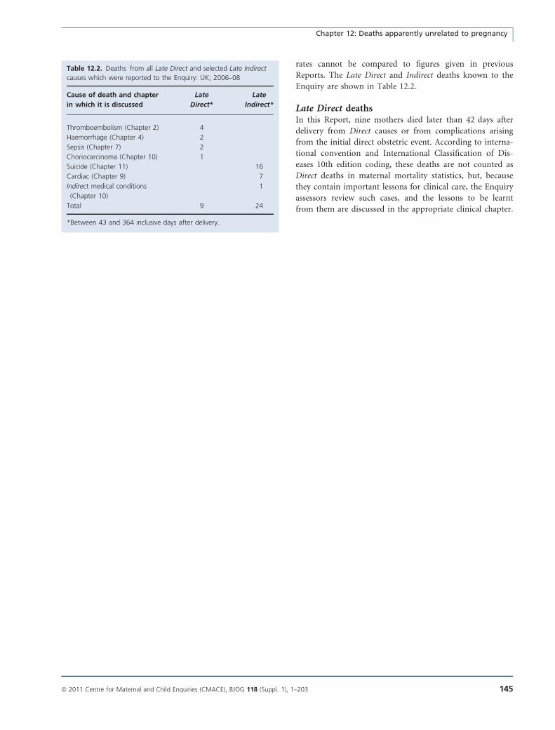
rates cannot be compared to figures given in previous
Reports. The Late Direct and Indirect deaths known to the
Enquiry are shown in Table 12.2.
Late Direct deathsIn this Report, nine mothers died later than 42 days after
delivery from Direct causes or from complications arising
from the initial direct obstetric event. According to interna-
tional convention and International Classification of Dis-
eases 10th edition coding, these deaths are not counted as
Direct deaths in maternal mortality statistics, but, because
they contain important lessons for clinical care, the Enquiry
assessors review such cases, and the lessons to be learnt
from them are discussed in the appropriate clinical chapter.
Table 12.2. Deaths from all Late Direct and selected Late Indirect
causes which were reported to the Enquiry: UK; 2006–08
Cause of death and chapter
in which it is discussed
Late
Direct*
Late
Indirect*
Thromboembolism (Chapter 2) 4
Haemorrhage (Chapter 4) 2
Sepsis (Chapter 7) 2
Choriocarcinoma (Chapter 10) 1
Suicide (Chapter 11) 16
Cardiac (Chapter 9) 7
Indirect medical conditions
(Chapter 10)
1
Total 9 24
*Between 43 and 364 inclusive days after delivery.
ª 2011 Centre for Maternal and Child Enquiries (CMACE), BJOG 118 (Suppl. 1), 1–203 145
Chapter 12: Deaths apparently unrelated to pregnancy
Annex 12.1. Domestic abuse
Domestic abuse: new and existing learning points from 2006 to 2008
This Enquiry continues to recommend that routine enquiry, ‘Asking the question’, should be made about domestic
abuse, either when taking a social history at booking or at another opportune point during a woman’s antenatal
period. Midwives should give high priority to ‘Asking the question’ and to giving information to all women about
domestic abuse. The antenatal booking appointment may be the appropriate time to ‘ask the question’ or the mid-
wife may decide to delay until the following appointment when a relationship has already been established.
All women should be seen alone at least once during the antenatal period to facilitate disclosure of domestic abuse.
Any member of the maternity team who notices that a woman has an injury, for example a black eye, should ask
sympathetically, but directly, about how this occurred and be prepared to follow up this enquiry with information,
advice and support as needed.
The recent report Responding to Violence against Women and Children recommended, as does this Report, that health
service providers and purchasers should have clear policies on the use of interpretation services that ensure that
women and children are able to disclose violence and abuse confidently and confidentially.1
When routine questioning is introduced, this must be accompanied by:
• The establishment of an appropriate method of recording the response on the woman’s records, in such a way that
protects her from further harm from the perpetrator, if abuse is disclosed.
• The development of local strategies for referral to a local multidisciplinary support network to which the woman can
be referred if necessary.
Information about local sources of help and emergency help lines, such as those provided by Women’s Aid, should be
displayed in suitable places in antenatal clinics, for example in the women’s toilets, or printed as a routine at the bottom
of hand-held maternity notes or cooperation cards.
Women who are known to suffer domestic abuse should not be regarded as ‘low risk’. They should be offered care that
involves other agencies and disciplines as needed for the individual’s situation, within a supportive environment. If they
choose midwifery-led care, the midwife should receive support and advice from an experienced colleague, for example the
Named Midwife for Safeguarding or a Supervisor of Midwives.
It must be remembered that health professionals, too, are victims of abuse and that domestic abuse occurs across all
social classes and within all ethnic groups.
Background
Domestic abuse has been the subject of separate chapters in
two previous Reports,2,3 and readers are referred to them for
a more detailed background information as well as the other,
more recent, documents that are referenced in this Chapter.
Domestic abuse has been defined as:
Any incident of threatening behaviour or abuse (psychologi-
cal, physical, sexual, financial or emotional) between adults
who are or have been intimate partners or family members,
regardless of gender or sexuality.4
The term ‘domestic abuse’ is used in preference to
‘domestic violence’ because the latter could be interpreted
as relating to physical abuse alone. It also covers issues that
mainly concern women from minority ethnic backgrounds,
such as forced marriage, female genital mutilation/cutting
and so-called ‘honour crimes’.
Previous Reports have highlighted the issue of domestic
abuse, and even murder, in pregnancy or after delivery. As
a result, a number of local and national initiatives were
introduced, including sensitive routine questioning about
existing abuse during the antenatal period. One publica-
tion, Responding to Domestic Abuse, a Handbook for Health
Professionals,4 arose directly from these recommendations.
The mothers affected by abuse: 2006–08During the 3 years 2006–08, 34 of the women who died
from any cause had features of domestic abuse. It is impor-
tant to remember that although the perpetrator is most
often the woman’s partner, it may also be other family
members. This was the case for the majority of the 11
women who were murdered, the abuse was fatal. Many of
Annex 12.1. Domestic abuse
146 ª 2011 Centre for Maternal and Child Enquiries (CMACE), BJOG 118 (Suppl. 1), 1–203
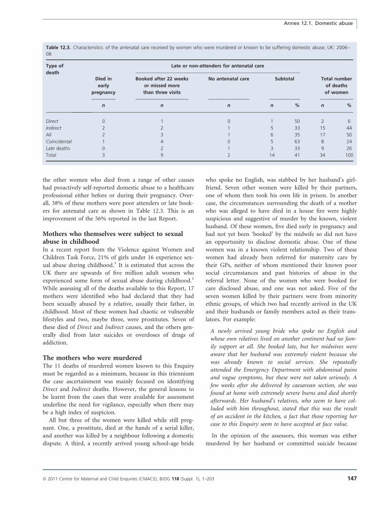
the other women who died from a range of other causes
had proactively self-reported domestic abuse to a healthcare
professional either before or during their pregnancy. Over-
all, 38% of these mothers were poor attenders or late book-
ers for antenatal care as shown in Table 12.3. This is an
improvement of the 56% reported in the last Report.
Mothers who themselves were subject to sexualabuse in childhoodIn a recent report from the Violence against Women and
Children Task Force, 21% of girls under 16 experience sex-
ual abuse during childhood.1 It is estimated that across the
UK there are upwards of five million adult women who
experienced some form of sexual abuse during childhood.1
While assessing all of the deaths available to this Report, 17
mothers were identified who had declared that they had
been sexually abused by a relative, usually their father, in
childhood. Most of these women had chaotic or vulnerable
lifestyles and two, maybe three, were prostitutes. Seven of
these died of Direct and Indirect causes, and the others gen-
erally died from later suicides or overdoses of drugs of
addiction.
The mothers who were murderedThe 11 deaths of murdered women known to this Enquiry
must be regarded as a minimum, because in this triennium
the case ascertainment was mainly focused on identifying
Direct and Indirect deaths. However, the general lessons to
be learnt from the cases that were available for assessment
underline the need for vigilance, especially when there may
be a high index of suspicion.
All but three of the women were killed while still preg-
nant. One, a prostitute, died at the hands of a serial killer,
and another was killed by a neighbour following a domestic
dispute. A third, a recently arrived young school-age bride
who spoke no English, was stabbed by her husband’s girl-
friend. Seven other women were killed by their partners,
one of whom then took his own life in prison. In another
case, the circumstances surrounding the death of a mother
who was alleged to have died in a house fire were highly
suspicious and suggestive of murder by the known, violent
husband. Of these women, five died early in pregnancy and
had not yet been ‘booked’ by the midwife so did not have
an opportunity to disclose domestic abuse. One of these
women was in a known violent relationship. Two of these
women had already been referred for maternity care by
their GPs, neither of whom mentioned their known poor
social circumstances and past histories of abuse in the
referral letter. None of the women who were booked for
care disclosed abuse, and one was not asked. Five of the
seven women killed by their partners were from minority
ethnic groups, of which two had recently arrived in the UK
and their husbands or family members acted as their trans-
lators. For example:
A newly arrived young bride who spoke no English and
whose own relatives lived on another continent had no fam-
ily support at all. She booked late, but her midwives were
aware that her husband was extremely violent because she
was already known to social services. She repeatedly
attended the Emergency Department with abdominal pains
and vague symptoms, but these were not taken seriously. A
few weeks after she delivered by caesarean section, she was
found at home with extremely severe burns and died shortly
afterwards. Her husband’s relatives, who seem to have col-
luded with him throughout, stated that this was the result
of an accident in the kitchen, a fact that those reporting her
case to this Enquiry seem to have accepted at face value.
In the opinion of the assessors, this woman was either
murdered by her husband or committed suicide because
Table 12.3. Characteristics of the antenatal care received by women who were murdered or known to be suffering domestic abuse; UK: 2006–
08
Type of
deathDied in
early
pregnancy
Late or non-attenders for antenatal care
Total number
of deaths
of women
Booked after 22 weeks
or missed more
than three visits
No antenatal care Subtotal
n n n n % n %
Direct 0 1 0 1 50 2 6
Indirect 2 2 1 5 33 15 44
All 2 3 1 6 35 17 50
Coincidental 1 4 0 5 63 8 24
Late deaths 0 2 1 3 33 9 26
Total 3 9 2 14 41 34 100
ª 2011 Centre for Maternal and Child Enquiries (CMACE), BJOG 118 (Suppl. 1), 1–203 147
Annex 12.1. Domestic abuse
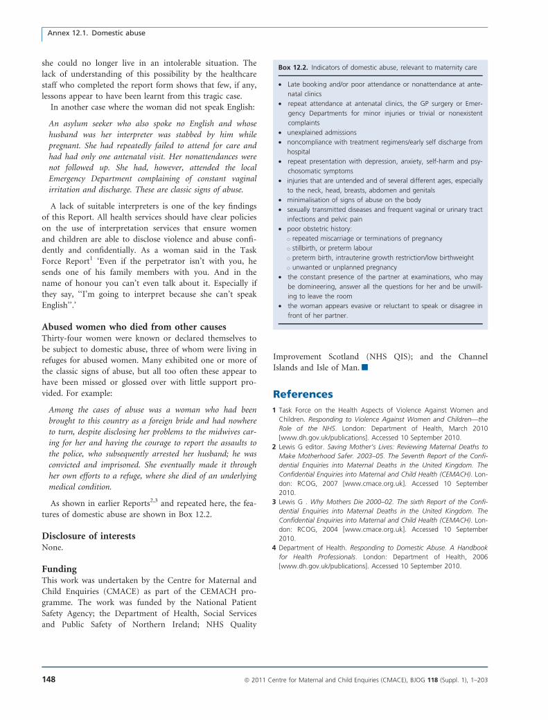
she could no longer live in an intolerable situation. The
lack of understanding of this possibility by the healthcare
staff who completed the report form shows that few, if any,
lessons appear to have been learnt from this tragic case.
In another case where the woman did not speak English:
An asylum seeker who also spoke no English and whose
husband was her interpreter was stabbed by him while
pregnant. She had repeatedly failed to attend for care and
had had only one antenatal visit. Her nonattendances were
not followed up. She had, however, attended the local
Emergency Department complaining of constant vaginal
irritation and discharge. These are classic signs of abuse.
A lack of suitable interpreters is one of the key findings
of this Report. All health services should have clear policies
on the use of interpretation services that ensure women
and children are able to disclose violence and abuse confi-
dently and confidentially. As a woman said in the Task
Force Report1 ‘Even if the perpetrator isn’t with you, he
sends one of his family members with you. And in the
name of honour you can’t even talk about it. Especially if
they say, ‘‘I’m going to interpret because she can’t speak
English’’.’
Abused women who died from other causesThirty-four women were known or declared themselves to
be subject to domestic abuse, three of whom were living in
refuges for abused women. Many exhibited one or more of
the classic signs of abuse, but all too often these appear to
have been missed or glossed over with little support pro-
vided. For example:
Among the cases of abuse was a woman who had been
brought to this country as a foreign bride and had nowhere
to turn, despite disclosing her problems to the midwives car-
ing for her and having the courage to report the assaults to
the police, who subsequently arrested her husband; he was
convicted and imprisoned. She eventually made it through
her own efforts to a refuge, where she died of an underlying
medical condition.
As shown in earlier Reports2,3 and repeated here, the fea-
tures of domestic abuse are shown in Box 12.2.
Disclosure of interestsNone.
FundingThis work was undertaken by the Centre for Maternal and
Child Enquiries (CMACE) as part of the CEMACH pro-
gramme. The work was funded by the National Patient
Safety Agency; the Department of Health, Social Services
and Public Safety of Northern Ireland; NHS Quality
Improvement Scotland (NHS QIS); and the Channel
Islands and Isle of Man. j
References
1 Task Force on the Health Aspects of Violence Against Women and
Children. Responding to Violence Against Women and Children—the
Role of the NHS. London: Department of Health, March 2010
[www.dh.gov.uk/publications]. Accessed 10 September 2010.
2 Lewis G editor. Saving Mother’s Lives: Reviewing Maternal Deaths to
Make Motherhood Safer. 2003–05. The Seventh Report of the Confi-
dential Enquiries into Maternal Deaths in the United Kingdom. The
Confidential Enquiries into Maternal and Child Health (CEMACH). Lon-
don: RCOG, 2007 [www.cmace.org.uk]. Accessed 10 September
2010.
3 Lewis G . Why Mothers Die 2000–02. The sixth Report of the Confi-
dential Enquiries into Maternal Deaths in the United Kingdom. The
Confidential Enquiries into Maternal and Child Health (CEMACH). Lon-
don: RCOG, 2004 [www.cmace.org.uk]. Accessed 10 September
2010.
4 Department of Health. Responding to Domestic Abuse. A Handbook
for Health Professionals. London: Department of Health, 2006
[www.dh.gov.uk/publications]. Accessed 10 September 2010.
Box 12.2. Indicators of domestic abuse, relevant to maternity care
• Late booking and/or poor attendance or nonattendance at ante-
natal clinics
• repeat attendance at antenatal clinics, the GP surgery or Emer-
gency Departments for minor injuries or trivial or nonexistent
complaints
• unexplained admissions
• noncompliance with treatment regimens/early self discharge from
hospital
• repeat presentation with depression, anxiety, self-harm and psy-
chosomatic symptoms
• injuries that are untended and of several different ages, especially
to the neck, head, breasts, abdomen and genitals
• minimalisation of signs of abuse on the body
• sexually transmitted diseases and frequent vaginal or urinary tract
infections and pelvic pain
• poor obstetric history:
s repeated miscarriage or terminations of pregnancy
s stillbirth, or preterm labour
s preterm birth, intrauterine growth restriction/low birthweight
s unwanted or unplanned pregnancy
• the constant presence of the partner at examinations, who may
be domineering, answer all the questions for her and be unwill-
ing to leave the room
• the woman appears evasive or reluctant to speak or disagree in
front of her partner.
Annex 12.1. Domestic abuse
148 ª 2011 Centre for Maternal and Child Enquiries (CMACE), BJOG 118 (Suppl. 1), 1–203
Chapter 13: MidwiferyD Garrod1, V Beale2, J Rogers3, A Miller4 (on behalf of the Centre for Maternal and Child Enquiries)1 Stepping Hill Hospital, Stockport, UK; 2 NHS South West, Taunton, UK; 3 Southampton University Hospitals Trust, Dublin, Ireland;4 Centre for Maternal and Child Enquiries, London, UK
Correspondence: Ms Debbie Garrod, Stepping Hill Hospital, 6th Floor Women’s Unit, Poplar Grove, Stockport SK2 7JE, UK. Email:
Keywords midwifery care, Confidential Enquiry, maternal,
mortality.
Midwifery practice: specific recommendations
• Carry out, record and act upon basic observations for both women at low and higher risk of complications.
• Recognise and act on symptoms suggestive of serious illness, including sepsis, as outlined in the Back to basics section
of this Report.
• Provide pregnant women and new mothers with information about the prevention and signs and symptoms of possi-
ble genital tract sepsis and the need to seek advice early if concerned, as well as the importance of good personal
hygiene.
• Assess the mother’s risk adequately throughout the continuum of pregnancy and the postnatal period, re-assessing as
needed if circumstances change.
• Refer and escalate concerns to a medical colleague of appropriate seniority.
• Make early referral to psychiatric services of women with serious mental health problems in line with the advice in
Back to basics.
• Ensure the availability and use of professional interpreting services for women who need them.
• Provide continuity of care for vulnerable women to promote engagement with the service.
Some key research questions also emerged during the assessment of the women who died this triennium. These are
listed in the Recommendations section of this Report but include research on assessing, what effect, if any, is the
reduced number of routine postnatal visits and clinical observations having on maternal health.
Introduction
This chapter identifies and highlights the key practice issues
for midwives raised by review of the circumstances and care
of the 261 women who died either of Direct or Indirect
causes of maternal death. There were many instances of
exemplary care but also others where lessons can be learned.
It is the aim of this Chapter to highlight both.
Midwives should read this Chapter in the context of the
Report as a whole and with particular reference to the new
section Back to basics. The nature of the midwife’s role, as
described below, means that midwifery care cannot be sep-
arated from the care provided by other members of the
maternity team. Many of the key issues raised in this
Report relate to effective team-working or multidisciplinary
care, as well as issues of clinical importance, so there is
much to be learnt from these other Chapters.
The role of the midwifeEvery pregnant woman has care provided by a midwife.
This places the midwife firmly by her side, as her compan-
ion throughout the childbearing continuum and the pro-
vider of care, advice, support and information appropriate
to her circumstances. The midwife is also her advocate in
ensuring that she receives the care best suited to meeting
her health and social needs. It was evident from some of the
situations reviewed for this Report that midwives need to
develop clear boundaries between advocacy and collusion.
There were instances where midwives should have taken a
Chapter 13: Midwifery
ª 2011 Centre for Maternal and Child Enquiries (CMACE), BJOG 118 (Suppl. 1), 1–203 149
supportive but challenging approach to ensure that women
received appropriate care that was in the best interests of
themselves and their babies. As examples:
A woman with a raised Body Mass Index (BMI) declined
to be weighed and, although she was booked for consultant-
led care, did not attend the appointments to see her obste-
trician. There was no evidence that her midwife had
explained the risks of her raised BMI or provided further
advice, information or support. She did not have thrombo-
prophylactic measures prescribed following a complicated
delivery requiring a caesarean section, and basic postnatal
observations were missed.
Although the deficiencies in her midwifery care cannot
be directly linked to her death from pulmonary embolism,
there is nevertheless no evidence that midwives addressed
the issue appropriately at the beginning of pregnancy, in
such a way that could have enabled a management plan to
be put into place.
A high-risk woman with learning disabilities and very high
levels of anxiety developed symptoms of pre-eclampsia, in
spite of which she continued to be inappropriately managed
in the community. Following the birth, she left hospital
against medical advice and, during the postnatal period,
became increasingly unwell. She died from disseminating
intravascular coagulation some days after birth.
Although her midwifery care was kind and supportive,
the midwives’ acceptance of her anxiety appeared to influ-
ence their decision to offer midwife-led care, which for this
woman was not appropriate. An alternative approach to
her care would have been to work with her to help her
overcome her anxieties, involving support from psychology
and/or learning disability services. This could have enabled
the woman to access the care she needed.
As illustrated by the vignettes above, the role of the mid-
wife is to become the woman’s ‘care navigator’1 working
within the context of the maternity team and other agen-
cies, providing both the continuity and the communication
links that will protect and ensure the mother’s health and
wellbeing. Similar lessons can be learned from some of the
25 Indirect deaths where the women were booked for mid-
wife-led care. Some of these women who died had co-mor-
bidities that were either missed by the midwife or deemed
to be unimportant.
The public-health role of the midwifeMidwives have the opportunity to make a substantial con-
tribution to the public-health agenda and to maximising
health gain and reducing general health inequalities. The
opening Chapter of this Report highlights yet again that
the most vulnerable mothers in society are at far higher
risk of maternal death, and it is imperative that they can
readily access maternity care which should, where possible,
be a positive experience. Women disadvantaged by social
deprivation, unemployment and minority ethnic status, for
example, are doubly disadvantaged by the fact that they
tend to book later for antenatal care. For some of the
women who died, earlier access to care could have made a
difference. The National Institute for Health and Clinical
Excellence (NICE) have recently issued a guideline on the
organisation of services for women with complex social
issues that may affect their pregnancy2 as a result of
recommendations made in several earlier Reports of this
Enquiry.
Chapter 1 highlights the key issue of obesity, a major
risk factor for maternal deaths and disability; helping and
supporting obese pregnant women requires urgent and co-
ordinated action. A systematic and pro-active approach to
addressing the problems associated with raised BMI is a
key priority for maternity services, and midwives are cen-
tral to implementing these approaches.
Learning from the mothers who died:issues for midwifery practice
Learning from substandard careOne of the main aims of this Enquiry is to learn lessons
from the management of the women who died to
improve care in future. The first step in doing this is to
identify any avoidable and substandard factors in the care
they received. This is done by both regional and central
Centre for Maternal and Child Enquiries midwifery asses-
sors. The detailed process is discussed in Appendix 1 of
this Report. At each stage, the case notes are reviewed by
midwives who specifically focus on the appropriateness of
the midwifery care throughout the continuum of care.
The factors identified by the assessors clearly demonstrate
that, in a small number of women, midwifery care was
not provided within the midwife’s contractual or statutory
requirements and has fallen outside the sphere of safe
practice. It is also worth noting that some of the internal
reviews considered for this Report demonstrated a failure
by midwives to recognise that care had been substandard.
During this triennium, a total of 261 women died from
Direct or Indirect causes. In 31 of the 107 Direct deaths
(29%), the midwifery assessors considered midwifery care
to be substandard, as well as in 27 of the 154 (16%) Indi-
rect deaths. This gives a rate of 22% overall for the 261
women who died of Direct and Indirect causes and whose
deaths comprise the numerator for the reported maternal
mortality rate. In a further seven women who were
excluded from the statistics because their deaths were
either unrelated to pregnancy or happened late in the
postnatal year, care was also less than optimal. Issues
Garrod et al.
150 ª 2011 Centre for Maternal and Child Enquiries (CMACE), BJOG 118 (Suppl. 1), 1–203
relating to substandard care can be broadly grouped into
poor midwifery care, failure to ensure appropriate senior
presence and input to care and failure to recognise, and
act on, high risk. In a majority of women, more than one
of these factors was present.
Poor general midwifery careThe poor care identified by the midwifery assessors was the
result of a number and combination of factors including:
• poor communication
• inadequate documentation
• failure to perform observations
• failure to follow up observations or to follow up the
woman reporting feeling unwell
• failure to visit or re-visit during the postnatal period.
Some high-risk women were not being correctly identi-
fied and managed in their antenatal period: there were
examples of delays in performing and acting on findings
such as proteinuria. In a case in the postnatal period, a
community midwife noted that a woman was experiencing
rigors; however, she failed to record the woman’s tempera-
ture and respiratory rate and to palpate the uterus,
although she did arrange a GP appointment.
Failure to ensure appropriate senior presence andinput to careIn 20 women there was a failure to ensure an appropriate
senior presence or referral. This was either a failure to refer
to specialist or obstetric care during the pregnancy or a fail-
ure to escalate concerns to a senior person, either medical
or senior midwifery staff, when ominous signs of illness
were evident. In addition to these, there were 11 women for
whom there was a failure by midwives to recognise and act
on obvious risk. For example, a delay in getting an urgent
appointment with a neurologist should not have just been
noted, it should also have been followed up. Similarly,
where a woman failed to attend for such specialist care, fur-
ther advice should have been sought.
Failure to recognise, and act on, high riskThere were several examples of midwives failing to act
appropriately on warning signs/risk factors:
A woman in the postnatal period saw a midwife for a
postnatal examination and reported having felt unwell for
a week with symptoms of breathlessness and pain on
breathing; she also had swelling in one leg and calf and
thigh pain. She was advised by the midwife to attend hos-
pital or a walk-in centre. Some hours later she arrived at
the Emergency Department where she collapsed, was intu-
bated, ventilated and transferred to the Intensive-Care
Unit. A diagnosis of pulmonary embolism/deep vein
thrombosis was made. She went on to have several cardiac
arrests later that day. She continued to deteriorate and
died some days later.
The midwife who saw this woman failed to recognise the
potential severity of her symptoms: she should have taken
urgent action to expedite her transfer to hospital via ambu-
lance. The fact that there was a delay of several hours
before she went to hospital could have made a difference
to the outcome.
Holistic careThe Encarta English Dictionary3 defines ‘holistic’ as ‘taking
into account all of somebody’s physical, mental, and social
conditions in the treatment of illness’.
Within the Enquiry, there were instances where midwives
did not provide such care; for example, situations in which
physical illness was attributed to a past history of mental
health problems:
A woman with a history of chronic physical illness and
panic attacks died as the result of a severe and undiagnosed
infection. Her symptoms and behaviour were attributed to
her past history of mental health issues, and, by the time
investigations were made and treatment was commenced, it
was too late.
The key message is that midwives must listen to women
and use their knowledge and clinical skills to identify and
meet all needs. This woman had repeatedly told the mid-
wives both in hospital and at home that she felt very ill
and was in considerable pain, but her concerns about phys-
ical symptoms were not heard or acted on. When a woman
voices concern about abnormal symptoms, basic observa-
tions must be taken, documented and, if abnormal, acted
on by appropriate referral.
There was also evidence of the converse. The care for a
woman whose death was associated with epilepsy placed
great emphasis on meeting her social and psychological,
but not clinical, needs. This meant that the midwife
neglected to ensure that she had appropriate care in place
to address the risk factors associated with her epilepsy.
Good midwifery practice: over-archingthemes
The over-arching themes for midwifery practice, based on
the stories of the women who died over the past 3 years,
are as follows:
• Provision of ‘basic’ midwifery care: the fundamentals
of practice
• Team work: referral pathways, communication, and
clear care planning
• Recognition of risk factors and serious illness and act-
ing on these
Chapter 13: Midwifery
ª 2011 Centre for Maternal and Child Enquiries (CMACE), BJOG 118 (Suppl. 1), 1–203 151
• Culture and systems within organisations, and new
patterns of care
• Public health issues
These five over-arching themes are explored within the
framework of the different ‘phases’ of the childbearing con-
tinuum: antenatal, intrapartum and postnatal care. In its
guidance for midwives, the Nursing and Midwifery Coun-
cil4 highlights that:
Childbirth is more than the act of giving birth. For a
woman it is a continuous process from conception,
through pregnancy, labour, birth and beyond. It is essen-
tial that anyone providing midwifery care during this
time has the appropriate skills, knowledge and compe-
tence to do so.4
Antenatal care
Antenatal bookingThe importance of the antenatal booking interview cannot
be overemphasised. It is at this point that midwives play a
key role in ensuring an appropriate pattern of care is
agreed with the woman and communicated clearly to her,
making referral to other services and agencies for care and
support. Poor risk assessment and inappropriate referral
pathways for care can and do increase poor outcomes, both
mortality and morbidity. Key issues identified are:
• When women present for care, they must be seen
promptly. There were multiple examples of long delays
between women seeing their GP at 6 weeks and then
having no contact with a midwife until 12 weeks or
later.
• Balancing the woman’s physical, social, psychological,
emotional and spiritual needs enables the midwife to
take into account the woman’s life circumstances,
which may impact on her pregnancy. For example,
there were instances of women experiencing significant
bereavement (loss of partners by suicide) and loss
(removal of previous children into the care of the
Local Authority), which should have been acknowl-
edged and appropriate support offered.
• As highlighted in Chapter 11, women must be asked
about previous history of psychiatric disorder as well
as their current mental health. Women with a history
of a serious mental illness should be referred for psy-
chiatric assessment and for a management plan to be
developed for the management of any acute postnatal
episode even if they are currently well.
• The question of domestic abuse should be raised at
booking or soon afterwards if the midwife is unable to
speak to the woman alone at booking. This issue is
discussed in Chapter 12.
• The definition of risk at booking and the ongoing
review of risk status are key in ensuring safe and
appropriate care. This includes the need to calculate
an accurate BMI at booking, which is of crucial
importance: midwives must not rely on mothers self-
reporting their weight or guessing their height.
• The current organisation of maternity care means that
in many areas women do not see their GP for antena-
tal care. Good communication systems between mid-
wife and GP are essential to ensure that information is
appropriately shared. Women should be advised to
take their hand-held records to GP appointments,
regardless of the reason for the consultation.
Lack of interpretersFailure in the provision of professional interpreting services
emerges as a significant theme, which recurs throughout all
of the chapters of this Report. It should be highlighted here
that there were too many examples of inappropriate use of
family members to interpret. Consequently, in these cases,
the midwife was unable to obtain a comprehensive booking
history, and some women were denied access to support
for domestic abuse. A further issue in relation to language
barriers was identified for those women who arrived in the
UK from abroad later in pregnancy: without interpreting
services, it was impossible to obtain a full booking history
and make an assessment of risk factors.
Ongoing antenatal careThe importance of performing basic observations, record-
ing these and acting on the findings is a key message for
midwives to take from this Report. In particular, there were
numerous instances of midwives failing to perform urinaly-
sis, sometimes with potentially disastrous consequences. Of
equal importance is the responsibility to make appropriate
referral when a deviation from the norm is detected. There
were examples of women with abnormal findings during
an antenatal examination (for example raised blood pres-
sure and glycosuria) being advised to see a practice nurse
for re-assessment. This is entirely inappropriate, as it is
outside the scope of their practice.
The provision of written information is crucial. When a
potential or actual problem is detected, clear written infor-
mation in the woman’s first language is of vital importance
to enable her to seek further help if ‘warning signs’ of a
worsening condition develop. The provision of this infor-
mation, in addition to verbal advice, should be docu-
mented.
A woman who booked late for antenatal care had symp-
toms of pre-eclampsia identified at a community antenatal
appointment in the third trimester. She was referred to the
obstetric unit the following day, reviewed and community
Garrod et al.
152 ª 2011 Centre for Maternal and Child Enquiries (CMACE), BJOG 118 (Suppl. 1), 1–203
midwife follow up was arranged. This did not take place as
planned, and some days later she self-referred to the hospi-
tal, arriving with a blood pressure of 215/115 mmHg. Her
blood pressure proved impossible to control; she had an
emergency caesarean section and died within a week as a
result of a brain haemorrhage. There was no evidence that
she had been given written information about the signs of
pre-eclampsia.
As highlighted in Chapter 2, women are at risk of
thromboembolism from the very beginning of pregnancy
until the end of the puerperium. Midwives must be aware
of this risk, carry out risk assessments, and act on them.
When obstetric input is indicated, midwives have a
responsibility to ensure that this input is at the correct level
of seniority. For example, a pregnant woman admitted to
hospital with complications and the potential for serious
illness requires early assessment by a senior obstetrician. In
at least one organisation where women died, the admission
policy was for all women to be reviewed by a senior house
officer before a registrar or above was called. This is
entirely inappropriate.
Women who have long-term co-existing medical condi-
tions need appropriate and consistent advice on managing
these: for example, women with epilepsy should be
advised against bathing or bathing their baby unattended;
women with asthma should continue with their medica-
tion if required. Midwives are advised to read Chapter 10
which considers issues relating to ‘Other Indirect deaths.’
Women who do not attend antenatal appointments are
at risk of developing complications that go undetected until
their condition becomes serious or life-threatening. These
women are also more likely to be socially vulnerable, in
poorer physical health and to have mental health problems,
as outlined in Chapter 1. All these factors increase their
vulnerability. There are many examples of midwives going
to great lengths to support these women and to provide
care tailored to meet their complex needs. For example:
A grand multiparous woman had a long history of drug
and alcohol abuse and chaotic lifestyle. She booked early in
her pregnancy and had appropriate referral to all relevant
agencies and services, some of which she accessed. She had
regular antenatal care, facilitated by the midwife’s efforts to
follow up when she did not attend. Despite this, she died of
a drug overdose.
Her midwife should be commended for her recognition
of this woman’s extreme vulnerability and her perseverance
in providing her with appropriate care. However, alongside
the positive examples of good practice such as this, there
were also instances where there was a lack of robust sys-
tems to follow up women who failed to attend for care.
The failure to record this, and so enable follow up, further
increased the likelihood of these women suffering severe
pregnancy-related complications or death.
Where there are safeguarding issues and social care
involvement, the decision may be taken to remove a baby
at birth to the care of the Local Authority. The period of
time ‘pre and post case conference’ should be recognised as
one of high vulnerability for women where vigilance is
vital. Support should be embedded into the system; multi-
agency liaison is essential to address the woman’s needs.
However, a midwife whom the woman knows and trusts
has a particular role to play in providing ongoing support,
and a follow-up visit or appointment should be planned
following the case conference decision.
Intrapartum care
The basics: communicating with womenListening carefully to women and assessing their physical
and emotional needs is a fundamental part of good mid-
wifery care. Good communication involves giving clear
explanations and checking that the woman has understood,
as well as listening. There were several examples in which
there was no evidence of women having been involved in
decisions that set in motion the chain of events that led to
their deaths. These include decisions to induce labour with
no clear indication. For example:
A multiparous woman was induced with prostaglandins for
‘post maturity’. Her first labour had been short, and this
time labour lasted for less than an hour. Following a nor-
mal delivery, she was transferred to the postnatal ward
where she collapsed and died a few hours after the birth of
a massive haemorrhage.
In this case, there was no clear rationale given for the
induction of labour and no evidence that the risks and
benefits of induction had been discussed with the mother.
Women of minority ethnic groups who have no or poor
use of English are particularly vulnerable during labour. As
in the antenatal period, the use of family members for
translation is not appropriate. Commissioners and provid-
ers of maternity services must ensure the 24-hour availabil-
ity of language support services to reduce women’s
vulnerability at this time. There were several examples
where the use of an interpreter would have greatly assisted
the mother during labour. For example:
A non-English-speaking woman planned a vaginal birth
following a previous caesarean section. Although she first
presented to her GP at 12 weeks, she failed to attend for
booking until her third trimester. At this stage, the mid-
wife she saw was unable to undertake her booking because
of pressure of work so this was carried out some weeks
later. At term she had an assisted delivery for delay in
Chapter 13: Midwifery
ª 2011 Centre for Maternal and Child Enquiries (CMACE), BJOG 118 (Suppl. 1), 1–203 153
the second stage and suspected fetal compromise. She col-
lapsed and died of a haemorrhage some hours later. The
only mention of an interpreter was when the woman was
in extremis and the staff wished to explain events to her
husband.
Basic observations and assessmentThere were many examples of failure to make or act
upon basic observations. For instance, a woman with sev-
eral risk factors for pre-eclampsia arrived at hospital with
a fully dilated cervix and promptly gave birth. She was
given Syntometrine and, over the next few hours, was
observed to have at least four abnormal features symp-
tomatic of pre-eclampsia. These were not acted upon
until she suffered a cerebral haemorrhage as a result and
died.
Appropriate risk assessment must be performed for all
women and risk status reviewed as necessary. For example:
A morbidly obese woman with additional risk factors for
hypertension had neither obstetric nor anaesthetic review in
either pregnancy or labour and so had no plan for delivery
care. She was induced at term, having had prelabour
rupture of membranes; the syntocinon rate was increased,
on the instruction of the senior midwife, when the cardioto-
cograph was already pathological. An emergency caesarean
section was carried out for suspected fetal compromise, and
this was followed by a fatal postpartum haemorrhage.
Another woman had a retained placenta following a nor-
mal delivery. There seemed to be no appreciation of the
change in her risk status, which led to failure to perform
appropriate observations and monitoring. It was several
hours before she was transferred to theatre. She suffered a
massive haemorrhage and underwent a hysterectomy but
died.
In these, and other cases, there seemed to be no
recognition that a further risk assessment needed to be
made in the light of an unforeseen complication and
that delay in responding might exacerbate the woman’s
condition.
Recognition of risk factors/signs of serious illnessand team workingWomen with risk factors need clear multidisciplinary care
plans for labour and birth. On occasion, midwives’ failure
to call for senior medical help illustrated that this is associ-
ated with a lack of recognition of the seriousness of the
condition or with an inadequate response, including poor
or no planning. For example:
A woman with severe pre-eclampsia was admitted to the
labour ward by an Senior House Officer but was not seen
by a registrar for some hours. Treatment eventually com-
menced but was inadequate, and by the time a consultant
was involved (several hours after admission), the woman
had sustained an intracerebral haemorrhage. She died the
following day.
The midwife should have escalated this woman’s referral
to the consultant obstetrician.
Staffing and SupervisionFor some women, documentation in case notes indicated
that the unit was very busy. However, it was apparent in
almost every case that escalation policies were not activated
appropriately, including involvement of the Supervisor of
Midwives.4 There were, however, some examples of exem-
plary practice:
A woman expecting her first baby was an inpatient at term;
she collapsed and required resuscitation, during which an
emergency caesarean was undertaken. Despite excellent care,
she did not survive.
The clinicians involved in this event responded in a
prompt, efficient and energetic manner, and excellent
teamwork was evident. The standard of the statement writ-
ing is such that a clear audit trail can be seen. In particular,
there was thorough support from the Supervisor of Mid-
wives, who was present for the emergency and also
arranged extensive support for the midwives during the
days and weeks following.
Postnatal care
Midwives should be aware that the postnatal period is
potentially a time of higher risk than pregnancy or labour.
As shown in Chapter 1, 63% of the 261 women who died
from Direct and Indirect causes did so following the birth
of their babies. There is a concern that the growing reduc-
tion in the frequency of direct postnatal visits and the drive
to replace direct contact with telephone communication
may be leading to less than optimal care. This is a research
recommendation in this Report.
Getting the basics rightAssessment of the woman’s health and wellbeing will
reduce the risk of the midwife focusing on emotional/men-
tal health issues that may be masking signs of physical ill-
ness. Having made an overall assessment of wellbeing, the
midwife should ensure that basic observations are per-
formed and recorded, acting on abnormal findings. There
were examples in the report of midwives failing in these
basic responsibilities, following both normal and operative
deliveries.
Garrod et al.
154 ª 2011 Centre for Maternal and Child Enquiries (CMACE), BJOG 118 (Suppl. 1), 1–203
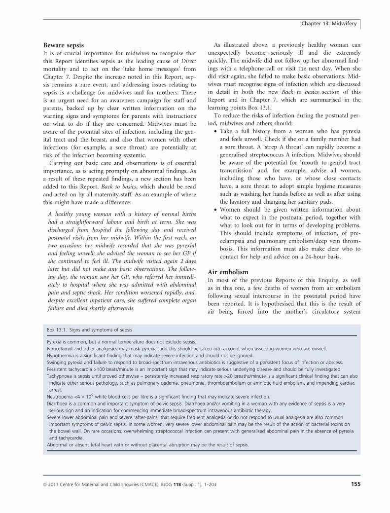
Beware sepsisIt is of crucial importance for midwives to recognise that
this Report identifies sepsis as the leading cause of Direct
mortality and to act on the ‘take home messages’ from
Chapter 7. Despite the increase noted in this Report, sep-
sis remains a rare event, and addressing issues relating to
sepsis is a challenge for midwives and for mothers. There
is an urgent need for an awareness campaign for staff and
parents, backed up by clear written information on the
warning signs and symptoms for parents with instructions
on what to do if they are concerned. Midwives must be
aware of the potential sites of infection, including the gen-
ital tract and the breast, and also that women with other
infections (for example, a sore throat) are potentially at
risk of the infection becoming systemic.
Carrying out basic care and observations is of essential
importance, as is acting promptly on abnormal findings. As
a result of these repeated findings, a new section has been
added to this Report, Back to basics, which should be read
and acted on by all maternity staff. As an example of where
this might have made a difference:
A healthy young woman with a history of normal births
had a straightforward labour and birth at term. She was
discharged from hospital the following day and received
postnatal visits from her midwife. Within the first week, on
two occasions her midwife recorded that she was pyrexial
and feeling unwell; she advised the woman to see her GP if
she continued to feel ill. The midwife visited again 2 days
later but did not make any basic observations. The follow-
ing day, the woman saw her GP, who referred her immedi-
ately to hospital where she was admitted with abdominal
pain and septic shock. Her condition worsened rapidly, and,
despite excellent inpatient care, she suffered complete organ
failure and died shortly afterwards.
As illustrated above, a previously healthy woman can
unexpectedly become seriously ill and die extremely
quickly. The midwife did not follow up her abnormal find-
ings with a telephone call or visit the next day. When she
did visit again, she failed to make basic observations. Mid-
wives must recognise signs of infection which are discussed
in detail in both the new Back to basics section of this
Report and in Chapter 7, which are summarised in the
learning points Box 13.1.
To reduce the risks of infection during the postnatal per-
iod, midwives and others should:
• Take a full history from a woman who has pyrexia
and feels unwell. Check if she or a family member had
a sore throat. A ‘strep A throat’ can rapidly become a
generalised streptococcus A infection. Midwives should
be aware of the potential for ‘mouth to genital tract
transmission’ and, for example, advise all women,
including those who have, or whose close contacts
have, a sore throat to adopt simple hygiene measures
such as washing her hands before as well as after using
the lavatory and changing her sanitary pads.
• Women should be given written information about
what to expect in the postnatal period, together with
what to look out for in terms of developing problems.
This should include symptoms of infection, of pre-
eclampsia and pulmonary embolism/deep vein throm-
bosis. This information must also make clear who to
contact for help and advice on a 24-hour basis.
Air embolismIn most of the previous Reports of this Enquiry, as well
as in this one, a few deaths of women from air embolism
following sexual intercourse in the postnatal period have
been reported. It is hypothesised that this is the result of
air being forced into the mother’s circulatory system
Box 13.1. Signs and symptoms of sepsis
Pyrexia is common, but a normal temperature does not exclude sepsis.
Paracetamol and other analgesics may mask pyrexia, and this should be taken into account when assessing women who are unwell.
Hypothermia is a significant finding that may indicate severe infection and should not be ignored.
Swinging pyrexia and failure to respond to broad-spectrum intravenous antibiotics is suggestive of a persistent focus of infection or abscess.
Persistent tachycardia >100 beats/minute is an important sign that may indicate serious underlying disease and should be fully investigated.
Tachypnoea is sepsis until proved otherwise – persistently increased respiratory rate >20 breaths/minute is a significant clinical finding that can also
indicate other serious pathology, such as pulmonary oedema, pneumonia, thromboembolism or amniotic fluid embolism, and impending cardiac
arrest.
Neutropenia <4 · 109 white blood cells per litre is a significant finding that may indicate severe infection.
Diarrhoea is a common and important symptom of pelvic sepsis. Diarrhoea and/or vomiting in a woman with any evidence of sepsis is a very
serious sign and an indication for commencing immediate broad-spectrum intravenous antibiotic therapy.
Severe lower abdominal pain and severe ‘after-pains’ that require frequent analgesia or do not respond to usual analgesia are also common
important symptoms of pelvic sepsis. In some women, very severe lower abdominal pain may be the result of the action of bacterial toxins on
the bowel wall. On rare occasions, overwhelming streptococcal infection can present with generalised abdominal pain in the absence of pyrexia
and tachycardia.
Abnormal or absent fetal heart with or without placental abruption may be the result of sepsis.
Chapter 13: Midwifery
ª 2011 Centre for Maternal and Child Enquiries (CMACE), BJOG 118 (Suppl. 1), 1–203 155
through the still healing placental bed. It is therefore rec-
ommended that:
• Midwives’ discussion of contraception and sexual
health issues after delivery should include clear advice
about sexual intercourse. The medical assessors to this
Enquiry recommend abstinence for 6 weeks, or gentle
intercourse and avoidance of positions where excess
air could be forced into the vagina.
Team work and care planningLow-risk women who are planning early discharge from
hospital or who have given birth at home need a thorough
and thoughtful first postnatal assessment, with awareness
that serious complications can develop rapidly in the early
postnatal period.
Midwives should be aware that common problems in the
postnatal period can occasionally become life threatening.
There is no room for complacency.
High-risk women need clear discharge planning with
appropriate senior obstetric input. Women who have had
problem-free pregnancies and births but who develop post-
natal complications should have senior obstetric review and
discharge care planning before going home.
It is essential to plan ongoing care and support for the
most vulnerable women, with documented pathways estab-
lished for transfer of care to the health visitor. For exam-
ple:
A woman with an unplanned first pregnancy booked late
for care. No assessment of her mental health was made at
booking or during the pregnancy. She had an emergency
caesarean section and expressed feelings of disappointment
and failure. She struggled with breastfeeding, and this com-
pounded her feelings of inadequacy.
Although the midwife recorded the woman’s emotional
distress, she did not communicate this to the GP or the
health visitor. There was no co-ordinated plan recorded for
her ongoing care and support. This woman committed sui-
cide some weeks after birth.
As highlighted in the previous section on antenatal care,
women with mental health problems and those whose
babies have been removed into the care of the Local
Authority for safeguarding reasons may be particularly vul-
nerable and must be offered ongoing support.
Organisational issuesOrganisational issues relating to postnatal care are of para-
mount importance, in particular full and timely communi-
cation of information and ensuring that there are clear
pathways for cross-boundary hand-over.
New patterns of postnatal care, for example the reduc-
tion in the number of routine home visits, additional clin-
ics/’drop-in’ sessions in Children’s Centres, and the use of
Midwifery Support Workers or other staff must have the
same rigorous follow up and recording systems for woman
who did not attend as should be in place for the antenatal
period. Failure to ensure such systems in place can mean
that women ‘slip through the net’: for example, a woman
did not attend her Children’s Centre postnatal appoint-
ment and was subsequently not seen for a significant per-
iod, during which time she had become seriously ill and
died as a result. Timely intervention from a midwife when
she became symptomatic might have saved her life.
Overall summary: take homemessages
Quality of internal reviewsA woman dying is fortunately a rare event within any sin-
gle organisation. It merits a thorough multidisciplinary
review, conducted in a spirit of open enquiry, to learn any
lessons that may help to save mothers’ lives in the future.
There was evidence of some thorough and excellent
reviews being carried out by Trusts; however, the converse
is also true. Given the rarity of the event, it was surprising
that some reviews were so cursory; others did not have
input from senior midwives into round-table discussions.
All such reviews must include input from the Head of
Midwifery and Supervisors of Midwives. If the arrange-
ments within individual Trusts do not support this, then
advice must be sought from the Local Supervisory Author-
ity.5
Supervision of midwivesMidwifery supervision has a central role to play in enabling
every midwife to reflect on her professional practice and to
learn from those situations where care has been well
planned and delivered as well as those where there have
been adverse outcomes. Supervision is therefore a key tool
in implementing the lessons learned from this Report.5
Final reflections
As described at the beginning of this Chapter, midwives in
the UK are in a unique position in relation to childbearing
women and their families, both in terms of their statutory
responsibilities and their ongoing relationship with women.
They are the healthcare professionals who can take the
overview of the woman’s health and wellbeing throughout
the childbearing continuum. It is imperative that the oper-
ational management of maternity services, as well as indi-
vidual midwives, recognise that ensuring continuity of care
and carer wherever possible plays a vital role in protecting
the wellbeing of women and their babies.
Midwives are the experts in the care of healthy, low-risk
women. They have a clear duty, however, to be equally
Garrod et al.
156 ª 2011 Centre for Maternal and Child Enquiries (CMACE), BJOG 118 (Suppl. 1), 1–203
skilled in the recognition of early signs of problems and to
make prompt referral for appropriate senior medical input.
Midwives accompany women at all stages of the journey
through pregnancy, birth and into new parenthood. The
ongoing nature of this relationship between midwives and
women means that midwives have a clear role as advo-
cates for women, particularly those vulnerable women
most in need of holistic support. However, at times, a
midwife may be the woman’s best advocate by challenging
her and helping her to see that the course of action she is
suggesting, although not the woman’s choice, is in her
best interest. If there is a single ‘take-home’ message for
midwives it is this: listen to the woman and act on what
she tells you.
Disclosure of interestsNone.
FundingThis work was undertaken by the Centre for Maternal and
Child Enquiries (CMACE) as part of the CEMACH pro-
gramme. The work was funded by the National Patient
Safety Agency; the Department of Health, Social Services
and Public Safety of Northern Ireland; NHS Quality
Improvement Scotland (NHS QIS); and the Channel
Islands and Isle of Man. j
References
1 Department of Health. Delivering High Quality Midwifery Care: The
Priorities, Opportunities and Challenges for Midwives. London:
Department of Health, 2009.
2 National Collaborating Centre for Maternal and Child Health. Preg-
nancy and Complex Social Factors: A Model for Service Provision for
Pregnant Women with Complex Social Factors. National Institute for
Health and Clinical Excellence and Social Care Institute for Excel-
lence. London: RCOG, 2010. [www.guidance.nice.org.uk/CG110].
Accessed 10 September 2010.
3 Encarta english dictionary, 2010. [encarta.msn.com/encnet/features/
dictionary/dictionaryhome.aspx]. Accessed 7 September 2010.
4 Nursing and Midwifery Council. Midwives Rules and Standards. Lon-
don: Nursing and Midwifery Council, 2010.
5 Nursing and Midwifery Council. Local Supervising Authority Mid-
wifery Officers Forum Strategic Direction 2008–2011. London: Nurs-
ing and Midwifery Council, 2007.
ª 2011 Centre for Maternal and Child Enquiries (CMACE), BJOG 118 (Suppl. 1), 1–203 157
Chapter 13: Midwifery
Chapter 14: General practiceJ Shakespeare (on behalf of the Centre for Maternal and Child Enquiries)
Summertown Health Centre, Oxford, UK
Correspondence: Dr Judy Shakespeare, Summertown Health Centre, 160 Banbury Road, Oxford OX2 7BS, UK. Email:
[email protected]; [email protected]
Keywords primary care, Confidential Enquiry, maternal,
mortality.
General practice (GP): specific recommendations
All GPs should make themselves aware of the following guidelines:
Venous thromboembolism and pulmonary embolus
• GPs should know the risk factors and warning signs of venous thromboembolism (VTE) and pulmonary embolus
(PE) in pregnancy and the puerperium.1 Risk factors include a past history of embolism, obesity or hyperemesis, and
warning signs include breathlessness.
Pre-eclampsia and eclampsia
• GPs need to ensure that they are up to date with guidelines for the detection and the referral indications for pre-
eclampsia or eclampsia.2 This should be actively excluded in women presenting with epigastric pain in pregnancy.
Asthma
• GP care should be consistent with recent guidelines.3 Steroids and beta-agonists are safe in pregnancy.
Mental health
• GPs are the lynch pin for multidisciplinary communication with the local perinatal mental health and general mental
health services and should follow the perinatal mental health guideline produced by the National Institute for Health
and Clinical Excellence (NICE).4
Obesity
• There are recent guidelines about the management of obesity in pregnancy, and all GPs should be aware of them.5
Information sharing: specificrecommendations
Whenever possible, a GP should give a named community
midwife confidential access to the woman’s full written
and electronic records, with the woman’s consent. If this
is not possible, the GP should supply a named commu-
nity midwife with a summary of the women’s past medi-
cal history, with the woman’s consent.
Introduction
This chapter identifies and highlights the key issues for
GPs raised by the 64 cases of maternal death that were
assessed for this chapter in this triennium. It should be
read in the context of the whole Report, especially the
‘Top ten’ recommendations and the Back to basics sections.
In this chapter, the key clinical issues are addressed in
the same order as the chapters in the Report, with some
additional GP-specific issues. The cases examined had sig-
nificant involvement of a GP in the care of the women
who died, or they were representative of a number of simi-
lar cases. For many women, the GP care was exemplary,
but, by the very nature of this Report, there were some
where the care was less than optimal. An assessment of
whether the care was substandard was a judgement made
in the light of the prevailing standards for good care at the
time of the death as described in Good Medical Practice for
GPs.6 In 45 of these 64 women, GP care was assessed as
Shakespeare
158 ª 2011 Centre for Maternal and Child Enquiries (CMACE), BJOG 118 (Suppl. 1), 1–203
substandard; in 37 women this was judged to be a major
factor, meaning that better care may have changed the
eventual outcome. In the other eight women it was judged
to be a minor factor, meaning that although lessons could
be learned, the care probably did not affect the eventual
outcome.
A significant cross-cutting and recurrent theme is obes-
ity. Obesity in pregnancy is an increasing problem and is
associated with adverse pregnancy outcomes for the
woman. Its prevalence and outcomes are discussed in more
detail in Chapter 1. There are recent guidelines about the
management of obesity in pregnancy.5
Clinical causes of death
Venous thromboembolismTwo of the seven women assessed for this section had sub-
standard GP care. For example, the following case of deep
vein thrombosis (DVT) with a subsequent PE was missed
by two GPs:
An obese woman with recognised needle phobia refused all
blood tests in pregnancy and declined enoxaparin (Clex-
ane) after a caesarean section. She discussed this with a
midwife, who gave her TED stockings and told her to
move her legs, but there was no discussion with a doctor
and no record of her refusal of thromboprophylaxis in the
discharge letter. She saw her GP some days later with
swollen legs but no pain, and her GP stated ‘not a DVT’.
Another GP reviewed her a few days later and prescribed
analgesics for severe leg pain. Some days later she died
from a PE.
This woman had several risk factors for a DVT: a recent
pregnancy, operative delivery and significant obesity. DVTs
cannot be excluded by looking at the legs. The second GP
should also have recognised the classic signs and symp-
toms. Her needle phobia could have been treated by psy-
chological help during pregnancy. It was mentioned in the
GP referral letter, but no action was taken. There should
also have been a discussion in pregnancy about her risk of
VTE, the importance of possible prophylaxis after delivery
and clear documentation in the hospital discharge letter
about her refusal of prophylaxis.
Breathlessness is a common symptom in pregnancy, but
it can be a symptom of PE, and premonitory symptoms
sometimes occur:
A woman with no known risk factors died in her third
trimester from a massive PE having seen her GP a few
days earlier complaining of shortness of breath. She had a
careful examination, including a peak flow and oximetry,
but her pulse was not recorded. A diagnosis of asthma
was made, and she was treated with a salbutamol
inhaler.
A significant event analysis was completed in the prac-
tice and returned to the Enquiry. The practice reflected
on the importance of observing the pulse rate and the
opportunity for the GP involved to be supported by col-
leagues.
Three other women who died of PE had also presented
to their GP with breathlessness shortly before their death.
It can be difficult to distinguish significant causes from
trivial ones, and this is discussed in more detail in the Back
to basics section of this Report. The inclusion of the signifi-
cant event analysis report was unique, and the GP practice
is to be commended for this.
Hyperemesis and PEHyperemesis is a recognised risk factor for VTE, and two
cases of PE associated with severe hyperemesis gravidarum
were assessed. These women had been treated with antie-
metics but had not been monitored for dehydration. Earlier
admission for rehydration and thromboprophylaxis might
have prevented their deaths.
VTE: learning points
• Making, or excluding, a diagnosis of DVT requires
hospital-based investigations.
• Shortness of breath may be the result of a PE, espe-
cially if a woman has risk factors.
• Women with severe hyperemesis are at higher risk of
VTE; they require close monitoring for dehydration
and should be admitted to hospital if they fail to
respond adequately to antiemetics.
• Needle phobic women can benefit from psychological
treatment during pregnancy to avoid compromising
their obstetric or emergency care.
EclampsiaThe cases of several of the 19 mothers who died from
eclampsia were reviewed for this chapter, although they are
counted and discussed in Chapter 3. In all cases, the GP
care was substandard. The main failings were a failure to
undertake routine observations of blood pressure and pro-
teinuria and a failure to understand the significance of, and
act on, significant abnormalities. For example:
A healthy primiparous woman developed proteinuria +++
in her third trimester, but her GP recorded a normal blood
pressure. She was referred urgently to a urology clinic. A
few weeks later she was admitted by a midwife with epigas-
tric pain, irritability and reduced fetal movements with
a blood pressure of >200/110 mmHg. She required an
Chapter 14: General practice
ª 2011 Centre for Maternal and Child Enquiries (CMACE), BJOG 118 (Suppl. 1), 1–203 159
emergency caesarean section after becoming confused
and drowsy. She died the next day from a cerebral
haemorrhage.
Her GP had failed to recognise the significance of new
onset proteinuria in pregnancy, and she should have been
urgently referred to the obstetric services, not urology.
In a further case a GP started treatment for pre-eclamp-
sia without hospital referral:
A woman with hypertension appropriately stopped her
enalapril at the start of pregnancy, but the midwife
booked for low-risk care, although the GP had mentioned
her hypertension in the referral letter. In the second
trimester, her blood pressure was 180/100 mmHg, and her
GP started methyldopa; her urine was not checked for
protein until some days later, when proteinuria was pres-
ent. A few days later she was admitted with epigastric
pain, catastrophic hypertension and heavy proteinuria. She
died of multi-organ failure and HELLP (haemolysis, ele-
vated liver enzymes and low platelet count) syndrome.
GPs should never start treatment for hypertension in pri-
mary care without specialist support. If this woman had
been referred immediately when her blood pressure was
raised, she would probably have survived. The community
management of hypertension in pregnancy has been
addressed in guidelines, and GPs should be aware of these.7
HELLP syndrome is part of the spectrum of pre-eclampsia:
A woman who developed gestational diabetes during preg-
nancy was under the care of the diabetic antenatal unit
(DAU). In her third trimester, she complained about
reduced fetal movements, generalised itching, oedema and
haematemesis. The DAU failed to recognise the significance
of these symptoms and recommended that she see her GP.
By now she also complained of smelly faeces, tiredness, slug-
gish fetal movements and belching. Her GP recorded that
she was jaundiced, with abdominal pain and bilateral leg
oedema; her blood pressure was normal. The GP performed
no investigations but advised her to see the midwife the
next day. By the next morning she was severely ill, with
hypertension, proteinuria +++ and deep jaundice. Despite
immediate delivery, she developed disseminated intravascu-
lar coagulation and died. At autopsy, the diagnosis was
HELLP syndrome.
HELLP syndrome may present before the other symp-
toms of pre-eclampsia. It has a poor prognosis and
requires immediate delivery to prevent rapid deterioration
of maternal liver function. Her specialist care was sub-
standard in failing to recognise the early symptoms of
HELLP syndrome. However, her GP should also have
recognised that severe jaundice in late pregnancy was sig-
nificant, even if she/he did not recognise HELLP syn-
drome, and should have referred her to hospital
immediately. This GP had no other contact with this
woman during pregnancy, was no longer carrying out any
routine antenatal care and was unaware of the serious sig-
nificance of her symptoms.
Pre-eclampsia and eclampsia:learning points
• Epigastric pain is a common symptom of significant
pre-eclampsia.
• Routine observation of urine and blood pressure
should be checked every time a pregnant woman is
seen by a GP, especially if she is feeling unwell.
• Jaundice in a pregnant woman requires immediate
investigation and admission.
• GPs should always refer a woman with pre-eclampsia
for specialist care and should only start antihyperten-
sives in primary care if a woman is severely unwell,
before her emergency admission; in this situation the
treatment of choice is oral labetolol.
Bleeding or abdominal pain in pregnancyDeaths in early pregnancy are counted and discussed in
Chapter 6. Unlike earlier reports, misdiagnosed women
who died from ectopic pregnancies were uncommon, but
they continue to occur:
A woman, unaware of her pregnancy, developed diarrhoea
and vomiting and spoke to an out-of-hours GP. A provi-
sional diagnosis of gastroenteritis was made. Her own GP
visited the next morning and admitted her to hospital
under the physicians, where the junior doctors continued to
treat her for gastroenteritis until she was in extremis. At
autopsy, a ruptured ectopic pregnancy was found.
The GP care was appropriate, but the diagnosis of a pos-
sible ectopic pregnancy was not considered either by the
GP or the hospital doctors. This presentation with diar-
rhoea and vomiting and abdominal pain but without
bleeding is typical of cases reported in the last few Reports.
If an ectopic had been considered, the outcome may have
been different. A higher level of suspicion and pregnancy
testing could make a difference.
A woman who spoke no English died after a miscarriage:
A woman went to see a practice nurse for a routine
immunisation and took her young daughter as an inter-
preter. The nurse elicited that her period was overdue and
declined to give the immunisation. It appeared from later
notes that she had been bleeding vaginally for some time
before this consultation. Later that night she collapsed. At
autopsy, there was evidence of chronic vaginal blood loss
and severe anaemia from an incomplete miscarriage.
Shakespeare
160 ª 2011 Centre for Maternal and Child Enquiries (CMACE), BJOG 118 (Suppl. 1), 1–203
If the nurse had asked more questions about the preg-
nancy, she may have discovered the history of bleeding.
The use of relatives, especially young children, as interpret-
ers is a recurring issue throughout this Report and is dis-
cussed in more detail under the key recommendations in
Chapter 1. GPs and practice staff need to know how to
access translating services rapidly within their area.
Pain and bleeding in earlypregnancy: learning points
• GPs should ask all women of reproductive age with
diarrhoea and vomiting and abdominal pain about
the risk of pregnancy. If there is any doubt, they
should carry out a pregnancy test in the surgery; they
should consider carrying a pregnancy test in their
emergency bags.
• GPs and practice staff should know how to access
professional translation services rapidly. This is a
major recommendation in this Report.
SepsisDeaths from sepsis are discussed in detail in Chapter 7. In
the last Report, the GP chapter warned that ‘puerperal fever
is not a disease of the past’. Deaths from genital tract sepsis
have risen rather than declined in this triennium. Genital
tract sepsis has become the leading cause of Direct maternal
deaths in the UK for the first time since these Confidential
Enquiries into Maternal Deaths commenced in 1952. Many
of these deaths were from community-aquired Group A
streptococcal disease, mirroring an overall background
increase in mortality from this disease in the general popula-
tion. For many of these women, the outcome was unavoid-
able despite excellent care because of the rapid course
and late presentation of the illness. However, in others, pos-
sible opportunities to save lives may have been missed and
lessons remain to be learnt. Many women were in contact
with primary care, of which the following case is typical:
A woman developed a temperature and tachycardia a few
days after delivery, and her midwife suggested that she speak
to her GP if she did not start to feel better, which she did
not do. The midwife saw her a few days later but did no
routine observations or examinations. The GP visited a day
or so later because of abdominal pain, by which time she
was very ill and breathless with signs of peripheral shut
down. The GP made the correct diagnosis of puerperal sep-
sis, spoke to the Emergency Department and faxed a letter
because of his concern, but arranged a nonurgent ambulance
transfer. In hospital, she developed disseminated intravascu-
lar coagulation and multiple organ failure, from which she
subsequently died. Blood culture was positive for group A
b-haemolytic streptococcus.
Although her GP care was generally good, her hospital
transfer should have been quicker. This was not the only
case of septicaemia where a GP did not arrange emer-
gency hospital transfer. The current management of sus-
pected meningococcal septicaemia is to give parenteral
penicillin before blue-light transfer. In puerperal sepsis,
the group A b-haemolytic streptococcus is the usual
pathogen and is sensitive to penicillin, so suspected cases
could also be managed in the same way. The shocking
speed with which this illness progressed from early
symptoms to death in a number of women cannot be
overemphasised.
There were cases when septicaemia developed after a
sore throat and upper respiratory tract infection. These
women had all seen their GP or practice nurse about the
symptoms, and none had been prescribed antibiotics, in
common with routine practice. The GP care was good,
but it may be that the threshold for taking a throat swab
and treating with antibiotics should be lowered in preg-
nancy. The Centor Criteria may be helpful in deciding
which women are more likely to be suffering from
Group A streptococcal infection.8 If any three of the fol-
lowing criteria are positive, then antibiotic prescribing is
indicated:
• history of fever
• tonsillar exudate
• no cough
• tender anterior cervical lymphadenopathy
There were two cases when women became septicaemic
after mastitis. In such cases, an antibiotic should be com-
menced immediately; oral flucloxacillin 500 mg four times
daily for 10–14 days is the treatment of choice.9 The man-
agement of sepsis is discussed in more detail in the Back to
basics section of this Report.
Sepsis: learning points
• All pregnant and recently delivered women should be
informed about the risks and signs and symptoms of
infection and how to take steps to prevent its trans-
misson (see Back to basics section).
• All healthcare professionals should be aware of the
classic signs and symptoms, and sepsis must be con-
sidered in all recently delivered women who feel
unwell and/or have pyrexia.
• Sepsis is often insidious in onset. In the community,
vital signs should always be checked in women who
have any signs or symptoms of possible infection.
• Women with sepsis can deteriorate and die rapidly.
Abdominal pain, fever and tachycardia are indications
for emergency admission by blue-light ambulance for
intravenous antibiotics.
Chapter 14: General practice
ª 2011 Centre for Maternal and Child Enquiries (CMACE), BJOG 118 (Suppl. 1), 1–203 161
Cardiac diseaseDeaths from cardiac conditions are discussed in more
detail in Chapter 9. Ischaemic heart disease is becoming
more common in pregnancy because of later motherhood,
less healthy lifestyles and obesity. However, even with clas-
sic symptoms, two GPs failed to recognise the significance
of this woman’s pain:
An older mother who smoked with a strong family history
of heart disease developed pain in her left arm some weeks
after delivery. Her GP diagnosed muscular pain, but some
days later she woke with retrosternal chest pain radiating to
her left arm and vomiting. The out-of-hours doctor recom-
mended paracetamol. She died at home shortly afterwards
of a myocardial infarction
Her GP and the out-of-hours GP both failed to consider
the diagnosis, despite a classic history, and there was a fail-
ure to make an adequate risk assessment because of inade-
quate telephone triage.
Other mothers with cardiac disease presented with
chest pain and/or breathlessness. Significant cardiac dis-
ease needs to be distinguished from trivial breathlessness
and investigated urgently as an inpatient. This is dis-
cussed in more detail in the Back to basics section of this
Report.
Cardiac disease: learning points
• Not all chest pain in pregnancy is heartburn: it may
be ischaemic, especially if a woman has risk factors.
• Breathlessness in pregnancy may be caused by cardiac
conditions and needs to be carefully assessed.
EpilepsyDeaths from neurological conditions are counted and dis-
cussed in Chapter 10. Several deaths from epilepsy were
reviewed for this Chapter, of whom three had substandard
GP care. For example:
A woman with well-controlled epilepsy had tailed off her
sodium valproate before a planned pregnancy on the advice
of a neurologist. When she had a fit, her GP referred her
back to neurology requesting an immediate appointment;
this was given over a month later. The neurologist recom-
mended starting lamotrigine and wrote to the GP but gave
no specific advice about how to introduce or monitor treat-
ment. The woman did not see her GP following the neurol-
ogy appointment. She was later found drowned in the bath.
At post-mortem her lamotrigine level was <1 mg/l, suggest-
ing that she had not taken the medication.
Here the GP provided good care by referring her
urgently to a neurologist. The neurologist should have
communicated with the GP by fax or telephone and given
explicit instructions about doses and monitoring of medi-
cation. There are specific issues about fluctuations of lamo-
trigine levels in pregnancy, as mentioned in Chapter 10; it
should be common practice to monitor these during preg-
nancy. The GP made no attempt to initiate the recom-
mended treatment. This lady was clearly ambivalent about
medication, perhaps because of worries of adverse effects
on the baby, but both her GP and neurologist should have
explored this with her in more detail.
Risks of bathing aloneAs has been seen in earlier Reports, and in this, women still
drown in the bath having had an epileptic seizure, some-
times with the door locked. This risk is generally not well
recognised, and all epileptic women should be advised to
shower, rather than bathe, and not to lock the bathroom
door. A safety leaflet from Epilepsy Action is available at
www.epilepsy.org.uk/info/safety.
Every epileptic woman of child-bearing age needs to
understand the need for pre-pregnancy counselling and
high-risk care during pregnancy. This is discussed in more
detail under ‘Managing pre-existing diseases’.
Epilepsy in pregnancy: learningpoints
• Epileptic women of child-bearing age need to be
informed of the risks of epilepsy in pregnancy before
conception.
• If fits increase or recur in pregnancy, a woman should
be referred to a neurologist who should see them
within a week.
• Lamotrigine levels need monitoring during pregnancy.
• Pregnant women should be advised to shower, rather
than bathe, and not to lock the bathroom door.
AsthmaThree women dying from asthma received substandard GP
care. They were all women with severe asthma who should
have been under the care of respiratory physicians during
pregnancy. For example:
A mother with severe asthma (history of several respiratory
arrests) stopped her inhaled corticosteroid, possibly on med-
ical advice, at the start of pregnancy. The GP included the
diagnosis of asthma, but not the severity, in his obstetric
referral letter. She was booked for low-risk maternity care.
She requested and obtained many repeat prescriptions for
salbutamol during pregnancy. A few days after delivery she
developed a chest infection and was treated with antibiotics;
her peak flow (PF) was 200 l/minute (44% of her predicted
Shakespeare
162 ª 2011 Centre for Maternal and Child Enquiries (CMACE), BJOG 118 (Suppl. 1), 1–203
PF 450 l/minute). She was subsequently treated with
prednisolone, but her PF, pulse and respiratory rate were
not recorded and no follow up was arranged. A few weeks
later, she suddenly became breathless, collapsed and died of
asthma.
She was inaccurately advised about stopping her inhaled
corticosteroid in pregnancy and was clearly requesting
excessive repeat prescriptions of salbutamol without the GP
noticing. Postnatally, the severity of her asthma was unrec-
ognised, inadequate observations were taken and no follow
up was arranged. If she had been treated using national
guidelines, her death may have been avoided.3 Short- and
long-acting beta agonists and inhaled corticosteroids are
safe in pregnancy.
In a review of the effects of pregnancy on asthma,10
increased hospital admission was associated with a lack of
use of inhaled corticosteroids. In general, the effect of preg-
nancy on known asthma was one-third improved, one-
third worse and one-third unchanged. Most women with
asthma have mild disease and require primary care during
pregnancy. GPs need to provide adequate detail about the
severity of asthma, so that maternity services can assess
women who need specialist care (see learning points).
Asthma: learning points
• GPs must inform maternity services about the severity
of pregnant women’s asthma.
• Most pregnant women with asthma can be cared for
in primary care. Only women with persistent poor
control, a history of hospital admission as an adult,
respiratory arrest or continuous or frequent courses
of oral steroids would usually require specialist refer-
ral to a chest physician during pregnancy.
Psychiatry and substance misuseDeaths from psychiatric causes are counted and discussed
in Chapter 11. As in the last Report, there were cases where
known previous psychiatric history and family history did
not prompt early psychiatric referral and care planning to
anticipate problems after delivery. It appears that GPs are
still not aware that the risk of recurrence of puerperal psy-
chosis is about 50% with a previous history.11 With plan-
ning, some of these deaths might have been prevented:
A pregnant woman had a history of severe postnatal depres-
sion that required hospital admission after her previous
pregnancy and a family history of psychotic illness. She was
taking venlafaxine before and during pregnancy. She was
booked for low-risk care because the midwife did not obtain
her mental health history at booking. In the third trimester,
she was admitted with severe depression but discharged
herself and failed to attend follow-up appointments. After
delivery, the midwife, as instructed, telephoned the psychia-
trist to say she had delivered, but no follow up was
arranged. She did not see her GP after delivery and died of
violent means some weeks later.
She should not have taken venlafaxine in pregnancy.
With her history, she should have been referred to a peri-
natal psychiatrist at the start of pregnancy to develop a
management plan. Following her admission for severe
depression in pregnancy and after delivery, the GP should
have contacted her. The GP did not communicate with the
midwife at any stage. As a result of strategic policy changes,
this GP and midwife were no longer working on the same
site and did not share computer systems; this was a barrier
to good communication.
In another case, despite a detailed postnatal mental-
health care plan, as recommended in previous Reports,
when the woman became psychotic she was treated at
home inappropriately rather than being admitted to hospi-
tal as stated in the agreed plan. A care plan needs to be a
practical document, rather than a ‘tick box’ exercise, other-
wise it is not worth the paper it is written on.
In three other cases where there was no specialist perina-
tal service, ill women were not accepted by psychiatric ser-
vices because they did not meet the local referral criteria,
having been labelled incorrectly as ‘anxious’. If specialist
perinatal services had been available, they may have been
more sensitive to the early symptoms of severe depression
or psychosis, and the outcome may have been different.
Vulnerable women can pose a challenge for health pro-
fessionals:
A woman with a history of domestic abuse and a chaotic
lifestyle had a previous partner who had died from a heroin
overdose. No personal history of drug use was obtained at
booking. She was referred to social services by the police,
having reported abuse. She attended the Emergency Depart-
ment in mid-pregnancy after an assault, intoxicated with
crack cocaine, but the letter to the GP recorded only the
assault. Several safeguarding conferences were held; drug
misuse was recorded in all the reports. The baby was placed
on the child protection register from birth. The GP started
her on citalopram for depression shortly after delivery, but
he did not arrange any follow up and did not inform the
health visitor, who worked on a different site. The woman
died of an overdose of alcohol and cocaine a few weeks
later.
The GP failed to consider the possibility of drug misuse,
despite clues from the history. This is a recurring theme of
cases examined, probably because women are afraid of the
consequences of disclosure. Maintenance treatment during
pregnancy by an addictions team or specialist GP may
Chapter 14: General practice
ª 2011 Centre for Maternal and Child Enquiries (CMACE), BJOG 118 (Suppl. 1), 1–203 163
reduce the risks to the woman and her child. The GP did
not attend or contribute to the safeguarding meetings or
read the reports that clearly mentioned drug misuse. GPs
need to be aware of their responsibilities in safeguarding
children.12 Finally, he did not arrange follow up or inform
the health visitor when he prescribed antidepressants. All
the health professionals involved in this woman’s care
worked from different sites, they had no meetings or mech-
anisms for communication and they were all overstretched.
This is a reflection of the current fragmentation of care.
As in the last Report, a number of deaths from overdoses
or suicide occurred around the time of a child protection
case conference. In this Report, ten women died in such
circumstances. Women are afraid of these meetings and
avoid services for fear that their child will be removed.
They need additional support and surveillance through
these difficult times, especially if the child is removed.
Mental health: learning points
• Women with a history of psychosis have at least a
50% risk of perinatal recurrence. A family history of
psychosis also increases the risk of puerperal psycho-
sis. Women with severe and enduring mental illness
should be referred by their GP to a mental health
team, preferably a perinatal service, for care planning
early in pregnancy.
• GPs need to have established channels of easy com-
munication with midwives, health visitors and mental
health teams.
• GPs should be proactive in maintaining contact with
women with significant mental health problems, rec-
ognising that the women may not initiate contact
themselves.
• GPs need a higher level of awareness for women who
may be misusing drugs.
Managing pre-existing diseaseOf the 64 cases examined for this chapter, 47 mothers
(73%) had pre-existing conditions. This included epilepsy,
diabetes, obesity and psychiatric conditions as well as rare
diseases. All these women, by definition, should receive
consultant-led care, although this may be shared with the
midwife and GP.
A key recommendation in the last Report, as in this, is
that women with pre-existing conditions should have
access to pre-conception care.13 While specialist consultants
have a responsibility for ensuring this happens, it is the GP
who should be central to coordination. This has been hap-
pening for conditions like bipolar disorder, but it seems
less likely to happen in conditions such as autoimmune or
cardiac disease and very rare conditions.
A woman with a rare form of autoimmune disease became
pregnant, and her GP referred her immediately to the local
obstetric and secondary-care services. He also wrote to the
tertiary centre responsible for her care asking for guidance
on management during pregnancy. The tertiary centre
failed to provide this. She developed significant and predict-
able complications in the third trimester and died as a
result.
This GP provided excellent anticipatory care, by making
specialist referrals at the same time as referring to an obste-
trician. The outcome from the tertiary referral should have
been a care plan available in her hand-held records antic-
ipating predictable clinical problems with advice about
appropriate action to take if problems arose. If this woman
had been delivered at the tertiary centre, she may have sur-
vived. No one except the GP considered this possibility.
GPs may need to make urgent referrals for these women
if their condition changes. There was evidence that consul-
tants did not always read the letters or make appropriately
requested urgent appointments. Choose and Book and
demand management systems to reduce GP referrals and
restrictions on inter-consultant referral should not compro-
mise referrals for the care of these women.
Written communications and urgent appointmentsInadequate written communication was again an issue. GP
referral letters did not always include significant details
about past medical history, such as a diagnosis of hyperten-
sion or an accurate record of severity of asthma.
There were instances where midwives failed to record
significant past medical history, such as severe mental ill-
ness or hypertension, even though the GP referral letter
was clear. Letters to GPs were sometimes inaccurate or
incomplete and directly contributed to some deaths; exam-
ples include a lack of instructions about blood pressure
monitoring after discharge in a woman with pre-eclampsia,
a diagnosis of ‘worsening peripheral oedema’ rather than
heart failure and vague instructions for starting antiepilep-
tics with no recommendation to monitor blood levels.
Consultants did not appear to read referral letters and
did not always respond to requests for urgent appoint-
ments; there were cases of a delay of 4 weeks with a history
of hypertension and subarachnoid haemorrhage, a delay of
6 weeks after a fit in pregnancy and a delay of 5 weeks for
a diabetic woman. This may be because this task is now
delegated to administrative staff.
In some situations, a phone call backed up by a letter
would be more appropriate; for example, in one case after
the death of a baby from sepsis, the GP might have been
more aware of the risk to the mother if the consultant had
spoken to him directly. Phone calls between GPs and con-
sultants and vice versa seem to be a disappearing feature of
Shakespeare
164 ª 2011 Centre for Maternal and Child Enquiries (CMACE), BJOG 118 (Suppl. 1), 1–203
the modern health service. Conversations take place
between GPs and junior staff, who may have insufficient
experience to offer adequate advice. Telephone communi-
cation needs encouragement in both directions; to facilitate
this, GPs need to be prepared to accept consultant calls
during surgery.
Referrals: learning points
• Remember, referral is not treatment.
• If you are concerned or think that an urgent response
is required, telephone a senior clinician.
• Always back up a fax, email or phone call with a writ-
ten letter (remember to copy in the midwife).
• Include in the referral letter details of:
s current problem and reason for referral
s details of any past medical history, including mental
health history, even if not directly relevant to the
presenting problem
s all medications she is currently on or has recently
stopped
s investigations so far.
Changes in service deliveryGPs are now rarely involved in routine maternity care.
Only three (5%) of the 64 women whose deaths were
examined for this chapter were in shared care between the
midwife and GP. However, GPs were often consulted in sit-
uations that led to death. It is worrying that some GPs
lacked basic knowledge about identifying and managing
certain conditions, such as DVT, PE or pre-eclampsia. GPs
are experts at dealing with uncertainty, especially with
complex problems. They are also the ‘experts’ in conditions
like upper respiratory tract infections and asthma and
should provide this care in pregnancy. The separation
between routine maternity care and routine primary care
may lead to de-skilling of GPs in obstetrics and put moth-
ers at risk.
In addition, midwives are working increasingly from
other premises, such as Children’s Centres. This makes it
more difficult to maintain good communication. There
were examples of this affecting care for the women who
died: for example, a woman failed to disclose a history of
severe depression to her midwife. Midwives in Children’s
Centres do not have access to GP computer records, which
give details of relevant past medical history, recent consul-
tations and results. In the last Report, we recommended
that ‘Whenever possible, a GP should give a named com-
munity midwife confidential access to the woman’s full
written and electronic records’ (preferably with the
woman’s consent). This recommendation should be
endorsed, but we need to recognise that this has become
more difficult since the last report because of geographical
separation. In this situation, formal ways of communicating
and sharing information need to be developed locally.
Information sharing: specificrecommendation
Whenever possible, a GP should give a named commu-
nity midwife confidential access to the woman’s full writ-
ten and electronic records, with the woman’s consent. If
this is not possible, the GP should supply a named com-
munity midwife with a summary of the women’s past
medical history, with the woman’s consent.
The impact of a maternal deathDealing with a maternal death is one of the most difficult
and upsetting situations that any healthcare worker, includ-
ing GPs, may face in the course of a professional life. There
may be extreme feelings of guilt, fear of anger from the
family and worry about blame from external investigations
or litigation. Sometimes these fears are borne out.
It is good practice (and encouraged by Quality and Out-
comes Framework) to conduct significant event audits
within practice after a maternal death; one example is
found among the vignettes about VTE. But there were also
examples where GPs were blamed in Trust enquiries, some-
times with justification. This should not happen unless the
GP has been invited to contribute and attend. Where a GP
who provided exemplary care was involved in a Trust
enquiry, it built understanding. For example:
A woman with presumed puerperal psychosis died in violent
circumstances a few days after her delivery. There were no
apparent risk factors, and her care had been excellent.
Excellent care was provided to her husband and family
after her death, with effective communication between the
GP and midwife. The GP was invited to attend the serious
untoward incident review meeting. All parties welcomed the
opportunity to reflect on the death and to ensure that path-
ways of communication and support were available for all
professionals.
Following a maternal death, a GP will be approached to
provide confidential information to the Enquiry. They are
expected to provide an extract of the clinical record, the
outcome from a significant event meeting and some per-
sonal reflection about their learning. In 34 cases, the GP
had provided adequate information, but in 15 there was
minimal information and in 15 there was nothing. This
does not seem adequate. Good Medical Practice for GPs6
states that an exemplary GP ‘co-operates with any investi-
gation arising from a complaint, and when appropriate
instigates changes to prevent any recurrence’.
Chapter 14: General practice
ª 2011 Centre for Maternal and Child Enquiries (CMACE), BJOG 118 (Suppl. 1), 1–203 165
Managing a maternal death ingeneral practice: learning points
• A maternal death should always be regarded as a sig-
nificant event within a practice.
• A GP should engage fully in any Trust enquiry into a
maternal death.
• GPs should forward to the Enquiry an anonymised
full copy of the woman’s pregnancy record, any rele-
vant past medical history, the results of a significant
event analysis for any maternal death and any other
reflections on the death.
Conclusion
GPs remain an important part of the teams that care for
pregnant women. This Report focused on tragic situations
when women died. There are many situations when deaths
are unavoidable and where GPs have provided exemplary
care and support. Most GPs are committed to providing
excellent care; they are the experts in managing uncertainty
in medicine. However, there are also cases contained in
this chapter where care has been less than good, and there
is real concern that GPs are becoming de-skilled in mater-
nity care. GPs need to demonstrate that they are compe-
tent in maternity care by updating their knowledge and
skills so as to manage the emergency situations they may
have to face.
Disclosure of interestsNone.
FundingThis work was undertaken by the Centre for Maternal and
Child Enquiries (CMACE) as part of the CEMACH pro-
gramme. The work was funded by the National Patient
Safety Agency; the Department of Health, Social Services and
Public Safety of Northern Ireland; NHS Quality Improve-
ment Scotland (NHS QIS); and the Channel Islands and Isle
of Man. Dr Judy Shakespeare was also funded by the Royal
College of General Practitioners (RCGP).
AcknowledgementsThis Chapter has been seen and discussed with Dr Anthony
Harnden, University Lecturer and Principal in General
Practice for the University of Oxford and Dr Jennifer
McAughey, General Practitioner at Skegoneill Health Cen-
tre. j
References
1 Royal College of Obstetrics and Gynaecology. Reducing the Risk of
Thrombosis and Embolism During Pregnancy and the Puerperium
Guideline No37. London: RCOG Press, 2009 [www.rcog.org.uk/
womens-health/clinical-guidance/reducing-risk-of-thrombosis-greentop37].
Accessed 9 October 2010.
2 APEC. Pre-Eclampsia Community Guideline (PRECOG guideline).
London: APEC, 2004 [www.apec.org.uk/pdf/guidelinepublishedvers04.
pdf]. Accessed 9 October 2010.
3 British Thoracic Society/Scottish Intercollegiate Guidelines Network.
British Guideline on the Management of Asthma. London: BTS,
2009.
4 National Collaborating Centre for Women’s and Children’s Health.
Antenatal and Postnatal Mental Health. Clinical Management and
Service Guidance. National Institute for Clinical Excellence. Clinical
Guideline 45. London: NICE, 2007 [guidance.nice.org.uk/CG45/nice-
guidance/pdf/English]. Accessed 9 October 2010.
5 Centre for Maternal and Child Enquires and the Royal College of
Obstetricians and Gynaecologists. CMACE/RCOG Joint Guideline:
Management of Women with Obesity in Pregnancy. London:
CMACE/RCOG, 2010.
6 Royal College of General Practitioners. Good Medical Practice for
General Practitioners. London: RCGP, 2008 [www.rcgp.org.uk/PDF/
PDS_Good_Medical_Practice_for_GPs_July_2008.pdf]. Accessed 9
October 2010.
7 National Collaborating Centre for Women’s and Children’s Health.
Hypertension in Pregnancy: the Management of Hypertensive Disor-
ders During Pregnancy. National Institute for Health and Clinical
Excellence Guideline 107. London: RCOG, August 2010 [guid-
ance.nice.org.uk/CG107/]. Accessed 5 October 2010.
8 Cooper RJ, Hoffman JR, Bartlett JG, Besser RE, Gonzales R, Hickner
JM, et al. Principles of appropriate antibiotic use for acute pharyngi-
tis in adults: background. Ann Intern Med 2001;134:509–17.
9 Guidelines and Audit Implementation Network. Guidelines on the
treatment, management and prevention of mastitis. 2009 [www.
gain-ni.org/Library/Guidelines/GAIN%20Mastitis%20Guidelines.pdf].
Accessed 9 October 2010.
10 Murphy VE, Smith R, Giles WB, Clifton VL. Asthma during preg-
nancy: mechanisms and treatment implications. Eur Respir J 2005;
25:731–50.
11 Oates M. Saving Mother’s Lives: Reviewing Maternal Deaths to Make
Motherhood Safer – 2003–2005. The Seventh Report of the Confi-
dential Enquiries into Maternal Deaths in the United Kingdom edited
by Lewis G. London: CEMACH, 2007 [www.cmace.org.uk].
Accessed 9 October 2010.
12 Royal College of General Practitioners. Safeguarding Children
and Young People: a Toolkit for General Practice. London: RCGP,
2009 [www.rcgp.org.uk/clinical_and_research/circ/safeguarding_
children_toolkit.aspx]. Accessed 9 October 2010.
13 Lewis G. Saving Mother’s Lives: Reviewing Maternal Deaths to Make
Motherhood Safer – 2003–2005. The Seventh Report of the
Confidential Enquiries into Maternal Deaths in the United Kingdom.
London: CEMACH, 2007 [www.cmace.org.uk]. Accessed 9 October
2010.
Shakespeare
166 ª 2011 Centre for Maternal and Child Enquiries (CMACE), BJOG 118 (Suppl. 1), 1–203
Chapter 15: Emergency medicineD Hulbert (on behalf of the Centre for Maternal and Child Enquiries)
Emergency Department, Southampton General Hospital, Southampton, UK
Correspondence: Dr Diana Hulbert, Emergency Department, Southampton General Hospital, C Level, East Wing, Mailpoint 56, Southampton
SO16 6YD, UK. Email: [email protected]
Keywords Confidential Enquiry, emergency medicine, maternal,
mortality.
Emergency Department: specific recommendations
• Emergency medicine clinicians should be trained in the identification and management of seriously ill pregnant or
recently delivered women, especially those with atypical presentations. They should be able to recognise obstetric emer-
gencies and to understand the physiological differences in pregnant women and how these interact with disease processes.
They should have a full knowledge of the obstetric aspects of emergency medicine.
• Emergency medicine clinicians should be aware of the signs and symptoms of the commoner obstetric conditions which
might be misattributed to other causes. These messages are contained in the Back to basics section of this Report. The fol-
lowing symptoms are important presenting features of potentially serious illness in pregnant and postpartum women:
Pyrexia Chest pain
Shortness of breath Proteinuria
Headache Hypertension
Diarrhoea Abdominal pain
Vomiting Tachycardia
Epigastric pain
• Pregnancy testing should be carried out in women of childbearing age who present to the Emergency Department
(ED) with unexplained symptoms that could be the result of pregnancy-related illness, including gastrointestinal
symptoms. The results must be communicated to the woman and acted upon if necessary.
• Women who attend the ED frequently should be seen by a senior clinician.
• A discharge summary should be sent to the woman’s GP following every ED attendance and particular issues should
be highlighted. These include women with multiple attendances for apparently minor complaints, which can indicate
domestic abuse.
• The woman’s handheld maternity notes should be written in following every ED attendance wherever possible.
• Equipment and drugs used in the ED should be familiar to all who use them, up to date and fit for purpose.
Introduction
This is the second Report in which a summary chapter has
been written especially for emergency medicine clinicians.
Once again, a consultant in emergency medicine has
reviewed the relevant maternal deaths where an Emergency
Department (ED) was involved and from which lessons
might be learnt. Although the specific recommendations
and learning points in this chapter are for emergency medi-
cine clinicians, all of the key recommendations, most of the
learning points in the other chapters and the new summary
of common clinical signs and symptoms which may be
overlooked as signs of significant maternal disease, Back to
basics, are also relevant to clinicians practising emergency
medicine. This chapter also endorses the recommendations
in the previous Report.1
Although in many instances the care provided for sick
pregnant or recently delivered women by members of staff
Chapter 15: Emergency medicine
ª 2011 Centre for Maternal and Child Enquiries (CMACE), BJOG 118 (Suppl. 1), 1–203 167
in the ED was of a high quality, issues of concern still
remain. In particular, these include the recognition of the
severity of illness and the management of women who
attend one or several EDs on a number of occasions.
Background
Emergency medicine servicesUrgent and emergency care in the UK is currently provided
by primary care, EDs, minor injury units, urgent care cen-
tres, walk-in centres and polyclinics. This multiplication of
points of access to the service tends to confuse patients.
The majority of patients attend the ED and primary care
for their emergency care. Out of hours and at weekends,
EDs are open and staffed 24 hours a day, 7 days a week,
whereas the other types of services tend to be open a vari-
able number of hours and days of the week and are incon-
sistently available. Local information about services, their
opening hours and what each can provide therefore needs
to be widely available. In the future, it may well be that a
single point of access is the most suitable model for
patients and clinicians.
During the period of this Report, 2006–08, in England,
the 4-hour emergency care operational standard stated that
98% of patients must be admitted, transferred or dis-
charged within 4 hours of their arrival at the ED.2 This put
tremendous pressure on already busy departments, but it
was crucial that safety was not compromised by a need to
get the patient through the system to fulfil the operational
standard. Even though it may appear that a patient’s care
is likely to fail to meet an operational target, they must be
treated in a timely and appropriate manner and referred
correctly, rather than discharged too early or sent to the
wrong ward just to tick a box. Fortunately, this did not
apply to many of the women in this Report. In April 2010,
the A & E Clinical Quality Indicators came into force.
Early pregnancy assessment unitsA number of hospitals now have dedicated early pregnancy
assessment units (EPAUs), which can assess potential prob-
lems in early pregnancy, such as bleeding, that used to be
performed by the ED staff. However, EPAUs are rarely
operational 24 hours a day, and it is extremely important
that women with problems know where to go, and when,
so that they do not end up in the wrong place at the wrong
time because of confusion. This is particularly the case if
those EPAUs which see women who are able to self-refer
are closed. There needs to be clear information for all
women about the alternative services available to them if
the unit is closed. These should be matched by protocols
between hospital units, EDs and EPAUs, which enable the
rapid transfer of any woman to the most appropriate place
for her care.
An additional issue is that many EDs do not see
women with pain or bleeding in early pregnancy, and so
this cohort of women, and the lessons they can provide,
is lost to many clinicians. An early pregnancy service that
is available 24 hours a day, 7 days a week and that pro-
vides a consistent and high-quality service to women is
vital.
Ambulances and paramedics: prehospital medicineWhen a pregnant or recently delivered woman collapses in
the community and someone calls 999, an ambulance has a
specific dispatch time target depending on the call category.
The ambulance will be staffed by at least one paramedic
and possibly an ambulance technician. In addition, in some
parts of the UK, rapid response vehicles can be deployed
that also carry paramedics, and there are also now some
services that offer first-response helicopters, with or with-
out doctors on board. Another growing trend has been the
employment of Emergency Care Practitioners—often para-
medics with extended clinical skills. These personnel work
autonomously and are able to keep a patient at home by
following certain protocols. Training for doctors in ‘pre-
hospital’ medicine is becoming more formalised, and there
is now a Faculty of PreHospital Medicine set up by the
College of Emergency Medicine in conjunction with the
Royal College of Surgeons of Edinburgh and the other
Royal Colleges. The Advanced Life Support Group have set
up a Prehospital Obstetric Emergencies Training Course
(POET), which is well-established in the UK and is also
being successfully used abroad.
Summary of key findings for 2006–08
The main causes of death for the pregnant or recently
delivered women who died and had contact with the ED
this triennium were thromboembolism, pre-eclampsia,
ectopic pregnancy and cardiac disease. Many of these
women received excellent care and moved swiftly through
the ED to other specialties.
Twenty-seven women died in the ED or shortly after
transfer to another critical-care area, the vast majority of
whom had suffered a cardiac arrest before arrival. The
remaining few arrested in the ED. In all women, cardiopul-
monary resuscitation took place. Ten women were then
moved to other critical-care areas following their successful
initial resuscitation, but, for the other 17, resuscitation was
discontinued while they were still in the ED. In general,
resuscitation guidelines were efficiently followed, even
though outcomes from prehospital arrests are universally
poor. It is undoubtedly the case that attempting to resusci-
tate a pregnant woman, with or without an associated peri-
mortem caesarean section, is extremely distressing for all
staff involved, partly because it happens so rarely. It is
Hulbert
168 ª 2011 Centre for Maternal and Child Enquiries (CMACE), BJOG 118 (Suppl. 1), 1–203
important that support and feedback are given to allow for
learning and reflection.
All the 17 women who died in the ED also underwent a
peri-mortem caesarean section. Most were already under-
going cardiopulmonary resuscitation on arrival, and the
chance of their own or their baby’s survival was already
poor. However, of the peri-mortem sections, there was one
live birth, five early neonatal deaths and the rest were still-
born. The gestational ages ranged between 20 and
39 weeks. As shown in Chapter 1 of this Report, no baby
this triennium survived a peri-mortem caesarean section
performed at 28 gestational weeks or less. However, sur-
vival rates increase with gestational maturity, and 47% of
babies born this way at 36 weeks of gestation or more sur-
vived this triennium. However, all but one of these fol-
lowed a collapse while the mother was already admitted to
hospital.
The care the women received
All the women who were transferred from the ED following
their emergency admission came into contact with critical-
care teams or general physicians, in addition to obstetri-
cians. In general, the care that they received in the ED was
of a high standard; nevertheless, there are some general
clinical and organisational themes.
Clinical practiceOne of the most important skills of an emergency medicine
clinician is the ability to recognise a sick woman at an early
stage, as once a woman is in extremis, their chances of sur-
vival are much lower. This skill is one which is repeatedly
taught to doctors in training in emergency medicine,
whether they are on an Acute Care Common Stem rotation
as junior doctors or undergoing higher specialist training
as a registrar. Basic skills in history-taking, examination
and the appropriate use of special investigations are the
building blocks of the recognition of the sick woman. In
addition, the management of these women should be based
on regular observations, modified early obstetric warning
scores (e.g. MEOWS) and, most importantly, the ability to
act on abnormalities in basic parameters such as heart rate,
respiratory rate, Glasgow Coma Score, temperature and
capillary refill. Parameters such as blood pressure can be
deceptive and should always be reviewed in context.
ThromboembolismIn both pregnant and nonpregnant women, the diagnosis of
pulmonary embolism continues to be challenging. Addi-
tional tests such as D-dimer measurement may help in cer-
tain cases. It is important not to place too much emphasis on
specialist investigations when the trigger to diagnose pulmo-
nary embolism is essentially a clinical one. Breathlessness is a
very significant presenting symptom, as also discussed in the
Back to basics section of this Report. Significant breathless-
ness of sudden onset in pregnant women, especially in the
presence of tachycardia, mandates that diagnosis of pulmo-
nary embolism must be high on the differential diagnosis
and excluded.3
Once pulmonary embolism is part of the differential
diagnosis, urgent referral to the physicians and the obstetri-
cians is indicated. Similarly, an urgent referral must also be
made if significant breathlessness is present. It is also extre-
mely important that any treatment takes account of a
woman’s individual circumstances. For example:
A woman with a psychiatric disorder and social problems
had a family history of thromboembolism. After delivery,
she was discharged home on ‘self-administered Clexane’ but
lacked the skills to give herself these injections. Four weeks
after delivery, she attended the ED with calf pain and
breathlessness. She was given an injection of low-molecular-
weight heparin and was asked to come back next day. She
failed to do so and 2 weeks later returned with a history of
pleuritic pain, developed severe breathlessness and died.
Venous thromboembolism: learningpoint
Symptoms of venous thromboembolism include: breath-
lessness, tachycardia and leg pain and early obstetric
referral should be considered.
Pre-eclampsiaWomen rarely present to the ED with pre-eclampsia, and
it may therefore be hard to recognise. Essentially, any
pregnant woman who presents with a headache or abdom-
inal pain (particularly epigastric pain) should have their
blood pressure recorded and urine tested for protein.
Raised blood pressure or proteinuria mandates a referral
to obstetric colleagues, as does the finding of abnormal
liver function or haematological tests. Once diagnosed,
careful fluid balance and judicious use of anti-hyperten-
sives is required.
All pregnant women with pre-eclampsia and a systolic
blood pressure of 150–160 mmHg or more require anti-
hypertensive treatment in line with the recent guidelines
from the National Institute for Health and Clinical
Excellence (NICE).4 If systolic hypertension is very severe
(e.g. 160 mmHg or more), treatment needs to be urgent.
Consideration should also be given to initiating treatment
at lower pressures if the overall clinical picture suggests
rapid deterioration or where the development of severe
hypertension can be anticipated. The target systolic blood
pressure after treatment is 150 mmHg.
Chapter 15: Emergency medicine
ª 2011 Centre for Maternal and Child Enquiries (CMACE), BJOG 118 (Suppl. 1), 1–203 169
Pre-eclampsia/eclampsia: learningpoints
Obstetric referral is recommended if any of the follow-
ing are present:
• hypertension
• proteinurea
• epigastric pain
• vomiting.
Ectopic pregnancyWhen the classical triad of symptoms (abdominal pain,
vaginal bleeding and syncope) is present, ectopic pregnancy
can still be difficult to diagnose. This is mainly because
these symptoms can be attributed to other less serious con-
ditions, including pregnancy itself. Furthermore, as has
been repeatedly stressed in earlier Enquiry reports, ectopic
pregnancy often presents with nonspecific symptoms
such as diarrhoea. Unless ectopic pregnancy is high on
the differential diagnosis list, it can be easily missed. For
example:
A woman was referred to hospital by her GP because of
diarrhoea, vomiting and abdominal pain, with suspected
gastroenteritis. Her haemoglobin value was 10.9 g/dl with
tachycardia on admission, but a pregnancy test was not
performed. She was then seen by several junior hospital
doctors and, during the following few hours, received several
litres of intravenous fluids with a urinary output of less
than 500 ml and a severe fall in haemoglobin. She died
before diagnosis. At autopsy, her abdominal cavity con-
tained about nine litres of bloody fluid and clot, together
with a ruptured tubal pregnancy.
In addition to the classical symptoms of vaginal bleeding,
abdominal pain and amenorrhoea, diarrhoea, vomiting and
fainting should all be taken seriously with a view to ectopic
pregnancy as part of the differential diagnosis.
Ultrasound scanning is now part of the curriculum for
higher specialist trainees in Emergency Medicine. However,
this is Focused Assessment Sonographic Trauma (FAST)
scanning, which will simply reveal free fluid in the abdo-
men, and not specialised abdominal or indeed transvaginal
scanning, which should only be carried out by a clinician
skilled in this diagnostic imaging technique.
Women who present with ectopic pregnancy-related
symptoms occasionally do not know their pregnant state,
and estimation of urine and serum levels of b-human chori-
onic gonadotrophin is required. Many departments include
pregnancy testing in all women of childbearing age who
present with abdominal and other nonspecific symptoms.
The Early Pregnancy deaths chapter of this Report, Chapter
6, also suggests that all women with gastrointestinal symp-
toms should be tested. Although this may result in many
negative results, it may well reveal previously unknown
positives. In some cases in this triennium, extremely young
and much older women were not tested and their preg-
nancy was missed as a result. In a few other cases, the
women were not informed of their pregnancy test results,
which may have contributed to their fatal outcome. There
were also cases of women who did not know that they were
pregnant even though this information was entered in the
notes. It is of crucial importance that, once a pregnancy test
has been found to be positive, action results:
A very young girl with a complex social history was unwell
for a year or so with vomiting and severe loss of weight.
Her symptoms were ascribed to an eating disorder, although
she did not seem to have been referred for psychiatric care.
Early in her illness, she attended the local ED with a history
of vomiting, abdominal pain and irregular periods. Her
positive pregnancy test at the ED was overlooked and not
followed up. During the succeeding months, she repeatedly
returned to the ED with similar symptoms, but no further
pregnancy test was done, perhaps because of her age. Her
symptoms were either ascribed to an eating disorder or gas-
tritis. Nearly a year after her positive pregnancy test, she
was admitted and died of a cerebrovascular accident as the
result of a disseminated choriocarcinoma.
Ectopic pregnancy: learning points
In addition to the classical symptoms of vaginal bleed-
ing, abdominal pain and amenorrhoea, the symptoms of
diarrhoea, vomiting and fainting should all be taken
seriously with a view to ectopic pregnancy as part of the
differential diagnosis. These features need to be emphas-
ised to all clinical staff.
All positive pregnancy tests carried out in the ED
should be followed up and acted upon by the relevant
clinician.
Cardiorespiratory diseaseThe diagnosis of pneumonia in pregnancy can also be chal-
lenging, as once again, breathlessness was too often wrongly
perceived to be a normal state in pregnancy.
Tachycardia is seen in both pneumonia and in other
forms of sepsis and may be ignored when other parameters
are stable. Tachycardia is particularly relevant in emergency
medicine and should never be allowed to exist in the
absence of a diagnosis.
Women who are pregnant but have a co-existing diagno-
sis of asthma should be carefully managed, as breathlessness
and wheeze are so often attributed to mild asthma when,
Hulbert
170 ª 2011 Centre for Maternal and Child Enquiries (CMACE), BJOG 118 (Suppl. 1), 1–203
in fact, these symptoms can be forerunners of moderate
and severe asthma. In this triennium, wheezing was also a
symptom of some women with pulmonary oedema, which
was missed, as the wheezing was wrongly attributed to
asthma. This is a key learning point in Chapter 9. Early
referral to the obstetricians is recommended for unex-
plained symptoms of wheeze and shortness of breath.
A woman who smoked presented to the ED with chest pain
and breathlessness some weeks after delivery. Acute coro-
nary syndrome was not considered in the differential diag-
nosis, possibly in view of a normal electrocardiogram
(ECG), her age and the quality of the pain. Serial ECGs
and Troponin were not requested and, even though the
working diagnosis was pulmonary embolus, a V/Q scan was
not performed either. She was discharged from the Clinical
Decision Unit and died shortly afterwards.
Cardiorespiratory disease: learningpoint
Wheeze can also mean cardiac disease.
In general, as with all other women, abnormal physical
findings in pregnant or recently delivered women should
not be ignored. A cause and source must be found to
explain all of these before discharge from the ED.
Domestic abuseThe ED is a clinical area where many women present hav-
ing been subject to domestic abuse. Repeated attendances
with apparently trivial conditions should arouse suspicion,
as should an inconsistent or variable history. This subject,
including the use of open questioning, is discussed in the
Annex to Chapter 12 of this Report.
All clinicians, especially those working in the ED, must
be trained in recognising undisclosed domestic abuse and
understand how to offer to help the woman if she is not
admitted. They should also appreciate the increased risk
of domestic abuse for pregnant and recently delivered
women.
Domestic abuse: learning point
Women are at increased risk of domestic abuse in preg-
nancy.
Women who frequently attend the ED with minor inju-
ries or apparently trivial complaints should be discussed
with their GP and midwife because these are classic
signs of domestic abuse.
TrainingIt is increasingly recognised that in secondary care there is
much subspecialisation. The only true hospital generalists
are emergency medicine clinicians. It is a huge challenge to
include teaching on all possible presenting complaints that
may come to EDs.
Emergency physicians need to be able to recognise a sick
woman, pregnant or not. They also need to understand the
main presenting features of pregnancy-related illness, how
to resuscitate a pregnant woman and carry out caesarean
section, if necessary. One of the most important things
about teaching in EDs is its multidisciplinary nature, and
certainly nurses and nurse practitioners are highly valued
in this environment. In addition, there have to be robust
measures to ensure that locum and agency staff can recog-
nise sick women, use the appropriate IT systems and
adhere to the highest clinical governance rules.
Service provision
StaffingIn addition to providing excellent training and teaching,
ED consultants bring high-level senior decision-making
skills to their departments. The College of Emergency Med-
icine has recently recommended a minimum of ten whole-
time-equivalent consultants in each Department, and this
will allow for extended hours of consultant shop-floor pres-
ence 7 days a week.5 However, to ensure 24-hour consul-
tant presence, the need is a minimum of 18 whole-time-
equivalent consultants. This is rare in the UK. Some
departments have 24-hour middle grade cover. It is crucial
that the junior doctors and nurses know who to call, how
to call and when to call if they are concerned about a
woman in their care.
All EDs should have protocols to ensure that certain
groups of pregnant women are always seen by an obstetri-
cian. These may include women in any trimester with spec-
ified symptoms, those in active labour and those who are
clearly unwell.
The delivery suite can be some distance from the ED,
and, although obstetric consultants may be available on the
labour ward, it is unusual to see an obstetric consultant in
the ED unless a peri-mortem caesarean section is being car-
ried out. Hence, EDs rely on the middle grade cover that
their obstetric unit can provide. This can be problematic in
a busy obstetric unit, especially if they are simultaneously
supposed to be in the delivery suite and on-call.
Isolated stand-alone obstetric units are unpopular with
ED clinicians, who believe that safe and high-quality care is
optimised by co-location of specialties. This may not
always be applicable for historical, organisational and cul-
tural reasons.
Chapter 15: Emergency medicine
ª 2011 Centre for Maternal and Child Enquiries (CMACE), BJOG 118 (Suppl. 1), 1–203 171
TransfersWhen the obstetric unit is geographically separate from the
ED, the woman will need to be transferred at some stage.
There is no doubt that the safest environment for the
woman who needs resuscitation, either pregnant or non-
pregnant, is the resuscitation room of a well-staffed ED.
Junior obstetricians may have fewer general resuscitation
skills than clinicians in the ED.
Tragedies do occasionally occur when a woman is trans-
ferred inappropriately. There is no point in transferring a
woman from a general ED to a specialist ward where either
there will be a delay in being seen or she will be seen by
someone less senior. It is much more important to keep
her in the ED resuscitation room and have a specialist
obstetrician come to her. It is also crucial that paramedic
crews know where to take pregnant women on arrival at
hospital and also to alert the hospital to the incoming
patient wherever possible.
EquipmentA number of the cases reviewed suggested that either the
wrong equipment was present, not enough equipment was
present or that the multidisciplinary team were not well
versed in the use of the equipment in the resuscitation
room. There is no point in having resuscitation equipment
that is unfamiliar, out of date or difficult to use, and this is
a failure of the system which can be easily overcome by
meticulous checking. The obstetric team and the ED team
should agree on the equipment which is required, and this
should be maintained at all times. In addition, any member
of the multidisciplinary team who is going to use the
equipment should be trained in its use.
Conclusions
Many of the maternal deaths that occurred in the ED were
unavoidable or untreatable, even with the best of care.
These were the result of sudden catastrophic cerebral
haemorrhage, massive pulmonary embolism or sudden car-
diac collapse. However, some other mothers had treatable
conditions, the early recognition of which was crucial to
the outcome.
Over the past 6 years, since these Reports have involved
the specialty of Emergency Medicine, it has become
increasingly clear that emergency medicine clinicians need
to be trained in the identification and management of
obstetric emergencies and to understand the physiological
differences in pregnant women and how these interact with
disease processes. They should also have greater knowledge
of obstetric medicine. In addition, protocols should be
developed, understood and acted on to enable clear lines of
referral to senior clinicians, both in their specialty and that
of obstetrics. And, to learn from past mistakes, all serious
incidents should be reviewed by the whole ED staff on a
regular basis. ED staff should also participate in audit and
research and organise or attend not only their own but also
the obstetric department mortality and morbidity meetings
if involved in the case. All of these measures will improve
the clinical practice of Emergency Medicine still further.
Disclosure of interestsNone.
FundingThis work was undertaken by the Centre for Maternal and
Child Enquiries (CMACE) as part of the CEMACH pro-
gramme. The work was funded by the National Patient
Safety Agency; the Department of Health, Social Services
and Public Safety of Northern Ireland; NHS Quality
Improvement Scotland (NHS QIS); and the Channel
Islands and Isle of Man.
AcknowledgementsThis Chapter has been seen and discussed with Dr John
Heyworth, President of the College of the Emergency
Medicine. j
References
1 Lewis G, editor. The Confidential Enquiry into Maternal and Child
Health (CEMACH). Saving Mothers’ Lives; Reviewing Maternal
Deaths to Make Motherhood Safer 2003–05. The Seventh Report of
the United Kingdom Confidential Enquiries into Maternal Deaths in
the United Kingdom. London, UK: CEMACH, 2007. [www.cmace.or-
g.uk]. Accessed 28 August 2010.
2 Emergency Care Operational Standard. Reforming Emergency Care.
London, UK: Department of Health, 2002.
3 de Swiet M. Management of pulmonary embolus. Eur Heart J
1999;20:1378–85.
4 National Collaborating Centre for Women’s and Children’s Health.
Hypertension in Pregnancy: The Management of Hypertensive Disor-
ders During Pregnancy. National Institute for Health and Clinical
Excellence (NICE) Guideline 107. London, UK: RCOG, August 2010.
[http://guidance.nice.org.uk/CG107). Accessed 28 August 2010.
5 College of Emergency Medicine. Workforce Document. London, UK:
CEM, April 2010.
Hulbert
172 ª 2011 Centre for Maternal and Child Enquiries (CMACE), BJOG 118 (Suppl. 1), 1–203
Chapter 16: Critical careT Clutton-Brock (on behalf of the Centre for Maternal and Child Enquiries)
University Department Anaesthesia and Intensive Care, Queen Elizabeth Hospital, Birmingham, UK
Correspondence: Dr Tom H Clutton-Brock, University Department Anaesthesia and Intensive Care, Queen Elizabeth Hospital, North 5a,
Edgbaston, Birmingham B15 2TH, UK. Email: [email protected]
Keywords critical care, sepsis, confidential enquiry, maternal
mortality.
Critical care: specific recommendations
• Early protocol-driven care should be adopted in the management of severe sepsis.
• The management of severe obstetric haemorrhage is complex and requires multidisciplinary input. Guidelines for the
rate and composition of fluid administration should be reviewed.
• Simulation training should be explored further as a method of improving performance in the management of life-
threatening emergencies.
Introduction
Over half of the all the woman who die each triennium
spend some time in a critical care unit, and many of those
will die there. Critical care makes advances year on year;
some of these really only applicable to critical care medi-
cine itself, while others are much more widely applicable
and should be adopted in the management of a variety of
life-threatening illnesses in mothers.
Critical care is, however, good at being critical ‘of’ care
provided to patients before admission. This may be appro-
priate, and examples of substandard care are regrettably
not hard to find. It must, however, be cautious; much of
the criticism is delivered with hindsight and is directed
only at patients who, almost by definition, have failed con-
ventional treatment. Seldom, if ever, does critical care look
at patients who, despite being very sick on the ward, have
made a recovery.
Critical care units are also a very privileged clinical envi-
ronment; few clinical areas can achieve the 1:1 ratio of
highly trained nursing staff to patients that level III care
provides. Critical care must therefore recognise the limita-
tions that inevitably restrict the care that can be provided
to sick mothers on the ward. Early referral and involve-
ment of senior staff from medicine, surgery, anaesthesia,
critical care and other specialties is essential, and there is
much that still can be done to improve maternal survival.
A recent survey by the Intensive Care National Audit and
Research Centre1 shows that 11.4% of all women aged
between 16 and 50 years admitted to critical care were
obstetric patients; over 80% were reported as ‘recently’
pregnant rather than ‘currently’ pregnant. Just under two-
thirds of obstetric patients were admitted for obstetric
reasons. Nearly 90% of ‘currently’ pregnant women were
admitted for non-obstetric reasons, 20% of these with
pneumonia. The most common pregnancy-related diagno-
sis was haemorrhage, which was the reason for 34% of
such admissions. The survey confirms previous findings
that overall obstetric patients admitted to critical care
have a much lower mortality than matched controls.
In slight contrast to critical care chapters in previous
Reports, this one selects some of the greatest challenges fac-
ing those caring for acutely sick mothers and explores how
advances in critical care can be more generally applied.
SepsisSepsis has, for the first time, become the leading cause of
Direct maternal deaths and remains an important cause of
deaths in critical care outside obstetrics. This raises several
important questions. Does pregnancy have a particular
impact on the susceptibility to severe sepsis? If severe sepsis
occurs, then does pregnancy influence the chances of sur-
vival? Can pregnancy mask the signs of impending sepsis
and so lead to delays in diagnosis and treatment? Or is it
Chapter 16: Critical care
ª 2011 Centre for Maternal and Child Enquiries (CMACE), BJOG 118 (Suppl. 1), 1–203 173
simply the case that severe sepsis has a high mortality
whether associated with pregnancy or not?
A survey of critical care admissions between 1995 and 2000
conducted by the Intensive Care National Audit and
Research Centre recorded 21 000 people with sepsis admit-
ted to critical care units each year with a mortality rate of
between 30 and 50%. This figure continues to rise, and
estimates of over 35 000 cases a year in 2009 and 2010 have
been proposed. In the USA, it is estimated that there will
be 934 000 new cases of sepsis in 2010. The estimated inci-
dence of bacteraemia in obstetric patients in the USA has
been quoted as around 7.5 per 1000, with approximately
8–10% of these going on to develop sepsis. The incidence
of septic shock, again from US figures, is quoted at 2.3 per
100 000 maternities.2 The maternal mortality rate from
sepsis in 2006–08 is 1.13 per 100 000 maternities (95% CI
0.77–1.67), as discussed in Chapter 7. For example:
A previously fit young mother had a normal vaginal deliv-
ery at term. She presented a few days later to her GP with
a sore and cracked nipple and was pyrexial and had a
tachycardia. She was prescribed oral flucloxacillin but
required admission to a gynaecology ward tachycardic and
hypotensive. She was given intravenous co-amoxiclav and
metronidazole, but hours later she collapsed very hypoten-
sive and tachycardic. A few hours after that, she was
admitted to the critical care unit and required endotracheal
intubation. Following extensive surgery, including a mastec-
tomy, she developed renal failure, acute respiratory distress
syndrome (ARDS) and was heavily vasopressor dependent.
She died some days later having had maximal critical care
treatment. Subsequent cultures grew Group A b-haemolytic
streptococcus.
The majority of sepsis seen in an obstetric population is
bacterial and typically from organisms sensitive to widely
available antibiotics. (Although penicillin would have been
a better choice in the case above, the organism was almost
certainly sensitive to co-amoxiclav). Why, then, does sepsis
present such a challenge to modern critical care both for
obstetric and non-obstetric admissions? Answering this
requires some exploration of both what is meant by sepsis
and the complex pathophysiology associated with the more
severe forms. This is necessarily a brief overview, and the
whole subject is one of rapidly changing theories as
research continues.
Over the last 20 years, our understanding of sepsis has been
helped by a clearer definition of the various stages encoun-
tered as the disease progresses.3 Bacteraemia occurs when
viable bacteria are present in the blood stream (fungaemia
or viraemia may be alternatives); this progresses to a
Systemic Inflammatory Response Syndrome (SIRS) when
the host mounts an acute inflammatory response. Sepsis per
se occurs when SIRS is present along with an identifiable
pathogenic organism. Sepsis progresses to Severe Sepsis
when organ dysfunction, hypoperfusion or hypotension is
present. Finally, the most severe form of sepsis, Septic
Shock, is defined as sepsis with refractory arterial hypoten-
sion despite adequate fluid resuscitation.
SIRS is defined as the presence of two or more of the fol-
lowing:
• body temperature <36�C or >38�C
• heart rate >90 beats per minute
• respiratory rate >20 breaths per minute
• white blood cell count <4 · 109 cells/l or
>12 · 109 cells/l in nonpregnant patients
The speed and intensity of progression through the various
stages is determined by a complex balance between the vir-
ulence and pathogenicity of the infecting organisms, the
nature of the host inflammatory response, co-existing clini-
cal conditions (which must include pregnancy), age and
the polymorphism in immune effector molecules and their
receptors.
In bacterial sepsis, cell wall components of Gram-positive
bacteria and components of outer cell membranes in
Gram-negative bacteria (lipopolysaccharides) bind to car-
rier proteins, which in turn bind to monocyte receptors
(CD14/TLR4/MD2 complex). This leads to transcription of
pro-inflammatory genes and to the subsequent secretion of
pro-inflammatory cytokines such as tumour necrosis fac-
tor-a (TNF-a) and interleukin-6 (IL-6).
Cytokines have a wide range of effects on cells and phys-
iological processes. The TNF-a is a potent chemoattractant
for neutrophils, helping them to stick to endothelial cells
(already upregulated in sepsis) to aid migration; it also
stimulates the liver to an acute-phase response with
increased C-reactive protein production. TNF-a also has
effects on the clotting cascade and downregulates the endo-
thelial expression of thrombomodulin, which in turn leads
to a reduction in activated protein C (a naturally occurring
anticoagulant). The net effect is procoagulant and leads to
the formation of microvascular clots, cell damage and dis-
ruption of mitochondrial function.
Given the unpleasant and widespread nature of such a
cytokine ‘storm’, it is perhaps not surprising that almost all
the organ systems are affected to a greater or lesser extent.
CardiovascularIn mild or early sepsis, the cardiovascular changes are
very similar to those seen later in pregnancy and in the
early puerperium with a rise in heart rate and cardiac
output and a modest fall in systemic vascular resistance.
As sepsis becomes severe, there is a loss of vasomotor
tone as large amounts of nitric oxide are produced from
the upregulation of the inducible form of nitric oxide
synthase. Myocardial depression also occurs with a reduc-
tion in both right and left ventricular ejection fractions
Clutton-Brock
174 ª 2011 Centre for Maternal and Child Enquiries (CMACE), BJOG 118 (Suppl. 1), 1–203
and ventricular dilatation; this, combined with a fall in
preload secondary to vasodilatation, can lead to dramatic
falls in cardiac output in severe sepsis. The damage to the
microcirculation probably combines with a direct toxic
effect from higher oxides of nitrogen to seriously impair
mitochondrial function, with a metabolic and lactic acido-
sis as the result.
RespiratoryThe adhesion of neutrophils to the pulmonary capillary
endothelium increases permeability and leads to interstitial
oedema and an increase in extravascular lung water. Preg-
nancy is known to reduce the colloid oncotic pressure of
blood, and this will further exacerbate the leakage of fluid
from the circulation. The effect of the oedema is to pro-
duce alveolar collapse leading to V:Q mismatch from intra-
pulmonary shunting and subsequent arterial hypoxaemia.
This process is commonly termed Acute Respiratory Dis-
tress Syndrome and, when severe in its own right, has a
mortality of between 30 and 60%.
RenalAcute renal failure is present in over 20% of patients with
severe sepsis and in almost all patients with septic shock.
The cause is usually attributed to acute tubular necrosis
secondary to renal hypoperfusion combined with increased
renal sympathetic activity and cytokine mediated renal cell
injury. Recently, the role of acute tubular necrosis has been
questioned; what is without doubt is that renal failure is
common in severe sepsis.
ClottingThrombocytopenia and a consumptive coagulopathy are
common in severe sepsis but are counterbalanced by the
hyper-coagulable state described above. The overall result is
a picture of disseminated intravascular coagulation (DIC).
Treatment of maternal sepsis
The increasing use of modified early obstetric warning
scoring systems (MEOWs) and collaboration between out-
reach and maternity teams will go some way to improving
earlier detection, although this remains a challenge for both
obstetric and non-obstetric patients. The complex and
widespread effects of sepsis described above hopefully go
some way to explain why effective treatments have proved
so elusive. The concept of ‘Care Bundles’ was mentioned
briefly in the previous report with a description of the Sur-
viving Sepsis Campaign (http://www.survivingsepsis.org).4
This remains the best evidenced package of treatment for
sepsis, but, despite its relative simplicity, uptake has been
slow. As is apparent from the more detailed description
below, time is of the essence in initiating treatment, and
the early stages should start before admission to critical
care. Clinical staff caring for mothers who demonstrate the
signs of sepsis should be aware of the recommended steps
and should be able to start treatment. Early and prompt
referral to critical care is paramount, and patient manage-
ment should be discussed at a senior clinical level even if
immediate admission is either not yet required or impossi-
ble because of bed availability.
The details and the evidence supporting the Surviving
Sepsis Campaign were extensively reviewed in 2008, and
International guidelines for the management of severe sep-
sis and septic shock derived from a consensus approach
were described in detail.5
There is increasing evidence that early protocol-based
resuscitation improves outcome;6 the currently recom-
mended steps are listed below and should be initiated when
the diagnosis of severe sepsis or septic shock has been
made, or is strongly suspected.
Here septic shock is defined as tissue hypoperfusion
(hypotension persisting after initial fluid challenge or a
blood lactate concentration equal to or >4 mmol/l).
Initial resuscitationEstablish intravenous access and take bloods for culture
and lactate
• intravenous antibiotics within 1 hour
• fluid resuscitation
• call for help from Critical Care/anaesthesia early
For the first 6 hours of resuscitation, attempts should be
made to achieve all of the following goals:
• central venous pressure (CVP): 8–12 mmHg
• mean arterial pressure (MAP) ‡65 mmHg
• urine output ‡0.5 ml/kg per hour 1 central venous
(superior vena cava) or mixed venous oxygen satura-
tion ‡70 or ‡65%, respectively
• central venous (superior vena cava) or mixed venous
oxygen saturation etc
Central venous saturations are increasingly being used as
a surrogate for true mixed venous saturations sampled
from the pulmonary artery. They are easy to perform: a
sample of blood is taken from a central venous catheter
into a heparinised blood gas syringe and analysed in a
blood gas analyser. More accurate results are obtained if
the blood gas analyser incorporates a CO-oximeter rather
than calculating the saturation. The central venous satura-
tion is typically around 5% higher than the mixed venous
saturation and represents the balance between global oxy-
gen delivery and consumption. Global oxygen delivery is
the product of haemoglobin concentration, arterial satura-
tion and cardiac output. Central venous saturation
therefore gives an insight into whole body cardiovascular
status.
Chapter 16: Critical care
ª 2011 Centre for Maternal and Child Enquiries (CMACE), BJOG 118 (Suppl. 1), 1–203 175
Diagnosis of sepsisTake blood cultures (preferably two), ideally before the first
dose of antibiotics is given, provided that this does not
delay antibiotic administration.
Take cultures from other sites such as vaginal swabs,
urine, cerebrospinal fluid, wounds, respiratory secretions,
or other body fluids that may be the source of infection
before antibiotic therapy, again provided that this does not
delay antibiotic administration.
Appropriate imaging should be arranged as early as pos-
sible; this may include chest X-rays, ultrasound and com-
puted tomography as appropriate.
Rapid antigen detection tests for urine are available for a
number of bacteria and have been described for group A
b-haemolytic streptococcus. These may prove useful in
adjusting antibiotic therapy in the future, but treatment
should not be delayed for test results to be available.
Antibiotic therapyIntravenous antibiotic therapy should be started as early as
possible and certainly within the first hour of recognition
of septic shock or severe sepsis without septic shock. As
described above, appropriate cultures should be obtained
before giving antibiotics, if possible, but should not delay
administration. Microbiology input should be sought as a
matter of urgency, and antibiotic regimens should be
adjusted as advised.
Source controlIdentifying and removing the source of the sepsis is a pri-
ority and ideally should be achieved within 6 hours of pre-
sentation. The genital tract is clearly an important site
during pregnancy and the puerperium, but other sites such
as chest or intra-abdominal area may be culprits. Line-
related sepsis is an important source in patients already in
critical care units.
Fluid therapyThere is no evidence to support the use of a particular type
of fluid in the resuscitation of sepsis (with the exception of
dextrose solutions, which should not be used). Several
studies have failed to show significant differences between
crystalloid solutions (Hartmann’s solution, saline solutions
etc.) and colloids (human albumin solution, starch solu-
tions, gelatin solutions, etc.). Concerns have been raised
about the effects of starch solutions on renal function, and
crystalloid solutions are universally cheaper.
Intravenous fluids should be given rapidly to achieve a
target central venous pressure of at least 8 mmHg
(12 mmHg in ventilated patients). Infusion rates of at
least 1000 ml of crystalloid or 300–500 ml of colloids
should be given over 30 minutes and continued provided
there is haemodynamic improvement. In the event that
the central venous pressure (or pulmonary capillary wedge
pressure, if measured) increases without significant haemo-
dynamic improvement, then fluid administration should
be slowed.
VasopressorsIn many sick patients, the target mean arterial pressure of
‡65 mmHg cannot be achieved by fluid infusion alone,
probably because of the extensive collapse in vascular tone
caused by nitric oxide release. Noradrenaline or dopamine
should be used as soon as central venous access has been
established. (As a guide, diluting 4 mg of noradrenaline to
50 ml with 5% dextrose and starting at an infusion rate of
5 ml/hour is equivalent to 0.1 lg/kg per minute in a 63-kg
woman and is a reasonable starting point. Similarly, dilut-
ing 200 mg of dopamine to 50 ml and starting at 5 ml/
hour is equivalent to 5 lg/kg per minute in a 63-kg
woman).
InotropesDobutamine is recommended as the inotrope of choice
where there is evidence of a reduced cardiac output in the
presence of elevated cardiac filling pressures. There is no
evidence to support the practice of trying to increase the
cardiac index to supranormal levels.
CorticosteroidsThe use of high-dose corticosteroid therapy no longer has
a place in the management of sepsis, however severe. There
is, however, some evidence to support the use of low-dose
(<300 mg/day hydrocortisone) treatment in septic women
who are unresponsive to fluid and vasopressors. Therapy
should be weaned off as soon as vasopressors are no longer
required.
Recombinant human activated protein CThe role of giving activated protein C in sepsis to address
the reduction from the downregulation of the endothelial
expression of thrombomodulin has been extensively investi-
gated by large randomised controlled trials. The current rec-
ommendations are that it should be reserved for patients
with a high risk of death (Acute Physiology and Chronic
Health Evaluation II [APACHE II] scores of ‡25). The risks
of bleeding outweigh its benefits in less seriously ill groups
and are of particular concern in postoperative sepsis.
Blood productsWhat evidence there is supports keeping the haemoglobin
between 7 and 9 g/dl. Platelets should be administered to
keep the count above 5 · 109/l at all times, at 5–30 · 109/l
if there is a significant risk of bleeding and at ‡50 · 109/l if
surgery is required. There is no evidence to support the use
of fresh frozen plasma in the absence of bleeding.
Clutton-Brock
176 ª 2011 Centre for Maternal and Child Enquiries (CMACE), BJOG 118 (Suppl. 1), 1–203
Respiratory supportAcute lung injury (ALI) or ARDS are commonly present in
patients with severe sepsis. Because they are also features of
several other conditions that bring mothers to critical care,
they are dealt with in a separate section.
Glucose controlConsiderable excitement resulted from the publication in
2001 of an apparent reduction in mortality in surgical criti-
cal care patients following tight glucose control by insulin
infusions. Subsequent studies have not found such an effect
and indeed have shown a significant rise in hypoglycaemic
episodes in patient with tight glycaemic control. Currently,
maintaining levels between 6 and 8 mmol/l is widely prac-
ticed.
The guidelines also contain advice about renal replace-
ment therapy, stress ulcer prophylaxis, prevention of deep
vein thrombosis and selective decontamination of the gas-
trointestinal tract. These, however, are part of longer-term
critical care management and so are not discussed in detail.
Many of the above treatment steps can be carried out
before admission to critical care; all could be carried out in
the emergency department, an obstetric high-dependency
unit or in the operating theatre. Time is of the essence: the
2005 SSC guidelines estimated that each hour’s delay in
appropriate antibiotic administration reduces the chance of
survival by 7.9%.
Exactly what the future holds for the management of
sepsis in mothers is unclear; research into trying to block
or impede the cytokine storm at several points, including
anti-TNF-a drugs and Toll-like receptor 4 antagonists, con-
tinues. In what may be a permanent absence of ‘wonder’
drugs, doing relatively simple things well and promptly will
save lives.
Acute respiratory syndrome
The development of ARDS is a feature of many of the
mothers admitted to critical care. ARDS is another complex
condition that is most easily described as the lungs’
response to systemic inflammation. The changes seen in
severe maternal sepsis have been described above,
but ARDS can also be a consequence of major haemorrhage,
blood product administration and amniotic fluid embolus.
The essentials of management remain the removal of the
triggering factor(s) and supportive treatments principally
centred around mechanical ventilation. ARDS and its less
severe form acute lung injury are now well defined. They
are both diseases with an acute onset, bilateral infiltrates on
chest X-ray, typically with sparing of the costophrenic
angles, and either a capillary wedge pressure of <18 mmHg
or other evidence of good cardiac function. If the PaO2:-
FiO2 ratio is <40 kPa (300 mmHg), then acute lung
infection is present; if the PaO2:FiO2 ratio is <26.7 kPa
(200 mmHg), then ARDS is diagnosed.
Pulmonary oedema from capillary endothelial damage
occurs as described above and is accompanied by dysfunc-
tion of type II alveolar epithelial cells with a concomitant
reduction in surfactant production. The extent of the
development of pulmonary fibrosis is a variable feature
and, unlike other forms of pulmonary fibrosis, appears to
be largely reversible.
Eradication of secondary pulmonary sepsis is essential
but can be challenging, and the major changes of the last
few years have centred around reducing the damage
caused by mechanical ventilation. The use of lower tidal
volumes, careful recruitment manoeuvres, high frequency
oscillation and a resurgence of interest in extracorporeal
support are all under investigation. As with sepsis, preg-
nancy has been an exclusion criterion for most of the
ARDS trials, including the important National Institutes
of Health (NIH) study from the USA,7 which led to the
adoption of the 6 ml/kg tidal volume. The reduction in
chest wall compliance seen in late pregnancy has led to
the suggestion that higher airway pressures may be better
tolerated, but, as the reduction in compliance has pulmo-
nary components as well, it seems safer to err on the
side of caution and adopt the same tidal volumes as for
non-pregnant patients.
Many patients requiring prolonged ventilation will have
a tracheostomy performed during their critical care stay,
typically performed by percutaneous dilatation; the opti-
mum timing is still undergoing considerable investigation.
Tracheostomy is not without its own hazards, however:
A young mother had a massive postpartum haemorrhage
following the removal of a retained placenta. She developed
ARDS in critical care and was managed with a range of
ventilatory strategies, including prone ventilation. Following
a trial of extubation, she had a surgical tracheostomy per-
formed on day 7: laryngeal and tracheal oedema were both
seen at this time. The following day, the tracheostomy tube
became dislodged and was replaced with difficulty, only to
be dislodged again within 24 hours. This time, not only
could the tracheostomy not be replaced but oral endotra-
cheal intubation proved impossible as well, and she died
from a hypoxic cardiac arrest.
The tragedy is all the more poignant here because it
appears that she had made an almost complete recovery
from her ARDS.
Amniotic fluid embolism
An older mother was delivered by ventouse and was apparently
well after delivery. However, she had a cardiac arrest within
half an hour and underwent cardiopulmonary resuscitation for
Chapter 16: Critical care
ª 2011 Centre for Maternal and Child Enquiries (CMACE), BJOG 118 (Suppl. 1), 1–203 177
pulseless electrical activity for 20 minutes, receiving both
boluses and an infusion of adrenaline. She began to bleed
torrentially and was transferred to the operating theatre where,
despite extensive transfusion, she developed a severe metabolic
acidosis and disseminated intravascular coagulation. A trans-
oesophageal echocardiogram showed good right ventricular
function, and she was transferred to the critical care unit.
Despite external iliac balloon occlusion, it proved impossible to
control the bleeding, and she died. Post-mortem examination
confirmed the diagnosis of amniotic fluid embolism (AFE) and
disseminated intravascular coagulation.
Although rare, AFE is of interest to the critical care
community because it contains many of the features com-
mon to other acute life-threatening illnesses. The incidence
quoted for AFE varies greatly, and figures between 1.2 and
12.5 per 100 000 maternities come out of the USA. The
UK figure for this triennium is close to 2 per 100 000
maternities. Case fatality rates vary between 61 and 86% in
the US literature, 34% being the figure from the UK in
this report.
Typically, AFE presents with acute shortness of breath fol-
lowed by cardiovascular collapse, so making the alternative
diagnosis of massive pulmonary embolus a common one.
There appears to be an acute ventilation–perfusion mismatch
with subsequent hypoxaemia presumed secondary to the em-
bolisation of material into the lungs. The respiratory effects
are accompanied by an acute reduction in left ventricular
function, often accompanied by cardiac arrhythmias. Animal
models show a transient rise in pulmonary artery pressures
followed by severe left ventricular dysfunction. This is in
contrast to massive pulmonary embolus where the right ven-
tricle is primarily affected. Isolated human case reports have
described acute right ventricular dysfunction as well. If the
mother survives the acute phase, then a condition similar to
severe sepsis ensues with an elevated cardiac output and
hypotension from a loss of vasomotor tone. Disseminated
intravascular coagulation is present in up to 83% of cases
and undoubtedly played a major role in the death of the
mother described above. Convulsions are also seen in the
acute phase of the illness.
The condition, then, has features of both anaphylaxis
and severe sepsis. Why this may be is still unclear, but ara-
chidonic acid metabolites are present in amniotic fluid and
may be the culprits.
Haemorrhage
Major obstetric haemorrhage (>1000 ml) occurs in approx-
imately 40 per 100 000 births in the UK, but the mortality
has fallen to just over 0.35 per 100 000 maternities (95%
CI 0.17–0.70), as discussed in Chapter 4. This success is
most likely to be a result of better multidisciplinary care,
better surgical intervention and the introduction of inter-
ventional radiology. Despite this, the management of the
acute and serious bleed remains a concern:
A mother had a normal vaginal delivery complicated by a
retained placenta. She had the placenta removed in the oper-
ating theatre under spinal anaesthesia, followed by brisk vagi-
nal bleeding. She returned to theatre for a second
examination, followed by a laparotomy under general anaes-
thesia. Eventually a hysterectomy was performed, the whole
procedure lasting more than 3½ hours. During the procedure,
she received 3.5 l gelatine-based colloids, 1 l starch-based col-
loids, 10 units of re-suspended red cells, 2.8 l of crystalloids
and 10 units of fresh frozen plasma (FFP). At the end of the
operation, her blood results showed: haemoglobin 5.0 g/dl,
ionised calcium 0.87 mmol/l, Base excess )4.6 mmol/l, Inter-
national Normalised Ratio (INR) 5.5; her platelet count is
not recorded. She went on to develop ARDS which largely
resolved. She died from airway obstruction after displacement
of her tracheostomy (described above).
Her low haemoglobin would suggest that either the
blood loss had been underestimated or that some difficulty
had been experienced in obtaining red cells. The Interna-
tional Normalised Ratio of 5.5 demonstrates just how
much FFP is required to correct the dilutional coagulopa-
thy that inevitably occurs when such large amounts of syn-
thetic colloids and crystalloid are given. No mention is
made of her platelet count: if platelets had not been given,
then this will have fallen precipitously as well.
In November 2009, the Royal College of Obstetricians
and Gynaecologists published a revised Green-top Guide-
line8 on the prevention and management of postpartum
haemorrhage. This proposes a useful division of major
haemorrhage into moderate (1000–2000 ml) and severe
(>2000 ml). The guideline also stresses the importance
of obtaining senior obstetric and anaesthetic help early,
as well as communication with blood transfusion and
portering.
The guideline also supports a structured approach to
resuscitation and the need to obtain good intravenous
access quickly while at the same time cross-matching
blood. The recommendation to cross-match just 4 units
initially is adequate for a major haemorrhage that is rapidly
controlled but will be inadequate for the magnitude of
bleeds typically reported to the Confidential Enquiry.
Blood can be cross-matched very quickly with modern
technology, particularly if the patient’s blood group is
already known (as it typically is), but significant delays can
be introduced during transport. The best approach is to be
asking for what will be required in 45 minutes time; you
should already have what you need now!
The guideline also recommends the widely accepted pro-
cedure of giving up to 3.5 l warmed crystalloid followed by
a maximum of 1.5 l warmed colloid if blood is still not
Clutton-Brock
178 ª 2011 Centre for Maternal and Child Enquiries (CMACE), BJOG 118 (Suppl. 1), 1–203
available. Even taking account of the increase in blood vol-
ume at term pregnancy by up to 50%, 5 l ‘clear’ fluids will
produce extreme haemodilution (haemoglobin <2 g/dl), a
dilutional coagulopathy and thrombocytopenia. The whole
subject of fluid resuscitation during major haemorrhage
continues to be controversial. The concept of ‘hypotensive
resuscitation’ has been widely accepted in trauma but less
so for other causes of major haemorrhage.
Fluid resuscitation in major haemorrhage should take
into account an understanding of the physiological
processes involved.
The body’s natural response to blood loss includes a rise
in heart rate, a fall in blood pressure and vasoconstriction
with diversion of cardiac output to vital organs. Clearly if
the blood pressure is allowed to fall too low, then organ
damage will result, but it is certainly unnecessary to restore
blood pressure levels to normal to prevent this. Indeed,
doing so will simply cause further bleeding unless the
source of the bleeding has been controlled. Stopping the
bleeding must therefore always be the priority once the cir-
culation has been restored to life-sustaining levels. The
National Institute for Health and Clinical Excellence guide-
lines for fluid resuscitation in trauma define this as the
presence of a radial pulse.9 Equally important is the under-
standing that fluid in the sucker bottle and on the swabs is
whole blood whereas the ‘blood’ being transfused is simply
a suspension of red cells in saline. FFP, cryoprecipitate and
platelets will all be needed to restore coagulation in severe
haemorrhage. The mother who is bleeding is losing whole
blood; we no longer have fresh whole blood to give back
and so must instead give back a suitable mix of compo-
nents if oxygen-carrying capacity and coagulation are to be
maintained. The move towards 1:1 red cells, FFP transfu-
sion both in the battle field and when massive transfusion
is expected, is a significant advance.
Our haematology colleagues are correctly protective of
blood products, because they are a finite and valuable
resource; however, preventing further whole blood loss
from extreme coagulopathy will reduce the overall usage of
blood products. Perhaps we should stop calling re-sus-
pended red cells ‘blood’?
Simulation
Simulation training in a variety of forms has been men-
tioned in previous Reports, so why discuss it again? It is
without doubt that simulation training is on the increase
across multiple specialties; most, if not all, medical students
have been exposed to more or less sophisticated simulation
during their training and increasingly it is being used in
the training of junior doctors. The word ‘simulation’ covers
a large range of very different activities, from ‘skills &
drills’ (Basic Life Support being a good example) right
through to very complex multidisciplinary team training in
purpose-built simulation suites. Very high levels of realism
can be achieved with high fidelity simulators, and the
extensive use of de-briefing is commonplace.
The detractors from simulation ask for level 1 evidence
that it improves clinical performance and saves lives; there
is little to say in reply. One suspects, though, that the gen-
eral public will need less convincing; the proposal that we
should all demonstrate proficiency in managing rare but
life-threatening emergencies relevant to our clinical practice
would seem to require little justification.
The expense of building and maintaining purpose-built
simulation facilities is considerable, and, more recently, a
new generation of medium-fidelity manikins have been
developed. These are highly transportable and make the
prospect of delivering simulation training in existing clini-
cal facilities much more feasible. As all the professional
bodies make the standards they expect their members to
perform clearer, and revalidation for doctors has now
arrived, there are opportunities to influence the minimum
standards to which all staff should perform.
Conclusions
Mothers still die every year in this country. Some die
despite having received the very best care we can offer;
some could no doubt have been saved if the care they
received had been delivered more promptly, in a more
structured fashion and by better trained staff. There is evi-
dence that some of the lessons from previous enquiries
have been put into practice, the use of the MEOWS charts
and outreach teams being good examples. There remain,
however, many significant challenges, the management of
severe sepsis and severe postpartum haemorrhage to name
just two.
There are lessons to be learnt from advances in critical
care medicine by caring for all mothers; in particular, the
delivery of protocol-driven treatment for life-threatening
illnesses can save lives but frequently needs to be started
before admission to a critical care unit.
Disclosure of interestsTom Clutton-Brock is a shareholder in Spectrum Medical
Ltd and is on the Medical Advisory Board of Sphere Medi-
cal Ltd, these are both medical device companies.
FundingThis work was undertaken by the Centre for Maternal and
Child Enquiries (CMACE) as part of the CEMACH pro-
gramme. The work was funded by the National Patient
Safety Agency; the Department of Health, Social Services
and Public Safety of Northern Ireland; NHS Quality
Chapter 16: Critical care
ª 2011 Centre for Maternal and Child Enquiries (CMACE), BJOG 118 (Suppl. 1), 1–203 179
Improvement Scotland (NHS QIS); and the Channel
Islands and Isle of Man.
AcknowledgementsThis Chapter has been reviewed by Dr John Clift, Consul-
tant in obstetric anaesthesia and critical care. j
References
1 Intensive Care and National Audit Research Centre (ICNARC) 2009
Female admissions (aged 16–50 years) to adult, general critical care
units in England, Wales and Northern Ireland, reported as ‘‘currently
pregnant’’ or ‘‘recently pregnant’’ 2007. Available from: http://
www.oaa-anaes.ac.uk/assets/_managed/editor/File/Reports/ICNARC_
obs_report_Oct2009.pdf. Last accessed 14/01/11.
2 Kankuri E, Kurki T, Carlson P, Hiilesmaa V. Incidence, treatment and
outcome of peripartum sepsis. Acta Obstet Gynecol Scand
2003;82:730–5.
3 Bone RC, Balk RA, Cerra FB, Dellinger RP, Fein AM, Knaus WA, et al.
American College of Chest Physicians/Society of Critical Care Medi-
cine Consensus Conference: definitions for sepsis and organ failure
and guidelines for the use of innovative therapies in sepsis. Crit Care
Med 1992;20:864–74.
4 Dellinger RP, Levy MM, Carlet JM, Bion J, Parker MM, Jaeschke R,
et al. Surviving Sepsis Campaign: international guidelines for man-
agement of severe sepsis and septic shock: 2008. Intensive Care
Med 2008;34:17–60.
5 Rivers E, Nguyen B, Havstad S, Ressler J, Muzzin A, Knoblich B, et al.
Early Goal-Directed Therapy Collaborative Group (2001) Early goal-
directed therapy in the treatment of severe sepsis and septic shock.
N Engl J Med 2001;345:1368–77.
6 Amato MB, Barbas CS, Medeiros DM, Magaldi RB, Schettino GP,
Lorenzi-Filho G, et al. Effect of a protective-ventilation strategy on
mortality in the acute respiratory distress syndrome. N Engl J Med
1998;338:347–54.
7 Royal College Obstetricians & Gynaecologists (RCOG) (2009) Postpar-
tum Haemorrhage, Prevention and Management (Green-top 52).
Available from http://www.rcog.org.uk/womens-health/clinical-gui-
dance/prevention-and-management-postpartum-haemorrhage-green-
top-52. Last accessed 14/01/11.
8 National Institute for Health & Clinical Excellence (2004) The clinical
and cost effectiveness of prehospital intravenous fluid therapy in
trauma: Guideline. Available from http://guidance.nice.org.uk/TA74.
Last accessed 14/01/11.
Clutton-Brock
180 ª 2011 Centre for Maternal and Child Enquiries (CMACE), BJOG 118 (Suppl. 1), 1–203
Chapter 17: Pathology overviewS Lucas1, H Millward-Sadler2 (on behalf of the Centre for Maternal and Child Enquiries)1 St Thomas’ Hospital, London, UK; 2 Departement of Pathology, General Hospital, Southampton General Hospital, Southampton, UK
Correspondence: Professor Sebastian Lucas, St Thomas Hospital, 2nd Floor, North Wing, London SE1 7EH, UK. Email:
Keywords autopsy, Confidential Enquiry, maternal, mortality,
pathology.
Pathology: specific recommendations
• The standard of the maternal autopsy must be improved. The number of locations where they are performed should
be reduced, with specialist pathologists taking them on as part of agreed job plans. More clinical discretion over
reporting maternal deaths to coroners is required, and there should be a complementary major input by clinicians
into obtaining more consented hospital autopsies.
Introduction
The Royal Colleges of Obstetricians and Gynaecologists and
of Pathologists both recommend that an autopsy be per-
formed on as many maternal deaths as possible to learn
from the results.
In this triennial report, the discussion of the pathology of
maternal death and autopsy issues is presented in a new for-
mat. This chapter gives a generic overview of the issues and
problems in providing optimum accounts, from the autopsy
process, of what happened when a mother died. The pathol-
ogy of maternal deaths may be varied but is not especially
difficult, and even those with minimal experience in this
area could produce a good quality report by following the
Royal College of Pathologists guidelines. These have just
been updated and are available online1 but for convenience
are included in part here at Annex 17.1. Maternal death
pathology has also been recently reviewed.2,3
The specific conditions of maternal morbidity and mor-
tality that are presented in the earlier chapters on Direct
and Indirect maternal deaths now have their own accompa-
nying pathological commentaries and pathological learning
points. These focus on the autopsy evaluation and changing
concepts of the clinical pathology and epidemiology. In par-
ticular, there are advances in the consideration of two
numerically increasing clinical pathologies: deaths from sep-
sis, and deaths from cardiac disease. They receive a more
detailed consideration than hitherto in these Reports, and
for sepsis, a new pathogenetic classification is presented to
stimulate new thinking on how these deaths might be pre-
vented. This is annexed to Chapter 7.
Cases reviewed
There were autopsies on 221 of the 261 (85%) Direct and
Indirect deaths that occurred in this 3-year period for 2006–
08. The pathology assessors have reviewed the Centre for
Maternal and Child Enquiries (CMACE) case files, the sum-
maries of the regional pathology assessors and the other
clinical assessors and the autopsy reports, with the intent to
determine the precise cause of death and the events that led
to the death. In a minority of cases, this involves disagreeing
with the diagnosis presented by the original pathologist and
concluding that a different cause of death was definitely rel-
evant or more likely. In nine cases, we concluded that the
cause of death is unascertained and so cannot be categorised
with certainty into any of the sections of this report except
as ‘Unresolved deaths’.
Such failures of the autopsy diagnostic process are regret-
table. Some follow from dreadfully inadequate autopsies
where not even the simplest observations and consideration
of events have been made and presented in the autopsy
report. But there are also cases where the scenario of death is
unclear and the clinical pathology is evidently difficult to
evaluate, and, despite doing an overall good job, the patholo-
gist has failed to test for or exclude a specific critical diagno-
sis that could alter the whole view of the case. For example:
A woman with type 2 diabetes was started on insulin in her
second trimester when her blood pressure was 145/
Chapter 17: Pathology overview
ª 2011 Centre for Maternal and Child Enquiries (CMACE), BJOG 118 (Suppl. 1), 1–203 181
95 mmHg. This was not checked over the next few weeks,
and there was no testing for proteinuria. She died suddenly
at home: a bitten tongue and subcutaneous oedema were the
only findings at autopsy, but no toxicology or biochemical
analyses were performed and there was no search for pre-
eclampsia or eclampsia on histology. The cause of death was
given as unascertained.
The medico-legal autopsy
The great majority of maternal death autopsies are
performed under the instruction of a coroner (England,
Wales and Northern Ireland) or Procurator Fiscal
(Scotland). In this triennium, only 10% were consented
autopsies where the only intent is to provide a full
explanation of the death, rather than satisfy a more
limited medico-legal inquiry.
Under current coronial legislation and regulations, a
death is reported to a coroner when it is suspected to be
unnatural (which includes iatrogenic factors), occurs in
custody or a doctor cannot compose a medical certificate
of cause of death ‘to the best of his knowledge and belief’,
that is the cause of death is not known. If the cause of
death is ‘natural’, as most maternal deaths are, then it is
not a legal requirement of the current coronial system that
the autopsy cause of death be accurate and true. And it is
not an expectation that such autopsy reports will be scruti-
nised in detail by numerous peers, as occurs in these
CMACE triennial reviews.
Another factor that militates against universal good prac-
tice is the enhanced coronial control over tissue sampling
and other analyses that take place after the gross autopsy.
The events of Alder Hey and Bristol at the turn of the mil-
lennium led to the subsequent Human Tissue Act 20044 and
the institution of the Human Tissue Authority (in 2006)5
with its compendious regulations on the working of the
post-mortem sector. As a result, there has been documented
increasing pressure on pathologists not to take tissue sam-
ples from dead people. That said, it is gratifying that mater-
nal autopsies defy that trend: the proportion of medico-
legal autopsies on Direct and Indirect maternal deaths that
had at least some histopathology performed was 191 of 219
(87%)—and in Scotland, the sampling rate was 100%.
These are significantly greater than the overall 19% rate
documented by the National Confidential Enquiry into
Patient Outcome and Death for all coronial autopsies sam-
pled in England and Wales in 2005.6
Another adverse factor that could affect the assessment
is a long delay between death and the autopsy. In seven
of the Direct deaths (9%), there was a delay of 4 days or
more. Autolysis probably adversely affected the assessment
in two deaths clinically attributed to amniotic fluid embo-
lism. In one with a 6-day delay, ‘squames’ were found
not only in pulmonary vessels but also in glomeruli. No
immunocytochemistry was performed. Because of the clin-
ical presentation, we accept that this death was the result
of amniotic fluid embolus but are concerned that a pro-
longed delay leading to misinterpretation of desquamating
endothelium can be a critical issue. In the second exam-
ple, following a 5-day delay and where the clinical diag-
nosis also was amniotic fluid embolus, macroscopically
there was a mottled liver and kidneys, and no squames
were found on routine microscopy. Unfortunately, there
was no immunocytochemical search for squames and no
renal histology. The assessors regard the cause as unascer-
tained.
Maternal autopsy quality in the past
The 5th Confidential Enquiry Report for 1997–997 was the
first of these triennial Enquiries to assess the quality of
autopsies in any great depth. It identified a high proportion
of very bad or appalling autopsy reports from the London
area, with many seemingly being conducted in public
mortuaries by forensic pathologists and, as such, isolated
from clinical information and from ready access to addi-
tional facilities such as microbiology. Since then, there has
been a steady but recently rapid improvement.8 The most
probable explanation is that most maternal death autopsies
from this area are now conducted in one hospital mortuary
by a team of pathologists, with a noticeable increase in the
quality as a consequence. The number of maternal deaths
in the UK is small: Direct, Indirect and Coincidental
together total around 150 a year, so that experience in
maternal death pathology is difficult to attain. Conse-
quently, having a small cadre of pathologists for each
region or area in the UK who are prepared to undertake
this work to a high standard would be helpful.
The Direct deaths covered all the major groups, but a
noticeable feature was the increase in deaths from sepsis
and, as noted in Chapter 2, fewer deaths from thromboem-
bolism. The reports in 18 of the 75 deaths (24%) were con-
sidered to be poor or worse, but there was a marked
increase to 61% in the good or excellent reports. The
remaining 14% of autopsies reviewed were considered ade-
quate. Across the spectrum of the different disorders, most
of the poor autopsy reports were in deaths from pulmo-
nary embolism (six of 14) and pre-eclampsia (six of 12). In
the deaths from pulmonary embolism, the most common
failings were a lack of clinico-pathological correlation and
no search for the source of the emboli in four cases each.
There were usually other deficiencies present: in two there
was no mention of the body habitus in morbidly obese
individuals, and there was even a totally inaccurate and
misleading clinical history given in one. Although not nec-
essarily a criterion for assessing the quality of the report,
Lucas, Millward-Sadler
182 ª 2011 Centre for Maternal and Child Enquiries (CMACE), BJOG 118 (Suppl. 1), 1–203
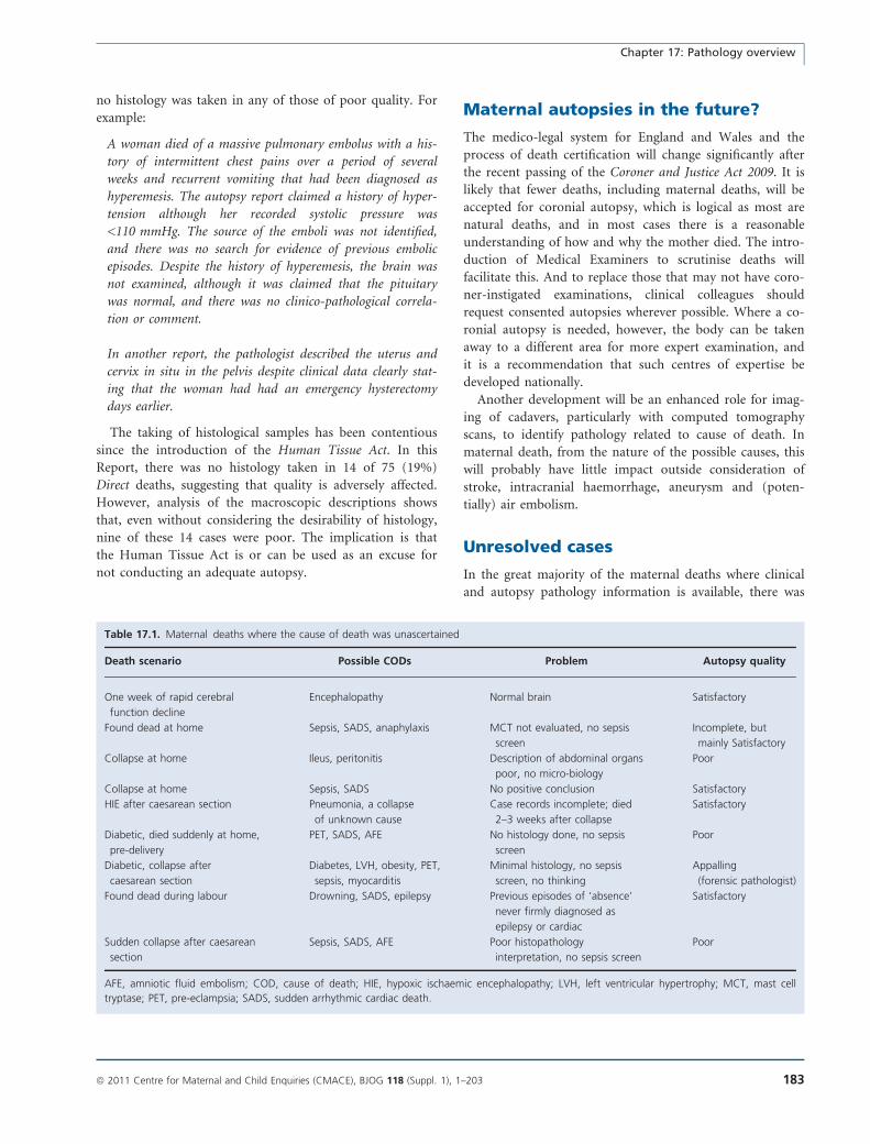
no histology was taken in any of those of poor quality. For
example:
A woman died of a massive pulmonary embolus with a his-
tory of intermittent chest pains over a period of several
weeks and recurrent vomiting that had been diagnosed as
hyperemesis. The autopsy report claimed a history of hyper-
tension although her recorded systolic pressure was
<110 mmHg. The source of the emboli was not identified,
and there was no search for evidence of previous embolic
episodes. Despite the history of hyperemesis, the brain was
not examined, although it was claimed that the pituitary
was normal, and there was no clinico-pathological correla-
tion or comment.
In another report, the pathologist described the uterus and
cervix in situ in the pelvis despite clinical data clearly stat-
ing that the woman had had an emergency hysterectomy
days earlier.
The taking of histological samples has been contentious
since the introduction of the Human Tissue Act. In this
Report, there was no histology taken in 14 of 75 (19%)
Direct deaths, suggesting that quality is adversely affected.
However, analysis of the macroscopic descriptions shows
that, even without considering the desirability of histology,
nine of these 14 cases were poor. The implication is that
the Human Tissue Act is or can be used as an excuse for
not conducting an adequate autopsy.
Maternal autopsies in the future?
The medico-legal system for England and Wales and the
process of death certification will change significantly after
the recent passing of the Coroner and Justice Act 2009. It is
likely that fewer deaths, including maternal deaths, will be
accepted for coronial autopsy, which is logical as most are
natural deaths, and in most cases there is a reasonable
understanding of how and why the mother died. The intro-
duction of Medical Examiners to scrutinise deaths will
facilitate this. And to replace those that may not have coro-
ner-instigated examinations, clinical colleagues should
request consented autopsies wherever possible. Where a co-
ronial autopsy is needed, however, the body can be taken
away to a different area for more expert examination, and
it is a recommendation that such centres of expertise be
developed nationally.
Another development will be an enhanced role for imag-
ing of cadavers, particularly with computed tomography
scans, to identify pathology related to cause of death. In
maternal death, from the nature of the possible causes, this
will probably have little impact outside consideration of
stroke, intracranial haemorrhage, aneurysm and (poten-
tially) air embolism.
Unresolved cases
In the great majority of the maternal deaths where clinical
and autopsy pathology information is available, there was
Table 17.1. Maternal deaths where the cause of death was unascertained
Death scenario Possible CODs Problem Autopsy quality
One week of rapid cerebral
function decline
Encephalopathy Normal brain Satisfactory
Found dead at home Sepsis, SADS, anaphylaxis MCT not evaluated, no sepsis
screen
Incomplete, but
mainly Satisfactory
Collapse at home Ileus, peritonitis Description of abdominal organs
poor, no micro-biology
Poor
Collapse at home Sepsis, SADS No positive conclusion Satisfactory
HIE after caesarean section Pneumonia, a collapse
of unknown cause
Case records incomplete; died
2–3 weeks after collapse
Satisfactory
Diabetic, died suddenly at home,
pre-delivery
PET, SADS, AFE No histology done, no sepsis
screen
Poor
Diabetic, collapse after
caesarean section
Diabetes, LVH, obesity, PET,
sepsis, myocarditis
Minimal histology, no sepsis
screen, no thinking
Appalling
(forensic pathologist)
Found dead during labour Drowning, SADS, epilepsy Previous episodes of ‘absence’
never firmly diagnosed as
epilepsy or cardiac
Satisfactory
Sudden collapse after caesarean
section
Sepsis, SADS, AFE Poor histopathology
interpretation, no sepsis screen
Poor
AFE, amniotic fluid embolism; COD, cause of death; HIE, hypoxic ischaemic encephalopathy; LVH, left ventricular hypertrophy; MCT, mast cell
tryptase; PET, pre-eclampsia; SADS, sudden arrhythmic cardiac death.
Chapter 17: Pathology overview
ª 2011 Centre for Maternal and Child Enquiries (CMACE), BJOG 118 (Suppl. 1), 1–203 183
an agreed main cause of death, and the case could be
assigned to the appropriate diagnostic category and type,
that is Direct, Indirect or Coincidental. However in a small
number, the final diagnosis and type could not be resolved
despite evaluation by many regional and central assessors
in the different specialties. The problems lie in (1) inade-
quate or inconclusive clinical data, (2) poor autopsy qual-
ity, and (3) the intractable nature of some deaths.
Table 17.1 highlights the main features of nine such cases,
combining the clinical and pathological data.
The possible diagnoses in these unresolved cases
encompass all the types of maternal death, and the
absence of conclusive diagnoses biases the calculated rates
of some important entities. The two most significant of
these are sepsis and sudden adult/arrythmic death syn-
drome; the current report has identified an increase in
these clinical pathologies, so it is unfortunate that the true
rates might be even higher if there had been better case
ascertainment.
Some deaths are never resolved at autopsy despite a full
examination and ancillary tests. One reason is that a week
or more of post-event intensive care can obliterate the
pathological evidence of what that critical event was. But
the main message from these deaths is that the maternal
autopsy deserves a full protocol approach. This includes
sepsis screen, retaining blood for later tests (e.g. mast cell
tryptase to prove or exclude anaphylaxis), careful evalua-
tion of the heart, full histopathology and, above all, think-
ing about the possible range of clinico-pathological
correlations.
Disclosure of interestsNone.
FundingThis work was undertaken by the Centre for Maternal and
Child Enquiries (CMACE) as part of the CEMACH pro-
gramme. The work was funded by the National Patient
Safety Agency; the Department of Health, Social Services
and Public Safety of Northern Ireland; NHS Quality
Improvement Scotland (NHS QIS); and the Channel
Islands and Isle of Man.
Acknowledgements
This Chapter has been reviewed by Dr R Nairn, Crosshouse
Hospital, Kilmarnock: Pathology Assessor for Scotland. j
References
1 Royal College of Pathologists: Guidelines on Autopsy Practice. Sce-
nario 5: Maternal Death. May 2010. [www.rcpath.org/resources/pdf/
g100autopsypracticesection5maternaldeathfinalmay2010.pdf].
Accessed 9 October 2010.
2 Millward-Sadler GH. Pathology of maternal deaths. In: Kirkham N,
Shepherd N, editors. Progress in Pathology, vol. 6. pp. 163–85. Lon-
don: Greenwich Medical Media Ltd; 2005.
3 Millward-Sadler GH. The maternal autopsy. In: Burton JL, Rutty GN,
editors. The Hospital Autopsy, 3rd edn. pp.203–15. London: Hodder
Arnold; 2010.
4 Human Tissue Act 2004. [www.opsi.gov.uk/acts/acts2004/ukp-
ga_20040030_en_1]. Accessed 9 October 2010.
5 Human Tissue Authority 2006. [www.hta.gov.uk]. Accessed 9 Octo-
ber 2010.
6 National Confidential Enquiry into Patient Outcome and Death (NCE-
POD). The Coronial Autopsy – do we deserve better?. 2006.
[www.ncepod.org.uk/2006.htm]. Accessed 9 September 2010.
7 Millward-Sadler GH. Pathology. In: Lewis G. editor. Why Mothers
Die. The 5th Report of the Confidential Enquiries into Maternal
Deaths in the United Kingdom 1997–99, pp. 276–306. The National
Institute of Clinical Excellence. London: RCOG Press; 2001.
8 Millward-Sadler GH. Pathology. In: Lewis G. editor. Saving Mother’s
Lives: Reviewing Maternal Deaths to Make Motherhood Safer.
2003–05. The 7th Report of the Confidential Enquiries into Maternal
Deaths in the United Kingdom, pp.184–98. London: RCOG; 2007.
9 Basso C, Burke M, Fornes P, Gallagher PJ, Henriques de Gouveia R,
Sheppard M, et al. Guidelines for autopsy investigation of sudden
cardiac death. Virchow Archiv 2008;452:11–8.
10 Royal College of Pathologists. Guidelines for Autopsy Practice. Sud-
den Death with Likely Cardiac Pathology. London: RCPath, 2005.
[www.rcpath.org/resources/pdf/AutopsyScenario1Jan05.pdf].
Accessed 9 September 2010.
Lucas, Millward-Sadler
184 ª 2011 Centre for Maternal and Child Enquiries (CMACE), BJOG 118 (Suppl. 1), 1–203

Annex 17.1. The main clinico-pathologiesencountered at autopsy in maternal death andspecific pathological scenarios (Adapted from RoyalCollege of Pathologists: Guidelines on AutopsyPractice. Scenario 5: Maternal Death. May 2010.)
1 Direct maternal death• Venous thrombosis and pulmonary embolism
• Hypertensive disease of pregnancy [pre-eclampsia, eclampsia]
a. Subtype of pre-eclampsia: HELLP syndrome in
pre-eclampsia (haemolysis, elevated liver enzymes, low
platelets)
• Peripartum haemorrhage
a. Uterine atony
b. Abruption of the placenta
c. Placenta praevia
d. Retained placenta and products of conception
e. Abnormally adherent placenta
(i) Placenta accreta, increta, percreta
f. Tear or rupture of genital tract
(i) Spontaneous
(ii) Iatrogenic
• Life support for peripartum haemorrhage
a. Transfusion-associated lung injury
b. Fluid overload
• Peripartum dilated cardiomyopathy (defined as cardiac
failure from last month of pregnancy up to 5 months
postpartum; other causes excluded)
• Amniotic fluid embolism
• Early pregnancy deaths
a. Ectopic pregnancy and haemorrhage
b. Spontaneous miscarriage
c. Legal termination
• Genital tract sepsis—puerperal sepsis
• Anaesthetic (general and regional anaesthesia)
a. general anaesthesia complications
b. regional anaesthesia complications (e.g. subdural hae-
matoma following inadvertent dural puncture during
epidural anaesthesia)
• Air embolism
• Ogilvie syndrome (pseudo-obstruction of the large
bowel)
• Choriocarcinoma and hydatidiform mole
• Ovarian hyperstimulation syndrome
• Acute fatty liver of pregnancy
2 Indirect maternal death• Cardiac
a. Congenital heart lesion with pulmonary hypertension
b. Inheritable cardiomyopathy, that is hypertrophic car-
diomyopathy (HOCM), arrhythmogenic right ventric-
ular cardiomyopathy (ARVCM)
c. Acquired cardiac muscle disease, that is ischaemic
heart disease, endocardial fibroelastosis
d. Obesity and sudden cardiac death
e. Valvular disease, that is in intravenous drug users,
rheumatic fever, mitral stenosis
• Systemic hypertension
• Idiopathic arterial pulmonary hypertension
• Pre-existing thrombophilia states, including antiphosp-
holipid syndrome
• Thrombotic thrombocytopaenic purpura
• Stroke
a. Subarachnoid haemorrhage
b. Cerebral infarction
c. Cerebral venous sinus thrombosis
• Other cardiovascular diseases
a. Dissection of aorta
b. Dissection of coronary artery
c. Dissection of splenic artery
• Psychiatric, including suicide related to pregnancy and
delivery
• Epilepsy (sudden unexplained death in epilepsy)
• Malignant disease worsened by pregnancy (breast, cervix)
• Community-acquired sepsis
• Acute anaphylaxis from drug treatment, that is antibiotics
• Other diseases
a. HIV/AIDS and tuberculosis
b. Sickle cell disease (HbSS and HbSC)
c. Connective tissue disease—systemic lupus erythemato-
sus
d. Diabetes mellitus—gestational and pre-existing diabe-
tes; this includes the hypoglycaemic ‘dead in bed’ syn-
drome
e. Influenza (e.g. epidemic type A—H1N1)
f. Cirrhosis
g. Any other clinico-pathological condition that the preg-
nant state makes worse; these include inherited and
acquired conditions, and the woman may have been spe-
cifically warned of the hazards of becoming pregnant
ª 2011 Centre for Maternal and Child Enquiries (CMACE), BJOG 118 (Suppl. 1), 1–203 185
Annex 17.1. The main clinico-thologies encountered at autopsy in maternal death and specific pathological scenarios
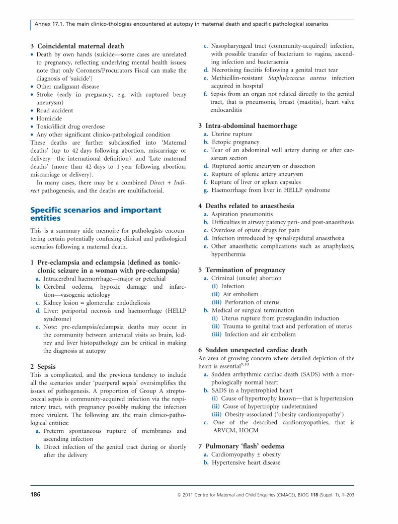
3 Coincidental maternal death• Death by own hands (suicide—some cases are unrelated
to pregnancy, reflecting underlying mental health issues;
note that only Coroners/Procurators Fiscal can make the
diagnosis of ‘suicide’)
• Other malignant disease
• Stroke (early in pregnancy, e.g. with ruptured berry
aneurysm)
• Road accident
• Homicide
• Toxic/illicit drug overdose
• Any other significant clinico-pathological condition
These deaths are further subclassified into ‘Maternal
deaths’ (up to 42 days following abortion, miscarriage or
delivery—the international definition), and ‘Late maternal
deaths’ (more than 42 days to 1 year following abortion,
miscarriage or delivery).
In many cases, there may be a combined Direct + Indi-
rect pathogenesis, and the deaths are multifactorial.
Specific scenarios and importantentities
This is a summary aide memoire for pathologists encoun-
tering certain potentially confusing clinical and pathological
scenarios following a maternal death.
1 Pre-eclampsia and eclampsia (defined as tonic-clonic seizure in a woman with pre-eclampsia)
a. Intracerebral haemorrhage—major or petechial
b. Cerebral oedema, hypoxic damage and infarc-
tion—vasogenic aetiology
c. Kidney lesion = glomerular endotheliosis
d. Liver: periportal necrosis and haemorrhage (HELLP
syndrome)
e. Note: pre-eclampsia/eclampsia deaths may occur in
the community between antenatal visits so brain, kid-
ney and liver histopathology can be critical in making
the diagnosis at autopsy
2 SepsisThis is complicated, and the previous tendency to include
all the scenarios under ‘puerperal sepsis’ oversimplifies the
issues of pathogenesis. A proportion of Group A strepto-
coccal sepsis is community-acquired infection via the respi-
ratory tract, with pregnancy possibly making the infection
more virulent. The following are the main clinico-patho-
logical entities:
a. Preterm spontaneous rupture of membranes and
ascending infection
b. Direct infection of the genital tract during or shortly
after the delivery
c. Nasopharyngeal tract (community-acquired) infection,
with possible transfer of bacterium to vagina, ascend-
ing infection and bacteraemia
d. Necrotising fasciitis following a genital tract tear
e. Methicillin-resistant Staphylococcus aureus infection
acquired in hospital
f. Sepsis from an organ not related directly to the genital
tract, that is pneumonia, breast (mastitis), heart valve
endocarditis
3 Intra-abdominal haemorrhagea. Uterine rupture
b. Ectopic pregnancy
c. Tear of an abdominal wall artery during or after cae-
sarean section
d. Ruptured aortic aneurysm or dissection
e. Rupture of splenic artery aneurysm
f. Rupture of liver or spleen capsules
g. Haemorrhage from liver in HELLP syndrome
4 Deaths related to anaesthesiaa. Aspiration pneumonitis
b. Difficulties in airway patency peri- and post-anaesthesia
c. Overdose of opiate drugs for pain
d. Infection introduced by spinal/epidural anaesthesia
e. Other anaesthetic complications such as anaphylaxis,
hyperthermia
5 Termination of pregnancya. Criminal (unsafe) abortion
(i) Infection
(ii) Air embolism
(iii) Perforation of uterus
b. Medical or surgical termination
(i) Uterus rupture from prostaglandin induction
(ii) Trauma to genital tract and perforation of uterus
(iii) Infection and air embolism
6 Sudden unexpected cardiac deathAn area of growing concern where detailed depiction of the
heart is essential9,10
a. Sudden arrhythmic cardiac death (SADS) with a mor-
phologically normal heart
b. SADS in a hypertrophied heart
(i) Cause of hypertrophy known—that is hypertension
(ii) Cause of hypertrophy undetermined
(iii) Obesity-associated (‘obesity cardiomyopathy’)
c. One of the described cardiomyopathies, that is
ARVCM, HOCM
7 Pulmonary ‘flash’ oedemaa. Cardiomyopathy ± obesity
b. Hypertensive heart disease
186 ª 2011 Centre for Maternal and Child Enquiries (CMACE), BJOG 118 (Suppl. 1), 1–203
Annex 17.1. The main clinico-thologies encountered at autopsy in maternal death and specific pathological scenarios

c. Pre-eclampsia and acute lung injury
d. Fluid overload
e. Transfusion-associated lung injury
8 Disseminated intravascular coagulation (DIC)A pathology with many causes and differential diagnoses.
a. Obtain the pre-mortem haematology laboratory results
b. Differentiate between DIC and thrombotic thrombo-
cytopaenic purpura from the laboratory results and
the histopathology
c. Consider to prove or exclude as causes of DIC
(i) Severe sepsis
(ii) Uterine atony and other causes of peripartum
haemorrhage
(iii) Amniotic fluid embolism
9 The fetusThere may be a fetus retained within the mother; or
accompanying the mother, following peri-mortem caesar-
ean section or other delivery.
a. If the fetus lived and then died, the coroner has
potential jurisdiction
b. If the fetus never lived, then the coroner has no juris-
diction; an autopsy would need consent from the rela-
tives, unless there are special circumstances
c. Autopsy of the fetus is usually unnecessary, as this will
contribute little or nothing to the understanding of
the mother’s cause of death. Exceptions to this are
possibly sepsis, when fetal skin or lung samples can
indicate severity and timing of ascending infection;
and the exceptionally rare event of a fetal malignant
tumour that might have spread to the mother
ª 2011 Centre for Maternal and Child Enquiries (CMACE), BJOG 118 (Suppl. 1), 1–203 187
Annex 17.1. The main clinico-thologies encountered at autopsy in maternal death and specific pathological scenarios
Appendix 1: The method of EnquiryA Miller
Centre for Maternal and Child Enquiries, London, UK
Correspondence: Mrs Alison Miller, Centre for Maternal and Child Enquiries, Chiltern Court, Lower Ground Floor, 188 Baker Street, London
NW1 5SD, UK. Email: [email protected]
Background
The UK Maternal Death Enquiry 2006–08 has been the
responsibility of the Confidential Enquiry into Maternal
and Child Health (CEMACH) until 1 July 2009, followed
by its successor organisation, the Centre for Maternal and
Child Enquiries (CMACE), to completion.
CMACE is a registered charity and company limited by
guarantee governed by its own Board of Trustees. The
Maternal Death Enquiry is part of the National Confiden-
tial Enquiry programme commissioned by the National
Patient Safety Agency (NPSA) for England and Wales, the
Northern Ireland Department of Health and NHS Quality
Improvement Scotland (NHSQIS) for their respective par-
ticipation in the Enquiry.
The Enquiry is carried out regionally through four
CMACE offices and two subcontracted offices in England,
the Public Health Agency team in Northern Ireland and
the Reproductive Health Programme team in Scotland. The
central coordination is carried out by the CMACE central
office, which is based in London.
Notification, surveillance and enquiryprocesses
It is a government requirement that all maternal deaths
should be subject to this Confidential Enquiry, and all
health professionals have a duty to provide the information
required. Additionally, the Clinical Negligence Scheme for
Trusts (CNST) in England includes contribution to the
enquiry within its criteria of standards.
In participating in the Maternal Death Enquiry, profes-
sionals concerned are asked for two things:
1 To provide a full and accurate account of the circum-
stances leading to the woman’s death, with supporting
records.
2 To reflect on any clinical or other lessons that have
been learned, either personally or as part of the
wider context.
The responsibility for initiating an enquiry into maternal
death lies with the CMACE regional manager or lead
within each office. Following a pregnancy-associated mater-
nal death, a notification is usually made by one of the
health professionals involved in the care of the woman to
the relevant CMACE regional manager. Cases are also
reported by coroners, Local Supervising Authority Mid-
wifery Officers (LSAMO) and others. This is followed up
with a request from the regional office for more detailed
surveillance data.
The Enquiry is then initiated by the regional office using
a standard data collection form (Maternal Death Report
[MDR1] pro forma), which is completed by obstetricians,
anaesthetists, pathologists, GPs, midwives and any other
professionals who were involved in the care of the woman.
Copies of case notes are obtained, and where relevant,
autopsy reports, internal reviews and, in some cases, addi-
tional written statements are supplied.
Each case is then prepared for a two-stage review and
assessment process, the first occurring at a regional level,
and the second occurring at a central level.
Regional assessment
Each CMACE region has one or more sets of regional
assessors, depending on requirements. Each set includes an
obstetric assessor, a midwifery assessor, a pathology asses-
sor, an anaesthesia assessor and a perinatal psychiatry asses-
sor. Nominations for medical assessors are sought from the
presidents of the Royal Colleges, and nominations for mid-
wives are sought from the LSAMO.
Regional assessors must be an active clinical practitioner
in the NHS, in the relevant specialty. If medical, the regio-
nal assessor should be at consultant level, or, if a midwife,
must be at supervisory or management level. The regional
assessors should have knowledge and experience of organi-
sation of care, as well as commanding the respect of their
peers. It is important that the regional assessor is able to
realistically commit enough time to assess and return
enquiry forms in a timely manner.
The role of the regional assessor is to review the infor-
mation reported in the MDR1 form and any other docu-
ments that have been assembled by the CMACE regional
manager. Every attempt is made to obtain full details of
any autopsy and pathological investigations.
Following collection of all the relevant information, the
records are anonymised and circulated by the CMACE
regional manager to regional assessors. The obstetric and
midwifery assessors review all cases. Anaesthesia and
188 ª 2011 Centre for Maternal and Child Enquiries (CMACE), BJOG 118 (Suppl. 1), 1–203
Miller
perinatal psychiatry assessors review all cases where there
was involvement of an anaesthetic or intensive care or a his-
tory of psychiatric illness, respectively. Cases where an
autopsy has been obtained are assessed by the regional
pathology assessor.
The assessors review the cases taking into account the case
history, the results of pathological investigations and find-
ings of autopsy that may have been conducted. They then
summarise in a short report, which includes their comments
and opinions, regarding the cause or causes of death and the
resources of the organisation responsible for the care of the
woman. The assessor is also asked to make a judgement as to
whether the care was substandard and, if so, if this was a
contributing factor in the death of the mother. The com-
pleted form is then returned to the regional office.
In Northern Ireland and Scotland, consensus case assess-
ment is made and the report is written within a meeting of
the regional assessors on a periodic basis within the trien-
nium.
Following regional assessment, the cases are sent from
the regional office to the CMACE central office for central
assessment.
Central assessment
The Director of the Maternal Death Enquiry reviews all
cases and allocates the cases for central assessment as
necessary. The central assessors cover a broad range of
specialties which include: obstetrics and gynaecology,
midwifery, anaesthesia, psychiatry, obstetric medicine
and cardiology, emergency medicine, general practice and
pathology. The Director will make a decision as to which
are the most appropriate central assessors to review each
case.
The central assessors review cases and provide a central
report, taking into account the regional assessors’ reports.
Following this detailed investigation, each case is allocated
to a specific chapter in the final report. All details regarding
the death, including the agreed clinical cause of death, are
recorded in a database.
In a small number of cases, the cause of death is assessed
to vary from the underlying cause of death as given on the
death certificate and classified by the registrars general
using the International Classification of Diseases 10th revi-
sion (ICD10). This is because a death may be coded for a
specific cause of death, but the pathogenesis of this condi-
tion may have been precipitated by an obstetric event. For
example, although a given death may be coded as multiple-
organ failure as the terminal event, it could have been
precipitated by an obstetric event such as septicaemia from
an infected caesarean section. Although each pregnancy-
associated maternal death reported to this Enquiry is only
counted once and assigned to one chapter, it may be
referred to in additional chapters. For example, a death
assigned to ‘hypertensive disorder of pregnancy’, in which
haemorrhage and anaesthesia also played a part, may be
discussed in all three chapters.
Authors
Chapters are initially drafted by individual central assessors
and then discussed in detail by the writing panel before the
report is finalised. Other acknowledged professionals, who
have a particular and expert interest in specific diseases or
areas of practice, will be asked to provide peer review and
comment on the content and recommendations made in
the chapter relevant to their expertise before publication.
Statistical analysis and data presentation advice is pro-
vided to each author by both an independent statistical
advisor and data analysts at CMACE.
These processes are summarised in Figure 1.
Verification of ascertainment
Ascertainment is checked with reference to data supplied
by the Office for National Statistics (ONS) for England and
Wales’ data. These data are supplied in two forms:
1 Direct notifications—these are deaths of women where
‘pregnancy’ is mentioned anywhere on the ONS death
registration. These cases are coded as a maternal death
according to the ICD10. This identifies all women
where pregnancy may have been a contributing factor
to their death.
2 Linkage notifications—these are deaths of women
where the name of the woman appeared on the regis-
tration of a birth in the current or preceding calendar
year. This allows for the identification of women who
have died up to 364 days following delivery.
These data are cross-matched with the data that have
been directly acquired by CMACE. Any cases that have
been identified by one organisation but not the other
would then be established. These outstanding cases are then
further investigated to ensure that they are pregnancy-asso-
ciated deaths and warrant inclusion in the Enquiry. The
enquiry process is then initiated as above using the stan-
dard data collection form.
Confidentiality
All data and databases are held in accordance with
CMACE information governance and security procedures
(not applicable in Scotland). These ensure anonymity at
different stages of the Enquiry process, guarantee confi-
dentiality and safeguard any identifiable information for
the duration it is held. These procedures meet with the
requirements set out by Cabinet Office Guidance on
Appendix 1: The method of Enquiry
ª 2011 Centre for Maternal and Child Enquiries (CMACE), BJOG 118 (Suppl. 1), 1–203 189
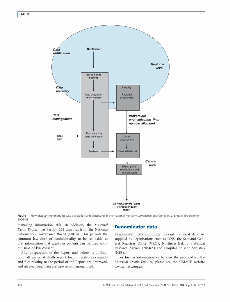
managing information risk. In addition, the Maternal
Death Enquiry has Section 251 approval from the National
Information Governance Board (NIGB). This permits the
common law duty of confidentiality to be set aside so
that information that identifies patients can be used with-
out next-of-kin consent.
After preparation of the Report and before its publica-
tion, all maternal death report forms, related documents
and files relating to the period of the Report are destroyed,
and all electronic data are irreversibly anonymised.
Denominator data
Denominator data and other relevant statistical data are
supplied by organisations such as ONS, the Scotland Gen-
eral Registrar Office (GRO), Northern Ireland Statistical
Research Agency (NISRA) and Hospital Episode Statistics
(HES).
For further information or to view the protocol for the
Maternal Death Enquiry, please see the CMACE website
www.cmace.org.uk.
Surveillancesystem
Data acquisitionanonymisation
Data cleaningData verification Central
assessment
Triennial dataset
Notification
Analysis
Expert panelinvestigation and
interpretation
Saving Mothers’ Livestriennial enquiry
report
Datarecovery
Datamanagement
Regionallevel
Centrallevel
ONSdata
Regionalassessment
Enquiry
Datanotification
Irreversibleanonymisation–finalnumber allocated
Figure 1. Flow diagram summarising data acquisition and processing in the maternal mortality surveillance and Confidential Enquiry programme
2006–08.
Miller
190 ª 2011 Centre for Maternal and Child Enquiries (CMACE), BJOG 118 (Suppl. 1), 1–203
Appendix 2A: Summary of United KingdomObstetric Surveillance System (UKOSS) Report onnear-miss studiesM Knight*
National Perinatal Epidemiology Unit, University of Oxford, Oxford, UK
Correspondence: Dr Marian Knight, National Perinatal Epidemiology Unit, University of Oxford, Old Road Campus, Headington, Oxford OX3
7LF, UK. Email: [email protected]
Surveillance of near-miss maternalmorbidity
The UK Obstetric Surveillance System (UKOSS) was set up
to study rare disorders of pregnancy1 on a national basis
throughout the UK. This has enabled serial surveillance of
a number of near-miss morbidities. It is increasingly being
recognised that study of near-miss morbidity can comple-
ment enquiries into maternal death and provide additional
information to guide prevention and treatment of poten-
tially life-threatening conditions.2 Maternal deaths represent
the tip of the iceberg of disease; a much larger number of
women suffer from near-miss morbidity, increasing the
power of these studies to investigate risk factors both for
the occurrence of disease and progression to death or other
severe complications. This summary presents the key find-
ings from UK surveillance of near-miss maternal morbidi-
ties and related conditions undertaken during the period of
the maternal death surveillance of this report and describes
ongoing studies of near-miss conditions. Further details are
available at www.npeu.ox.ac.uk/ukoss.
Surveillance methodsThe UKOSS methods are described in detail in the Intro-
duction to this Report. Each month, reporting cards with a
tick-box list of conditions are sent to nominated reporting
clinicians (midwives, obstetricians, risk management mid-
wives and anaesthetists) in all consultant-led maternity
units in the UK. Inclusion of all consultant-led maternity
units enables UKOSS near-miss surveillance to cover the
entire cohort of women giving birth in the UK, as women
who experience these events at home or in a non-consul-
tant unit will be transferred to a consultant unit. The sys-
tem, however, does not allow for detailed surveillance of
near-miss conditions occurring in early pregnancy, such as
ectopic pregnancy, when women will be cared for in gynae-
cology units. Similarly, conditions which present later in
the postnatal period, for example puerperal psychosis, are
not surveyed, because presenting women are most likely to
be cared for in settings other than obstetric units.
UKOSS is an active, negative surveillance system, that is,
zero reports are sought in addition to reports of cases.
Hence, all participating hospitals will return a report card
each month, whether or not there have been any cases,
enabling continual monitoring of response rates and confir-
mation of the denominator number of women for the calcu-
lation of disease incidence. In response to a report of a case,
clinicians are sent a data collection form requesting further
details of a woman’s demographic, pregnancy and other
characteristics, as well as management and pregnancy out-
comes. All information collected is anonymous. In addition,
for some studies, collaborating clinicians may be asked to
provide similar details for a comparison woman. This allows
UKOSS to conduct case–control and cohort studies as well
as descriptive studies and this considerably widens the range
of research questions that can be addressed using the system.
Examples of questions that can be addressed using
UKOSS studies include:
• estimating disease incidence; for example UKOSS sur-
veillance of eclampsia demonstrated a 45% reduction
in incidence between 1992 and 20053
• describing the prevalence of factors associated with
near-miss maternal morbidity; for example a UKOSS
study estimated that more than one in every 1200
women delivering in the UK is extremely obese (body
mass index [BMI] ‡ 50 kg/m2)4
• quantifying risk factors for severe morbidity; for exam-
ple UKOSS surveillance of peripartum hysterectomy
for severe haemorrhage showed a significant associa-
tion with previous delivery by caesarean section5
• auditing of national guidelines; for example UKOSS
surveillance of antenatal pulmonary embolism showed
that very few women were not receiving thrombopro-
phylaxis according to Royal College of Obstetricians
and Gynaecologists guidelines6,7* Head of the United Kingdom Obstetric Surveillance System, National
Perinatal Epidemiology Unit.
Appendix 2A: Summary of United Kingdom Obstetric Surveillance System (UKOSS) Report on near-miss studies
ª 2011 Centre for Maternal and Child Enquiries (CMACE), BJOG 118 (Suppl. 1), 1–203 191
• investigating different management techniques; for
example the use of total versus subtotal hysterectomy
was examined in the UKOSS study of peripartum hys-
terectomy for severe haemorrhage, but no significant
differences in complication rates between the two
techniques were found8
• describing the outcomes of severe morbidity; for
example UKOSS surveillance of acute fatty liver of
pregnancy showed that both maternal and infant out-
comes were better than suggested by previous hospi-
tal-based historical studies9
UKOSS can, in addition, be used to conduct studies rap-
idly in response to emerging public health issues; hence, in
response to the influenza A/H1N1v (‘swine flu’) pandemic,
surveillance of women admitted to hospital with confirmed
infection was initiated to inform ongoing clinical guidance
during the course of the pandemic.10
Surveillance of specific near-missmorbidities—key points fromcompleted UKOSS studies
Haemorrhage and associated conditions
Peripartum hysterectomyThe incidence of peripartum hysterectomy in the UK is 4.1
cases per 10 000 maternities (95% CI 3.6–4.5) with a case
fatality of 0.6% (95% CI 0–1.5%). Peripartum hysterec-
tomy is strongly associated with previous delivery by cae-
sarean section (adjusted odds ratio [aOR] 3.52, 95% CI
2.35–5.26), and the risk rises with increasing number of
previous caesarean section deliveries (aOR 2.14 with one
previous delivery, 95% CI 1.37–3.33; 18.6 with two or
more, 95% CI 7.67–45.4)). Maternal age and parity are also
important risk factors.5
The majority of cases occur in association with either
uterine atony or a morbidly adherent placenta (placenta ac-
creta). The associated haemorrhage is managed in a variety
of ways and not universally according to existing guide-
lines; some women who have a hysterectomy for haemor-
rhage caused solely by uterine atony are not fully treated
with uterotonic drugs to control the haemorrhage. Very
few women were reported to have had a hysterectomy fol-
lowing treatment with some of the more innovative thera-
pies for control of haemorrhage, including uterine artery
embolisation and factor VII.8 However, this study did not
collect information on women who had a postpartum
haemorrhage successfully controlled using these therapies,
and therefore further surveillance was undertaken specifi-
cally to describe all women managed with specific second-
line therapies to fully describe outcomes following their use
(see below).
Management with specific second-line therapies forpostpartum haemorrhageInformation was collected on all women in the UK man-
aged with uterine compression sutures, pelvic vessel em-
bolisation or ligation or factor VII for severe postpartum
haemorrhage between September 2007 and March 2009.
An estimated 2.6 women per 10 000 maternities were
managed with these therapies.11 Almost a quarter of
women subsequently underwent a hysterectomy following
failure of control of the haemorrhage. Uterine compres-
sion sutures were the most frequently used of these spe-
cific second-line therapies; the compression sutures failed
to control the haemorrhage in 29% of cases (95% CI 26–
36%). Treatment with uterine artery embolisation failed
to control haemorrhage in 21% of cases (95% CI 6–
46%), surgical ligation of pelvic vessels was performed in
a very small number of cases and was associated with a
failure rate of 68% (95% CI 43–87%) and factor VII,
used in 12% of cases, unsuccessfully controlled haemor-
rhage for 71% of women (95% CI 52–85%). Women
whose haemorrhage was not successfully controlled with a
uterine compression suture were significantly more likely
to have had a later placement of the suture (more than
2 hours following delivery) than women whose haemor-
rhage was controlled (aOR 3.86, 95% CI 1.65–8.99), high-
lighting the importance of early recognition and
treatment of postpartum haemorrhage to improve out-
comes for women.
Hypertension and related disorders
EclampsiaThe incidence of eclampsia has decreased significantly in
the UK from 4.9 per 10 000 maternities in 199212 to 2.7
cases per 10 000 maternities (95% CI 2.4–3.1)3 in 2005.
The majority of women in the UK are managed with mag-
nesium sulphate according to national protocols, and com-
parison between the 1992 and 2005 data suggest that
maternal morbidity has been significantly reduced as a con-
sequence. No women in the study died (case fatality 0%,
95% CI 0–1.7%). Fifty-four (26%) had recurrent fits.
Twenty-two women (10%) were reported to have other
severe morbidity after the eclamptic episode. Outcomes
were known for 222 infants (204 singletons, 18 twins).
Eight infants were stillborn, and five died in the neonatal
period (perinatal mortality 59 per 1000 births, 95% CI
32–98). This study showed the practical benefits of the
incorporation of research evidence into practice, improving
outcomes for women through the widespread use of
evidence-based guidelines.
Knight
192 ª 2011 Centre for Maternal and Child Enquiries (CMACE), BJOG 118 (Suppl. 1), 1–203
Acute fatty liver of pregnancyNationally acute fatty liver of pregnancy is rare, with an
estimated incidence of 5.0 cases per 100 000 maternities
(95% CI 3.8–6.5/100 000) or 1 in 20 000 maternities.9 The
‘Swansea’ diagnostic criteria previously proposed agree sub-
stantially with clinical diagnosis.13 Eighteen percent of
women had twin pregnancies, and 20% were underweight
(BMI < 20 kg/m2). This suggests that women with twin
pregnancies appear to be at higher risk, but further studies
are needed to investigate the risk associated with low BMI.
One woman out of 57 received a liver transplant. One
woman died (case fatality rate 1.8%, 95% CI 0–9.4%).
There were seven deaths among 67 infants (perinatal mor-
tality rate 104 per 1000 births, 95% CI 43–203). The inci-
dence estimate from this study is lower than documented
by earlier hospital-based studies, but maternal and neonatal
outcomes are better than previously reported, possibly
related to improved ascertainment.
Thrombosis and thromboembolism
Antenatal pulmonary embolismA total of 143 women with antenatal pulmonary embolism
were reported, representing an estimated incidence of 1.3
per 10 000 maternities (95% CI 1.1–1.5).6 There were a few
cases where thromboprophylaxis was not provided accord-
ing to national guidelines, and there may be scope for fur-
ther work on guideline implementation. The main risk
factors for pulmonary embolism were multiparity (aOR
4.03, 95% CI 1.60–9.84) and obesity (BMI ‡ 30 kg/m2)
(aOR 2.65, 95% CI 1.09–6.45); however, without additional
studies of cost-effectiveness, we are unable to recommend
any changes to current guidelines which would aid preven-
tion of this serious condition. Nearly a third of women
with antenatal pulmonary embolism had no classical risk
factors, and only nine women with classical risk factors
were eligible for prophylaxis under 2007 guidelines. Further
work is needed to assess how information about these risk
factors may be used to guide prophylaxis. Two women had
recurrent pulmonary emboli (1.4%, 95% CI 0.2–5.1%), and
five women died (case fatality 3.5%, 95% CI 1.1–8.0%).
Hence, significant severe morbidity from thromboembolic
disease underlies the maternal deaths from antenatal pul-
monary embolism reported in the UK, with approximately
30 women diagnosed with the condition for each woman
who died.
Amniotic fluid embolismAnalysis of data reported over 4 years to UKOSS shows an
estimated incidence of amniotic fluid embolism (AFE) of
2.0 cases per 100 000 maternities (95% CI 1.5–2.5/
100 000).14 Occurrence of AFE was significantly associated
with induction of labour (aOR 3.86, 95% CI 2.04–7.31)
and multiple pregnancy (aOR 10.9, 95% CI 2.81–42.7); an
increased risk was also noted in older women from ethnic
minorities (aOR 9.85, 95% CI 3.57–27.2). Caesarean deliv-
ery was associated with postnatal amniotic fluid embolism
(aOR 8.84, 95% CI 3.70–21.1). Twelve women died (case
fatality 20%, 95% CI 11–32%); five of 37 infants of women
with antenatal AFE died (perinatal mortality 135/1000 total
births, 95% CI 45–288). Women who died were signifi-
cantly more likely to be from ethnic minority groups (aOR
11.8, 95% CI 1.40–99.5). In view of the extreme rarity of
this condition and the significant associated mortality, sur-
veillance through UKOSS is ongoing to further investigate
risk factors and describe outcomes following the use of dif-
ferent management techniques.
Ongoing surveillanceOngoing surveillance of near-miss maternal morbidity is
clearly important to complement the comprehensive studies
of maternal death as presented in this report. The following
studies are ongoing (2010 onwards); additional studies will
be introduced as part of the UK National Maternal Near-
miss Surveillance Programme funded by the National Insti-
tute for Health Research.
Uterine ruptureUterine rupture is associated with significant maternal
and fetal morbidity, and a decrease in the number of
women attempting vaginal birth after caesarean section
may be the result of concerns about the risk of uterine
rupture. There are, however, no systematic data available
at a population level to quantify the incidence of uterine
rupture and to assess the risks associated with induction
and augmentation of labour in women who have had a
previous caesarean delivery. This study will investigate the
incidence, risk factors and outcomes of uterine rupture in
the UK.
Placenta accretaPlacenta accreta is thought to be becoming more common
for a number of reasons, including rising maternal age at
delivery and an increasing proportion of deliveries by cae-
sarean section. There is at the same time a debate about
the optimal diagnostic and management techniques. This
study will describe the current management of placenta ac-
creta in the UK and associated outcomes for women and
their infants. In addition, this study will estimate the
national incidence of placenta accreta in the UK and iden-
tify the extent to which previous caesarean section and
older maternal age are risk factors in this population. This
will enable appropriate future service planning, provide
accurate information, which can be used when counselling
women about the risks associated with caesarean section
and developing management guidelines, and provide a
Appendix 2A: Summary of United Kingdom Obstetric Surveillance System (UKOSS) Report on near-miss studies
ª 2011 Centre for Maternal and Child Enquiries (CMACE), BJOG 118 (Suppl. 1), 1–203 193
baseline incidence against which future trends can be mon-
itored if caesarean delivery rates continue to rise nationally.
Antenatal strokeStroke is an important cause of severe maternal morbidity
and mortality in the UK. The increasing age of women at
childbirth, along with other risk factors, may lead to an
increase in the incidence of stroke associated with preg-
nancy. There have to date been no prospective national
studies to estimate the incidence or outcomes of this con-
dition. This study will investigate the incidence, risk fac-
tors, management and outcomes of stroke in pregnancy in
the UK to inform future guidelines for prevention and
treatment.
Myocardial infarctionMyocardial infarction in pregnancy is known to be associ-
ated with significant maternal and fetal mortality. The cur-
rent incidence estimate is based on a study from 1970.
Current trends in lifestyle factors and increasing age at
childbirth are likely to be leading to an increase in inci-
dence. This study will provide an estimate of the incidence
of the disease, its epidemiology and its management.
Pulmonary vascular diseasePulmonary vascular disease in pregnancy is widely consid-
ered to pose an extreme risk of maternal death, but there
have been no recent prospective case series to assess this
risk. Novel methods of management may also impact on
outcomes. This study will provide a national picture of the
incidence of the disease, its epidemiology and management.
Aortic dissectionAortic dissection in pregnancy is a significant cause of
maternal morbidity and mortality. Changes in birth pat-
terns, with a rise in older mothers and increased prevalence
of obesity, may contribute to an increased occurrence of
aortic dissection in the UK. There have been no prospective
studies to estimate the incidence of this disease and its
investigation and management during pregnancy. This
study will estimate the national incidence of aortic dissec-
tion in the pregnant population in the UK and use these
national data to investigate and quantify risk factors for
aortic dissection in pregnancy.
Failed intubationAlthough anaesthetic-related maternal deaths have
decreased in number in recent years, hypoxia related to
failed intubation remains a consistent cause of mortality.
The incidence of failed intubation in the obstetric popula-
tion is thought to be higher than in the nonpregnant popu-
lation, but the reasons for this higher incidence in the
obstetric population are thought to be multiple. This study
will investigate the incidence, risk factors, management and
outcomes of failed intubation in the obstetric population
in the UK to inform future guidelines for prevention and
treatment.
Conclusions
National studies of near-miss maternal morbidity con-
ducted through UKOSS provide a range of information to
complement the national Confidential Enquiries into
Maternal Death contained in this report. The UKOSS
network of collaborating clinicians in all consultant-led
maternity units allows for ongoing surveillance of specific
near-miss conditions as well as rapidly responsive studies
where there is an urgent public-health need. The UK Con-
fidential Enquiries into Maternal Death are cited worldwide
for their role in improving maternity care and pregnancy
outcomes for women, and the model has been adopted in
many countries. In a similar manner, the UKOSS model is
now being adopted internationally15,16 with the potential
for multinational collaborative surveillance studies giving
added benefit to women and their families.
FundingThis is an independent report from a study that is part-
funded by the Policy Research Programme in the Depart-
ment of Health. The views expressed are not necessarily
those of the Department. Studies are additionally funded
by Wellbeing of Women, the Obstetric Anaesthetists Asso-
ciation and Heart UK.
AcknowledgementsThis work would not have been possible without the con-
tribution and enthusiasm of the UKOSS collaborating clini-
cians who notified cases and completed data collection
forms. The following members of the National Perinatal
Epidemiology Unit contributed to study design, analysis
and data collection: Peter Brocklehurst, Jane Forrester-Bar-
ker, Carole Harris, Gilles Kayem, Jenny Kurinczuk, Matth-
ias Pierce and Patsy Spark. j
References
1 Knight M, Kurinczuk JJ, Tuffnell D, Brocklehurst P. The UK Obstetric
Surveillance System for rare disorders of pregnancy. BJOG
2005;112:263–5.
2 Pattinson RC, Hall M. Near misses: a useful adjunct to maternal
death enquiries. Br Med Bull 2003;67:231–43.
3 Knight M. Eclampsia in the United Kingdom 2005. BJOG
2007;114:1072–8.
4 Knight M, Kurinczuk JJ, Spark P, Brocklehurst P. Extreme obesity in
pregnancy in the UK. Obstet Gynecol 2010;115:989–97.
5 Knight M, Kurinczuk JJ, Spark P, Brocklehurst P. Cesarean delivery
and peripartum hysterectomy. Obstet Gynecol 2008;111:97–105.
Knight
194 ª 2011 Centre for Maternal and Child Enquiries (CMACE), BJOG 118 (Suppl. 1), 1–203
6 Knight M. Antenatal pulmonary embolism: risk factors, manage-
ment and outcomes. BJOG 2008;115:453–61.
7 Nelson-Piercy C. Thromboprophylaxis during pregnancy, labour and
after vaginal delivery. RCOG Green-top guideline no. 37. July 2007
London: RCOG; 2007.
8 Knight M. Peripartum hysterectomy in the UK: management and
outcomes of the associated haemorrhage. BJOG 2007;114:1380–7.
9 Knight M, Nelson-Piercy C, Kurinczuk JJ, Spark P, Brocklehurst P. A
prospective national study of acute fatty liver of pregnancy in the
UK. Gut 2008;57:951–6.
10 Yates LM, Pierce M, Stephens S, Mill AC, Spark P, Kurinczuk JJ,
et al. Influenza A/H1N1v in pregnancy: an investigation of the char-
acteristics of affected women and the relationship to pregnancy out-
comes for mother and infant. Health Technol Assess 2010;114:109–
82.
11 Knight M, Spark P, Pierce M on behalf of UKOSS. UKOSS Annual
Report 2010. Oxford: National Perinatal Epidemiology Unit; 2010.
12 Douglas KA, Redman CW. Eclampsia in the United Kingdom. BMJ
1994;309:1395–400.
13 Ch’ng CL, Morgan M, Hainsworth I, Kingham JGC. Prospective
study of liver dysfunction in pregnancy in Southwest Wales. Gut
2002;51:876–80.
14 Knight M, et al. Amniotic fluid embolism in the UK. Obstet Gynecol
2010;115:910–17.
15 The Australasian Maternity Outcomes Surveillance System (AMOSS).
2009 [www.amoss.com.au/]. Accessed 27 November 2009.
16 NOSS-Nordic Obstetric Surveillance Study. 2009 [www.noss.nu/
index.html]. Accessed 27 November 2009.
ª 2011 Centre for Maternal and Child Enquiries (CMACE), BJOG 118 (Suppl. 1), 1–203 195
Appendix 2A: Summary of United Kingdom Obstetric Surveillance System (UKOSS) Report on neariss studies
Appendix 2B: Summary of Scottish ConfidentialAudit of Severe Maternal Morbidity Report 2008C Lennox, on behalf of the Reproductive Health Programme of NHS Quality Improvement
Scotland
NHS Quality Improvement Scotland, Edinburgh, UK
Correspondence: Dr Chris Lennox, NHS Quality Improvement Scotland, Elliot House, Hillside Crescent, Edinburgh EH7 5EA, UK. Email:
Background and methods
Since 2003, following a successful pilot,1 a continuous audit
of severe maternal morbidity in Scotland has been con-
ducted, collecting data on consistently defined events in all
consultant-led maternity units in Scotland. The included
events and their definitions are based on pilot work by Man-
tel et al.2 The audit is administered by the Reproductive
Health Programme of NHS Quality Improvement Scotland
(NHS QIS), and an annual report is produced. The report
for 20083 includes definitions of events, a detailed descrip-
tion of the methodology, an analysis of trends over the
6 years of the audit to date and web links to previous annual
reports. The main findings for the triennium 2006–08 are
presented here, with reference to earlier years where appro-
priate. It can be downloaded from www.nhshealthquali-
ty.org/nhsqis/files/SCASMM_REP_APR10.pdf.
In each consultant-led maternity unit, a designated mid-
wife coordinator notifies the NHS QIS Reproductive Health
Programme of all women meeting one or more of the
severe morbidity definitions. Minimum data are collected
for all categories of morbidity, and detailed information is
recorded for cases of major obstetric haemorrhage and of
eclampsia, including a detailed within-unit assessment of
the quality of care. No named patient data are submitted
to NHS QIS.
Rates of morbidity in this summary are calculated from
birth statistics published by the General Register Office for
Scotland4 and include all live births and stillbirths. Stan-
dards of care are derived from guidelines current during
the triennium.5,6
Results
During the triennium 2006–08, 1025 women were
reported to the confidential audit, experiencing a total of
1237 morbidities. The rate of severe maternal morbidity
was 5.88 per 1000 births (95% CI 5.52–6.25) based on
174 430 births in Scotland.4 Most women (832) had a
single morbidity, but 177 suffered two, 12 had three and
four women had four identified morbidities. In the same
time period, there were 13 Direct and Indirect maternal
deaths in Scotland, giving a mortality/morbidity ratio of 1
in 79. The ratio in the previous triennium was 1 in 56.
The distribution of reported morbidities in each category
is shown in Table 1.
Since the audit commenced in 2003, the rate of reported
morbidity has varied from a low of 4.50 per 1000 births
(95% CI 3.96–5.09) in 2004 to a high of 6.25 per 1000 births
(95% CI 5.63–6.94) in 2006. Almost all of this variation has
been the result of changes in the reported rate of major
obstetric haemorrhage (MOH), which has always been the
most frequent cause of morbidity. The combined rates of all
other causes reported to the audit have changed little over
the years (Figure 1). An apparent rise in the rate of MOH in
the middle of the decade appears to have halted.
An analysis of intensive-care unit (ICU) admissions was
not carried out until 2008. In that year, only 29 (8%)
women were admitted to ICU for reasons not otherwise
within the audit criteria. The most frequent reason overall
for ICU admission was for MOH (47% of ICU admis-
sions, 16% of all cases of MOH), and the most frequent
‘uncategorised’ reason for admission was for a cardiac
condition, both congenital and acquired (12% of ICU
admissions).
The perinatal mortality rate among women suffering
severe morbidity is high. In 2006–08, the rate was 57.2 per
1000 births (95% CI 41.7–77.0) compared with a perinatal
mortality rate of 7.6 per 1000 (95% CI 7.2–8.0) for all
births in Scotland in the same period.7
The most frequent cause of MOH is uterine atony
(46.9% of all cases of MOH in 2006–08). Although the
incidence of a morbidly adherent placenta as a cause is rel-
atively low (40 cases, 5.5% of MOH in 2006–08), the audit
confirmed its association with a previous caesarean section.
Also confirmed was the association between MOH and cae-
sarean section delivery: 44.3% of cases of MOH were deliv-
ered by emergency caesarean section, rising to 55.2% when
elective caesarean sections are included. In 2006–08 in
Scotland, the overall emergency caesarean section rate was
16%, rising to 26% when all caesarean sections were
included.8 One in five (21.8%) emergency caesarean
Lennox
196 ª 2011 Centre for Maternal and Child Enquiries (CMACE), BJOG 118 (Suppl. 1), 1–203
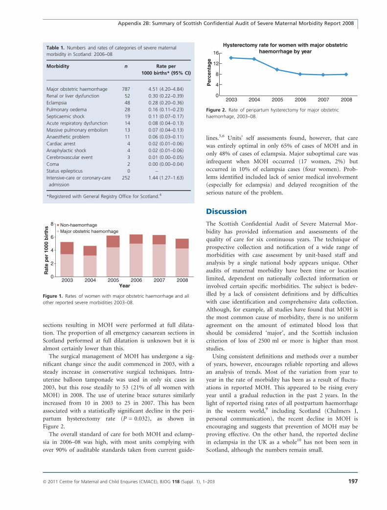
sections resulting in MOH were performed at full dilata-
tion. The proportion of all emergency caesarean sections in
Scotland performed at full dilatation is unknown but it is
almost certainly lower than this.
The surgical management of MOH has undergone a sig-
nificant change since the audit commenced in 2003, with a
steady increase in conservative surgical techniques. Intra-
uterine balloon tamponade was used in only six cases in
2003, but this rose steadily to 53 (21% of all women with
MOH) in 2008. The use of uterine brace sutures similarly
increased from 10 in 2003 to 25 in 2007. This has been
associated with a statistically significant decline in the peri-
partum hysterectomy rate (P = 0.032), as shown in
Figure 2.
The overall standard of care for both MOH and eclamp-
sia in 2006–08 was high, with most units complying with
over 90% of auditable standards taken from current guide-
lines.5,6 Units’ self assessments found, however, that care
was entirely optimal in only 65% of cases of MOH and in
only 48% of cases of eclampsia. Major suboptimal care was
infrequent when MOH occurred (17 women, 2%) but
occurred in 10% of eclampsia cases (four women). Prob-
lems identified included lack of senior medical involvement
(especially for eclampsia) and delayed recognition of the
serious nature of the problem.
Discussion
The Scottish Confidential Audit of Severe Maternal Mor-
bidity has provided information and assessments of the
quality of care for six continuous years. The technique of
prospective collection and notification of a wide range of
morbidities with case assessment by unit-based staff and
analysis by a single national body appears unique. Other
audits of maternal morbidity have been time or location
limited, dependent on nationally collected information or
involved certain specific morbidities. The subject is bedev-
illed by a lack of consistent definitions and by difficulties
with case identification and comprehensive data collection.
Although, for example, all studies have found that MOH is
the most common cause of morbidity, there is no uniform
agreement on the amount of estimated blood loss that
should be considered ‘major’, and the Scottish inclusion
criterion of loss of 2500 ml or more is higher than most
studies.
Using consistent definitions and methods over a number
of years, however, encourages reliable reporting and allows
an analysis of trends. Most of the variation from year to
year in the rate of morbidity has been as a result of fluctu-
ations in reported MOH. This appeared to be rising every
year until a gradual reduction in the past 2 years. In the
light of reported rising rates of all postpartum haemorrhage
in the western world,9 including Scotland (Chalmers J,
personal communication), the recent decline in MOH is
encouraging and suggests that prevention of MOH may be
proving effective. On the other hand, the reported decline
in eclampsia in the UK as a whole10 has not been seen in
Scotland, although the numbers remain small.
Table 1. Numbers and rates of categories of severe maternal
morbidity in Scotland: 2006–08
Morbidity n Rate per
1000 births* (95% CI)
Major obstetric haemorrhage 787 4.51 (4.20–4.84)
Renal or liver dysfunction 52 0.30 (0.22–0.39)
Eclampsia 48 0.28 (0.20–0.36)
Pulmonary oedema 28 0.16 (0.11–0.23)
Septicaemic shock 19 0.11 (0.07–0.17)
Acute respiratory dysfunction 14 0.08 (0.04–0.13)
Massive pulmonary embolism 13 0.07 (0.04–0.13)
Anaesthetic problem 11 0.06 (0.03–0.11)
Cardiac arrest 4 0.02 (0.01–0.06)
Anaphylactic shock 4 0.02 (0.01–0.06)
Cerebrovascular event 3 0.01 (0.00–0.05)
Coma 2 0.00 (0.00–0.04)
Status epilepticus 0 –
Intensive-care or coronary-care
admission
252 1.44 (1.27–1.63)
*Registered with General Registry Office for Scotland.4
0
2
4
6
8
2003 2004 2005 2006 2007 2008
Rat
e p
er 1
000
bir
ths
Year
Non-haemorrhageMajor obstetric haemorrhage
Figure 1. Rates of women with major obstetric haemorrhage and all
other reported severe morbidities 2003–08.
0
4
8
12
16
2003 2004 2005 2006 2007 2008
Per
cen
tag
e
Hysterectomy rate for women with major obstetrichaemorrhage by year
Figure 2. Rate of peripartum hysterectomy for major obstetric
haemorrhage, 2003–08.
Appendix 2B: Summary of Scottish Confidential Audit of Severe Maternal Morbidity Report 2008
ª 2011 Centre for Maternal and Child Enquiries (CMACE), BJOG 118 (Suppl. 1), 1–203 197
Evidence of effective management is seen in the signifi-
cant decline in peripartum hysterectomies despite a pre-
dicted rise as caesarean section rates rise.11 The potential
place of prophylactic interventional radiology in the pre-
vention of anticipated major haemorrhage and of red blood
cell salvage in its management is described in recent guide-
lines,12 and data on these important developments will be
collected in future years of the audit and may provide valu-
able evidence as to their efficacy.
All of the women studied in this audit survived episodes
of severe morbidity, and it is, therefore, unsurprising that
their care was, in general, good. Although major subopti-
mal care of cases of MOH was uncommon, minor or inci-
dental problems were frequent, occurring in over one-third
of cases. Even units caring for 1000 births per year can
expect four or five episodes of MOH annually. Practice
drills including education in the recognition of risk factors
may help, and the early involvement of senior medical staff
when MOH is anticipated (for example where placenta ac-
creta is suspected or emergency caesarean section at full
dilatation is performed) or occurs should be encouraged.
Direct consultant involvement in the immediate care of
women experiencing eclampsia was particularly lacking.
With a mean of only 16 cases annually of eclampsia in
Scotland in 2006–08, some units may see no cases for sev-
eral years. This is reflected in the reported failure to recog-
nise prodromal signs and symptoms. Despite these
reported deficiencies, the severe morbidity/maternal mortal-
ity ratio in Scotland in 2006–08 improved to 1/79 from 1/
56 in 2003–05, although the small numbers of maternal
deaths make it difficult to form firm conclusions. In addi-
tion, although there were two deaths from MOH in Scot-
land in 2006, there were none in 2007 or 2008, and no
deaths from eclampsia occurred in the triennium. The
value of measuring morbidity and its care is clear.
AcknowledgementsLeslie Marr, Reproductive Health Programme coordinator
of NHS Quality Improvement Scotland is the major con-
tributor to the overall management of the audit. The hard
work and cooperation of all unit coordinators are gratefully
acknowledged, as is the statistical and analytical input from
Dr AK McFadyen of Glasgow Caledonian University. j
References
1 Brace V, Penney G, Hall M. Quantifying severe maternal morbidity:
a Scottish population study. BJOG 2004;1:481–4.
2 Mantel GD, Buchmann E, Rees H, Pattinson RC. Severe acute mater-
nal morbidity: a pilot study of a definition for a near-miss. BJOG
1998;105:985–90.
3 NHS Quality Improvement Scotland. Scottish Confidential Audit of
Severe Maternal Morbidity. 6th Annual Report. Edinburgh. NHS QIS;
2010.
4 General Register Office for Scotland. Vital events reference tables
2008. [www.gro-scotland.gov.uk/files2/stats/vital-events-ref-tables-
2008/ve-2008-t3-1.pdf]. Accessed 15 February 2010.
5 Scottish Obstetric Guidelines and Audit Project. The Management of
Postpartum Haemorrhage 1998 and Guideline Update 2002. Scottish
Programme for Clinical Effectiveness in Reproductive Health, Publica-
tion No. 6.
6 Royal College of Obstetricians and Gynaecologists. The Manage-
ment of Severe Pre-eclampsis/Eclampsia. Green Top Guideline 2006.
London: RCOG, 2006.
7 Information Services Division, NHS Scotland. Scottish perinatal and
infant mortality and morbidity report 2008. [www.isdscotland.org/
isd/3112.html]. Accessed 15 February 2010.
8 Information Services Division, NHS Scotland. Live births by mode of
delivery and induced. [www.isdscotland.org/isd/files/mat_bb_table
5.xls]. Accessed 15 February 2010.
9 Joseph KS, Rouleau J, Kramer MS, Young DC, Liston RM, Baskett
TF, et al. Investigation of an increase in postpartum haemorrhage in
Canada. BJOG 2007;114:751–9.
10 Knight M, on behalf of UKOSS. Eclampsia in the United Kingdom
2005. BJOG 2007;114:1072–8.
11 Knight M, Kurinczuk JJ, Tufnell D, Brocklehurst P. The UK Obstetric
Surveillance System for rare disorders of pregnancy. BJOG
2005;112:263–5.
12 Royal College of Obstetricians and Gynaecologists. Prevention and
Management of Postpartum Haemorrhage. Green-top Guideline no.
52. London: RCOG, 2009.
Lennox
198 ª 2011 Centre for Maternal and Child Enquiries (CMACE), BJOG 118 (Suppl. 1), 1–203
Appendix 3: Contributors to the Maternal DeathEnquiry for the triennium 2006–08 and CMACEpersonnel
Director of Maternal Death Enquiry
Professor Gwyneth Lewis OBE MSc MRCGP FFPH FRCOG FACOG DSc
Central Assessors and Authors
Anaesthetics Dr G Cooper OBE FRCA FRCOG
Dr JH McClure FRCA
Emergency Medicine Dr D Hulbert FRCS FCEM
General Practice Dr J Shakespeare MRCP FRCGP
Intensive Care Dr T Clutton-Brock FRCP FRCA
Medicine Professor M de Swiet MD FRCP FRCOG
Professor C Nelson-Piercy FRCP FRCOG
Professor C Williamson MD FRCP
Midwifery Ms D Garrod RM DPSM BA MA PGCE
Dr J Rogers BA PhD DPSM SRN RM
Mrs V Beale RN RM Dip Man MSc
Obstetric Mr A Dawson MD FRCOG
Professor J Drife MD FRCOG FRCP (Ed) FRCS (Ed)
FCOG (SA) FFSRH
Dr A Harper OBE MD FRCOG FRCPI FFSRH
Professor J Neilson MD FRCOG
Professor Jane Norman MDFROG
Professor C O’Herlihy MD FRCPI FRCOG FRANZCOG
Pathology Dr GH Millward-Sadler FRCPath MHSM
Professor S Lucas FRCP FRCPath
Psychiatry Dr M Oates OBE FRCPsych FRCOG
Dr R Cantwell FRCPsych
CMACE personnel involved in Enquiry work
Central Office
Chief Executive Mr R Congdon
Director of Research and Development Ms S Golightly
Programme Director and Midwifery Lead Mrs A Miller
Project Manager Ms I Mortagy
Data Manager Mrs D Parmar
Senior Data Analyst Ms A Springett
Administrative Officer Mrs M Wilson
Administrative Assistant Mrs S Sanyaolu
CMACE Regional Offices
Cambridge office Mrs C Hay
Sheffield office Mrs S Wood/Mrs C Wade
London office Ms R Thomas/Ms S Akhtar/Ms C Haken
Bristol office Mrs R Thompson/Ms L Howarth
ª 2011 Centre for Maternal and Child Enquiries (CMACE), BJOG 118 (Suppl. 1), 1–203 199
Appendix 3: Contributors to the Maternal Death Enquiry for the triennium 2006–08 and CMACE personnel

Newcastle office Mrs M Renwick
Belfast office Dr A Bell/Ms J Gluck
Manchester office Mrs J Maddocks/Ms P Norris
Cardiff office Mrs D Roberts/Mrs J Maddocks
Cork office Ms E Manning
Regional Assessors in Obstetrics (England)
East of England Mr B Lim FRCOG FRANZCOG
East Midlands, Yorkshire and Humber Miss HJ Mellows FRCOG (until 2008)
Mr SF Spooner FRCOG
Mr D Tuffnell FRCOG
Professor J Walker MD FRCP FRCOG
London and South East Miss G Henson FRCOG
Miss P Hurley FRCOG
Miss C Iffland FRCOG
Dr K Morton FRCOG
Mr ME Setchell FRCS FRCOG
Mr GHW Ward FRCOG FRANZCOG
North East Professor JM Davison MD FRCOG
North West and West Midlands Miss P Buck FRCOG (until 2008)
Mr SJ Duthie FRCOG
Mrs G Masson FRCOG
Mr DHA Redford FRCOG
Mr S Walkinshaw FRCOG
South West Miss I Montague MRCOG
Miss S Sellers MD FRCOG
Regional Assessors in Anaesthetics (England)
East of England Dr J Bamber FRCA
East Midlands, Yorkshire and Humber Dr D Bogod FRCA
Dr G Kesseler FFARCS (until 2008)
Dr IF Russell FRCA
Dr R Wilson MRCP FRCA (from 2008)
London and South East Dr H Adams FRCA
Dr W Aveling DRCOG FRCA
Dr MB Dobson MRCP FRCA
Dr I Findley DRCOG FRCA
Dr S Yentis MD FRCA
North East Dr D Hughes FRCA
North West and West Midlands Dr J Clift FRCA (from 2006)
Dr K Grady FRCA
Dr M Lewis FRCA (until 2007)
Dr S Wheatly FRCA
Dr A Wilkey FRCA (from 2007)
South West Dr L Shutt FRCA (until 2007)
Dr M Wee FRCA (from 2008)
Regional Assessors in Pathology (England)
East of England Dr A Cluroe FRCPA Dip Forens Path
East Midlands, Yorkshire and Humber Dr LJR Brown FRCPath
Dr DR Morgan FRCPath (until 2008)
Dr S Knight MRCPath (from 2006)
London and South East Dr A Bates MD PhD MRCPath
Dr C Corbishley FRCPath
Dr M Hall FRCPath
Dr M Jarmulowicz FRCPath
Dr H Millward-Sadler FRCPath MHSM (until 2008)
North West and West Midlands Dr R Hale FRCPath
Dr L Hammond PhD MRCPath (from 2007)
Dr T Rollason FRCPath (until 2006)
Dr G Wilson FRCPath
Dr A Yoong FRCPath (from 2007)
200 ª 2011 Centre for Maternal and Child Enquiries (CMACE), BJOG 118 (Suppl. 1), 1–203
Appendix 3: Contributors to the Maternal Death Enquiry for the triennium 2006–08 and CMACE personnel
North East Dr JN Bulmer PhD FRCPath
South West Dr L Hirschowitz FRCPath (until 2008)
Dr C Keen FRCPath
Dr J McCarthy FRCPath (from 2008)
Regional Assessors in Midwifery (England)
East of England Mrs J Driver RN, RSCN, RM, ADM, SOM, PGCert
Supervision of Midwives (from 2008)
Miss J Fraser MSc DPSM RM RN (until 2008)
East Midlands, Yorkshire and Humber Mrs JM Jackson RN RM
Ms J Lovett RGN RM (until 2006)
Miss JM Savage RN RM
Ms D Watkins RN RM ADM MSc
London and South East Ms P Cooke RM ADM MSc PGCEA
Mrs P Cox RM, RN, SOM (from 2008)
Ms A Cuthbertson MSc RM RGN (until 2007)
Ms C Drummond RGN RM (until 2007)
Ms J Hughes RGN RM (from 2007)
Ms D Phelan RM RN (from 2007)
Ms S Sauter RM DNLS ADM Dip Ethics
Ms S Truttero MBA LLB ADM RN RM (until 2008)
Ms A Weavers RM
North East Ms K Mannion RM RN ADM MSc
North West and West Midlands Ms D Birchall RGN RM LLB MSc (until 2007)
Mrs S Brown RM RN (from 2008)
Mrs H Jackson SRN SCM ADM BSc (Hons) (from 2008)
Mrs E Newell RGN RM DPSM BSc (Hons)
Ms N Parry RGN RM BSc
Ms A Pedder RN RM BSc MA (until 2006)
Mrs S Smithson BSc (Hons) RN ADM ONC
South West Mrs V Beale RN RM Dip Man MSc (until 2006)
Mrs J Drury RM RN MSc DpSM SEN (from 2006)
Ms D Morrall RN RM
Mrs A Remmers RGN RM MSc
Regional Assessors in Psychiatry (England)
East England Dr F Blake MRCGP MRCPsych
East Midlands, Yorkshire and Humber Dr P Brownsett MRPsych
Dr S Shaw MRCPsych MMedSci
London and South East Dr A Gregoire DRCOG MRCPsych
Dr L McDonald MRCPsych MPhil Dip CBT
North East Dr A Walsh MRCP MRCPsych
North West and West Midlands Dr K Hofberg MMedSci(Psych) MRCPsych
Dr A Wieck MD FRCPsych
South West Dr S Oke MRCPsych
Other UK nations (Northern Ireland, Scotland and Wales) and Ireland involved in the Maternal Death Enquiry
Northern Ireland
Department of Health and Social Services:
Northern Ireland (Health Promotion Agency)
Dr C Willis MFPH
Obstetrics Assessor Dr A Harper OBE MD FRCOG FRCPI FFSRH
Anaesthetic Assessor Dr D Hill FCARCSI
Pathology Assessor Dr G McCusker FRCPath
Midwifery Assessor Mrs E Bannon RN RM MSc
Psychiatry Assessor Dr J Lynch MRCPsych
Scotland
Chairman of Enquiry Dr J McClure FRCA
Clinical Adviser to Reproductive Health
Programme, NHS Quality Improvement Scotland
Dr C Lennox FRCOG
ª 2011 Centre for Maternal and Child Enquiries (CMACE), BJOG 118 (Suppl. 1), 1–203 201
Appendix 3: Contributors to the Maternal Death Enquiry for the triennium 2006–08 and CMACE personnel
Reproductive Health Programme Co-ordinator,
NHS Quality Improvement Scotland
Ms L Marr MA RGN RM ADM MSc
CMO’s Representative: Scottish Executive
Health Department
Dr L Smith PhD MSc(LSHTM) MFPH
Obstetrics Assessors Dr DAR Lees FRCOG
Dr H MacPherson FRCOG
Professor J Norman MD FRCOG
Anaesthetic Assessor Dr JH McClure FRCA
Pathology Assessor Dr R Nairn FRCPath DipFM
Midwifery Assessors Ms A Lobo MSc RGN RM ADM
Mrs J Reekie RGN RM
Psychiatry Assessor Dr R Cantwell FRCPsych
Wales
Welsh Assembly Government Dr J Ludlow FFPHM
Obstetrics Assessor Mr A Dawson MD FRCOG
Anaesthetic Assessor Dr P Clyburn FRCA
Pathology Assessor Dr L Murray FRCPath
Midwifery Assessor Mrs K McGrath SRN RM Dip Mid MSc (Health Management)
Psychiatry Assessor Dr S Smith MRCPsych
Ireland
Institute of Obstetricians and Gynaecologists Dr M F O’Hare MD FRCPI FRCOG
Obstetric Assessor Professor C O’Herlihy MD FRCPI FRCOG FRANZCOG
Anaesthetic Assessor Dr J Loughrey MRCPI FCARCSI FFPMCAI
Pathology Assessor Dr P Kelehan MSc MRCPath FRCPath
Midwifery Assessors Ms S Canny RGN RM BMS
Ms M Doyle RGN RM ADM BEd(Hons) MSc
Ms F Hanrahan RGN RM MSc
Psychiatric Assessor Dr A McCarthy MRCPsych FRCPI MMedSc
202 ª 2011 Centre for Maternal and Child Enquiries (CMACE), BJOG 118 (Suppl. 1), 1–203
Appendix 3: Contributors to the Maternal Death Enquiry for the triennium 2006–08 and CMACE personnel
Appendix 4: CMACE Governance
CMACE Board of Trustees
Patron: Baroness Julia Cumberlege
Professor JW Walker, Chair
Professor Sir Sabaratnam Arulkumaran
Dr J Chapple
Professor P Fleming (from July 2010)
Dr A Harnden (until June 2010)
Ms S Jenkinson (from July 2010)
Ms L Sellers (from July 2010)
Ms PA Seymour
Ms L Silverton
Professor I Greer (Chair)—National Advisory Committee
ª 2011 Centre for Maternal and Child Enquiries (CMACE), BJOG 118 (Suppl. 1), 1–203 203
Appendix 4: CMACE Governance