Embed Size (px)
Citation preview

1
MOLECULAR ENGINEERING OF MULTIFUNCTIONAL NUCLEIC ACID PROBES FOR BIOANALYTICAL AND BIOMEDICAL APPLICATIONS
By
YOUNGMI KIM
A DISSERTATION PRESENTED TO THE GRADUATE SCHOOL OF THE UNIVERSITY OF FLORIDA IN PARTIAL FULFILLMENT
OF THE REQUIREMENTS FOR THE DEGREE OF DOCTOR OF PHILOSOPHY
UNIVERSITY OF FLORIDA
2008

2
© 2008 Youngmi Kim

3
To my family

4
ACKNOWLEDGMENTS
I am deeply indebted to a long list of people who made this dissertation possible. First, I
wish to express my gratitude to my advisor, Dr. Weihong Tan. His advice, suggestions, and
encouragement were greatly helpful for advancing and progressing projects and were the driving
sources that kept me challenging to be a better scientist every day. In addition, I thank Dr.
Charles Martin, Dr. Nicole Horestein, Dr. David Powell and Dr. Powell Holloway on my
graduate committee. The advice, assistance, and encouragement from my committee are highly
appreciated.
This dissertation is a result of successful collaborations with scientists in different areas. I
would like to thank Dr Chaoyong James Yang for guiding me when I started the very first
research project. I greatly appreciate Dr. Zeihui Charles Cao for initiating the molecular
assembly project and thought me fluorescence spectroscopy. I appreciate Dr. Haipeng Liu and
Joeshep Philips for the critical comments and help in development of azobenzene-modified
nucleic acid probes. I appreciate Dr. Donn Dennis in department of anesthesiology for initiating
selection project using bivalirudine. I am very thankful for Hui Wang’s hard working on the
unimolecular nucleic acid sensor project. I also would like to thank Colin Medley and Joshua
Smith for their special friendship.
The Tan research group is a special place to work. The help and friendship from former
and current group members make my memory of Gainesville enjoyable and unforgettable. I
would like to thank Dr. Ronghua Yang, Dr. Charles Lofton, Dr. Dihua Shangguan, Dr. Zhiwen
Tang, Dr. Lin Wang, Dr. Alina Munteanu , Dr Prabodhika Mallikaratchy, Dr. Carmen Maria
Estevez , Dr. Yufen Huang, Dr. Liu Yang, Dr. Xiaoling Zhang, Karen Martinez, Hui Chen,
Kwame Sefah, Parag Parekh, Eunjung Lee, Yanrong Wu, Huaizhi Kang, Yan Chen, Meng Ling,
Megan O’Donoghue, Dalia Lopez-Colon, Jennifer Martin, Suwussa Bamrungsap, Zhi Zhu, Xu

5
Ye, Xiaolan Chen, Sewon Bae, Dimitri Simaeys Van , Basri Gulbakan, Elizabeth Jimenez,
Xiangling Xiong for their friendship, encouragement, and help.
I am deeply indebted to my mother for her unconditional love, support, and guidance and
strong spirit. I thank my brothers for their love. I am grateful to my husband, Dosung Sohn for
being a wonderful friend, helpful colleague, and supportive spouse who enable me to succeed. I
thank my lovely daughter, Mia Sohn, for being such a great daughter and bringing every kind of
joy to my life every moment.

6
TABLE OF CONTENTS
page
ACKNOWLEDGMENTS ...............................................................................................................4
LIST OF TABLES .........................................................................................................................10
LIST OF FIGURES .......................................................................................................................11
ABSTRACT ...................................................................................................................................14
CHAPTER
1 INTRODUCTION ..................................................................................................................16
Review of Nucleic Acid Structures ........................................................................................16 Chemical Synthesis of Nucleic Acids .....................................................................................18 Fluorescence Methods for Signal Transduction .....................................................................22
Fluorescence Mechanism ................................................................................................22 Fluorescence Quenching Mechanism ..............................................................................24 Fluorescence Resonance Energy Transfer (FRET) .........................................................25
Nucleic Acid in Biology .........................................................................................................27 Using MBs for RNA Monitoring in Living Cells ...........................................................30 Engineering MBs for Intracellular Analysis ....................................................................33
Systematic Evolution of Ligands by Exponential Enrichment (SELEX) ...............................35 Polyvalent Interactions ...........................................................................................................38
Thermodynamic Model to Describe Cooperativity .........................................................39 Kinetics and Enhanced Affinity ......................................................................................41
DNAzymes .............................................................................................................................41 Blood Coagulation and Thrombin ..........................................................................................43
Coagulation ......................................................................................................................43 Thrombin Structure .........................................................................................................44
Photochromic Switches ..........................................................................................................45 Photochromism ................................................................................................................45 Azobenzene .....................................................................................................................48
2 SUPERIOR STRUCTURAL STABILITY AND SELECTIVITY OF HAIRPIN NUCLEIC ACID PROBES WITH AN L-DNA STEM .........................................................50
Introduction .............................................................................................................................50 Experimental Section ..............................................................................................................54
Synthesis of MBs and Their Targets ...............................................................................54 Hybridization Experiments ..............................................................................................55 Melting Temperature (Tm) Measurement of MBs ..........................................................56 Protein Sensitivity Tests ..................................................................................................56
Results and Discussion ...........................................................................................................57

7
Stability and Sensitivity of L-DNA Stem MBs (LS MBs) ..............................................57 Improved Structural Stability. .........................................................................................59 Elimination of Intramolecular Interaction .......................................................................60 Elimination of False Positive Signal ...............................................................................63 Biostablility of LS MBs ...................................................................................................65
Conclusions .............................................................................................................................69
3 MOLECULAR ASSEMBLY FOR HIGH PERFORMANCE BIVALENT NUCLEIC ACID INHIBITOR .................................................................................................................70
Introduction .............................................................................................................................70 Experimental Section ..............................................................................................................73
Chemicals and Reagents: .................................................................................................73 Synthesis and Purification of Mono- and Bivalent NA Ligands and Their Targets .......73 Clotting Time Tests .........................................................................................................75 Real-Time Monitoring of the Clotting Reaction .............................................................75 Monitoring of Apparent kon and koff .................................................................................76 Reversible Binding Reaction Using Target DNAs ..........................................................76 Human Plasma Tests .......................................................................................................77
Results and Discussion ...........................................................................................................77 Thrombin Aptamers and Their Properties .......................................................................77 Design of Bivalent NA Inhibitors ....................................................................................79 Monitoring Inhibitory Functions Using Light Scattering ................................................81 Binding Kinetics Studies .................................................................................................83 Antidote Effect of Binding Aptamers ..............................................................................88 Antithrombin Potency of Bi-8S .......................................................................................89
Conclusions .............................................................................................................................91
4 DEVELOPMENT OF DENDRITIC APTAMER ASSEMBLIES AS SUPERIOR INHIBITORS ..........................................................................................................................93
Introduction .............................................................................................................................93 Experimental Section ..............................................................................................................94
Synthesis of Multimeric Assemblies ...............................................................................94 Real-Time Monitoring of Coagulation Process ...............................................................94 PT Measurement ..............................................................................................................94
Results and Discussion ...........................................................................................................95 Design of Multimeric Assemblies ...................................................................................95 Validation of Products .....................................................................................................97 Superior Anticoagulation Potency ...................................................................................97 Real-Time Monitoring of Clotting Reaction ...................................................................99
Conclusions ...........................................................................................................................100

8
5 USING PHOTONS TO MANIPULATE ENZYME INHIBITION BY AN AZOBENZENE-MODIFIED NUCLEIC ACID PROBE ....................................................102
Introduction ...........................................................................................................................102 Experimental Section ............................................................................................................104
Chemicals and Reagents ................................................................................................104 Synthesis of Azobenzene Phosphoramidite ...................................................................105 Synthesis and Purification of Photochromic Self-regulating Inhibitor .........................106 Real-Time Monitoring of Clotting Reaction .................................................................106 Human Plasma Tests .....................................................................................................108 Monitoring Site-Specific Activation of Enzymatic Reaction in Microfluidic
Channel ......................................................................................................................109 Results and Discussion .........................................................................................................110
Optimization of Probe Designs .....................................................................................110 Function as Photo-Switching Anticoagulant .................................................................114 Dynamic Photoconversion to Restore Coagulation Process .........................................116 Spatially Controllable Activation of Coagulation Reaction in Microfluidic Channel ..118
Conclusions ...........................................................................................................................122
6 UNIMOLECULAR CATALYTIC-DNA SENSOR FOR ULTRASENSITIVE DETECTION ........................................................................................................................124
Introduction ...........................................................................................................................124 Experimental Section ............................................................................................................127
Chemicals and Reagents ................................................................................................127 Synthesis and Purification of Fluorescently Labeled Oligonucleotides ........................128 Determination of Melting Temperature .........................................................................128 Hybridization Assay ......................................................................................................129 Single Molecular Reaction ............................................................................................130 Gel-Based Activity Assay .............................................................................................131
Results and Discussion .........................................................................................................131 Optimization of the Hairpin Structure Sensor Design ...................................................132 Excellent Sensitivity ......................................................................................................134 Superior Selectivity .......................................................................................................136 Single Ion Reaction .......................................................................................................137
Conclusions ...........................................................................................................................140
7 SUMMARY AND FUTURE DIRECTIONS .......................................................................141
Molecular Engineering of Multifunctional Nucleic Acid Probes for Bioanalytical and Biomedical Applications ...................................................................................................141
Future Directions ..................................................................................................................143 Developing High Throughput Metal-Screening Chip ...................................................143 Pharmaceutical Application of Multifunctional Drugs .................................................145

9
LIST OF REFERENCES .............................................................................................................147
BIOGRAPHICAL SKETCH .......................................................................................................161

10
LIST OF TABLES
Table page
1-1 Comparisons between aptamer and antibody ....................................................................38
1-2 Thermodynamic parameters to describe cooperativity. .....................................................41
2-1 Copy number of each sequence in biological nucleic acid sequences ...............................51
2-2 Sequences of hairpin probes and their targets ....................................................................55
2-3 Signal to background (S/B) of each MB was calculated and compared. ...........................58
2-4 Comparisons of stem melting temperatures of MBs ..........................................................60
2-5 MB 1-1 and its target sequences ........................................................................................62
2-6 Calculated melting temperature of each target with its complementary sequence. ...........64
3-1 DNA sequences. S means one unit of spacer phosphoramidite (hexaethylene glycol). ....74
4-1 Sequences of multimeric assemblies. ................................................................................96
5-1 Sequences of probes. A in red refers to azobenzene. .......................................................107
5-2 Characterized properties of each probe ............................................................................112
6-1 Names and sequences of DNA. .......................................................................................128
6-2 Comparison of melting temperature and fluorescence enhancement among different design sequences. .............................................................................................................133

11
LIST OF FIGURES
Figure page
1-1 Components of nucleic acids .............................................................................................16
1-2 Nucleotide structure and linkage via phosphate groups. ...................................................17
1-3 Base pairing of DNA. ........................................................................................................17
1-4 Structure of cytosine phosphoramidite (top) and four monomers of nucleic acid phosphoramidite (bottom). .................................................................................................19
1-5 Nucleic acid synthesis. .......................................................................................................20
1-6 Jablonski diagram for fluorescence mechanism. ...............................................................23
1-7 Spectral overlap between donor’s emission and acceptor’s excitation (left) and description of fluorescence resonance energy transfer (right). ..........................................26
1-8 Working principle of molecular beacons. ..........................................................................29
1-9 Intracellular imaging of single cells using MB probes ......................................................32
1-10 Molecular structures of 2’-OMe RNA, locked nucleic acid (LNA), and peptide nucleic acid (PNA). ............................................................................................................33
1-11 Background signal of LNA-MB and DNA-MB as a function of time after being injected into cells. ..............................................................................................................35
1-12 Systematic Evolution of Ligands by Exponential Enrichment (SELEX). .........................37
1-13 Multivalent interactions. ....................................................................................................39
1-14 Structure of thrombin. ........................................................................................................44
1-15 Light induced hydrogen transfer tautomerization ..............................................................46
1-16 Photocyclization. ................................................................................................................47
1-17 Cis-trans isomerization ......................................................................................................48
1-18 Molecular structure of azobenzene and energy diagram. ..................................................49
2-1 Molecular beacon design and interactions .........................................................................51
2-2 Responses of LS MBs and DS MBs to the targets.............................................................58
2-3 Melting temperature profiles of DS and LS MB 1 ............................................................60

12
2-4 Elimination of stem and loop interaction using a L-DNA stem ........................................62
2-5 Comparison of selectivity of LS MB 1 and DS MB 1 .......................................................64
2-6 Nuclease resistance of LS and DS MB. .............................................................................66
2-7 Characterization of biostability of LS OMe MBs(green) compared to DS MBs(blue) .....67
2-8 Characterization of biostability of LS LNA MBs (red) compared to DS MBs(blue) ........68
3-1 Working principles of monovalent and bivalent NA ligands. ...........................................78
3-2 Comparison of the normalized clotting times of thrombin bound to different NA inhibitors ............................................................................................................................79
3-3 Real-time monitoring of scattered light generated by the coagulation process in the presence of different monovalent or bivalent NA ligands (Bi-xSs) ...................................82
3-4 Comparison of koff’. Real-time fluorescence signal change of koff’ measurement .............84
3-5 Investigation of concentration effect of T-15Apt in binding comparison .........................85
3-6 Comparison of kon’ .............................................................................................................86
3-7 Investigation of the dissociation of T’-15Apt ....................................................................87
3-8 Reversible inhibitory function ...........................................................................................89
3-9 Comparison of anticoagulant potency of Bi-8S and 15Apt using human plasma and aPTT ...................................................................................................................................90
3-10 Comparison of anticoagulant potency of Bi-8S and 15Apt using human plasma and PT measurements ...............................................................................................................91
4-1 The sequences and schematic of dendritic aptamer assemblies .........................................96
4-2 Agarose gel image with ethidium bromide (EB) staining. ................................................97
4-3 Dose-dependent prothrombin time (PT) ............................................................................98
4-4 Real-time monitoring of thrombin activity ......................................................................100
5-1 Photoisomerization of azobenzene in nucleic acid chain (a) and working principle of Xc-Yazo probes (b) ..........................................................................................................103
5-2 Synthesis of azobenzene phosphoramidite. .....................................................................106
5-3 Insertion of azobenzene phosphoramidite to DNA chains. .............................................107

13
5-4 Relationship between melting temperature and the number of base pairs and azobenzene insertions (a) and (b) and the photo-regulating inhibitory function (c) of probes. ..............................................................................................................................113
5-5 PT measurement using each probe- cis and -trans (a), (b), (c), and (d) and IC200 of each probe’s state (e) .......................................................................................................115
5-6 Dynamic alteration of thrombin’s activity by switching 9c-8azo-cis to -trans. ..............117
5-7 Imaging clotting reaction in a microfluidic device ..........................................................120
5-8 Fluorescence intensity alteration of different zones in real-time. ....................................121
5-9 Site-specific activation of thrombin’s activity using a laser pointer in a microfluidic device ...............................................................................................................................122
6-1 The hairpin structure DNAzyme-based Pb2+ sensor and the working principle ..............127
6-2 Fluorescence signal in the absence and presence of 10uM Pb2+ after 10 min. ................133
6-3 PAGE-gel image of probes in the absence and presence of 10uM Pb2+ after 10 min .....134
6-4 Fluorescence increase over background at varying Pb2+ concentrations .........................135
6-5 Kinetics of fluorescence increase for D10 in 5uM Pb2+ ..................................................135
6-6 Selectivity of D10 Pb2+ sensor .........................................................................................137
6-7 Single ion reaction kinetics inside polycabornate membrane vials .................................139
7-1 Surface-immobilized Pb2+ chip. (a) microarray and anticipated results. (b) shows the cartoon to describe working principle of the metal sensor chip. .....................................144
7-2 Mouse model to show photo-controllable anticoagulation. .............................................146

14
Abstract of Dissertation Presented to the Graduate School of the University of Florida in Partial Fulfillment of the Requirements for the Degree of Doctor of Philosophy
MOLECULAR ENGINEERING OF MULTIFUNCTIONAL NUCLEIC ACID PROBES FOR
BIOANALYTICAL AND BIOMEDICAL APPLICATIONS
By
Youngmi Kim
December 2008 Chair: Weihong Tan Major: Chemistry
Over the past few decades, the role of nucleic acids including deoxyribonucleic acids and
ribonucleic acids in purely biological systems has been defined, and these versatile compounds
have also been integrated into nanoscience. As a result, many different types of DNA probes and
architectures have been proposed to detect a wide range of targets, such as ions, small molecules,
nucleic acids, proteins, and even living organisms. These developments have been accelerated by
advanced technologies for modifying the nucleic acid-based probes with different signaling
mechanisms and bases of nucleic acids with organic functional groups. The overall goal of this
doctoral research is dedicated to engineering these biomolecular components with the objective
of building novel molecular tools and devices for biological studies, biomedical research and
therapeutic applications.
In cell biology, alteration of the expression levels of nucleic acids or spatial localization
often reflects important cellular events. Tracking such alterations in the native cellular
environment can be one of the most effective ways to advance biology, drug discovery and
therapeutic treatment of injury and infection. Because this requires robust probes for reliable
intracellular studies, we have been designing molecular probes using nonstandard nucleic acids,

15
such as L-DNAs, locked nucleic acids, 2’-OMe RNAs, to extend probe lifetimes and enhance the
sensitivity and selectivity.
The second phase of this research has involved the molecular assembly of artificially
selected functional ssDNA sequences called aptamers. To amplify the inhibitory functions of
aptamer, the multivalent interaction concept has been applied to engineer aptamers with high
affinity and specificity for a given target. The process involves assembly of multiple ligands and
target binding via multiple sites. The binding of the resulting assembly is much stronger than that
of the individual ligands, resulting in excellent inhibition of target proteins.
The third phase of this research has involved the development of photo-regulating nucleic
acid probes, in which photochromic compounds are incorporated into the DNA chains to alter
the hybridization characteristics. We have utilized these compounds to design nucleic acid
inhibitors which can regulate functions with photons, and we have developed a nanoarchitecture
made of DNA and photochromic compounds.
The final project was development of an ultra-sensitive heavy metal sensor. Since metal
ion detection is of great importance in human health and environmental monitoring, better
sensing technology for heavy metal ions is sorely needed. With this aim, we developed lead-
specific ultrasensitive nucleic acid sensor with subnanomolar detection limits, superior
sensitivity, and excellent selectivity.
In summary, this research has focused on the design, synthesis and investigation of
multifunctional and advanced nucleic acid probes, with the ultimate goal of increasing the
understanding of biological processes and the development of advanced molecules for nucleic
acid-based detection and therapy.

16
CHAPTER 1 INTRODUCTION
Nucleic acids carry genetic information from parent cell to daughter cell and from
individuals to their offspring. These universal cellular components also regulate many biological
reactions. Nucleic acids are largely divided into two groups: deoxyribonucleic acid (DNA) and
ribonucleic acid (RNA). Since their functions were elucidated in the mid-twentieth century,
nucleic acids have been transformed and diversified into many research areas, from basic biology
to computational and materials science engineering. For readers unfamiliar with this subject, the
first section of this dissertation gives a brief overview of nucleic acid structure and function.
Review of Nucleic Acid Structures
The molecular building blocks for DNA and RNA polymers are shown in Figures 1-1 and
1-2. The monomer units, called nucleotides, which contain a sugar (2-deoxyribose in DNA and
ribose in RNA) are esterified to a phosphate. The sugar is also linked to a cyclic base via a β-N-
glycosidic bond. The nucleotides are bonded by phosphate linkages between the 3′ and 5′
carbons of the sugars.
Figure 1-1.Components of nucleic acids

17
O
OHO
HHHH
PO
O
O-
O
OHO
HHHH
PO
O
O-
O
OHO
HHHH
PO
O-
O
O-
Base
Base
Base
O
HO
HHHH
PO
O
O-
O
HO
HHHH
PO
O
O-
O
HO
HHHH
PO
O-
O
O-
Base
Base
Base
Deoxyribonucleic acid (DNA)Ribonucleic acid (RNA)
Figure 1-2. Nucleotide structure and linkage via phosphate groups
Figure 1-3. Base pairing of DNA
The secondary structure of DNA was formulated in the 1950s by Rosalind Franklin, James
Watson and Francis Crick (Figure 1-3).1 X-ray data obtained by Rosalind Franklin showed that
DNA has a double helix formation. In 1953, Watson and Crick proposed an ingenious double

18
helix model for DNAs, in which complementary bases (C and G; A and T) on each strand are
linked by hydrogen bonds (3 H-bonds for C-G; 2 H-bonds for A-T). The two strands align anti-
parallel to each other in the coil, leading to a double helix structure. This high molecular weight
nucleic acid (109 Daltons or greater) is found primarily in the nuclei of complex cells, known as
eukaryotic cells, or in the nucleotide regions of prokaryotic cells, such as bacteria.
Biological information is encoded in the DNA polymers via successive groups of three
nucleotides. Each 3-base code corresponds to a certain amino acid in the coded protein. To
access this information the pattern is transcribed to RNA templates, a lower molecular weight,
but much more abundant nucleic acids. Three kinds of RNAs have been identified. The largest
subgroup (85 to 90%) is ribosomal RNAs (rRNAs), the major component of ribosomes. The
sizes of rRNA molecules vary, but they are generally smaller than DNAs. The other forms of
RNAs are messenger RNAs (mRNAs), which carry the transcribed code to the ribosomes, and
transfer RNAs (tRNAs), which carry the correct amino acid corresponding to the 3-base code to
the growing polypeptide chain. Both mRNAs and tRNAs have transient lifetimes. Recently,
regulatory RNAs, such as microRNAs and siRNAs, were newly discovered.
Chemical Synthesis of Nucleic Acids
After complete structures of nucleic acids were clearly elucidated, the phosphoramidite
method was developed to synthesize artificial nucleic acids by an automated system. Thus,
nucleic acids have become popular building blocks for designing molecular probes due not only
to their capability for selective recognition against a wide range of targets but also automated
DNA synthesis technology for highly efficient and reproducible synthesis with a variety of
modifications for numerous applications in biological studies. The basis of the synthetic
chemistry is solid-support synthesis of oligonucleotide via phosphoramidite chemistry.2,3

19
Phosphoramidites are composed of different functional groups (Figure 1-4). To eliminate
side reactions during the synthesis of nucleic acids, primary amines of bases are blocked by
appropriate protecting groups that are vulnerable to basic condition, such that they can be
effectively removed by strong bases. 5’-O is capped by dimethoxytrityl (DMT) group to
selectively activate 5’OH under strong acidic condition. The phosphate is also protected by
diisopropylamino and 2-cyanoethyl groups for selective activation under appropriate condition.
Any modifiers, such as fluorophore, biotin, amine, and polyethylene glycol linker, share the
same strategy to design functional phosphoramidites.
Figure 1-4. Structure of cytosine phosphoramidite (top) and four monomers of nucleic acid phosphoramidite (bottom)
The synthesis is carried out in a column containing a solid controlled-pore glass (CPG)
support, where the 3'-hydroxyl of the first nucleotide or modified functional group is attached

20
through a long spacer arm. This support allows excess reagents to be removed by washing and
eliminates the need for tedious purification steps during the synthesis process. The synthesis
requires four chemical reaction steps: 1) detritylation, 2) coupling, 3) capping and 4) oxidation
(Figure 1-5). In the first step, detritylation, the trityl protecting group on the 5′ oxygen is
removed to activate the 5' hydroxyl on the growing oligonucleotide attached to CPG, by passing
strong acidic reagents, such as 3% of dichloroacetic acid (DCA) or trichloroacetic acid (TCA) in
dichloromethane (DCM) through the column. After deprotection and removal of excess acid with
acetonitrile, coupling takes place (step 2) by adding a phosphoramidite derivative of the next
Figure 1-5. Nucleic acid synthesis

21
nucleotide in the presence of tetrazole, a weak acid. The tetrazole plays two important roles in
conjugating the monomers. It protonates the nitrogen of the diisopropylamine group on the 3'-
phosphorous of the incoming nucleotide to produce a tetrazolyl phosphoramidite, which is
susceptible to nucleophilic attack by the activated 5’ hydroxyl group to form a phosphite linkage.
After coupling, the column is washed to remove any extra tetrazole followed by the capping step.
Since the coupling yield cannot reach 100%, there are always a small percentage of failure
sequences, which must be capped to prevent further coupling and the need to remove DNAs
having one or more sequences missing. Capping (step 3) is accomplished by adding acetic
anhydride and N-methylimidazole in tetrahydrofuran to the reaction column. Only residual bare
5’-hydroxyl groups are acetylated, but not the DMT-protected 5’ hydroxyl groups. Capping is
followed by oxidation (step 4), in which the less stable phosphite is oxidized to the stable
pentavalent phosphate tri-ester using iodine as the oxidizing agent and water as the oxygen
donor. After completion of these four steps, one nucleotide has been added to the chain, which is
now ready for the next round of conjugation.
Following the complete synthesis, post-treatment cleaves the product from the solid
support and deprotects the nitrogen bases by reaction with ammonium hydroxide at high
temperature, normally above 50oC. However, if fragile modifiers are present, this procedure is
changed to avoid such a harsh condition. For example, 1-[3-(4-monomethoxytrityloxy)propyl]-
1'-[3-[(2-cyanoethyl)-(N,N-diisopropyl phosphoramidityl]propyl]-3,3,3',3'-
tetramethylindodicarbocyanine chloride (Cy5 phosphoramidite) and 6-(4-
monomethoxytritylamino) hexyl-(2-cyanoethyl)-(N,N-diisopropyl)-phosphoramidite (MMT-NH2
modifier) are vulnerable to strong basic conditions and often decompose or lose the 5’-protecting
group. After the completion of cleavage and base deprotection, the oligonucleotide is

22
precipitated in ethanol/NaCl. After incubation at -20oC for 30 minutes, the precipitated solid is
obtained with high speed centrifugation.
The solid is re-dissolved in 0.1 M triethylammonium acetate (TEAA), pH 7, for the further
high-performance liquid chromatography (HPLC) purification. HPLC purification strongly relies
on the hydrophobic protecting group (generally DMT) remains on the 5′ position after successful
synthesis of the desired oligonucleotide. The typical mobile phase for oligonucleotide
purification for HPLC is 0.1 M TEAA, pH 7, and acetonitrile. After the purified oligonucleotide
is dried, the DMT group is removed by incubation with 80% acetic acid. The reaction is
quenched with ethanol followed by vacuum drying. The pure oligonucleotide is then quantified
by UV measuring absorption at 260nm.
Fluorescence Methods for Signal Transduction
Fluorescence measurement has been the most popular and convenient analytical method
for a variety of investigations in bioanalytical, biochemical, and chemical research due to its high
sensitivity, nondestructive nature, and multiplexing capabilities. Fluorescence can be easily
incorporated into other signaling mechanisms, such as fluorescence quenching, fluorescence
resonance energy transfer (FRET), fluorescence lifetime and fluorescence anisotropy to monitor
a molecular recognition event. Thus, modification of nucleic acid probes with fluorescent
molecules and incorporation of various signaling transductions have been the most popular
approaches in the design of nucleic acid probes.
Fluorescence Mechanism
Fluorescence is one of the relaxation mechanisms for electronically excited molecules.4,5
There are a number of pathways by which the excited molecules can return to ground state, and
Figure 1-6, termed a Jablonski diagram, shows a few of these processes. The singlet (no unpaired
electrons) ground state is labeled S0, with S1 and S2 as singlet excited states. The labels T1 and

23
T2 refer to triplet excited states (2 unpaired electrons). When exposed to electromagnetic
radiation (EMR), the chromophore is excited to an upper vibrational level in S1 (or in S2). Then,
the excited molecule drops to the ground vibrational level of the S1 state via vibrational
relaxation. If conditions are favorable, the molecule returns to an upper vibrational level in S0 by
emission of visible EMR. This process, called fluorescence, occurs in the 10−9 to 10−5 seconds of
the time range. The energy lost through vibrational relaxation causes the red shift (longer
wavelength), which allows the clear differentiation of the emission signal from the excitation
signal. Besides this fluorescence emission, there are several other pathways for returning to the
ground state from the excited singlet state, including non-radiative decays (generally thermal
relaxation) and intersystem crossing to a triplet excited state (phosphorescence).
Figure 1-6. Jablonski diagram for fluorescence mechanism

24
Fluorescence Quenching Mechanism
Among the various non-radiative processes for releasing energy, fluorescence quenching is
one of the most popular signaling mechanisms for studying molecular interactions between
nucleic acid probes and their targets. Fluorescence quenching occurs via two major mechanisms:
collisional quenching and static quenching. In collisional (or dynamic) quenching, the excited
molecule collides with other molecules or ions in the solution, including solvent molecules,
resulting in energy loss by the fluorophore. As a result, the fluorophore returns to the ground
state without generating photons. The decay of fluorescence intensity caused by collisional
quenching can be described using the Stern-Volmer equation:
]Q[k1]Q[K1FF
0q0 τ+=+= (1-1)
in which
F0 = Initial fluorescence intensity without quencher F = Fluorescence intensity with quencher K = Stern-Volmer quenching constant (M−1) kq= Bimolecular quenching rate constant (M−1s−1) τ0 = Fluorescence lifetime in the absence of the quencher (s) [Q] = quencher concentration (M)
In aqueous solutions at room temperature, a fluorophore with a lifetime, τ0, of 1 ns generally has
a Stern-Volmer quenching constant of about 10 M−1 for a typical quencher. This estimation
suggests that, for quencher concentrations in the millimolar range, the effect of quenching is not
significant. However, when the fluorophore and the quencher are covalently linked to each other,
the collision rate can be dramatically elevated due to the close proximity, and not thereby
depending on diffusion, which results in significant quenching.

25
In static quenching, the quencher forms a non-fluorescent complex (dark complex), FQ,
with the fluorophore in the ground state. The relationship between the fluorescence decrease and
the quencher concentration is given by:
]F[]FQ[1]Q[K1
FF
f0 +=+= (1-2)
in which
Kf = Formation constant for FQ [FQ] = Concentration of the dark complex [F]: Concentration of fluorophore [Q]: Concentration of quencher.
Unlike dynamic quenching, fluorescence lifetime does not change in static quenching, because
the only observed fluorescence is from the free fluorophore, which possesses the same lifetime
as before quenching. In contrast, dynamic quenching causes the lifetime to decrease by the same
factor as the fluorescence intensity. In addition, the effect of temperature is different for the two
types of quenching. In static quenching, elevated temperature causes the quenching efficiency to
decrease due to dissociation of the weakly bound FQ complex. On the other hand, accelerated
diffusion rate at elevated temperature increases the collisional quenching rate, resulting in a
significant decrease of fluorescence. Static quenching can be incorporated into the design of
molecular probes for studying molecular recognition. The typical example is the molecular
beacon, which is described later in this chapter.
Fluorescence Resonance Energy Transfer (FRET)
In FRET, the excitation energy is transferred from an initially excited donor (D) molecule
to an acceptor (A) molecule via a long range dipole-dipole interaction.5 FRET generally requires
spectral overlap between the emission of the donor and the absorption of the acceptor and close
proximity of D and A (less than 10nm) to allow coupling by dipole-dipole interaction (Figure 1-

26
7). FRET often results a decrease of the donor‘s fluorescence and an increase of the acceptor’s
fluorescence. The rate of energy transfer depends upon several factors, such as the spectral
overlap between the emission spectrum of the donor and the absorption spectrum of the acceptor,
the quantum yield of the donor and acceptor, and the relative orientation of the donor and
acceptor transition dipoles. The FRET efficiency, E, also depends strongly on the distance
between the two molecules, as described in the following equation:
660
60
rR
RE
+= (1-3)
In which
R0 = Förster radius where energy transfer is 50% efficient r = distance between the donor and the acceptor
Figure 1-7. Spectral overlap between donor’s emission and acceptor’s excitation (left) and description of fluorescence resonance energy transfer (right)

27
The strong distance dependency of FRET efficiency has been widely exploited in molecular
structure and dynamics studies, intermolecular association detection, intermolecular binding
assays, as well as molecular probe design.
Nucleic Acid in Biology
One of the largest fields in which nucleic acids are utilized is biology. As described above,
the cell’s genetic information is stored in the DNAs, and RNAs play critical roles in processing
the genetic code and regulation of protein synthesis. Of the two types of nucleic acids, RNAs are
often used as biological targets, since their expression levels directly reflect the status of
biological reactions. In addition, appropriate regulation of RNA concentration can be used to
manipulate living systems for various purposes, such as disease treatments or biological studies.
Of all areas where genomics has the potential for huge impact, nothing will be more
meaningful than its affect on health research. By revealing the secrets of the genome, scientists
will be one step closer to learning the origins of certain diseases. In living systems, genomic
information and disease are tightly correlated with each other; one is the cause, and the other is
the consequence. For example, many diseases, such as autism, mental disorders, cancer, stroke,
and diabetes,6 can be explained by a single gene mutation, called a Single Nucleotide
Polymorphism (SNP). In addition, any disease process can ultimately be explained in
biochemical terms which reflect gene function and expression. Genetic variations also provide
the blueprint to understand individual’s susceptibility to certain environmental factors.7 There are
now four major emerging technologies which make use of genomic information: diagnostics,
bioinformatics, proteomics and drug development, and antisense therapy.
Gene expression is the process by which a gene’s DNA sequence is converted into cell’s
functional proteins. Thus, a blueprint for understanding cellular processes can be obtained by
gene expression profiling, which is the simultaneous measurement of the cellular concentrations

28
of different messenger RNAs, often representing thousands of genes in biological samples. This
process often utilizes DNA microarrays, which contain representative sequences of all the genes
to be measured at one time and provide the relative abundance information. Recently, the use of
gene expression profiling has expanded to the later stages in the drug discovery pipeline,
including pharmacogenomics-based assessment of the efficacy and safety of novel compounds.
The use of gene expression for clinical applications, such as patient classification and
diagnostics, is also growing. Many groups are working on the classification of different types of
cancer and on the development of gene expression-based diagnostic tools to select the best
treatment.
Although the most common way to study gene expression is via use of DNA microarrays
to collect massive amounts of data, this technique often gives false results due to the complexity
of ex vivo samples composed of different cell types, making it difficult to interpret the data.
Thus, analysis of single-cell gene expression can provide a more precise understanding of human
disease pathogenesis and can lead to important diagnostic applications. Applications of
functional genomics to crucial biological processes, such as response to stimuli or drugs and the
determination of cellular destiny, would be greatly facilitated by direct monitoring of gene
expression in a single cell. However, monitoring gene function and activity in single living cells
has long been a problem of great interest and difficulty in clinical and basic life science research
and development.
Molecular Beacons
Molecular beacons (MBs) shown in Figure 1-8 are single-stranded DNA probes composed
of three different functional domains: stem, loop, and fluorophore/quencher pair.8 The stem
sequences (4-7 base pairs) are complementary to each other, and the loop is complementary to
the target. The fluorophore/quencher (F/Q) signaling element, switches between the on and off

29
states, depending on the conformational state of the MBs. In the absence of targets, the energy
absorbed by F is transferred via FRET to Q, which is spatially very close to F due to stem
hybridization, and fluorescence is not observed (off state). When the target is present, the loop
and target hybridize (generally 15 to 25 base pairs), and the distance between F and Q greatly
increases (approximately longer than 10nm). Thus, FRET no longer occurs, and strong
fluorescence is observed (on state).
Figure 1-8. Working principle of molecular beacon
The unique hairpin structure and on/off signaling mechanism endow the MBs with several
advantages. First of all, the light-up signaling mechanism allows it to perform highly sensitive
detections for nucleic acid monitoring in real time. Because the unbound MBs stay in the off
state, fluorescence is produced only when target is added, and the intensity is proportional to the
target concentration. Such a detection-without-separation property is particularly useful for the
MBs in situations where it is either impossible or undesirable to extract the probe/target hybrids
from an excess of the unbound probes. Another advantage of the MBs is their relatively high
signal-to-background ratio (S/B), which provides high sensitivity. Upon hybridization of its
target, a well designed MB can generate a fluorescence enhancement as high as 200-fold under

30
optimal conditions.9 This provides the MBs with a significant advantage over other fluorescent
probes in ultra-sensitive analysis. In addition to its sensitivity, the MBs offer excellent
selectivity. They are extraordinarily target-specific and are able to differentiate changes as small
as single-mismatched sequences. The selectivity of the MBs is a direct result of its hairpin
conformation because the stem hybrid acts as an activation energy barrier to the loop-target
hybrid. The remarkable selectivity has been demonstrated in a variety of biological environments
where a large number of different non-target nucleic acid sequences are present. Since they were
first created in 1996, the MBs have been utilized in many research fields and applications,
including intracellular monitoring, biosensor development and clinical diagnosis.
Using MBs for RNA Monitoring in Living Cells
As described above, one of the primary advantages of the MBs is their inherent capability
of detection without separation. This property is especially important in intracellular
applications, where any type of separation is unlikely to be applicable without damaging the
living system. For this reason, the MBs are able not only to detect RNAs in their native
environment, but also to visualize and track their sub-cellular localization in real time. To
accomplish such a goal, design of high-performance MBs is critical.
The major concern in design of the MBs for intracellular applications is the selection of an
appropriate target region, because most of regions of RNA targets are present in double strands.
The selection of target sites starts with the prediction of possible RNA secondary structures. The
target sites are chosen around the regions that are likely to be single-stranded, in order to assure
that the native mRNA structure competes minimally with the proposed MB. In addition, the site
should be unique to represent the specific target. For the chosen regions, high affinity
oligonucleotides of different lengths that are complementary to the regions are used as the loop
sequences of the MBs. Each loop sequence is then flanked with two complementary arm

31
sequences for the stem, which is usually four to seven base pairs long and has a very high G and
C content (higher than 65 percent), to ensure that the hairpin conformation is stable in the living
environment. However, the stem interaction cannot be too strong since it can prevent the binding
of the MB to its target sequence, resulting in low signal enhancement.
Currently, the application of the MBs for intracellular analysis is a rather young field.
Initial studies of the MBs concentrated on detection of the MB hybridization to RNA10-12 In
2003, Tyagi et al demonstrated that MBs could be used to visualize the distribution and transport
of oskar mRNA in Drosophila melanogastar oocytes.13 To eliminate background exhibited from
the MBs, a binary MB approach was developed, which utilized two MBs that targeted adjacent
positions on the mRNA. Only when both MBs were hybridized to the mRNA sequence, the
donor and acceptor fluorophore were brought into close proximity. This allowed FRET to take
place to generate a new signal (at the emission wavelength of the acceptor), indicating
hybridization of the MBs with the mRNA. In addition to visualizing the mRNA distribution, they
were also able to track the migration of the mRNA inside of the cell and even into adjacent cells
in the oocyte. Other studies have imaged MBs on viral mRNA inside of host cells to investigate
the localization of the mRNA inside of cells. The study also utilized photobleaching of the
fluorophore on the MB in order to study the diffusion of the MB-mRNA hybrids. In 2005, Bao et
al expanded on mRNA visualization by showing the co-localization of mRNA and intracellular
organelles in human dermal fibroblasts.14 Their observation was confirmed by several control
experiments, including the use of negative control MBs and fluorescent in situ hybridization
(FISH), as well as detection of colocalization of 28S ribosomal RNA with the rough endoplasmic
reticulum. The authors suggested that the observation of subcellular associations of mRNA with

32
organelles such as mitochondria may provide new insight into the transport, dynamics, and
functions of mRNA and mRNA-protein interactions.
Instead of focusing on localization and distribution, expression levels of mRNA have also
been studied inside of living cells using MBs. The binary MB approach was explored to
determine relative expression levels of K-ras and surviving mRNA in human dermal
fibroblasts.12 According to the report, the expression level of K-ras mRNA is 2.25 time higher
than normal, a result comparable to the ratio of 1.95 using RT-PCR. Recently, the stochasticity
of manganese superoxide dismutase (MnSOD) mRNA expression in human breast carcinoma
cells was studied using MBs with an internal standard reference probe to allow ratiometric
analysis (Figure 1-9).15 By using this method, many of experimental and instrumental variations
Figure 1-9. Intracellular imaging of single cells using MB probes. A ratiometric approach was used to minimize experimental variations and to enable more reliable data collection. The top row shows the cellular response for ‘closed’ MBs. The bottom row shows the cellular responses for ‘open’ MBs. (a) and (d) are the fluorescence emission images of a reference probe; (b) and (e) correspond to fluorescence emission images of the MB probe; (c) and (f) are representative ratiometric images of the MB response, obtained by dividing the image from the MB by the image of the reference probe.

33
were compensated, making direct comparisons of different cells possible. The study showed that
the stochasticity of gene expression for MnSOD was different for the basal, lipopolysaccharide
(LPS)-treated, and the transfected cells, while there was little or no difference in β-actin mRNA
in the three groups. This represents a novel means for direct examination of the stochasticity of
transcription of MnSOD and other genes implicated in cellular phenotype regulation.
Engineering MBs for Intracellular Analysis
Base
Base
Base
Figure 1-10. Molecular structures of 2’-OMe RNA, locked nucleic acid (LNA), and peptide nucleic acid (PNA)
While the researches cited above demonstrate that the MBs are useful in intracellular
monitoring, problems have occurred, mostly due to the complexity of the living environment or
the inherent properties of the MBs. For example, the MBs are vulnerable to intracellular
enzymatic reactions, such as digestion by nuclease, RNase cleavage of MB-bound RNA targets
nucleases, and non-specific opening by single-strand binding proteins (SSBs),15 causing false-
positive or false-negative signals. For example, it has been reported that unmodified

34
phosphodiester oligonucleotides may possess a half-life as short as 15-20 min in living cells.16 In
addition, the high background signal from autofluorescence and the MBs themselves decrease
the resolution. To solve the stability problem, non-standard nucleic acids have been explored to
design MBs with improved resistance to enzymatic activity. These include 2-OMe-modified
RNAs,17-21 peptide nucleic acids (PNAs),22-25 and locked nucleic acids (LNAs).26,27 The 2-OMe-
modified MBs show good nuclease resistance, higher target affinity, increased specificity, faster
hybridization kinetics, and protection of bound RNA targets from RNase H cleavages.28
However, 2-OMe-modified MBs open non-specifically in cells, possibly due to protein
binding.13,20 The peptide backbones of PNAs are not degraded by nucleases, they have a neutral
charge, and hybrids with RNA are thermally more stable compared with DNA-RNA and RNA-
RNA duplexes. Xi et al27 reported that the use of PNA-MBs instead of traditional fluorescent in
situ hybridization probes or DNA-MBs can be better under a wide range of environmental
conditions. However, PNAs have not been widely used, mainly because limited solubility
causing aggregation in biological environment. Recently, Tan et al investigated a locked nucleic
acid molecular beacon (LNA-MB) and demonstrated the great potential of these probes.26,28
LNAs are conformationally restricted nucleic acid analogues, in which the ribose ring is locked
into a rigid C3'-endo (or Northern-type) conformation by a simple 2'-O, 4'-C methylene
bridge.29,29-31 These compounds, as well as LNA-MBs, have many attractive properties,29,31 such
as high binding affinity, excellent mismatch-discrimination capability, and decreased
susceptibility to nuclease digestion (Figure 1-11). The high structural stability of LNA-MBs
results in a significantly lower background compared to DNA-MBs delivered into the cancer
cells. In the living environment, the LNA-MB showed no fluorescence increase over a period of
one hour in the absence of target, while the DNA-MB exhibited a dramatic increase in signal
35
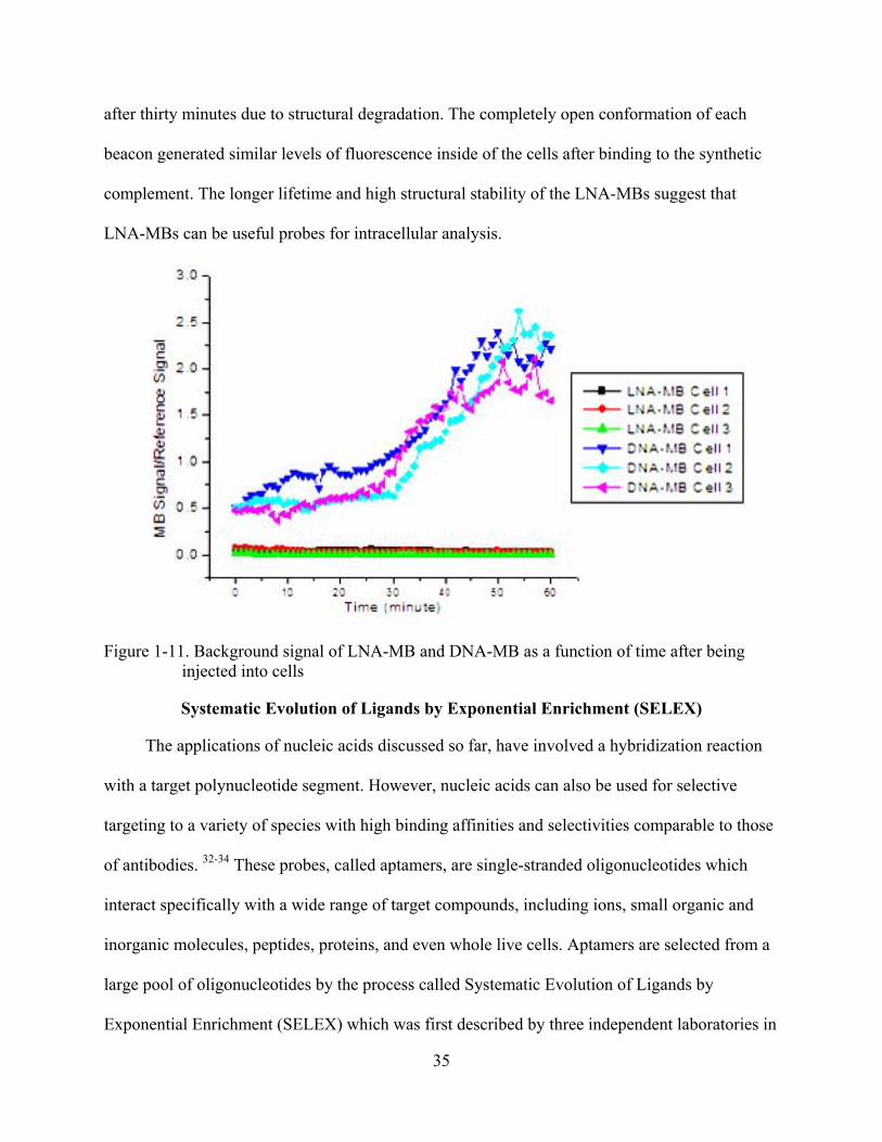
after thirty minutes due to structural degradation. The completely open conformation of each
beacon generated similar levels of fluorescence inside of the cells after binding to the synthetic
complement. The longer lifetime and high structural stability of the LNA-MBs suggest that
LNA-MBs can be useful probes for intracellular analysis.
Figure 1-11. Background signal of LNA-MB and DNA-MB as a function of time after being injected into cells
Systematic Evolution of Ligands by Exponential Enrichment (SELEX)
The applications of nucleic acids discussed so far, have involved a hybridization reaction
with a target polynucleotide segment. However, nucleic acids can also be used for selective
targeting to a variety of species with high binding affinities and selectivities comparable to those
of antibodies. 32-34 These probes, called aptamers, are single-stranded oligonucleotides which
interact specifically with a wide range of target compounds, including ions, small organic and
inorganic molecules, peptides, proteins, and even whole live cells. Aptamers are selected from a
large pool of oligonucleotides by the process called Systematic Evolution of Ligands by
Exponential Enrichment (SELEX) which was first described by three independent laboratories in

36
1990.32-34 It is based on the concept that the unique configuration of small oligonucleotides,
typically 80-100mers, can be suitable for recognition of a specific target with high selectivity. To
maximize the number of potential binding sequences, the oligonucleotide pool contains a
tremendously large number of various sequences, normally 1015 to 1016, flanked by defined
primer binding sites for amplification by polymerase chain reaction.
As shown in Figure 1-12, in each round of SELEX, the first step is incubation of library
sequences with the target molecule in the desired buffer condition. During this step, some
sequences of the library bind to target molecules tightly, while other sequences are weakly bound
or do not interact. The second step is the purification to remove both unbound and weakly bound
sequences from the few high affinity sequences. This is the most critical step of SELEX, because
narrowing the pool to high affinity sequences will allow rapid enrichment. Otherwise, if a few
nonbinding sequences remain, they will subsequently be amplified during the later PCR reaction
and will need to be removed in the next rounds of SELEX. For this reason, there are many
strategies established to improve the purification efficiency at step 2, including nitrocellulose
filtration, affinity chromatography, immunoprecipitation using magnetic beads, gel
electrophoresis and capillary electrophoresis (CE).
After the undesired sequences are removed, the bound sequences are eluted using
denaturing conditions. For example, heating or adding a high pH solution (0.1M NaOH) causes
the oligonucleotides to lose their binding potency by destroying the proper configurations for the
interaction with target. The collected oligonucleotides are then amplified via the polymerase
chain reaction (PCR) process for the next round of SELEX. During the incubating and washing
step, the stringency of the binding condition is generally increased in later rounds to obtain high
affinity sequences with fewer total rounds. In general, 20-30 rounds of SELEX are sufficient to

37
enrich the pool. Once the pool is enriched, the sequences are identified by a sequencing process
and screened to verify their binding potencies.
Figure 1-12. Systematic Evolution of Ligands by Exponential Enrichment (SELEX)
The selected aptamers can be applied in many scenarios. First of all, their high selectivity
and affinity are easily incorporated into the design of bioanalytical tools with an appropriate
signal transduction mechanism, such as fluorescence or radioactive decay. Fluorophores can be
covalently linked to either the ends of nucleic acids or in the middle of sequences based on
phosphoramidite chemistry, and isotope (P32) labels can be also be added using enzymatic
reactions. With fluorescence as the signal mechanism, fluorescence anisotropy, fluorescence
lifetime, fluorescence quenching, fluorescence resonance energy transfer (FRET), and excimer
formation can be used to monitor the binding of aptamers to targets.
Another common application of aptamers is in therapeutics. Compared to antibodies,
aptamers possess many benefits as future therapeutic agents, as summarized in Table 1-1.35
Aptamers can be selected in a timeframe of two months due to the automated in vitro SELEX

38
procedure. On the other hand, antibodies can only be selected via a more time-consuming in vivo
process. Although aptamer and antibody affinities are comparable, aptamers show superior
advantages in terms of high inhibitory potential, low molecular weight, and low to no
immunogenicity or toxicity. Unlike antibodies, an aptamer target can be any extra- and
intracellular protein. In addition to the higher physciochemical stability, the easy chemical
modification of aptamers allows further engineering to improve their targeting potency and in
vivo lifetimes. Thus, many aptamers are undergoing clinical trials for drug commercialization.
Table 1-1. Comparisons between aptamer and antibody35
Polyvalent Interactions
In the field of biochemistry, multivalent interactions are ubiquitous and of high
interest.36,37 Multivalency refers to the binding of two (or more) entities via the simultaneous
interaction of multiple and complementary ligands (Figure 1-13). The valency of a substance is
the number of individual ligand/receptor interactions. The positive outcome of the multivalent
interaction is enhanced binding avidity. Multivalent binding governs many interactions between
proteins and small molecules, between proteins or antibodies and cell membranes, between
Feature Aptamer Antibody
Production < 8 weeks (automated, in vitro) >10 weeks (in vivo)
Specificity and affinity High, Kd: pico to low nanomolar
High, Kd : pico to low nanomolar
Inhibitory potential High Low, 1 out of 200
Molecular weight 5-25 kDa ~150 kDa Immunogenicity and
toxicity Not observed Immune reaction observed
Target space Extra- and intracellular proteins Extracellular proteins only
Chemical modification Easy Difficult
Physicochemical stability Stable Labile

39
viruses and cells.36 In particular, protein–carbohydrate interactions are being intensively
investigated, because of their pivotal roles in the binding of the influenza virus to cell
membranes and in membrane recognition events mediated by carbohydrate-binding proteins
(lectins).38
Figure 1-13. Multivalent interactions
The concept of cooperativity is frequently used to describe polyvalent interactions.
Cooperativity refers to the binding of multiple single-dentate ligands to a polydentate target, such
that an early binding event increases the affinity of later binding interactions.39,40 The enhanced
binding potency often plays a critical role in protein-protein interactions, such as those between
an antibody and an antigen or between a virus and its host.39 The enhanced binding potency may
result from secondary ligand interactions or from allosteric effects. A common example is the
binding of oxygen to hemoglobin; the binding of the first O2 molecule to one subunit of the
hemoglobin tetramer enhances the affinity of the remaining subunits for O2. To understand
cooperativity in detail, a thermodynamic model is introduced.
Thermodynamic Model to Describe Cooperativity
The binding reaction takes place between two types of molecules and is described by the
lock and key model. In the case of a polyvalent interaction, N ligands and N receptors are
involved, as shown in Figure 1-13. Thus, if N is equal to 1, 2, or 3, the interactions are termed
monovalent, bivalent, or trivalent, respectively. Table 1-2 summarizes the nomenclature of the
thermodynamic parameters which relate the free energies of binding (ΔG) to the affinity

40
constants (Ki) for both monovalent and polyvalent systems. Considering the contribution of a
single ligand to a polyvalent interaction, the average free energy of interaction, ΔGpolyaverage , is
equal to ΔGpoly/N. A monovalent ligand-receptor interaction occurs with a free energy change of
ΔGmono, and N independent (no cooperativity) monovalent ligands interact with N receptors to
give a free energy change of NΔGmono. The relation between ΔGpolyaverage and ΔGmono can be
described by ΔGpolyaverage = αNΔGmono. Depending on the value of parameter α, there is three
types of cooperativity: greater than, equal to, or less than the free energy in the analogous
monovalent interaction for α greater than, equal to, or less than 1, respectively. These classes of
cooperativity are termed positively cooperative (synergistic), non-cooperative (additive), or
negatively cooperative (interfering), respectively. Positive cooperativity, in which initial binding
makes the later binding events more favorable, is not commonly observed. The well studied
example of four O2 molecules binding to tetrameric hemoglobin is really the result of an
allosteric effect. A possible example of a positively cooperativity derived from polyvalent
interaction (a>1) is the association of pentameric cholera toxin with GM1, an oligosaccharide
portion of the GM1 ganglioside.41 On the contrary, negative cooperativity, in which initial
binding makes binding of the second ligand to be less favorable, is common in the binding of a
polyvalent antibody to ligands that are densely packed on a biological surface, such as a
mammalian cell or a virus, or a solid support for an enzyme-linked immunosorbent assay
(ELISA) or a polymeric matrix.42 Lee et al. studied the binding of di- and trivalent galactose-
containing ligands, which bind C-type lectins on the surface of hepatocytes. Although the density
of these receptors is unknown, the observation that Kbi 2 = 3x10⁷/M < (Kmono = 7x10⁴/M)2 and
Ktri 3 = 2x10⁸/M < (Kmono)3 clearly indicates that these di- and trivalent ligands show negative
cooperativity.43
41
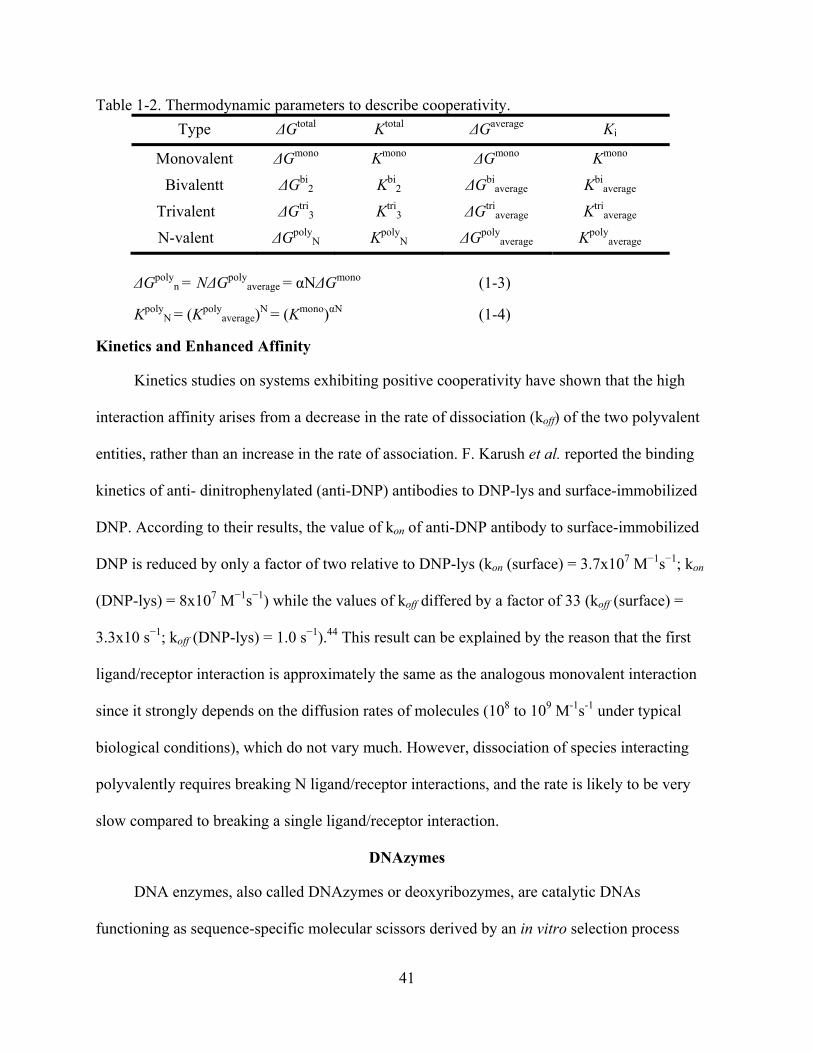
Table 1-2. Thermodynamic parameters to describe cooperativity. Type ΔGtotal Ktotal ΔGaverage Ki
Monovalent ΔGmono Kmono ΔGmono Kmono
Bivalentt ΔGbi2 Kbi
2 ΔGbiaverage Kbi
average
Trivalent ΔGtri3 Ktri
3 ΔGtriaverage Ktri
average
N-valent ΔGpolyN Kpoly
N ΔGpolyaverage Kpoly
average
ΔGpoly
n = NΔGpolyaverage = αNΔGmono (1-3)
KpolyN = (Kpoly
average)N = (Kmono)αN (1-4)
Kinetics and Enhanced Affinity
Kinetics studies on systems exhibiting positive cooperativity have shown that the high
interaction affinity arises from a decrease in the rate of dissociation (koff) of the two polyvalent
entities, rather than an increase in the rate of association. F. Karush et al. reported the binding
kinetics of anti- dinitrophenylated (anti-DNP) antibodies to DNP-lys and surface-immobilized
DNP. According to their results, the value of kon of anti-DNP antibody to surface-immobilized
DNP is reduced by only a factor of two relative to DNP-lys (kon (surface) = 3.7x10⁷ M−1s−1; kon
(DNP-lys) = 8x10⁷ M−1s−1) while the values of koff differed by a factor of 33 (koff (surface) =
3.3x10 s−1; koff (DNP-lys) = 1.0 s−1).44 This result can be explained by the reason that the first
ligand/receptor interaction is approximately the same as the analogous monovalent interaction
since it strongly depends on the diffusion rates of molecules (108 to 109 M-1s-1 under typical
biological conditions), which do not vary much. However, dissociation of species interacting
polyvalently requires breaking N ligand/receptor interactions, and the rate is likely to be very
slow compared to breaking a single ligand/receptor interaction.
DNAzymes
DNA enzymes, also called DNAzymes or deoxyribozymes, are catalytic DNAs
functioning as sequence-specific molecular scissors derived by an in vitro selection process

42
similar to SELEX. The best characterized DNAzyme is probably the “10-23” subtype, which
contains a catalytic loop that is the interaction site for cations and two flanking arms that
specifically recognize their target RNA.45 After a cation binds to the catalytic loop, hydrolysis of
the target RNA proceeds via a de-esterification reaction, to produce a 2’,3’-cyclic phosphate
terminus and a 5’-hydroxyl terminus. For catalytic activity, the core sequence of the DNAzyme
should be conserved, but the arm sequences can be any sequences complementary to the
intended RNAs. This unique hydrolysis function has been of great interest for developing
biomedical and analytical tools. Numerous investigations have involved inhibition of gene
expression using this DNAzyme with a number of structural modifications to enhance the
stability and to improve the potency.
As a convenient and rapid analytical method, DNAzymes have been coupled with a
signaling mechanism for the design of heavy metal sensors. One of the successful examples is
the 8-17 DNAzyme-based lead sensor.46,47 Yi, Lu at al prepared many different types of Pb2+
sensors using gold nanoparticle aggregation, lateral flow strip, and FRET mechanism. All of
these techniques have been successful for convenient and rapid analysis of Pb2+ in environmental
samples. The selectivity for Pb2+ arises from the two-step hydrolysis mechanism of the 8-17
DNAzyme.48 According to MALDI-MS analysis, the 8-17 DNAzyme produces a 2',3'-cyclic
phosphate by an internal transesterification mechanism, in which the 2'-OH group at the cleavage
site undergoes in-line attack on the scissile phosphorus, forming a penta-coordinated phosphate
intermediate, followed by loss of the 5'-oxygen.49,50 Then, Pb2+ further catalyzes a second step,
hydrolysis of the 2',3'-cyclic phosphate. This reaction is observed by presenting the presences of
Pb2+ but not other divalent cations including Mg2+ or Zn2+.

43
Blood Coagulation and Thrombin
Coagulation
Alpha-thrombin (EC3 .4.21.5), a trypsin-like serine proteinase, plays a vital role in blood
coagulation and other physiological processes involving catalytic functions and enzymatic
intermolecular interactions.51,52 Thrombin cleaves or interacts with many different blood
components, including fibrinogen, fibrin, platelets, coagulation factors V, VIII, and XIII,
thrombomodulin, and protein C. Blood coagulation is a part of an important host defense
mechanism termed hemostasis. Coagulation is a complex process by which the blood forms clots
to cover a damaged blood vessel by a platelet and fibrin, such that bleeding stops from the
damaged vessel to begin repairing. Disorders of coagulation may cause severe bleeding or
strokes. Coagulation is highly conserved throughout biology. There are two major cascades of
coagulation: the intrinsic cascade (contact activation) and the extrinsic cascade (tissue factor
pathway) both of which lead to fibrin formation. Each pathway involves a series of protein
activations, in which a zymogen (inactive enzyme precursor) of a serine protease and its
glycoprotein co-factor are activated to become active components. They then catalyze the next
reaction in the cascade, ultimately resulting in formation of cross-linked fibrin. The major role of
tissue factor pathway (extrinsic) is to generate a "thrombin burst", a process by which
prothrombin is instantaneously activated to thrombin. It is initiated by release of tissue factor (a
specific cellular lipoprotein), and the efficiency of its pathway can be measured by the
prothrombin time (PT) test.53 The contact activation pathway (intrinsic) begins with formation of
the primary complex of high-molecular-weight kininogen (HMWK), prekallikrein, and FXII
(hageman factor) on collagen. The efficiency of this pathway can be determined by the activated
partial thromboplastin time (aPTT).54 After the clotting is initiated by either or both pathway(s),
thrombin finally plays a key role in forming blood clots. Its primary role is the hydrolysis

44
of fibrinogen to fibrin, the building block of a hemostatic plug. In addition, it activates other
proteins, such as Factor XIII cross-linking fibrin monomers to form polymers.
Thrombin Structure
Alpha-Thrombin contains two polypeptide domains that are covalently linked with a
disulfide bond (Figure 1-14): the A chain, which is non-essential for proteolytic activity,
composed of 36 residues, and the B chain composed of 259 residues .55,56 The B chain, which is
derived from the carboxyl terminal sequence of prothrombin, carries the functional epitopes of
the enzyme with the typical fold of serine proteases.57 The active site has three functional amino
acids, His57, Asp102, and Ser195, which are responsible for thrombin’s trypsin-like behavior of
cleaving its substrates after arginine residues. The catalytic site polarizes the side chain of
Ser195 for nucleophilic attack on the C atom in the scissile bond of the substrate. This results in
Figure 1-14. Structure of thrombin

45
the C atom’s forming a tetrahedral intermediate stabilized by hydrogen bonds with surrounding
amino acids. The substrate is then acylated by the O atom of Ser195, and finally, its C-terminal
fragment is released. There are two known positively charged loops on α-thrombin: exosite I, a
Ca2+ binding loop and exosite II, a Na+ binding loop. The positively charged loops eventually
provide electrostatic interactions for recognition. Exosite I is the binding site of fibrinogen, 51,58
thrombomodulin,59,60 hirudin,58,61,62 and the thrombin receptors.63-66 Exosite II, which is on the
opposite side of the molecule from exosite I, is the binding site for polyanionic ligands, for
instance heparin and the chondroitin sulfate moiety of thrombomodulin.67-69 Binding of the
molecules to exosite II may influence the catalytic activity of the enzyme allosterically.
Photochromic Switches
Photochromism
Recently, a number of reports have appeared on the inclusion of photochromic molecules
into DNA chains to incorporate photo-regulation into nucleic acid probes. Compounds which
undergo reversible photochemical reactions are said to be photochromic. Examples of
photochromic equilibria are: pericyclic reactions, cis-trans isomerizations, intramolecular
hydrogen transfers, intramolecular group transfers, dissociation processes and electron transfers
(oxidation-reduction).70 Photochromic compounds have two molecular states that are
interchangeable by irradiation. When the ground-state molecule is excited by UV light, it
undergoes a conformational change, resulting in formation of an unstable configuration. The
excited molecule can return to the ground state by thermally releasing the energy or by emitting
visible radiation. The two molecular states are very different from each other in terms of both
their physical and chemical properties; for example, their absorption spectra, refractive indices,
dielectric constants, geometrical structures, oxidation/ reduction potentials, etc... Photochromic

46
molecules can be divided into several classes depending on their transformation mechanisms; for
example, phototautomerizations, cis-trans isomerizations, and photocyclizations.
In photo-tautomerization, the structural isomers of an organic compound are reversibly
interconverted by absorption of UV/Vis light. Figure 1-15 shows a hydrogen-transfer
photochromic tautomerism. The best known examples is the salicylidene-aniline tautomerism.71
After irradiation with UV light, the hydrogen on the oxygen is transferred to nitrogen. The enol-
form is pale yellow, and the keto-form appears in reddish or brown. This tautomer can further
exist in two forms: an intramolecular hydrogen bonded cis-keto form and a rotated trans-keto
form.
Figure 1-15. Light induced hydrogen transfer tautomerization
Photocyclization involves an electrocyclic reaction. Typical examples involving
derivatives of 1,3,5-hexatriene, fulgides, diarylethenes or spiropyran, are shown in Figure 1-16.
These photochromic compounds have been incorporated to design molecules with
photocontrolled magnetism, liquid crystalline phase switching, gated reactivity, fluorescence,
and/or reversible surface morphology for the development of photomodulating devices.
The most well known examples of cis-trans isomerization involve the stilbenes Figure 1-
17. After the trans compound is excited to the singlet excited state, the excess energy is released
via either fluorescence or photocyclization to the cis form. The isomerization in n-pentane is
highly efficient, when the sample is irradiated at λ = 313 nm. Since cis-stilbenes are not

47
thermally stable, they return to the trans-form in the dark. For most of stilbene derivatives, the
thermal activation barrier lays around 154 kJ/mol.
Figure 1-16. Photocyclization.

48
Figure 1-17. Cis-trans isomerization
Azobenzene
Among stilbene derivatives, azobenzene and its analogues have shown the most desirable
photoconversion properties for many applications. As shown in Figure 1-18, azobenzene is
composed of two phenyl rings linked by an azo (N═N) group. The cis and trans isomers are
reversibly interconverted by UV and visible light. The conversion is highly efficient; 91% of cis-
isomer can be obtained with 365 nm excitation of the trans isomer. Return to the trans form
occurs by either thermally or by emission of visible light. Electron donating groups on the
benzene ring cause a red-shift of the excitation radiation into the visible region. The quantum
yield is 0.20 for trans- to- cis conversion and 0.70 for cis-to- trans conversion.
Even though there are many applications of azobenzenes, the detailed mechanism of the
isomerization is somewhat unclear. The trans isomer has a planar structure with two absorption
bands: a strongly allowed transition to S2 (ππ*, ε317 = 16 980 M-1 cm-1, Φt-c= 0.10) and a weakly
allowed transition to S1 (nπ*, ε449= 405 M-1 cm-1, Φt-c=0.25). The ground state of cis-azobenzene
is twisted with an angle of 10° and has two absorption bands: a moderately allowed transition to
S2 ( ε290 = 4980 M-1 cm-1, Φc-t= 0.27) and a weak transition to S1 (ε434 =1295 M-1 cm-1, Φc-t =
0.56)72. In the recent report, C. Stuart et al. made use of Resonance Raman analysis to reveal the
detailed mechanism of the photoisomerizaion.72 According to their results, the trans-to-cis
conversion can be explained via inversion mechanism, because upon UV irradiation, the CN

49
bond is shortened, and the N═N bond is elongated. However, the large change of the CNNC
torsion angle suggests that torsional motion contributes to the cis-to-trans conversion. This is
attributed to the twisted nature of the cis-azobenzene ground state, which is therefore susceptible
to rotation.
Figure 1-18. Molecular structure of azobenzene and energy diagram

50
CHAPTER 2 SUPERIOR STRUCTURAL STABILITY AND SELECTIVITY OF HAIRPIN NUCLEIC
ACID PROBES WITH AN L-DNA STEM
Introduction
Molecular beacons (MBs) are single-stranded nucleic acid probes composed of three
different functional domains: stem, loop, and fluorophore/quencher pair (Figure 2-1d).8 A stem
functions as a lock that maintains the closed hairpin structure when there is no hybridization with
a complementary target, so that the fluorescence is quenched with high quenching efficiency.
Upon hybridization between the loop and its complementary target, the MB undergoes a
conformational change from hairpin to linear structure, resulting in an increased fluorescence
due to the increased physical separation of the fluorophore and quencher. Various
fluorophore/quencher pairs can be incorporated into experimental designs so that multiple targets
can be monitored simultaneously. MBs have been used for real time monitoring of polymerase
chain reaction (RT-PCR)73,74 and mRNA expression inside living cells.11,12,75 Unlike traditional
mRNA analyses, MBs do not require pre- or post-treatment of cells because, theoretically, MBs
will not give any fluorescence signal unless hybridized with their complementary targets. Indeed,
MBs are selective enough to distinguish single-mismatched targets, since the principle of MB’s
target recognition is based on Watson-Crick base pairing.
Although initial MB applications demonstrated wide feasibility and great potential,
challenges in probe design and applications still remain. Since stems are critical for maintaining
a stable hairpin structure, major difficulties often arise from the interruption of the stem
structure. There are two common causes of stem disruption which severely affect MB’s
sensitivity and selectivity. One problem is the undesired intermolecular interactions between
stems that will hybridize with their complementary sequences , called stem invasion (Figure 2-
1b). This stem invasion by non-target sequences should not be underestimated in real biological

51
Figure 2-1. Molecular beacon design and interactions. (a) MB binding to target mRNA ; (b) MB opened by non-specific mRNA that binds to MB stem; (c) MB mis-folds into non-hairpin structure through the hybridization of loop sequence with stem sequence. (d) A newly designed MB using L-DNA for the stem (red) to enforce a hairpin structure, and D-DNA for the loop (blue) for nucleic acid recognition effectively avoids the situations depicted in (b) and (c).
Table 2-1. Copy number of each sequence in biological nucleic acid sequences
samples since there could be high copy numbers of short sequences complementary to part of
the MB stems. In theory, the expressed frequency of occurrence of any particular stretch of RNA
or DNA sequence with n bases is 4-n.76 The average number of occurrences of each particular
sequence in human genome is 5x109 x4-n.76 Table 2-1 summarizes the occurrence of a nucleic
Sequence Length Number of Occurrence in DNA (5 Billion Bp)
Number of Occurrence in RNA (~0.3 Billion Bp)
10 4768 286
11 1192 72
12 298 18
13 74 4
14 18 1
15 4 1
16 1 1

52
acid sequence with n base pairs. This theoretical calculation indicates that if the base parings
between the short sequences can take place, MBs can be vulnerable to false positive signals
arising from stem-involved recognition, even though each MB is designed to target only one
complementary nucleic acid sequence.
Another problem arising from the stems is the thermodynamic conformational switch
between hairpin and non-hairpin structures. This is a result of the unwanted intramolecular
hybridization between the stem and part of the loop (Figure 2-1c). In the case of MBs, this can
cause a significant fluorescence background due to incomplete quenching. This problem
increases the amount of effort required in designing and testing MBs for new target sequences
with acceptable performance (Figure 2-1c). Sometimes the stem invasion cannot be avoided at
all, resulting in low performance hairpin structures in nucleic acid monitoring.
There have been developments aimed at minimizing the aforementioned problems. For
example, strengthening stem stability by increasing stem length or the G and C content can
increase the dominance of the hairpin structure, so that the background signal of MBs remains
low.77 Unfortunately, a strong stem leads to slow hybridization kinetics and a high tendency of
forming sticky-end pairing, reducing the fluorescence signal enhancement upon target
hybridization.78 The typical example is the locked nucliec acid (LNA), or LNA MB.26 LNA is
known as an extraordinarily strong nucleic acid binder and LNA MBs show ultra-high thermal
stability, resulting in a low background signal. However, LNA MBs tend to have extremely slow
hybridization kinetics, which limits their usefulness.
In order to minimize unnecessary intermolecular interactions, MBs have been created in
which the stems have structural features that inhibit stem-loop hybridization. In one study, the
sense of the stem DNA was inverted to the loop, i.e. 3’-stem-5’-5’-loop-3’-3’-stem-5’, to ensure

53
that any stem/loop hybridization would be disrupted at the 5’5’ and 3’3’ junctions.79
Unfortunately, the usefulness of these MBs is limited because they require longer stems causing
increased sticky-end-pairing problems. These examples showcase the underlying complexity of
designing MBs and other DNA hairpin probes. There has yet to be a MB design that solves the
problem of stem invasion without causing other performance-limiting effects.
Ideally, the MB stem should not interact with the loop or any other complementary nucleic
acid sequences. If the stem can be immune to naturally occurring nucleic acids either within the
probe or in the sample matrix, there will be no stem invasion, resulting in highly stable hairpin
nucleic acid probes. This thought has led us to consider non-standard bases like L-DNA which
do not hybridize with natural nucleic acids. Our hypothesis is that stems made of such bases will
hybridize with each other and not to the loop or any other natural nucleic acids in the sample. In
this way, a stable hairpin structure can be formed in which the stem and loop have no innate
ability to form a stable structure. This idea has been explored using homo DNA( oligo(2′,3′-
dideoxy-â-D-glucopyranosyl)nucleotides)-stem MBs.80 Unfortunately, the synthesis of such MBs
is not easy and requires optimization of the stem design. Here we propose to use non-natural
enantiomeric DNA, termed L-DNA, in the stem, and natural D-DNA in the loop (Figure 2-1d) as
a model system for MB design. Since L-DNA is the mirror-image form of the naturally occurring
D-DNA, its duplexes have the same physical characteristics in terms of solubility and stability as
D-DNA hybrids, except for forming a left-handed double-helix.81-84 L-DNA was examined as a
potential antisense reagent, but it failed to perform adequately because there is no interaction
between L-DNA and D-DNA due to the chiral difference.85 By taking advantage of these
features of L-DNA to build stems we can prevent intramolecular and intermolecular nonspecific
interactions in any hairpin-structured DNA probes. In addition, the easy incorporation of L-DNA
54
stems to other non-standard bases, such as 2-OMe-modified RNA and LNA, can be attractive to
design bio-compatible MBs. In the present report, we have designed, prepared, and studied MBs
based on this strategy, and will show that this design can effectively minimize stem-related
problems in MBs.
Experimental Section
Synthesis of MBs and Their Targets
The three oligonucleotide sequences shown in Table 2-2 were synthesized. The MB1
sequence, which does not have any biological targets, was randomly designed with high signal
enhancement and moderate hybridization kinetics. MB2 and MB3 were specially designed to
target β-actin mRNA (GenBank Accession No. BC014861) and MnSOD mRNA (Gen-Bank
Accession No. NM-000636), respectively.86 The L-deoxyphosphoramidites were obtained from
ChemGenes Corporation (Wilmington, MA). The other synthesis reagents, such as 3’-dabcyl
(quencher) CPG, 6-fluorescein phosphoramidite (fluorophore), 2-OMe-modified RNA
phosphoramidite, and D-deoxyphosphoramidites, were from Glen Research Corporation
(Sterling, VA). All MBs and their targets were synthesized using an ABI 3400 DNA/RNA
synthesizer (Applied Biosystems, Foster City, CA) at 1μmol scale with the standard synthesis
protocol. Dabcyl was placed at the 3’ end to ensure that every DNA strand contains a quencher.
For high coupling yield, the coupling reaction time of 6-fluorescein phosphoramidite to the 5’
end of each molecular beacon was extended to 15 min. For the complete cleavage and
deprotection, overnight incubation with ammonia was used. After ethanol precipitation, the
precipitates were then dissolved in 0.5ml of 0.1 M triethylammonium acetate (TEAA, pH7.0) for
further purification with high-performance liquid chromatography (HPLC) using a ProStar
HPLC Station (Varian, CA) equipped with a fluorescence detector, a photodiode array detector
and C-18 reverse phase column (Alltech, C18, 5μM, 250x4.6mm).

55
Table 2-2. Sequences of hairpin probes and their targets. Black letters indicate natural DNA bases; red letters are L-DNA bases; light blue letters are lock nucleic acids; and green letters are 2’-O-Me-modified bases. Underlined regions are stems.
Name Sequence
LS MB 1 5’-FAM-CCTAGCTCTAAATCACTATGGTCGCGCTAGG-Dab-3’
LS MB 2 5’-FAM-CCGAGCCAGTTACATTCTCCCAGTTGATTGCTCGG-Dab-3’
LS MB 3 5’-FAM-CCGTCGAGGAAGGAAGGCTGGAAGAGCGACGG-Dab-3’
LS OMe MB 1 5’-FAM-CCTAGCTCTAAATCACTATGGTCGCGCTAGG-Dab-3’
LS OMe MB 2 5’-FAM-CCGAGCCAGTTACATTCTCCCAGTTGATTGCTCGG-Dab-3’
LS OMe MB 3 5’-FAM-CCGTCGAGGAAGGAAGGCTGGAAGAGCGACGG-Dab-3’
DS MB 1 5’-FAM-CCTAGCTCTAAATCACTATGGTCGCGCTAGG-Dab-3’
LS LNA MB 5’-FAM-CCTAGCTCTAAATCACTATGGTCGCGCTAGG-Dab-3’
DS MB 2 5’-FAM-CCGAGCCAGTTACATTCTCCCAGTTGATTGCTCGG-Dab-3’
DS MB 3 5’-FAM-CCGTCGAGGAAGGAAGGCTGGAAGAGCGACGG-Dab-3’ MB 1 target
(LCD) 5’-GCGACCATAGTGATTTAGA-3’
MB 2 target 5'- CCGAGCAATCAACTGGGAGAATGTAACTG-3'
MB 3 target 5'- CCGTCGCTCTTCCAGCCTTCCTTCCT -3'
Hybridization Experiments
Fluorescence measurements were performed with a Fluorolog-3 Model FL3-22
spectrofluorometer (JOBIN YVON-SPEX Industries, Edison, NJ) using a quartz cuvette. All
hybridizations were performed at room temperature. First, the background fluorescence from
200μL of the buffer containing 20mM of Tris-HCl (pH7.5), 50mM NaCl and 5mM MgCl2,
designated as MB buffer, was monitored for about 1 minute, and then each stock MB solution
(20 μM) was added to the hybridization buffer to reach final concentration of 65nM and the
fluorescence was monitored. After a stable fluorescence signal was obtained from the MB, an
excess of target oligonucleotide (650nM) was added. The level of fluorescence intensity was
recorded until the signal reached plateau. The excitation and emission wavelengths were set to

56
488nm and 520nm, respectively. Signal enhancement was calculated using the following
equation:
)F(F)F(F
BS
bufferclosed
bufferopen
−−
= (2-1)
In which
Fopen: fluorescence signals from MBs with target Fclosed : fluorescence signals from MBs without target Fbuffer: fluorescence signal of buffer
Melting Temperature (Tm) Measurement of MBs
Thermal denaturing profiles of each MB were recorded to evaluate the stability of the
stems using by a BioRad RT-PCR thermal cycler. The solution was prepared with 100nM final
concentration of each MB in MB buffer. The fluorescence intensity of each MB in buffer at
temperatures ranging from 10°C to 95 °C in 1°C intervals was measured and plotted as a
function of the temperature to generate the melting temperature curve.
Protein Sensitivity Tests
To test the nuclease digestion of MBs, deoxynuclease I (DNase I) from Sigma-Aldrich,
Inc. (St. Louis, MO) was chosen as a standard nuclease. The fluorescence of 65 nM MBs in MB
buffer was measured as a function of time at room temperature. Once the fluorescence stabilized,
two units of ribonuclease-free DNase I was added, and the fluorescence change was monitored
until it reached to plateau.
To test the vulnerability of MB-RNA duplexes to ribonuclease H digestion (Sigma-
Aldrich, Inc., St. Louis, MO), 65 nM MBs and RNA targets were incubated in the MB buffer
while the fluorescence intensity was monitored. After the hybridization reached equilibrium, two

57
units of ribonuclease H were added, and the change in fluorescence was recorded as a function of
time.
Cell lysate was prepared using CCRF-CEM (CCL-119, T cell line, human ALL) obtained
from American Type Culture Collection (Manassas, VA) and cell culture lysis buffer containing
25mM Tris (pH 7.8 with H3PO4), 2mM 1,2-Cyclohexylenedi nitrilo-tetraacetic Acid (CDTA),
2mM dithiothreitol (DTT), 10%glycerol and 1% Triton®X-100 purchased from Promega
(Madison, WI) with the protocol recommended by manufacturer. CCRF-CEM cells were
cultured in RPMI medium 1640 (American Type Culture Collection) supplemented with 10%
fetal bovine serum (FBS, heat-inactivated; GIBCO) and 100 units/ml penicillin–streptomycin
(Cellgro). Briefly, approximately 6 million cells were centrifuged to remove cell culture media
and then washed twice with 500μL of 5mM MgCl2 in Dulbecco’s PBS with calcium chloride and
magnesium chloride purchased from Sigma-Aldrich, Inc. (St. Louis, MO). All these steps
performed at 4°C. After the buffer was removed, the cells were incubated with 200μL of cell
lysis buffer for 3 minutes at room temperature. Finally, the mixture was centrifuged to remove
any cell debris. To determine biostability of each MB, 2μL of cell lysate was applied to the MB-
containing solution.
Results and Discussion
Stability and Sensitivity of L-DNA Stem MBs (LS MBs)
To show the feasibility of an L-DNA stem, we synthesized three L-DNA stem MBs using
the sequence MB1, MB2 and MB3 as shown in Table 2-2. The same sequences were used to
prepare control MBs, called DS MBs, entirely made of D-DNA bases. Hybridization of each MB
to its corresponding natural DNA target was performed under the same conditions. In order to
ensure all MBs opened, we used a 10 fold excess of the target DNA. The signal enhancement
was calculated using the equation 3-1. As shown in Figure 2-2a, the L-DNA stem duplex was

58
able to maintain the hairpin conformation and dehybridize when the loop bound to its target with
moderate hybridization kinetics. More interestingly, compared to DS MB 1, LS MB 1 produced
a lower fluorescence signal in the absence of its target, which leads to a signal enhancement ratio
of more than twice that of the DS MB 1, 46 compared to 21, respectively (Table 2-3). High
signal enhancements were consistently observed from all three LS MBs as shown in Table 2-3.
Figure 2-2. Responses of LS MBs and DS MBs to the targets. The hybridization of LS MB1 and DS MB1 took place with 10 fold excess of the target DNA, and their fluorescence change was recorded.
Table 2-3. Signal to background (S/B) of each MB was calculated and compared
S/B
LS MB1 46 21 DS MB1
LS MB2 18 9 DS MB2
LS MB3 30 18 DS MB3

59
LS MB 2 and LS MB 3 had 18 and 30 times enhancement compared to their counterparts
MB 2 and MB 3, which had 9 and 18 times, respectively (Table 2-3). Thus, the better S/B of L-
DNA stem MBs over regular MBs can be generalized regardless of oligo sequences. The
improved sensitivity is believed to be a result of the enhanced stability of the hairpin
conformation in L-DNA MBs due to the lack of stem-loop interactions which could otherwise
contribute to a significant background. The better structural stability causing higher S/B was
supported by the higher melting temperature of stems (Tm) for LS MBs compared to those of DS
MBs.
Improved Structural Stability
To evaluate the stem stability of MBs, the melting temperatures (Tm) of all probes were
determined and compared. For all MB sequences prepared, LS MBs showed higher melting
temperatures than their DS counterparts. Typical melting temperatures are shown in Table 2-4
for LS and DS MBs. Tms of LS MBs exceeded those of the corresponding DS MBs by at least
4oC. This difference is well above the uncertainty in Tm measurement in our instrument (<0.5 °C).
Because L-DNA:L-DNA base pairs have stabilities comparable to those D-DNA counterparts,
such an increase in Tm is more likely due to a more stable hairpin conformation in the LS MB
rather than stronger base pairing between L-DNA bases.87 The improved stability of the L-DNA
stem is coherent with the enhanced sensitivity of the probes observed in our hybridization
experiments. In an earlier report, the partial replacement of D-DNA bases with L-DNA bases in
DNA duplexes caused lower melting temperatures.87 Our results do not contradict this work
since the double-stranded stem of the LS MBs is made entirely of L-DNA and therefore does not
have to accommodate both helicities simultaneously. Thus, the L-DNA stems are strong enough

60
to maintain the stable hairpin structure, and actually show better stability than the pure D-DNA
stems.
Table 2-4. Comparisons of stem melting temperatures of MBs. LS MBs generally showed much higher melting temperature, about 5 to 10 °C than their counterparts.
Tm
LS MB1 62.0 °C 58.0 °C DS MB1
LS MB2 71.6 °C 60.0°C DS MB2
LS MB3 67.5 °C 62.3 °C DS MB3
Figure 2-3. Melting temperature profiles of DS and LS MB 1
Elimination of Intramolecular Interaction
As discussed above, intramolecular interactions between stem and loop domains can cause
a high background due to thermal fluctuations between the hairpin structure and other
conformations. In the case of FRET-based linear nucleic acid probes, conformational changes

61
due to thermal fluctuations are not critical, since linear probes do not require any proper
conformation to minimize FRET background prior to target binding. However, in the case of
hairpin structures like MBs, the minor contribution of non-hairpin conformations can affect S/B
more dramatically due to incomplete quenching. Thus, the addition of an L-DNA stem
eliminates unwanted intramolecular interactions between the stem and loop, favoring the hairpin
conformation and therefore improving sensitivity of MBs, as well as easing restrictions in stem
design. This situation can be understood thermodynamically in the following way. Free of target,
the unwanted intramolecular interactions give the DS MB higher entropy than the LS MB while
the entropies of the MBs hybridized to targets are similar. This causes the difference in the free
energy between open or random coil states and closed states to be more negative for the LS MB.
This argument correlates well with the higher Tms. Conversely, the DS MB requires less energy
input to open since the hairpin structure is not as dominant and the less stable intramolecular
configurations melt at lower temperatures.
To prove the hypothesis that the low affinity of L-DNA to D-DNA can eliminate the stem-
loop interaction and stabilize hairpin structure more efficiently, a specific MB, called MB 1-1
(Table 2-5), was designed such that the non-hairpin structure is dominant for DS MB 1-1, as
shown in Figure 2-4. Thus, DS MB 1-1 can have two dominant conformations, hairpin and non-
hairpin structures, and their distributions are dependent on thermodynamic stability. On the other
hand, replacing the MB stem with an L-DNA stem can ensure a hairpin structure as a dominant
structure as shown in Figure 2-4, since the L-DNA cannot hybridize with D-DNA. The major
difference between the two MBs is observed in the kinetics of hybridization with their targets.
The DS MB 1-1 shows a noticeably slow hybridization rate (Figure 2-4). We believed that there
are two major reasons for the kinetic effect. One is the reduction of exposed loop sequences of

62
the DS MB 1-1to its target, causing minimized chance of interactions between a probe and its
target. The other is the extra energy required for dehybridization of the partially hybridized loop
region in the DS MB 1-1 prior to binding to targets. In contrast, the LS MB 1-1 hybridizes with
Table 2-5. MB 1-1 and its target sequences. The underlines at the end for sequences represent stems and the middle of the loop is the complementary to the 5’ stem.
Name Sequence
LS MB 1-1 5’-FAM-CCTAGCTCTAAATCAGCTAGGTCGCGCTAGG-Dabcyl-3’
DS MB 1-1 5’-FAM-CCTAGCTCTAAATCAGCTAGGTCGCGCTAGG-Dabcyl-3’
MB 1-1 target 5’-GCGACCTAGCTGATTTAGAGCTAGG-3’
Figure 2-4. Elimination of stem and loop interaction using a L-DNA stem. Possible conformations of MB1-1 sequence as predicted by DNA/RNA folding program mfold. DS MB 1-1 folds into non-hairpin structure because of the stem-loop interaction. Use of L-DNA in the stem of LS MB 1-1 removes stem-loop interaction, forcing the probe to form a hairpin structure. Hybridization curves showing faster hybridization kinetics for LS MB 1-1 compared to DS MB 1-1.

63
the target much faster. This is because the fully open loop region can be easily accessed for
interaction with its target, resulting in a rapid dynamic response. These results confirm that using
L-DNA stem in designing MBs can effectively eliminate undesired stem-loop interactions in
hairpin structured nucleic acid probes.
Elimination of False Positive Signal
The other major advantage of using an L-DNA stem in MB construction is elimination of
false reports coming from the stem and non-target sequence interactions (Figure 2-1b). This is an
intrinsic problem of all DS MBs. Presumably, a short sequence with only 5 to 6 base pairs might
not contribute significantly to the stem invasion, but the extended sequences partially matched to
the loop can have high probability of non-selective opening of MB and false positive signals. In
order to evaluate the extent of such non-specific interactions, we designed oligo-D-DNA targets
with different lengths that had 6 bases complementary to the stem of the MB while the remaining
sequence matched the loop region: 5’-CCTAGC-3’, 5’-CCTAGCGC-3’, 5-CCTAGCGCGA-3’,
and 5’-CCTAGCGCGACC-3’ (underline is complementary to the 3’ stem sequence of the MBs
in Table 2-2). Using the mfold software, the calculated thermodynamic stabilities of each target
with its complementary sequence are shown in Table 2-6. The loop complementary DNA target,
5’-GCGACCATAGTGATTTAGA-3’ was also prepared as a reference. These sequences were
separately incubated with either LS MB 1or DS MB 1 for 1 hr. The responses of the MBs were
recorded and the fluorescence signal of each sample was compared to the fluorescence signal of
MB with loop-target DNA mixture (Figure 2-5). For DS MB 1, incubation with a 10-fold excess
of the 6mer DNA target failed to open the hairpin structure. This is expected since the
intramolecular binding constant between the stem sequences is far greater than the
intermolecular interaction between one arm of the stem and the 6mer DNA target. Similar results
were obtained for the 8mer sequence. On the contrary, in the presence of the 10mer DNA, which

64
was complementary to one arm of the stem and 4 adjacent bases in the loop, approximately 55%
of the DS MB 1 opened. The 12mer sequence, in a 10-fold excess, was able to fully open the DS
MB 1 (Figure 2-5). This result clearly demonstrates how severely the stem invasion sequence
can compromise the MB’s function and its selectivity in biological
applications. There are tremendously diversified sequences in a real sample matrix, and the copy
number of this type of short complementary sequences could be very high. The chance of such a
Table 2-6. Calculated melting temperature of each target with its complementary sequence
Targets Tm
6mer target (5’-CCTAGC-3’) <10 ºC
8mer target (5’-CCTAGCGC-3’) 17.5 ºC
10mer target (5-CCTAGCGCGA-3’) 25.1 ºC
12mer target (5’-CCTAGCGCGACC-3’) 31 ºC
Figure 2-5. Comparison of selectivity of LS MB 1 and DS MB 1. A final concentration of 100nM MB was incubated with 1μM of each target for 1hr and the fluorescence signal was measured. The experiment was repeated 5 times and the average value and standard deviations were calculated.
stem invasion is thus equally high and this is one of the reasons that limit the usage of MBs in
quantitative analysis. In contrast, an L-DNA MB does not have such a problem. None of the

65
mock target sequences except the full length target sequence was able to open LS MB 1,
indicating superior selectivity and stability. This excellent selectivity coming from the L-DNA
stem will be useful when performing quantitative analysis on raw biological samples as well as
for intracellular studies.
Biostablility of LS MBs
In general, degradation of D-DNA MBs is one of the most significant problems in intracellular
applications.88 This problem cannot be avoided with LS MBs since the loop is still made of
standard nucleic acids (Figure2-6). In order to solve this problem, non-standard nucleic acid
bases have been explored to design MBs, such as peptide nucleic acids (PNAs),22-24 locked
nucleic acids (LNAs),26,31,89,90 and 2’-OMe-modified RNAs.18-20,25,91 Most of these bases show
greatly improved biostability. In addition, they form stronger duplexes with their targets, leading
to more stable signal production. The 2’-OMe-modified RNAs have methoxyl groups at the 2’
carbon of the ribose sugar. They can recognize natural nucleic acid targets but are not bio-
degradable. As a result, these advantageous features of 2’-OMe-modified RNAs as well as their
well characterized properties have often been used as MB building blocks. In order to
demonstrate the feasibility of MBs composed of L-DNA stems and 2’-OMe-modified RNA
loops, called LS OMe MBs, in intracellular measurement, we investigated the biostability,
sensitivity, and selectivity of these probes. We prepared three different sequences of LS OMe
MBs (Table 2-2) and the characterization was performed as described previously.
As shown in Figure 2-7a, b, and c, such a replacement of the loops did not affect structural
stability and selectivity regardless of sequences. LS OMe MBs still showed higher S/B and Tm
compared to DS MBs. In addition, when DNase I was added, LS OMe MBs did not show any
fluorescence increase which means much better resistance to nuclease digestion (Figure 2-7a).

66
DS MBs showed immediate signal increase with the addition of DNase I, whereas the LS OMe
MBs did not show such an increase with DNase I (Figure 2-7a). The nuclease resistance
Figure 2-6. Nuclease resistance of LS and DS MB
of LS OMe MBs can be more beneficial to detect low copy number of mRNAs since the false-
positive signal coming from MB degradation will not be the case for LS-OMe MBs. Next, the
stability of DS and LS OMe MB/RNA target duplexes were inspected using RNase H, an
enzyme that selectively cleaves RNA/DNA duplexes. In the case of DS MBs, dramatic signal
decrease was observed after RNase H was added to the duplexes, indicating that that the DS
MB/RNA target duplexes are extremely vulnerable to RNase H digestion, resulting in signal
fluctuation for intracellular measurements. On the contrary, LS OMe MBs/RNA target duplexes
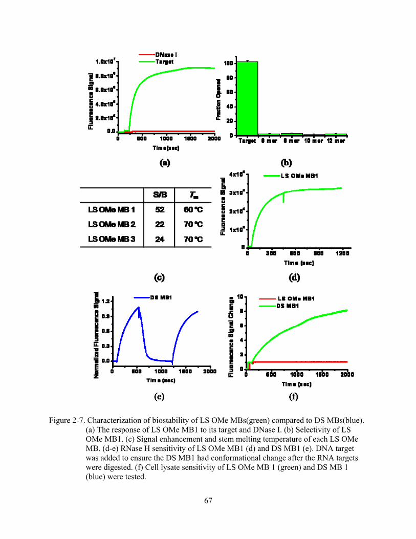
67
Figure 2-7. Characterization of biostability of LS OMe MBs(green) compared to DS MBs(blue). (a) The response of LS OMe MB1 to its target and DNase I. (b) Selectivity of LS OMe MB1. (c) Signal enhancement and stem melting temperature of each LS OMe MB. (d-e) RNase H sensitivity of LS OMe MB1 (d) and DS MB1 (e). DNA target was added to ensure the DS MB1 had conformational change after the RNA targets were digested. (f) Cell lysate sensitivity of LS OMe MB 1 (green) and DS MB 1 (blue) were tested.

68
Figure 2-8. Characterization of biostability of LS LNA MBs (red) compared to DS MBs(blue). The response of LS LNA MB to its target (a), DNase I (b), and Cell lysate(c) was tested. RNase H sensitivity of LS LNA MB (d) was experimented.
are stable and resistant to RNase H activity due to the 2-OMe RNA loops. Even though LS OMe
MBs are resistant to DNase I and RNase H digestion, this does not mean that they are stable in
true cellular environments. Thus, we tested LS OMe MBs in cell lysates, and the results are
shown in Figure 2-7f. The stability of LS OMe MBs in the cell lysate clearly proves that the LS
OMe MB design eliminate many sources of false positive or negative signals in intracellular

69
measurements. The same investigation was applied to LS MBs containing LNAs of the loops as
well (Figure 2-8). As shown, LS LNA MB can also resist the nuclease digestion and protect the
RNA targets from RNase H digestion. Thus, we can conclude that the incorporation of L-DNA
stems in MBs with loops composed of other non-standard nucleic acids does not alter the
stability of the hairpin structure and takes full advantage of the biostability features of each
component.
Conclusions
In summary, we have established that the use of an L-DNA stem in a hairpin structured
DNA probes, like MBs, eliminates unwanted intra- and intermolecular interactions. The
exclusion of stem-loop interaction in an L-DNA stem MB ensures that the designed hairpin
structure is dominant thereby improving the sensitivity and stability of hairpin DNA probes. The
use of an L-DNA stem prevents the probe from being opened by non-specific D-DNA sequences
that contain short sequences complementary to the stem. Thus, stem invasion causing false
positive signals is eliminated. In addition, L-DNA stems can be used to improve any known MB
design because L-DNA stems can universally stabilize hairpin conformations of nucleic acid
probes, as long as D-type nucleic acids are used the loop. Moreover, the combination of a
nuclease resistant loop with L-DNA stems is a powerful molecular beacon design since it
removes the possibility of false positive and negative reports coming from intracellular
enzymatic activities and non-specific interactions of MBs and targets. While there are other
approaches to design stable hairpin DNA probes, the L-DNA stem strategy is the simplest, most
direct, and most effective way to develop hairpin structured DNA probes with desired properties.
Our results also suggest that enantiomeric DNA (L-DNA) is useful for designing structured
functional nucleic acid probes for a variety of biological and biotechnological applications.

70
CHAPTER 3 MOLECULAR ASSEMBLY FOR HIGH PERFORMANCE BIVALENT NUCLEIC ACID
INHIBITOR
Introduction
In contrast to monovalent interactions, multivalent (polyvalent) interactions involve the
binding of multiple ligands, such as small molecules, oligosaccharides, proteins, nucleic acids,
lipids, or aggregates of these molecules, to multiple binding pockets or receptors of a target, e.g.,
a protein, virus, bacterium or cell.36 Polyvalency is ubiquitous in biology and has a number of
benefits over monovalent interactions. For instance, polyvalent interactions collectively possess
higher binding affinity than the corresponding monovalent interactions. That is, polyvalency
results in a “cooperative” configuration in which the probability of re-binding of a dissociated
monomer to the target is increased by the presence of other monomers bound to the same target.
A classical example of this is demonstrated by the binding of Gal-terminated oligosaccharides to
C-type mammalian hepatic lectins.43
In addition to increased binding affinity, polyvalent interactions also stand a better chance
of providing higher selectivity in target recognition. A multivalent binder, despite being
composed of weak homo- or heterogeneous ligands, can still have stronger binding property due
to multiple binding events.92 A well known example of this phenomenon is taken from the
biology of gene regulation by oligomeric transcription factors. Specifically, the retinoid X
receptor (RXR) functions as a transcription factor in the presence of its ligand.93 Each RXR-
ligand complex (RXR-L) binds to single strand DNA called the cellular retinol-binding protein II
element (CRBP-II element). Interestingly, while the intrinsic affinity of one or more units of
RXR-L for one CRBP-II element (i.e., di-, tri- or tetravalent interaction) is insufficient to initiate
transcription, more than five of these complexes adjacent to CRBP-II elements can, in fact,
initiate the transcription. As a result, transcriptive response is well regulated depending on the

71
concentration of the transcription factor. Furthermore, this activity demonstrates the
“cooperative” configuration, as noted above, which gives polyvalent interactions the potential for
considerably increased binding affinity.
A number of recent studies have systematically reported the unique properties of
multivalent interactions. Investigators have attempted to mimic the mechanisms underlying such
interactions in order to design new therapeutic entities, particularly those using repetitive
epitopes of antibodies.36 By designing more efficient targeting reagents with potentially viable
therapeutic applications, all of these attempts have clearly shown promising results. A typical
example is the single chain variable fragment (scFv) constructed by linking the antigen binding
VH and VL domains of an antibody with a flexible polypeptide linker.94 The combinatory
configurations of scFvs can be designed and investigated to optimize the functionality. Another
successful therapeutic design, which takes advantage of polyvalent interactions, is the bi-specific
T-cell engager molecule (BiTE).95 A BiTE molecule is a bi-specific antibody that is constructed
by linking the binding domains of two antibodies with different specificities via short, flexible
peptides and is, therefore, expressed as a single polypeptide chain. The typical working principle
is that BiTEs bind with one arm to a target cell and the other arm to a T cell, consequently
activating the T cell. This unique mode of action results in increasing the cytotoxic potency of
BiTE molecules by at least 10,000-fold higher than that of conventional human IgG1
antibodies.96 These two achievements demonstrate how biomolecular engineers have exploited
the potential of multivalent binding motifs. However, the genetic engineering required to mimic
the mechanisms underlying multivalent interactions is time consuming and prone to many
technical difficulties. For instance, expressed proteins may not fold into the expected tertiary
structures, leading to non-functional products. Also, the heavy molecular weight of the final

72
product can be a limitation for future therapeutic applications. For these reasons, alternative
ligands that have functionalities similar to those of antibodies, but without the limitations, are
clearly attractive. In the present study, we demonstrate how the nucleic acid aptamer (NA
aptamer) is a strong candidate for such multivalent applications.
Aptamers are nucleic acid sequences selected by the SELEX (Systematic Evolution of
Ligands by Exponential Enrichment) method.32-34 They specifically recognize targets ranging
from ions, and small organic or inorganic molecules, to proteins and living cells, with binding
affinities and selectivities to targets comparable to those of antibodies. NA aptamers have
markedly lower molecular weights (usually below 20 kDa) than antibodies, and their secondary
structures are easily predictable. In addition, aptamers are not prone to the irreversible
denaturation that affects antibodies. Moreover, they can be synthesized efficiently and reliably
using established phosphoramidite chemistry. Thus far, many aptamers have been identified, and
some of them are very close to becoming marketable drugs.97-101 Nevertheless, engineering these
aptamers for enhanced performance or new functions remains virtually unexplored.
Here, we demonstrate that rationally designed aptamer assemblies can combine the
functionality and binding affinity of different aptamers to achieve greatly enhanced enzymatic
inhibition with potential medical significance. Using fluorescence resonance energy transfer
(FRET)-based molecular probes,102 we have also revealed the kinetic properties of the aptamer
assembly-protein interaction that underlie this improved inhibition. A molecular assembly of two
separate aptamers is expected to have even more advantages: stronger binding affinity, enhanced
inhibitory function, high intrinsic selectivity, minimal host immune response, and low to no
cytotoxicity.103 In addition, a unique feature of DNA-based therapy is that the antidotes of
aptamer drugs are readily available from their target DNAs.104 We have demonstrated the use of

73
these antidotes to effectively reverse the activity of the aptamer assembly because of the strong
and fast aptamer/DNA hybridization. With this set of investigations, we have proved that the
careful and rational design of aptamer assemblies can exploit multivalent interactions with the
protein target to produce enhanced enzymatic inhibition in a simple, effective, and practical way.
Experimental Section
Chemicals and Reagents:
All DNA synthesis reagents, including 6-fluorescein phosphoramidite, 5'-dabcyl
phosphoramidite, spacer phosphoramidite 18 (hexaethylene glycol linker) and D-
deoxyphosphoramidite, were purchased from Glen Research. All reagents for buffer preparation
and HPLC purification were from Fisher Scientific Company L.L.C. (Pittsburgh, PA). The buffer
resembling physiological conditions used for the buffer experiment contained 25 mM Tris-HCl
at pH 7.4, 150 mM NaCl, 5 mM KCl, 1 mM MgCl2, 1mM CaCl2, and 5% (V/V) glycerol.
Human α-thrombin was purchased from Haematologic Technologies, Inc. (Essex Junction, VT).
Fibrinogen and the sulfated hirudin fragment were obtained from Sigma-Aldrich, Inc. (St. Louis,
MO). Universal Coagulation Reference Plasma (UCRP) and thromboplastin-DL for human
sample testing were purchased from Pacific Hemostasis (Cape Town, South Africa). The
activated partial thromboplastin time (aPTT) assay and prothrombin time (PT) assay reagent was
from Trinity Biotech USA (Berkeley Heights, NJ). The bivalirudin was obtained from The
Medicines Company (Parsippany, NJ).
Synthesis and Purification of Mono- and Bivalent NA Ligands and Their Targets
To optimize the length of two different aptamers for the best binding affinity toward the
target protein, we designed multiple bivalent nucleic acid ligands having different linker lengths,
for example, 4, 6, 8, and 10 spacer phosphoramidite 18. All sequences of nucleic acid ligands are
presented in Table 3-1. All of them were synthesized using an ABI 3400 DNA/RNA synthesizer

74
Table 3-1. DNA sequences. S means one unit of spacer phosphoramidite (hexaethylene glycol). Dabcyl is a quencher and FAM is a fluorophore.
Name Sequence (5’-3’)
15Apt GGTTGGTGTGGTTGG
27Apt GTCCGTGGTAGGGCAGGTTGGGGTGAC
Bi-4S GTCCGTGGTAGGGCAGGTTGGGGTGACT-(S)4-TGGTTGGTGTGGTTGG
Bi-6S GTCCGTGGTAGGGCAGGTTGGGGTGACT-(S)6-TGGTTGGTGTGGTTGG
Bi-8S GTCCGTGGTAGGGCAGGTTGGGGTGACT-(S)8-TGGTTGGTGTGGTTGG
Bi-10S GTCCGTGGTAGGGCAGGTTGGGGTGACT-(S)10-TGGTTGGTGTGGTTGG
Bi’-8S AGGCGTGGTACCGTAGGATGGGGTGGTT-(S)8-TGGTTGGTGTGGTTGG
15Apt MBA Dabcyl-GGTTGGTGTGGTTGG-FAM
Bi-8S MBA1 Dabcyl-GGTTGGTGTGGTTGGT-FAM-(S)8-TGTCCGTGGTAGGGCAGGTTGGGGTGACT
T-15Apt CCAACCACACCAACC
T-27Apt GTCACCCCAACCTGCCCTACCACGGAC
T’-15Apt CACACCAACC
F-T’-15Apt FAM--CACACCAACC
Short 15Apt-Q GGTTGGTGT-Dabcyl (Applied Biosystems, Foster City, CA) at 1μmol scale with the standard synthesis protocol. For
high coupling yield, the coupling time of special modifiers, including 6-fluorescein
phosphoramidite, 5'-dabcyl phosphoramidite and spacer phosphoramidite 18, was extended to 15
min. For complete cleavage and deprotection, overnight incubation with ammonia was used.
After ethanol precipitation, the precipitates were then dissolved in 0.5ml of 0.1 M
triethylammonium acetate (TEAA, pH7.0) for further purification with high-performance liquid
chromatography (HPLC) performed on a ProStar HPLC Station (Varian Medical Systems, Palo
Alto, CA) equipped with a fluorescence detector and a photodiode array detector. A C-18 reverse
phase column (Alltech, C18, 5μM, 250x4.6mm) was used.

75
Clotting Time Tests
To perform the clotting time comparison for the optimization of linker length, we designed
a simple buffer experiment that contained only thrombin, each nucleic acid ligand, and
fibrinogen substrate in physiological buffer. The theory behind the experiment is that the mixture
of sample becomes non-fluidic when the fibrinogen is digested by thrombin. As a result, the
different time points of this transition can be used as an indicator. Briefly, 1μL of 10 μM
thrombin and 1 μL of 100 μM monovalent or bivalent nucleic acid ligand were added to a
disposable transparent plastic cuvette (Fisher Scientific Company L.L.C., Pittsburgh, PA)
containing 200μL physiological buffer and then incubated for 15 minutes. In the case of non-
linked 15Apt and 27Apt mixture, 1 μL of 100 μM of each probe was applied. Following that, 4
μL of 20 mg/mL fibrinogen was added, and samples in the cuvette were carefully examined by
tilting the cuvette to record the time when the sample becomes non-fluidic. Each experiment was
performed in tandem. A reaction mixture containing only thrombin and fibrinogen was always
tested together with other samples as an internal standard. All clotting times were normalized
based on the internal standard and compared to it.
Real-Time Monitoring of the Clotting Reaction
To monitor the clotting event in real time, we utilized scattering light. When fibrinogen is
digested by thrombin, the mixture of sample becomes not only non-fluidic, but also cloudy.
Turbidity of samples can be measured using either absorption or scattering light. To monitor the
clot formation, we chose scattering light. Briefly, reaction mixtures were prepared in the same
way as the clotting tests described above, except that the reaction took place in a 100 μL quartz
fluorescence cuvette (Starna Cells, Inc., Atascadero, CA), and the scattering light was monitored
on a Fluorolog-3 spectrofluorometer (Jobin Yvon, Inc., Edison, NJ). For scattering monitoring,
the excitation and emission wavelengths were both set at 580 nm, and the emission was detected

76
at the right angle relative to the light excitation, so that the excitation light did not interfere with
the light scattering signal. The initial rate of scattering increase represented the relative
thrombin-inhibition strength of the tested sample. Initial rates were calculated from the linear
range of the early slopes of the scattering profiles.
Monitoring of Apparent kon and koff
Monitoring the binding kinetics (kon’) was accomplished by modifying each inhibitor with
fluorescein and dabcyl. The sequence of each probe is shown in Table 1. To compare the kon’ of
each inhibitor with thrombin, we pre-incubated 100 nM of each inhibitor with 100 nM of T’-
15Apt. Then, a 5 times excess of thrombin (500nM) was added, and the fluorescence decay was
monitored on a Fluorolog-3 spectrofluorometer. Obtained data were used to calculate the kinetic
parameters in the following way:
)F(F)F(Fk
ThrombinMBAAptTMBA
ThrombinMBApointtimeeachon
/15'/
/'−−
=−
(3-1)
To monitor the koff’ of each inhibitor, 500nM of T-15Apt was added to the pre-incubated
mixture of thrombin (500nM) and each ligand (100nM). To generate the values of koff’, the
following equation was applied:
)F(F)F(Fk
MBAfreeAptTMBA
MBAfreepointtimeeachoff
−−
=−15'/
' (3-2)
Reversible Binding Reaction Using Target DNAs
To test the reversible binding of Bi-8S, we treated the sample mixture with target DNAs of
27Apt or 15Apt. The mixture of sample, including fibrinogen, was prepared in the same way as
indicated for the clotting time. About 500 seconds after fibrinogen was added to the reaction
mixture, the appropriate target of either 15Apt or 27Apt, was added to reach a final concentration
of about 5 times that of Bi-8S while the intensity of scatter light was monitored.

77
Human Plasma Tests
To evaluate the feasibility of the bivalent nucleic acid ligand as a potential anticoagulant
reagent, we determined aPTT and PT for each ligand using human plasma samples. Procedures
applied were those recommended by the manufacturer. For aPTT determination, 50µL of UCRP
was pre-incubated at 37°C with a different amount of each ligand for 2 minutes; then 50µL of
aPTT-L was added and incubated for another 200 seconds. Next, 50µL of pre-warmed CaCl2
was added to initiate the intrinsic clotting cascade. Finally, the scattering signal was monitored
until the signal was stabilized at the maximum. For PT determination, 50µL of UCRP was pre-
incubated at 37°C with a different amount of each ligand for 2 minutes; then 50µL of
thromboplastin-L was added to initiate the extrinsic clotting cascade. Finally, the scattering
signal was monitored until the signal was stabilized at the maximum. For the calculation of aPTT
and PT, the end time was determined to be the point where scattering signal reached half
maximum between lowest and maximum points. Each measurement was repeated twice, and
each set of experiments was performed with a single batch of plasma.
Results and Discussion
Thrombin Aptamers and Their Properties
Thrombin is a multifunctional protease involved in homeostasis. As an initiator of blood
clot formation, thrombin hydrolyzes fibrinogen and activates platelets and some blood
coagulation factors that have procoagulant activity. Disorders in blood clotting are tightly linked
to many serious health issues, including heart attack and stroke. Therefore, thrombin is typically
the target in anticoagulation therapy for these diseases. However, anticoagulant drugs currently
on the market often suffer from indirect inhibition and sub-optimum selectivity, which can lead
to side effects including bleeding.105,106

78
There are two known thrombin NA aptamers. One is 15 bases long (15Apt) and binds to
exosite 1, while the other, called 27Apt, is 27 bases long and interacts with exosite 299,107 as
shown in Figure 3-1. The dissociation constant Kd of 15Apt tends to be very high (up to 450
nM), depending on measurement methods,108-110 and Kd of 27Apt is approximately 0.7 nM.107 As
a potential anticoagulant, only 15Apt should have the enzymatic inhibitory functions required for
thrombin-mediated coagulation, since it occupies the fibrinogen-binding exosite 1. However,
efforts to explore the anticoagulant effect of this aptamer have shown only limited progress due
to the lack of sufficient binding strength to the exosite 1 on the target protein. Modifications on
the existing aptamer itself have also been explored in order to generate an enhanced functional
NA ligand of thrombin. However, this type of construct is likely to be even more
Figure 3-1. Working principles of monovalent and bivalent NA ligands. (a) 15Apt, monovalent ligand, has constant ON and OFF and diffuses into bulk solution immediately after dissociation from thrombin. Thus, thrombin can be readily reactivated to cleave fibrinogen. (b) In contrast, when linked to 27Apt to form a bivalent ligand, 15Apt can rapidly return to the binding site after dissociation due to confined molecular diffusion by 27Apt that is still bound to thrombin. As a result, the equilibrium of the reaction is shifted to the left.

79
unpredictable, as its unique conformational structure can be disrupted, resulting in the loss of its
binding property.111 Therefore, instead of modifying the aptamer sequence itself, we have
linearly assembled two existing NA aptamers of thrombin to form a molecular assembly of
thrombin aptamers (Figure 3-1b). This assembly specifically improves the inhibitory function of
thrombin due to the multivalent interaction mentioned above.
Design of Bivalent NA Inhibitors
We hypothesized that a linear molecular assembly of two monovalent NA aptamers would
be a superior functional NA inhibitor of enzymatic reactions with multivalent binding properties.
However, first we needed to find out whether we could achieve enhanced inhibition even in the
absence of multivalent interactions, by simply mixing these two aptamers without a covalent
linkage. The second important question would be whether these two aptamers could interact with
Figure 3-2. Comparison of the normalized clotting times of thrombin bound to different NA inhibitors. Clotting time of thrombin alone was defined as 1, and the relative values are based on it. 15Apt alone showed a threefold increase of the clotting time, but no delay was observed from the 27Apt-treated sample. Among bivalent NA candidates (Bi-xS), Bi-8S is the best inhibitor with the longest clotting time. The replacement of 27Apt by a random sequence, as in Bi’-8S, causes almost complete loss of anticoagulant function, mainly due to the lack of bivalency and interaction between the scrambled sequence and 15Apt.

80
each other and subsequently lose their inhibitory properties. To address these issues, we
performed typical clotting tests using no aptamer, 15Apt alone, 27Apt alone, and a mixture of
15Apt and 27Apt. As shown in Figure 3-2, 15Apt alone delayed the coagulation time by nearly
three-fold, while 27Apt had no significant observed inhibition. This result was expected since
27Apt does not target the critical exosite 1. The non-linked 15Apt/27Apt mixture showed an
inhibitory effect similar to the 15Apt alone. These findings indicate that 15Apt and 27Apt act
independently from each other and thus do not promote or interfere with each other’s activities.
Next, several candidate bivalent NA ligands were designed and evaluated with the purpose
of optimizing the distance between the two different NA aptamers. This step is particularly
critical in designing bivalent ligands, and we initially assumed that a shorter distance between
the two aptamers would result in disruption of their simultaneous binding and lead to less
effective binding and inhibition. Therefore, we designed several potential bivalent NA ligands
with linkers of different lengths composed of 4, 6, 8, or 10 spacer phosphoramidites and
designated Bi-xSs in Table 1. Considering that one spacer is about 2.1 nm long and that the inner
diameter of thrombin is several nanometers, this represents a sufficiently ample range of lengths;
i.e., from 8.4 to 21.0 nm. Then, a clotting test was carried out to evaluate the effect of linker
length on thrombin inhibition, and the results were then compared with the clotting times of the
monovalent 15Apt. In this test, the thrombin converts soluble fibrinogen into insoluble strands of
fibrin, resulting in increasing turbidity and decreasing fluidity. The recorded time when it
became completely non-fluidic was normalized and compared among the series of potential
bivalent ligands.
The results in Figure 3-2 show that the number of spacers increased, the inhibition activity
first increased and then maximized with 8 spacers. After that, a decline of inhibition was

81
observed. Interestingly, the Bi-4S displayed a worse anticoagulation efficiency than free 15Apt.
Competition between the two aptamers readily explains this phenomenon. If the distance
between 15Apt and 27Apt is not sufficiently long, the binding of 27Apt will surpass that of
15Apt due to its stronger affinity to thrombin. This, in turn, will prevent 15Apt from reaching the
binding pocket of thrombin and lead to a drastically reduced anticoagulation capability.
However, with a sufficient linker length, Bi-8S inhibited thrombin nine times better than 15Apt
alone. Contrary to our initial assumption, and considering that the size of thrombin is about 3-4
nm in diameter, it was unexpected to see that a linker as long as ~16 nm (8 spacers) was needed
for the best inhibition. Based on these findings, we therefore hypothesized that some extra linker
length was required for both aptamers to wrap around thrombin and assume the optimum 3-
dimensional position and orientation to interact with thrombin. This extra linker is most likely
needed to minimize interference originating from thrombin/linker contact. In spite of these
results, an even longer linker length, such as in Bi-10S, was not as efficient as Bi-8S or Bi-6S as
shown in Figure 3-2. Taken together, these results support the fact that simultaneous binding
through bivalent interaction does take place and does result in improved inhibitory effect.
Additional evidence was obtained from the clotting test using Bi’-8S in which 15Apt was linked
to a scrambled DNA sequence with 8 spacers. The results showed very limited thrombin
inhibition. Therefore, the nine-fold increase of clotting inhibition by Bi-8S over 15Apt alone
must be a direct result of bivalent interaction realized by molecular assembly. This indicates that
aptamers are ideal for the design of multivalent ligands through molecular assembly.
Monitoring Inhibitory Functions Using Light Scattering
To obtain real-time kinetics of coagulation, we designed a more quantitative experiment
based on the nephelometric measurements of the turbidity caused by gel. The intensity of the
scattered radiation during coagulation was monitored by a spectrofluorometer, and the results are

82
Figure 3-3. Real-time monitoring of scattered light generated by the coagulation process in the presence of different monovalent or bivalent NA ligands (Bi-xSs). After the coagulation was initiated by adding fibrinogen to each sample, the reaction kinetics varied depending on the ligands. The initial reaction rate of each sample was calculated (slope of the initial linear part of the curve is in cps/s) and then plotted in the inset. These results are consistent with those of the clotting test. As the number of spacers increased, the reaction rate went down and then up (inset). Results show that the Bi-8S is the best design for a bivalent NA inhibitor.
shown in Figure 3-3. The background scattering of the thrombin-inhibitor mixture was stable
until the addition of fibrinogen. The increased intensity of scattering light reflected the net rate of
the coagulation reaction, as this was the direct result of fibrinogen cleavage. The relative
inhibition strengths of the monovalent and bivalent NA ligands were estimated from the initial
reaction rates, with higher rates representing weaker inhibition and the reverse for lower rates.
As expected, Bi-4S produced an initial rate of 2903 cps per second calculated from the part of
the slope of the scattering profile. This result, which 2.8 times faster than that of 15Apt alone,
1049 cps per second indicates that 27Apt interferes with 15Apt binding when the linker is only

83
8.4nm long. In contrast, the initial reaction rates of Bi-8S and Bi-6S were much slower than the
others, at 63 and 97 cps per second, respectively. Since Bi-8S demonstrates an initial rate close
to 16.6 times slower than free 15Apt, it also represents a considerable improvement of
antithrombin efficacy. The anticoagulation trends among the tested inhibitors correlate very well
with the results from the clotting tests. As shown in the clotting test, Bi-10S did not function as
well as Bi-8S, resulting in an increased initial reaction rate. Likewise, Bi’-8S, with a scrambled
sequence in the 27Apt domain showed no improvement in inhibiting the clotting process.
Therefore, based on the evidence gathered from both clotting tests and turbidity measurements,
we conclude that Bi-8S is the best design for improved thrombin inhibition.
Binding Kinetics Studies
Because the bivalent interaction of the aptamer assembly with thrombin increases the
overall binding affinity, it is proposed that enhanced affinities of NA inhibitors caused the
stronger inhibition. Since the binding affinity is directly related to kinetic parameters, such as kon
and koff, of the thrombin/inhibitor interaction, we carried out experiments to investigate the
impact of the molecular assembling on kon and koff to determine what actually causes the
improved inhibition. One important feature of aptamers is their binding to the target is often
accompanied by changes in tertiary structures. This allows researchers to build various signal
transduction mechanisms, such as fluorescence resonance energy transfer (FRET), into aptamers
for sensitive target detection. In fact, 15Apt was among the first aptamers to be built into a
molecular beacon aptamer (MBA) for protein detection based on FRET.112 In the present work,
we labeled 15Apt and the 15Apt domain of Bi-8S with a fluorophore and quencher pair to
form15Apt MBA and Bi-8S MBA 1 (Table 3-1). The compact structure of the 15Apt bound to
thrombin was expected to differ considerably from the random coil structure in solution, thus

84
giving different fluorescence intensities. We then used these modified aptamers to study the
kinetics of interactions with thrombin under different conditions.
To compare koff of the 15Apt MBA and the 15Apt domain of Bi-8S MBA 1, the full
complementary target DNA of 15Apt (T-15Apt) was added to the sample solution to make the
15Apt, when released from thrombin, inactive by forming a duplex with it (Figure 3-4). At the
same time, opening of the MBA should give intensive fluorescence. 100nM of each MBA probe
and 500nM of thrombin were pre-incubated for half an hour to complete the binding reaction.
Then, T-15Apt was added to the mixture while the fluorescence signal was monitored. To
normalize the fluorescence signal, MBAs fully opened by T-15Apt were used as the reference.
Figure 3-4. Comparison of koff’. Real-time fluorescence signal change of koff’ measurement. Free 15Apt MBA (green line) showed very rapid hybridization kinetics with its target DNA. Thrombin-bound 15Apt MBA (blue) showed slower hybridization kinetics compared to the free form.
One might question whether T-15Apt could induce the release of 15Apt from the binding
pocket. Our investigation of concentration effects of T-15Apt (Figure 3-5) shows that this was

85
not the case. In our tests, regardless of the concentration of T-15Apt, the reaction kinetics was
consistent, which means that T-15Apt does not induce the release, but rather captures the
released 15Apt as a separate step. Finally, the initial rate of each reaction was calculated using
the linear part of the slope. Even though values calculated this way are not the absolute koff’ rates,
they can still be useful for comparing kinetic parameters among different thrombin ligands.
Figure 3-5. Investigation of concentration effect of T-15Apt in binding comparison. 15Apt/thrombin complex was treated with different amounts of T-15Apt (left). MA MB1/thrombin complex was treated with different amounts of T-15Apt (right). As shown in the figure, there was no noticeable kinetics change.
Determination of kon’ for each MBA was done in a similar way (Figure 3-6)). Competition
between thrombin and a short target DNA of 15Apt, called T’-15Apt, was studied. Briefly, each
MBA was incubated with T’-15Apt, and then thrombin was added while the fluorescence signal
was monitored. Due to the weak binding affinity between T’-15Apt and 15Apt, thrombin would
compete with T’-15Apt for binding to 15Apt or the 15Apt domain of the bivalent ligands. This
study was based on three assumptions: 1) that T’-15Apt only interacted with 15Apt, 2) that the
binding affinity of the T’-15Apt is identical for both MBAs, and 3) that binding of 15Apt to

86
thrombin is more favorable than to T’-15Apt. Optimization of the T’-15Apt sequence revealed
that 10 complementary bases gave the best results in terms of binding to 15Apt and detectability
Figure 3-6. Comparison of kon’. Each inhibitor MBA was incubated with T’-15Apt, and then thrombin was added to the hybrid mixture while fluorescence signal was recorded. The florescence was decreased after each MBA was bound to thrombin. Real-time fluorescence signal change of kon’ measurement. After thrombin was added, each sample mixture showed fluorescence decay. The decreasing rate was comparable in both cases. According to the calculation of the initial reaction rate, Bi-8S exhibited a 1.2 times faster kon’ than 15Apt did.
of the kinetic parameters. The reason that we concluded that T’-15Apt works the best is the
disassociation between 15Apt and T’-15Apt is not the rate limiting step but the association
between 15Apt and thrombin target so that what we observed is the association rate between
15Apt and thrombin (Figure 3-7). The experiment result showed that such dissociation was very
fast (few tens seconds to the competition). Specifically, 100nM of each MBA and the 10-base
long T’-15Apt were pre-incubated to complete the hybridization. Then, 5 times excess of

87
thrombin was added to the mixture while the fluorescence was monitored. Immediately
following that, there was sharp fluorescence signal decay. The obtained plot was normalized, and
the data points for the first 100 sec were used to calculate relative reaction rates.
Figure 3-7. Investigation of the dissociation of T’-15Apt. The pre-incubated mixture of F-T’-15Apt and short 15Apt-Q(Dabcyl) for 30 mins was treated with 15Apt. The increased fluorescence signal of the sample was obtained from the dissociation of T’-15Apt, which is very rapid reaction. Since this dissociation between 15Apt and T’-15Apt is much faster than the association of 15Apt to thrombin, it does not interfere the measurement of kon’.
The relative koff’ values obtained for monovalent and bivalent 15Apts were 1.5 and
0.029 %/sec, respectively (Figure 3-4) (all measured by percentage of changes in fluorescence
intensity). This means that koff’ of monovalent 15Apt is 51.7 times faster than that of bivalent
15Apt. 15Apt MBA and Bi-8S MBA 1 have relative kon’ values of approximately negative
0.00424 and 0.00498 %/sec, respectively (Figure 3-6). In other words, the kon’ of bivalent 15Apt
is about similar to that of monovalent 15Apt. Finally, we obtained the relative Ka’ values by
dividing kon’ by koff’, which revealed that binding affinity of bivalent 15Apt to thrombin is about
62.5 or more times higher than that of monovalent 15Apt. These results agree with the binding

88
properties of other reported multivalent ligands. It is believed that, while multivalent interaction
does not affect kon’ significantly, it does alter koff’ considerably. By using the FRET strategy, we
were able to measure the changes in kinetics of a single domain rather than the whole molecule,
observing about 50 times higher binding affinity for the 15Apt domain. This confirms that the
increased thrombin inhibition potency of the aptamer assembly originated from the kinetic
changes caused by cooperative binding. As a byproduct of the study, we have also demonstrated
that aptamer-based multivalent ligands make the study of kinetics using FRET quite convenient
compared to antibody or small molecule based ligands.
Antidote Effect of Binding Aptamers
One of the unique properties of NA aptamers is that the binding can be readily regulated
using complementary sequences.104 Strong binding affinity of a target is critical, but reversibility
of binding is equally, if not more, important. Reversibility of binding directly impacts the
pharmacology of drug treatments, in the sense that the side effects of drugs may be quenched by
antidotes. Based on their binding affinity as measured by the Watson-Crick base pairing of
complementary sequences, NA ligands and their complementary NAs can be very effective
drug/antidote pairs. To demonstrate this, the antidote effects of two aptamers’ target sequences,
T-15Apt and T-27Apt, were investigated. Clotting mixtures, including thrombin and fibrinogen
with Bi-8S, were treated with excess T-15Apt and T-27Apt separately while the scattering was
monitored (Figure 3-8). With the treatment of T-15Apt, immediate scattering increase was seen,
and the extent of final scattering intensity was comparable to that observed without any thrombin
inhibitors (Figure 3-8), indicating that the activity of thrombin is readily recovered by
inactivation of 15Apt, and the response is rapid. On the contrary, the clotting mixture treated
with target DNA of 27Apt showed slower change in the scattering signal (data not shown),
suggesting that its effectiveness in reversing the inhibition of thrombin is limited. We later used

89
molecular beacon assay to confirm that the hybridization of 27Apt to its target was much slower
than that of 15Apt and its target. It is clear that the secondary structure of 27Apt is quite stable,
leading to slower duplex formation. The second reason is that 15Apt domain was still active even
when 27Apt was hybridized to its target. In conclusion, the target DNA of 15Apt is an effective
antidote, even for aptamer assembly-based therapy.
Figure 3-8. Reversible inhibitory function. Red: T-15Apt was added at around 200 seconds to the incubation of Bi-8S, thrombin and fibrinogen. Black: fibrinogen was added to thrombin at 0 seconds in the absence of any inhibitors. Blue: Bi-8S incubated with thrombin and fibrinogen (no T-15Apt).
Antithrombin Potency of Bi-8S
Recent studies related to its biological functions have shown that thrombin is critical in
blood clotting disorders and influences a tumor angiogenesis.111 Thus, after we demonstrated that
Bi-8S is the best inhibitor of thrombin in buffer system, we tested Bi-8S in human plasma

90
samples to further demonstrate the potency of the anticoagulant. Standard activated partial
thromboplastin time (aPTT)113 and prothrombin time (PT)53 tests were utilized as described in
Materials and Methods. The aPTT and PT are performance indicators, which measure the
efficacy of the contact activation pathway and extrinsic pathway of coagulation, respectively, as
well as the common coagulation pathways. In each test, a different amount of each inhibitor was
treated, and the obtained results were plotted using a sigmoid fit. The dosage dependence is
shown in Figure 3-9 and 3-10. Enhancements in the delay of coagulation triggered by both
contact activation and extrinsic pathway were consistently observed. The results show that the
plasma samples treated with Bi-8S showed approximately five to six times longer PT and aPTT
than those without any treatments, while the delay by 15Apt alone was only two to three times
longer. Even though the enhancement obtained using human plasma samples was not as great as
Figure 3-9. Comparison of anticoagulant potency of Bi-8S and 15Apt using human plasma and aPTT. It shows dosage-dependent aPTT plotted for each NA inhibitor, and the maximal aPTT is shown inside the figure.

91
the delay in a buffer system, it proved that the bivalent NA ligand can still function well in
human biological fluid and give enhanced anticoagulation efficacy.
Figure 3-10. Comparison of anticoagulant potency of Bi-8S and 15Apt using human plasma and PT measurements. It shows dosage-dependent PT, and the maximal PT recorded appears inside the figure.
Conclusions
In summary, by assembling two thrombin-binding aptamers with optimized linker and
linker length, we have developed an NA-based high-performance bivalent ligand, which can be
applied as a potential anticoagulant. This new design has the combined advantages of both
ligands and has achieved enhanced thrombin inhibition capability, indicating its potential in
biomedical applications for treating various diseases related to blood clotting disorders.
Moreover, the molecular assembly approach offers a simple and noninvasive way to accomplish
higher performance with a known protein inhibitor. It is worth noting that aptamers are ideal

92
candidates for constructions of such assemblies because they can be easily linked in a predictable
way using reliable synthetic chemistry mechanisms. In addition, binding of aptamers to different
epitopes of one protein is quite common, and aptamers can theoretically be obtained using a
variety of suitable selection strategies for targets ranging from small molecules to complex
organisms.32-34,114

93
CHAPTER 4 DEVELOPMENT OF DENDRITIC APTAMER ASSEMBLIES AS SUPERIOR INHIBITORS
Introduction
The concept of molecular assembly is found in nature.36 Standard examples include amino
acids, nucleic acids and carbohydrates, which assemble in different sequences or geometries,
resulting in the creation of unique functions. In bioengineering, assembling molecules with
distinctive functions into a new structure is highly advantageous in creating new properties or
amplyfying existing ones. An important example is the enhancement of binding properties,
which was discussed in the previous chapters, by the phenomenon of multivalency (or
polyvalency).36 Multivalency is defined as the simultaneous multiple interactions between two
molecules, creating a “cooperative” effect among multiple binding domains, which is the key
source of a dramatic increase of binding affinities. For this reason alone, much effort has recently
focused on taking advantage of polyvalency in the design of inhibitors by linearly assembling
capture molecules, such as repetitive epitopes of antibodies, sugars or small molecules, that
recognize specific cell receptors. These efforts have all shown progress.115 However, demand
remains high for better molecular recognition elements and strategies. This chapter reports the
assemblies of various aptamers for improved inhibitory efficacy to protein activity.
The advantages of using DNA aptamers are the binding units and have been described in
previous chapters, such as relatively small molecular weight (making multiple-molecule
assembly feasible) and have high binding affinity. Structurally, DNA-aptamers are easily
modifiable with robust structural restoration under ambient conditions.116 As a model, we use
thrombin and its two binding aptamers, 15Apt binding to exosite 1 and 27Apt recognizing
exosite 2.97,107 Their binding specificities are respectively very high, but their affinities are rather
different, sub-nanomolar Kd for 27Apt and sub-micromolar Kd for 15Apt.108,109 Although a series

94
of trials demonstrated the efficacy of 15Apt as a potential anticoagulant, its insufficient binding
affinity has often been a significant drawback. To solve this problem, recent studies reported the
engineering of a molecular assembly to link 15 Apt and 27 Apt.117 However, the final results did
not reach what we would consider to be the limits of inhibitory potency for aptamer assemblies.
Therefore, we took a dendritic approach to the design of an effective multi-ligand assembly and
tested its inhibitory efficacy against thrombin.
Experimental Section
Synthesis of Multimeric Assemblies
All DNA synthesis reagents were from Glen Research. All DNA probes (Table 1) were
synthesized with an ABI3400 DNA/RNA synthesizer using the standard procedure descried in
the previous chapter.
Real-Time Monitoring of Coagulation Process
A well known mechanism during the clotting process is the turbidity of sample, which is
dramatically increased due to the cleavage of fibrinogen to fibrin. Thus, UV-VIS measurement is
used to conduct real-time monitoring of the clotting process. A Cary 100 Bio UV-VIS
spectrophotometer (Varian Inc. Palo Alto, CA) was used. Briefly, a mixture of 200 microliters of
physiological buffer (25 mM Tris-HCl at pH 7.4, 150 mM NaCl, 5 mM KCl, 1 mM MgCl2, 1mM
CaCl2, and 5% (V/V) glycerol) containing 1nM thrombin and 1nM each inhibitor was incubated
for a half hour. Fibrinogen was then added to the sample mixture while the absorption was
recorded at 500nm.
PT Measurement
To evaluate anticoagulation efficacy, we determined PT for each ligand using human
plasma sample, following the manufacturer’s instructions. For PT determination, 50µL of UCRP
was pre-incubated at 37°C with different amounts of each ligand for 2minutes, and then 50µL of

95
thromboplastin-L was added to initiate the extrinsic clotting cascade. Finally, the scattering
signal was monitored until the signal reached a plateau. For the calculation of aPTT and PT, the
end time was determined at the point where scattering signal reached half maximum between
lowest and maximum points. It was repeated twice, and each set of experiments was conducted
with a single batch of plasma.
Results and Discussion
Design of Multimeric Assemblies
To improve the anticoagulation potency of DNA-based molecules, we designed a unique
assembly composed of two different functional domains: an anchor and a dendrimer-like
inhibitor, in this case, 27Apt and 15Apt, respectively (Table 4-1). The schematic structure of this
assembly is shown in Figure 4-1. as shown in Chapter 3, an anchoring molecule, 27Apt in this
case, is highly beneficial in overcoming the low binding affinity of 15Apt.117 The dendrimer-like
configuration of 15Apt functions as a highly branched inhibitory domain that is composed of two
or three 15Apts.118 Recalling that 15Apt binds to exosite 1, the engineering of this heterogeneous
ligand assembly activates the “cooperative” effect described previously. That is, because the
process results in the gain of extra ligands, the concentration of inhibitors around exosite 1 is
artificially, but effectively, increased. The anchor domain (27Apt) ensures that the dendrimer-
like assembly of 15Apts stays around the thrombin so that the effective local concentration is
extremely high compared to the same non-linked 15Apts.119 Each assembly contains an anchor
domain, which is 27Apt. In the middle of the linker region, a dendritic modifier is introduced to
covalently link the inhibitory aptamer (15Apt). In the absence of a dendritic modifier, only one
15Apt is linked to 27Apt, which we term mono-15Apt assembly. In contrast, doubler or trebler
dendritic modifiers allow two and three 15Apts to be linked to 27Apt at the identical distance
from the anchor. These are called dual-15Apt and triple-15Apt, respectively.

96
.
Figure 4-1. The sequences and schematic of dendritic aptamer assemblies. Each assembly contains an anchor domain, which is 27Apt. In the middle of the linker region, a dendritic modifier is introduced to covalently link the inhibitory aptamer (15Apt). In the absence of a dendritic modifier, only one 15Apt is linked to 27Apt, which called the mono-15Apt assembly. In contrast, doubler or trebler dendritic modifiers allow two and three 15apts to be linked to 27Apt with the identical distance. These are called dual-15Apt and triple-15Apt, respectively.
Table 4-1. Sequences of multimeric assemblies
To design the assemblies, we applied two different dendritic modifiers, a doubler and a
trebler (Figure 4-1). These are the functional groups that provide the branched assemblies of
15Apts. New assemblies were then based on the optimal design of the Mono-15Apt assembly
Name Sequence (5’-3’)
15Apt GGTTGGTGTGGTTGG
27Apt GTCCGTGGTAGGGCAGGTTGGGGTGAC
Mono-15Apt Assembly
Dabcyl-TGGTTGGTGTGGTTGG-(S)8-GTCCGTGGTAGGGCAGGTTGGGGTGACT
Dual-15Apt Assembly
Dabcyl-TGGTTGGTGTGGTTGG-(S)5-(doubler)-(S)3-GTCCGTGGTAGGGCAGGTTGGGGTGACT
Dual-15Apt Assembly 2
Dabcyl-TGGTTGGTGTGGTTGG-(S)3-(doubler)-(S)5-GTCCGTGGTAGGGCAGGTTGGGGTGACT
Triple-15Apt Assembly
Dabcyl-TGGTTGGTGTGGTTGG-(S)5-(trebler)-(S)3-GTCCGTGGTAGGGCAGGTTGGGGTGACT

97
controls, with each dendritic modifier inserted between the third and fourth hexaethylene glycol
(HEG). This site was randomly chosen because the inhibitory efficiency of the assemblies is not
dependent on the position of the dendritic modifiers since the molecules are quite small
compared to the hexaethylene linker, but rather the number of assembled 15Apts data not
shown).
Validation of Products
To verify the final products after HPLC purification, the length of each DNA probe was
estimated using agarose gel electrophoresis because HPLC cannot completely purify the
dendritic products due to the highly branched structures. Thus, each peak from HPLC
purification was collected and analyzed by gel electrophoresis since the new assemblies
contained either two or three 15Apts and were named Dual-15Apt or Triple-15Apt Assembly,
respectively, which means that the length are different. The results are shown in Figure 4-2.
According to the gel image, we were able to observe that the molecular weight of each assembly
is increased by the number of 15Apts. Thus, we concluded that the desired assemblies of DNA
aptamer probes were observed. In addition, the length of each product was as expected.
Figure 4-2. Agarose gel image with ethidium bromide (EB) staining
Superior Anticoagulation Potency
The target protein, thrombin, is a serine protease (EC 3.4.21.5) that hydrolyzes soluble
fibrinogen into insoluble strands of fibrin, as well as catalyzes many other coagulation-related

98
reactions. To evaluate the inhibitory potency of the newly designed probes, prothrombin time
(PT) was determined by using a human plasma sample.53 PT is the clotting time when the
extrinsic pathway is activated. Thus, determining PTs with varying concentrations of each probe
can provide an effective way of comparing inhibitory potency (Figure 4-3). The results showed
that the Mono-15Apt Assembly could prolong PT two-fold over the 15Apt alone. The Dual-
15Apt Assembly showed about 260% and 130% longer PT compared to 15Apt and Mono-15Apt
Assembly, respectively.
Actually, the IC200, the concentration needed to delay clotting by a factor of two, is a more
accurate measure of inhibition in the blood clotting test. The IC200 of Dual-15Apt Assembly is
Figure 4-3. Dose-dependent prothrombin time (PT). Real human plasma sample was used to determine the anticoagulant efficacy of each assembly. The Dual-15Apt and Triple-15Apt Assemblies show the best anticoagulant efficacy, although Triple-15Apt did not show much improvement when compared to that of Dual-15Apt assembly. The inset box summarizes PT maxima and IC200, which is the concentration to prolong the clotting time twice longer.

99
0.2 μM, which is five times lower than that of Mono-15Apt Assembly at 1 uM. This dramatic
improvement of anticoagulation is quite surprising because even when the concentration of
15Apt or Mono-15Apt Assembly is doubled, their inhibition of thrombin activity cannot be as
efficient as the Dual-15Apt assembly. Thus, the results partially support the success of the
dendrimeric approach in assembling highly efficient functional aptamers. However, triple-15Apt
assembly did not demonstrate any significant improvement over Dual-15Apt Assembly. This can
be explained by the steric hindrance and heavy molecular weight of such an assembly. Possibly,
the high molecular weight may increase the diffusion time, which is one of the main factors
affecting kon in aptamer binding with thrombin, likely resulting in lower inhibition than expected.
Moreover, the dense configuration of three or more 15Apts hampers each other from freely
reaching the binding site. According to the anticoagulation comparisons, dual-15Apt Assembly
is the optimal design for maximizing anticoagulation and minimizing structural complexity.
Real-Time Monitoring of Clotting Reaction
In the case of Dual- and Triple-15Apt Assemblies, it may be argued that the enhanced
anticoagulation effect arises simply from the doubled or tripled concentration of 15Apt in the
mixture of samples, rather than significant increase of 15Apt’s local concentration around
thrombin. To investigate such a possibility, we used a simplified testing system that contained
physiologically conditioned buffer, thrombin, and substrate (fibrinogen). The clotting process
was monitored by a change in optical density. To minimize the tri-molecular interactions
involving two thrombin proteins and a single Dual-15Apt assembly, we chose a concentration of
1 nM for each component, far below the Kd of 15Apt. Thus, the observed result should reflect
only the bi-molecular interaction. The results, as shown in Figure 4-4, indicate limited inhibition
by 1 nM, 2 nM and 3 nM 15Apt-treated samples. The reaction rate of each sample was compared
to the rate of the blank sample (VB). As the concentration of 15Apt is increased, the initial rate is

100
slowed to 72, 68, and 44% of VB. At the same time, the rates are 5.2% and 0.92% of VB for the
Mono-15Apt and Dual-15Apt assemblies, respectively, clearly demonstrated that the assemblies
produce greatly increased inhibition. Interestingly, Dual-15Apt Assembly has about a 6-fold
slower reaction rate than Mono-15Apt. We believe that this observation results from the
enhanced local concentration of 15Apts and not from the doubled or tripled actual concentration
of 15Apt in the mixture of the samples. Assembly improves this because there is one extra 15Apt
nearby. Therefore, the significant improvement of anticoagulation by Dual-15Apt Assembly
mainly results from the increased effective concentration of 15Apts around thrombin.
Figure 4-4. Real-time monitoring of thrombin activity. When treated with each inhibitor, the reaction rate is compared. Thrombin activity, pre-incubated with each inhibitor, was monitored after the substrate was added. Left: 15 Apt alone at various concentrations; Right: Mono-15Apt and Dual-15Apt assemblies at 1 nM concentration.
Conclusions
In conclusion, we have demonstrated that the dendritic approach for molecular assembly
shows significantly enhanced molecular function. By designing a series of assemblies and testing

101
them relative to each other in real human plasma samples, we have shown that Dual-15Apt
Assembly is the best inhibitor for thrombin because it possesses; 1) a higher effective local
concentration, and 2) a more flexible steric space for inhibition. Aside from the proof-of-concept
demonstrated by this antithrombin model, the actual experimental results demonstrate the
efficacy of the molecular assembly as a novel treatment modality for many diseases involving
abnormal coagulation. In addition, recent studies reported positive anticoagulation therapy
outcomes in patients whose tumor growth and hematosis were linked to thrombosis.121 Therefore,
the dendritic molecular assembly of aptamers can be one of the most effective approaches for use
in aptamer-based drug development.

102
CHAPTER 5 USING PHOTONS TO MANIPULATE ENZYME INHIBITION BY AN AZOBENZENE-
MODIFIED NUCLEIC ACID PROBE
Introduction
Over the last decade, extensive research has been devoted to the study of photo-
transformable molecules based on photochromism chemistry. This science has been applied to
photo-optical technology and photo-modulated devices, such as eyeglasses called
“Transitions.”119-122 A photochromic compound has two molecular states: stable and relatively
unstable. The two states are interchangeable by the effect of irradiation using different
wavelengths and differ from one another in terms of both physical and chemical properties. The
photoconversion mechanism can largely be divided into three transformation types:
photochromic tautomerism, cis-trans isomerization, and photocyclization. Since isomerization
causes a conformational change that can change the overall structure of a molecule, cis-trans
isomerization is an intriguing mechanism that can be used to regulate mechanical devices and
biological reactions.70,123,124
As the most popular photo-transformable molecules in use today, azobenzene and its
derivatives belong to the cis-trans isomerization category and are composed of two phenyl rings
linked by a N=N double bond (Figure 5-1a).125 The two isomers can be switched with particular
wavelengths of light: ultraviolet light at 365nm, corresponding to the trans-to-cis conversion,
and visible light at 465nm, corresponding to the cis-to-trans isomerization. There are many
reports that demonstrate the possible applications of such a feature in the development of
sensors,126 nanomotors,127-129 and even peptide engineering.130 In this chapter, we incorporated
azobenzene molecules into DNA sequences131,132 in order of the conformational switch, as
described above, to regulate the function of aptamers particularly recognizing aptamers.

103
Figure 5-1. Photoisomerization of azobenzene in nucleic acid chain (a) and working principle of Xc-Yazo probes (b). X and Y indicate the number of bases and azobenzene in the regulatory-domain sequences. The trans- and cis- conformation is reversibly regulated by different input of enectromagnetic radiation of the energy (a). When probes are treated with visible light, the regulatory domain is hybridized and the probe is released from its target, resulting in activation of the enzyme (thrombin). When treated with ultraviolet light, probes form the open conformation, and the inhibitory domain can bind to the target, causing low enzymatic activity (b).
The many describable features if aptamers are described in the previous chapters.
Particularly, the properties of aptamers targeted to thrombin are described in Chapter 3 and 4.
Due to the many advantages of thrombin binding aptamers and the importance of thrombin in the
regulation of homeostasis and relation to many types of cancers, engineering probes targeting
thrombin whose function can be precisely regulated by photonic energy can advance
anticoagulation therapy for many diseases.99,111
Our photon-controllable inhibitor design has three domains based upon function:
inhibitory, linking, and regulatory (Figure 5-1b). The inhibitory domain is a 15Apt sequence.

104
The regulatory domain is composed of the complementary sequence to 15Apt with azobenzene
molecules inserted into the phosphate backbone of the DNA sequence using an automated DNA
synthesizer (Figure 1-5 and 5-3).133 The linking domain ties the other two domains together.
Since the regulatory domain is composed of photochromic molecules, the affinity of the
inhibitory domain can be altered by different light treatments, as discussed above. Briefly, UV
light induces trans-to-cis isomerization, resulting in a low binding affinity of the regulatory
domain to 15Apt. This alteration frees 15Apt for binding to exosite 1 of thrombin. Conversely,
visible light reverses the conformation of the regulatory domain allowing it to hybridize 15Apt.
This results in the low affinity of 15Apt for thrombin, thus enabling thrombin to hydrolyze
fibrinogen for coagulation.
To optimize the photon-controllable inhibitor, we designed and characterized a series of
candidate molecules with different lengths of cDNA and different amounts of azobenzene in the
inhibitory domain. The inhibitory potency of these probes in both the ground and excited states
was measured using prothrombin time (PT) measurement in human plasma. The results
demonstrate that the real-time ability of the cis-form of these probes to switch-on thrombin’s
activity under visible illumination. Spatial control of thrombin inhibition can be achieved using a
microscopic fluorescence-based PT assay, which allows sequential site-specific activation of
coagulation within a microfluidic channel.
Experimental Section
Chemicals and Reagents
Most DNA synthesis reagents, such as 6-fluorescein phosphoramidite, 5'-dabcyl
phosphoramidite, spacer phosphoramidite 18 and D-deoxyphosphoramidite, were purchased
from Glen Research. All chemical reagents for buffer preparation, post-treatment for oligo
preparation and HPLC purification were from Fisher Scientific Company, L.L.C. (Pittsburgh,

105
PA). A physiological buffer that resembles physiological conditions contained 25 mM Tris-HCl
at pH 7.4, 150 mM NaCl, 5 mM KCl, 1 mM MgCl2, 1mM CaCl2, and 5% (V/V) glycerol for the
buffer experiment. Human α-thrombin was obtained from Haematologic Technologies, Inc.
(Essex Junction, VT). Fibrinogen was obtained from Sigma-Aldrich, Inc. (St. Louis, MO).
Universal coagulation reference plasma (UCRP) and thromboplastin-DL for human sample
testing were purchased from Pacific Hemostasis (Cape Town, South Africa).
Synthesis of Azobenzene Phosphoramidite
The azobenzene phosphoramidite monomer is synthesized in three steps from D-threoninol
by following the procedure described previously. Briefly, D-threoninol (0.91 g, 9.0 mmol) and 4-
(phenylazo) benzoic acid (2.25 g, 10.0 mmol) were mixed in dimethylformamide (DMF) and
coupled in the presence of N,N'-dicyclohexylcarbodiimide (DCC) (2.05 g, 10.0 mmol) and N-
hydroxybenzotriazole (HOBt) (1.32 g, 10.0 mmol). The intermediate was purified by column
chromatography (ethyl acetate/methanol 20/1), yield 83% (2.34 g, 7.48 mmole). The first step
intermediate (0.8 g, 2.4 mmol) was then mixed with 4-dimethylaminopyridine (DMAP) (0.015 g,
0.12 mmol) in pyridine. To this mixture was added 4,4'-dimethoxytrityl chloride (DMT-Cl) (1.0
g, 3.0 mmol) in dichloromethane (4 mL). After 2 hours the solvent was evaporated, and the
residue was purified by column chromatography (ethyl acetate/hexane/triethylamine 50:50:3)
and dried to afford the second step intermediate compound (0.76 g, 1.24 mmol, 52%) as an
orange-red solid. The monomer was synthesized by adding 2-cyanoethyl diisopropyl
chlorophosphoramidite (290 uL, 1.3 mmol) in the presence of N, N'-diisopropylethylamine
(DIPEA) (0.39 g, 3.0 mmol) and dichloromethane (20 mL). The final compound was isolated by
column chromatography (ethyl acetate/hexane/ triethylamine 40:60:3), yield: 0.52 g, 0.64 mmol,
64%. The monomer purity was confirmed by 1H and 31P NMR.

106
Figure 5-2. Synthesis of azobenzene phosphoramidite
Synthesis and Purification of Photochromic Self-regulating Inhibitor
To optimize the number of complementary base pairs and azobenzenes incorporated into
the complementary sequences of 15Apt, we designed, prepared, and characterized a series of
molecular probes. These sequences are shown in Table 5-1. The candidates were synthesized and
post-treated as described in the previous chapters. Azobenzene phosphoramidite was inserted to
DNA chains using the standard oligo synthesis protocol as well (Figure 1-5 and 5-3).
Real-Time Monitoring of Clotting Reaction
To monitor the clotting time alteration in response to different light treatments, we
designed a simple buffer experiment that contained only thrombin, one of nucleic acid inhibitors,
and fibrinogen substrate in physiological buffer. The underlying principle of the experiment is
based on the mixture of sample which becomes non-fluidic and tends to scatter light by fibrin

107
Table 5-1. Sequences of probes. A in red refers to azobenzene Probe Sequence
16c-15azo 5’-Dabcyl-AACACAAAAACACAAACAAACACAAAAACAC-(S)5-GGTTGGTGTGGTTGGT-Fam-3’
16c-14azo 5’-Dabcyl-ACACAAAAACACAAACAAACACAAAAACAC-(S)5-GGTTGGTGTGGTTGGT-Fam-3’
14c-13azo 5’-Dabcyl-AACACAAAAACACAAACAAACACAAAA-(S)5-GGTTGGTGTGGTTGGT-Fam-3’
12c-11azo 5’-Dabcyl-AACACAAAAACACAAACAAACAC-(S)5-GGTTGGTGTGGTTGGT-Fam-3’
10c-9azo 5’-Dabcyl-AACACAAAAACACAAACAA-(S)5-GGTTGGTGTGGTTGGT-Fam-3’
10c-4azo 5’-Dabcyl-ACACAAACACAACA-(S)5-GGTTGGTGTGGTTGGT-Fam-3’
9c-8azo 5’-Dabcyl-AACACAAAAACACAAAC-(S)5-GGTTGGTGTGGTTGGT-Fam-3’
9c-7azo 5’-Dabcyl-ACACAAAAACACAAACAA-(S)5-GGTTGGTGTGGTTGGT-Fam-3’
8c-4azo 5’-Dabcyl-AACACAAACACA-(S)5-GGTTGGTGTGGTTGGT-Fam-3’
7c-6azo 5’-Dabcyl-AACACAAAAACAC-(S)5-GGTTGGTGTGGTTGGT-Fam-3’
7c-4azo 5’-Dabcyl-AACACAAAACC-(S)5-GGTTGGTGTGGTTGGT-Fam-3’
7c-3azo 5’-Dabcyl-AACCAAAACC-(S)5-GGTTGGTGTGGTTGGT-Fam-3’
6c-5azo 5’-Dabcyl-AACACAAAA-(S)5-GGTTGGTGTGGTTGGT-Fam-3’
6c-4azo 5’-Dabcyl-AACACAAAAC-(S)5-GGTTGGTGTGGTTGGT-Fam-3’
6c-3azo 5’-Dabcyl-ACACAAAAC-(S)5-GGTTGGTGTGGTTGGT-Fam-3’
Figure 5-3. Insertion of azobenzene phosphoramidite to DNA chains

108
aggregation, the product of thrombin’s catalytic reaction. As a result, the clotting process can be
measured by the decrease of light penetration through the sample cell. To compare the efficacy
of inhibition, each probe was pre-treated with UV (365nm) or Vis (white light). The mixture
containing 1μL of 10 μM thrombin and 1 μL of 100 μM each inhibitor in 200μL physiological
buffer was then incubated for 10 minutes under either UV (365nm) or Vis (500nm) light in the
spectrometer. In sequence, 4 μL of 20 mg/mL fibrinogen was quickly added and mixed while the
UV spectrometer was monitoring either the UV or the Vis wavelength as a function of time.
Reaction mixtures containing only thrombin and fibrinogen with or without 15Apt were always
tested together with other samples as internal standards. All clotting times were normalized
based on the internal standard and compared to it.
Human Plasma Tests
To evaluate the feasibility of the inhibitor as a potential anticoagulant reagent, we
determined PT for each ligand using human plasma samples. Reaction mixtures were prepared in
a manner similar to the clotting tests described above, except that the initially scattered light at
90oC was monitored on a Fluorolog-3 spectrofluorometer (Jobin Yvon, Inc., Edison, NJ)
equipped with the circulating temperature controller (Portsmouth, NH) to maintain a 37°C
reaction temperature. A 100 μL quartz fluorescence cuvette (Starna Cells, Inc., Atascadero, CA)
was used as a sample container, and the excitation and emission wavelengths were both set at
500nm for Vis-treated sample and 365nm for UV-treated sample to minimize unwanted
isomerization. Procedures applied were those recommended by the manufacturer. For PT
determination, 50µL of UCRP was pre-incubated at 37°C with a different amount of each ligand
for 2 minutes; then 50µL of thromboplastin-L was added to initiate the extrinsic clotting cascade,
and the scattering intensity was monitored until the signal reached plateau. For the calculation of
PT, the end time was determined to be the point where scattering signal was the half way

109
between the lowest and maximum values. Each sample was repeated twice, and each set of
experiments was performed with a single batch of plasma.
Monitoring Site-Specific Activation of Enzymatic Reaction in Microfluidic Channel
To demonstrate site-specific activation of thrombin’s activity, we used a microfluidic
channel to create a two-dimensional model of the clotting process using fluorescent fibrinogen to
visualize fibrin networks. Fibrinogen was labeled with Alexa Flour 488. Fibrinogen (10mg) was
dissolved in 1ml of 0.1 M sodium bicarbonate buffer. Then, 0.5mg of Alexa Flour 488 in
dimethylsulfoxide (DMSO) was added to the protein followed by stirring for one hour. The
conjugated protein was separated from unreacted Alexa Flour 488 using a Nap-25 column (GE
Healthcare Bio-Sciences Corp., Piscataway, NJ). The purified protein had about a single
fluorophore on each protein.
Microfluidic channels were prepared by the following procedure. Microscope slides and
18 mm square No. 1 cover glasses (Fisher) were rinsed with deionized-H2O and dried under
nitrogen. Strips of double-sided tape (3M) were placed 4 mm apart on a microscope slide, and
the cover glass was placed on top. Devices were filled by capillary action. Solution exchange
was performed by simultaneously pipetting solution at one end and withdrawing fluid from the
other end with P8 filter paper (Fisher). An Olympus FV500-IX81 confocal microscope was used
to both illuminate and image specific regions of the channel using a 10x objective and the 494
nm laser line. The length scale of the 10x field of view was calibrated using a micrometer and
the image was 1.286 x 1.286 mm. A Human plasma sample (10µL) was prepared for activated
partial thromboplastin time (aPTT) measurement containing 10µM of cis-probes and Alexa-488-
labeled fibrinogen, and the mixture was loaded into the channel and incubated at room
temperature. The aPTT is a performance indicator for measuring the efficacy of both the
"intrinsic" (now referred to as the contact activation pathway) and the common coagulation

110
pathways. The fluorescent time-lapse images were obtained every fifteen seconds until the
reaction was completed.
Results and Discussion
Optimization of Probe Designs
To design effective photon-controllable inhibitors, the complementary sequences to 15Apt
was modified with azobenzenes, and the inhibitory and regulatory domains was linked using a
polyethylene glycol linker (PEG) composed of five units of hexa-ethylene glycols conjugated
using phosphoramidite chemistry. The decision to incorporate azobenzene to the complementary
sequences of 15Apt, rather than the 15Apt sequence itself, was based on the fact that the
complementary sequence of 15Apt can function as an excellent antidote for 15Apt, as a result of
its high binding affinity via Watson-Crick base pairing.104,134 In addition, minor modifications to
15Apt can result in significant disruption of interacting configurations,110 which are generally
unpredictable without computational simulation or experimental investigation. Thus, modifying
the complimentary 15Apt sequence with azobenzene is a more feasible approach. Five units of
hexa-ethylene glycol (approximately 10nm) were used to ensure close proximity of the inhibitory
and regulatory domains.117 Unimolecular reaction was allowed to occur between the regulatory
and inhibitory domains in a manner of maximized the efficiency of dissociation and association
between them with azobenzene insertions. This was expected since small alterations are enough
to disrupt intramolecular interactions, while more significant alterations are required to break
intermolecular interactions. Furthermore, the unimolecular nature of our design allows the
molecule to switch between active and inactive inhibitory states without the addition of another
strand of DNA.
Optimization of probe design also depends on maximizing the efficacy of
photoisomerization, which involves both the number and position of azobenzene molecules and

111
base pairs in the regulatory domain. Previous reports have shown that the insertion of
azobenzene molecules (trans conformation) to the DNA chain can destabilize or stabilize
duplexes of DNAs depending on their positions. The most common method of regulating DNA
duplex conformations is to alternate every two bases with a single azobenzene phosphoramidite.
Although this strategy works well at high temperatures, a maximum of 7 azobenzene molecule
insertions did not result in a kinetically favorable duplex transition within the 15 base-pair stem
under the reaction conditions necessary to perform the PT assay ( 37C and physiological salt).
Therefore, we investigated the feasibility of alternating azobezene moieties between every other
nucleotide. Using this protocol, we could potentially have a probe with 15 or 16 azobenzene
incorporations within the regulatory domain.
To investigate the ability of azobenzenes to destabilize our probe design, a series of
molecular probes having different numbers of azobenzene and base pairings were created (Table
5-1). Each probe contained a FRET pair (fluorescein and dabcyl) as a signaling element to
monitor the hybridization and dehybridization between the regulatory and inhibitory domains.135
The working principle is that dissociation and association of the two domains report high and
quenched fluorescence signal, respectively. The probes are named Xc-Yazo where X is the
number of complementary sequences, and Y is the number of azobenzenes. These probes were
characterized by melting temperature measurement as described in Chapter 2, binding to
thrombin, and clotting assay (Table 5-1). Depending on the number of azobenzene
incorporations, these molecular probes had significantly different properties, as summarized in
Table 5-1. Probes with ≤ 40% azobenzenes per nucleotide (10c-4azo, 8c-4azo, 7c3azo, and 6c-
3azo) showed higher melting temperatures than non-azobenzene incorporated probes as shown in
Figure 4. It is a well-known phenomenon that the insertion of azobenzenes between at least two

112
Table 5-2. Characterized properties of each probe. Each probe was named Xc-Yazo; X is the number of complementary sequences, and Y means the number of azobenzenes in the regulatory domain.
Name Azobenzenes per target (%) Tm average (ºC) Response
16c-15azo 48 71+/- 0 O
16c-14azo 47 69+/-1 O
14c-13azo 48 63+/-1.5 O
12c-11azo 48 65+/-0.71 O
10c-9azo 47 62+/-0.71 O
10c-4azo 29 76+/-0 X
9c-8azo 47 51+/-0 O
9c-7azo 44 52+/-0.71 O
8c-4azo 33 75+/-0.71 X
7c-6azo 46 48+/-2.9 O
7c-4azo 36 55+/-0 X
7c-3azo 30 66+/-1.2 X
6c-5azo 45 43+/-2.5 O
6c-4azo 40 51+/-1.7 Δ
6c-3azo 33 66+/-1.7 X
nucleotides can enhance duplex stability by enhanced π-π stacking interaction. The continued
insertion of azobenzenes, however, causes significantly reduced stability. The reduced thermal
stability is clearly shown by comparing the Tms of probes with the same number of base pairs,
but different percentages of azobenzene incorporations: e.g., 7c-6azo< 7c-4azo< 7c-3azo and 6c-
5azo < 6c-4azo < 6c-3azo (Figure 5-4b). Molecular probes containing a low percentage of
azobenzenes did not respond differentially to UV or Vis light in the fluorescent assay, indicating
that the duplex had not dissociated. On the contrary, probes possessing the maximum number of
azobenzenes per regulatory domains (16c-15azo, 14c-13azo, 12c-11azo, 10c-9azo, 9c-8azo, 7c-
6azo, and 6c-5azo) showed dramatic decreases in duplex stability after illumination with UV

113
Figure 5-4. Relationship between melting temperature and the number of base pairs and azobenzene insertions (a) and (b) and the photo-regulating inhibitory function (c) of probes.
light (Figure 5-4c). In addition, they showed sufficient conversion of binding to non-binding to
thrombin. Thus, it was concluded that to design photoconvertable probes that work as room
temperature, the regulatory domain should contain an azobenzene between two nucleic acids at
least. Depending on the number of base pairings, the probes also demonstrate different ranges of
photoalteration.

114
Function as Photo-Switching Anticoagulant
The results showed that the duplex structure of specific probe designs can be regulated
with ultraviolet and visible light under physiological conditions. To demonstrate that these
probes are also capable of regulating human blood clotting reaction, the prothrombin time (PT)
of each probe was measured in the cis- or trans-conformations.136 PT measures how long it takes
for blood to clot, and measuring PT is used as a method by which the efficacy of the extrinsic
pathway (common coagulation pathway) is defined. Thus, PT testing is frequently used as a
standard method to check bleeding problems and the efficiency of anticoagulants. The results
obtained from the PT tests are plotted verse concentration and are fitted to a sigmoidal curve.
Then, the concentration effective in doubling the plasma clotting time, the IC200, for each probe
is calculated. It was expected that probes in the trans state would show shorter PTs and higher
IC200, whereas probes in the cis state would have relatively longer PTs with lower IC200 under
identical sample preparation. Typical results are shown in Figure 5-5a and b. As expected the
PTs of trans probe samples are much shorter than those of cis probe samples. A general trend of
increased clotting time correlated with increased number of base pairs as can be seen by
comparing probes 6c-5-azo to 10c-9azo (Figure 5-5a and d). However, probes 12c-11azo through
16c-15azo showed only negligible difference in PT (Figure 5-3c and d.
The difference in probes is more obvious when comparing IC200 values (Figure 5-5e). As
expected, the IC200 values of both states of the 6c-5azo probe samples were the lowest and IC200
values for the cis forms increased with an increasing number of base pairs. The difference
between IC200-cis and IC200-trans was maximized with the 10c-9azo and 9c-8azo probe (Figure
5-5e). On the other hand, probes with more than 10 base pairs were too stable to have a
detectable difference in IC200-between the trans and cis states. We concluded that the 10c-9azo
and 9c-8azo probes were the optimal probe designs since they showed the highest differential

115
Figure 5-5. PT measurement using each probe- cis and -trans (a), (b), (c), and (d) and IC200 of each probe’s state (e). Dose-response of 6c-5azo or 7c-6azo -cis and -trans was plotted in each graph (a) or (b), respectively. Dose-responses of 10c-9azo, 12c-11azo, 14c-13azo and 16c-14azo -cis or -trans are compared (c) or (d), respectively. We the obtained dose-response curve; IC200s of each -cis and -trans are summarized in the table (e).

116
IC200, thereby showing the largest difference in anticoagulation response upon treatment with
UV or Vis light. In subsequent experiments on real-time and sub-millimeter control of clotting
activity, we chose probe 9c-8azo over 10c-9azo because probe 9c-8azo had a lower melting
temperature and faster overall kinetics than 10c-9azo probe (51 oC and 60 oC, respectively).
Dynamic Photoconversion to Restore Coagulation Process
In most cases, it is difficult to reverse an enzyme’s activity once a drug molecule has
bound, but this conversion can be useful in many respects, especially in clinical applications. For
instance, immediate deactivation of drugs can limit side effects in medical treatment and
potentially save lives. In some cases, there are antidotes that disrupt inhibitor-enzyme
interactions, but there is still no general way of finding molecules that can perform this function.
In this sense, aptamers can be advantageous compared to other small molecule- or antibody-
based enzyme inhibitors since aptamer’s complementary sequences naturally function as
effective antidotes. One potential drawback of this approach is that a large quantity of antidote
sequences must be administered to overcome the slow reaction time of diffusion and
hybridization in the case of in vivo applications. Another attempt that used the caged thrombin
aptamers to deactivate thrombin by photons does not fit to the concept of antidote since this
mechanism is opposite to restoring the protein’s function.137 With this in mind, the covalent
linkage of the inhibitor to its antidote, which corresponds to the regulatory domain and inhibitory
domain in our design, is highly beneficial in maximizing the hybridization efficiency between an
aptamer and its complementary sequence. Therefore, we tested whether the inhibitory nature of
our probes could be controlled in real-time, a capability that would be necessary to realize the
clinical inhibitor-antidote applications.
To show the dynamic transition of aptamer’s function from active to nonactive, the
following experiment was designed. First, the 9c-8azo -cis probe was incubated with thrombin

117
for five minutes under 365 nm excitation in the UV spectrometer. To initiate the clotting
reaction, fibrinogen substrate was added, and the reaction was monitored by measuring the
decrease in light intensity due to scattering (Figure 5-6). After 100 seconds, visible light (553
nm) using a regular laser pointer was directed onto the sample in the UV spectrometer for 30
seconds. Then, the clotting reaction was continuously monitored until a plateau was reached. The
Figure 5-6. Dynamic alteration of thrombin’s activity by switching 9c-8azo-cis to -trans
samples that did not contain probes, 9c-8azo-trans and 9c-8azo-cis without light treatment, were
used as references. As shown in Figure 5-6, the blank sample showed fast reaction kinetics. The
sample containing cis-form probes showed greatly inhibited reaction kinetics and took much
longer time to reach plateau compared to the sample containing trans-form probes. Especially,
during the first 100 seconds, the reaction rate is very slow. Thus, this time point was chosen to
show the dramatic alteration of a reaction rate. As shown, the 30-second treatment with visible

118
light after 100sec, makes the clotting reaction accelerated considerably. The reaction after light
treatment had the same slope as the trans probe; thus we can see that the cis-state and thrombin
inhibition can be efficiently controlled in real time. This experiment clearly demonstrated that
such a molecular probe can quickly response to the light so that deactivated coagulation process
can be restored in a short amount of time.
Spatially Controllable Activation of Coagulation Reaction in Microfluidic Channel
The regulation of reactions using photons in the UV/Vis region, instead of other
mechanisms such as heat, is advantageous because light of these wavelengths allows high spatial
resolution, measuring the ability to activate a molecule’s function in a spatially confined area.
This can be therapeutically beneficial as side effects and unnecessary drug activation in
undesired areas can be eliminated. For example, the activity of a specific enzyme that causes
cell death could be up-regulated within a tumor, but not changed within healthy tissue. In the
case of thrombin, when the -cis/probes are introduced into the body, local visible light treatment
can activate the clotting reaction, resulting in selective clotting of blood vessels, while non-
targeted sites (no visible light) are not prone to the clot formation. In general, the spatial
resolution of this method is limited to the resolution of the wavelength of light and optics used to
deliver the visible light. For example, near field optics allow spatial resolution by photons in the
nanometer range.138 This means that sub-micron spatial resolution should be achievable, a limit
that confocal microscopes can easily achieve.
To investigate if our probes were capable of spatially controlled clotting, we made use of a
microfluidic system to model two-dimensional clot formation. First, we demonstrated that the
clotting reaction could be visualized with fluorescent fibrinogen (Figure 5-7a).139 Fluorescent
fibrinogen and the 10c-9azo-cis probe were mixed with the standard aPTT136 assay solutions and
injected into a microfluidic channel. After the serine protease hydrolyzed soluble fibrinogen, the

119
insoluble strands of fibrin could be visualized as a fluorescent protein network, Figure 5-7a. The
time required to form this network is an indication of the clotting reaction. Figure 5 depicts
fluorescent time-lapse images of clotting assays, which clearly show the difference in clotting
times from solutions with no probe, 10c-9azo-trans, and 10c-9azo-cis probes. Figure 5-7b shows
the results using a blank sample without probes. In the beginning, there was homogeneous
fluorescence because fibrinogen is a highly water soluble protein. Thirty seconds after the
reaction was initiated, the fluorescence signal became heterogeneous as a result of fibrin
aggregation. This result is consistent with the data obtained using UV absorption measurement.
Figure 5-7c shows the same time-lapse using the 10c-9azo-trans sample. The clotting reaction
was started at around 105 seconds. Figure 5-7d shows the results for the 10c-9azo-cis sample. As
shown, the heterogeneous fluorescence took place around 135 seconds. Interestingly, we noted
that there was an observable asymmetry in the time-lapse corresponding to the 9c-8azo-cis
probes. Figure 5-8 shows the real-time fluorescence signal change of the left (a blue box in
Figure 5-7d) and right ( a red box in Figure 5-7d) sides of the images, with a longer aPTT on the
right side of about 40 sec more than the left side. We speculated that this difference arose from a
problem intrinsic to the device and measurement method. Specifically, the residual fluid flow
allows a small volume of solution to pass from left to right during the time-lapse recording.
Thus, once cis-probes enter the field of view from the left flow, they undergo conformation
switch from opened to closed hairpin structure during the time-lapse, causing an asymmetric
clotting pattern.
After observing the asymmetry noted above, we designed experiments to test whether we
could initiate clotting in multiple regions of the channel in a temporally and spatially resolved
manner. To accomplish this, the first region of the channel was imaged until fibrin fibers were

120
Figure 5-7. Imaging clotting reaction in a microfluidic device. The cartoon describes the experiment (a), the obtained image using no, 10c-9azo-trans, 10c-9azo-cis-containing human plasma sample (b), (c), and (d), respectively.
135sec

121
Figure 5-8. Fluorescence intensity alteration of different zones in real-time
observed. The stage was then moved manually to a different region of the channel in order to
image a second zone that had not been illuminated by visible light during the time-lapse of the
first region. In the first region, the clot formed within 117 seconds, and the second region did
not show signs of clotting (Figure 5-9b). After observing the second region for over 30 seconds,
a clot finally formed at 153 seconds. These experiments were repeated several times, and other
experiments that used the confocal microscope software to illuminate a small region of the field
of view before time-lapse also showed spatial resolution of clot formation. With this
investigation, we were able to regulate the clotting reaction in a site-specific manner with sub-
millimeter resolution using visible light.

122
Figure 5-9. Site-specific activation of thrombin’s activity using a laser pointer in a microfluidic device. The cartoon to describe the experiment (a) and the obtained image (b).
Conclusions
We have engineered photo-switchable nucleic acid probes modified with azobenzene
using photon-controllable energy. Taking advantage of the principles of photoisomerization, we
optimized our aptamer probes to inhibit and restore clotting reaction reversibly, depending on
photon energy, by a self-regulating property. In fact, the conversion of cis to trans was so
noticeably rapid that the activity of thrombin could be instantly restored by visible light. This
quick restoration of activity allowed us to demonstrate that the clotting reaction within a
microfluidic channel could be temporally and spatially regulated by photons in a site-specific
manner. We anticipate that such high resolutions potentially allow fabricating small scale
architectures. Furthermore, in terms of therapeutic point of view, manipulating the clotting
reaction with light can provide a sophisticated tissue-specific clotting activation and inhibition.
In detail, when cis-probes/thrombin complexes are introduced to body, the local visible light

123
treatment can activate the clotting reaction, resulting in selectively blocking the blood vessels
while non-targeting sites (no visible light) are not prone to the clot formation. If this technique is
applied to tumor site, we can virtually disconnect the delivery of nutrition to the tumor site. As a
new class of cancer treatment, regulating angiogenesis has been highlighted to minimize the
nutrition delivery to tumor site. The possible problems, for example, ultraviolet light is not
biocomparable, and low conversion efficiency can be easily solved with modification of
azobenzene to tune the absorption spectrum and extinction coefficient140-142. Thus, we believe
that this novel strategy can be a useful tool for developing multifunctional drugs.

124
CHAPTER 6 UNIMOLECULAR CATALYTIC-DNA SENSOR FOR ULTRASENSITIVE DETECTION
Introduction
Nucleic acid sensor is a new class of detection tools with unique features.143-145 First,
targets of the nucleic acids can be anything ranging from ions to small organic and inorganic
molecules, nucleic acids, peptides, proteins, and even whole living cells selected by in vitro
selection process.32-34,112 Second, the selected sequences are generally very specific with the
excellent binding affinity to targets. Finally, the capability of easy chemical modifications to
nucleic acids via highly advanced phosphoramidite chemistry allows them to be coupled to
various signal transduction mechanisms, for example, fluorescence, anisotropy, lifetime
measurement, fluorescence resonance energy transfer, surface plasmon resonance, and
radiolabeling, to develop better sensors.8,135,146-148 Thus, there are many reports that demonstrate
the great potentials of nucleic acid sensors over others, including peptide, protein, and small
organic molecules. Among them, catalytic DNAs called DNAzymes especially possess
interesting properties.149
A DNAzyme is defined as a deoxyribonucleic acid sequence as described in Chapter 1 and
like protein enzymes, it catalyzes chemical reactions, for example, cleavage of ribonucleic acid
targets. DNAzyme is composed of two functional domains: the catalytic loop, which recognizes
specific ions used as coenzymes, and the binding arm that targets its complementary sequence
(the substrate). When the DNAzyme is hybridized with its target and desired cation binds to the
catalytic loop, it is active now to accelerate the hydrolysis of the target sequences. Due to the low
affinity of the cleaved substrate, the DNAzyme can bind to another substrate so that it can be
recycled for hydrolysis of multiple substrates. This unique enzyme-like feature has been
highlighted by researchers, who try to develop DNAzymes as a new class of therapeutic agents

125
for selective down-regulation of target mRNAs translation.45,150,151 Another interesting
application is the development of 8-17 DNAzyme-based sensors for metal detection.152-156
Design of fluorescent metal sensors has recently become one of the most active research areas
because the sensors can provide in situ and real-time information for a number of applications
including environment monitoring, industrial process control, metalloneurochemistry, and
biomedical diagnostics. DNAzymes have been shown to catalyze many of the same reactions as
RNA or protein enzymes.157 In addition, DNAzymes are relatively less expensive to produce and
more stable to hydrolysis than RNA and protein enzymes.158,159 Unlike proteins, most
DNAzymes can be denatured and renatured many times without losing binding ability or
activity.
Among those metal ion sensors, the development of a lead ion sensor using 8-17
DNAzyme is an attractive area. Lead is a common environmental contaminant, and low-level
lead exposure can lead to a number of adverse health effects.160-162 When exposed to lead, infants
and children may have delayed physical or mental development; children can show deficits in
attention span and learning abilities; adults may have kidney problems and high blood pressure.
Announced by the EPA, the lead level in the blood is considered toxic when it is ≥480 nM, and
the drinking water lead action level is 72nM.163 This avenue of research demonstrated that Pb2+
specific 8-17 DNAzyme can be coupled to many different signaling mechanisms to improve the
sensitivity, and the outcomes seem reasonably acceptable to function as metal sensor. The
working principle is that the hybridization of the DNAzyme and target DNA strand equilibrated
in buffer brings Q and F close together, and the fluorescence intensity is low. Pb2+ binds to the
catalytic loop activates the hydrolysis reaction, and then the cleaved product is released, resulting
in restoration of fluorescence. The sensor shown in Fig 6-1(a) has high selectivity for Pb2+, but

126
the detect limits are poor due to the low affinity of 8-17 for target.164 It is obvious that
developing a sensitive sensor without complicated modifications to 8-17 DNAzyme is somewhat
difficult concept to be realized.149,152 We analyze the problem of such a less attractive feature of
8-17 DNAzyme as a Pb2+ sensor in terms of the intrinsic property. As shown, the poor
hybridization efficiency leads to incomplete quenching and a high background signal because of
the low affinity arising from the loop structure, which causes a bubble in the middle of the
DNAzyme chain. Thus, the simple quenching mechanism was not good enough to detect the low
concentrations of Pb2+ although the selectivity is superior.
To address this problem, we proposed to covalently link the DNAzyme and substrate
strand to make a use of a strong unimolecular interaction (Figure 6-1). Compared to
intermolecular hybridization, intramolecular interaction of nucleic acids tends to be much
stronger. This allows the hybridization of short base-paired sequences that cannot form strong
duplexes at room temperature via bimolecular interaction. The new type of probe has four
functional domains: catalytic DNAzyme sequence, linker that ties catalytic and substrate
sequences, substrate sequence that is hybridized with binding arms of the DNAzyme sequence,
and signaling domain composed of fluorophore and quencher. When there is no Pb2+, the probe
forms stable hairpin structure such that the fluorescence is quenched. Upon the presence of Pb2+
ion, the substrate sequence is cleaved, and the piece containing fluorophore is released due to the
lack of sufficient binding affinity that is required to maintain the duplex. The strong quenching
and high hydrolysis efficiency allows the better sensitivity to detect Pb2+. This probe design
utilizes intramolecular hybridization/dehybridization, and doesn’t change the catalytic core of the
DNAzyme, which maintains the ribonucleotide cleavage efficiency and simplifies probe
optimization. Theoretically, the intramolecular design can be used for any two-pieces
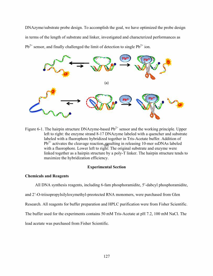
127
DNAzyme/substrate probe design. To accomplish the goal, we have optimized the probe design
in terms of the length of substrate and linker, investigated and characterized performances as
Pb2+ sensor, and finally challenged the limit of detection to single Pb2+ ion.
Figure 6-1. The hairpin structure DNAzyme-based Pb2+ sensor and the working principle. Upper left to right: the enzyme strand 8-17 DNAzyme labeled with a quencher and substrate labeled with a fluorophore hybridized together in Tris-Acetate buffer. Addition of Pb2+ activates the cleavage reaction, resulting in releasing 10-mer ssDNAs labeled with a fluorophore. Lower left to right: The original substrate and enzyme were linked together as a hairpin structure by a poly-T linker. The hairpin structure tends to maximize the hybridization efficiency.
Experimental Section
Chemicals and Reagents
All DNA synthesis reagents, including 6-fam phosphoramidite, 5′-dabcyl phosphoramidite,
and 2’-O-triisopropylsilyloxymethyl-preotected RNA monomers, were purchased from Glen
Research. All reagents for buffer preparation and HPLC purification were from Fisher Scientific.
The buffer used for the experiments contains 50 mM Tris-Acetate at pH 7.2, 100 mM NaCl. The
lead acetate was purchased from Fisher Scientific.
(a)
(b)

128
Synthesis and Purification of Fluorescently Labeled Oligonucleotides
To optimize the design of the hairpin probe, multiple candidates were designed and
prepared as shown in Table 6-1. All were synthesized and post-treated as described in Chapter 2
using an ABI 3400 DNA/RNA synthesizer (Applied Biosystems) at 1-μmol scale with the
standard phosphoramidite chemistry. To deprotect a RNA base, the precipitates were dissolved
in 0.5 ml tetrabutylammonium fluoride solution, with shaking for 6 hours at 35°C after the
complete cleavage and deprotection and ethanol precipitation. Then the desalting was performed
with the 0.1 M triethylammonium acetate (TEAA, pH 7.0) as elution buffer. The HPLC was
performed on a ProStar HPLC Station (Varian Medical Systems) equipped with a fluorescence
detector and a photodiode array detector. A C18 reverse-phase column (Alltech, C18, 5 μm, 250
× 4.6 mm) was used.
Table 6-1. Names and sequences of DNA
Name Sequences
D10 5’-/Dabcyl/-TATCTCTTCTCCGAGCCGGTCGAAATAGTGAG-(T)10ACTCACTATrAGGAAGAGATA-/FAM/-3’
D7 5’-/Dabcyl/-ATCTTCCGAGCCGGTCGAAATAGTGAG-(T)10-ACTCACTATrAGGAAGAT-/FAM/-3’
D5 5’-/Dabcyl/-ATTCCCCGAGCCGGTCGAAATAGTGAG-(T)10-ACTCACTATrAGGAAT-/FAM/-3’
Determination of Melting Temperature
Using a BioRad RT–PCR thermal cycler, thermal denaturizing profiles of each probe were
measured to study their thermostabilities. D10, D7 and D5 sequences were dissolved to the final
concentration 50 nM each in 50 mM Tris-acetate buffer, pH 7.2, with 50 mM NaCl. The mixture
was annealed by heating to 90 °C for 5 min and subsequently cooled to 4 °C in the 1 oC intervals
after each change equilibrated at the new temperature for at least 1 min. Upon melting, the
substrate strand was dissociated from the enzyme strand as the temperature was decreased,

129
resulting in an increased fluorescence signal. The fluorescence intensity of each probe was
measured and plotted against the temperature to generate the melting temperature curve.
Hybridization Assay
D10 sequence was annealed to final concentration 200nM using the same procedure
performed in the melting temperature determination. The annealed sample was then taken to
room temperature for subsequent assays. A 90-µL aliquot of the 100 nM hybridized
DNAzyme/substrate solution was loaded in each well of a 96-well plate. A 10-µL aliquot of
concentrated metal ions stock solution was then added to the DNA solution to initiate the
cleavage reaction. The fluorescence intensity was recorded for 100 μl buffer containing 50 mM
Tris–Acetate (pH7.2), 100 mM NaCl, the 200nM DNAzyme/substrate solution without lead, and
the 100 nM DNAzyme/substrate solution with 100X concentrated lead contamination. The
excitation and emission wavelengths were set to 473 nm and 520 nm, respectively. Signal
enhancement was calculated using the following equation;
)F(F)F(F
bufferIIwithoutead
bufferIIleadwith
−−
=)(
)(tenahncemenSignal (6-1)
In which
F with lead (II): fluorescence signal containing both a probe and Pb2+ F without lead(II): fluorescence intensity with a probe only Fbuffer: fluorescence intensity of buffer
The substrate fluorescence detection was performed with a Tecan Safire microplate reader
with 96 well plates. The excitation laser wavelength was set at 473 nm, and the emission
wavelength was set at 520 nm, respectively, to monitor the fluorescence of FAM. Data
quantification was performed using XFluor software. For each experiment 90 μL of the D10
solution was placed in the well and the fluorescence of the sample was measured immediately as

130
Fannealed. Then 10uL concentrated Pb2+ was added, followed by 10 minutes incubation at room
temperature. The fluorescence was measured, and then the solution in the well was collected and
heating up to 90oC for 5 minutes. The fluorescence will be measured again as Fcleaved. The RNase
free water was used as the internal standard to minimize the differences between each scan.
Other metal salts used included the following: MgCl2, CaCl2, SrCl2, BaCl2, Mn(OAc)2,
Fe(NH4)2(SO4)2, CoCl2, NiCl2, CuCl2, ZnCl2, Cd(ClO4)2, and Hg(ClO4)2.
Single Molecular Reaction
We used the Nuclepore polycarbonate membranes (Fisher Scientific) to form reactors of
femtoliter volumes, which are used in filtration. We used membranes with pore diameters of
5um. These pore sizes are uniform, and the material is confirmed in bulk studies to be chemically
inactive in the enzymatic reaction in this work. The thickness is ~6 um, producing volumes
between 110 and 200 fL for each vial. Liquid filling of nanoscopic volumes has been a difficult
technological problem in many fields, including biological studies where micromanipulators
coupled with microinjection have been employed. Since we are studying more than 100 vials
simultaneously, it is impossible to fill the vials one by one. The techniques applied here combine
ultrasonic vibration with vacuum degassing.165 We used rhodamine 6G dye solution to test this
procedure. From the fluorescence image, we conclude that this procedure ensured that this
surface was filled with the desired solution. The membrane was placed onto a glass slide, and
then a small drop of the D10/Pb2+ solution was added onto the top of the membrane. A very thin
quartz coverslip (0.08 mm) was put on the top to cover the liquid-filled vials. The polycarbonate
was sandwiched between two quartz plates to create the vials. A tight seal is expected to form
between the glass slides and the quartz coverslip. The quartz coverslip prevented any evaporation
from these small vials and mixing among the vials during the monitoring of the enzyme
reactions.

131
Gel-Based Activity Assay
2μM D10 solution was annealed in 50 mM Tris-acetate buffer, containing 100 mM NaCl
(pH 7.2). After taking a 5-μl aliquot out as a zero time point, Pb2+ was added to a final
concentration of 200 nM to the remaining solution, and aliquots were taken out at 10 minute
intervals. The D10 only sample was also loaded on the gel to measure the effect in the absence of
Pb2+. The cleaved and uncleaved substrates were separated using 20% PAGE, and the gel was
analyzed by a fluorescence imager (FLA-3000G; Fuji, Tokyo, Japan) using an excitation
wavelength of 473 nm. Gels containing 20% polyacrylamide were run on a FB-VE10-1
electrophoresis unit (FisherBiotech) at room temperature (200 V, constant voltage). The running
buffer was TAE/Mg2+ buffer.
Results and Discussion
Lead-dependent, site-specific cleavage of RNA has been the focus of many research
endeavors.166,167 The results may lead to a better understanding of lead toxicity in environmental
and biological systems and provide a model system for studying the role of metal ions in
DNA/RNAzymes.168 The demonstration of a lead-dependent DNAzyme as a highly sensitive and
selective lead sensor may stimulate further study in this field, as insight gained from the structure
and mechanism of the DNAzyme may allow better design of metal biosensors.
For this purpose, we analyzed 8-17 DNAzyme profile and designed the hairpin structural
D10 Pb2+ probe. This 8-17 DNAzyme preformed the catalytic cleavage reaction by two-step
mechanism.48 Product analysis by MALDI-MS demonstrated that Pb2+ catalyzes the cleavage
reaction of the 8-17 DNAzyme by the formation of a product containing a 2',3'-cyclic phosphate.
Then, Pb2+ catalyzes a second step of further hydrolysis of the 2',3'-cyclic phosphate. To increase
the cleavage reaction efficiency, the hairpin structure lead sensor probe was designed, in which

132
the substrate is held close to DNAzyme, ensuring high hybridization efficiency. In addition, the
lead binding pocket is pre-organized, thus increasing the apparent cleavage efficiency.
Optimization of the Hairpin Structure Sensor Design
In order to maximize the performance of the probes, the effects of several design
parameters were studied; the length of linker between the catalytic and substrate domain and the
substrate fragment design. The latter consideration is particularly critical because the substrate
fragment must be not only long enough to form a stable duplex with catalytic domain but also
short enough to be dissociated well after the hydrolysis reaction. Thus, we investigated the
leaving substrate design by varying the substrate sequence (Table 6-1). The background
fluorescence and melting temperatures of the hairpin structures were compared to evaluate the
hybridization efficiencies, and the gel electrophoresis was used to compare the hydrolysis
efficiencies (Table 6-2, Figure 6-2, and Figure 6-3, respectively)).
We designed probes containing 5, 7, and 10 base-paired leaving sequence called D5, D7
and D10 (Table 6-1). Probes containing longer substrates were not considered, because the
dissociation may not be favorable for more than 10 base pairs. As shown in Figure 6-2, the
fluorescence background of probes without lead ion was significantly reduced by increasing the
number of base pairings. Probes can have quenched fluorescence signal only when they form
stable hairpin structures, which means that the probes are in the active state to catalyze the
hydrolysis reaction in the presence of Pb2+. Thus, D10 with the lowest fluorescence background
and the highest signal enhancement achieved the maximum efficiency in the catalytic reaction,
giving approximately 16-fold signal enhancement when the lead ion was added. This
improvement is outstanding, considering that the previous work reported that the single quencher
design showed only 4-fold enhancement,46 and the dual quencher probe slightly improved the
enhancement to 6-fold.164

133
The reduced background is easily explained by the enhanced duplex stability indicated by
thermal denaturing profiles which correlated well with quenching efficiency because the
quencher/fluorophore pairs must be in close proximity. As shown in Table 6-2, the melting
Figure 6-2. Fluorescence signal in the absence and presence of 10uM Pb2+ after 10 min
Table 6-2. Comparison of melting temperature and fluorescence enhancement among different design sequences
Hairpin sequence Tm (°C) S/B ratio
D5 45 4
D7 48 7
D10 54 16 temperature of D5 was only 45 °C, and that of D10 increased to 54ºC. This enhanced duplex
stability is quite phenomenal because the Tm of unlinked DNAzyme and its substrate is 34°C
which is only slightly above room temperature.

134
This enhanced duplex stability also resulted in the better performance in the hydrolysis
reaction. According to the result obtained from naturing PAGE-gel electrophoresis, only D10
showed the efficient cleavage reaction (Figure 6-3). Each set of the samples was composed of
probe only, probe with lead ion, and probe with target DNA. The D5 probe only showed a
smeared band unlike D10 and D7. This was caused by the incomplete hybridization of substrate
with the catalytic domain, which resulted in the heterogeneous distribution of nucleic acid
conformations. Likewise, the hydrolysis yield was not favorable, which contains mostly intact
targets. Similarly, noticeable cleavage was not observed for D7 (lane 4 and 5) even though D7
sample did not show a smeared band. In contrast, D10 in the absence of Pb2+ (lane 7) showed a
clear single band. Upon the addition of Pb2+ (2 μM), the fluorescence of an intact probe band
decreased while a clear increase of cleaved piece carrying fluorophore was observed. This
experiment shows that the increased fluorescence was indeed due to DNAzyme-catalyzed
cleavage with Pb2+ as a cofactor.
Figure 6-3. PAGE-gel image of probes in the absence and presence of 10uM Pb2+ after 10 min. Lanes 1, 4,7: DNAzyme alone; lanes 2, 5, 8: time 10 min after adding Pb2+, to the DNAzyme. The upper and lower bands are uncleaved and cleaved product, respectively. Lanes 3, 6, 9: 10 min after adding cDNA.
Excellent Sensitivity
To obtain the full profile of D10 performance, we determined the dose response,
calibration curve including the limit of detection, and reaction kinetics (Figure 6-4 and 6-5,

135
Figure 6-4. Fluorescence increase over background at varying Pb2+ concentrations. The DNAzyme sensor concentration was 200 nM. (Inset) Sensor response to low concentrations of Pb2+.
Figure 6-5. Kinetics of fluorescence increase for D10 in 5uM Pb2+. The background-corrected intensity in each sample has been normalized.

136
respectively). Figure 6-4 shows the full profile of fluorescence signal enhancement verse Pb2+
concentration and the linear response in the presence of low concentration of Pb2+. According to
the full profile, D10 can have up to 20 times signal enhancement. This large signal enhancement
allows us to detect lead ion in large dynamic range in nanomolar concentration with surprisingly
low detection limits, approximately 3nM (3σ/slope), which is about 167 times lower than the
previously reported result. When we consider that U.S. Environmental Protection Agency (EPA)
defined higher than 15 ppb Pb2+ level in drinking water considered to be toxic, D10 can function
well to monitor environmental samples, since the minimum toxicity level to be toxic is well
within the sensor dynamic range. In addition to the excellent sensitivity of D10, the kinetics of
D10 is also rapid. As shown in Figure 6-5, the signal reached a plateau after about 15 min. the
time required for bimolecular sensors is more than 30 minute (data not shown). The superior
performance of D10 is attributable to several factors associated with the intramolecular
interaction-based sensing strategy. First, the DNAzyme itself binds Pb2+ very strongly and
rapidly since most of D10 is in the active state. Second, the sensor design has very low
background fluorescence or background variation, which allows higher signal-to-background
ratio. Finally, and probably most unique, the signal can be amplified through fast turnovers of
Pb2+ to cleave multiple fluorescent target strands. This increases the sensitivity while still
maintaining a very rapid response.
Superior Selectivity
Probably, one of the most unique features of DNAzyme-based metal sensors is the
absolute selectivity. This excellent selectivity of artificial nucleic acid sequences to their targets
originates from the unique secondary configurations, which can discriminate against non-targets.
Likewise, 8-17 DNAzyme also shows excellent selectivity for Pb2+ compared to all other
divalent ions. To demonstrate the excellent selectivity of D10, the fluorescence signal changes

137
upon 9 competing divalent metals at concentrations of 10 μM, 5 μM, and 1 μM were obtained
(Figure 6-5). As expected, they induced little to no fluorescence change. Slight decreases of
Figure 6-6. Selectivity of D10 Pb2+ sensor. Sensor responses to potential competing metal ions at four concentrations (10 μM, 5 μM, 2 μM and 0.5 μM) with 10 min reaction time.
fluorescence from some ions were observed probably due to metals-induced quenching to
fluorophores. This result indicates that the selectivity of 8-17 DNAzyme was not affected by
construction of D10 probe.
Single Ion Reaction
According to the preliminary investigation, D10 is a highly sensitive and selective probe
for Pb2+. To further challenge the limit of D10’s detection capability, we thus designed a single-
Pb2+ detection using a micro-well reactor. Each well contains a membrane well with 5 μm
diameter and approximate volume of ~120 femtoliter. When a well contains a single ion, the
concentration is about 14 pM, which is well below the limit of detection of D10. We employed a

138
Pb2+ concentration corresponding to statistically filling of only 25% of the wells to minimize the
chance of having more than 2 ions in a single well. The fluorescence signal was recorded using
D10 only (200nM) and D10 with Pb2+, and the obtained results is summarized in Figure 6-7.
The single-molecule reaction was monitored every 10s for 1400s. Since each image contained
about 30 to 60 wells in most of the experiments, fluorescence signal of each vial was analyzed,
and a reaction rate curve was constructed. When the membrane vials were filled with only the
D10 solution, we did not observe any significant fluorescence intensity change during the
monitoring. This means that the D10 can form a stable hairpin structure without significant
surface interaction. After the fluorescence signal had somewhat stabilized, we added the desired
amount of Pb2+ and the fluorescence monitoring was continued. In contrast to the blank sample
containing only D10, we could observe three typical patterns of fluorescence increase: negligible,
slight, and large fluorescence change (blue, red, and green, respectively in Figure 6-7 (a)).
Interestingly the greatest fluorescence enhancement was achieved for the lowest number of
membrane pores while most of the fluorescent pores showed limited fluorescence enhancement
(Figure 6-7 (b)). Thus, we speculated that the difference in rate of signal production was
originated from the different number of Pb2+ ions in the wells. The negligible increase of
fluorescence came from fluctuations of D10 itself probably due to surface interactions or local
heating by the laser rather than interaction with Pb2+. The slight increase of fluorescence with
time was from wells containing a single Pb2+. Likewise, the wells showing the fastest increasing
rate contained several Pb2+s. Despite of the low concentration of Pb2+ ion, D10 was able to bind
the single target at a concentration of 14pM and generate a detectible fluorescence signal under
the microscope. In addition, the fluorescence signal reached to a plateau in a relatively short
amount of time (less than 15min). Thus, we conclude that the optimized probe was able to detect

139
as low as a single Pb2+ within 15 minutes.
Figure 6-7. Single ion reaction kinetics inside polycabornate membrane vials. Individual Pb2+ ions were trapped inside membrane holes with the D10 solution. The fluorescence enhancement was monitored over time. The background-corrected intensity in each well has been normalized to the signal prior to adding to Pb2+.
(a)
(b)

140
Conclusions
To conclude, we have employed a Pb2+-specific DNAzyme to design a highly sensitive and
selective Pb2+ sensor. With a detection limit (3 nM) approaching those of the most sensitive
instruments, and with higher selectivity over other metal ions (better than instruments such as
ICP-AES), the sensor can be a simple portable alternative to instrumental methods. This results
show that the DNAzyme sensing platform has an enormous potential for detection and
quantification of many metal ions. With wide availability of portable fluorometers, such a highly
sensitive and selective Pb2+ sensor will find widespread application in on-site and real-time
environmental monitoring.
141
CHAPTER 7 SUMMARY AND FUTURE DIRECTIONS
Molecular Engineering of Multifunctional Nucleic Acid Probes for Bioanalytical and Biomedical Applications
Last decades, many different types of nucleic acid probes and architectures have been
proposed and developed in recent years. Among them, DNA probes have served as a novel class
of recognition elements to detect a wide variety of targets; for example, ions, small molecules,
nucleic acids, proteins, and even living cells. These developments have been accelerated by
advanced technologies for modifying DNA-based probes with different signaling mechanisms.
The overall direction of this doctoral research has been dedicated to engineering such
biomolecular components with the goal of building novel molecular tools and devices for
biological science, biomedical research and therapeutic applications. This work has encompassed
three main avenues of research: development of molecular probes for improved detection,
engineering nucleic acid binders to enhance inhibitory functions, and incorporation of light
sensitive molecules into DNA sequences to develop molecular tools for light regulation.
The first phase of this research was development of multifunctional molecular tools for
bioanalytical applications. In cell biology, alteration of nucleic acid expression levels or spatial
localization often reflects important cellular events, and tracking such alterations in native
cellular environments can be one of the most effective ways to advance biological science, drug
discovery, and therapeutic treatment of injury and infection. However, the complexity of living
cells often challenges the measurements due to degradation of nucleic acid probes by nuclease
digestion. Thus, it requires the development of robust probes for reliable intracellular studies. To
meet this goal, we designed robust molecular probes using nonstandard nucleic acids, such as
locked nucleic acid, 2-O-methyl modified RNAs, and L-DNA. One of such innovations is the
development of chimeric nucleic acid probes using locked nucleic acids (LNA) or 2-O-methyl

142
modified RNAs and L-DNA. This probe design showed superior biostability and the least
tendency for false-positive or false-negative reporting.
Heavy metal contamination in the human body or environment is a significant problem,
and there is a clear demand for ultrasensitive detection methods to monitor such a contamination
for high throughput and rapid screening. To meet the goal, we have developed a Pb2+-specific
unimolecular ultrasensitive nucleic acid sensor using catalytic DNAzyme. The optimized sensor
demonstrated detection limits in the nanomolar range (3nM) with superior sensitivity and
excellent selectivity when used with a routine fluorospectrometer. Use of a confocal microscope
and microwell array provided a clear positive report with only 14pM of Pb2+, ie, a single Pb2+ ion.
Another research project involved the molecular assembly of functional ssDNA sequences
called aptamers, selected by a method known as cell-based SELEX ("Systematic Evolution of
Ligands by Exponential Enrichment"). Although this selection strategy produces molecular
probes with high affinity for and specific binding to a given target, we have discovered a process
to further increase binding capacity several-fold over conventional aptamers: multivalent
interaction. The process involves the assembly of multiple ligands for multiple binding with
target(s). The overall binding of the assembly becomes much stronger than that of the individual
ligand. Using this as the working principle, we assembled two aptamers targeted to thrombin to
create a bivalent ligand. As an anticoagulant, the bivalent ligand showed a 30-fold increase in
clotting time compared to individual aptamers. In addition, the multimeric assemblies showed
increased clotting times 2-fold when human plasma sample was applied. This concept was
generalized using PDGF and its aptamer. We believe that this concept can be effectively applied
to the design of nucleic acid probe-based drugs for pharmaceutical applications.

143
The third phase of this research was the development of a photo-regulating nucleic acid
inhibitor. Photochromic compounds can be successfully incorporated into the DNA chains to
alter the hybridization of DNA duplexes. Thus, we utilized these compounds to design nucleic
acid inhibitors regulated by UV and visible photons. We used thrombin and its aptamer to
engineer such a probe and tested the photo-converting inhibitory functions. The results showed
that inhibition of thrombin’s protease activity can be effectively switched on and off depending
on the photon excitation energy. In addition, the experiment using a microfluidic device revealed
that such photocontrolling enzymatic reactions can be spatially regulated. We expect that this
strategy can be highly useful in manipulating biological reactions by light.
In summary, this research has focused on the design, synthesis and investigation of
multifunctional and advanced nucleic acid probes for the biological sciences, biomedical
research, and therapeutic and clinical applications. The successful outcome to these studies will
lead to a better understanding of biological processes and the development of advanced
molecules for nucleic acid-based detections and medicines.
Future Directions
Developing High Throughput Metal-Screening Chip
To screen multiple heavy metal contamination, it is also possible to develop unimolecular
sensors for other targets. So far, there are many DNAzymes already known, including those
targeting Cu2+, UO2+, Cu2+ and Zn2+, and there is no doubt that more DNAzymes will be
selected, since the selection of target-specific DNAzyme is a very active research area. Thus, we
can use the same manner of ultrasensitive Pb2+ sensor to develop sensitive sensors targeting
different ions.
As a high throughput screening method, the Pb2+ (or other metals) sensor can be
immobilized on the surface of a microarray chip via the streptavidine/biotin method. After

144
coating the surface with spreptavidine, biotinylated sensor probes can be incubated and captured.
After careful washing, the chip will be ready for detection. Since the sensor molecules contain
fluorophore and quencher, but no target, each spot will produce a fluorescence signal. When the
sample is applied to each spot, depending on the concentration of the target, the spots will show
different fluorescence signals. Thus, multiple samples can be tested at once in a short amount of
time. In addition, we expect that the array may be able to show concentration dependent response
due to the probe’s high signal enhancement ratio.
Figure 7-1. Surface-immobilized Pb2+ chip. (a) microarray and anticipated results. (b) shows the cartoon to describe working principle of the metal sensor chip.
There will be major advantages to the proposed design. First, detection without sample
washing can save time and effort. Second, multiple samples and ions can be screened using a
small device. Finally, the chips can be used for quantitative analysis. Therefore, this approach
will be an interesting future project to make general use of DNAzyme-based sensors.
(a)
145
Pharmaceutical Application of Multifunctional Drugs
According to our in vitro investigation, ssDNA assemblies show dramatic improvements in
the inhibition of enzymatic functions. Thus, in vivo investigation of such pharmaceutical potency
can be another promising area.
First, the anticoagulant potency of each assembly of thrombin aptamers can be investigated
using a mouse model. Coagulation is highly conserved biological process from one species to
another species. Thus, these aptamers should also be able to bind to mouse thrombin protein.
Dose response, cytotoxicity and other pharmaceutical parameters can be investigated using
mouse model experiments.
Second, the ability of the antithrombin approach to cure cancer by controlling angiogenesis
will be demonstrated. According to recent reports, thrombin functions as an important factor to
regulate the tumor growth. Thus, we speculate that antithrombin can be a general approach to
regulate the growth of tumors. To do so, we need to adopt the tumor model and investigate the
tumor growth. As a selective delivery method, we can attach cancer-specific aptamers to the
assemblies or nanoparticles. Then, we may be able to improve the efficiency of regulating tumor
growth.
Third, it will demonstrate that the photo-controllable inhibitors can selectively regulate
coagulation reactions in living system. There will be two types of samples: one is injected with
UV- treated probes; and another mouse is injected with Vis-treated probes. When both mice are
bleeding, we expect that the one injected with UV- treated probes will bleed for a longer time
than the one injected with Vis-treated probes, due to the better anticoagulation potency of UV-
treated probes. In addition, we can also demonstrate that the mouse injected with UV- treated
probes can show different clotting times with post-light treatments.

146
Figure 7-2. Mouse model to show photo-controllable anticoagulation

147
LIST OF REFERENCES
1. Watson, J. D. & Crick, F. H. C. Molecular Structure of Nucleic Acids: A structure for deoxyribose nucleic acid. Nature 171, 737-738 (1953).
2. Beaucage, S. L. & Iyer, R. P. The synthesis of modified oligonucleotides by the phosphoramidite approach and their applications. Tetrahedron 49, 6123-6194 (1993).
3. Beaucage, S. L. & Iyer, R. P. Advances in the synthesis of oligonucleotides by the phosphoramidite approach. Tetrahedron 48, 2223-2311 (1992).
4. Ingle, J. D. & Crouch, S. R. Spectrochemical analysis. (1988).
5. Lakowicz, J. R. Principles of fluorescent spectroscopy. Kluwer Academic /Plenum Publishers, New York. (1999).
6. Chakravarti, A. Single nucleotide polymorphisms to a future of genetic medicine. Nature 409, 822-823 (2001).
7. Altmuller, J., Palmer, L. J., Fischer, G., Scherb, H. & Wjst, M. Genomewide scans of complex human diseases: True linkage is hard to find. American Journal of Human Genetics 69, 936-950 (2001).
8. Tyagi, S. & Kramer, F. R. Molecular beacons: probes that fluoresce upon hybridization. Nat. Biotechnol. 14, 303-308 (1996).
9. Tyagi, S., Bratu, D. P. & Kramer, F. R. Multicolor molecular beacons for allele discrimination. Nat. Biotech. 16, 49-53 (1998).
10. Perlette, J. & Tan, W. H. Real-time monitoring of intracellular mRNA hybridization inside single living cells. Anal. Chem. 73, 5544-5550 (2001).
11. Sokol, D. L., Zhang, X. L., Lu, P. Z. & Gewitz, A. M. Real time detection of DNA RNA hybridization in living cells. Proceedings of the National Academy of Sciences 95, 11538-11543 (1998).
12. Santangelo, P. J., Nix, B., Tsourkas, A. & Bao, G. Dual FRET molecular beacons for mRNA detection in living cells. Nucl. Acids Res. 32, (2004).
13. Bratu, D. P., Cha, B. J., Mhlanga, M. M., Kramer, F. R. & Tyagi, S. Visualizing the distribution and transport of mRNAs in living cells. Proceedings of the National Academy of Sciences 100, 13308-13313 (2003).

148
14. Santangel, P. J., Nitin, N. & Bao, G. Direct visualization of mRNA colocalization with mitochondria in living cells using molecular beacons. Journal of Biomedical Optics 10, (2005).
15. Drake, T. J., Medley, C. D., Sen, A., Rogers, R. J. & Tan, W. H. Stochasticity of manganese superoxide dismutase mRNA expression in breast carcinoma cells by molecular beacon imaging. Chembiochem 6, 2041-2047 (2005).
16. Fang, X. H., Li, J. J. & Tan, W. H. Using molecular beacons to probe molecular interactions between lactate dehydrogenase and single-stranded DNA. Anal. Chem. 72, 3280-3285 (2000).
17. Uchiyama, H., Hirano, K., KashiwasakeJibu, M. & Taira, K. Detection of undegraded oligonucleotides in vivo by fluorescence resonance energy transfer. Journal of Biological Chemistry 271, 380-384 (1996).
18. Kehlenbach, R. H. In vitro analysis of nuclear mRNA export using molecular beacons for target detection. Nucl. Acids Res. 31, e64 (2003).
19. Mhlanga, M. M., Vargas, D. Y., Fung, C. W., Kramer, F. R. & Tyagi, S. tRNA-linked molecular beacons for imaging mRNAs in the cytoplasm of living cells. Nucl. Acids Res. 33, 1902-1912 (2005).
20. Molenaar, C., Marras, S. A., Slats, J. C., Truffert, J. C., Lemaître, M., Raap, A. K., Dirks, R. W. , Tanke, H. J., Linear 2 ' O-Methyl RNA probes for the visualization of RNA in living cells. Nucl. Acids Res. 29, art-e89 (2001).
21. Marras, S. A. E., Gold, B., Kramer, F. R., Smith, I. & Tyagi, S. Real-time measurement of in vitro transcription. Nuc. Acids Res. 32, e72 (2004).
22. Kuhn, H., Demidov, V.V, Gildea, B. D., Fiandaca, M. J., Coull, J. C., & Frank-Kamenetskii, M. D. PNA beacons for duplex DNA. Antisense & Nucleic Acid Drug Development 11, 265-270 (2001).
23. Petersen, K., Vogel, U., Rockenbauer, E., Nielsen, K. V., Kølvraa, S., Bolund, L., Nexø, B. Short PNA molecular beacons for real-time PCR allelic discrimination of single nucleotide polymorphisms. Molecular and Cellular Probes 18, 117-122 (2004).
24. Seitz, O. Solid-phase synthesis of doubly labeled peptide nucleic acids as probes for the real-time detection of hybridization. Angewandte Chemie-International Edition 39, 3249-3252 (2000).
149
25. Tsourkas, A., Behlke, M. A. & Bao, G. Hybridization of 2 '-O-methyl and 2 '-deoxy molecular beacons to RNA and DNA targets. Nucl. Acids Res. 30, 5168-5174 (2002).
26. Wang, L., Yang, C. Y. J., Medley, C. D., Benner, S. A. & Tan, W. H. Locked nucleic acid molecular beacons. J. Am. Chem. Soc. 127, 15664-15665 (2005).
27. Xi, C. W., Balberg, M., Boppart, S. A. & Raskin, L. Use of DNA and peptide nucleic acid molecular beacons for detection and quantification of rRNA in solution and in whole cells. Applied and Environmental Microbiology 69, 5673-5678 (2003).
28. Yang, C. J., Wang, L., Wu, Y., Kim, Y., Medley, C. D., Lin, H. & Tan, W. Synthesis and investigation of deoxyribonucleic acid/locked nucleic acid chimeric molecular beacons. Nucl. Acids Res. 35, 4030-4041 (2007).
29. Orum, H. & Wengel, J. Locked nucleic acids: A promising molecular family for gene-function analysis and antisense drug development. Current Opinion in Molecular Therapeutics 3, 239-243 (2001).
30. Vissing, H. et al. High throughput multiplex genotyping using chimeric LNA (Locked Nucleic Acids)/DNA oligos immobilized polymer microchip. American Journal of Human Genetics 69, 470 (2001).
31. Vester, B. & Wengel, J. LNA (Locked nucleic acid): High-affinity targeting of complementary RNA and DNA. Biochemistry 43, 13233-13241 (2004).
32. Ellington, A. D. & Szostak, J. W. Invitro Selection of RNA Molecules That Bind Specific Ligands. Nature 346, 818-822 (1990).
33. Robertson, D. L. & Joyce, G. F. Selection Invitro of An RNA Enzyme that specifically cleaves single-stranded-DNA. Nature 344, 467-468 (1990).
34. Tuerk, C. & Gold, L. Systematic Evolution of ligands by exponential enrichment - RNA ligands to bacteriophage-T4 DNA-polymerase. Science 249, 505-510 (1990).
35. Mayor, G., Grattinger, M. & Blind, M. Aptamers: multifunctional tools for target validation and drug discovery. Drug Plus Int. 2, 6-10 (2003).
36. Mammen, M., Choi, S. K. & Whitesides, G. M. Polyvalent interactions in biological systems: Implications for design and use of multivalent ligands and inhibitors. Angewandte Chemie-International Edition 37, 2755-2794 (1998).
37. Mulder, A., Huskens, J. & Reinhoudt, D. N. Multivalency in supramolecular chemistry and nanofabrication. Organic & Biomolecular Chemistry 2, 3409-3424 (2004).

150
38. Varki, A. Biological Roles of Oligosaccharides - All of the theories are correct. Glycobiology 3, 97-130 (1993).
39. Kohli, P. Ligand-receptor energetics. John Wiley & Sons, New York (1997).
40. Hammes, G. G. Thermodynamics and kinetics for the biological sciences. John Wiley & Sons, New York (2000)
41. Schon, A. & Freire, E. Thermodynamics of intersubunit interactions in cholera-toxin upon binding to the oligosaccharide portion of its cell-surface receptor, ganglioside-Gm1. Biochemistry 28, 5019-5024 (1989).
42. Tanaka, T., Suzuno, R., Nakamura, K., Kuwahara, A. & Takeo, K. Thermodynamic analysis of the interactions of a mouse dinitrophenyl-specific myeloma protein, mopc-315, with immobilized dinitrophenyl and trinitrophenyl ligands by affinity electrophoresis. Electrophoresis 7, 204-209 (1986).
43. Lee, Y. C. & Lee, R. T. Carbohydrate-potein interactions - basis of glycobiology. Acc. Chem. Res. 28, 321-327 (1995).
44. Karush, F., Chua, M. M. & Rodwell, J. D. Interaction of a bivalent ligand with Igm anti-lactose antibody. Federation Proceedings 37, 1279 (1978).
45. Santoro, S. W. & Joyce, G. F. A general purpose RNA-cleaving DNA enzyme. Proceedings of the National Academy of Sciences 94, 4262-4266 (1997).
46. Li, J. & Lu, Y. A highly sensitive and selective catalytic DNA biosensor for lead ions. J. Am. Chem. Soc. 122, 10466-10467 (2000).
47. Breaker, R. R. & Joyce, G. F. A DNA enzyme that cleaves RNA. Chemistry & Biology 1, 223-229 (1994).
48. Brown, A. K., Li, J., Pavot, C. M. B. & Lu, Y. A lead-dependent DNAzyme with a two-step mechanism. Biochemistry 42, 7152-7161 (2003).
49. Zhou, D. M. & Taira, K. The Hydrolysis of RNA: From theoretical calculations to the hammerhead ribozyme-mediated cleavage of RNA. Chem. Rev. 98, 991-1026 (1998).
50. Kuimelis, R. G. & McLaughlin, L. W. Mechanisms of ribozyme-mediated RNA cleavage. Chem. Rev. 98, 1027-1044 (1998).
51. Berliner, L. J. Structure-function-relationships in human alpha-thrombins and gamma-thrombins. Molecular and Cellular Biochemistry 61, 159-172 (1984).

151
52. Mann, K. G. & Lundblad, R. L. Hemostasis and thrombosis: basic principles and clinical practice. J. B. Lippincott Co., Philadelphia (1987).
53. Quick, A. J., Stanley-Browne, M. & Bancroft, F. W. A study of the coagulation defect in hemophilia and in jaundice. American Journal of the Medical Sciences 190, 501-511 (1935).
54. Mischke, R. Activated partial thromboplastin time as a screening test of minor or moderate coagulation factor deficiencies for canine plasma: sensitivity of different commercial reagents. Journal of Veterinary Diagnostic Investigation 12, 433-437 (2000).
55. DiCera, E., Dang, Q. D. & Ayala, Y. M. Molecular mechanisms of thrombin function. Cellular and Molecular Life Sciences 53, 701-730 (1997).
56. Bode, W., Turk, D. & Karshikov, A. The refined 1.9-angstrom X-ray crystal-structure of D-Phe-Pro-Arg chloromethylketone-inhibited human alpha-thrombin - structure-analysis, Protein Science 1, 426-471 (1992).
57. Lesk, A. M. & Fordham, W. D. Conservation and variability in the structures of serine proteinases of the chymotrypsin family. Journal of Molecular Biology 258, 501-537 (1996).
58. Rydel, T. J., Ravichandran, K. G., Tulinsky, A., Bode, W., Huber, R., Roitsch, C. & Fenton, J. W. 2nd. The Structure of A Complex of recombinant hirudin and human alpha-thrombin. Science 249, 277-280 (1990).
59. Mathews, I. I., Padmanabhan, K. P., Tulinsky, A. & Sadler, J. E. Structure of a nonadecapeptide of the 5th EGF domain of thrombomodulin complexed with thrombin. Biochemistry 33, 13547-13552 (1994).
60. Srinivasan, J., Hu, S., Hrabal, R., Zhu, Y., Komives, E. A. & Ni, F. Thrombin-bound structure of an Egf subdomain from human thrombomodulin determined by tansferred nuclear overhauser effects. Biochemistry 33, 13553-13560 (1994).
61. Rydel, T. J., Tulinsky, A., Bode, W. & Huber, R. Refined structure of the hirudin-thrombin complex. Journal of Molecular Biology 221, 583-601 (1991).
62. Vijayalakshmi, J., Padmanabhan, K. P., Mann, K. G. & Tulinsky, A. The isomorphous structures of pre-thrombin2, hirugen-thrombin, and pack-thrombin - changes accompanying activation and exosite binding to thrombin. Protein Science 3, 2254-2271 (1994).

152
63. Lerner, D. J., Chen, M., Tram, T. & Coughlin, S. R. Agonist recognition by proteinase-activated receptor 2 and thrombin receptor - Importance of extracellular loop interactions for receptor function. J. Biol. Chem. 271, 13943-13947 (1996).
64. Ishihara, H., Connolly, A. J., Zeng, D., Kahn, M. L., Zheng, Y. W., Timmons, C., Tram, T. & Coughlin, S. R. Protease-activated receptor 3 is a second thrombin receptor in humans. Nature 386, 502-506 (1997).
65. Ni, F., Ripoll, D. R., Martin, P. D. & Edwards, B. F. P. Solution structure of a platelet receptor peptide bound to bovine alpha-thrombin. Biochemistry 31, 11551-11557 (1992).
66. Mathews, I. I., Padmanabhan, K. P., Ganesh, V., Tulinsky, A., Ishiim, M., Chen, J., Turck, C. W., Coughlin, S. R. & Fenton, J. W. Crystallographic structures of thrombin complexed with thrombin receptor peptides - existence of expected and novel binding modes. Biochemistry 33, 3266-3279 (1994).
67. Vindigni, A., White, C. E., Komives, E. A. & DiCera, E. Energetics of thrombin-thrombomodulin interaction. Biochemistry 36, 6674-6681 (1997).
68. Ye, J., Esmon, C. T. & Johnson, A. E. The chondroitin sulfate moiety of thrombomodulin binds A 2nd molecule of thrombin. J. Biol. Chem. 268, 2373-2379 (1993).
69. Ye, J., Rezaie, A. R. & Esmon, C. T. Glycosaminoglycan contributions to both protein-C activation and thrombin inhibition involve a common arginine-rich site in thrombin that includes residues arginine-93, arginine-97, and arginine-101. J. Biol. Chem. 269, 17965-17970 (1994).
70. Katsonis, N., Lubomska, M., Pollard, M. M., Feringa, B. L. & Rudolf, P. Synthetic light-activated molecular switches and motors on surfaces. Progress in Surface Science 82, 407-434 (2007).
71. Horspool, W. M. CRC Handbook of organic photochemistry and photobiology. CRC press, Boca Raton (1995).
72. Stuart, C. M., Frontiera, R. R. & Mathies, R. A. Excited-state structure and dynamics of cis- and trans-azobenzene from resonance Raman intensity analysis. Journal of Physical Chemistry A 111, 12072-12080 (2007).
73. Giesendorf, B.A., Vet, J.A., Tyagi, S., Mensink, E.J., Trijbels, F.J. & Blom, H.J. Molecular beacons: a new approach for semiautomated mutation analysis. Clinical Chemistry 44, 482-486 (1998).

153
74. Kostrikis, L. G., Tyagi, S., Mhlanga, M. M., Ho, D. D. & Kramer, F. R. Molecular beacons - Spectral genotyping of human alleles. Science 279, 1228-1229 (1998).
75. Matsuo, T. In situ visualization of messenger RNA for basic fibroblast growth factor in living cells. Biochimica et Biophysica Acta-General Subjects 1379, 178-184 (1998).
76. Leal, N. A., Sukeda, M. & Benner, S. A. Dynamic assembly of primers on nucleic acid templates. Nucl. Acids Res. 34, 4702-4710 (2006).
77. Tsourkas, A., Behlke, M. A., Rose, S. D. & Bao, G. Hybridization kinetics and thermodynamics of molecular beacons. Nucl. Acids Res. 31, 1319-1330 (2003).
78. Li, J. W. J. & Tan, W. H. A real-time assay for DNA sticky-end pairing using molecular beacons. Analytical Biochemistry 312, 251-254 (2003).
79. Browne, K. A. Sequence-specific, self-reporting hairpin inversion probes. J. Am. Chem. Soc. 127, 1989-1994 (2005).
80. Crey-Desbiolles, C., Ahn, D. R. & Leumann, C. J. Molecular beacons with a homo-DNA stem: improving target selectivity. Nucl. Acids Res. 33, e77 (2005).
81. Urata, H., Shinohara, K., Ogura, E., Ueda, Y. & Akagi, M. Mirror-image DNA. J. Am. Chem. Soc. 113, 8174-8175 (1991).
82. Ashley, G. W. Modeling, Synthesis, and hybridization properties of (L)-ribonucleic acid. J. Am. Chem. Soc. 114, 9731-9736 (1992).
83. Damha, M. J., Giannaris, P. A., Marfey, P. & Reid, L. S. Oligodeoxynucleotides containing unnatural L-2'-deoxyribose. Tetrahedron Letters 32, 2573-2576 (1991).
84. Urata, H., Ogura, E., Shinohara, K., Ueda, Y. & Akagi, M. Synthesis and properties of mirror-image DNA. Nucl. Acids Res. 20, 3325-3332 (1992).
85. Garbesi, A., Capobianco, M. L., Colonna, F. P., Tondelli, L., Arcamone, F., Manzini, G., Hilbers, C. W., Aelen, J. M., & Blommers, M. J. L-DNAs as potenital antimessenger oligonucleotides: a reassessment. Nucl. Acids Res. 21, 4159-4165 (1993).
86. Medley, C. D., Drake, T. J., Tomasini, J. M., Rogers, R. J. & Tan, W. Simultaneous monitoring of the expression of multiple genes inside of single breast carcinoma cells. Anal. Chem. 77, 4713-4718 (2005).
87. Hauser, N. C., Martinez, R., Jacob, A., Rupp, S., Hoheisel, J. D. & Matysiak, S. Utilising the left-helical conformation of L-DNA for analysing different marker types on a single universal microarray platform. Nucl. Acids Res. 34, 5101-5111 (2006).

154
88. Li, J. J., Geyer, R. & Tan, W. Using molecular beacons as a sensitive fluorescence assay for enzymatic cleavage of single-stranded DNA. Nucl. Acids Res. 28, e52 (2000).
89. Koshkin, A. A., Rajwanshi, V. K. & Wengel, J. Novel convenient syntheses of LNA [2.2.1]bicyclo nucleosides. Tetrahedron Letters 39, 4381-4384 (1998).
90. Wengel, J. Synthesis of 3 '-C- and 4 '-C-branched oligodeoxynucleotides and the development of locked nucleic acid (LNA). Acc. Chem. Res. 32, 301-310 (1999).
91. Marras, S. A. E., Gold, B., Kramer, F. R., Smith, I. & Tyagi, S. Real-time measurement of in vitro transcription. Nucl. Acids Res. 32, e72 (2004).
92. Rao, J., Lahiri, J., Isaacs, L., Weis, R. M. & Whitesides, G. M. A trivalent system from vancomycin D-Ala-D-Ala with higher affinity than avidin/biotin. Science 280, 708-711 (1998).
93. Chen, H. W. & Privalsky, M. L. Cooperative Formation of high-order oligomers by retinoid-X receptors - an unexpected mode of DNA Rrecognition. Proceedings of the National Academy of Sciences 92, 422-426 (1995).
94. Kortt, A. A., Dolezal, O., Power, B. E. & Hudson, P. J. Dimeric and trimeric antibodies: high avidity scFvs for cancer targeting. Biomolecular Engineering 18, 95-108 (2001).
95. Wolf, E., Hofmeister, R., Kufer, P., Schlereth, B. & Baeuerle, P. A. BiTEs: bispecific antibody constructs with unique anti-tumor activity. Drug Discovery Today 10, 1237-1244 (2005).
96. Wolf, E. & Baeuerle, P. A. Micromet: engaging immune cells for life. Drug Discovery Today 7, S25-S27 (2002).
97. Bock, L. C., Griffin, L. C., Latham, J. A., Vermaas, E. H. & Toole, J. J. Selection of single-stranded DNA molecules that bind and inhibit human thrombin. Nature 355, 564-566 (1992).
98. DeAnda, A., Coutre, S. E., Moon, M .R., Vial, C. M., Griffin, L. C ., Law, V. S., Komeda, M., Leung, L. L., & Miller, D. C. Pilot study of the efficacy of a thrombin inhibitor for use during cardiopulmonary bypass. Ann Thorac Surg 58, 344-350 (1994).
99. Griffin, L. C., Tidmarsh, G. F., Bock, L. C., Toole, J. J. & Leung, L. L. In vivo anticoagulant properties of a novel nucleotide-based thrombin inhibitor and demonstration of regional anticoagulation in extracorporeal circuits. Blood 81, 3271-3276 (1993).

155
100. Huang, J. et al. Highly specific antiangiogenic therapy is effective in suppressing growth of experimental Wilms tumors. Journal of Pediatric Surgery 36, 357-361 (2001).
101. Ruckman, J., Green, L. S., Beeson, J., Waugh, S., Gillette, W. L., Henninger, D. D., Claesson-Welsh, L. & Janjic, N. 2'-Fluoropyrimidine RNA-based aptamers to the 165-amino acid Form of vascular endothelial growth factor (VEGF165). J. Biol. Chem. 273, 20556-20567 (1998).
102. Li, J. W. J., Fang, X. H. & Tan, W. H. Molecular aptamer beacons for real-time protein recognition. Biochemical and Biophysical Research Communications 292, 31-40 (2002).
103. Nimjee, S. M., Rusconi, C. P., Harrington, R. A. & Sullenger, B. A. The potential of aptamers as anticoagulants. Trends in Cardiovascular Medicine 15, 41-45 (2005).
104. Rusconi, C. P., Scardino, E., Layzer, J., Pitoc, G. A., Ortel, T. L., Monroe, D. & Sullenger, B. A. RNA aptamers as reversible antagonists of coagulation factor IXa. Nature 419, 90-94 (2002).
105. Di Nisio, M., Middeldorp, S. & Buller, H. R. Direct thrombin inhibitors. N Engl J Med 353, 1028-1040 (2005).
106. Hirsh, J. Current anticoagulant therapy--unmet clinical needs. Thrombosis Research 109, S1-S8 (2003).
107. Tasset, D. M., Kubik, M. F. & Steiner, W. Oligonucleotide inhibitors of human thrombin that bind distinct epitopes. Journal of Molecular Biology 272, 688-698 (1997).
108. Berezovski, M., Nutiu, R., Li, Y. & Krylov, S. N. Affinity analysis of a protein-aptamer complex using nonequilibrium capillary electrophoresis of equilibrium mixtures. Anal. Chem. 75, 1382-1386 (2003).
109. German, I., Buchanan, D. D. & Kennedy, R. T. Aptamers as ligands in affinity probe capillary electrophoresis. Anal. Chem. 70, 4540-4545 (1998).
110. Ikebukuro, K., Okumura, Y., Sumikura, K. & Karube, I. A novel method of screening thrombin-inhibiting DNA aptamers using an evolution-mimicking algorithm. Nucl. Acids Res. 33, e108 (2005).
111. Rickles, F. R., Patierno, S. & Fernandez, P. M. Tissue factor, thrombin, and cancer. Chest 124, 58S-68S (2003).
156
112. Nimjee, S. M., Rusconi, C. P. & Sullenger, B. A. Aptamers: An emerging class of therapeutics. Annual Review of Medicine 56, 555-583 (2005).
113. Langdell, R. D., Wagner, R. H. & Brinkhous, K. M. Effect of antihemophilic factor on one-stage clotting tests - A presumptive test for hemophilia and a simple one-stage Aantihemophilic factor assay procedure. Journal of Laboratory and Clinical Medicine 41, 637-647 (1953).
114. Shangguan, D., Li, Y., Tang, Z., Cao, Z. C., Chen, H. W., Mallikaratchy, P., Sefah, K., Yang, C. J. & Tan, W. Aptamers evolved from live cells as effective molecular probes for cancer study. Proceedings of the National Academy of Sciences 103, 11838-11843 (2006).
115. Filpula, D. Antibody engineering and modification technologies. Biomolecular Engineering 24, 201-215 (2007).
116. Bunka, D. H. J. & Stockley, P. G. Aptamers come of age - at last. Nature Reviews Microbiology 4, 588-596 (2006).
117. Kim, Y., Cao, Z. & Tan, W. Molecular assembly for high-performance bivalent nucleic acid inhibitor. Proceedings of the National Academy of Sciences 105, 5664-5669 (2008).
118. Yang, C. J., Lin, H. & Tan, W. Molecular assembly of superquenchers in signaling molecular interactions. J. Am. Chem. Soc. 127, 12772-12773 (2005).
119. New Research on Optical materials. 119-156. Nova Science Publishers, Inc., Hauppauge, N. Y
120. Heinz D. & Henri B.-L.. Photochromism: Molecules and systems. Elsevier Science & Technology (Netherlands) (2003).
121. Pala, R. A., Shimizu, K. T., Melosh, N. A. & Brongersma, M. L. A nonvolatile plasmonic switch employing photochromic molecules. Nano Letters 8, 1506-1510 (2008).
122. Raymo, F. M. & Tomasulo, M. Optical processing with photochromic switches. Chemistry-A European Journal 12, 3186-3193 (2006).
123. Ando, R., Mizuno, H. & Miyawaki, A. Regulated fast nucleocytoplasmic shuttling observed by reversible protein highlighting. Science 306, 1370-1373 (2004).
157
124. Habuchi, S., Ando, R., Dedecker, P., Verheijen, W., Mizuno, H., Miyawaki, A. & Hofkens, J. Reversible single-molecule photoswitching in the GFP-like fluorescent protein Dronpa. Proceedings of the National Academy of Sciences 102, 9511-9516 (2005).
125. Yager, K. G. & Barrett, C. J. Novel photo-switching using azobenzene functional materials. Journal of Photochemistry and Photobiology A-Chemistry 182, 250-261 (2006).
126. Mohr, G. J. A tricyanovinyl azobenzene dye used for the optical detection of amines via a chemical reaction in polymer layers. Dyes and Pigments 62, 77-81 (2004).
127. Browne, W. R. & Feringa, B. L. Making molecular machines work. Nat. Nano. 1, 25-35 (2006).
128. Liang, X. G., Nishioka, H., Takenaka, N. & Asanuma, H. A DNA nanomachine powered by light irradiation. Chembiochem 9, 702-705 (2008).
129. Yamada, M., Kondo, M., Mamiya, J. I., Yu, Y. L., Kinoshita, M., Barrett, C. J. & Ikeda, T. Photomobile polymer materials: Towards light-driven plastic motors. Angewandte Chemie-International Edition 47, 4986-4988 (2008).
130. Christophe Dugave. Cis-trans isomerization in biochemistry. Wiley-VCH (2006).
131. Matsunaga, D., Asanuma, H. & Komiyama, M. Photoregulation of RNA digestion by RNase H with azobenzene-tethered DNA. J. Am. Chem. Soc. 126, 11452-11453 (2004).
132. Yamada, M. D., Nakajima, Y., Maeda, H. & Maruta, S. Photocontrol of kinesin ATPase activity using an azobenzene derivative. J. Biochem 142, 691-698 (2007).
133. Asanuma, H., Liang, X., Nishioka, H., Matsunaga, D., Liu, M. & Komiyama, M. Synthesis of azobenzene-tethered DNA for reversible photo-regulation of DNA functions: hybridization and transcription. Nature Protocols 2, 203-212 (2007).
134. Tuddenham, E. Medicine: RNA as drug and antidote. Nature 419, 23-24 (2002).
135. Fang, X. H., Sen, A., Vicens, M. & Tan, W. H. Synthetic DNA aptamers to detect protein molecular variants in a high-throughput fluorescence quenching assay. Chembiochem 4, 829-834 (2003).
136. Proctor, R. R. & Rapaport, S. I. Partial thromboplastin time with k aolin - A simple screening test for first stage plasma clotting factor deficiencies. American Journal of Clinical Pathology 36, 212-& (1961).

158
137. Heckel, A. & Mayer, G. Light regulation of aptamer activity: An anti-thrombin aptamer with caged thymidine nucleobases. J. Am. Chem. Soc. 127, 822-823 (2005).
138. Tan, W., Shi, Z. Y., Smith, S., Birnbaum, D. & Kopelman, R. Submicrometer intracellular chemical optical fiber sensors. Science 258, 778-781 (1992).
139. Guthold, M., Liu, W., Stephens, B., Lord, S. T., Hantgan, R. R., Erie, D. A., Taylor, R. M. & Superfine, R. Visualization and mechanical manipulations of individual fibrin fibers suggest that fiber cross section has fractal dimension 1.3. Biophysical Journal 87, 4226-4236 (2004).
140. Nishioka, H., Liang, X. G., Kashida, H. & Asanuma, H. 2 ',6 '-Dimethylazobenzene as an efficient and thermo-stable photo-regulator for the photoregulation of DNA hybridization. Chemical Communications 4354-4356 (2007).
141. Zhang, F. Z., Sadovski, O. & Woolley, G. A. Synthesis and characterization of a long, rigid photoswitchable cross-linker for promoting peptide and protein conformational change. Chembiochem 9, 2147-2154 (2008).
142. Beharry, A. A., Sadovski, O. & Woolley, G. A. Photo-control of peptide conformation on a timescale of seconds with a conformationally constrained, blue-absorbing, photo-switchable linker. Organic & Biomolecular Chemistry. 10.1039/b810533b (2008)
143. Jenison, R., Yang, S., Haeberli, A. & Polisky, B. Interference-based detection of nucleic acid targets on optically coated silicon. Nat. Biotech. 19, 62-65 (2001).
144. Song, S., Wang, L., Li, J., Fan, C. & Zhao, J. Aptamer-based biosensors. TrAC Trends in Analytical Chemistry 27, 108-117 (2008).
145. Potyrailo, R. A., Conrad, R. C., Ellington, A. D. & Hieftje, G. M. Adapting selected nucleic acid ligands (aptamers) to biosensors. Anal. Chem. 70, 3419-3425 (1998).
146. Han, M. S., Lytton-Jean, A. K. R., Oh, B. K., Heo, J. & Mirkin, C. A. Colorimetric screening of DNA-binding molecules with gold nanoparticle probes. Angewandte Chemie-International Edition 45, 1807-1810 (2006).
147. Cao, Y. W. C., Jin, R. C. & Mirkin, C. A. Nanoparticles with raman spectroscopic fingerprints for DNA and RNA detection. Science 297, 1536-1540 (2002).
148. Gokulrangan, G., Unruh, J. R., Holub, D. F., Ingram, B., Johnson, C. K. & Wilson, G. S. DNA aptamer-based bioanalysis of IgE by fluorescence anisotropy. Anal. Chem. 77, 1963-1970 (2005).

159
149. Lu, Y. & Liu, J. Functional DNA nanotechnology: emerging applications of DNAzymes and aptamers. Current Opinion in Biotechnology 17, 580-588 (2006).
150. Santoro, S. W. & Joyce, G. F. Mechanism and utility of an RNA-cleaving DNA enzyme. Biochemistry 37, 13330-13342 (1998).
151. Cairns, M. J., King, A. & Sun, L. Q. Nucleic acid mutation analysis using catalytic DNA. Nucl. Acids Res. 28, e9 (2000).
152. Liu, J. & Lu, Y. Colorimetric Biosensors Based on DNAzyme-assembled gold nanoparticles. Journal of Fluorescence 14, 343-354 (2004).
153. Liu, J. & Lu, Y. A Ccolorimetric lead biosensor using DNAzyme-directed assembly of gold nanoparticles. J. Am. Chem. Soc. 125, 6642-6643 (2003).
154. Liu, J. & Lu, Y. Optimization of a Pb2+-directed gold nanoparticle/DNAzyme assembly and Its application as a colorimetric biosensor for Pb2+. Chem. Mater. 16, 3231-3238 (2004).
155. Liu, J. & Lu, Y. Accelerated color change of gold nanoparticles assembled by DNAzymes for simple and fast colorimetric Pb2+ detection. J. Am. Chem. Soc. 126, 12298-12305 (2004).
156. Liu, J. & Lu, Y. Stimuli-responsive disassembly of nanoparticle aggregates for light-up colorimetric sensing. J. Am. Chem. Soc. 127, 12677-12683 (2005).
157. Lu, Y. New transition-metal-dependent DNAzymes as efficient endonucleases and as selective metal biosensors. Chemistry 8, 4588-4596 (2002).
158. Pan, T. & Uhlenbeck, O. C. A small metalloribozyme with a two-step mechanism. Nature 358, 560-563 (1992).
159. Li, J. & Lu, Y. A highly sensitive and selective catalytic DNA biosensor for lead ions. Journal of the American Chemical Society 122, 10466-10467 (2000).
160. Needleman, H. Lead poisoning. Annual Review of Medicine 55, 209-222 (2004).
161. Gidlow, D. A. Lead toxicity. Occupational Medicine-Oxford 54, 76-81 (2004).
162. He, Q., Miller, E. W., Wong, A. P. & Chang, C. J. A selective fluorescent sensor for detecting lead in living cells. J. Am. Chem. Soc. 128, 9316-9317 (2006).
163. Drinking water contaminants. EPA (http://www.epa.gov/ogwdw/contaminants/index.html) (2003).

160
164. Liu, J. W. & Lu, Y. Improving fluorescent DNAzyme biosensors by combining inter- and intramolecular quenchers. Anal. Chem. 75, 6666-6672 (2003).
165. Tan, W. H. & Yeung, E. S. Monitoring the reactions of single enzyme molecules and single metal ions. Anal. Chem. 69, 4242-4248 (1997).
166. David, L., Lambert, D., Gendron, P. & Major, F. Leadzyme. Ribonucleases, Pt A 341, 518-540 (2001).
167. Westhof, E. & Hermann, T. Leadzyme RNA catalysis. Nature Structural Biology 6, 208-209 (1999).
168. Brown, R. S., Hingerty, B. E., Dewan, J. C. & Klug, A. Pb(II)-catalysed cleavage of the sugar-phosphate backbone of yeast transfer rnaphe - implications for lead toxicity and self-splicing RNA. Nature 303, 543-546 (1983).

161
BIOGRAPHICAL SKETCH
Youngmi Kim was born in South Korea in 1979. She was graduated in 1998 from
Myoungduck High School. While in high school, she was inspired to become a chemist and thus
entered Inha University to study chemistry. After earning her B.S degree in chemistry at Inha
University, South Korea, she began her doctorate study under the supervision of Dr. Weihong
Tan at the University of Florida in 2004. Youngmi Kim completed her Doctor of Philosophy in
analytical chemistry in 2008.