Embed Size (px)
Citation preview
8/6/2019 2010 Fry Heloderma
http://slidepdf.com/reader/full/2010-fry-heloderma 1/13
8/6/2019 2010 Fry Heloderma
http://slidepdf.com/reader/full/2010-fry-heloderma 2/13
et al. 1997; Cantrell 2003) with symptoms of Heloderma en-venomation including extreme pain, acute local swelling,nausea, fever, faintness, myocardial infarction, tachycardia,hypotension, and inhibition of blood coagulation.
Reptile venom proteins are the result of the duplicationof an ordinary body protein, often one involved in a keyphysiological process, with the subsequent tissue-specificexpression of the new gene (Fry et al. 2005). It has beenpreviously demonstrated that a core set of venom genesevolved in the common ancestor of the Toxicofera (Fryet al. 2006). Additional toxin recruitment events facilitated
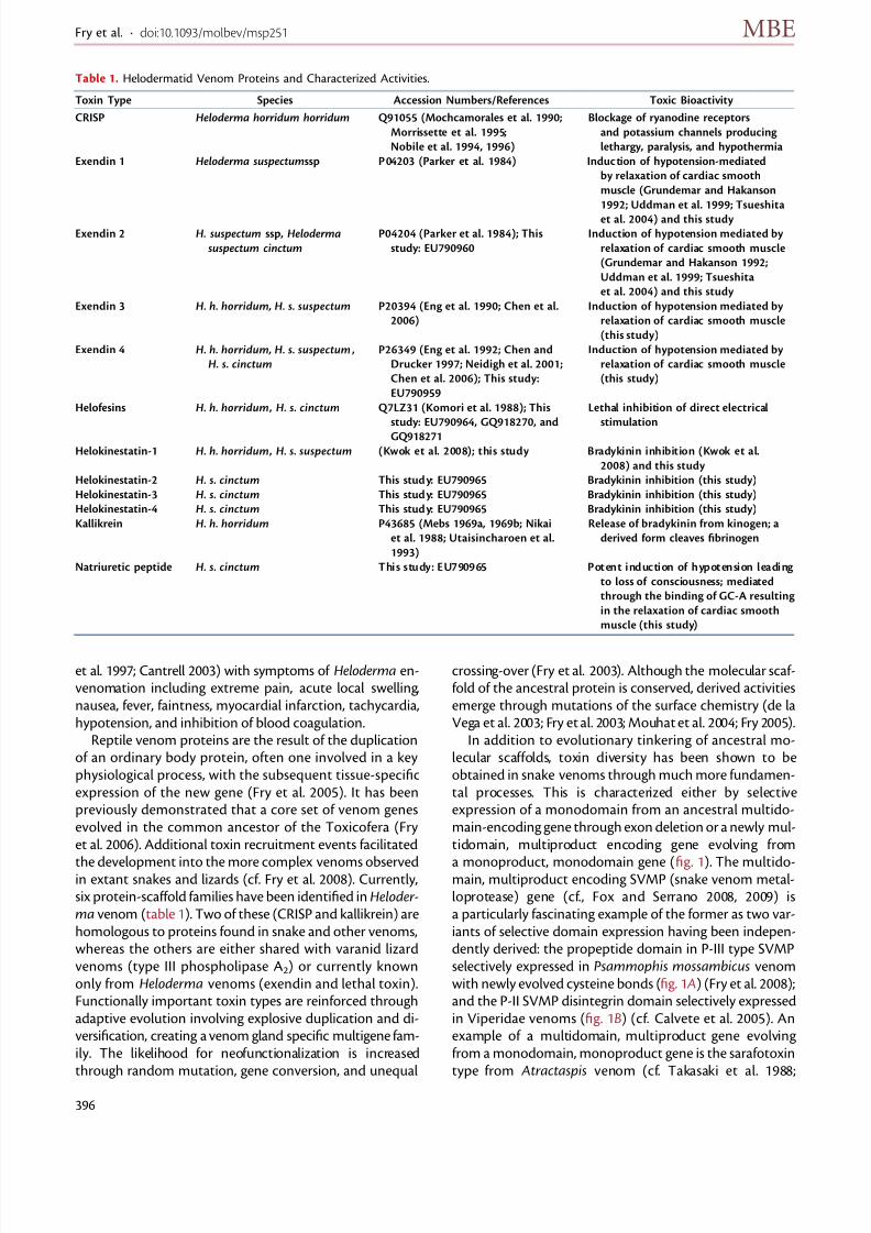
the development into the more complex venoms observedin extant snakes and lizards (cf. Fry et al. 2008). Currently,six protein-scaffold families have been identified in Heloder-ma venom (table 1). Two of these (CRISP and kallikrein) arehomologous to proteins found in snake and other venoms,whereas the others are either shared with varanid lizardvenoms (type III phospholipase A2) or currently knownonly from Heloderma venoms (exendin and lethal toxin).Functionally important toxin types are reinforced throughadaptive evolution involving explosive duplication and di-versification, creating a venom gland specific multigene fam-ily. The likelihood for neofunctionalization is increasedthrough random mutation, gene conversion, and unequal
crossing-over (Fry et al. 2003). Although the molecular scaf-fold of the ancestral protein is conserved, derived activitiesemerge through mutations of the surface chemistry (de laVega et al. 2003; Fry et al. 2003; Mouhat et al. 2004; Fry 2005).
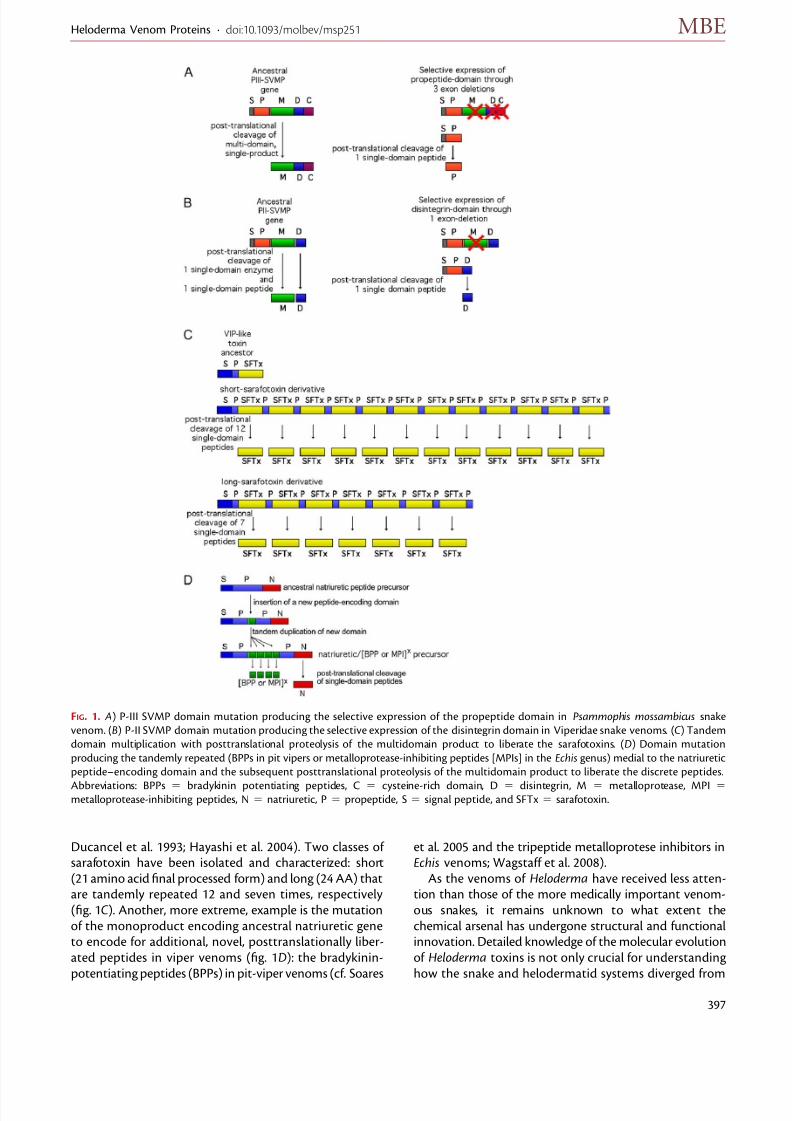
In addition to evolutionary tinkering of ancestral mo-lecular scaffolds, toxin diversity has been shown to beobtained in snake venoms through much more fundamen-tal processes. This is characterized either by selectiveexpression of a monodomain from an ancestral multido-main-encoding gene through exon deletion or a newly mul-tidomain, multiproduct encoding gene evolving from
a monoproduct, monodomain gene (fig. 1). The multido-main, multiproduct encoding SVMP (snake venom metal-loprotease) gene (cf., Fox and Serrano 2008, 2009) isa particularly fascinating example of the former as two var-iants of selective domain expression having been indepen-dently derived: the propeptide domain in P-III type SVMPselectively expressed in Psammophis mossambicus venomwith newly evolved cysteine bonds (fig. 1 A) (Fry et al. 2008);and the P-II SVMP disintegrin domain selectively expressedin Viperidae venoms (fig. 1B) (cf . Calvete et al. 2005). Anexample of a multidomain, multiproduct gene evolvingfrom a monodomain, monoproduct gene is the sarafotoxintype from Atractaspis venom (cf. Takasaki et al. 1988;
Table 1. Helodermatid Venom Proteins and Characterized Activities.
Toxin Type Species Accession Numbers/References Toxic Bioactivity
CRISP Heloderma horridum horridum Q91055 (Mochcamorales et al. 1990;
Morrissette et al. 1995;
Nobile et al. 1994, 1996)
Blockage of ryanodine receptors
and potassium channels producing
lethargy, paralysis, and hypothermia
Exendin 1 Heloderma suspectumssp P04203 (Parker et al. 1984) Induction of hypotension-mediated
by relaxation of cardiac smooth
muscle (Grundemar and Hakanson
1992; Uddman et al. 1999; Tsueshita
et al. 2004) and this studyExendin 2 H. suspectum ssp, Heloderma
suspectum cinctum
P04204 (Parker et al. 1984); This
study: EU790960
Induction of hypotension mediated by
relaxation of cardiac smooth muscle
(Grundemar and Hakanson 1992;
Uddman et al. 1999; Tsueshita
et al. 2004) and this study
Exendin 3 H. h. horridum, H. s. suspectum P20394 (Eng et al. 1990; Chen et al.
2006)
Induction of hypotension mediated by
relaxation of cardiac smooth muscle
(this study)
Exendin 4 H. h. horridum, H. s. suspectum ,
H. s. cinctum
P26349 (Eng et al. 1992; Chen and
Drucker 1997; Neidigh et al. 2001;
Chen et al. 2006); This study:
EU790959
Induction of hypotension mediated by
relaxation of cardiac smooth muscle
(this study)
Helofesins H. h. horridum , H. s. cinctum Q7LZ31 (Komori et al. 1988); This
study: EU790964, GQ918270, and
GQ918271
Lethal inhibition of direct electrical
stimulation
Helokinestatin-1 H. h. horridum , H. s. suspectum (Kwok et al. 2008); this study Bradykinin inhibition (Kwok et al.
2008) and this study
Helokinestatin-2 H. s. cinctum This study: EU790965 Bradykinin inhibition (this study)
Helokinestatin-3 H. s. cinctum This study: EU790965 Bradykinin inhibition (this study)
Helokinestatin-4 H. s. cinctum This study: EU790965 Bradykinin inhibition (this study)
Kallikrein H. h. horridum P43685 (Mebs 1969a, 1969b; Nikai
et al. 1988; Utaisincharoen et al.
1993)
Release of bradykinin from kinogen; a
derived form cleaves fibrinogen
Natriuretic peptide H. s. cinctum This study: EU790965 Potent induction of hypotension leading
to loss of consciousness; mediated
through the binding of GC-A resulting
in the relaxation of cardiac smooth
muscle (this study)
Fry et al. · doi:10.1093/molbev/msp251 MBE
396
8/6/2019 2010 Fry Heloderma
http://slidepdf.com/reader/full/2010-fry-heloderma 3/13
Ducancel et al. 1993; Hayashi et al. 2004). Two classes of sarafotoxin have been isolated and characterized: short(21 amino acid final processed form) and long (24 AA) thatare tandemly repeated 12 and seven times, respectively(fig. 1C ). Another, more extreme, example is the mutationof the monoproduct encoding ancestral natriuretic geneto encode for additional, novel, posttranslationally liber-ated peptides in viper venoms (fig. 1D): the bradykinin-potentiating peptides (BPPs) in pit-viper venoms (cf. Soares
et al. 2005 and the tripeptide metalloprotese inhibitors inEchis venoms; Wagstaff et al. 2008).
As the venoms of Heloderma have received less atten-tion than those of the more medically important venom-ous snakes, it remains unknown to what extent thechemical arsenal has undergone structural and functionalinnovation. Detailed knowledge of the molecular evolutionof Heloderma toxins is not only crucial for understandinghow the snake and helodermatid systems diverged from
FIG. 1. A) P-III SVMP domain mutation producing the selective expression of the propeptide domain in Psammophis mossambicus snake
venom. (B) P-II SVMP domain mutation producing the selective expression of the disintegrin domain in Viperidae snake venoms. (C ) Tandem
domain multiplication with posttranslational proteolysis of the multidomain product to liberate the sarafotoxins. (D) Domain mutation
producing the tandemly repeated (BPPs in pit vipers or metalloprotease-inhibiting peptides [MPIs] in the Echis genus) medial to the natriuretic
peptide–encoding domain and the subsequent posttranslational proteolysis of the multidomain product to liberate the discrete peptides.
Abbreviations: BPPs 5 bradykinin potentiating peptides, C 5 cysteine-rich domain, D 5 disintegrin, M 5 metalloprotease, MPI 5metalloprotease-inhibiting peptides, N 5 natriuretic, P 5 propeptide, S 5 signal peptide, and SFTx 5 sarafotoxin.
Heloderma Venom Proteins · doi:10.1093/molbev/msp251 MBE
397
8/6/2019 2010 Fry Heloderma
http://slidepdf.com/reader/full/2010-fry-heloderma 4/13
8/6/2019 2010 Fry Heloderma
http://slidepdf.com/reader/full/2010-fry-heloderma 5/13
8/6/2019 2010 Fry Heloderma
http://slidepdf.com/reader/full/2010-fry-heloderma 6/13
Results
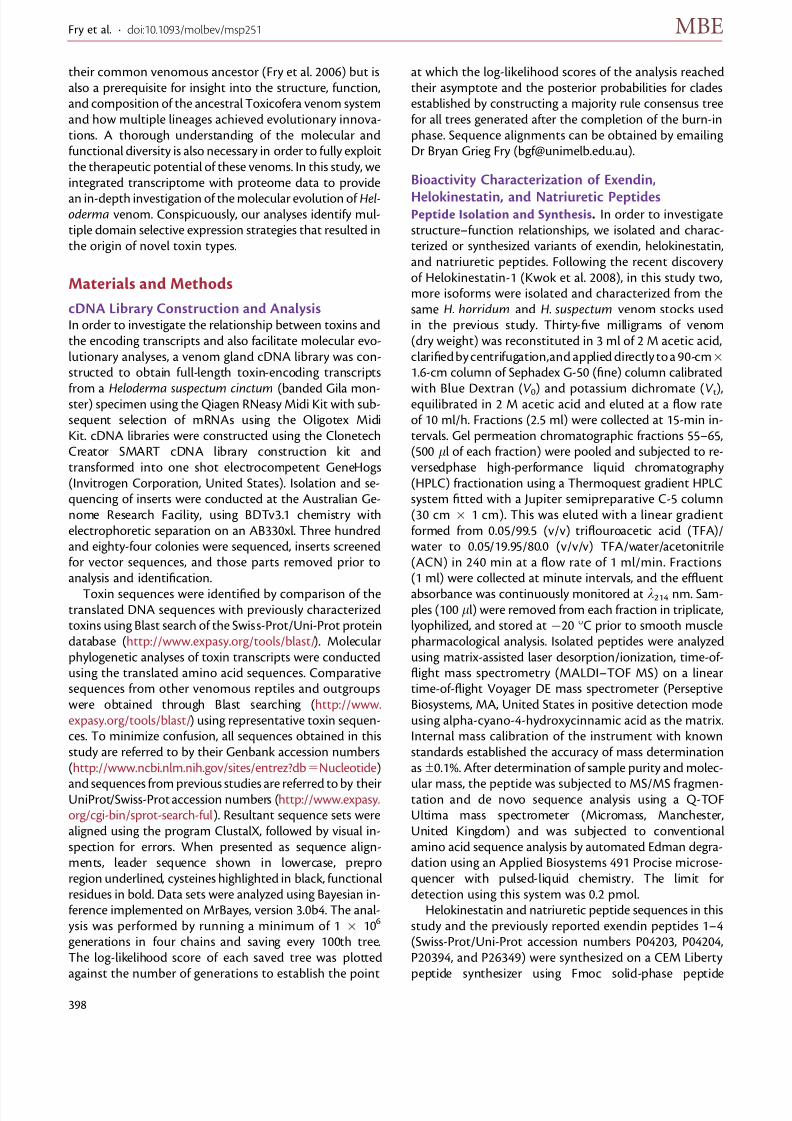
Novel Toxin Isoforms and Precursors in theHelodemma Venom TranscriptomecDNA clones were found that covered the entire precursorsequences of orthologs of the short lethal toxin-1 fragment(Q7LZ31) previously isolated from the venom of Heloder-ma horridum Horridum (Komori et al. 1988), showing it tobe constructed of beta-defensin domain repeats (fig. 2),with us thus giving this class of toxins the name ‘‘helofen-
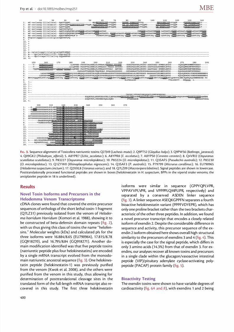
sins.’’ Molecular weights (kDa) and calculated pIs for thethree isoforms were 16.884/8.65 (EU790964), 17.815/8.78(GQ918270), and 16.795/8.84 (GQ918271). Another do-main modification identified was that five peptide toxins(natriuretic peptide plus four helokinestatins) are encodedby a single mRNA transcript evolved from the monodo-main natriuretic ancestral sequence (fig. 3). One helokines-tatin peptide (helokinestatin-1) was previously purifiedfrom the venom (Kwok et al. 2008), and the others werepurified from the venom in this study, thus allowing fordetermination of posttranslational cleavage sites in thetranslated form of the full-length mRNA transcript also re-covered in this study. The first three helokinestatin
isoforms were similar in sequence (GPPYQPLVPR,VPPAYVPLVPR, and VPPPPLQMPLIPR, respectively) andseparated by a conserved ASDEN linker sequence(fig. 3). A linker sequence ASEQKGPPFN separates a fourthbioactive helokinestatin variant (PPPFVDYEPR), which hasonly one proline bracket rather than the two brackets char-acteristic of the other three peptides. In addition, we founda novel precursor transcript that encodes a closely relatedisoform of exendin 2. Despite the considerable difference insequence and activity, this precursor sequence of the ex-
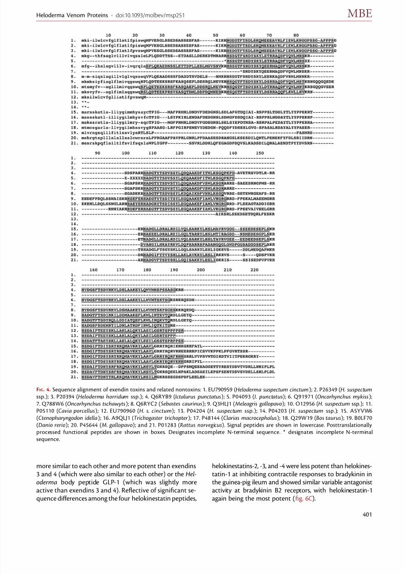
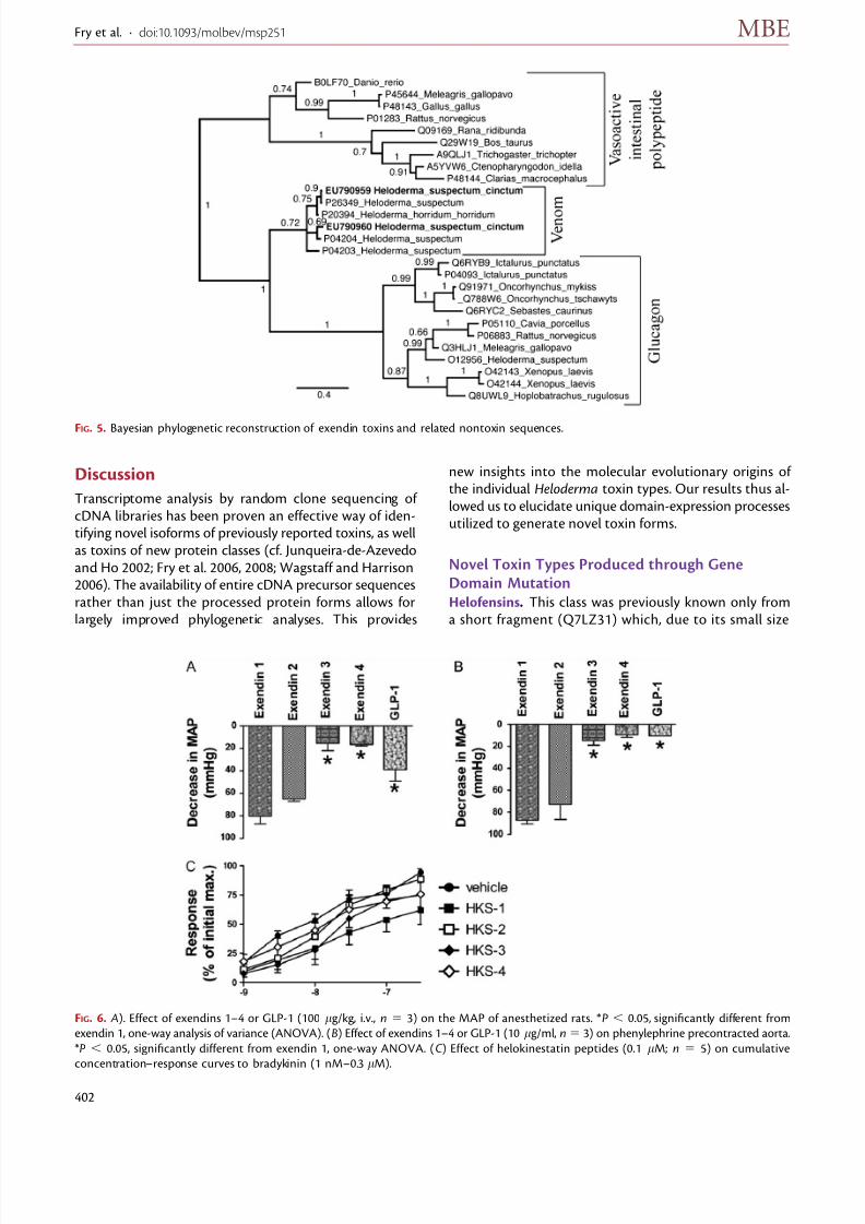
endin 2 isoform obtained here shows overall high structuralsimilarity to the precursors of exendins 3 and 4 (fig. 4). Thisis especially the case for the signal peptide, which differs inonly 3 amino acids (14.3%) from that of exendin 3. For ex-endins, our analyses recover all known toxins and precursorsin a single clade within the glucagon/vasoactive intestinalpeptide (VIP)/pituitary adenylate cyclase-activating poly-peptide (PACAP) protein family (fig. 5).
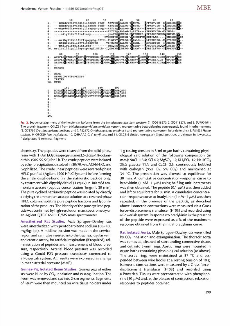
Bioactivity Testing The exendin toxins were shown to have variable degrees of cardioactivity (fig. 6 A and B), with exendins 1 and 2 being
FIG. 3. Sequence alignment of Toxicofera natriuretic toxins: Q27J49 (Lachesis muta); 2. Q9PT52 (Gloydius halys); 3. Q9PW56 (Bothrops_jararaca);
4. Q09GK2 (Philodryas_olfersii); 5. A8YPR7 (Echis_occelatus); 6. A8YPR8 (E. occelatus); 7. A8YPS0 (Cerastes cerastes); 8. Q4VRI2 (Oxyuranusscutellatus scutellatus); 9. P83227 (Oxyuranus microlepidotus); 10. P83224 (O. microlepidotus); 11. Q3SAF5 (Pseudechis australis); 12. P83230
(O. microlepidotus); 13. Q1ZYW0 (Rhinoplocephalus nigrescens); 14. Q3SAF3 (P. australis); 15. P79799 (Micrurus corallinus); 16. EU790965
(Heloderma suspectum cinctum); 17. Q2XXL8 (Varanus varius); and 18. Q7LZ09 (Macrovipera lebetina). Signal peptides are shown in lowercase.
Posttranslationally processed functional peptides are shown in boxes (helokinestatin in H. suspectum , BPPs in the viperid snake venoms; the
antiplatelet peptide in 18 is underlined).
Fry et al. · doi:10.1093/molbev/msp251 MBE
400
8/6/2019 2010 Fry Heloderma
http://slidepdf.com/reader/full/2010-fry-heloderma 7/13
8/6/2019 2010 Fry Heloderma
http://slidepdf.com/reader/full/2010-fry-heloderma 8/13
Discussion
Transcriptome analysis by random clone sequencing of cDNA libraries has been proven an effective way of iden-tifying novel isoforms of previously reported toxins, as wellas toxins of new protein classes (cf. Junqueira-de-Azevedoand Ho 2002; Fry et al. 2006, 2008; Wagstaff and Harrison2006). The availability of entire cDNA precursor sequencesrather than just the processed protein forms allows forlargely improved phylogenetic analyses. This provides
new insights into the molecular evolutionary origins of the individual Heloderma toxin types. Our results thus al-lowed us to elucidate unique domain-expression processesutilized to generate novel toxin forms.
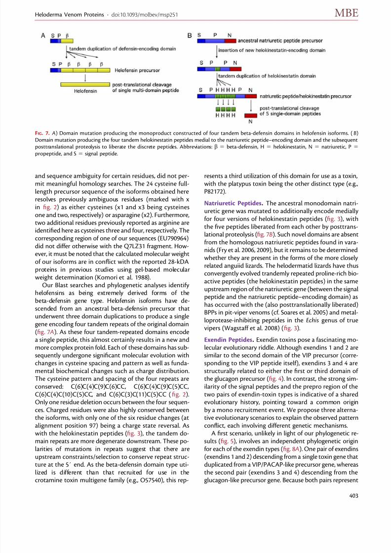
Novel Toxin Types Produced through GeneDomain MutationHelofensins. This class was previously known only froma short fragment (Q7LZ31) which, due to its small size
FIG. 5. Bayesian phylogenetic reconstruction of exendin toxins and related nontoxin sequences.
FIG. 6. A). Effect of exendins 1–4 or GLP-1 (100 lg/kg, i.v., n 5 3) on the MAP of anesthetized rats. *P , 0.05, significantly different from
exendin 1, one-way analysis of variance (ANOVA). (B) Effect of exendins 1–4 or GLP-1 (10 lg/ml, n5 3) on phenylephrine precontracted aorta.
*P , 0.05, significantly different from exendin 1, one-way ANOVA. (C ) Effect of helokinestatin peptides (0.1 lM; n 5 5) on cumulative
concentration–response curves to bradykinin (1 nM–0.3 lM).
Fry et al. · doi:10.1093/molbev/msp251 MBE
402
8/6/2019 2010 Fry Heloderma
http://slidepdf.com/reader/full/2010-fry-heloderma 9/13
and sequence ambiguity for certain residues, did not per-mit meaningful homology searches. The 24 cysteine full-
length precursor sequence of the isoforms obtained hereresolves previously ambiguous residues (marked with xin fig. 2) as either cysteines (x1 and x3 being cysteinesone and two, respectively) or asparagine (x2). Furthermore,two additional residues previously reported as arginine areidentified here as cysteines three and four, respectively. Thecorresponding region of one of our sequences (EU790964)did not differ otherwise with the Q7LZ31 fragment. How-ever, it must be noted that the calculated molecular weightof our isoforms are in conflict with the reported 28-kDAproteins in previous studies using gel-based molecularweight determination (Komori et al. 1988).
Our Blast searches and phylogenetic analyses identify
helofensins as being extremely derived forms of thebeta-defensin gene type. Helofensin isoforms have de-scended from an ancestral beta-defensin precursor thatunderwent three domain duplications to produce a singlegene encoding four tandem repeats of the original domain(fig. 7 A). As these four tandem-repeated domains encodea single peptide, this almost certainly results in a new andmore complex protein fold. Each of these domains has sub-sequently undergone significant molecular evolution withchanges in cysteine spacing and pattern as well as funda-mental biochemical changes such as charge distribution.The cysteine pattern and spacing of the four repeats are
conserved: C(6)C(4)C(9)C(6)CC, C(6)C(4)C(9)C(5)CC,C(6)C(4)C(10)C(5)CC, and C(6)C(3)C(11)C(5)CC (fig. 2).Only one residue deletion occurs between the four sequen-ces. Charged residues were also highly conserved betweenthe isoforms, with only one of the six residue changes (atalignment position 97) being a charge state reversal. Aswith the helokinestatin peptides (fig. 3), the tandem do-main repeats are more degenerate downstream. These po-larities of mutations in repeats suggest that there areupstream constraints/selection to conserve repeat struc-ture at the 5# end. As the beta-defensin domain type uti-lized is different than that recruited for use in thecrotamine toxin multigene family (e.g., O57540), this rep-
resents a third utilization of this domain for use as a toxin,with the platypus toxin being the other distinct type (e.g.,
P82172).Natriuretic Peptides. The ancestral monodomain natri-uretic gene was mutated to additionally encode mediallyfor four versions of helokinestatin peptides (fig. 3), withthe five peptides liberated from each other by posttrans-lational proteolysis (fig. 7B). Such novel domains are absentfrom the homologous natriuretic peptides found in vara-nids (Fry et al. 2006, 2009), but it remains to be determinedwhether they are present in the forms of the more closelyrelated anguiid lizards. The helodermatid lizards have thusconvergently evolved trandemly repeated proline-rich bio-active peptides (the helokinestatin peptides) in the same
upstream region of the natriuretic gene (between the signalpeptide and the natriuretic peptide–encoding domain) ashas occurred with the (also posttranslationally liberated)BPPs in pit-viper venoms (cf. Soares et al. 2005) and metal-loprotease-inhibiting peptides in the Echis genus of truevipers (Wagstaff et al. 2008) (fig. 3).
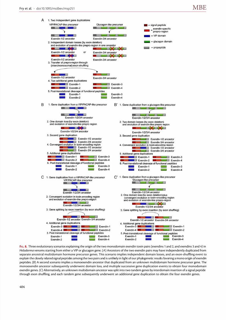
Exendin Peptides. Exendin toxins pose a fascinating mo-lecular evolutionary riddle. Although exendins 1 and 2 aresimilar to the second domain of the VIP precursor (corre-sponding to the VIP peptide itself), exendins 3 and 4 arestructurally related to either the first or third domain of the glucagon precursor (fig. 4). In contrast, the strong sim-
ilarity of the signal peptides and the prepro region of thetwo pairs of exendin-toxin types is indicative of a sharedevolutionary history, pointing toward a common originby a mono recruitment event. We propose three alterna-tive evolutionary scenarios to explain the observed patternconflict, each involving different genetic mechanisms.
A first scenario, unlikely in light of our phylogenetic re-sults (fig. 5), involves an independent phylogenetic originfor each of the exendin types (fig. 8 A). One pair of exendins(exendins 1 and 2) descending from a single toxin gene thatduplicated from a VIP/PACAP-like precursor gene, whereasthe second pair (exendins 3 and 4) descending from theglucagon-like precursor gene. Because both pairs represent
FIG. 7. A) Domain mutation producing the monoproduct constructed of four tandem beta-defensin domains in helofensin isoforms. (B)
Domain mutation producing the four tandem helokinestatin peptides medial to the natriuretic peptide–encoding domain and the subsequent
posttranslational proteolysis to liberate the discrete peptides. Abbreviations: b 5 beta-defensin, H 5 helokinestatin, N 5 natriuretic, P 5
propeptide, and S 5 signal peptide.
Heloderma Venom Proteins · doi:10.1093/molbev/msp251 MBE
403
8/6/2019 2010 Fry Heloderma
http://slidepdf.com/reader/full/2010-fry-heloderma 10/13
FIG. 8. Three evolutionary scenarios explaining the origin of the two monodomain exendin toxin pairs (exendins 1 and 2, and exendins 3 and 4) in
Heloderma venoms starting from either a VIP or glucagon gene. ( A) Ancestors of the two exendin pairs may have independently duplicated from
separate ancestral multidomain hormone precursor genes. This scenario implies independent domain losses, and an exon-shuffling event to
explain the closely related signal peptides among the two pairs and is unlikely in light of our phylogenetic results favoring a mono origin of exendin
peptides. (B) A second scenario implies a monoexendin ancestor that duplicated from an unknown multidomain hormone precursor gene. The
monoexendin ancestor subsequently underwent domain loss, and multiple successive gene duplication events to obtain four monodomain
exendin genes. (C ) Alternatively, an unknown multidomain ancestor was split into two tandem genes by interdomain insertion of a signal peptide
through exon shuffling, and each tandem gene subsequently underwent an additional gene duplication to obtain the four exendin genes.
Fry et al. · doi:10.1093/molbev/msp251 MBE
404
8/6/2019 2010 Fry Heloderma
http://slidepdf.com/reader/full/2010-fry-heloderma 11/13
truncated (monodomain) precursors while all known hor-mone precursors in this protein family have multipledomains (VIP/PACAP precursors typically contain two do-mains; glucagons precursors typically have three), this im-plies that their respective ancestral genes independentlylost one and two domain-encoding exons, respectively,to obtain their monodomain-encoding genes. In addition,dual origins would imply that the nearly identical signal
peptides shared by all the exending peptides resulted froman exon-shuffling event, that is, the transfer of the signalpeptide-encoding exon from one ancestral exendin to thatof the other, followed by another duplication round foreach exendin type. The most common mechanisms cur-rently implicated to explain exon shuffling in eukaryotesare interlocus gene conversion (nonreciprocal recombina-tion) and retrotransposition. Recent studies have suggestedthat interlocus gene conversion is most common betweenlinked loci (on the same chromosome), and its frequency isinversely proportional to the distance between the in-volved loci (Ezawa et al. 2006). However, if the two pairs
of exendin evolved independently from VIP and glucagonancestors, it is unlikely that they are spatially linked. Screen-ing of the genome maps of various vertebrates reveals thatthe glucagon and VIP genes are situated on differentchromosomes in zebra fish (chromosomes 22 and 13, re-spectively), chicken (chromosomes 7 and 3), rat (chromo-somes 3 and 1), and human (chromosomes 2 and 6),suggesting an ancient genomic separation of the two genes.This would imply that the VIP and glucagon genes (and anyrecently derived paralogs) are likely to lie on different chro-mosomes in Heloderma as well. An exon shuffling origin forthe exendin signal peptides would therefore imply an in-terchromosomal (ectopic) gene-conversion event or a ret-
rotransposition event. In summary, a dual origin of the twoexendin gene pairs would not only imply two independenttoxin recruitment events and subsequent domain lossesbut also an extremely rare interchromosomal exon-shuffling event.
A second scenario implicates a single evolutionary originof exendins, as suggested by our phylogenetic analyses, withthe sole founding gene either VIP (fig. 8B) or glucagon de-rived (fig. 8B#). Under this scenario, a single ancestral mul-tidomain precursor gene lost one (VIP as the startingmaterial) or two domains (glucagon as the starting mate-rial) through exon deletion(s). The structural similarities of
the two exendin pairs with different hormone peptidessuggest substantial convergent evolution within the pep-tide-encoding regions to explain their present structuralvariation. The timing of this would be best explained asoccurring to one duplicate after the first gene duplicationround subsequent to the toxin recruitment event, withanother round of gene duplication producing the twoexendin pairs
The third scenario, like scenario two, also implicatesa single origin of exendins, again consistent with our phy-logenetic analyses. This scenario involves a different typeof domain mutation to explain the currently observedpeptide variation, again with the sole founding gene either
VIP (fig. 8C ) or glucagon derived (fig. 8C#). Under thesescenarios, the single ancestral multidomain precursorgene was split into two tandem genes by insertion of its signal peptide in between two of the peptide-encodingdomains through exon shuffling (e.g., as a result of un-equal crossing-over). The two tandem genes subsequentlyduplicated again, each giving rise to one of the presentlyobserved exendin pairs. This scenario as well requires sub-
stantial convergent amino acid substitutions to explaintheir similarities with different hormones, with the timingoccurring either just before or just after the gene splittingbut before the first gene duplication round subsequent tothe toxin recruitment event. This scenario would also re-quire an exon deletion if glucagon is the starting material(fig. 8C #).
The first scenario is excluded by our phylogenetic anal-yses and also the unlikeliness of interchromosomal exonshuffling. However, the final distinction between the sec-ond and third scenarios will require insights into the geno-mic organization of exendins and their closely related
glucagon family genes. Regardless, both the second andthird scenarios would require that the ancestor of at leastone of the two exendin pairs underwent substantial con-vergent evolution with either of the hormone clades to ob-tain their present structural variation. Endogenous VIPhormones have been shown to be more potent vasodila-tors than endogenous glucagon hormones (Ezawa et al.2006). As a consequence, a peptide that mimics a general-ized VIP hormone may provide a more effective toxin. Theresults of this study demonstrate that exendins 1 and 2have a higher cardioactivity than exendins 3 and 4 and alsothe nontoxin peptide GLP-1 (O12956) (fig. 6 A and B). Thisis in agreement with other studies on exendins 1 and 2 that
demonstrated vasodilatory actions similar to VIP (Uddmanet al. 1999; Tsueshita et al. 2004). The molecular evolutionfrom a less potent (glucagon-like) form toward a more po-tent (VIP-like) form is more plausible than the converse.The origin of exendin peptides thus favors a glucagon-likeancestor with subsequent divergence of one duplicate toconvergently become a VIP-like toxin (fig. 8B# or C #).The timing of these molecular evolutionary events howeverremains to be resolved. Glucagon-like exendins 3 and 4 havebeen isolated from both H. suspectum and H. horridum ,whereas the VIP-like 1 and 2 have only been isolated fromH. suspectum. This would thus favor a basal glucagon-
derived exendin type as shown in figure 8B#
with a sub-sequent VIP-like convergence specific to H. suspectum.However, the absence of evidence is not necessarilyevidence of absence. Thus, exhaustive sequencing of theH. horridum venom transcriptome must be undertakenbefore the concluded absence of the VIP-like exendins1 and 2 peptides can be considered as adequately sup-ported. If exendins 1 and 2 peptides are in fact presentin the venom of H. horridum , this would still supporta glucagon basal peptide but would not allow for distin-guishing between scenario B# or C# in figure 8 withoutgenomic understanding of the toxins and their nontoxinancestors.
Heloderma Venom Proteins · doi:10.1093/molbev/msp251 MBE
405
8/6/2019 2010 Fry Heloderma
http://slidepdf.com/reader/full/2010-fry-heloderma 12/13
8/6/2019 2010 Fry Heloderma
http://slidepdf.com/reader/full/2010-fry-heloderma 13/13
phylogenetic analysis of toxin sequences and related body
proteins. Genome Res. 15:403–420.
Fry BG, Scheib H, van der Weerd L, Young B, McNaughtan J,
Ramjan SFR, Vidal N, Poelmann RE, Norman JA. 2008. Evolution
of an arsenal. Mol Cell Proteom. 7:215–246.
Fry BG, Vidal N, Norman JA, et al. (14 co-authors). 2006. Early evolution
of the venom system in lizards and snakes. Nature 439:584–588.
Fry BG, Wickramaratana JC, Lemme S, Beuve A, Garbers D,
Hodgson WC, Alewood P. 2005. Novel natriuretic peptides
from the venom of the inland taipan (Oxyuranus micro-lepidotus): isolation, chemical and biological characterisation.
Biochem Biophys Res Commun. 327:1011–1015.
Fry BG, Wroe S, Teeuwisse W, et al. (27 co-authors). 2009. A central
role for venom in predation by Varanus komodoensis (Komodo
Dragon) and the extinct giant Varanus (Megalania) priscus. ProcNatl Acad Sci USA. 106:8969–8974.
Fry BG, Wuster W, Kini RM, Brusic V, Khan A, Venkataraman D,
Rooney AP. 2003. Molecular evolution and phylogeny of elapid
snake venom three-finger toxins. J Mol Evol. 57:110–129.
Grundemar L, Hakanson R. 1992. Unlike vip, the vip-related peptides
pacap, helodermin and helospectin suppress electrically evoked
contractions of rat vas-deferens. Regul Peptides. 40:331–337.
Hayashi MAF, Ligny-Lemaire C, Wollberg Z, et al. (12 co-authors).
2004. Long-sarafotoxins: characterization of a new family of endothelin-like peptides. Peptides 25:1243–1251.
Hooker KR, Caravati EM. 1994. Gila monster envenomation. AnnEmerg Med. 24:731–735.
Junqueira-de-Azevedo IDM, Ho PL. 2002. A survey of gene
expression and diversity in the venom glands of the pitviper
snake Bothrops insularis through the generation of expressed
sequence tags (ESTs). Gene 299:279–291.
Komori Y, Nikai T, Sugihara H. 1988. Purification and characteriza-
tion of a lethal toxin from the venom of Heloderma horridumhorridum. Biochem Biophys Res Commun. 154:613–619.
Kwok HF, Chen T, O’Rourke M, Ivanyi C, Hirst D, Shaw C. 2008.
Helokinestatin: a new bradykinin B-2 receptor antagonist
decapeptide from lizard venom. Peptides 29:65–72.
Mebs D. 1969a. Purification and properties of a kinin liberating
enzyme from venom of Heloderma suspectum. Naunyn-Schmiedebergs Archiv Pharmakol. 264:280.
Mebs D. 1969b. Isolation and properties of kallikrein from venom of
gila monster (Heloderma suspectum). Hoppe-Seylers Z PhysiolChem. 350:821.
Mochcamorales J, Martin BM, Possani LD. 1990. Isolation and
characterization of helothermine, a novel toxin from Heloderma
horridum horridum (Mexican beaded lizard) venom. Toxicon28:299–309.
Morrissette J, Kratzschmar J, Haendler B, et al. (11 co-authors).
1995. PRIMARY Structure and properties of helothermine,
a peptide toxin that blocks ryanodine receptors. Biophys J. 68:
2280–2288.
Mouhat S, Jouirou B, Mosbah A, De Waard M, Sabatier JM. 2004.
Diversity of folds in animal toxins acting on ion channels.
Biochem J. 378:717–726.
Neidigh JW, Fesinmeyer RM, Prickett KS, Andersen NH. 2001.
Exendin-4 and glucagon-like-peptide-1: NMR structural compar-
isons in the solution and micelle-associated states. Biochemistry40:13188–13200.
Nikai T, Imai K, Sugihara H, Tu AT. 1988. Isolation and
characterization of horridum toxin with arginine ester hydrolase
activity from Heloderma horridum (beaded lizard) venom. ArchBiochem Biophys. 264:270–280.
Nobile M, Magnelli V, Lagostena L, Mochcamorales J, Possani LD,
Prestipino G. 1994. The toxin helothermine affects potassiumcurrents in newborn rat cerebellar granule cells. J Membr Biol.139:49–55.
Nobile M, Noceti F, Prestipino G, Possani LD. 1996. Helothermine,
a lizard venom toxin, inhibits calcium current in cerebellar
granules. Exp Brain Res. 110:15–20.
Parker DS, Raufman JP, Odonohue TL, Bledsoe M, Yoshida H,
Pisano JJ. 1984. Amino-acid-sequences of helospectins, new
members of the glucagon superfamily, found in gila monster
venom. J Biol Chem. 259:1751–1755.
Soares MR, Oliveira-Carvalho AL, Wermelinger LS, Zingali RB, Ho PL,
Junqueira-de-Azevedo IDM, Diniz MRV. 2005. Identification of
novel bradykinin-potentiating peptides and C-type natriuretic
peptide from Lachesis muta venom. Toxicon 46:31–38.
Strimple PD, Tomassoni AJ, Otten EJ, Bahner D. 1997. Report onenvenomation by a Gila monster (Heloderma suspectum) with
a discussion of venom apparatus, clinical findings, and
treatment. Wild Environ Med. 8:111–116.
Takasaki C, Tamiya N, Bdolah A, Wollberg Z, Kochva E. 1988.
Sarafotoxins-s6-several isotoxins from Atractaspis engaddensis(burrowing asp) venom that affect the heart. Toxicon 26:
543–548.
Tsueshita T, Onyukusel H, Sethi V, Gandhi S, Rubinstein I. 2004.
Helospectin I and II evoke vasodilation in the intact peripheral
microcirculation. Peptides 25:65–69.
Uddman R, Goadsby PJ, Jansen-Olesen I, Edvinsson L. 1999.
Helospectin-like peptides: immunochemical localization and
effects on isolated cerebral arteries and on local cerebral blood
flow in the cat.J Cereb Blood Flow Metab
. 19:61–67.
Utaisincharoen P, Mackessy SP, Miller RA, Tu AT. 1993. Complete
primary structure and biochemical-properties of gilatoxin,
a serine-protease with kallikrein-like and angiotensin-degrading
activities. J Biol Chem. 268:21975–21983.
Vidal N, Hedges SB. 2005. The phylogeny of squamate reptiles
(lizards, snakes, and amphisbaenians) inferred from nine nuclear
protein-coding genes. C R Biol. 328:1000–1008.
Wagstaff SC, Favreau P, Cheneval O, Laing GD, Wilkinson MC,
Miller RL, Stocklin R, Harrison RA. 2008. Molecular character-
isation of endogenous snake venom metalloproteinase inhib-
itors. Biochem Biophys Res Commun. 365:650–656.
Wagstaff SC, Harrison RA. 2006. Venom gland EST analysis of the
saw-scaled viper, Echis ocellatus , reveals novel alpha(9)beta(1)
integrin-binding motifs in venom metalloproteinases and a new
group of putative toxins, renin-like aspartic proteases. Gene
377:21–32.
Heloderma Venom Proteins · doi:10.1093/molbev/msp251 MBE