Embed Size (px)
Citation preview
Contact: Dr. Bernhard Steiner, Médecin Adjoint, Children‘s [email protected]
Chinderarztpraxis, Ruopigenring 37, 6015 [email protected]
Clinical approach to the dysmorphic newborn
Genetics in Neonatology
Cantonal Hospital Lucerne, 15th of January 2013
Clinical approach to the dysmorphic newborn
�Major congenital anomalies
At birth: 2 – 3 %At age 5: 4 – 6 %
� Minor congenital anomalies
At birth: 15 %
Major versus minor anomalies
From J. Graham: Smith‘s Recognizable Patterns of Human Deformation (2007)
Major malformations.
Those that have medical & /or social implications.
Often require surgical repair.
Minor malformations.
Have sometimes cosmetic significance.
Normal variants.
The importance of minor anomalies
From J. Graham: Smith‘s Recognizable Patterns of Human Deformation (2007)
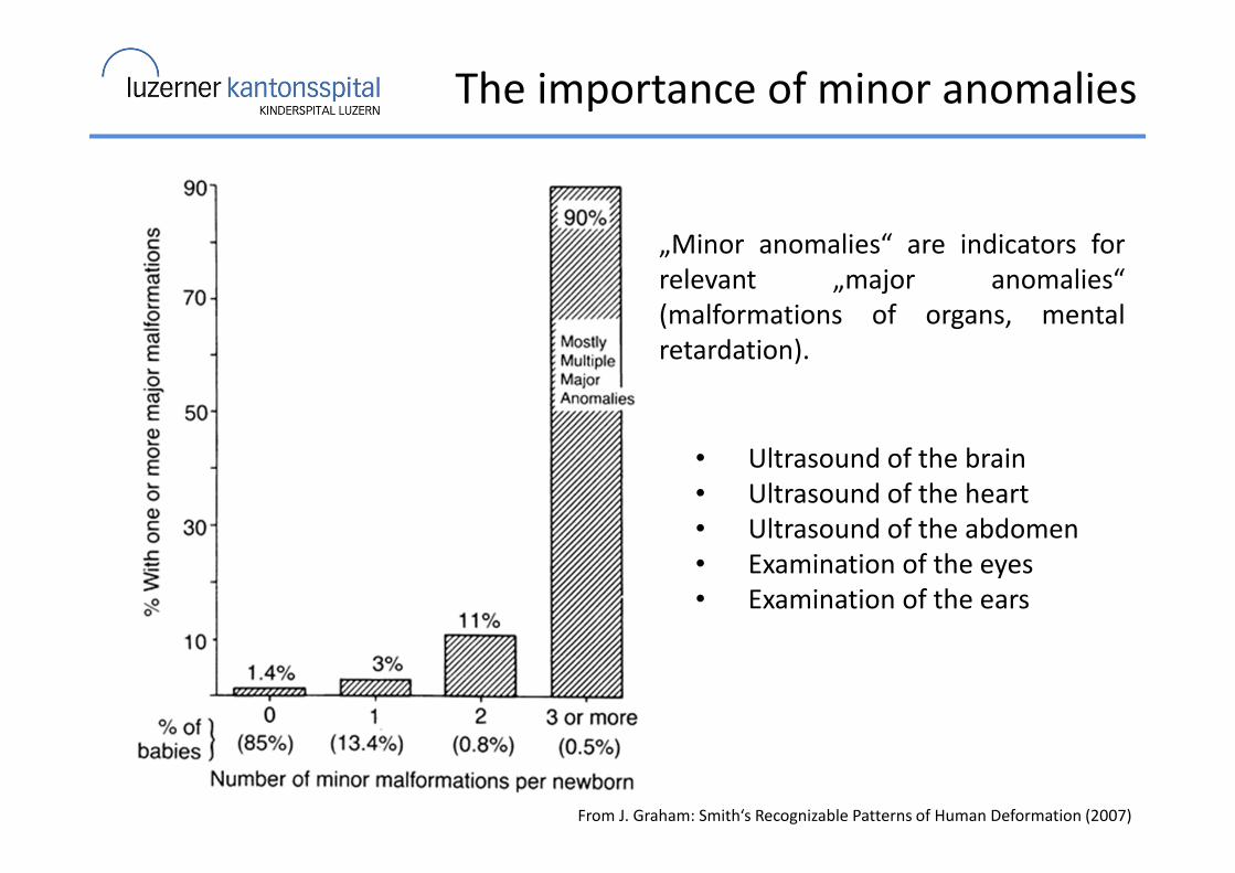
„Minor anomalies“ are indicators for
relevant „major anomalies“
(malformations of organs, mental
retardation).
• Ultrasound of the brain
• Ultrasound of the heart
• Ultrasound of the abdomen
• Examination of the eyes
• Examination of the ears
Causes of human malformations
From Stevenson and Hall: Human Malformations And Related Anomalies (2006)
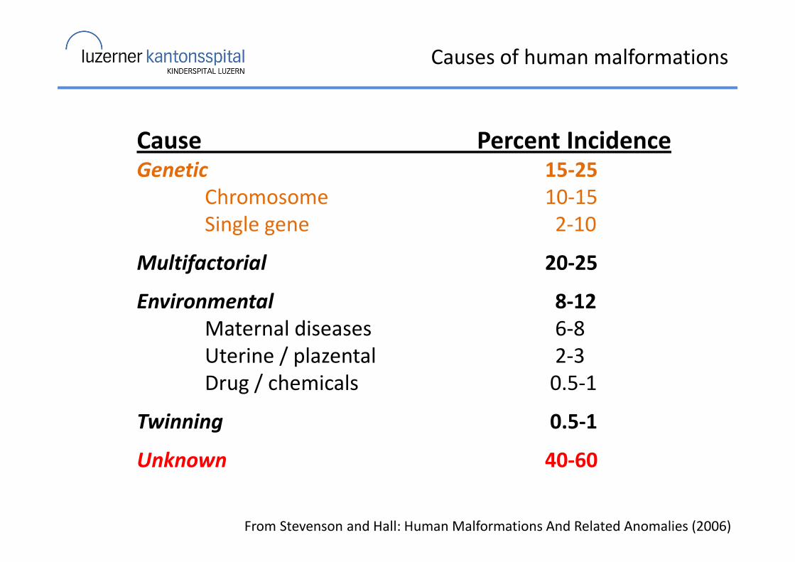
Cause Percent IncidenceGenetic 15-25
Chromosome 10-15
Single gene 2-10
Multifactorial 20-25
Environmental 8-12
Maternal diseases 6-8
Uterine / plazental 2-3
Drug / chemicals 0.5-1
Twinning 0.5-1
Unknown 40-60
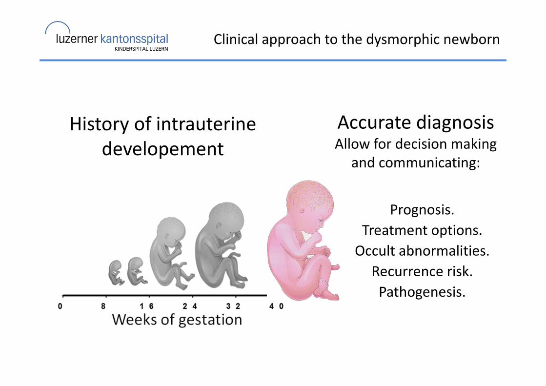
Clinical approach to the dysmorphic newborn
History of intrauterine
developement
Accurate diagnosisAllow for decision making
and communicating:
Prognosis.
Treatment options.
Occult abnormalities.
Recurrence risk.
Pathogenesis.
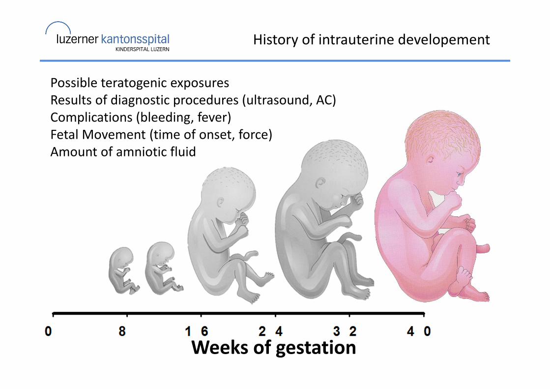
History of intrauterine developement
Weeks of gestation
Possible teratogenic exposures
Results of diagnostic procedures (ultrasound, AC)
Complications (bleeding, fever)
Fetal Movement (time of onset, force)
Amount of amniotic fluid
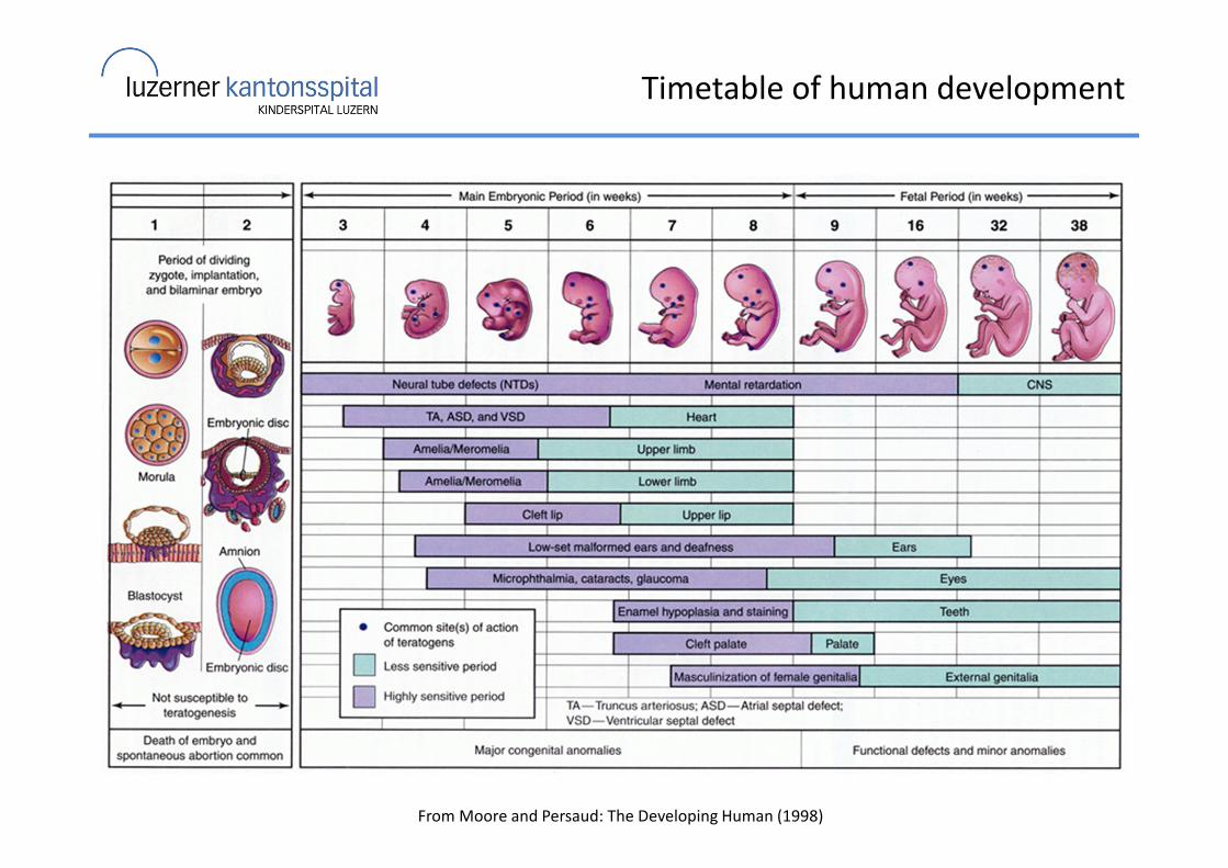
Timetable of human development
From Moore and Persaud: The Developing Human (1998)
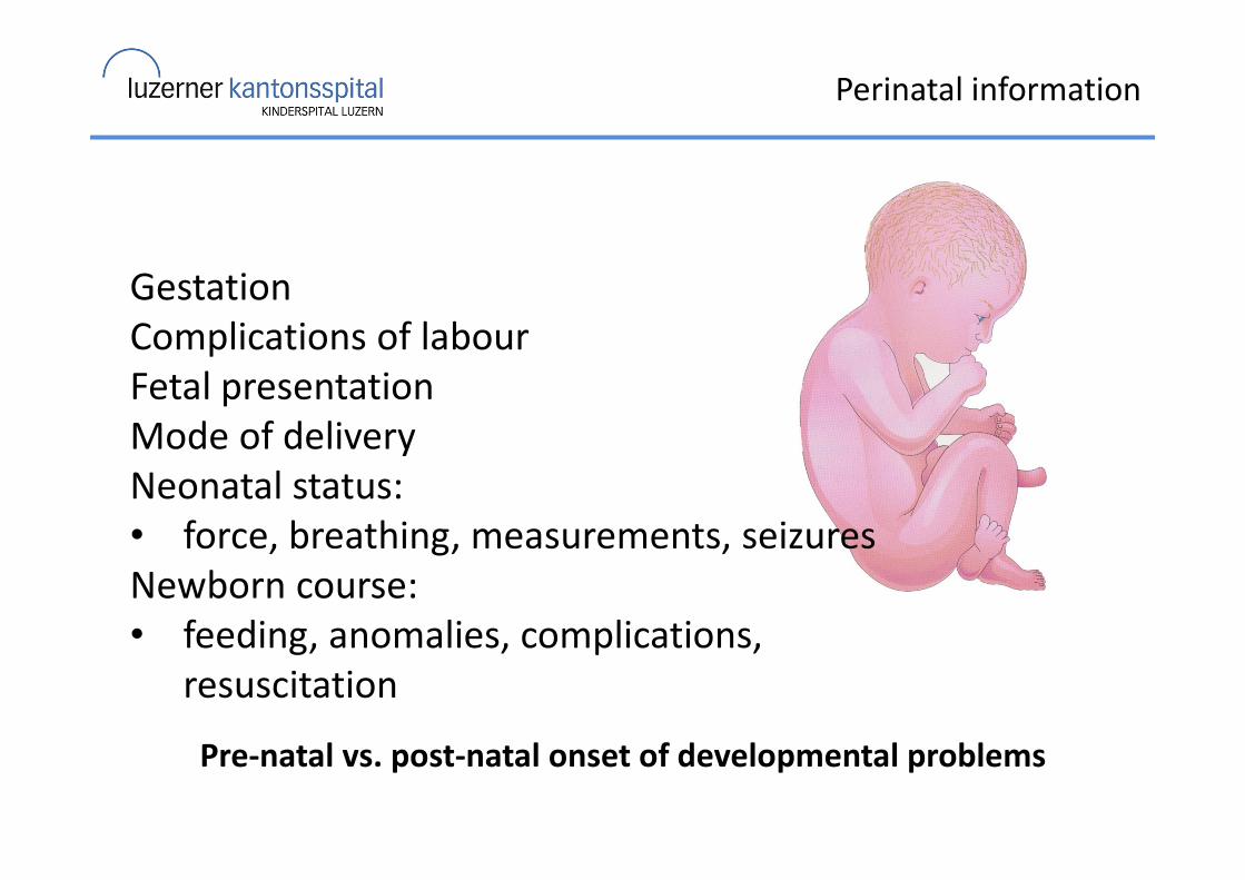
Perinatal information
Gestation
Complications of labour
Fetal presentation
Mode of delivery
Neonatal status:
• force, breathing, measurements, seizures
Newborn course:
• feeding, anomalies, complications,
resuscitation
Pre-natal vs. post-natal onset of developmental problems
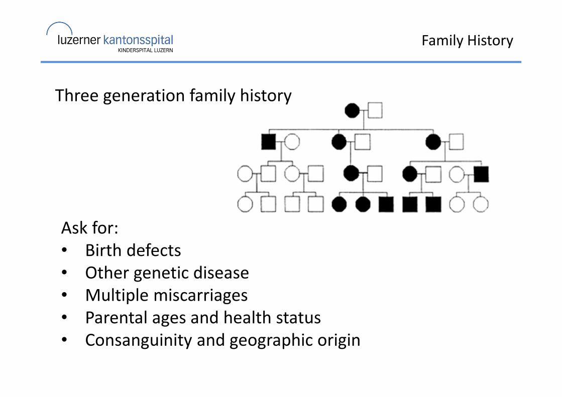
Family History
Ask for:
• Birth defects
• Other genetic disease
• Multiple miscarriages
• Parental ages and health status
• Consanguinity and geographic origin
Three generation family history
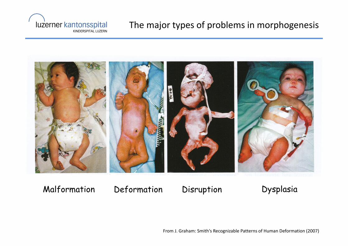
The major types of problems in morphogenesis
Malformation Deformation Disruption Dysplasia
From J. Graham: Smith‘s Recognizable Patterns of Human Deformation (2007)
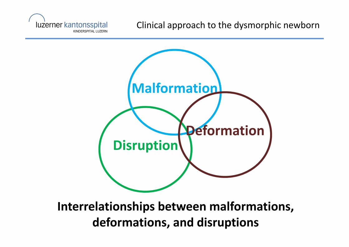
Clinical approach to the dysmorphic newborn
Disruption
Interrelationships between malformations,
deformations, and disruptions
Malformation
Deformation
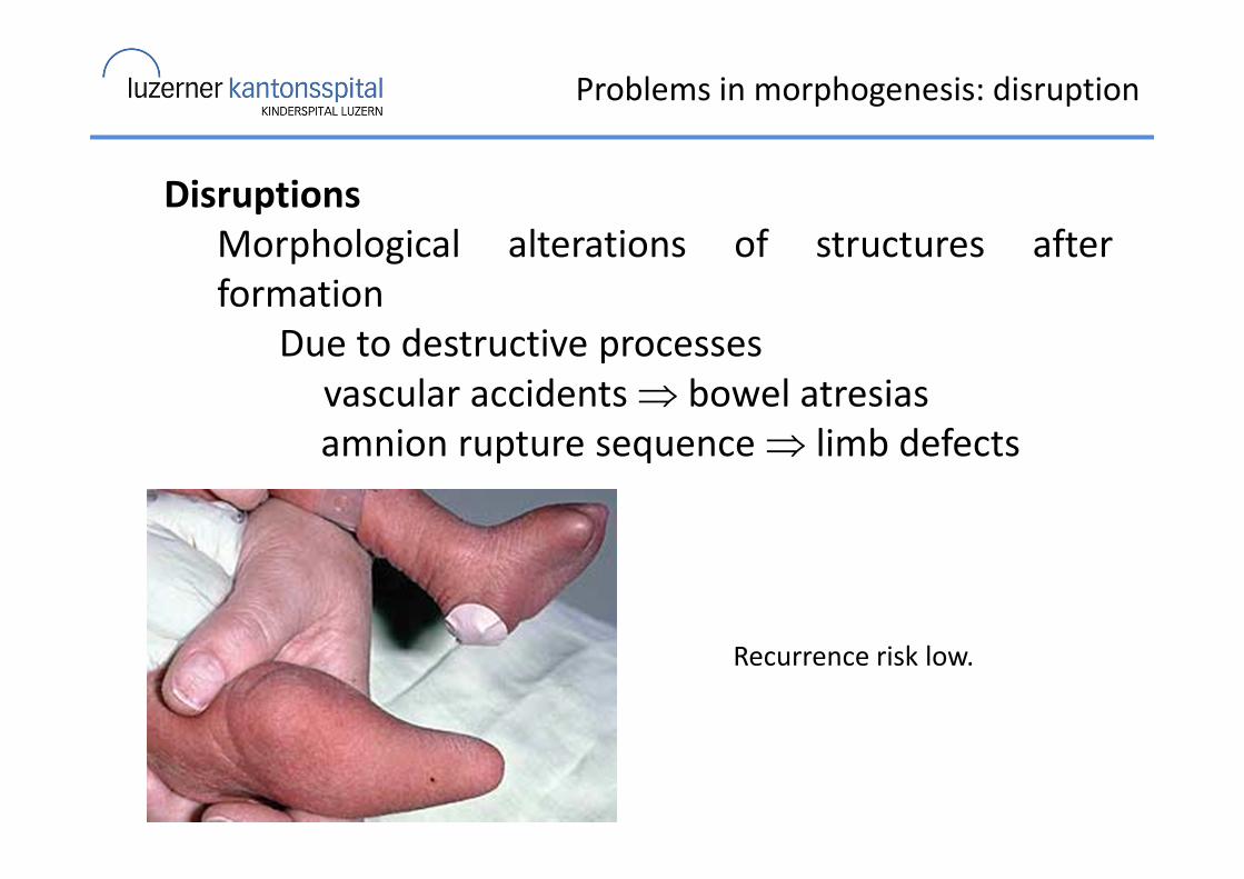
Problems in morphogenesis: disruption
Disruptions
Morphological alterations of structures after
formation
Due to destructive processes
vascular accidents ⇒ bowel atresias
amnion rupture sequence ⇒ limb defects
Recurrence risk low.
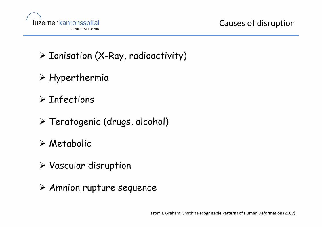
Causes of disruption
From J. Graham: Smith‘s Recognizable Patterns of Human Deformation (2007)
� Ionisation (X-Ray, radioactivity)
� Hyperthermia
� Infections
� Teratogenic (drugs, alcohol)
� Metabolic
� Vascular disruption
� Amnion rupture sequence
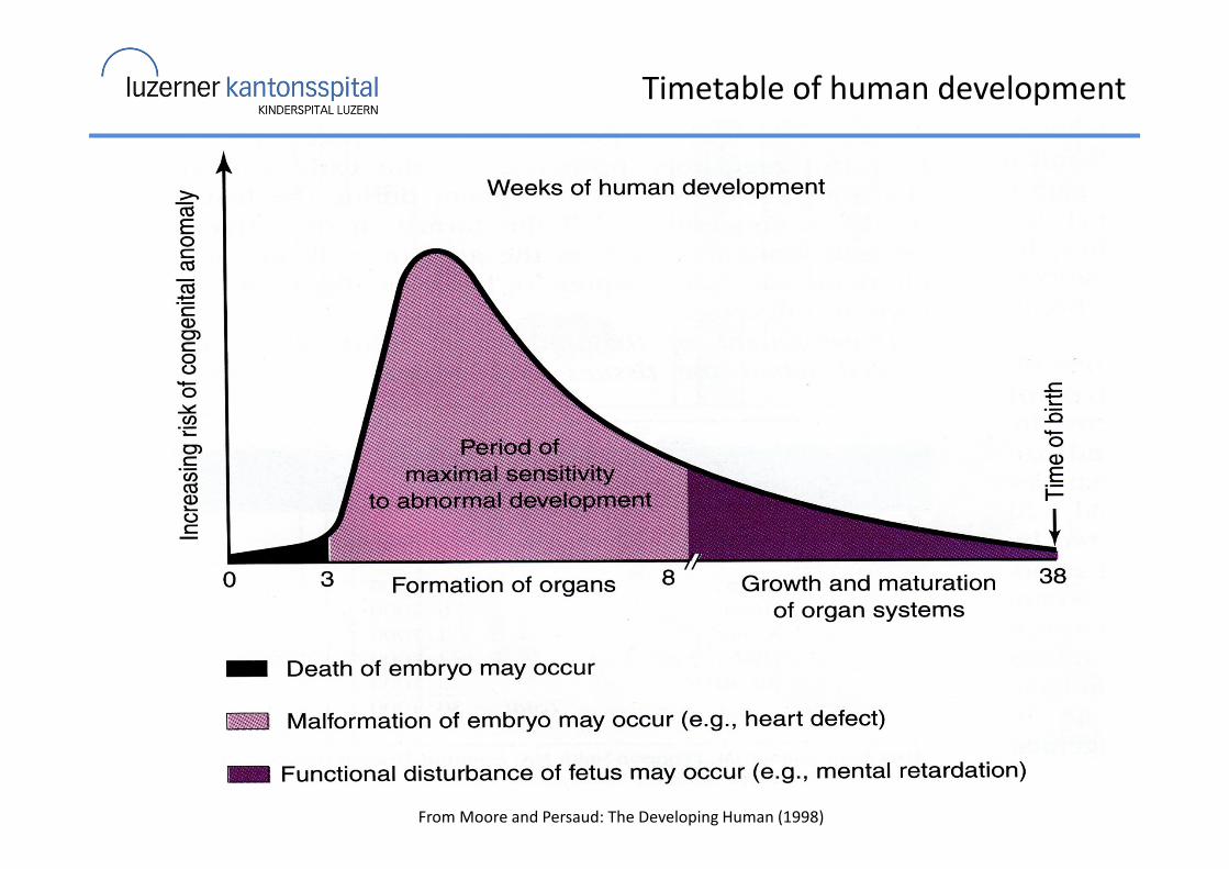
Timetable of human development
From Moore and Persaud: The Developing Human (1998)
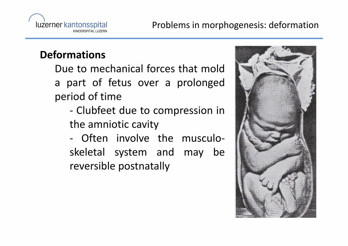
Problems in morphogenesis: deformation
Deformations
Due to mechanical forces that mold
a part of fetus over a prolonged
period of time
- Clubfeet due to compression in
the amniotic cavity
- Often involve the musculo-
skeletal system and may be
reversible postnatally
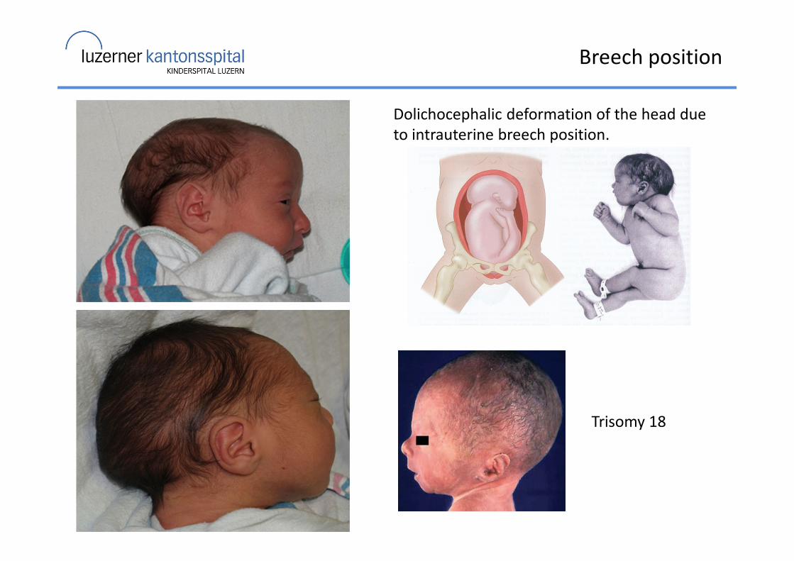
Breech position
Dolichocephalic deformation of the head due
to intrauterine breech position.
Trisomy 18

Risk factors for fetal constraint
Maternal risk factors
� Primigravida
� Small maternal size
� Small uterus
� Uterine malformation
� Uterine fibromata
� Small maternal pelvis
Fetal risk factors
� Oligohydramnios
� Large fetus
� Multiple fetuses
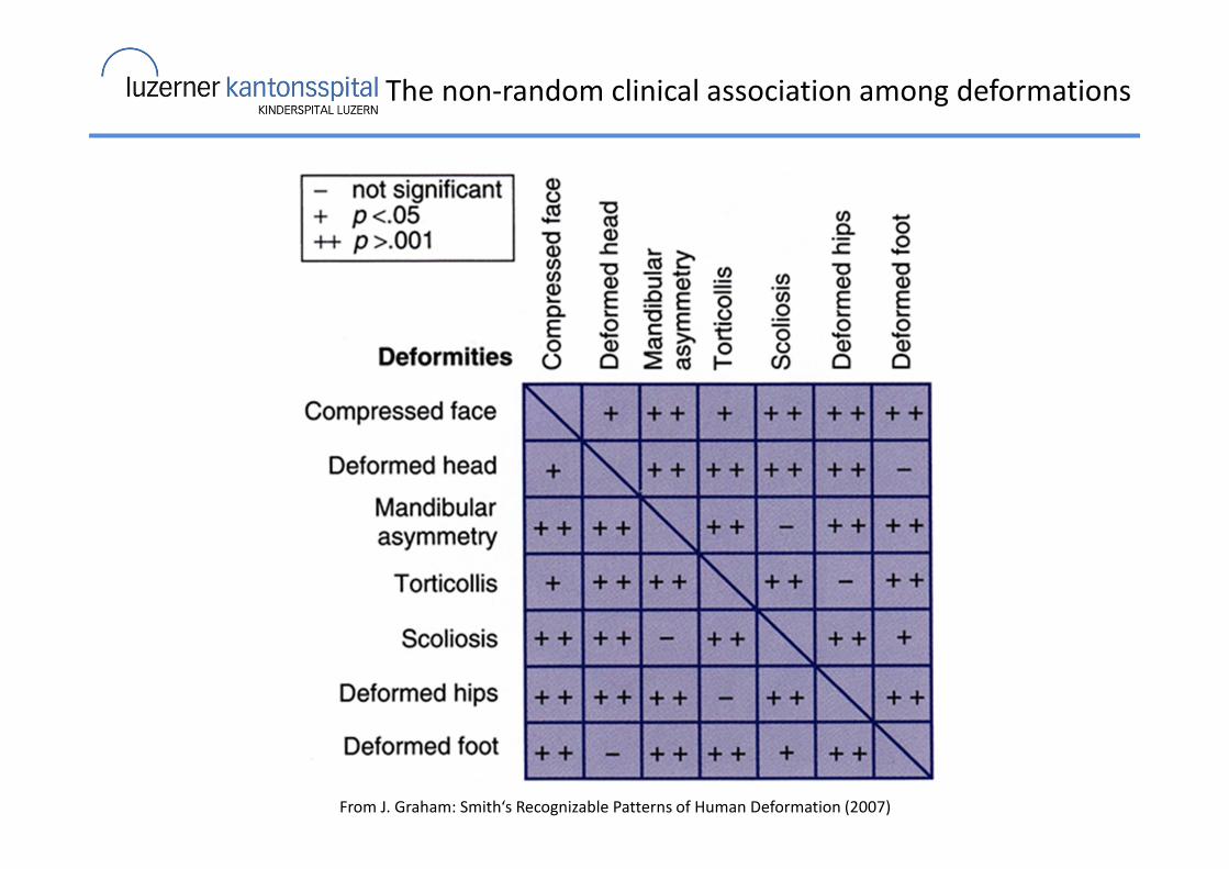
The non-random clinical association among deformations
From J. Graham: Smith‘s Recognizable Patterns of Human Deformation (2007)
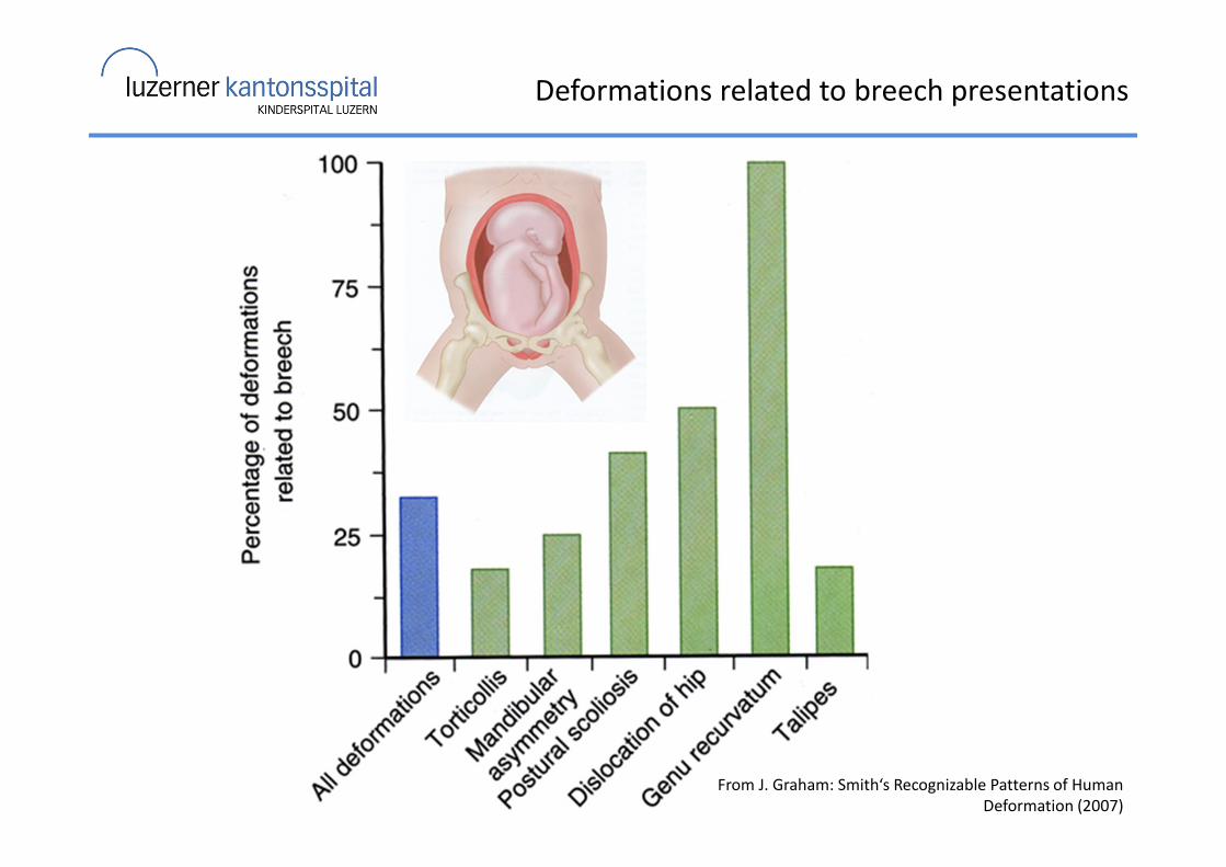
Deformations related to breech presentations
From J. Graham: Smith‘s Recognizable Patterns of Human
Deformation (2007)
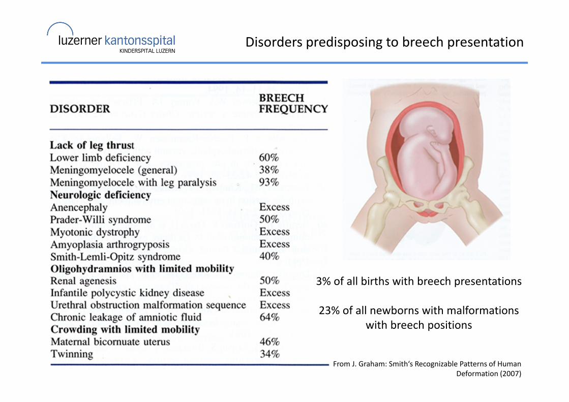
Disorders predisposing to breech presentation
From J. Graham: Smith‘s Recognizable Patterns of Human
Deformation (2007)
3% of all births with breech presentations
23% of all newborns with malformations
with breech positions
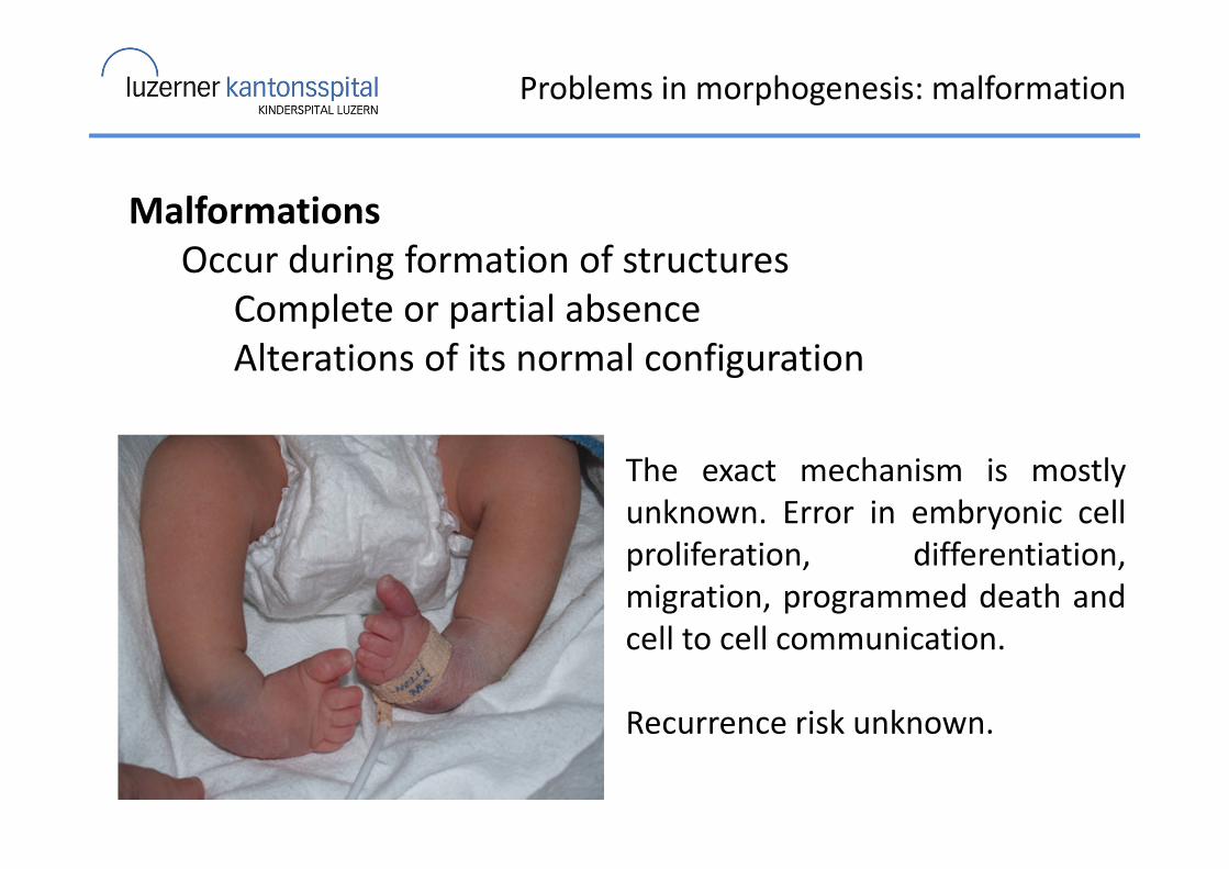
Problems in morphogenesis: malformation
Malformations
Occur during formation of structures
Complete or partial absence
Alterations of its normal configuration
The exact mechanism is mostly
unknown. Error in embryonic cell
proliferation, differentiation,
migration, programmed death and
cell to cell communication.
Recurrence risk unknown.
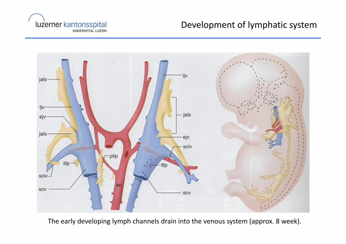
Development of lymphatic system
The early developing lymph channels drain into the venous system (approx. 8 week).
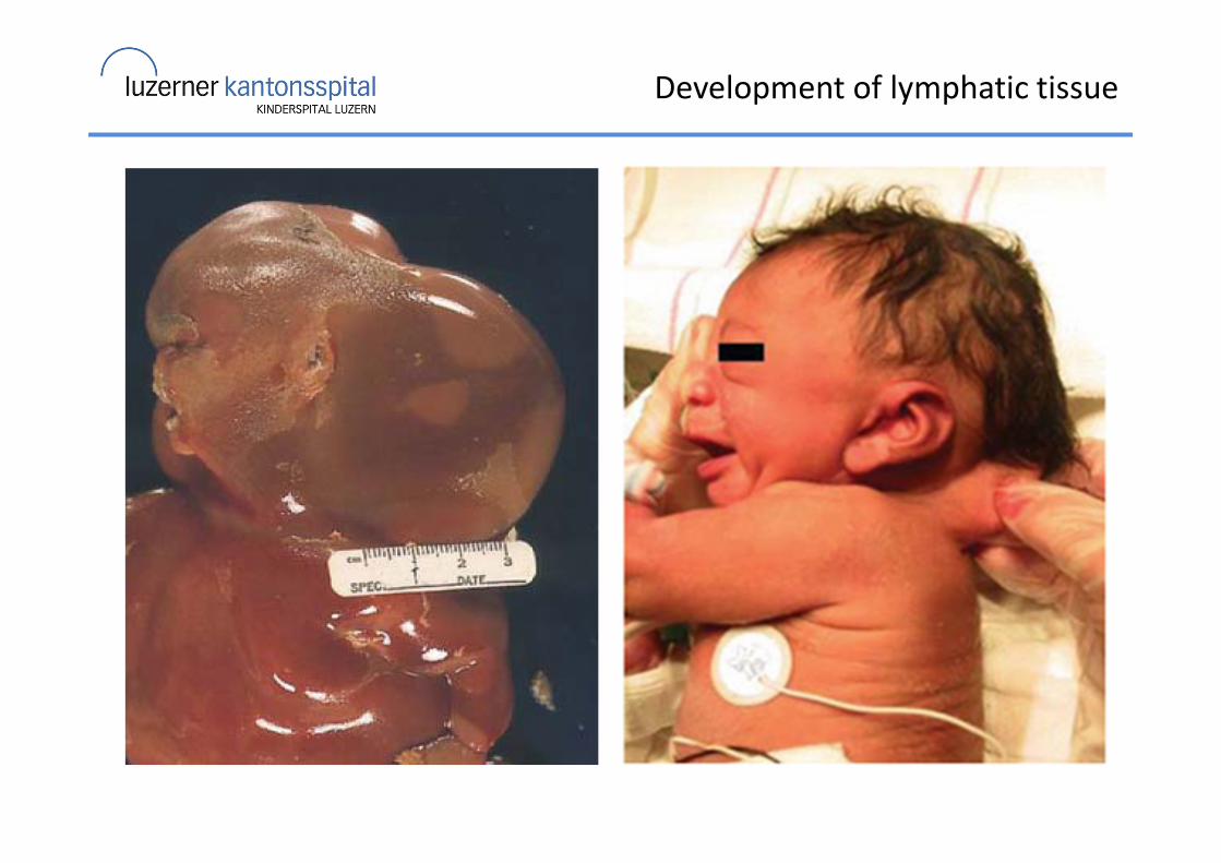
Development of lymphatic tissue

Development of lymphatic tissue
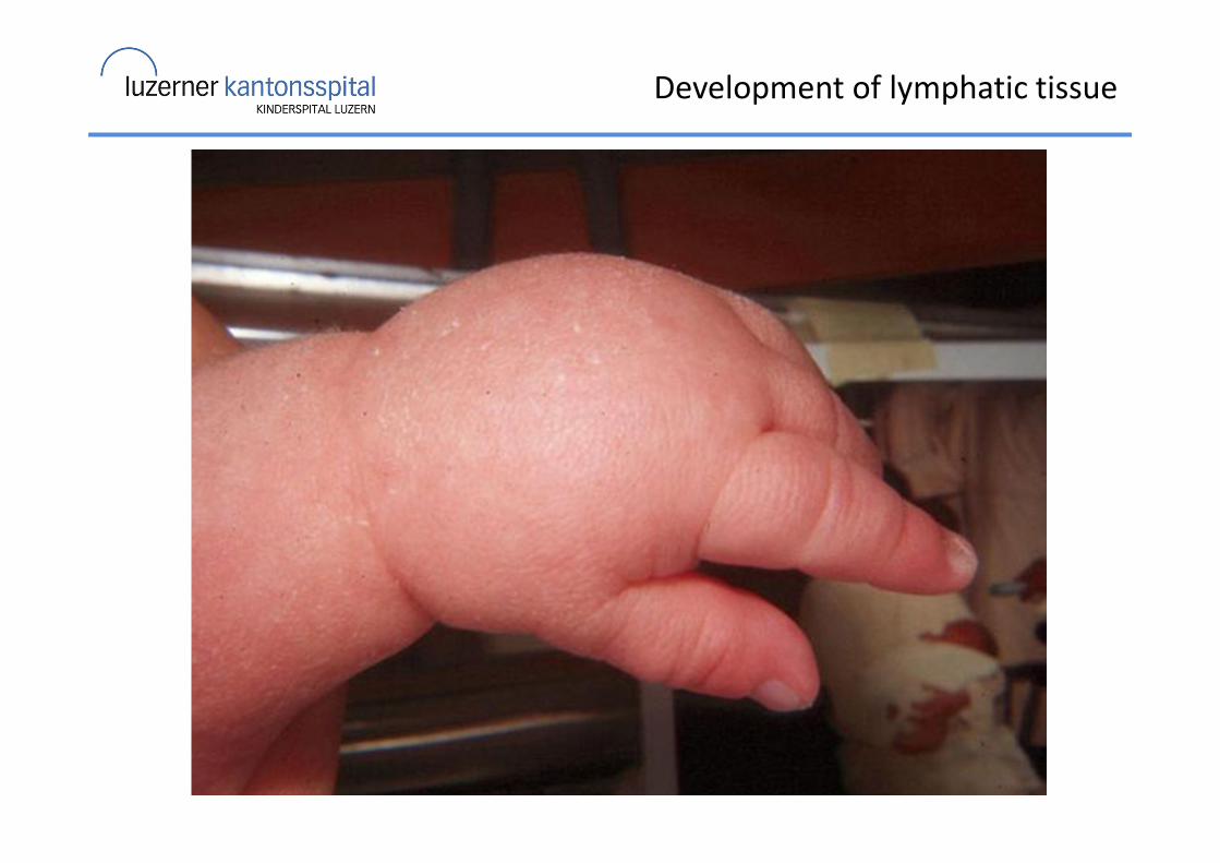
Development of lymphatic tissue
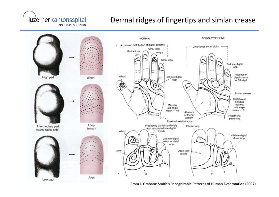
Dermal ridges of fingertips and simian crease
From J. Graham: Smith‘s Recognizable Patterns of Human Deformation (2007)
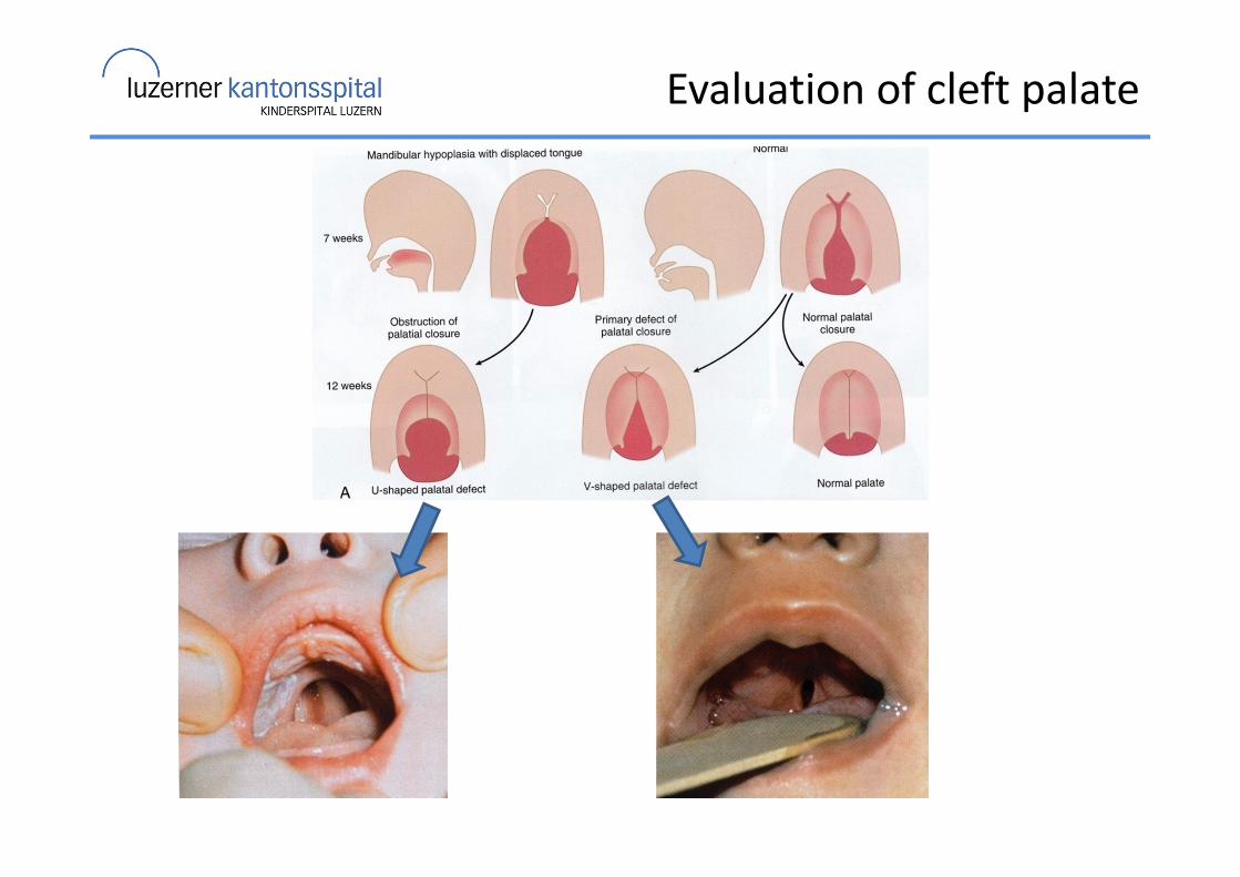
Evaluation of cleft palate
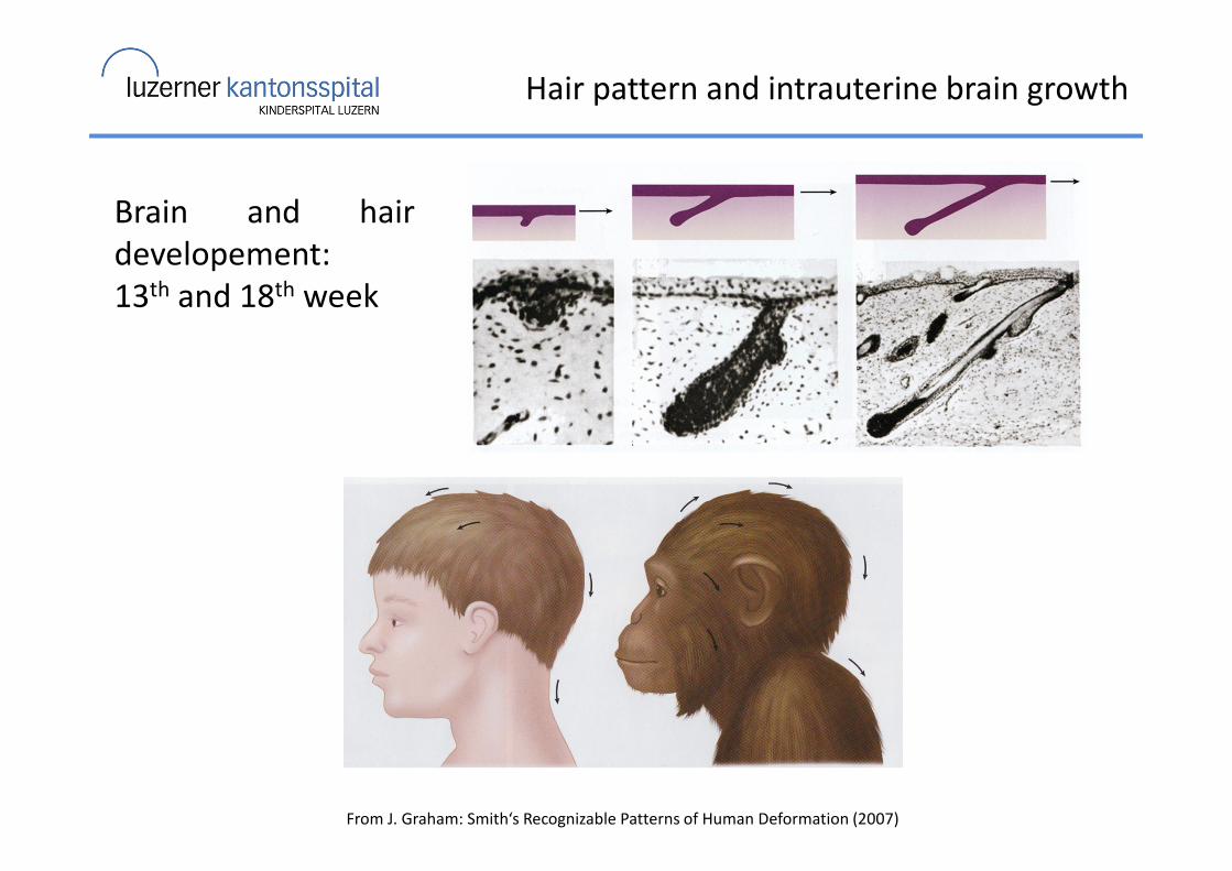
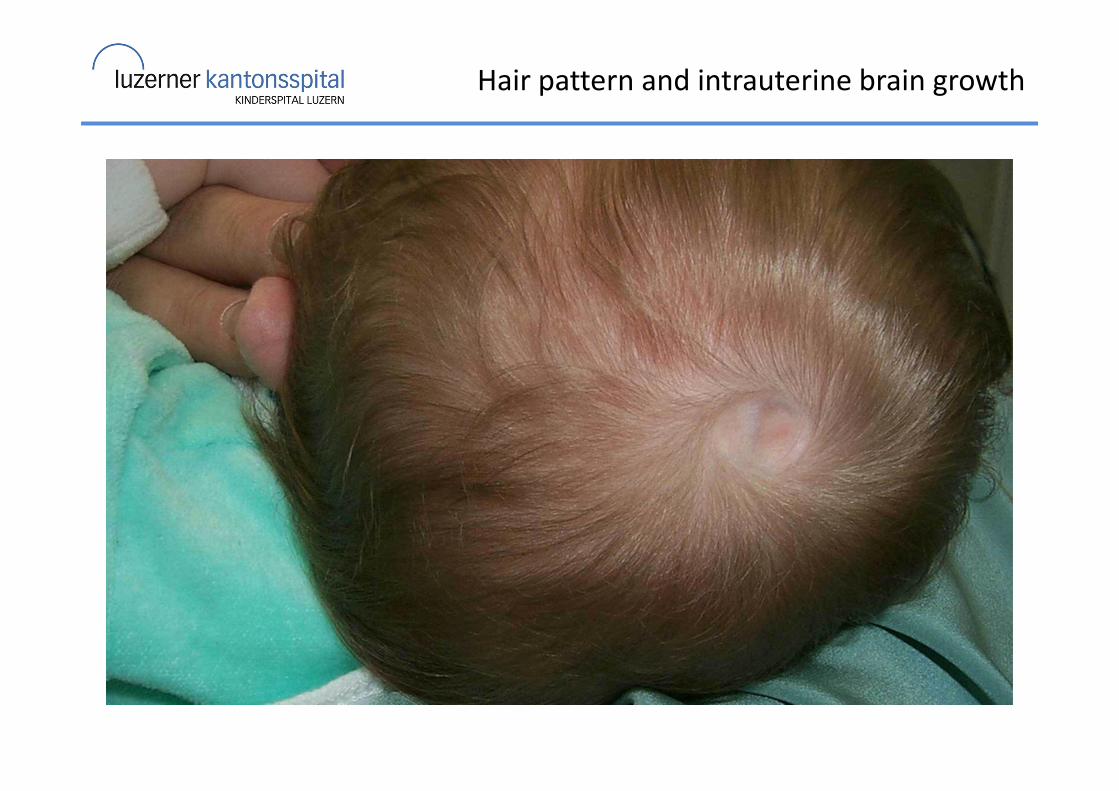
Hair pattern and intrauterine brain growth
From J. Graham: Smith‘s Recognizable Patterns of Human Deformation (2007)
Brain and hair
developement:
13th and 18th week
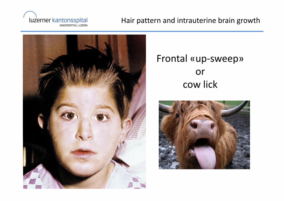
Hair pattern and intrauterine brain growth
Frontal «up-sweep»
or
cow lick
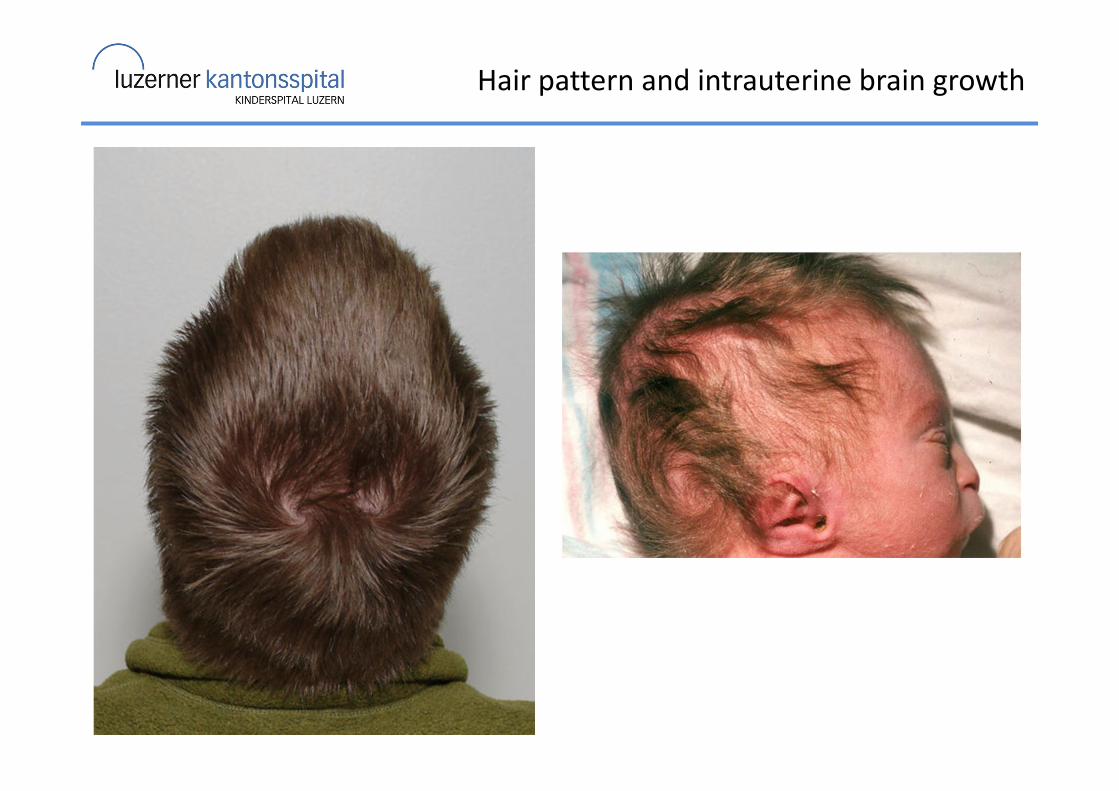
Hair pattern and intrauterine brain growth
Hair pattern and intrauterine brain growth
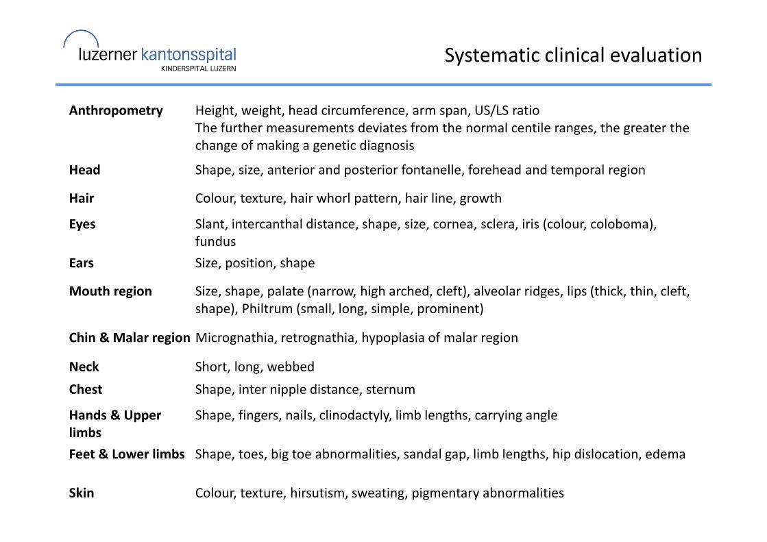
Systematic clinical evaluation
Anthropometry Height, weight, head circumference, arm span, US/LS ratio
The further measurements deviates from the normal centile ranges, the greater the
change of making a genetic diagnosis
Head Shape, size, anterior and posterior fontanelle, forehead and temporal region
Hair Colour, texture, hair whorl pattern, hair line, growth
Eyes Slant, intercanthal distance, shape, size, cornea, sclera, iris (colour, coloboma),
fundus
Ears Size, position, shape
Mouth region Size, shape, palate (narrow, high arched, cleft), alveolar ridges, lips (thick, thin, cleft,
shape), Philtrum (small, long, simple, prominent)
Chin & Malar region Micrognathia, retrognathia, hypoplasia of malar region
Neck Short, long, webbed
Chest Shape, inter nipple distance, sternum
Hands & Upper
limbs
Shape, fingers, nails, clinodactyly, limb lengths, carrying angle
Feet & Lower limbs Shape, toes, big toe abnormalities, sandal gap, limb lengths, hip dislocation, edema
Skin Colour, texture, hirsutism, sweating, pigmentary abnormalities
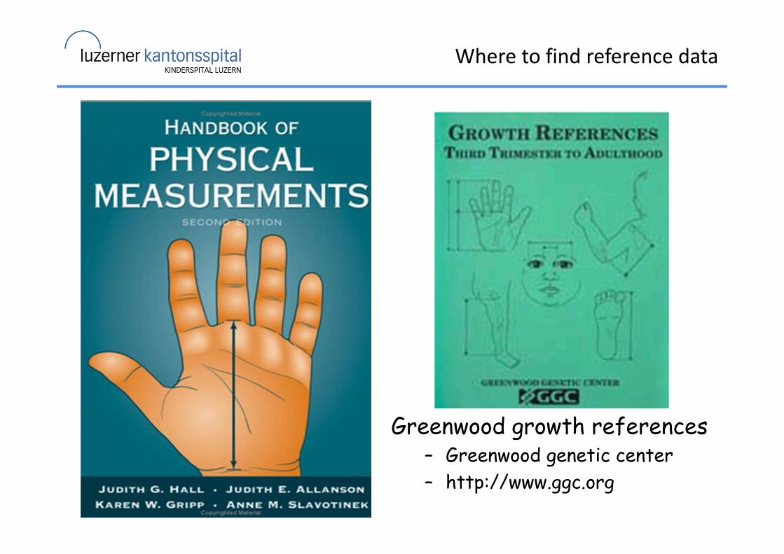
Where to find reference data
Greenwood growth references– Greenwood genetic center
– http://www.ggc.org
ABase
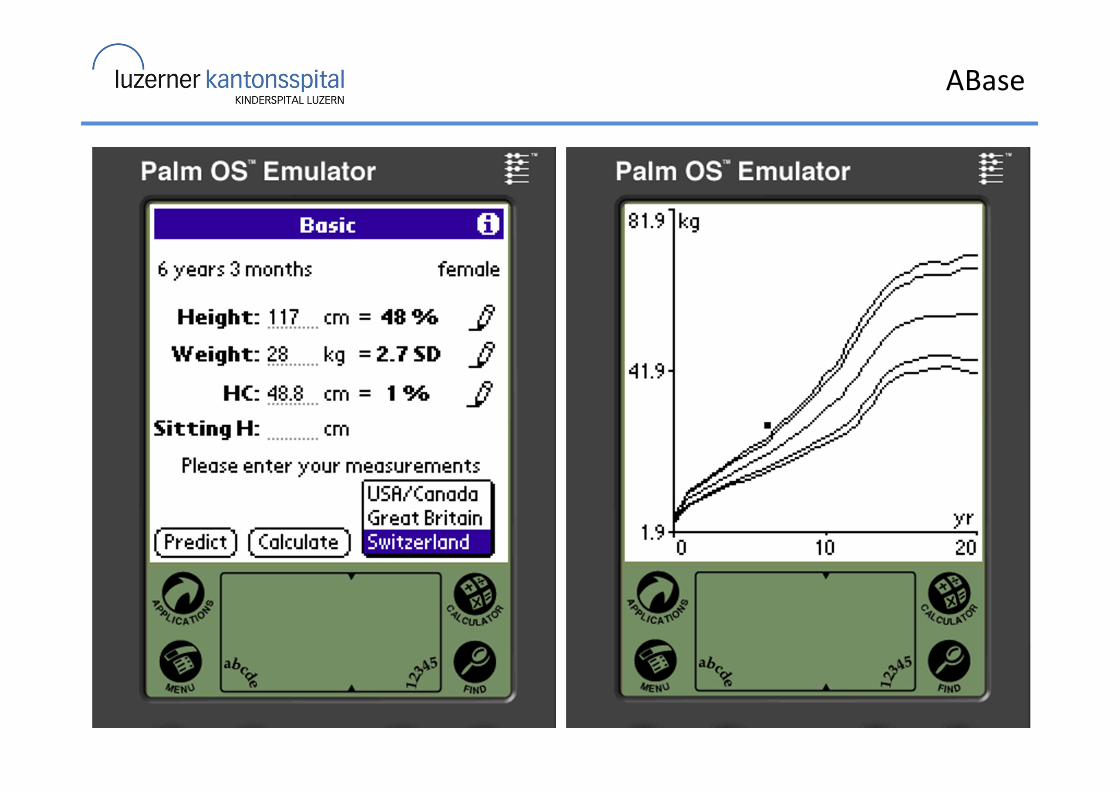
ABase
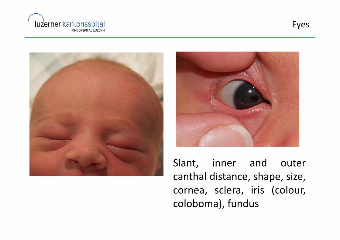
Eyes
Slant, inner and outer
canthal distance, shape, size,
cornea, sclera, iris (colour,
coloboma), fundus
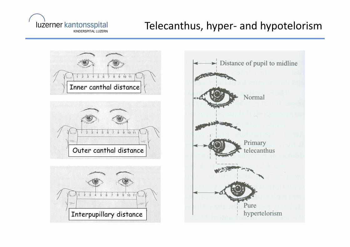
Inner canthal distance
Outer canthal distance
Interpupillary distance
Telecanthus, hyper- and hypotelorism
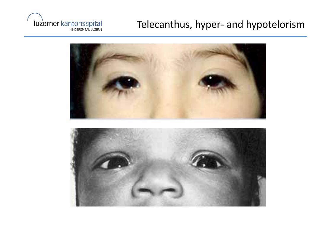
Telecanthus, hyper- and hypotelorism
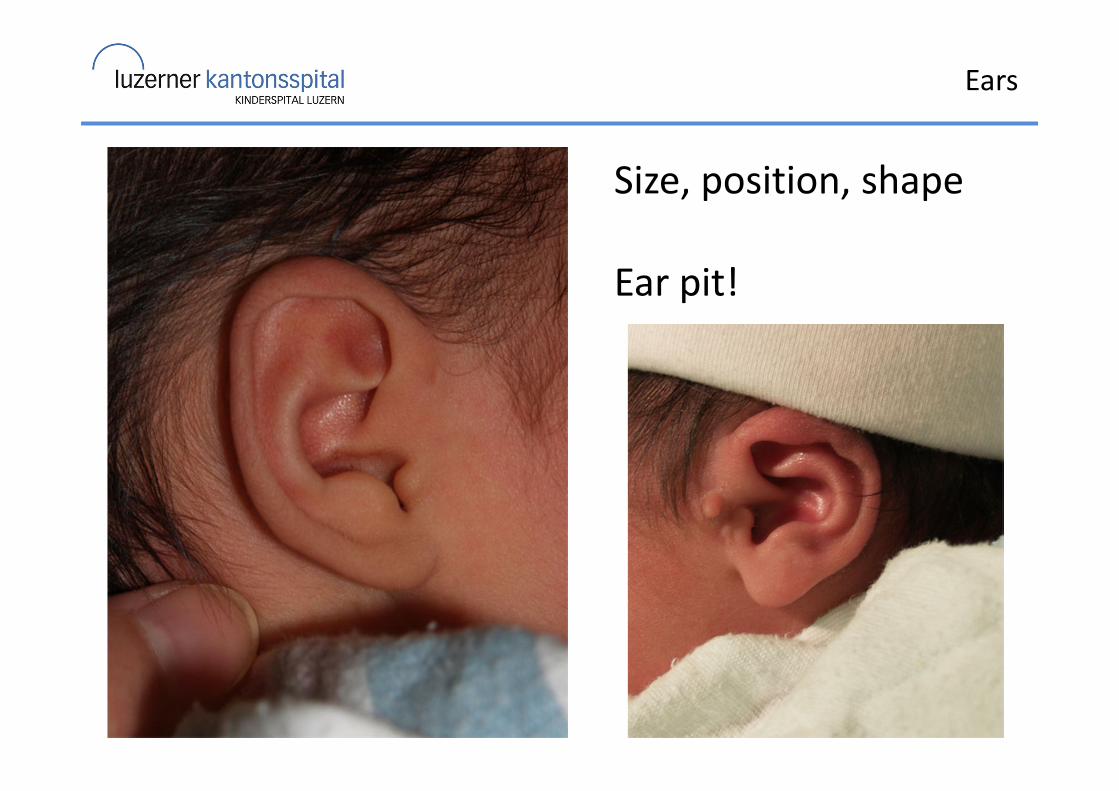
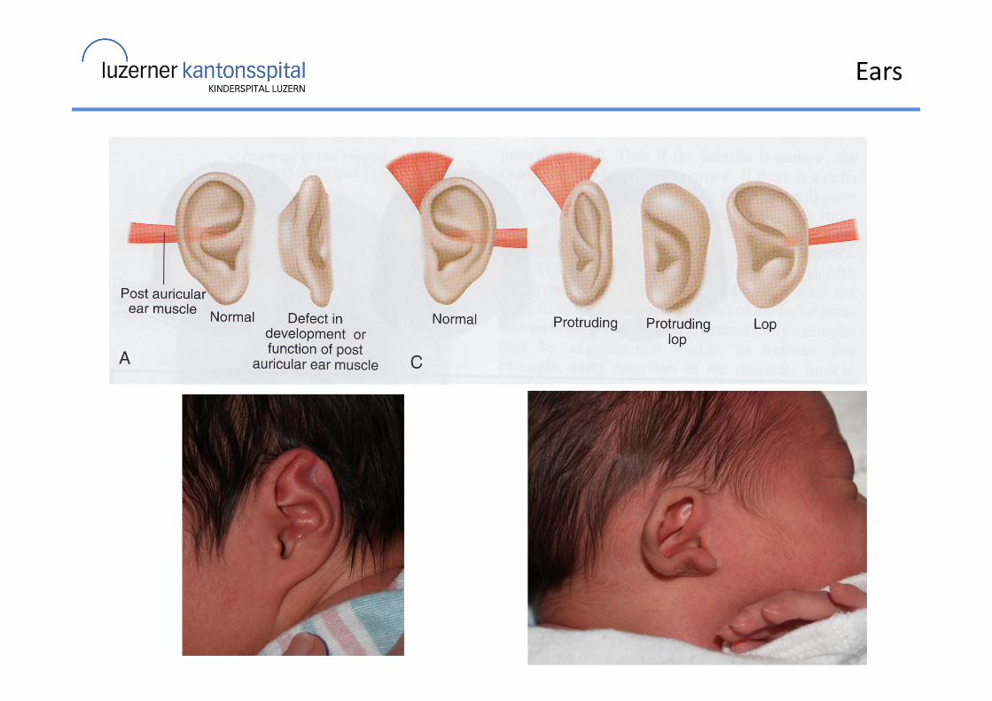
Ears
Size, position, shape
Ear pit!

Ears
MicrotiaHypoplastic ear
Ears
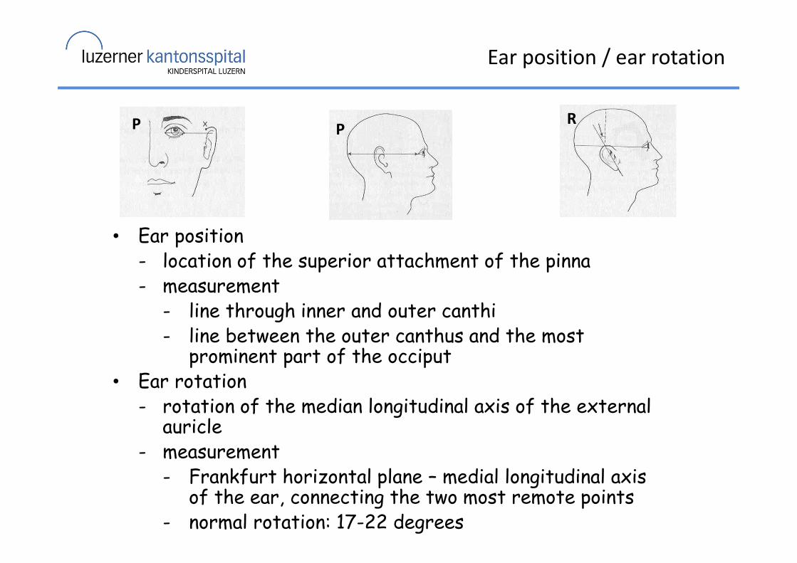
Ear position / ear rotation
• Ear position- location of the superior attachment of the pinna- measurement
- line through inner and outer canthi - line between the outer canthus and the most
prominent part of the occiput• Ear rotation
- rotation of the median longitudinal axis of the external auricle
- measurement- Frankfurt horizontal plane – medial longitudinal axis
of the ear, connecting the two most remote points- normal rotation: 17-22 degrees
P PR
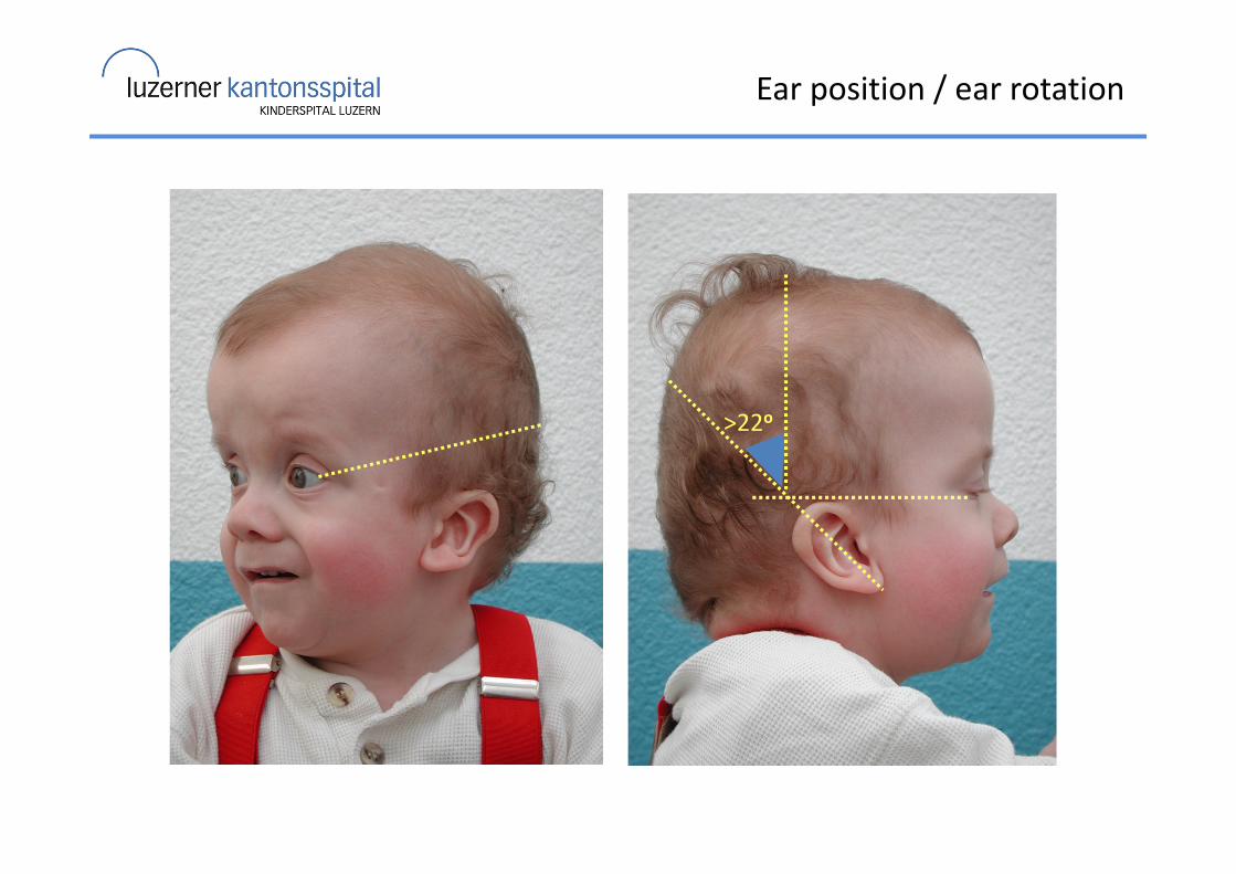
Ear position / ear rotation
>22o
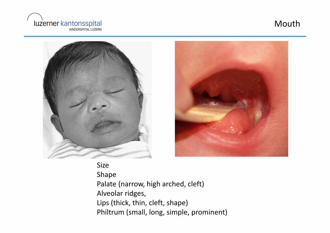
Mouth
Size
Shape
Palate (narrow, high arched, cleft)
Alveolar ridges,
Lips (thick, thin, cleft, shape)
Philtrum (small, long, simple, prominent)
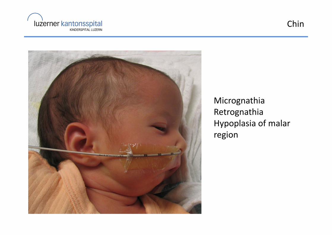
Chin
Micrognathia
Retrognathia
Hypoplasia of malar
region
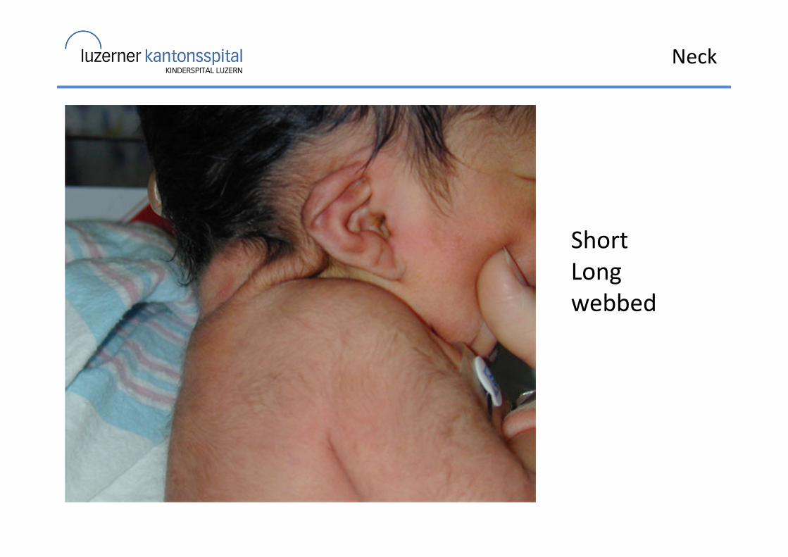
Neck
Short
Long
webbed
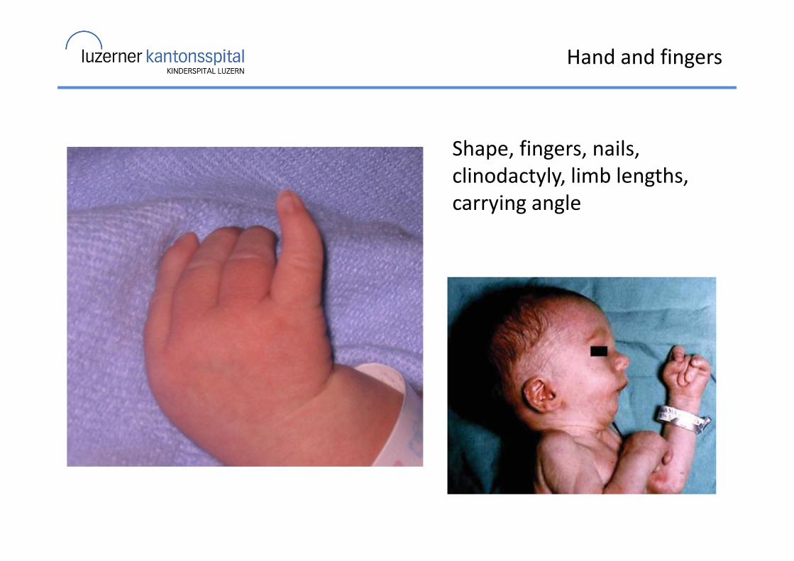
Hand and fingers
Shape, fingers, nails,
clinodactyly, limb lengths,
carrying angle
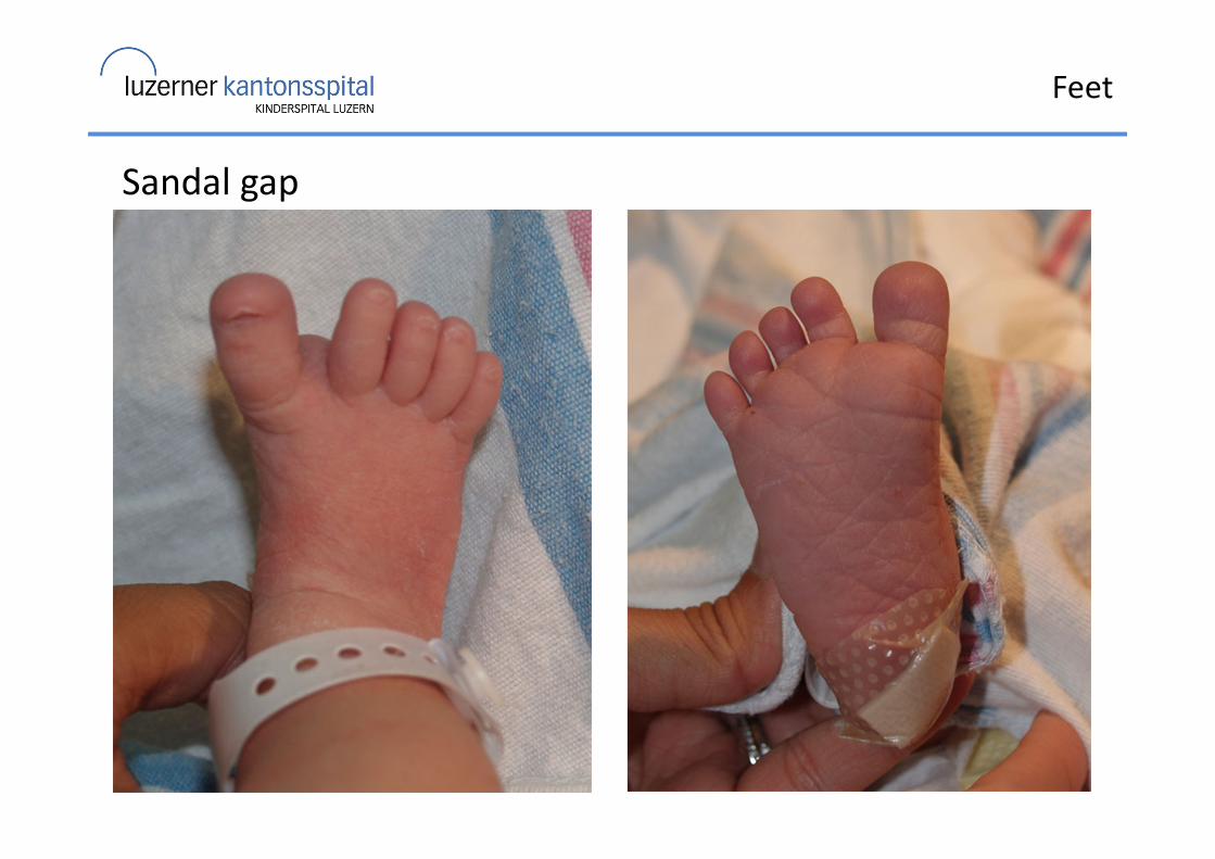
Feet
Sandal gap
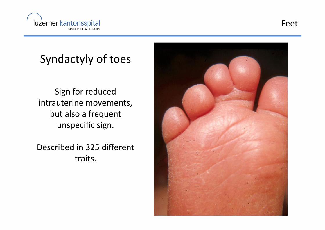
Feet
Syndactyly of toes
Sign for reduced
intrauterine movements,
but also a frequent
unspecific sign.
Described in 325 different
traits.
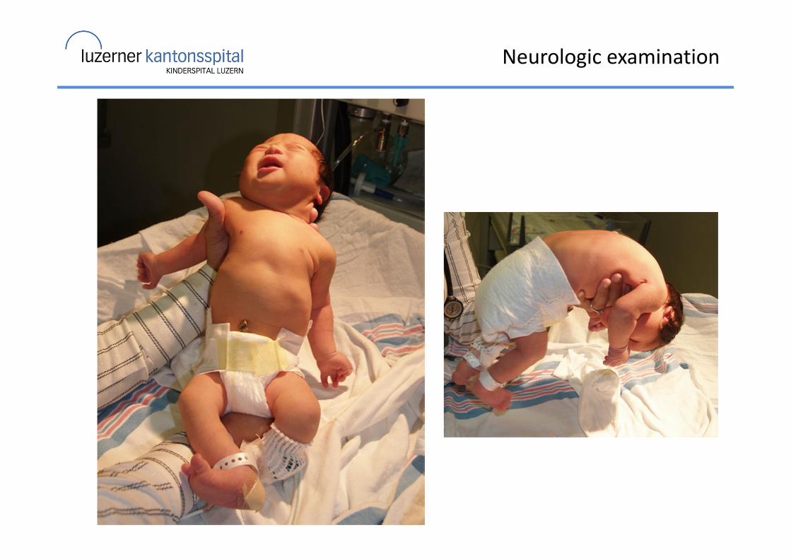
Neurologic examination
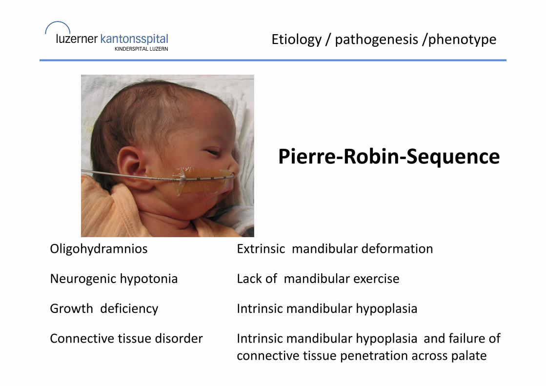
Etiology / pathogenesis /phenotype
Oligohydramnios Extrinsic mandibular deformation
Neurogenic hypotonia Lack of mandibular exercise
Growth deficiency Intrinsic mandibular hypoplasia
Connective tissue disorder Intrinsic mandibular hypoplasia and failure of
connective tissue penetration across palate
Pierre-Robin-Sequence
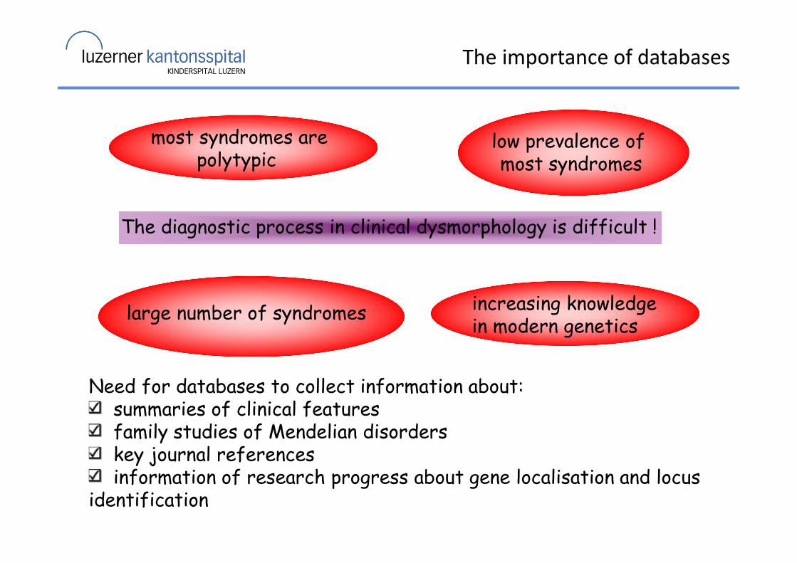
The importance of databases
The diagnostic process in clinical dysmorphology is difficult !
large number of syndromes
low prevalence of most syndromes
increasing knowledge in modern genetics
Need for databases to collect information about:summaries of clinical featuresfamily studies of Mendelian disorderskey journal referencesinformation of research progress about gene localisation and locus
identification
most syndromes arepolytypic
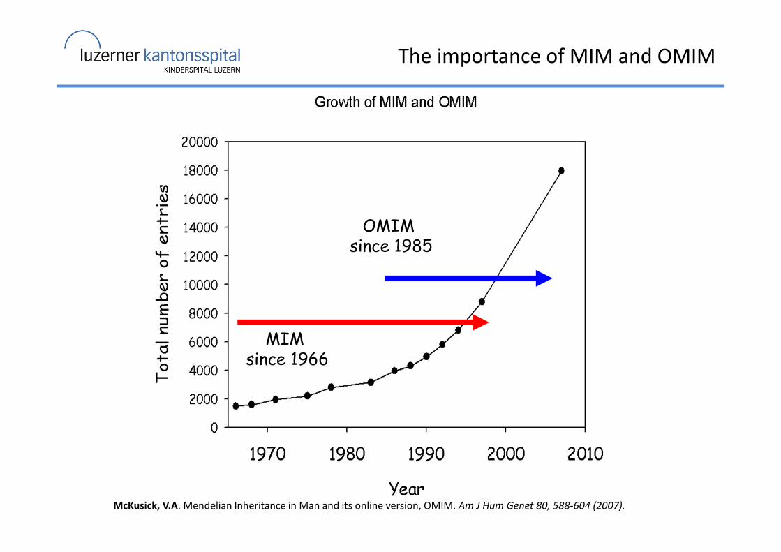
OMIM since 1985
MIM since 1966
McKusick, V.A. Mendelian Inheritance in Man and its online version, OMIM. Am J Hum Genet 80, 588-604 (2007).
The importance of MIM and OMIM
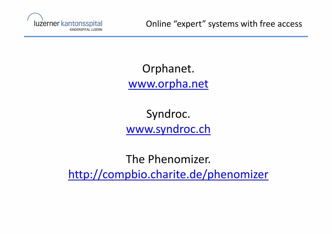
Online “expert” systems with free access
Orphanet.
www.orpha.net
Syndroc.
www.syndroc.ch
The Phenomizer.
http://compbio.charite.de/phenomizer
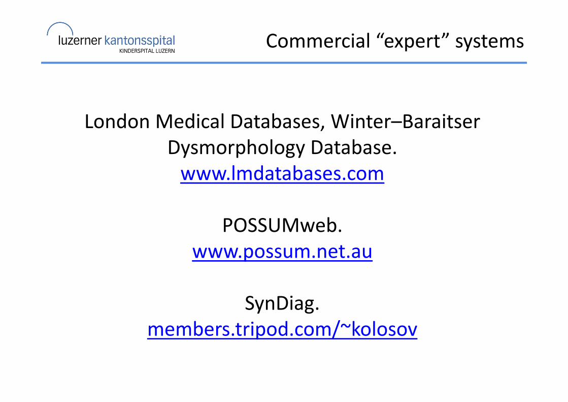
Commercial “expert” systems
London Medical Databases, Winter–Baraitser
Dysmorphology Database.
www.lmdatabases.com
POSSUMweb.
www.possum.net.au
SynDiag.
members.tripod.com/~kolosov
Clinical approach to the dysmorphic newborn
Thank you for your attention!