Embed Size (px)
Citation preview

2013 SUMMER RESEARCH PROGRAM
STUDENT ABSTRACTS

2
This page left blank

3
Contents
Preface………………………………………………………… 5 Acknowledgements ………………………………………… 7 Lab Research Ownership …………………………………. 9 Index Medical Students ………………………………………... 11 Undergraduate Students ……………………………….. 12 International Medical Students ………………………… 13 Abstracts – Medical Students …………………………… 14 Abstracts – Undergraduates ……………………………. 82 Abstracts – International Medical Students ……..…….. 123

4
This page left blank

5
Preface
The University of Texas Medical School at Houston (UTMSH) Summer Research Program provides intensive, hands-on laboratory research training for MS-1 medical students and undergraduate college students under the direct supervision of experienced faculty researchers and educators. These faculty members’ enthusiasm for scientific discovery and commitment to teaching is vital for a successful training program. It is these dedicated scientists who organize the research projects to be conducted by the students. The trainee’s role in the laboratory is to participate to the fullest extent of her/his ability in the research project being performed. This involves carrying out the technical aspects of experimental analysis, interpreting data and summarizing results. The results are presented as an abstract and are written in the trainees’ own words that convey an impressive degree of understanding of the complex projects in which they were involved. To date, more than 1,800 medical, college, and international medical students have gained research experience through the UTMSH Summer Research Program. Past trainees have advanced to pursue research careers in the biomedical sciences, as well as gain an appreciation of the relationship between basic and clinical research and clinical practice. UTMSH student research training is supported by a grant from the National Institute of Neurological Disorders and Stroke (NINDS), and/or by financial support from the Dean and the departments and faculty of the medical school and School of Dentistry. Biomedical science education remains a vital and integral part of our nation’s interests. The UTMSH Summer Research Program, and the dedication of our faculty and administration exemplify the institution’s commitment to training and educating the future leaders in our biomedical scientific communities.
Gary C. Rosenfeld, Ph.D. Director, Summer Research Program Assistant Dean for Educational Programs

6
This page left blank

7
Acknowledgements
This publication marks the completion of the twenty-fifth year of The University of Texas Medical School at Houston (UTMSH) Summer Research Program. The longevity and success of the program are rooted in the overwhelming support received from the deans, faculty, staff and students of the medical school. Indicative of this support is the administrative assistance and financial support for the Program’s college and medical students provided by UTMSH. Sincere appreciation is expressed to Dean Giuseppe Colasurdo M.D. and Patricia M. Butler, M.D., Associate Dean, Office of Educational Programs who continue to ensure the yearly success of the Summer Research Program. Major financial assistance for medical students has also been provided through a short term research grants by the National Institute for Neurological Disorders and Stroke (NINDS; 5 T35 NS064931). Negotiated cooperative agreements with several international medical schools have been set up to offer tailored research programs at UTMSH for selected foreign medical students who interact fully with the other students in the Summer Research Program. The success of the Summer Research Program depends primarily on the faculty who volunteer to mentor the trainees. These dedicated educators organize and guide the research projects that includes for each student data analysis, preparation of an abstract and public presentation of results. Our sincere appreciation to all faculty mentors.

8
This page left blank
9
Lab Research Ownership
Publication and/or Disclosure Each student participating in this program is required to read, agree to, and sign this disclosure form. The original signed copy is on file in the Summer Research Program office; the student and their faculty mentors are each furnished with a copy. “In reference to the laboratory research you will perform this coming summer through The University of Texas Medical School at Houston’s Summer Research Program, you are required to comply with the standard restrictions regarding participation in the Summer Research Program: “All of your laboratory research is CONFIDENTIAL and although your abstract will be available through our website, you cannot independently disclose or publish any research findings or data in any form (including at meetings or conferences) without the express prior written approval of The University of Texas Medical School at Houston. If you wish to submit your abstract to any third party, you must first contact your faculty mentor no less than three (3) weeks prior to any deadlines in order to obtain the necessary written approvals. “Because your research was generated from ideas and funds that originated with your faculty mentor and The University of Texas Medical School at Houston, ownership of any data generated by you during the Summer Research Program belongs to The University of Texas Medical School at Houston or the Principle Investigator (PI).”

10
This page left blank
11
Medical Students
Last Name First Name
Page #
Last Name First Name Page
# Adams Bradley 15
Karri Jay 50
Aziz Shahroz 16
Kwater Andrzej 51 Booth Michael 17
LeBlanc Anthony 52
Brown Charles 18
Loh Jonash 53 Brown Kendra 19
McBride Cameron 54
Caplan Henry 20
Mena Stephanie 55 Crenwelge Tiffany 21
Messer Jay 56
Davis Elizabeth 23
Mount Andrew 57 DiCicco Beau 24
Mulanovich Eduardo 58
Dubuisson Danielle 25
Mulcahy Collin 59 Ekhlassi Erfon 26
Nguyen Adam 60
Elhorr Feryal 27
Novak Matthew 61 Feng Kimberly 28
Oyeniyi James 62
Fraivillig Kurt 29
Patel Kishan 63 Frank Thomas 30
Philip Grace 65
Fu Chen 37
Poddar Keshav 66 Gilbert Blaine 32
Reynolds Jake 67
Goerlich Corbin 33
Rogers Nathan 68 Gonzales Omar 35
Roper Brennan 69
Goodman Casey 36
Segal Graeme 70 Hacopian Alexander 37
Seidel Hudson 71
Haight Derek 38
Sloan Duncan 72 Hanania Alexander 39
Srikrishnan Anand 73
Holloway Steven 40
Villegas Inurrigarro Joaquin 74 Holmes Genevieve 41
Vo Jonathan 75
Hoover Katlyn 42
Wallace Nicholas 76 Huang Michael 44
Waller-Delarosa John 77
Hutto Jake 45
Waters David 78 Huynh Douglas 46
Wilcoxson Brandon 79
Irani Malcolm 47
Witkov Richard 80 Jawad Adbul 48
Zebda Denna 81
Kansara Sagar 49

12
Undergraduate Students
Last Name First Name Page
#
Last Name First Name Page
#
Abraham Annie 83
McMenemy Madison 101
Abu-Shamat Farid 84
McNulty Catherine 102
Ahn Stephanie 85
Nguyen Tran 103
Ajluni Steven 86
Patel Anjali 104
Alapati Nitheesha 88
Putman Jordan 105
Ayoub Christopher 89
Randall James 106
Bricker Madeleine 90
Rosenthal Nathan 107
Brooks Sarah 91
Sajid Madiha 108
Cheng Yanshu 92
Schoenemann Kara 109
Chua Cherie 93
Song Ye-Jin 110
Dadashazar Samareh 94
Taylor Ellen 112
Hoang Ellen 88
Thomas Carrisa 113
Jebreen Osama 95
Travis Kate 114
Kirchgessner Megan 96
Trevino Viviana 115
Lerner Michelle 97
Wright Robin 116
Lima Maria 98
Wu Patrick 117
Lin Andy 99 Yao Zhixing 119
Maddox Jessica 88 Zhu Lawrence 120
Mathews Stacy 100 Zimmerman Rachel 122

13
International Medical Students
Last Name First Name Page
#
Jiang Shutian 124
Li Yike 125
Lin Chyauwen 126
Morimoto Kana 127
Shimozono Komei 128
Wei Yahui 131
Wu Ziping 132
Yeh Tingyu 133
Zhu Youwei 134

14
Medical
Students

15
ABSTRACT
2013 Summer Research Program Office of Educational Programs
Medical Student
MSCs Inhibit Microglia and T-cell Inflammatory Response In Vitro
BRADLEY D. ADAMS The University of Texas at Houston Medical School Class of 2016
Sponsored by: Scott D. Olson, PhD, Department of Pediatric Surgery
Supported by: Scott D. Olson, PhD, Department of Pediatric Surgery; The University of Texas at Houston Medical School—Office of the Dean
Key Words: TBI, MSCs, Leukocytes, Anti-Inflammatory Traumatic brain injury (TBI) is recognized as a leading cause of mortality and permanent neurological disability worldwide, yet efforts to develop protective therapies have had no success in human trials. Along with the initial physical damage that defines TBI, it often results in a delayed secondary inflammatory injury to the patient, brought on by over-recruitment and activation of both central and peripheral inflammatory cells, such as T-cells, microglia and other leukocytes. Multipotent Stromal Cells (MSCs), formerly-known as mesenchymal stem cells, have been shown to have modulatory effects on inflammatory activation of various immune cells. There's no current treatment for the secondary inflammatory injury following TBI, which presents an opportunity to develop therapeutic approaches that may prevent further neurological damage and loss of function following injury. We hypothesize that MSCs will be able to inhibit the secretion of inflammatory cytokines from activated microglia and T-cells to prevent further injury in TBI. Co-culture of bone marrow-derived MSCs with mouse microglia cell line and human T-cell line showed a decrease in the expression of TNF-α and IL-2 by 32.4% and 50.6%, respectively. This aligns well with our previous co-cultures of MSCs with raw mouse splenocytes and human peripheral blood mononuclear cells (PBMCs), where there were decreases in TNF-α, IFN-γ and other interleukins. These results suggest that MSCs inhibit secretion of inflammatory cytokines, and that MSCs are a promising approach to TBI therapy.

16
ABSTRACT
2013 Summer Research Program Office of Educational Programs
Medical Student
Pneumatosis Intestinalis and Portal Venous Gas In The Low Birth Weight Infant: Is There an Indication for Surgical Intervention?
SHAHROZ K. AZIZ The University of Texas at Houston Medical School Class of 2016
Sponsored by: Stacey D. Moore-Olufemi, MD, Department of Pediatric Surgery
Supported by: Stacey D. Moore-Olufemi, MD, Department of Pediatric Surgery
Key Words: Necrotizing Enterocolitis, Portal Venous Gas, Pneumotosis Intestinalis, Indications for Surgery
Necrotizing enterocolitis (NEC) is an abdominal catastrophe in the low-birth weight, preterm infant. To date, free air in the peritoneal cavity seen on x-ray is the only immediate indication for surgical intervention. While pneumatosis intestinalis (PI) is considered diagnostic for NEC, the role of portal venous gas (PVG) as an indicator for surgical intervention and mortality remains controversial. Purpose: The objective of this study was to determine if PVG is a useful indicator of outcome for low-birth weight, preterm infants with NEC. Methods: A retrospective chart review was performed on infants diagnosed with NEC between 2004 and 2009 at Children’s Memorial Hermann Hospital. After studying the demographic data, we evaluated the following outcomes: presence of PI ± PVG, initial surgical treatment, time until discharge or death, overall mortality, mortality within 30 days of birth, birth weight and gestational age upon consult. Outcomes for surgical NEC cases were further evaluated for mortality outcomes. Results are expressed as means, and logistical regression analysis was used to calculate odds ratios and confidence intervals. Results: 218 infants were identified as having NEC. 24% of patients developed PI + PVG, of which 85% went on to surgery. Infants with PI + PVG had lower birth weight and gestational age when compared to infants with PI alone. The overall and 24-hour morality rates for PI + PVG (73% and 43%) vs. PI alone (37% & 16%) were higher. The odds of overall mortality with PI + PVG was 4.02 (95%CI: 2.09-7.78) vs. 1.40 (95%CI: 0.80-2.43) with PI alone. Conclusion: Very-low and low birth weight infants with PI + PVG have higher 24-hour and overall mortality rates than infants with either radiological finding alone. Based on this information, we have changed our surgical practice and intervene surgically in this population. We are prospectively following our outcomes in this group.

17
ABSTRACT
2013 Summer Research Program Office of Educational Programs
Medical Student
Regulation of Muscle Regeneration by SIK1 MICHAEL BOOTH The University of Texas at Houston Medical School Class of 2016
Sponsored by: Rebecca Berdeaux, PhD, Department of Integrative Biology and Pharmacology
Supported by: National Institutes of Health, National Institute of Arthritis and Musculoskeletal and Skin Diseases R01-AR059847
Key Words: Sik1, skeletal muscle regeneration, muscle progenitor cells, Pax7 After skeletal muscle injury, quiescent muscle progenitor cells become activated and can differentiate into myocytes, which are committed to the myogenic lineage. These cells fuse with one another and to damaged myofibers to form regenerated myofibers. The serine/threonine kinase salt inducible kinase 1 (SIK1) has been shown to phosphorylate and inhibit class II histone deacetylases, thereby promoting activity of myocyte enhancement factor 2 (MEF2), a transcription factor required for myogenic differentiation. Moreover, inhibition of Sik1 expression in primary muscle progenitor cells profoundly impairs differentiation ex vivo. Because myogenic differentiation is required for muscle regeneration, we hypothesized that Sik1 is required in muscle progenitor cells for efficient regeneration after injury in vivo. To test this, we evaluated regenerative capacity of muscle progenitor cell-specific Sik1 knockout mice. We utilized mice with a conditional allele of Sik1 that can be deleted by Cre recombinase (Cre). In our system, tamoxifen-activated Cre is under the control of the Pax7 locus, which is only expressed in muscle progenitor cells. By injecting tamoxifen in young mice, we bypass possible developmental effects of Sik1 deletion and knock out Sik1 only in Pax7-positive cells days prior to injury. Cardiotoxin (CTX) was used to induce necrosis in tibialis anterior (TA) muscle of wild type and knockout mice. 5-ethynyl-2’-deoxyuridine (EdU), a nucleoside analog of thymidine, was administered for two days after injury to mark newly synthesized DNA of proliferating cells, allowing us to quantify the extent of cell proliferation after injury. Five days after injury, TA muscles were collected, cryosectioned, and stained to evaluate histological appearance and quantify the percentage of Pax7-positive and EdU-positive cells. We found that Sik1fl/flPax7ERCre/+ TM+ mice (muscle progenitor cell Sik1 knockout) had the same percent of Pax7-positive nuclei (2.71%±.70) as control Sik1+/+Pax7ERCre/+ TM+ mice (2.69%±1.29). However, Sik1+/+Pax7ERCre/+ TM+ had fewer Pax7-positive nuclei (2.69%±1.29) compared to Sik1+/+Pax7+/+ TM+ (5.31%±1.37), indicating that cell proliferation was impaired by expression of Cre-ER or by haploinsufficiency of Pax7. In addition, tamoxifen-treated mice had higher percentages of EdU-positive nuclei than untreated mice, indicating that tamoxifen could have an independent effect on muscle regeneration. We will identify an alternate method to delete Sik1 without tamixofen in the future. Histological examination confirmed that Sik1 deletion did not result in noticeable impairment in early stages of muscle regeneration. We conclude that Sik1 is not required for proliferation after injury. In future experiments we will analyze effects of Sik1 deletion on later stages of regeneration, as proliferation is only one step in the regenerative process.

18
ABSTRACT
2013 Summer Research Program Office of Educational Programs
Medical Student
Evolution of Infarcts in Patients Treated with Autologous Bone Marrow Mononuclear Cells
CHARLES A. BROWN The University of Texas at Houston Medical School Class of 2016
Sponsored by: Sean Savitz, MD, Department of Neurology
Supported by: Sean Savitz, MD, Department of Neurology; The University of Texas at Houston Medical School – Office of the Dean
Key Words: Stem cells, ischemic stroke, infarct expansion ratio, MRI
Background and Purpose: An accurate measurement of lesion volume is crucial to assess progression or regression of ischemic stroke (IS). Magnetic resonance imaging (MRI) is routinely applied to measure lesion volume longitudinally. This measurement uses high resolution T2-weighted anatomical and diffusion weighted imaging (DWI) to precisely outline the lesion boundary. The change in lesion volume over time paired with clinical assessment can be used to assess efficacy of a new therapeutic intervention. In a current clinical trial, where we are investigating the safety of autologous bone marrow mononuclear cell (BM-MSC) infusion as a possible new treatment for patients with IS, we are measuring lesion volume over the time span of two years to investigate the effects of the cell therapy on the size of the stroke lesion. Methods and Materials: BM-MSCs were harvested from the iliac crest and transplanted intravenously within 72 hours of the onset of stroke. We pooled a total of 27 IS patients, where 17 received BM-MSC therapy and 10, from another comparable group, did not receive therapy as a control. Patients that underwent hemicraniectomy were excluded from the study. We obtained DWI, T1-, T2-weighted, and FLAIR images at 30, 90, 180, 360, and 720 days from onset of the stroke, for stem cell treated patients, and at 30, 90, and 180 days for the control group. We used MRIcron software (http//:www.cabiatl.com/mricro/mricron), to measure lesion volume by outlining abnormal hyperintensity on each slice. The software determined lesion volume based on the slice thickness and interslice gap. The change in volume over time of the stroke lesions was measured for the stem cell treated patients and was compared to the control group, indicating the effect of the stem cells on the lesion size. We also calculated infarct expansion ratio (IER), i.e. lesion volume at 3 month over baseline. Results: The treated group had an average volume of 62.05 mL, 55.68 mL, 47.42 mL, 45.38 mL, 43.87 mL, and 43.64 mL at baseline, 30, 90, 180, 360, and 720 days, respectively. The control group had an average volume of 23.45 mL, 20.70 mL, 20.50 mL, and 19.09 mL at baseline, 30, 90, and 180 days, respectively. In the group treated with stem cells, the IER (0.72) was lower compared to the non-treated group (0.87). Conclusions: The IER value shows that the lesion size, on average, may be smaller at 3 months than at baseline. This trend continues to the final measuring point of 2 years. While the IER values show that the lesion size is getting smaller in both groups, the lower IER value in the treated group shows that there may be a larger decrease in volume size over time. However, these results are limited by the image quality, human error in volumetry measurements, and small sample size.

19
ABSTRACT
2013 Summer Research Program Office of Educational Programs
Medical Student
The Role of PAK2 in Inhibiting Myosin Light Chain Phosphorylation in Ileus
KENDRA BROWN The University of Texas at Houston Medical School Class of 2016
Sponsored by: Karen Uray, PhD, Department of Pediatric Surgery
Supported by: Karen Uray, PhD, Department of Pediatric Surgery, The University of Texas at Houston Medical School – Office of the Dean
Key Words: PAK2, MLC, MYPT1, Ileus Intestinal dysmotility, or ileus, is often due to intestinal edema and leads to delayed enteral feeding and prolonged hospital stay. The mechanism of ileus is complex and likely to involve the enteric nervous system and intestinal smooth muscle. Smooth muscle cell contractility is regulated by myosin light chain (MLC) phosphorylation. Edema causes increased intestinal smooth muscle cell stretch and decreases MLC phosphorylation leading to decreased contractility. MLC phosphatase activity increases in edema, and is regulated by inhibitory phosphorylation of the myosin phosphatase target subunit 1 (MYPT1). A decrease in phosphorylation of this subunit leads to greater phosphatase activity. P21-activated kinase 1 (PAK1) plays a significant role in regulating MLC phosphorylation; the role of PAK2 in the regulation of MLC phosphorylation is unclear. PAK1 inhibits MYPT1 phosphorylation and, therefore, MLC phosphorylation, under edematous conditions. I hypothesized that PAK2 will also inhibit MLC phosphorylation by inhibiting phosphorylation of MYPT1 of MLC phosphatase. Knockdown of PAK2 expression was performed in human intestinal smooth muscle cells then subjected to basal cyclical stretch or increased cyclical stretch (edematous conditions). Under control conditions, PAK2 appears to increase MLC phosphorylation, similar to PAK1; however, in contrast to PAK1, under edematous conditions (increased cyclical stretch) PAK2 had little effect of MLC phosphorylation. PAK1 co-immunoprecipitates with PAK2 raising the possibility that PAK2 regulates MLC phosphorylation via PAK1. In future experiments, we hope to determine the effect of PAK2 on MYPT1 phosphorylation. We will also construct a constitutively active PAK2 and dominant negative PAK2 to further explore the role of PAK2 in the regulation of MLC phosphorylation.
20
ABSTRACT
2013 Summer Research Program Office of Educational Programs
Medical Student
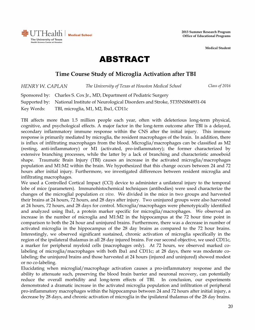
Time Course Study of Microglia Activation after TBI
HENRY W. CAPLAN The University of Texas at Houston Medical School Class of 2016
Sponsored by: Charles S. Cox Jr., MD, Department of Pediatric Surgery
Supported by: National Institute of Neurological Disorders and Stroke, 5T35NS064931-04
Key Words: TBI, microglia, M1, M2, Iba1, CD11c
TBI affects more than 1.5 million people each year, often with deleterious long-term physical, cognitive, and psychological effects. A major factor in the long-term outcome after TBI is a delayed, secondary inflammatory immune response within the CNS after the initial injury. This immune response is primarily mediated by microglia, the resident macrophages of the brain. In addition, there is influx of infiltrating macrophages from the blood. Microglia/macrophages can be classified as M2 (resting, anti-inflammatory) or M1 (activated, pro-inflammatory); the former characterized by extensive branching processes, while the latter by a lack of branching and characteristic amoeboid shape. Traumatic Brain Injury (TBI) causes an increase in the activated microglia/macrophages population and M1:M2 within the brain. We hypothesized that this change occurs between 24 and 72 hours after initial injury. Furthermore, we investigated differences between resident microglia and infiltrating macrophages. We used a Controlled Cortical Impact (CCI) device to administer a unilateral injury to the temporal lobe of mice (parameters). Immunohistochemical techniques (antibodies) were used characterize the changes of the microglial population ex vivo. We divided in the mice in two groups and harvested their brains at 24 hours, 72 hours, and 28 days after injury. Two uninjured groups were also harvested at 24 hours, 72 hours, and 28 days for control. Microglia/macrophages were phenotypically identified and analyzed using Iba1, a protein marker specific for microglia/macrophages. We observed an increase in the number of microglia and M1:M2 in the hippocampus at the 72 hour time point in comparison to both the 24 hour and uninjured brains. Furthermore, there was a decrease in number of activated microglia in the hippocampus of the 28 day brains as compared to the 72 hour brains. Interestingly, we observed significant sustained, chronic activation of microglia specifically in the region of the ipsilateral thalamus in all 28 day injured brains. For our second objective, we used CD11c, a marker for peripheral myeloid cells (macrophages only). At 72 hours, we observed marked co-labeling of microglia/macrophages with both Iba1 and CD11c; at 28 days, there was moderate co-labeling; the uninjured brains and those harvested at 24 hours (injured and uninjured) showed modest or no co-labeling. Elucidating when microglial/macrophage activation causes a pro-inflammatory response and the ability to attenuate such, preserving the blood brain barrier and neuronal recovery, can potentially reduce the overall morbidity and long-term effects of TBI. In conclusion, our experiments demonstrated a dramatic increase in the activated microglia population and infiltration of peripheral pro-inflammatory macrophages within the hippocampus between 24 and 72 hours after initial injury, a decrease by 28 days, and chronic activation of microglia in the ipsilateral thalamus of the 28 day brains.

21
ABSTRACT
2013 Summer Research Program Office of Educational Programs
Medical Student
Does Extent of Insurance Coverage Influence Therapeutic Choices in Patients with Lower Extremity Ulcers?
TIFFANY B. CRENWELGE The University of Texas at Houston Medical School Class of 2016
Sponsored by: Adelaide A. Hebert, MD, Department of Dermatology
Supported by: The University of Texas at Houston Medical School – Office of the Dean
Key Words: Diabetes, lower extremity ulcers, venous insufficiency, insurance Background: The purpose of this database analysis was to determine whether type of insurance coverage (Medicare vs Medicare/Medicaid +secondary insurance vs./Medicare+Medicaid only) influenced therapeutic choices and consequently management strategies for patients with lower extremity ulcers. A consortium of 5 hospital based outpatient wound centers in the northeastern USA operates under a management agreement with Precision Healthcare and thus has a protocol based approach to the care of leg ulcer patients. The physicians are employees of the hospital and are compensated on a fixed salary and thus are not motivated to utilize costly interventions on the basis of their potential revenue generation. This seemed an ideal environment in which to evaluate whether insurance type affects patient treatment decisions for costly healthcare interventions. Bioengineered skin (BioSkin) is a treatment for chronic non-healing wounds. Medicare Administrative Carriers (MACs) determine the specifics of Medicare coverage for BioSkin in terms of which products are covered and how many applications are allowed per wound. Within the jurisdiction of this regional MAC, BioSkin is covered for diabetic foot ulcers and venous leg ulcers, but Medicare pays only 80% of charges leaving the patient responsible for 20%. Average patient responsible charges are $348.69 per application if the patient has no secondary insurance. Patients may undergo up to 5 applications in a course of treatment. The purpose of this project was a high level determination of whether BioSkin was more often utilized among patients with Medicare plus a secondary insurance which would cover the remaining 20%. Methods: With the permission of the 5 hospital facilities and Precision Healthcare, protected health information (PH) was redacted by the electronic health records vendor (Intellicure, Inc., The Woodlands, Texas) and data pertaining to lower extremity ulcer patients were combined and transferred to an Excel spreadsheet for this project. Data from 9/28/2011 to 12/31/2012 were evaluated and 5,487 total patients were identified, of which 851 had diabetic foot ulcers (DFUs) and 1,847 had venous stasis ulcers (VSUs). Results: Preliminary data review of these 5 clinics suggested that 75 patients (1.37%) in this combined hospital wound care clinic database had Medicare as their only type of insurance with no secondary insurance provider.

22
A total of 169 patients (3.08%) received BioSkin therapy of whom 36.09% had Medicare plus a secondary insurance whereas only 1.78% had Medicare only insurance. Among the patients receiving BioSkin, 68 had an underlying diagnosis of diabetes and 26 of those (38.24%) had Medicare plus a secondary insurance. Only one patient with diabetes that received BioSkin therapy had Medicare only insurance, constituting 1.47% of the diabetic patient population receiving BioSkin. In the subset that received BioSkin, 77 patients had venous insufficiency/ulceration. Twenty-eight patients (36.36%) with venous ulceration had Medicare as well as supplementary insurance. Only one patient with venous stasis ulceration who received BioSkin therapy had Medicare as their sole insurance provider, constituting 1.299% of the patient population with the underlying diagnosis of venous insufficiency. Conclusions: In a clinic setting with uniform practice patterns, among patients cared for by physicians on a straight salary, the use of expensive products for non-healing wounds like BioSkin is significantly less likely if Medicare patients have no secondary insurance to cover the 20% “patient responsible” portion. Among patients with VSUs and DFUs, only 1.18% of Bioskin applications were among patients with “Medicare only” insurance. This substantiates the preliminary suggestion that level of insurance coverage may influence therapeutic strategies for patients attending hospital based wound care clinics. As a result of this review of a large wound care database, further research is warranted to determine whether the decreased likelihood of advanced therapeutics among these patients is associated with a difference in healing outcomes (e.g. time to heal or likelihood of healing). We are aware of no similar analyses in the literature.
23
ABSTRACT
2013 Summer Research Program Office of Educational Programs
Medical Student
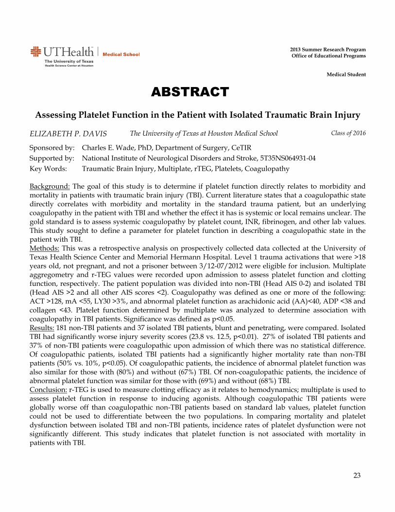
Assessing Platelet Function in the Patient with Isolated Traumatic Brain Injury
ELIZABETH P. DAVIS The University of Texas at Houston Medical School Class of 2016
Sponsored by: Charles E. Wade, PhD, Department of Surgery, CeTIR
Supported by: National Institute of Neurological Disorders and Stroke, 5T35NS064931-04
Key Words: Traumatic Brain Injury, Multiplate, rTEG, Platelets, Coagulopathy
Background: The goal of this study is to determine if platelet function directly relates to morbidity and mortality in patients with traumatic brain injury (TBI). Current literature states that a coagulopathic state directly correlates with morbidity and mortality in the standard trauma patient, but an underlying coagulopathy in the patient with TBI and whether the effect it has is systemic or local remains unclear. The gold standard is to assess systemic coagulopathy by platelet count, INR, fibrinogen, and other lab values. This study sought to define a parameter for platelet function in describing a coagulopathic state in the patient with TBI. Methods: This was a retrospective analysis on prospectively collected data collected at the University of Texas Health Science Center and Memorial Hermann Hospital. Level 1 trauma activations that were >18 years old, not pregnant, and not a prisoner between 3/12-07/2012 were eligible for inclusion. Multiplate aggregometry and r-TEG values were recorded upon admission to assess platelet function and clotting function, respectively. The patient population was divided into non-TBI (Head AIS 0-2) and isolated TBI (Head AIS >2 and all other AIS scores <2). Coagulopathy was defined as one or more of the following: ACT >128, mA <55, LY30 >3%, and abnormal platelet function as arachidonic acid (AA)<40, ADP <38 and collagen <43. Platelet function determined by multiplate was analyzed to determine association with coagulopathy in TBI patients. Significance was defined as p<0.05. Results: 181 non-TBI patients and 37 isolated TBI patients, blunt and penetrating, were compared. Isolated TBI had significantly worse injury severity scores (23.8 vs. 12.5, p<0.01). 27% of isolated TBI patients and 37% of non-TBI patients were coagulopathic upon admission of which there was no statistical difference. Of coagulopathic patients, isolated TBI patients had a significantly higher mortality rate than non-TBI patients (50% vs. 10%, p<0.05). Of coagulopathic patients, the incidence of abnormal platelet function was also similar for those with (80%) and without (67%) TBI. Of non-coagulopathic patients, the incidence of abnormal platelet function was similar for those with (69%) and without (68%) TBI. Conclusion: r-TEG is used to measure clotting efficacy as it relates to hemodynamics; multiplate is used to assess platelet function in response to inducing agonists. Although coagulopathic TBI patients were globally worse off than coagulopathic non-TBI patients based on standard lab values, platelet function could not be used to differentiate between the two populations. In comparing mortality and platelet dysfunction between isolated TBI and non-TBI patients, incidence rates of platelet dysfunction were not significantly different. This study indicates that platelet function is not associated with mortality in patients with TBI.

24
ABSTRACT
2013 Summer Research Program Office of Educational Programs
Medical Student
miRNA Regulation of 1 and 1 sGC Subunits in Human Umbilical Vein Endothelial Cells
BEAU A. DICICCO The University of Texas at Houston Medical School Class of 2016
Sponsored by: Iraida G. Sharina, PhD, Department of Internal Medicine
Supported by: The University of Texas at Houston Medical School – Office of the Dean
Key Words: Soluble Guanylyl Cyclase (sGC), Nitric Oxide (NO), miRNA
The role of Nitric Oxide (NO) as a potent vasodilator has been both extensively characterized in the literature as well as frequently employed in clinics for its ameliorative effects during acute congestive heart failure. Increasing shear stress within blood vessels and signaling by a broad array of humoral agents facilitate the conversion of L-Arginine into NO by endothelial NO Synthase (eNOS). NO rapidly diffuses into surrounding vascular smooth muscle where it binds to and
activates soluble guanylyl cyclase, a heterodimer composed of and subunits. Upon NO binding
to a pocket in the subunit, GTP is allowed to interact with the enzyme’s active site where it is converted to the second messenger molecule cGMP. Finally cGMP interacts with various gated ion channels and protein kinases modulating, among other factors, intracellular Ca2+ levels and cross bridge cycling activity. In acute scenarios NO is extremely efficacious in increasing blood vessel diameter, facilitating perfusion of cardiac muscle and reducing the pressure against which the heart must pump. Unfortunately, physiological tolerance for NO develops rapidly, and thus it lacks efficacy as a long term therapeutic in the treatment of chronic hypertension. A more thorough understanding of regulation within the NO cascade may yield alternative drug targets and
therapeutic strategies in the treatment of hypertension. According to a recent publication, sGC 1
expression is regulated by a specific micro-RNA transcript, miR 34c-5p, in the lung tissue of mice. Additionally, bioinformatic analysis of miR 34c-5p has revealed significant complementarity
between the transcript and 1 and 1 gene sequences. Our aim with this study has been to elucidate
the role of this miRNA transcript in regulation of 1 and 1 subunit expression in human endothelium. Primary Human Umbilical Vein Endothelial Cells (HUVECs) were cultured through four passages whereupon they were transfected with a 100nM solution of miR 34c-5p mimic and
incubated for 24 hours. RNA purified from cell lysate was converted to cDNA and levels of 1/1 coding nucleic acids from the transfection group were analyzed against nucleic acids from a sham group grown in adjacent wells; levels of 18s RNA were measured as an internal control. miRNA regulates protein expression through both targeted degradation of complimentary transcripts as well as without eliminating the transcript through translational obstruction. With this in mind,
western blots were performed to directly compare the levels of 1/1 subunit protein expression between sham and transfection groups. Results thus far are inconclusive in implicating miR 34c-5p
in the down regulation of sGC 1 and 1 subunits. Repetition of qPCR and western blot experiments under optimized conditions and with alternative reagents is still necessary and currently underway.

25
ABSTRACT
2013 Summer Research Program Office of Educational Programs
Medical Student
Does Case Duration and/or Specialty Affect Adherence to the Pre-Incisional Surgical Safety Checklist?
DANIELLE DUBUISSON The University of Texas at Houston Medical School Class of 2016
Sponsored by: KuoJen Tsao, MD, Department of Pediatric Surgery
Supported by: KuoJen Tsao, MD, Department of Pediatric Surgery; The University of Texas Medical School at Houston – Office of the Dean
Key Words: Surgical Safety Checklist (SSCL), Perioperative care, Safety Culture
Introduction: Children’s Memorial Hermann Hospital (CMHH) put into effect a surgical safety checklist (SSCL) in 2011 after the World Health Organization demonstrated that SSCL’s improve patient morbidity and mortality. Soon after implementation of the SSCL, trained observers noted low adherence to the checklist checkpoints. Targeted educational interventions were enacted and follow-up observations took place over the next two summers. Adherence was noted to dramatically improve. Given that the adherence was still not 100%, we hypothesized that factors such as surgical specialty and duration of operations may be influencing its completion. Methods: An observational study was conducted measuring the completion of 22 points on the pre-incisional portion of the SSCL within the pediatric operating rooms at CMHH over an eight week observational period. Specialties chosen for analysis included Dental, ENT, General, Neurosurgery, Orthopedics, Plastics and Urology. Each specialty’s adherence was analyzed. Case durations were divided into intervals (in minutes) of less than 10, 11-30, 30-60, 60-120 and greater than 120 minutes to help categorize adherence. Regression analysis was used to determine statistical significance. Results: A total of 358 cases were observed (see Table 1). There was no significant difference in adherence between any single specialty (Dental: p= 0.253, ENT: p= 0.960, General: p= 0.459, Neurosurgery: p= 0.860, Orthopedics: p= 0.573, Plastics: p=0.471, Urology: p=0.214) or case duration interval (11-30 minutes: p=0.126, 31-60 minutes: p=0.281, 61-120 minutes: p=0.324, >120 minutes: p=0.063). Conclusion: While no significant difference was observed between adherence and specialty nor case duration, we did note a difference in both that could help direct more targeted educational interventions going forward. Additional interventions, such as focusing on adherence to specific checklist checkpoints or encouraging specific lower-adherence surgeons may also help achieve 100% checklist adherence.
Specialty Number of Cases Observed
Avg Case Length
Dental 12 1:17
ENT 67 1:19
General 147 1:20
Neurosurgery 16 1:32
Orthopedics 30 1:05
Plastics 50 1:12
Urology 10 0:40
Table 1

26
ABSTRACT
2013 Summer Research Program Office of Educational Programs
Medical Student
Detecting In Vitro Biofilm Formation on Different Biomaterials Utilizing qPCR and Salvaging Previously Infected Biomaterials
ERFON EKHLASSI The University of Texas at Houston Medical School Class of 2016
Sponsored by: Heidi B. Kaplan, PhD, Department of Microbiology and Molecular Genetics
Supported by: N/A
Key Words: Biofilm, diabetes, biomaterials, Enterococcus faecalis Biofilm formation on in-dwelling devices constitutes the most common cause of nosocomial infections in the United States and can be attributed as a major cause of chronic infections. Diabetics are particularly susceptible to these infections due to compromised vascularity in their extremities and a weakened immune response. We have adapted an in vitro biofilm model developed in our lab to evaluate biofilm growth on different orthopaedic biomaterials that had been previously used for biofilm growth. Previously used and cleaned steel, TMZF alloy, and Grade 2 and 5 anodized titanium alloy discs served as growth substrates for Enterococcus faecalis, which is one of the two most common etiological agents of orthopaedic biofilm infections. Unused polymethylmethacrylate (PMMA) bone cement discs served as a control substrate. Each E. faecalis and disc combination was incubated statically in 24-well plates at 37°C in a synthetic interstitial fluid, which was exchanged daily. On days 1 – 7, the DNA from two discs of each combination was extracted and subjected to quantitative PCR (qPCR) to determine the number of cells attached to each disc. One disc of each combination was stained with cell viability dyes and imaged using a fluorescence microscope. The results of these experiments will be compared to previous results in which we used confocal microscopy to quantitate the bacterial attachment to unused surfaces. Previously we found that titanium discs supported the most biofilm growth and TMZF alloy the least biofilm growth.

27
ABSTRACT
2013 Summer Research Program Office of Educational Programs
Medical Student
Morphological Changes of Medial Prefrontal Neurons in Mice Exposed to Chronic Stress and Repeated Concussive Injury
FERYAL ELORR The University of Texas at Houston Medical School Class of 2017
Sponsored by: Pramod K. Dash, PhD, Department of Neurobiology and Anatomy
Supported by: National Institute of Neurological Disorders and Stroke, 5T35NS064931-04
Key Words: Mild traumatic brain injury, concussion, chronic stress, repeat mTBI, dendritic spines
Mild traumatic brain injury (mTBI) or concussion is a trauma applied to the brain, either directly by an external force or indirectly from sudden acceleration or deceleration. The frontal and temporal lobes are highly vulnerable to TBI. Ultra-structural and/or functional damages to these structures, in the absence of overt damage, can result in profound behavioral and cognitive dysfunctions. The severity of these deficits is usually increased in cases of repeated concussion, an occurrence often seen in athletes and military populations. Though chronic stress, commonly found in mTBI-prone environments, has been implicated in influencing the physiology of a wide variety of diseases, its influence on the pathobiology of repeated concussion is not well understood. As previous studies have established that the medial prefronal cortex (mPFC) in mice is critical for working memory (an ability to hold and integrate information in mind in order to guide goal-directed behavior; a feature often negatively affected by repeated mTBI) and morphological changes can alter neuronal function, we examined morphology of pyramidal neurons in layers II/III of mPFC using Golgi staining and analysis. The purpose of this study was to determine if repeated concussion causes morphological changes of these neurons, and whether these changes are influenced in the context of chronic stress. Four groups of mice were generated: 1) control, 2) mice exposed to repeated mild closed head injury (rmCHI) and killed after 24 hrs, 3) mice exposed to rmCHI and killed after a month, and 4) mice subjected to chronic restraint stress, then given rmCHI and killed after 24 hrs. We used a cortical impactor to cause closed head injuries that resulted in a 1 mm skull deformity midway between bregma and lambda once a day for 4 days. After brain extractions, we sectioned the tissue and processed it using Golgi staining. We found that rmCHI resulted in a significant increase in the density of basal, but not apical, dendrite spines by 24 hrs after injury. This increase was no longer observed at the 1 month time point, and was significantly reduced in mice pre-exposed to chronic restraint stress. As previous studies have demonstrated that increased spine density can have a significant influence on neuronal function and communication, these results may provide a structural mechanism for working memory dysfunction often seen in repeated concussions.

28
ABSTRACT
2013 Summer Research Program Office of Educational Programs
Medical Student
Prospective Assessment of Medical Decision-Making Capacity in Stroke Patients using a Rapid Standardized Questionnaire: Can My Patient
Consent for Treatment or Clinical Trial? KIMBERLY L. FENG The University of Texas at Houston Medical School Class of 2016
Sponsored by: Andrew D. Barreto, MD, Department of Neurology
Supported by: N/A
Key Words: Acute stroke, decision making capacity, vascular cognitive impairment Objective: Stroke deficits frequently alter patient medical decision-making capacity (MDC) resulting in lost trial recruitment and reducing validity of qualitative outcome measures. Since no standardized tool exists for MDC evaluation in stroke, we tested a validated standardized questionnaire used for medical patients, the Aid to Capacity Evaluation (ACE), vs. independent clinician assessment in mild-to-moderate severity stroke patients. We hypothesized that the ACE would show similar agreement with clinicians and therefore be appropriate for rapid bedside screening for MDC.
Methods: Ischemic or hemorrhagic stroke patients underwent 3 independent capacity assessments by a medical student (ACE), psychiatrist (PS) and neuropsychologist (NP). Inter-rater reliability was assessed using intraclass correlation (ICC) and Cohen’s kappa. Assuming the clinician as the gold-standard, we tested sensitivity and specificity vs. ACE. Results: All planned 30 patients (90% ischemic; mean age 67.8; 60% male; median NIHSS = 6) were prospectively enrolled between 7/13- 8/13. The median time from stroke onset to first capacity assessment was 3.3 days. 11 (37%) had aphasia and/or neglect and 38% had left hemispheric stroke. ACE agreed with PS and NP in 59% (kappa 0.293; 95% CI 0.08-0.51) and 76% (kappa 0.494; 95% CI 0.19-0.80) of cases, respectively. Despite low sensitivity and NPV, specificity and PPV of ACE vs. clinicians ranged 88-100%; only classifying 1 patient capable when clinicians scored incapable. ICC among all raters was 0.474 (95% CI 0.25-0.68).
Conclusions: There was fair overall agreement between a standardized questionnaire and expert clinicians. The ACE was highly specific in identifying mild-to-moderate severity stroke patients who lacked MDC. The ACE might be a useful screening tool to determine lack of capacity in stroke patients, but low sensitivity for identifying presence of capacity warrant caution and further study.

29
ABSTRACT
2013 Summer Research Program Office of Educational Programs
Medical Student
Simulation of a Neuron and a Neural Network that are modified by Operant Conditioning
KURT L. FRAIVILLIG The University of Texas at Houston Medical School Class of 2016
Sponsored by: John H. Byrne, PhD, Department of Neurobiology & Anatomy
Supported by: John H. Byrne, PhD, Department of Neurobiology & Anatomy; The University of Texas at Houston Medical School – Office of the Dean
Key Words: Aplysia, reward learning, excitability Operant conditioning (OC) is a form of learning in which an animal learns the consequences of its behavior and is thought to play an important role in drug addiction and substance abuse (Koob and Volkow, 2010). Despite its ubiquitous nature, little is known about the underlying mechanisms of OC. One attractive model system to study OC is the feeding neural circuit of the marine mollusc Aplysia. This circuit mediates two types of motor activity patterns: ingestive buccal motor programs (iBMPs) which bring food into the animal’s mouth and rejection buccal motor programs (rBMP) which move food out of its mouth. After operant conditioning, the animal generates more iBMPs compared to controls (Nargeot et al., 1997, 1999a,b,c). Neuron B4 is an important cell in the circuit for selecting iBMPs (Dacks and Weiss 2013) and shows a decrease in excitability following OC (Neveu et al. 2013, in preparation). Therefore, understanding B4’s properties is central to understanding the mechanisms of OC. Using SNNAP (Simulator for Neural Networks and Action Potentials) (Ziv et al. 1994), I developed a mathematical model of B4 and one in which its properties were changed by OC to help understand its effects on BMP selection. Empirical data from Neveu et al. (2013, in preparation) and Jing and Weiss (2001) were used to constrain various parameters and keep the models’ properties within physiological range. A slow potassium channel was also added to the model to mimic the adaptation seen in B4 firing observed in vitro. The B4 model was then incorporated into the feeding network model developed by Baxter and Byrne (2013) and BMPs were simulated. Next OC-induced changes in B4 were incorporated by increasing the conductance of the slow potassium channel and decreasing the strength of the inhibitory synaptic connection between B4 and B51. After stimulating BMPs, we found that these simple changes mimicked OC by increasing the number of iBMPs. However, these changes also resulted in a decrease in total BMP frequency which is inconsistent with observations in Nargeot et al. (1997). Therefore, additional changes need to be incorporated in the feeding network model in order to fully account for OC (e.g. increases in excitability of pattern initiators B65, B30, B35). In future experiments, it will be possible to determine the individual effects of B4’s OC-induced changes and how they affect BMP frequency.
30
ABSTRACT
2013 Summer Research Program Office of Educational Programs
Medical Student
ZIP4 Expression in Pancreatic Ductal Adenocarcinoma as a Novel Molecular Marker in Endoscopic Ultrasound Guided Fine Needle
Aspiration (EUS-FNA) THOMAS FRANK The University of Texas at Houston Medical School Class of 2016
Sponsored by: Min Li, PhD, The Vivian L. Smith Department of Neurosurgery
Supported by: The Vivian L. Smith Department of Neurosurgery; The University of Texas at Houston Medical School – Office of the Dean
Key Words: Pancreatic Cancer, ZIP4, Fine Needle Aspiration Over the last decade, the pancreatic cancer (PC) incidence rate has remained close to its mortality rate because of difficulties in early stage detection and a lack of competent therapies. Conventional chemotherapy, surgery, and radiation are not sufficient, therefore the identification of novel molecular markers and drug targets are important in treating this disease. Our lab previously identified that aberrant expression of ZIP4, a membrane bound zinc transporter, in pancreatic ductal adenocarcinoma (PDA) contributes to its pathogenesis and progression. The purpose of this study was to compare the ZIP4 expression level in pre-operative endoscopic ultrasound guided fine needle aspiration (EUS-FNA) specimens to corresponding surgical specimens. Also, the correlation between ZIP4 expression and the clinicopathological features of PDA was assessed. Immunohistochemistry was used to compare the EUS-FNA specimens to the surgically resected specimens (parallel control). A total of 23 cases with both FNA and surgical specimens were evaluated. ZIP4 was significantly over-expressed in tumor cells in both sets of samples. The sensitivity, specificity, positive predictive value (PPV), and negative predictive value (NPV) of ZIP4 for PDA diagnosis in EUS-FNA samples were 72.9%, 72.5%, 76.1%, and 69.0%, respectively. In surgical specimens they were 97.9%, 65.4%, 83.9%, and 94.4%, respectively. The ZIP4 intensity level in both EUS-FNA and surgically resected samples were significantly correlated with tumor stage, tumor differentiation, and patient survival. ZIP4 could serve as a novel molecular marker for PDA diagnosis and prognosis through IHC staining of EUS-FNA samples, which is quicker and less invasive than obtaining samples surgically.

31
ABSTRACT
2013 Summer Research Program Office of Educational Programs
Medical Student
Matrixmetalloproteinases Enhance the Metastatic Capacity of Osteosarcoma Cells
CHEN FU The University of Texas at Houston Medical School Class of 2016
Sponsored by: Yong Li, PhD, Department of Pediatric Surgery
Supported by: Yong Li, PhD, Department of Pediatric Surgery; The University of Texas at Houston Medical School – Office of the Dean
Key Words: Matrix Metalloproteinase, Sarcoma, MG63, Migration, Metastasis Introduction: Osteosarcoma is a malignant mesenchymal neoplasm and is the most common type of primary bone cancer in the US. It is extremely prone to metastasis, with over 80 percent of patients having metastatic disease at the time of diagnosis. However, the mechanism for this increased metastatic potential has yet to be fully resolved. It has long been known that osteosarcoma cells overexpress matrixmetalloproteinases (MMP) 2 and 9, which belong to a family of zinc-dependant endopeptidases that play a major role in the degradation of extracellular proteins, as well as in various cell signaling pathways including apoptosis. The current experiment attempts to resolve the role that MMPs play in the metastatic potential of osteosarcomas. Methods: The primary osteosarcoma cell line MG63, purchased from ATCC (USA), was cultured in the proper cancer cell media. Experimental groups were cultured with the MMP inhibitor GM6001 in a 6 well and a 24 well plate with various conditions. Immunocytochemistry was applied to check for the expression of pluripotency markers in these cells. Cell migration assays were performed with cells incubated in GM6001 and with the addition of MMP1 by a bioreactor system, in which each singe cell migration can be tracked and recorded with real-time imaging. Quantitative real-time PCR was performed to determine the expression of pluripotency and migration markers. Results: Cells showed significant expression of Oct4 marker, but no other pluripotency markers, as is consistent with the literature. Cell migration assays showed a significant decrease in mobility with the addition of inhibitor and a significant increase in migration speed/distance with the addition of MMP1. Conclusion: It is apparent that the matrixmetalloproteinase family plays a significant role in cell migration, which may explain why many highly metastatic cancers exhibit an overexpression of MMPs. Our pilot study indicated that MMP1 plays essential role during osterosarcoma metastases, which may include accelerating cell migration as well as promote tumor stem cell activation. MMP inhibitors or blockers should be studied for possible therapeutic benefits in patients with highly metastatic cancers that overexpress MMP.

32
ABSTRACT
2013 Summer Research Program Office of Educational Programs
Medical Student
Lipophagy Reverses Lipid Accumulation in Rat L6 Myocytes BLAINE C. GILBERT The University of Texas at Houston Medical School Class of 2016
Sponsored by: Heinrich Taegtmeyer, MD, DPhil., Department of Internal Medicine
Supported by: The University of Texas at Houston Medical School – Office of the Dean
Key Words: Autophagy, Lipids, Skeletal Muscle The metabolic derangements of obesity and type 2 diabetes mellitus result in ectopic lipid accumulation and lipotoxicity of heart and skeletal muscle - “fatty atrophy” of muscle, first described by Virchow in 1856. My study aimed to determine the impact of autophagy on lipid stores in myocytes. Autophagy is a basic catabolic mechanism that involves the intracellular degradation of dysfunctional cellular components through the lysosomal machinery. Rat L6 skeletal myocytes were incubated with or without 1.0mM long chain fatty acids (equimolar mixture of oleate and palmitate) and treated with sirolimus (1 µM) or Bafilomycin A1 (200 nM) to activate or inhibit autophagy, respectively. Changes in autophagic flux were confirmed by immunofluorescence and immunoblotting with primary antibodies for p62 and LC3. Cell death was also measured by immunofluorescence and immunoblotting of cleaved Caspase-3. Intracellular triglyceride (TG) accumulation was assessed by Oil Red O staining and immunofluorescence and confirmed by direct quantification with an enzymatic assay. In a large series of experiments, I have shown a direct correlation between autophagy and the disappearance of lipids in the cell. I found that chronic treatment with fatty acids increased intracellular TG accumulation; cells treated with Bafilomycin A1 further increased TG accumulation and cell death, whereas Rapamycin reduced both TG accumulation and cell death. However, the exact mechanism of lipid disappearance still remains unknown. Future studies in the lab will now elucidate the nature of lipid disappearance (e.g. increased β-oxidation, lipid sequestration, decreased uptake), a process termed “lipophagy”. In conclusion, lipophagy, or the autophagic removal of lipids, is a main mechanism involved in the reversal of lipid accumulation in myocytes.

33
ABSTRACT
2013 Summer Research Program Office of Educational Programs
Medical Student
Validation of Sepsis Screening Tool Utilizing StO2 in Emergency Department Patients
CORBIN E. GOERLICH The University of Texas at Houston Medical School Class of 2016
Sponsored by: Laura J. Moore, MD, FACS, Department of Surgery, Division of Acute Care Surgery
Supported by: Department of Surgery, Center for Translational Injury Research (CeTIR)
Key Words: Sepsis, Screening, StO2, Emergency Department
Background: Sepsis accounts for over 1,141,000 cases, 193,970 deaths and 16.4 billion dollars in health care costs annually in the United States. However, despite clinical and research achievements in reducing complications and improving evidence-based treatment of sepsis, its early identification remains difficult due to the ambiguous nature of its manifestation. The purpose of this study was to develop a screening tool for the early identification of sepsis in emergency department (ED) patients using readily available information at triage. Methods: This prospective, observational study took place at Memorial Hermann Hospital, a tertiary referral hospital in Houston, TX. Over a 10-week period, all patients that were seen at triage were screened for study enrollment, in accordance with IRB-approved protocol. Inclusion criteria were adult (age ≥18 years), non-trauma patients and exclusion criteria were prisoners and pregnant woman. Additionally, patients were excluded if they bypassed the typical hospital triage station and received intervention in a location other than the emergency department (e.g., STEMI or t-PA protocol patients). An InSpectra™ Near Infrared Spectroscopy StO2 Spot Check device (Model 300) was used to obtain StO2 measurements on the thenar eminence of patients’ hands in a relaxed position on their lap. Vital signs were obtained by triage staff and recorded by research staff. Data was then used to develop a screening tool, utilizing the Spot Check StO2 device and readily available 2001 ACCP/CCMP Systemic Inflammatory Response Syndrome (SIRS) criteria at triage: heart rate, respiratory rate and temperature. These values were then used to generate a cumulative screening score indicating whether a patient may have sepsis at triage. Results: Over a 10-week period 500 patients were screened. The incidence of sepsis in the study population was 8.4%. The screening tool yielded a sensitivity of 85.71%, specificity of 78.38%, a positive predictive value of 26.67%, and a negative predictive value of 98.36%. There was a difference between non-septic, sepsis, severe sepsis and septic shock patients’ mean StO2 values at triage: 77.77% ± 8.39, 76.18% ± 1.71, 70.25% ± 5.69, 69% ± 11.65, respectively. Moreover, there is an increase in abnormal StO2 values between non-septic, sepsis, severe sepsis and septic shock patients’ at triage: 37.12%, 48.48%, 75.00%, 80.00%, respectively.

34
Conclusion: Heart rate, respiratory rate and temperature have good diagnostic potential for the early identification of sepsis among emergency department triage personnel. Additionally, early evidence suggests StO2 may play a complimentary and synergistic role in the early identification of sepsis by triage personnel. However, characterization of StO2 in this population needs to be investigated further. Moreover, the screening tool must be validated in a larger prospective study, which is currently underway.

35
ABSTRACT
2013Summer Research Program Office of Educational Programs
Medical Student
Distinct metabolic pathways for alkalinization in the fungal pathogen Candida albicans
OMAR GONZALES The University of Texas at Houston Medical School Class of 2016
Sponsored by: Michael C. Lorenz, PhD, Department of Microbiology and Molecular Genetics
Supported by: Michael C. Lorenz, PhD, Department of Microbiology and Molecular Genetics; The University of Texas at Houston Medical School – Office of the Dean
Key Words: Candida albicans, alkalinization, alternative carbon metabolism Candida albicans is an opportunisitic pathogen of the gastrointestinal flora that accounts for the majority of fungal infections and is the fourth most common cause of nosocomial blood stream infections. Mortality rates for systemic infections are estimated around 40%.
The ability to change its morphology has been shown to be critical to virulence and escape from the macrophage in C. albicans. In nutritional conditions nominally similar to the phagosome in vitro, C. albicans alters the pH of its environment ex vivo, raising it from 4-5 (approximately the phagolysosomal pH) to pH 7 or above in a short time. This autoinduces hyphal morphogenesis, and allows phaocytosed cells to escape from the macrophage. Alkalinization is accomplished by extrusion of volatile ammonia derived from amino acid catabolism.
A previous study identified several mutants defective in alkalinization when cultured on low-glucose, acidic media, two of which were Δstp2 and Δptr3. Stp2p controls the expression of several amino acid permeases and Ptr3p is an amino acid sensor that helps mediate the nuclear localization of Stp2p. The results from the mutant library screen suggest that the mechanism of alkalinization is the uptake and catabolism of amino acids.
Previous observation has demonstrated a related phenomenon in which alkalinization is driven by catabolism of dicarboxylic acids such as α-ketoglutarate. This observation forces to expand our model of alkalinization in C. albicans because the ammonia extruded in glucose-poor environments is thought to be derived from the amine group of imported amino acids and is not present in α-ketoglutarate. Our goal is to identify genes involved in this alternative alkalinization mechanism. We have developed a 96-well plate assay to screen mutation-enriched C. albicans libraries for the inability to alkalinize on α-ketoglutarate. These colonies were identified by their inability to elicit a color-change in the pH indicator bromocresol purple. Mutants of interest were cultured again in α-ketoglutarate – pH and OD (600nm) measurements were taken over 24 hours. These data will be compared to a plot of the change in pH vs. OD in WT C. albicans under similar conditions. This will allow us to eliminate mutants that fail to alkalinize because of growth defects caused by their mutation.
36
ABSTRACT
2013Summer Research Program Office of Educational Programs
Medical Student
Medications and Weight Gain in Adults CASEY A. GOODMAN The University of Texas at Houston Medical School Class of 2016
Sponsored by: Kevin Hwang, MD, Department of Internal Medicine
Supported by: The University of Texas at Houston Medical School – Office of the Dean
Key Words: Medication induced weight gain Obesity and being overweight leads to health risks such as type 2 diabetes, heart disease, high blood pressure, osteoarthritis, stroke, and some forms of cancer. When medications that are needed over substantial lengths of time cause patients to gain weight, not only do they put themselves at greater risk for these diseases, but also compliance with their current medication regime decreases. The purpose of this study was to retrospectively analyze the association between certain medications (i.e. beta-blockers, sulfonylureas, thiazolinediones, corticosteroids, generic antidepressants) and weight change in routine clinical care. Data was obtained from the UTP Allscripts EMR, which includes data from all satellite UTP clinics. A literature search was conducted to ascertain what medications may cause weight gain or what medications would be fit for study. A query was run on the database to obtain the weight/height of any individual taking any of these medications for at least one month, their initial weight, and any subsequent weight measurements recorded. To eliminate patients such as pregnant women who may skew the data, we implemented an algorithm similar to Manson, et. al (Use of an automated database to evaluate markers for early detection of pregnancy), using ICD9 and CPT codes to identify pregnancy markers and pregnancy outcomes. Once these persons are eliminated from the study, the data can then be analyzed to for associations between use of these medications and weight gain in patients.

37
ABSTRACT
2013Summer Research Program Office of Educational Programs
Medical Student
The Effects of Sparring and Exercise on Executive Function in a Cohort of Professional Boxers
ALEXANDER HACOPIAN The University of Texas at Houston Medical School Class of 2016
Sponsored by: Anne B. Sereno, PhD, Department of Neurobiology and Anatomy
Supported by: Anne B. Sereno, PhD, Department of Neurobiology and Anatomy; The University of Texas at Houston – Office of the Dean
Key Words: Mild Traumatic Brain Injury (mTBI), iPad, Boxers The purpose of this study is to test the effects of exercise versus full-contact sparring on reflexive and voluntary attention of professional boxers. Previous research indicates reflexive motor movement can reflect subcortical and brainstem sensorimotor integrity while voluntary motor movement can gauge executive function integrity. It has also been shown that exercise improves performance on executive functions, including memory and reaction time tasks. Our goal was to see how repeated blows to the head during exercise affected performance on motor and cognitive tasks. Using a novel iPad app, the reflexive and voluntary motor responses of five professional boxers (1 female) were recorded before and after a sparring workout. A subset of the boxers (n=4; 1 female) also performed the iPad test before and after a vigorous workout with a punching bag that mimicked the work to rest ratio of the sparring match. Order of testing (sparring/workout) and tasks (reflexive/voluntary) were counterbalanced. Each boxer wore standard mouthpiece, head gear and body protector and sparred an average of 5.5 3-minute rounds separated by 1-minute rest periods. Subjects involved in the non-sparring workout exhibited significant improvement in both the reflexive (mean diff: -70 ms; p < .05) and voluntary tasks (mean diff: -91 ms; p < .05). Subjects involved in the sparring workout improved marginally in the reflexive task (mean diff: -40 ms; p = .13) and significantly in the voluntary task (mean diff: -91 ms; p < .01). Although the design was not complete, order of testing or task did not affect these preliminary findings. Since we did not observe a significant difference between the two conditions (sparring and non-sparring) we combined the data and found that overall, exercise improves performance on both reflexive and voluntary tasks. While the repeated head blows may have impaired cognition, it is possible that the increased state of arousal immediately following the sparring session masked any deficits. We are currently obtaining data from subjects performing the reflexive and voluntary tasks pre-, post-, as well as three hours after both forms of exercise in an effort to investigate any residual changes once athletes have had time to recover.
38
ABSTRACT
2013 Summer Research Program Office of Educational Programs
Medical Student
Chronic Hyperinsulinemia Impairs Insulin Signaling and Downregulates UCP3 in the Heart
DEREK L. HAIGHT The University of Texas at Houston Medical School Class of 2016
Sponsored by: Heinrich Taegtmeyer, MD, DPhil, Department of Internal Medicine
Supported by: The University of Texas at Houston Medical School – Office of the Dean
Key Words: Heart, insulin resistance, uncoupling protein 3, hyperinsulinemia Myocardial insulin resistance is a proposed cause for mitochondrial dysfunction and heart failure in diabetes. However, the mechanisms linking altered insulin signaling to impaired myocardial function are still unclear. The lab I chose recently reported that cardiac Uncoupling Protein 3 (UCP3) levels are dramatically reduced in a rat model of diet-induced insulin resistance (Harmancey R. et al., FASEB J. 2013; in press). UCP3 is an inner mitochondrial membrane protein which promotes fatty acid oxidation and decreases reactive oxygen species generation. I therefore hypothesized that the decrease in UCP3 levels is a primary mechanism
linking insulin resistance to mitochondrial dysfunction. I established two in vivo protocols in mice to test our hypothesis. I first investigated the effect of acute insulin stimulation on cardiac UCP3 levels with a single injection of fast-acting insulin (1 mU/kg body weight). My second protocol investigated the effect of chronic hyperinsulinemia, a consequence of insulin
resistance and one of the main features of type 2 diabetes. For that purpose, mice were injected twice daily with gradually increasing concentrations of intermediate-acting insulin (from 0.14 to 1.68U/day) for 15 days. In addition to their normal rodent chow, mice had free access to sugar cubes and 5% glucose drinking water to prevent hypoglycemia (group INS + GLU). Control groups included untreated mice maintained on rodent chow (CONT) and mice injected with saline and also fed with extra sugar (GLU). At the end of both protocols, hearts were quickly recovered and total RNA and proteins were extracted to quantify UCP3 mRNA and protein levels by real-time PCR and immunoblot, respectively. The phosphorylation status of the protein kinase Akt was taken as an index of insulin signaling activity. I found that acute insulin injection rapidly stimulated insulin signaling through Akt in the heart. Akt activity peaked 30 minutes after insulin injection, which was followed by an increase in UCP3 protein levels in the heart. While chronic insulin treatment raised plasma insulin levels by 235%, it
conversely decreased Akt activity by 39% when compared to the CONT group (P < 0.05). This decrease in Akt activity was accompanied by a decrease in both UCP3 mRNA (-51%; P < 0.001)
and protein levels (-28%; P < 0.05). Interestingly, increased consumption of sugar in the GLU group had a similar effect. In conclusion, I have demonstrated that chronic hyperinsulinemia impairs insulin signaling in the heart and causes a decrease in UCP3 levels.

39
ABSTRACT
2013 Summer Research Program Office of Educational Programs
Medical Student
Calprotectin: A Novel Biomarker for the Diagnosis of C.difficile Associated Diarrhea
ALEXANDER N. HANANIA The University of Texas at Houston Medical School Class of 2016
Sponsored by: Herbert L. DuPont, MD; UTSPH Center for Infectious Diseases
Supported by: The University of Texas at Houston Medical School – Office of the Dean
Key Words: Clostridium difficile, calprotectin, diarrhea
Due to the high sensitivity of qPCR it has become the standard method used for the diagnosis of Clostridium difficile associated diarrhea (CDAD). However, the major problem with qPCR is that it detects genes of toxigenicity and not functional toxins and therefore does not distinguish between colonization and infection. Furthermore, only 10% of cases with hospital and antibiotic-associated diarrhea are thought to be due to true CDAD. Therefore, rapid diagnostic aids are required in differentiating patients who are only colonized with C. difficile versus those who have true C. difficile associated diarrhea and to predict more severe disease. Fecal calprotectin (FC), a biomarker of intestinal inflammation, has become increasingly promising in the diagnosis of inflammatory bowel disease; however, the utility of such a marker in the diagnosis of CDAD has not been examined. We hypothesized that because C. difficile toxins A and B in patients with CDAD produce mucosal inflammation, FC would be elevated in the stool of these patients. We conducted a retrospective study, testing for FC via enzyme-linked immunosorbent assay (ELISA) on 100 patient samples of Clostridium difficile-associated diarrhea with varying severity (50 patients with severe CDAD and 50 with non-severe CDAD). Severity was based on elevated white blood cell count (≥ 15,000 wbc/µL) and/or fever with temperature (≥ 101 °F). FC was compared in CDAD groups along with 50 patients who tested negative for C. difficile. Significantly higher levels of FC were found in severe CDAD with mean and median levels of calprotectin of 710.5 µg/g, 275.7 µg/g respectively, versus in the non-severe CDAD 125.6 µg/g, 11.2 µg/g and the control groups 32.5 µg/g, 15.73 µg/g (p<.0001). Our results indicate that calprotectin is a sensitive marker of CDAD in patients with antibiotic-associated diarrhea and correlates with CDAD severity.

40
ABSTRACT
2013 Summer Research Program Office of Educational Programs
Medical Student
Determining TUTase-dependence of LIN28B-mediated let-7 Repression in Medulloblastoma
STEVEN B. HOLLOWAY The University of Texas at Houston Medical School Class of 2016
Sponsored by: John P. Hagan, PhD, Department of Neurosurgery
Supported by: John P. Hagan, PhD; The University of Texas at Houston Medical School – Office of the Dean
Key Words: Medulloblastoma, TUTase, LIN28B, let-7 Medulloblastoma is a leading cause of cancer death in children. Despite relative success in certain molecular subgroups, even successful treatment with the current regimen often leaves patients with lifelong developmental and neurocognitive defects. Therefore, there is a dire need to develop better treatment options in order to improve survivability as well as the overall quality of life of patients. My research focuses on defining the currently unidentified TUTase that interacts with oncogenic LIN28B to deplete tumor-suppressor let-7 levels, as well as developing screens for identification of small molecule inhibitors that ultimately function to restore physiological levels of let-7. We have designed and created a recombination-based system for LIN28B-positive medulloblastoma lines to introduce shRNAs specific for seven candidate TUTases in order to elucidate the TUTase required with LIN28B to inactivate let-7, thereby identifying a novel target for rational drug design. Furthermore, I have developed two reporter systems for screening large libraries of small molecules for their ability to restore mature let-7 levels. One system utilizes a dual-luciferase set of reporters for measuring relative changes in let-7, while a second takes advantage of differential survivability of LIN28B-positive cancer cells in the presence of ganciclovir. At present time, experiments are ongoing and show promising potential for the expansion of understanding this disease and for identifying alternative therapeutic options that improve survivability and reduce incidence of co-morbidities in medulloblastoma patients.

41
ABSTRACT
2013 Summer Research Program Office of Educational Programs
Medical Student
Saccadic Eye Movement Reaction Times Support Attentional Bias Towards Cocaine Cues in Cocaine Dependent Subjects
GENEVIEVE M. HOLMES The University of Texas at Houston Medical School Class of 2017
Sponsored by: Scott D. Lane, PhD, Vice Chair for Research, Department of Psychiatry and Behavioral Sciences
Supported by: The Bernard Saltzberg Summer Research Fellowship, Department of Psychiatry and Behavioral Sciences
Key Words: Cue reactivity, attentional bias, eye tracking, cocaine dependence Chronic drug abuse has been shown to alter prefrontal cortical areas, which are involved in executive functions such as inhibitory control and cue reactivity to salient stimuli (e.g drug-related). Eye tracking of saccadic movements provides a means to probe these prefrontal areas, which are sites of integration for the visual control of attention towards stimuli, in this case drug-specific stimuli, in cocaine dependent subjects. Cocaine dependent (46) and control subjects (33) completed an attentional bias computer task while an eye tracker measured saccadic eye movements in response to neutral, shape and cocaine stimuli (pictures). Both pro-saccade and anti-saccade trails were presented. Reaction time distributions in response to each of the three stimulus types were analyzed graphically and statistically, comparing the cocaine dependent subjects to controls in order to understand the mechanisms of attentional processing to the different cue types. We hypothesized differential attention to drug (vs. neutral and shape) cues between the two groups. Preliminary results reveal: (1) an ex-Gaussian distribution of reaction times in cocaine dependent subjects in response to cocaine related stimuli, indicating a disruption in the cognitive processing to inhibit a reflexive prosaccade and generate a successful antisaccade; and (2) more rapid reaction times in cocaine-dependent subjects on pro-saccade trials with cocaine stimuli, indicating an attentional bias towards the cocaine cues. Eye tracking is an effective, non-invasive method of measuring subtle neurobehavioral changes that can occur in the human brain. Our results support an attentional bias towards salient (cocaine-specific) stimuli in cocaine dependent subjects and indicate a deficit in the top-down pre-frontal cortical control over voluntary attention and inhibitory control involving the visual circuitry.

42
ABSTRACT
2013 Summer Research Program Office of Educational Programs
Medical Student
Effects of Changing the Resuscitation Paradigm on the Development of Multiple Organ Failure (MOF) in Trauma Patients Admitted to the
Shock Trauma Intensive Care Unit (STICU) KATLYN HOOVER The University of Texas at Houston Medical School Class of 2016
Sponsored by: John B. Holcomb, M.D., FACS, Department of Surgery, CeTIR
Supported by: John B. Holcomb, M.D., FACS, Department of Surgery, CeTIR
Key Words: transfusion; multiple organ failure; trauma BACKGROUND: The most recent reported rates of MOF in seriously injured and transfused trauma patients range from 15 to 30% and are associated with high mortality. In 2008, the standard resuscitation for trauma patients admitted to the STICU at MHH-TMC was a large amount of crystalloid and fewer blood products. Over the last five years, we have changed our standard of care; minimizing the use of crystalloid and beginning resuscitation with balanced ratios of blood products. While we have previously demonstrated this approach is associated with a reduction in mortality from hemorrhage, others have suggested that survivors of this strategy would suffer from higher rates of MOF, potentially negating its benefits. The aim of this study was to determine if our change in resuscitation strategy had increased the incidence of MOF. METHODS: A retrospective cohort study was performed at MHH-TMC. Data were collected on all trauma patients meeting highest-level activation criteria and admitted to the STICU during two separate time periods, November 2008-October 2009 and January-December 2012. Patients were excluded if they were pregnant, prisoners, age <18 years, survived <48 hours, transferred from another hospital, had an Injury Severity Score <15, did not receive any blood within the first 24 hours of admission, or if they had sustained a mechanical injury such as burns or hanging. Patient demographics, amount of crystalloid, red blood cells, plasma and platelets, as well as MOF parameters as defined by the Denver score were collected. Univariate and purposeful multivariate analyses were performed. RESULTS: The results of this study show a trend towards decreased mortality in 2012 compared to 2008-2009 (8 vs. 12%, p=0.084). Therefore, and not surprisingly, the incidence of MOF was higher in 2012 (17 vs. 11%, p=0.013). In 2012, trauma patients received significantly less crystalloid within the first 24 hours compared to patients admitted in 2008-2009 (1965 mL vs. 2625 mL, p=<0.001). 2012 patients experienced significantly more ventilator-free days (25 vs. 18, p=<0.001), ICU-free days (20 vs. 15, p=<0.001), and more patients had a positive discharge disposition directly to home (74 vs. 63%, p=0.013). Multivariate linear regression demonstrated an increase in MOF-free days in 2012 (95% C.I. 0.060-3.219, p=0.042). CONCLUSION: While higher ratios of plasma: platelets: RBC and lower crystalloid volumes (balanced resuscitation) are associated with reduction in mortality following hemorrhage, many

43
have suggested that survivors suffer from (or succumb to) inflammatory complications related to this approach. However, contrary to conventional wisdom, our change in resuscitation paradigm resulted in a trend towards lower mortality, fewer inflammatory complications and more ventilator and ICU free days among patients admitted to our STICU. In addition, when controlling for age, race, injury severity and shock, patients cared for in 2012 had more MOF-free days.

44
ABSTRACT
2013 Summer Research Program Office of Educational Programs
Medical Student
Are Emergency Room Visits and Readmissions in Medically Underserved Gastrointestinal Cancer Surgery Patients a Result of Poor
Surgical Care? MICHAEL H. HUANG The University of Texas at Houston Medical School Class of 2016
Sponsored by: Lillian S. Kao, MD, MS, Department of Surgery
Supported by: Lillian S. Kao, MD, MS, Department of Surgery
Key Words: Gastrointestinal cancer, readmissions, ER visits, surgical complications BACKGROUND: Post-operative complications predict unplanned emergency room (ER) visits and readmissions. In addition, gastrointestinal (GI) cancer surgery patients, particularly those who are uninsured or of racial minorities, may have preventable causes for frequent ER visits that require identification. We hypothesized that among GI cancer surgery patients treated at a safety-net hospital, the majority of ER visits and readmissions are related to post-operative complications. METHODS: A retrospective review was performed of GI cancer patients at a large safety-net hospital who underwent surgery between April 2012 and March 2013. Data was collected on patient demographics, cancer surgery, post-operative complications, ER visits and readmissions up to a year after surgery, and readmission length of stay (LOS). Univariate analysis was performed using chi-square or Fischer exact test for categorical variables and Two-sample t-test or Kruskal-Wallis test for continuous variables (STATA). RESULTS: Of 116 GI cancer surgery patients, the majority had colorectal cancer (72, 62%) followed by hepatobiliary tumors (liver 8, 6.9%; pancreas 4, 3.5%; biliary 4, 3.4%). Forty-two patients (36%) had 71 post-op ER visits; almost half of those patients were readmitted (20, 48%). Female and older patients were more likely to have a post-operative ER visit. The most common reasons for ER visits were complications related to surgery (44%), cancer progression (24%), or comorbidities (14%); 20% of ER visits were for a new and unrelated diagnosis. Four ER visits (6%) were related to lack of follow up scheduling or adequate discharge instructions. Complications related to surgery were the most common reason for ER visits within 30 days (67% of ER visits), while cancer progression or unrelated diagnoses were the common reasons for ER visits beyond 30 days (29% of ER visits each). CONCLUSION: Prevention of post-operative complications could reduce ER visits and readmissions within the first 30-days after GI cancer surgery. However, this patient population is still at high risk for readmissions up to one year post-surgery due to their underlying cancer and comorbidities. Reduction of ER visits and readmissions in medically underserved patients requires multi-disciplinary coordination of care with primary care providers and oncologists, not just improved surgical care.

45
ABSTRACT
2013 Summer Research Program Office of Educational Programs
Medical Student
Do Repeated Doses of LPS Up-regulate Leukocyte Expression of Tissue Factor in a Mouse Model of Hemophilia A?
JAKE C. HUTTO The University of Texas at Houston Medical School Class of 2016
Sponsored by: Keri Smith, PhD, Department of Pathology and Laboratory Medicine
Supported by: Keri Smith, PhD, Department of Pathology and Laboratory Medicine; The University of Texas at Houston Medical School – Office of the Dean
Key Words: FVIII, hemophilia A, Tissue Factor, LPS
Hemophilia A is an X-linked hereditary disorder in which a patient, after withstanding trauma or injury, does not have efficient blood clotting. This puts them at risk for serious bleeding episodes, especially in joint spaces, muscles, and the digestive tract. Hemophilia A patients lack functional Factor VIII (FVIII), a protein essential to the intrinsic clotting cascade. The most common treatment for this condition is administration of recombinant FVIII to replace the deficit. In 30% of Hemophilia A patients, however, an inhibitor antibody response develops against recombinant FVIII, complicating treatment. It has been assumed that concurrent inflammation is responsible for increased inhibitor antibody generation, and studies in the Smith lab have confirmed this by inducing inflammation with low-dose lipopolysaccharide (LPS) administration in FVIII knockout mice (FVIII-KO). It was also observed that LPS injected mice, despite high functional inhibitor titer, were capable of blood clotting, and that expression of tissue factor (TF), a protein involved in the extrinsic clotting cascade, was increased on B-lymphocytes and monocytes. We hypothesized that the repeated LPS dosing stimulated TF expression on leukocytes, which allowed clotting to occur. Therefore, we aimed to confirm expression of TF on these cells in response to low-dose LPS administration. Methods: FVIII-KO mice were i.v. injected with 10 μg LPS + 2 µg FVIII 1X/week, and experiments terminated at designated times. Splenocytes were extracted and isolated, and then characterized for expression of TF on B-lymphocytes, dendritic cells, and macrophages via flow cytometry (FACS). Prior to spleen extraction, bleeding times following tail-snip were noted to test for effects of LPS injection on clotting times. Results and Discussion: FACS analysis of splenocytes did not reveal definite evidence of splenocyte TF
expression following LPS stimulation. The percentage of CD19+TF+ splenocytes was 4.65% compared to
4.48% of splenocytes in mice immunized with FVIII alone and 2.99% of splenocytes in mice immunized with LPS alone. Interestingly, a slight increase in TF expression on peripheral blood mononuclear cells was observed at day 28 post-primary injection in mice injected with FVIII and LPS. Long term exposure to either FVIII or FVIII + LPS increased the percentage of B lymphocytes expressing TF (35.14%) in peripheral blood compared to control mice (1.64%). Clotting times in FVIII deficient mice did not decrease following LPS injection when compared with clotting times in mice receiving FVIII alone. Therefore, these results suggested that low-dose LPS injection increased inhibitor antibody production but did not alter leukocyte expression of TF.
46
ABSTRACT
2013 Summer Research Program Office of Educational Programs
Medical Student
Lipiodol Trans-arterial Chemoembolization Coupled with Radiofrequency Field-induced Cytotoxicity for Destruction of
Hepatocellular Carcinoma with Gold Nanoparticles DOUGLAS C. HUYNH The University of Texas at Houston Medical School Class of 2016
Sponsored by: David Volk, PhD, Institute of Molecular Medicine; Steven Curley, MD,
MDACC, Department of Surgical Oncology
Supported by: David Volk, PhD, Institute of Molecular Medicine
Key Words: Lipiodol, TACE, Gold Nanoparticle, Radiofrequency, Thermal Ablation, Sorafenib, Hepatocellular Carcinoma
Trans-arterial chemoembolization is a treatment for hepatocellular carcinoma that simultaneously delivers chemodrugs to the targeted tumor and occludes the main artery supplying the tumor. The drug delivery and occluding agent is typically Lipiodol or a drug eluting bead. For patients with advanced unresectable primary hepatocellular carcinomas, trans-arterial chemoembolization has been a promising treatment option. Our goal is to couple radiofrequency field-induced cytotoxicity utilizing intracellular gold nanoparticles as a way to enhance TACE treatment in patients. Gold nanoparticles heat when exposed to a radiofrequency field and can be targeted to a specific cell type. Because of these properties, gold nanoparticles are currently used in specific thermal ablation of tumors. This technique is thought to complement TACE as induced hyperthermia of cells leads to improved drug efficacy and cytotoxicity. Herein, we have shown that Lipiodol improves internalization of gold nanoparticles into cells and displays an increase in heating rate when exposed to a radiofrequency field compared to gold alone.
47
ABSTRACT
2013 Summer Research Program Office of Educational Programs
Medical Student
The Safety of Pioglitazone for Hematoma Resolution In IntraCerebral Hemorrhage (SHRINC) Trial: Radiographic Results
MALCOLM IRANI The University of Texas at Houston Medical School Class of 2016
Sponsored by: Nicole Gonzales, MD, Department of Neurology
Supported by: Nicole Gonzales, MD, Department of Neurology; The University of Texas at Houston Medical School – Office of the Dean
Key Words: Intracerebral Hemorrhage, MRI, Hematoma Volume Background and Purpose Preclinical work suggests that faster hematoma resolution is associated with improved functional recovery in our animal model of intracerebral hemorrhage (ICH). Using serial MRI, we describe the change in hematoma volumes over time in patients with spontaneous intracerebral hemorrhage (ICH) enrolled in the Safety of Pioglitazone for Hematoma Resolution in ICH (SHRINC) trial (NCT00827892). Methods Treatment duration for patients enrolled into the SHRINC trial was determined by the time to resolve 75% of the hematoma or 10 weeks, whichever came first. Patients underwent serial MRI at baseline, days 2, 7 or 14, 28, and every two weeks thereafter until 75% of the hematoma had resolved or until 10 weeks. MRI acquisitions were performed on a 3-Tesla scanner. Hematoma volumes were calculated using a published method of hand drawn region of interests (ROI) using a DICOM Viewer (Sante DICOM Viewer free version 7.2) or a semi-automated program that relies on a single ROI using Analyze 10.0. Hematoma resolution over time will be compared between treatment groups. We will evaluate whether the speed of hematoma resolution is associated with clinical outcome and evaluate possible predictors of hematoma resolution. Survival analyses utilizing the Kaplan-Meier method with censored data will be performed to determine the treatment duration by exploring the time it takes for 75% of the hematoma to resolve. Results From March 2009 to April 2013, 84 patients were enrolled into the SHRINC trial. Long term follow-up will be complete in October 2013 and investigators remain blinded to treatment allocation. Seventy-five (89%) patients had a baseline MRI. An average of 5 MRIs per patient was performed. Speed of hematoma resolution, association with clinical outcome, and predictors of hematoma resolution will be reported. Conclusions The radiographic results of the first prospective translational clinical trial to evaluate hematoma resolution as a potential treatment target for ICH will be presented.
48
ABSTRACT
2013 Summer Research Program Office of Educational Programs
Medical Student
The Effect of Cumulative Dose of Bortezomib in Producing Chemotherapy-induced Peripheral Neuropathy in Multiple Myeloma Patients
A. BASIT JAWAD The University of Texas at Houston Medical School Class of 2017
Sponsored by: Patrick M. Dougherty, PhD, Professor, Department of Pain Medicine; Edgar T. Walters, PhD, Professor, Department of Physiology and Cell Biology
Supported by: Mechanisms of Lymphoid Cancer-Related Chemoneuropathy, Project 3 in Mechanisms of Symptoms of Multiple Myeloma; National Cancer Institute, CA124787
Key Words: CIPN, Bortezomib, QST, Multiple Myeloma, Stem Cell Transplant
Purpose: Peripheral neuropathy is the major side effect of all of the frontline chemotherapeutic agents used to treat the most common cancers. Chemotherapy-induced Peripheral Neuropathy (CIPN) causes such painful (e.g. burning pain) and non-painful (e.g. numbness and tingling) symptoms in patients that these often result in dose reduction or discontinuation of optimal treatment, hence negatively impacting survival. Additionally, CIPN often persists in cancer survivors and is refractory to medical management thus impacting rehabilitation and return to productivity and quality of life. Bortezomib (Velcade) is the frontline chemotherapeutic for the management of multiple myeloma and is reported to often produce severe chronic CIPN. Although Bortezomib neuropathy has been claimed as positively associated to cumulative dose, there is as yet no objective quantitative sensory data to support this claim. This gap in knowledge was addressed in the present study. Methods: All Patients were either recruited through the Multiple Myeloma Clinic when treatment naive (n=25) or on referral to the Bone Marrow Transplant Center following induction therapy with Bortezomib (n=48) and signed informed consent to participate. Quantitative Sensory Test was conducted in a dedicated laboratory space and included the following assays: Touch, Sharpness, Bumps, and thermal detection; and sensory motor function. In addition, patients completed maps of any sensory disturbance that might be present and visual analog scales and word descriptor lists if pain was present. Results: Patients receiving bortezomib showed a significant dysfunction in all sensory modalities except sharpness detection. Indices of dysfunction in Aβ fiber function included a significant increase in slotted peg board time, an increase in bumps detection threshold and an increase in VonFrey detection threshold. Signs of dysfunction in Aδ and C-fiber function include a significant deficit in cool detection and increase in cold pain threshold, whereas deficits in warm and heat pain threshold indicate deficits in C-fiber function. No deficits in sharpness detection, suggestive of further deficit in Aδ function were observed. The correlations of sensory deficits to cumulative dose of Bortezomib were variable among the various QST measures. Conclusion: Our results suggest that cumulative dose is associated with deterioration of sensory and sensorimotor function. As cumulative dose increases deficits become more pronounced.

49
ABSTRACT
2013 Summer Research Program Office of Educational Programs
Medical Student
A Genetic Model of Heart Failure: Tsc2 Deletion Mediated mTORC1 Overactivation
SAGAR G KANSARA The University of Texas at Houston Medical School Class of 2016
Sponsored by: Heinrich Taegtmeyer, MD, D Phil, Department of Internal Medicine
Supported by: Heinrich Taegtmeyer, MD, D Phil, Department of Internal Medicine
Key Words: Heart failure, mTOR, tuberin, cardiac hypertrophy Heart disease is the leading cause of death and disability in the western world, yet many new therapies have failed clinical trials in the past two decades. Causes of heart failure include neurohumoral, hemodynamic, and metabolic stresses that result in cardiac hypertrophy. At the same time, increased stress on the heart activates a well-characterized insulin signaling pathway that leads to the activation of the mammalian target of rapamycin complex 1, or mTORC1, a master regulator of protein translational machinery. Indeed, heart samples from heart failure patients demonstrate an increase in mTORC1 activity that is attenuated by load reduction with a left-ventricular assist device. Sustained mTORC1 activation can cause ER stress by dysregulating protein translation, ultimately leading to cardiac contractile dysfunction. TSC, or tuberous sclerosis complex, is a heterodimer composed of Tsc1 (hamartin) and Tsc2 (tuberin) that normally inhibits mTORC1. In order to generate a murine model to study the link between heart failure and mTORC1, I elected to utilize a Cre/loxP system that is both inducible and cardiomyocyte specific to conditionally knock out Tsc2, which results in constitutively active mTORC1. Once mTORC1 activity is induced, weekly echocardiogram was performed to track in vivo cardiac function and structural changes. Additionally, proteomic studies and histologic analysis were employed to characterize the hypertrophic changes. My initial results show increased protein translation as evidenced by increased levels of phospho-S6 - a ribosomal translational machinery that is directly downstream of mTORC1 - on western blot as well as immunohistochemistry staining throughout the myocardium. Histologically, my data show enlarged hearts and increased left ventricular wall thickness and cardiomyocyte size. The phenotypic characterizations demonstrate this novel murine model recapitulates many features of cardiac hypertrophy. The lab now intends to utilize it as a platform for investigating new avenues for more effective treatment and management of cardiac failure.

50
ABSTRACT
2013 Summer Research Program Office of Educational Programs
Medical Student
Assessing the Concentration and In-Vitro Efficacy of Human Fibrinogen Concentrate RiaSTAP® after Rapid Reconstitution
JAY KARRI The University of Texas at Houston Medical School Class of 2016
Sponsored by: John B. Holcomb, MD, FACS, Department of Surgery and CeTIR
Supported by: The Center for Translational Injury Research (CeTIR)
Key Words: trauma, transfusion, human fibrinogen concentrate, coagulopathy
Introduction: Fibrinogen is the first coagulation factor to reach critical levels during hemorrhage. Consequently, reestablishing normal fibrinogen levels is necessary for adequately achieving hemostasis. Fibrinogen is supplemented through administration of Fresh Frozen Plasma, cryoprecipitate, or Human Fibrinogen Concentrate RiaSTAP. RiaSTAP is most advantageous for fibrinogen replacement because it offers the highest fibrinogen concentration, the lowest volume, and the most accurate dose. Unfortunately, RiaSTAP is limited by a protocol reconstitution of 20 minutes. Therefore, physicians in emergency settings resort to a forceful and rapid reconstitution, which causes foaming and possible protein loss and/or damage. This study aims to address the in vitro effectiveness of protocol reconstituted RiaSTAP vs. rapidly reconstituted RiaSTAP® vs. thawed cryoprecipitate. Methods: Three fibrinogen treatments were prepared: protocol reconstituted RiaSTAP, rapid reconstituted RiaSTAP, and thawed cryoprecipitate. Each treatment was added in doses of 0-600 mg/dL to fibrinogen-depleted plasma (normal fibrinogen levels are 150-450mg/dL). Samples were generated in triplicate and each sample was subjected to Rapid TEG and CLAUSS assays. The Rapid TEG assay measures the hemostatic potential of a blood/plasma sample. The maximum amplitude (MA) parameter indicates overall clot strength and is a reflection of fibrinogen efficacy. The CLAUSS assay measures the time to clot formation in response to a known concentration of thrombin. The amount of functional fibrinogen is then determined from a standard curve. Results: For all fibrinogen treatments, increasing fibrinogen dose yielded an increase in MA. There was no significant difference in MA between both RiaSTAP reconstitutions (slope of RiaSTAP(Protocol): 10.85 mm/[100mg/dL], slope of RiaSTAP(Rapid): 10.54 mm/[100mg/dL]). However, both protocol and rapidly reconstituted RiaSTAP have higher MA values than cryoprecipitate in doses ≥100mg/dL. Moreover, each replicate of cryoprecipitate showed high variance in fibrinogen efficacy (CV= 44.7%) at fibrinogen dose 300mg/dL. RiaSTAP, however, showed a lower variance in fibrinogen efficacy, for both reconstitutions (RiaSTAP(Protocol): CV=3.3% and RiaSTAP(Rapid): CV=2.7%). Conclusions: RiaSTAP (either reconstitution) has greater hemostatic potential and less variability in fibrinogen concentration compared to cryoprecipitate. Rapidly Reconstituted RiaSTAP does not compromise hemostatic potential and can be used to facilitate hemostasis in trauma patients with major hemorrhage.

51
ABSTRACT
2013 Summer Research Program Office of Educational Programs
Medical Student
The Evaluation of a Respiratory Monitor in Surgical Patients with a BMI>35 Undergoing Elective Surgery under General Anesthesia
ANDRZEJ P. KWATER The University of Texas at Houston Medical School Class of 2016
Sponsored by: Evan G. Pivalizza, MBChB, FFA, Department of Anesthesiology
Supported by: Evan G. Pivalizza, MBChB, FFA, Department of Anesthesiology; The University of Texas at Houston Medical School – Office of the Dean
Key Words: Obesity, respiratory monitor, anesthesia, obstructive sleep apnea
Respiratory assessment is important during postoperative care when the surgical patient has been extubated and is no longer supported on a ventilator. During this recovery phase, respiratory depression and subsequent adverse outcomes can arise due to residual anesthetics and/or opioid administration. Obese patients in particular have an increased risk for postoperative respiratory complications secondary to anatomical and physiological differences. Currently, there are limited options for accurately measuring respiratory competence in non-intubated patients, especially in this at-risk population. A novel, non-invasive Respiratory Volume Monitor (RVM) has been developed that provides real-time measurements of minute ventilation (MV), tidal volume (TV), and respiratory rate (RR) for evaluation of respiratory status. Although the device is not intended for intraoperative use, this study was designed to validate the accuracy and utility of the monitor by comparing its respiratory measurements to those obtained from the anesthesia ventilator in the operating room. To evaluate the RVM, an adhesive electrode PadSet was fitted on the patient’s thorax in the preoperative area after obtaining written informed consent. Spirometry testing and a STOP-Bang questionnaire (for obstructive sleep apnea risk assessment) were also completed. Data was then continuously gathered from the device as well as from the anesthesia ventilator during surgery. During the immediate postoperative period, the device obtained additional data until the patient met recovery room discharge criteria. The subject population for this study is adult patients undergoing elective surgery under general anesthesia with a BMI greater than 35 kg/m2. During a two-month period, 21 subjects participated in the study. Because the study is in progress (anticipated enrollment of 100 subjects), data analysis has not been completed. However, minute ventilation data from 7 subjects was used for preliminary analysis and these results demonstrate a close correlation between the RVM and the ventilator (r=0.85). The hypothesis that respiratory measurements gathered by the RVM correlate with anesthesia ventilator and spirometry data will be tested more extensively once a larger sample size has been obtained. In conclusion, further analysis will continue to assess the accuracy of the device as well as test its ability to detect the post-extubation effect of opioids on respiratory status in patients. Data from the device will also be used to determine the correlation between apnea episodes, as detected by the RVM, and individual risk for obstructive sleep apnea. The RVM can potentially guide clinical decision-making in the postoperative setting and allow for early intervention when needed for patients at risk of respiratory depression.
52
ABSTRACT
2013 Summer Research Program Office of Educational Programs
Medical Student
Flow Cytometric Analysis of Lymphocyte Activation Markers in Patients with Early Reoccurrence Hepatitis C vs. Patients without Early
Reoccurrence Hepatitis C ANTHONY F. LEBLANC The University of Texas at Houston Medical School Class of 2016
Sponsored by: Wasim A. Dar, MD, PhD, Department of Surgery- Organ Transplantation
Supported by: The University of Texas at Houston Medical School – Office of the Dean
Key Words: early reoccurrence, hepatitis C, transplant Hepatitis C is a viral infection caused by the hepatitis C virus (HCV) of the family Flaviviridae that predominantly affects the liver. Chronic infection can lead to scarring of the liver and cirrhosis, which can ultimately lead to end-stage liver disease (ESLD). Hepatitis C is the leading cause of patients requiring liver transplantation, which is the most effective treatment of ESLD caused by infection with hepatitis C. Unfortunately, the outcomes for patients requiring liver transplantation due to hepatitis C are poorer as compared to patients who undergo liver transplantation for other causes. The primary reason for these poorer outcomes is due to the recurrence of active HCV infection in the liver and development of severe fibrosis, resulting in graft failure and loss. Chronic infection with HCV is known to cause activation of T and B cell function. This effect on lymphocytes is thought to lead to a number of pathologies related to chronic hepatitis C infection such as potentiating liver damage through development of an autoimmune hepatitis phenotype. We have identified a cohort of patients who have received liver transplants due to ESLD from HCV infection who demonstrate clinical signs of early, progressive allograft fibrosis. Based on the effects of HCV on lymphocyte activation and function in pre-transplant patients, we hypothesize that altered lymphocyte function in post-transplant patients may play a role in the development and rapid progression of allograft fibrosis. Flow cytometry was used to analyze T and B cell activation markers in patients with early allograft fibrosis after liver transplantation for ESLD due to HCV and compare these patients to those transplanted for the same reason but without fibrosis in their allografts. By evaluating cell-signaling events associated with lymphocyte activation, we inquire to determine differences in lymphocyte function and activation between these two patient populations. In addition, understanding these differences may provide crucial insight into the nature of fibrosis following liver transplantation for ESLD for HCV.

53
ABSTRACT
2013 Summer Research Program Office of Educational Programs
Medical Student
Improvements in the Efficiency and Quality of Osteoporosis Care at UTHealth
JONASH LOH The University of Texas at Houston Medical School Class of 2016
Sponsored by: Nahid Rianon, MD, DrPH, Department of Internal Medicine
Supported by: The University of Texas at Houston Medical School – Office of the Dean
Key Words: older patients, bone mineral density, osteoporosis care
Disease recognition and appropriate treatment by health care providers are known challenges in improving management of osteoporosis, a common bone health problem of old age. Recent quality improvement studies have been aimed at addressing these very complications and preventing fragility fractures, a devastating consequence of osteoporosis. Studies revealed that an emphasis on increasing the number of bone mineral density (BMD) scans for at-risk patients leads to increased frequency of osteoporosis diagnosis, which is the first crucial step in disease management and fracture prevention. Once osteoporosis was identified, the immediate prescription for treatment has proven to significantly decrease the rate of expected hip fracture, by up to 40%. A specialized osteoporosis clinic was established in July 2011 within the UTHealth Center for Healthy Aging (CHA) in an effort to improve the quality of osteoporosis care in older patients. The purpose of this study was to determine whether more patients were being diagnosed and treated for osteoporosis ever since the clinic’s establishment in 2011. We performed a chart review of 229 patients seen in the CHA from August 2010 until May 2013. A bivariate analysis described number of osteoporosis cases and patient characteristics including medical information relevant to osteoporosis care before and after establishment of the geriatric osteoporosis clinic. More than 80% of the 229 total osteoporosis cases were seen after establishing the clinic with dedicated days for osteoporosis care. Mean (±SD) age of all patients was 79±9 years (range 48 to 101 years). The majority (88%) were women with 72% being Caucasian. There were no significant differences in age, height, weight, co-morbidities, femoral and spinal BMD, alcohol or tobacco use between patients seen before or after establishing the osteoporosis clinic. Compared to patients seen before, significant increases (p ≤0.05) were noted in having a BMD report in chart, use of osteoporosis medication, and bone metabolic work up to determine the cause of osteoporosis in patients seen after establishing the osteoporosis clinic. Establishing the specialty clinic for osteoporosis care increased case identification considerably. Furthermore, its marked improvements in osteoporosis screening, work up, and treatment should help to lower the frequency of future fragility fractures.

54
ABSTRACT
2013 Summer Research Program Office of Educational Programs
Medical Student
Left Subclavian Artery Coverage During Thoracic Endovascular Aortic Repair (TEVAR) for Traumatic Aortic Injury: A Quality of Life Study
CAMERON L. MCBRIDE The University of Texas at Houston Medical School Class of 2016
Sponsored by: Ali Azizzadeh, MD, Department of Cardiothoracic and Vascular Surgery
Supported by: N/A
Key Words: TEVAR, left subclavian artery coverage, traumatic aortic injury Objective: Thoracic endovascular aortic repair (TEVAR) is widely used for traumatic aortic injury (TAI). Stent graft coverage of the left subclavian artery (LSA) may be required in up to 40% of cases. We evaluated long term effects of intentional LSA coverage on symptoms and return of functionality for these patients, compared to a similarly treated group without LSA coverage. Methods: Cases were identified from an institutional trauma registry. TAI requiring TEVAR was confirmed using computed tomographic angiography (CTA). Medical records were reviewed and telephone interviews conducted after hospital discharge using the SF-12v2® to assess quality of life. An additional questionnaire assessed specific symptoms and return to normal activities. Data were analyzed using SAS 9.2. Results: TEVAR was used in 82 patients (57 male, mean age 49.5 +/- 20 years, mean ISS 34 +/- 10.0); 32 (39.5%) with LSA coverage (LSAC) and 50 with LSA uncovered (LSAU). The baseline njury severity (ISS) was not different between groups. We found no statistically significant difference in SF-12® physical health scores (ρ = -.08, P = .62) between LSAC and LSAU patients was identified. LSAC patients had slightly better mental health scores (ρ = .62, P = .037) than LSAU. Symptom severity and return to activity were not different between groups. Moreover, symptoms and activity limitation were strongly correlated with overall quality of life. Conclusion: Covering the LSA during TEVAR does not compromise mental and physical health outcomes for patients with TAI. LSAC does not increase risk for symptoms affecting the left arm or impairment of normal activities.
55
ABSTRACT
2013 Summer Research Program Office of Educational Programs
Medical Student
Effects of Extended Release Methylphenidate (ER-MPH) on Inhibition in Children with ASD and ADHD
STEPHANIE MENA The University of Texas at Houston Medical School Class of 2016
Sponsored by: Deborah A. Pearson, PhD, Department of Psychiatry and Behavioral Sciences
Supported by: The Bernard Saltzberg Summer Research Fellowship, Department of Psychiatry and Behavioral Sciences
Key Words: ADHD, ASD, Autism, Inhibition, Stimulant Many children (14-75%) with an Autism Spectrum Disorder (ASD) are reported to have ADHD symptomatology including deficits in inhibition (ability to suppress impulsive responses). A formal diagnosis of ADHD is now permitted with ASD in the new DSM-5 and many children with ASD are placed on stimulant medications for their ADHD symptoms. Although most studies demonstrate that higher doses of stimulant medication are correlated with better outcomes, some studies have reported a curvilinear response to stimulant medication. A curvilinear response is one in which lower doses produce initial improvements but higher doses are not associated with better outcomes. The purpose of this study was to determine if treatment with the stimulant ER-MPH was associated with improved inhibition and to determine if this improvement was linear (consistent gains in attention as the ER-MPH dose is increased) or curvilinear. During the medication trial, each child received one week of four doses (placebo, low, medium and high) of ER-MPH. At the end of each week, inhibition was measured using a delay of gratification task (DOG) and a stop signal task (SST). Improvements progressed in a linear fashion with successively higher doses of MPH. These findings suggest inhibition, as assessed by stop signal and DOG tasks, improves with MPH treatment (p<.001 and p<.005, respectively) in children with an ASD and ADHD. Together with concurrent data on selective and sustained attention, our findings suggest treatment with ER-MPH in children with an ASD and ADHD symptomatology is associated with significant improvements in cognitive task performance.

56
ABSTRACT
2013 Summer Research Program Office of Educational Programs
Medical Student
Investigation of the Cleavage and Polyadenylation Complex in Neural Differentiation
JAY A. MESSER The University of Texas at Houston Medical School Class of 2016
Sponsored by: Eric J. Wagner, PhD, Department of Biochemistry and Molecular Biology
Supported by: Eric J. Wagner, PhD, Department of Biochemistry and Molecular Biology; The University of Texas at Houston Medical School – Office of the Dean
Key Words: PC12, Differentiation, Cleavage and Polyadenylation Complex Background: The protein complex that carries out cleavage and polyadenylation (CAP) of pre-mRNAs in the nucleus can be modified depending on the state of the cell, which can change the site of the poly-A tails. Recent evidence has demonstrated the importance of alternative cleavage and polyadenylation (APA) in transformation of gliomas and activation of T-cells. One of the proteins in the CAP complex, CFIm25, plays a critical role in the survival of patients with glioblsatoma multiforme (GBM) and has been implicated in neural differentiation. Purpose: We aim to determine the changes in mRNA levels of CFIm25 and the rest of the CAP protein complex before and after nerve growth factor (NGF) induced neural differentiation of rat PC12 cells. Methods: The PC12 cells were grown on plastic petri dishes in a 15% FBS DMEM media. Differentiation was achieved by growing cells in 1% FBS DMEM media plus NGF for 7 days at a high density (150,000 cells/well) and a low density (25,000 cells/well). The RNA was harvested and isolated from undifferentiated and the two types of differentiated cells. Primers were developed to amplify the mRNA of the 15 proteins in the CAP complex. The mRNA levels of the three types of cells were analyzed using real time reverse transcriptase PCR (qRT-PCR). Results: The cells plated at a lower density had a higher percentage of differentiation when compared to the cells plated at a higher density. Therefore these cells had a more exaggerated change in mRNA levels when compared to the undifferentiated cells. The general trend was a decrease in the amount of mRNA for the CAP complex except for 1 protein which increased. Cstf 77 had the biggest decrease of approximately 4-fold. Conclusion: The results did not demonstrate a significant decrease in CFIm25 like we thought in our hypothesis. However, one of the proteins associated with CFIm25 did decrease significantly so it is still possible that it plays a role regulating neural differentiation. CstF 77 should be investigated further into its role in neural differentiation. Future studies should include an optimized protocol to achieve close to 100% differentiation since only about 25% was reached in these experiments. A greater change would give more significant changes in the mRNA levels. Western blots should also be conducted on these proteins to assess their expression levels between the differentiated and undifferentiated PC12 cells.
57
ABSTRACT
2013 Summer Research Program Office of Educational Programs
Medical Student
The Effect of Implementing a 1:1 Transfusion Protocol with Decreased Crystalloid Usage on ARDS Rates
ANDREW MOUNT The University of Texas at Houston Medical School Class of 2016
Sponsored by: Laura Moore, MD, Bryan Cotton, MD, Department of Surgery
Supported by: Laura Moore, MD, Bryan Cotton, MD, Department of Surgery
Key Words: ARDS, transfusions, crystalloid, trauma Background: This study focuses on ARDS resulting from trauma and utilized the Berlin Definition as the criteria for diagnosing ARDS. This study was performed to view how the ARDS rates changed over a 4-year period that coincided with a changing of transfusion protocol at a level-1 university trauma center. The transfusion protocol was shifted from one consisting heavily of crystalloid resuscitation to one more heavily weighted towards early blood product use with minimal crystalloid use. The hypothesis was that the decrease in crystalloid usage and the aggressive 1:1:1 transfusions at this trauma center would result in decreased ARDS rates. Methods: The study focused on adult (18+) patients admitted to the STICU. 2 groups were used for comparison: November 1, 2008 through October 31, 2009 (676 patients) and 2012 (865 patients). Patients who died within the first 24 hours we excluded. P/F values were compiled for the first 10 days of the patients hospital stay. If the P/F value was below 300 on a given day, x-ray reports were consulted to look for bilateral infiltrates and perihilar haze/opacities. Echocardiograms were obtained, if applicable, to rule out heart failure (EF<40%). Results: The average crystalloid usage for the first 24 hours was found to decrease by 30% from 08/09 to 2012 (p-value<0.001). The total ARDS incidence rate (mild, moderate, and severe) decreased from 4.14% in 08/09 to 1.5% in 2012. Conclusion The 64% decrease in ARDS incidence that occurred over the observed time frame coincided with decreased crystalloid usage. The new resuscitation protocol focused on early transfusions did not significantly increase RBC and FFP usage, which could be attributed to quicker replenishment of coagulation factors. More studies and analysis are needed to determine causality of the decreased ARDS rates.
58
ABSTRACT
2013 Summer Research Program Office of Educational Programs
Medical Student
Reducing Anti-FVIII Inhibitor through TLR2 Stimulation: Modification of Antigen
Presenting Cell Activation and Co-stimulation
EDUARDO MULANOVICH The University of Texas at Houston Medical School Class of 2016
Sponsored by: Keri C. Smith, PhD, Department of Pathology and Laboratory Medicine
Supported by: The University of Texas at Houston Medical School – Office of the Dean
Key Words: Anti-Factor VIII, Toll-Like Receptor 2, Antigen Presenting Cell
Management of bleeding episodes for congenital hemophilia A using injections of anti-hemophilic Factor VIII (FVIII) may result in “non-self” recognition of this protein by the adaptive immune response. About 30% of congenital hemophilia A patients produce anti-FVIII inhibitor antibodies (Abs) that neutralize FVIII, which greatly reduces the efficacy of the treatment of subsequent bleeding episodes. This complication is remarkably expensive, as treatment costs average $500,000/patient/year, and Medicaid costs are estimated at $3.9 billion/ year to treat the estimated 8,100 patients with inhibitor. The immunologic factors that drive inhibitor formation are not well defined, though it has been hypothesized that therapeutic injections of FVIII under inflammatory conditions may increase antibody formation.
Ongoing work in our laboratory is investigating this possibility by testing the effects of stimulation of innate immune Toll-like receptors (TLRs) during exposure to FVIII in a FVIII-deficient (FVIII-KO) mouse model. We have demonstrated that stimulation through TLR4 increases total anti-FVIII antibody as well as inhibitor titer. Surprisingly, stimulation with a TLR2 agonist resulted in an overall decrease in inhibitor titer. In this study, we investigated the possible mechanisms by which TLR2 stimulation reduces anti-FVIII inhibitor titer.
Development of inhibitor antibody was monitored over time in mice injected every 7 days with FVIII +/- TLR2 agonist (Pam(3)Cys) and spleens isolated after 1, 2, 3, and 4 injections of FVIII. Expression of activation markers (CD80/CD86, CD40, and PDL1) was determined on CD11b+ macrophages and CD11c+ dendritic cells via flow cytometry. The effects of TLR2 stimulation on anti-FVIII antibody production were determined via ELISA for total IgG, and the IgG1, IgG2a, and IgG3 subclasses. Activation of TLR2 current with FVIII injection did not significantly change total anti-FVIII IgG antibody titer but did decrease functional inhibitor, especially in comparison to our TLR4-stimulating LPS positive control which increased both inhibition and total antibody titers. Flow analysis of the APCs showed marked increase in the percentage of macrophages (CD11b) expressing the PDL1 co-receptor following TLR2 activation, vs response to FVIII alone or FVIII + LPS. The percentage of dendritic cells (CD11c) was also increased. TLR2 stimulation did not have an appreciable effect on CD80/86 or CD40 expression compared to FVIII alone or FVIII + LPS. All things considered, these findings suggest an important role of TLR2 in expanding a population of PDL1+ macrophages that may regulate response to FVIII.

59
ABSTRACT
2013 Summer Research Program Office of Educational Programs
Medical Student
Might Misdiagnosis of Multiple Sclerosis Compromise Outcomes of Clinical Trials?
COLLIN F. MULCAHY The University of Texas at Houston Medical School Class of 2016
Sponsored by: Jerry S. Wolinsky, MD, Department of Neurology
Supported by: Foundation of the Consortium of Multiple Sclerosis Centers Teva Research Scholarship
Key Words: Multiple sclerosis, MRI, lesion patterns
Purpose: Multiple sclerosis (MS) is a neurological disorder characterized by inflammatory demyelination and abnormal neurological function. MRI is the image technique of choice, and provides an essential component of modern diagnosis of MS (the McDonald criteria). These guidelines have been periodically revised. This study was designed to categorize and quantify lesion patterns in patients enrolled into a phase III clinical drug trial for MS. We suspected that patients with atypical lesion patterns upon entry into the trial might have different on-study outcomes than those with more typical MRI findings. Methods: MRI scans from 1008 randomized patients were reviewed. The scans were processed in the UT MRI Analysis Center, and consisted of a dual T2 weighted echo, FLAIR images, pre and post Gd-enhanced T1 weighted images for each patient. Scans were reviewed, and lesions categorized and counted, and any “special features” were noted. Lesions were grouped into categories as prescribed by the McDonald criteria: T2 hyperintense lesions, infratentorial, juxtacortical, periventricular, and Gd-enhanced. Calculations were made to determine whether a particular patient met the 2005 and or 2010 McDonald criteria. In this initial phase of analysis, we evaluated on-study behavior differences between patients who did and did not meet MRI diagnostic criteria. Results: 83.43% of the scans met the 2005 criteria, 16.57% did not. 90.18% of the scans met the 2010 criteria, 9.82% did not. Major comparisons were made between outcomes of whether patients had any new lesion activity on-study (combined unique activity or CUA), protocol-defined exacerbations (PDEs), clinical progression (measured by the Expanded Disability Status Scale or EDSS), or combinations of these, including disease activity-free status (DAFS) and clinical activity-free status (CAFS)1. Significant differences were found for CUA and DAFS. 64.92% of patients who met the 05 McDonald criteria had on study CUA while 35.93% of patients who did not meet the criteria had CUA. 2010 McDonald criteria patients were similar with 62.38% and 39.39%. 23.07% of patients who met the 05 McDonald criteria had DAFS, while 38.32% who did not meet the criteria had DAFS. Conclusion: Consistent with earlier analysis2, having more MRI features at study entry corresponded with more on-study activity in the face of partially effective treatments, suggesting that more stringent entry criteria assures more on-study events. This held true for both MRI-based activity (CUA), and a combination of MRI and clinical-based activity (DAFS).
References: 1Lublin et. al, Ann Neurol 73:327, 2013. 2Barkhof et. al, Ann Neurol 53:718, 2003
60
ABSTRACT
2013 Summer Research Program Office of Educational Programs
Medical Student
A Comparison of the Microbiome of a Diabetic Foot Ulcer with the Microbiome of the Normal Skin of the Contralateral Foot
ADAM V. NGUYEN The University of Texas at Houston Medical School Class of 2016
Sponsored by: Adelaide A. Hebert, M.D., Department of Dermatology
Supported by: The University of Texas at Houston Medical School – Office of the Dean
Key Words: Diabetic foot ulcer, Skin microbiome
Diabetes is a disease that affects approximately 24 million people in the U.S. currently. In this country, estimated expenditures exceed 1.5 billion dollars each year on one common complication of diabetes, the chronic non-healing wound or ulcer of the foot. Bacterial colonization exacerbates the severity of these wounds, potentially leading to chronic infection. Since the vascular supply to the diabetic foot may be compromised, antimicrobial therapy of infected wounds may be ineffective in this population. The purpose of this research was to determine the differences between the microbiome of a patient's diabetic foot ulcer versus that of the patient’s normal cutaneous surface of the foot. Polymerase chain reaction of the 16S ribosomal RNA gene for genomic characterization of the entire microbiome of the diabetic foot ulcer was compared to normal skin of the unaffected foot. This process was to be performed on 6 to 10 patients, and the microbiome of these patients would be subsequently compared to those of healthy patients in the US National Institutes of Health Human Microbiome Project or the currently available publications that delineate the microbiome of the diabetic foot. Data obtained thus far from the Common Pathogens Rapid Diagnostic Results (Pathogenius Laboratories, Lubbock, TX) of three subjects demonstrated the presence of detectable methicillin-resistant bacteria for both feet. All three patients also showed Enterococcus faecalis in their ulcerated foot, although this bacterial species was undetectable on the cultures of the normal feet. Two of three patients had Serratia marscescens in their ulcer as well. The E. faecalis was pan-sensitive to all antibiotics tested, but the S. marcescens was sensitive to tigecycline, fosfomycin, trimethoprim-sulfamethoxasole, gentamicin, tobramycin, amikacin, rifaximin. The S. marcescens was resistant to vancomycin, nitrofurantoin, trimethoprim, daptomycin, linezolid, rifampin, and fusidic acid. Currently only three recent publications elucidating the microbiome of the diabetic foot in the English language exist, all of which highlight increased diversity of microbial colonization of the ulcer as compared to predominantly Staphylococcus spp.-colonized normal foot skin. The enrolled subjects exemplify the same trends with those reported in the available literature with regard to the microbial milieu of diabetic feet. This study is unique in that both PCR and 16S rRNA gene sequencing are employed to evaluate the microbiome of the diabetic ulcers with the normal foot serving as a comparator. A better understanding of the bacteria that colonize the diabetic wound has the potential to advance therapeutic strategies for this challenging patient population.

61
ABSTRACT
2013 Summer Research Program Office of Educational Programs
Medical Student
Evaluation of the Non-adherent Telfa Dressing on Intraoperative Hemostasis
MATTHEW D. NOVAK The University of Texas at Houston Medical School Class of 2016
Sponsored by: David J. Wainwright, MD, Division of Plastic and Reconstructive Surgery
Supported by: David J. Wainwright, MD, Division of Plastic and Reconstructive Surgery
Key Words: hemostasis, Telfa, non-adherent dressing, burn BACKGROUND: Blood loss following burn wound excision or skin grafting can be severe, leading to hemodynamic instability and poor adherence of grafted skin to the wound surface. This study sought to evaluate the efficacy of a non-adherent bandage (Telfa) as an intraoperative dressing, compared to a laparotomy sponge.
METHODS: Data was collected on burn patient demographics, injury details, and surgery methods/results. After an excision or graft site was established, a laparotomy sponge and Telfa, saturated with a topical thrombin-epinephrine solution, were applied in parallel to the site. After a period of five minutes, photos were taken at specified time intervals and three blinded observers evaluated which site showed less bleeding.
RESULTS: On 14 patients, 17/34 (50%) of the sites were determined to have less bleeding with Telfa as compared to the laparotomy sponge. When the sites were sorted by the amount of bleeding, 1/10 (10%) of the sites with minimal bleeding, 15/18 (83.33%) of sites with moderate bleeding, and 1/6 (16.67%) of the sites with extensive bleeding showed better hemostasis with Telfa. Another interesting correlation was that the average days between injury and surgery for all 34 sites was 14 days, while the average for the sites with extensive bleeding was 29 days.
CONCLUSION: The results indicate that the amount of bleeding at the wound site determines whether Telfa will be beneficial or ineffective. Donor and excision sites on which Telfa produced the best hemostatic results were those that demonstrated moderate bleeding. In conjunction with established indicators of blood loss such as amount of days post injury, Telfa can be predicted to be beneficial and efficient on a patient with moderate blood loss.

62
ABSTRACT
2013 Summer Research Program Office of Educational Programs
Medical Student
Treatment of Carcinomatosis Using Cytoreductive Surgery and Hyperthermic Intraperitoneal Chemotherapy (HIPEC) in Adolescents
and Young Adults (AYA) with Colon Carcinoma JAMES OYENIYI The University of Texas at Houston Medical School Class of 2016
Sponsored by: Andrea Hayes-Jordan MD, Department of Pediatric Surgery
Supported by: Andrea Hayes-Jordan MD, Department of Pediatric Surgery
Key Words: HIPEC, colon, carcinoma, cancer, BRAF, KRAS Introduction: It is estimated that only approximately ~150 patients per year are diagnosed with colon cancer who are less than 25 years old. However, due to lack of screening in their age group, they are at risk to have more advanced disease. Objective: We sought to determine the outcome by stage in AYA patients. Methods: Clinical characteristics for 28 patients under the age of 26 were retrospectively reviewed from a single institution. Clinical histologic and surgical were evaluated by log-rank test. Results: The median overall survival for the cohort was 3.2 years. Overall 5 year survival was 42%. Histology, BRAF and KRAS mutation status, micro- satellite instability and presence of carcinomatosis did not have a statistically significant impact on survival. Stage 1 or 2 patients had superior overall survival. (p=0.035) Patients age 14 to 21 years had a worse overall survival at 27% compared to 50% in older patients. (p=0.057) Conclusion: As expected less advanced cancers have better prognosis than later stages. However, there is a trend to patients less than 21 years having a worse outcome. Novel more aggressive therapy may be necessary in these patients. A larger sample size is needed for confirmation.

63
ABSTRACT
2013 Summer Research Program Office of Educational Programs
Medical Student
Effect of Therapeutic Hypothermia on Survival and Neurological Outcomes of Patients with Out-of-Hospital Cardiac Arrest Secondary to
Pulseless Electrical Activity or Asystole KISHAN G. PATEL The University of Texas at Houston Medical School Class of 2016
Sponsored by: Pratik Doshi, MD, Department of Emergency Medicine
Supported by: Pratik Doshi, MD, Department of Emergency Medicine; The University of Texas at Houston Medical School – Office of the Dean
Key Words: therapeutic hypothermia, asystole and PEA, out-of-hospital cardiac arrest Introduction: Post-cardiac arrest syndrome (PCAS) is a phenomenon that occurs from return of spontaneous circulation (ROSC) following cardiac arrest and is characterized by a combination of pathophysiological processes including brain injury, myocardial dysfunction, and a systemic response, all which potentially lead to detrimental effects on patient mortality and morbidity. Therapeutic hypothermia (TH) has been shown to improve neurological outcome and survival in out-of-hospital cardiac arrest (OHCA) following return of spontaneous circulation (ROSC) in patients presenting with ventricular fibrillation (VF) and pulseless ventricular tachycardia (VT). Current American Heart Association (AHA) guidelines recommend the use of TH for all comatose survivors of OHCA. However, recommendations for non-shockable rhythms are not as strongly supported. Our study aims to provide further evidence on the use of TH in non-shockable rhythms, particularly asystole and PEA. Our hypothesis is that TH improves neurological outcome and survival following OHCA in non-shockable cases versus patients who do not receive TH. Methods: A multivariate analysis with propensity score matching will be performed using the cardiac arrest registry maintained by the Houston Fire Department. The analysis is limited to adult patients (>18 y.o.) that have ROSC following OHCA secondary to non-shockable rhythms in the Houston area from 2007 to 2012. Use of TH was defined as either initiating therapy in the hospital or initiating therapy in the field with continuation in the hospital. Only patients that had TH initiated/continued in the hospital were included in the analysis. The primary outcomes measured are survival to hospital discharge and neurological performance, as measured by the five-point cerebral performance score (CPC). Results: A total of 9479 records were identified for analysis, of these, 7839 had an initial non-shockable rhythm. 2609(33.3%) had sustained ROSC with 1768(22.6%) being admitted to the hospital. 715 patients had data on TH in the hospital, with 337(47.1%) being treated with TH and 378(52.9%) not being treated with TH. 190 patients survived to discharge from the hospital, 88(26.1%) in the TH group and 102(27.0%) in the non TH group. We are still pending the results
64
of the multivariate analysis with propensity score matching to determine the true effect of TH on survival after OHCA secondary to non-shockable rhythms. Currently, we have neurologic outcomes on 25 patients, and are in the process of collecting further data to complete the analysis of the effects of TH on neurologic outcomes for patients with OHCA secondary to non-shockable rhythms. Conclusions: Based on the results of our analysis, we hope to add the largest dataset to the literature assessing the effect of TH on survival and neurologic outcomes in patients with OHCA secondary to non-shockable rhythms. Our study will still have limitations of being a retrospective review of registry data and thus will not be as strong as a prospective randomized controlled trial, but will hopefully add further evidence to the need for such a trial. Additionally, as described earlier, post cardiac arrest syndrome is a complex pathophysiologic state and there are significant variables that need to be measured and corrected for to determine the true effect of TH in these patients.

65
ABSTRACT
2013 Summer Research Program Office of Educational Programs
Medical Student
Role of Multipotent Adult Progenitor Cell Treatment Inducing Neurogenesis after Traumatic Brain Injury
GRACE PHILIP The University of Texas at Houston Medical School Class of 2016
Sponsored by: Charles S. Cox, Jr., MD, Department of Pediatric Surgery
Supported by: Department of Pediatric Surgery; The University of Texas at Houston Medical School – Office of the Dean
Key Words: Traumatic brain injury, MAPC, neurogenesis, Doublecortin, microglia Multipotent adult progenitor cells (MAPC) are found in the postnatal bone marrow. In addition to the ability to differentiate into mesenchymal, endothelial, and neuroectodermal phenotypes, MAPC promote an influx of T-regulatory lymphocytes to dampen the pro-inflammatory (M1) response as a result of an injury. In this manner, MAPC may be a potential therapy for individuals afflicted with traumatic brain injuries (TBI). MAPC may attenuate the M1 phenotype of microglia following a TBI to provide a milieu favorable for neurogenesis in the adult brain. We hypothesized that MAPC treatment will promote long term neurogenesis in the hippocampus of treated animals following a TBI. Using a rodent model, we tested our hypothesis by intravenous administration of MAPC 2 hours and 24 hours following a cortical contusion injury. 120 days after the injury, the rat brains were collected and stained with immunohistochemistry for the presence of Doublecortin (DCX) in the ipsalateral and contralateral hippocampus, one of two sites of neurogenesis in the adult brain. DCX is a microtubule-associated protein expressed in neuroblasts for regulation and stability during nuclear translocation, neuronal migration, and growth cone development. DCX is expressed during the rapid proliferative stage of neuronal precursors and ceases at maturation. We tested four groups of rats: sham (uninjured), injured but untreated (CCI), injured and treated at a dose of 2 million cells/kg (CCI-2), and injured and treated at a dose of 10 million cells/kg (CCI-10). CCI-2 and CCI-10 showed an increase in neurogenesis in a dose dependent manner in both the ipsalateral and contralateral hippocampus as compared to CCI alone. The greatest increase in DCX stained cell bodies was detected in the contralateral hippocampus for CCI-10. These results suggest that MAPC treatment promotes neurogenesis 120 days after a TBI. Future directions would be to measure the level of neurogenesis at 48 hours and 28 days for short-term time studies. Additionally, it will be necessary to elucidate the role MAPC play in microglial polarization from the M1 phenotype to the anti-inflammatory M2 phenotype by measuring the differences in hippocampal microglia population through morphologic analysis.

66
ABSTRACT
2013 Summer Research Program Office of Educational Programs
Medical Student
Retrospective Chart Review of Pain Management Outcomes with Liposomal Bupivacaine Infiltration Versus Disposable Elastomeric Pumps with Indwelling Catheters in Laproscopic Bariatric Surgery
Patients KESHAV PODDAR The University of Texas at Houston Medical School Class of 2016
Sponsored by: Dr. Jaideep Mehta, MD, MBA, Department of Anesthesiology
Supported by: N/A
Key Words: Exparel, laparoscopy, bupivacaine, liposome, opioid, bariatric Opioid drugs have many deleterious side effects, and limiting their use improves the quality of care for patients. Side effects include: addiction, vomiting, nausea and respiratory depression. Bupivacaine is a local anesthetic that, through multimodal analgesia, has provided an alternative to high doses of opioids for years. One very common bupivacaine infiltration technique is an elastomeric ON-Q pump. It delivers a continuous flow of anesthetic for 72 hours and improves patient outcomes over opioids alone. A few years ago, new drug named Exparel was developed that seems to be an improvement over ON-Q pumps. Exparel is a liposomal bupivacaine formulation that supplies a continuous infusion of the anesthetic over a period of 96 hours. In laparoscopies, liposomal bupivacaine is administered at the incision site. In contrast, ON-Q requires catheterization of the anterior abdominal wall lateral to the incision site, which is much more invasive. In bunionectomies and hemorrhoidectomies, Exparel is associated with reduced pain scores and opioid use, as well as lower cost and time of hospitalization. Although the data on Exparel has been promising no one has studied stomach surgeries such as gastric sleeve or gastric bypass yet. ON-Q, at about $320 per patient including device, local anesthetic, and labor charges, is currently more expensive than liposomal bupivacaine at $275. Due to its effectiveness, ease of use, and reasonable price we think liposomal bupivacaine may be able to replace ON-Q for pain control in postsurgical laparoscopic surgery patients. This study required the chart review of two patient cohorts of 35 patients each. The patients were inpatients at Memorial Hermann Hospital that had one of a number of stomach surgeries performed on them. One group was treated with Exparel and the other with ON-Q elastomeric pumps. Different endpoints of care were compared: nausea, emesis, and length of stay among others. As of yet, we have not analyzed enough patients to draw a reliable conclusion from the data.

67
ABSTRACT
2013 Summer Research Program Office of Educational Programs
Medical Student
The Incidence of Transfusion-Related Acute Lung Injury (TRALI) at a Large, Urban, Tertiary Medical Center: A Decade’s Experience
JACOB W. REYNOLDS The University of Texas at Houston Medical School Class of 2016
Sponsored by: Bryan A. Cotton, MD, MPH, Dep’t of Surgery, UTHSC-H
Supported by: Center for Translational Injury Research (CeTIR)
Key Words: TRALI, transfusion, plasma, acute lung injury BACKGROUND: Many surgeons and physicians have shied away from the aggressive use of plasma transfusions due to the supposed risk of transfusion-related acute lung injury (TRALI). Recent reports estimate the incidence of TRALI at 1 in 4000 blood units transfused. However, some have suggested that the incidence of TRALI is overstated and overemphasized. As Blood Banks have moved towards male-only plasma (or at least nulliparous female plasma) and the trauma community has become more judicious in its use of crystalloids for resuscitation, the prevalence of TRALI appears to have been reduced to an extremely rare event. HYPOTHESIS: We hypothesized that the incidence of TRALI has become so rare that clinicians should no longer use its potential occurrence as reason not to embrace earlier, aggressive plasma use, especially in cases of life-threatening hemorrhage. METHODS: The Memorial Hermann Hospital (MHH) registry was queried for transfused patients (admitted September 2002- March 2013) demonstrating signs of TRALI. The incidence of TRALI for this time period was calculated. Cases of TRALI were analyzed based on demographics, outcome, blood types, observed symptoms and their duration, and type of product transfused. RESULTS: Only fifteen (15) cases of TRALI were identified at MHH for the indicated time period. Only four (4) of these occurred in trauma patients. The incidence of TRALI was estimated to be 1 in 47,000 blood units. A total of 714,757 units of blood products were transfused at MHH between September 2002- March 2013. TRALI patients showed an acute duration of symptoms and only one death was reported. CONCLUSIONS: This study demonstrates that while TRALI still exists, clinically meaningful cases are rare. The acute duration of symptoms and low death rate amongst TRALI patients suggests that this rare reaction is treatable and that prognosis is not poor. Physicians should consider these findings when contemplating earlier, aggressive plasma use, especially in cases of life-threatening hemorrhage.

68
ABSTRACT
2013 Summer Research Program Office of Educational Programs
Medical Student
Surgical Antibiotic Prophylaxis: Good to Not Great NATHAN B. ROGERS The University of Texas at Houston Medical School Class of 2016
Sponsored by: KuoJen Tsao MD, Department of Pediatric Surgery
Supported by: KuoJen Tsao MD, Department of Pediatric Surgery; The University of Texas at Houston Medical School – Office of the Dean
Key Words: Antibiotic prophylaxis, SCIP, adherence, surgical safety checklist, quality improvement
Purpose: Children’s Memorial Hermann Hospital (CMMH) follows nationally recommended surgical antibiotic prophylaxis guidelines determined by the Surgical Care Improvement Project (SCIP). Before this study, nursing and anesthesia initiated pre-operative antibiotic discussion almost equally. After the 2012 summer, CMMH anesthesia was given full responsibility of antibiotic discussion and delivery based on SCIP guidelines in order to reduce confusion and increase patient safety. However, data from our institution has shown that multi-faceted educational interventions are required to change safety culture in the OR. The hypothesis of this study was that giving anesthesia complete control of antibiotic administration, without any educational interventions, would not increase adherence to SCIP guidelines. Methods: With IRB approval, a retrospective review was carried out to determine adherence to SCIP guidelines during the summer of 2012. Then, a prospective observational study was carried out between June and July 2013, during which time data from the operating room and electronic chart reviews were collected and analyzed. Summary: Of 185 cases where antibiotics were administered in 2012 and 215 in 2013, the study showed a decrease from 94% to 92% in the percentage of surgical procedures where the correct type of antibiotic was administered/withheld. The correct timing interval of administration of prophylactic antibiotic before the start of a procedure increased from 82% to 84%. The percentage of cases where the correct weight-based dose of antibiotic was given decreased from 75% to 61%. There was no change from 26% in the percentage of cases requiring an antibiotic redose that correctly received it. Finally, the overall percentage of cases where all four aspects of SCIP antibiotic guidelines were met decreased from 49% to 47%. Conclusion: The results of this study are consistent with the hypothesis. Giving anesthesia full control pre-operative antibiotic discussion and administration, without any educational interventions, resulted in no significant improvement in adherence to SCIP guidelines. Education and training are required to increase adherence to evidence-based guidelines and ensure patient safety. Future research should focus on adherence to SCIP antibiotic prophylaxis guidelines after educational experiences have occurred.

69
ABSTRACT
2013 Summer Research Program Office of Educational Programs
Medical Student
Examining the Effects on Cognitive Performance of a post-Maximal Exertion State in Middle School and High School Student-Athletes
BRENNAN P. ROPER The University of Texas at Houston Medical School Class of 2016
Sponsored by: Summer D. Ott, PsyD, Department of Orthopaedic Surgery
Supported by: National Institute of Neurological Disorders and Stroke, T35 NS064931-04
Key Words: Sports-related concussion (SRC), ImPACT, baseline Purpose: Over the past decade the rate of sports-related concussions (SRC) has doubled in ages 8-13 and increased by more than 200% in ages 14-19. In addition to increased rates of adolescent SRC, this age group is at higher risk for negative consequences following concussion as well as longer recovery periods. Computerized neurocognitive test batteries provide an objective assessment and have been shown to increase the diagnostic sensitivity by 19%. However, a definitive timeline for the proper use of computerized neurocognitive assessment has yet to be established. It has been demonstrated in college athletes that maximal exertion may negatively interfere with performance on computerized neurocognitive test batteries. Specifically, Covassin et al (2007) showed decreased verbal memory performance immediately following achievement of VO2max. The purpose of this study is to determine whether neurocognitive declines such as those demonstrated in college athletes immediately after maximal exertion are also experienced, and to the same degree, in an adolescent population. Methods: Immediate Post-Concussion Assessment and Cognitive Testing (ImPACT Applications, INC. Pittsburg, PA) was utilized in the evaluation of participants. ImPACT assesses Verbal Memory, Visual Memory, Visual Motor (processing) Speed, Reaction Time, and Impulse Control as markers of neurocognitive performance. Several studies have demonstrated ImPACT to be both valid and reliable, and alternate versions of the test exist in order to minimize potential practice effects. Healthy 12-18 year old participants were randomly assigned to either the experimental or control group. Each participant was administered a neurocognitive baseline assessment. Following baseline testing the experimental group was taken to maximal exertion, as defined by achievement of VO2max, on a treadmill and the control group was asked to rest for 15 minutes. Each participant was then administered a post-test immediately following the treadmill or resting period, and again 3 days later. Reported markers were compared across the 3 tests in order to determine what effect maximal exertion had on neurocognitive performance. Results: Data collection is currently underway. Preliminary data analysis suggests a difference in neurocognitive performance between the control and experimental group, however it is not yet known if the results will replicate those found by Covassin et al. Conclusion: A definitive trend representing the effect of maximal exertion on adolescent neurocognitive performance will not be identifiable until data collection is complete.

70
ABSTRACT
2013 Summer Research Program Office of Educational Programs
Medical Student
Effects of AHCC (Active Hexose Correlated Compound) on Inflammation-Induced Oxidative Stress in Rats
GRAEME SEGAL The University of Texas at Houston Medical School Class of 2016
Sponsored by: Marie-Franciose Doursout, PhD, Department of Anesthesiology
Supported by: Marie-Franciose Doursout, PhD, Department of Anesthesiology; The University of Texas at Houston Medical School – Office of the Dean
Key Words: Active Hexose Correlated Compound (AHCC), Inflammation, Nitric Oxide Hypertension and cancer are major causes of mortality. Prior studies suggest a link between inflammation and both cancer and hypertension. Additionally, endothelial dysfunction is known to be an early marker in the development of such diseases. Active Hexose Correlated Compound (AHCC) is a purified extract of the Lentinula elodes fungi, Shiitake mushroom and is used as a supplement for the treatment of hypertension and cancer in Japan and China. AHCC is thought to inhibit oxidative stress through its affect on the nitric oxide signaling pathway. We hypothesize that AHCC could be an effective therapeutic intervention in diseases inducing endothelial dysfunction, e.g. hypertension; inflammation and/or cancer, especially when endogenous NO-sGC-cGMP signaling pathway is impaired. Twelve male Sprague-Dawley rats were instrumented under anesthesia to implant catheters into the femoral artery and jugular vein to record mean arterial blood pressure (MAP) and heart rate (HR), and for drug administrations, respectively. Following recovery from surgery, animals were divided into 2 groups. Group 1 (n=6) was treated with lipopolysaccharide (LPS) alone (20 mg/kg IV) to induce inflammation and Group 2 (n=6) was treated with AHCC by gavage (10%) prior to be subjected to LPS. MAP and HR were continuously recorded for 3 hrs following LPS administration. Blood samples were collected for nitric oxide (NO) and cytokine production. Animals were sacrificed at 3 hrs and lungs harvested for further determination of oxidative stress markers, protein of the NO-sGC-cGMP signaling pathway, edema production and histology. Preliminary data indicate that AHCC delivered at 10% by gavage inhibited the LPS-decrease in blood pressure by 50% suggesting that AHCC modulates the vascular tone in vivo studies, while heart rate remained unchanged. Wet/dry mass ratios indicate a reduction in lung tissue edema in the group treated with AHCC. Inflammatory cytokine and nitric oxide assays are currently being run.

71
ABSTRACT
2013 Summer Research Program Office of Educational Programs
Medical Student
Delayed Application of Pelvic Binder in the Emergent Setting HUDSON H. SEIDEL The University of Texas at Houston Medical School Class of 2016
Sponsored by: Dr. Joshua L. Gary, MD, Department of Orthopaedic Surgery Dr. Andrew R. Burgess, MD, Department of Orthopaedic Surgery
Supported by: Center for Translational Injury Research (CeTIR); Andrew R. Burgess, MD, Department of Orthopaedic Surgery; The University of Texas at Houston Medical School – Office of the Dean
Key Words: Pelvic ring disruption, pelvic binders, hemorrhage
Pelvic ring disruption (PRD) has been shown to be a significant risk factor for morbidity and mortality in critically injured patients. Despite improvements in trauma care, mortality among PRD patients remains high and the optimal pre-hospital and immediate trauma resuscitation management is undetermined. The purpose of this study was to determine if the use of provisional pelvic stabilization with circumferential compression (i.e. pelvic binder) is associated with a reduction in mortality. The study was conducted at a level I trauma center over a four-year period. All trauma patients ages 16 years and older with documented PRD (ICD9Code) were included. Overall mortality rate associated with binder use was evaluated using multivariate logistic regression modeling and a propensity score model was used with 1:1 matching and replacement. Of the 22,968 trauma admissions during the study period, 1639 (7.3%) met inclusion criteria. Overall mortality was 11%. Binders were used in 130 (7.9%) patients. Binder application and time of injury were documented in 33 patients (25%) with an average placement 3.16 hours post-injury. Patients receiving binders were younger (38 vs. 42); p=0.04) with higher injury severity scores (ISS 29 vs. 18; p<0.001), more severe physiologic disturbance, and a higher incidence of shock (SBP 102 vs. 121; p<0.001). After adjusting for injury severity (AIS scores), demographics (age), ED vitals (HR and SBP), and incidence of shock (SBP<90 or BV<-4), pelvic binders were not found to be associated with a significant reduction in mortality (OR 0.96; 95% CI 0.49-1.88); p=0.91). The use of pelvic binders in trauma patients with PRD represents a potential method to reduce massive pelvic exsanguination and potentially death. Due to the limited application of pelvic binders and the delayed post-injury application, further prospective studies are needed to elucidate if binders are associated with a reduction in mortality. In addition, other important outcome variables including transfusion requirements should be investigated to further determine binder efficacy.

72
ABSTRACT
2013 Summer Research Program Office of Educational Programs
Medical Student
Association between Admission TEG Values and Worsening of Intracranial CT Scan Findings in Trauma Patients
DUNCAN W. SLOAN The University of Texas at Houston Medical School Class of 2016
Sponsored by: John B. Holcomb, MD, FACS, Center for Translational Injury Research (CeTIR)
Supported by: John B. Holcomb, MD, FACS, Center for Translational Injury Research (CeTIR)
Key Words: Thrombelastography, CT, TBI, Trauma Background: Head computed tomography (CT) scans have proven very useful in monitoring the progression of intracranial injury. Thrombelastography (TEG) has been used to measure systemic coagulation in trauma patients; providing speed and dynamic testing of coagulation compared to the conventional coagulation tests. Admission TEG values have been successfully used to guide massive transfusion protocol in trauma patients; indicating the need for specific blood products. The purpose of this study was to determine any association between admission TEG values and head CT progressions of intracranial pathology in trauma patients. Correlation between TEG and head CT would provide prognostic significance to admission TEG values; highlighting patients in need of immediate intervention. We hypothesized that TEG values would be associated with worsening head CT progression. Methods: A retrospective study included patients from January 2011 to December 2012 meeting the criteria: (1) 18 years of age or older, (2) admission TEG, (3) admission and 24-hr follow-up head CT and (4) admitted as a maximum level trauma patient. Demographic, physiologic, and TEG data were collected from the Memorial Hermann trauma registry. CT progression was determined by comparing admission and follow-up CT scans. STATA 12 software was used to determine 95% confidence interval; as well as, the two sample-mean t-test analyses for comparison of data sets between the stable and worsening populations. Results: 535 patients met the study criteria with 271 stable follow-up CT patients and 264 worsening follow-up CT patients. 160 patients who died prior to receiving their follow-up CT and were used as a negative control group to demonstrate critically injured patients. Comparing stable and worsening follow-up groups, TEG values were not different for ACT (p=0.124), R (p=0.055), SP (p=0.056), K (p=0.524), α-angle (p=0.264), MA (p=0.179), G (p=0.555), and LY30 (p=0.639). However, worsening patients did show poorer injury scores when compared to stable patients with respect to GCS, RTS, AIS head, and ISS (p<0.001). Worsening CT cases were also associated with increased cranial operative intervention (6.3% vs 24.6%) and greater 24-hour (1.1% vs 4.2%) and 30-day mortality (2.2% vs 13.6%). Conclusion: TEG values are a global measure of functional coagulation. Even though injury scores, operative intervention and mortality were higher in patients with worsening CT scans, TEG values were not associated with worsening intracranial injury.

73
ABSTRACT
2013 Summer Research Program Office of Educational Programs
Medical Student
Cerebral Vasculature Pathology in Acta2-/- Mice ANAND SRIKRISHNAN The University of Texas at Houston Medical School Class of 2016
Sponsored by: Dianna Milewicz, MD, PhD, Department of Medical Genetics
Supported by: Dianna Milewicz, MD, PhD, Department of Medical Genetics; The University of Texas at Houston Medical School – Office of the Dean
Key Words: ACTA2, Moyamoya Disease (MMD) Moyamoya disease (MMD) is a rare genetic condition characterized by bilateral occlusion of the distal internal carotid arteries (ICAs), along with abnormal collateral vessel formation. Occlusion of the ICAs and weakness of the collateral circulation can lead to ischemic and hemorrhagic strokes as early as the first decade of life. Previous studies have shown that mutations in the gene ACTA2, which encodes the smooth muscle cell (SMC)-specific isoform of α-actin (α-SMA), predisposes patients to aortic disease and, occlusive vascular diseases, including Moyamoya-like cerebrovascular disease. Patients with ACTA2 mutations and Moyamoya-like cerebrovascular disease, suffer early onset ischemic strokes, similar to classic MMD due to occlusion of the internal carotid artery, but have distinctive pathologic features characterized by dilatation of proximal ICA, abnormally straight course of intracranial arteries, and an absence of basal collateral formation. Analysis of the small arteries in the brain show pathological thickening of the medial layer with increased number of smooth muscle cells, leading to stenosis or occlusion of small arteries. To better understand the underlying cerebral vascular pathology in patients with ACTA2 mutations, we studied cross sections from the anterior brain of the Acta2-/- mouse model. CT imaging of the brain vasculature shows that Acta2+/- mice appear to have increased straightening of the superior cerebellar, posterior cerebral, and posterior communicating arteries. Calponin and α-actin stains of the cerebral vasculature showed medial thickening by SMCs, indicating SMC hypertrophy or hyperplasia in the intracranial vasculature in both, Acta2+/- and Acta2-/-, when compared to wildtype mice. We developed a numbering system to quantify the thickening seen in hemotoxylin and eosin stains, and used this system to analyze the pathology seen. Analysis of the data shows a statistically significant (p<0.01), 21% increase in thickened arteries in Acta2+/- mice compared to wildtype mice. We also report a statistically significant 23% increase in thickened arteries in Acta2-/- mice compared to wildtype (p=0.05). We suggest that medial thickening leads to the straightened vessel pathology observed in both mice and humans. In the future, imatinib treatment trials can be performed as a possible preventative measure for medial thickening in the cerebral vasculature in Acta2+/- and Acta2-/- mice.

74
ABSTRACT
2013 Summer Research Program Office of Educational Programs
Medical Student
Localization of the Virulence Regulator AtxA in Bacillus anthracis Cells JOAQUIN A. VILLEGAS INURRIGARRO
The University of Texas at Houston Medical School Class of 2016
Sponsored by: Theresa M. Koehler, PhD, Department of Microbiology and Molecular Genetics
Supported by: N/A
Key Words: Bacillus anthracis, virulent gene regulation, microscopy Bacillus anthracis is a spore-forming Gram-positive bacterium that resides in soil but can cause anthrax disease upon entry into mammal hosts. The anthrax toxin proteins are critical virulence factors of the bacterium. Transcription of the toxin genes requires the trans-acting regulator AtxA (anthrax toxin activator). Ongoing studies of AtxA function suggest that protein activity is controlled by the phosphoenolpyruvate: carbohydrate phosphotransferase system (PTS), a system used by many bacteria for sugar uptake. The central amino acid sequence of AtxA is comprised of putative PTS-regulatory domains that are thought to be phosphorylated by the PTS. The carboxy-terminal region of AtxA shows amino acid sequence similarity to protein EIIB. EIIB is a component of the PTS that is well-studied in the non-pathogen B. subtilis. EIIB in B. subtilis and other bacteria is membrane-associated, and part of the sugar permease complex. I hypothesized that AtxA associates with the membrane via its EIIB domain. I investigated the localization of AtxA using direct fluorescence microscopy and immunofluorescence microscopy. To asses AtxA localization, I constructed a B. anthracis strain carrying a recombinant gene encoding a green fluorescent protein (GFP)-tagged AtxA. Unfortunately, AtxA activity assays showed that the GFP-AtxA protein was inactive. Also the recombinant protein formed inclusion bodies within cells. I also attempted to detect His- and Flag-tagged AtxA proteins using immunofluorescence microscopy. However, the fluorescence of cells expressing these proteins was indistinguishable from background fluorescence. In future studies, we will use anti-AtxA antibody and/or try an alternative imaging protocol.

75
ABSTRACT
2013 Summer Research Program Office of Educational Programs
Medical Student
Lipiodol Trans-arterial Chemoembolization Coupled with Radiofrequency Field-induced Cytotoxicity for Destruction of
Hepatocellular Carcinoma with Gold Nanoparticles JONATHAN C. VO The University of Texas at Houston Medical School Class of 2016
Sponsored by: Steven Curley, MD, Department of Surgical Oncology, University of Texas MD Anderson Cancer Center
Supported by: Steven Curley, MD, Department of Surgical Oncology, University of Texas MD Anderson Cancer Center
Key Words: Lipiodol, TACE, Gold Nanoparticle, Radiofrequency, Thermal Ablation, Sorafenib, Hepatocellular Carcinoma
Trans-arterial chemoembolization is a treatment for hepatocellular carcinoma that simultaneously delivers chemodrugs to the targeted tumor and occludes the main artery supplying the tumor. The drug delivery and occluding agent is typically Lipiodol or a drug eluting bead. For patients with advanced unresectable primary hepatocellular carcinomas, trans-arterial chemoembolization has been a promising treatment option. Our goal is to couple radiofrequency field-induced cytotoxicity utilizing intracellular gold nanoparticles as a way to enhance TACE treatment in patients. Gold nanoparticles heat when exposed to a radiofrequency field and can be targeted to a specific cell type. Because of these properties, gold nanoparticles are currently used in specific thermal ablation of tumors. This technique is thought to complement TACE as induced hyperthermia of cells leads to improved drug efficacy and cytotoxicity. Herein, we have shown that Lipiodol improves internalization of gold nanoparticles into cells and displays an increase in heating rate when exposed to a radiofrequency field compared to gold alone.

76
ABSTRACT
2013 Summer Research Program Office of Educational Programs
Medical Student
Effects of Non-Steroidal Anti-Inflammatory Drugs (NSAID) on Osteoblast Proliferation and Function
NICHOLAS B. WALLACE The University of Texas at Houston Medical School Class of 2016
Sponsored by: Catherine G. Ambrose, PhD, Department of Orthopedic Surgery
Supported by: The University of Texas at Houston Medical School – Office of the Dean
Key Words: NSAID, human osteoblast, bone fracture In the United States, more than 48 million surgeries are performed each year, with 621,000 of those being bone fracture reductions (Centers for Disease Control and Prevention, 2009). NSAIDs are extensively used as analgesics for the treatment of post-surgical and post-traumatic pain in a motimodal drug regimen. The purpose of this experiment was to determine the dose-dependent effects of various NSAIDs on the viability and function of cultured primary osteoblasts and their possible implications on the bone healing process. Primary osteoblast cells were harvested from de-identified bone specimens and plated onto tissue culture flasks with Dulbecco's Modified Eagle Medium containing 10% fetal bovine serum. Cells were incubated at 37° C and 5% CO2 until reaching 80% confluence. Osteoblasts were then plated onto 96-well plates and treated with a 2 fold serial dilution of an NSAID ranging from 1 mg/ml to 1.96 µg/ml, along with positive and negative controls. After 4 days of treatment, cell viability was tested using fluorescence of Resazurin blue dye, excited at 530 and emitting at 590. Total protein and alkaline phosphatase activity were measured on the same cells and visualized at 562 and 405 respectively to determine the effects on cell functionality. Our findings were as follows: each of the six tested NSAIDS showed increasing lethality on hOBs with increasing drug concentration. The LD50 for the drugs ranged from approximately 0.5 mg/ml to 0.125 mg/ml. Similar to cell viability, the hOBs showed an inverse relationship between NSAID concentration and alkaline phosphatase activity, though less pronounced than the former relationship. The investigation is still continuing with the effects of NSAIDS on hOB mineralization.
77
ABSTRACT
2013 Summer Research Program Office of Educational Programs
Medical Student
The Role of Anti-Epstein-Barr Virus Antibodies in Relapsing-Remitting Multiple Sclerosis Disease Activity
JOHN WALLER-DELAROSA The University of Texas at Houston Medical School Class of 2016
Sponsored by: John W. Lindsey, M.D., Department of Neurology
Supported by: FCMSC Genzyme Corporation Research Scholarship
Key Words: Multiple Sclerosis, Epstein-Barr virus, Antibodies, Correlation Purpose: The objective of this study was to determine if there is a correlation between the humoral immune response against Epstein-Barr virus (EBV) and relapsing-remitting multiple sclerosis (MS) disease activity in MS patients. Methodology: Plasma samples that had been collected over time from 6 MS patients were analyzed with quantitative ELISAs to determine the levels of the anti-EBV antibodies nuclear antigen-1 (EBNA1) IgG, viral capsid antigen (VCA) IgM, and early antigen (EA) IgG and IgA. The resulting data was correlated with lesion volume and number from MRI scans at various time intervals. Results: One patient had significant correlations (CC>0.7) between EBNA1 IgG and both lesion gadolinium volume (0.73), and T2 volume (0.77) as well as between EA IgA and both lesion gadolinium (0.87) and T2 volume (0.73). But there were no consistent correlations for the group as a whole. Conclusion: Only one of the patients displayed the ideal pattern of an absence of any lesions, followed by significant lesion development. In this patient, there was significant correlation between specific anti-EBV antibody levels and lesion activity. The other patients all had either little lesion activity or continuous lesion activity making it more difficult to discern the etiology of the pathogenesis. We did not find a consistent correlation between MS activity on MRI scans and anti-EBV antibody titers.

78
ABSTRACT
2013 Summer Research Program Office of Educational Programs
Medical Student
The Effect of a GI-safer Aspirin on Platelets and the Development of Colon Cancer
DAVID WATERS The University of Texas at Houston Medical School Class of 2016
Sponsored by: Lenard M. Lichtenberger, PhD, Department of Integrative Biology & Pharmacology
Supported by: Lenard M. Lichtenberger, PhD, Department of Integrative Biology & Pharmacology; The University of Texas at Houston Medical School – Office of the Dean
Key Words: Aspirin, platelets, NSAIDs, colon cancer, sulindac, ibuprofen
Background: Low dose aspirin is recommended for patients at risk for cardiovascular disease/stroke due to its ability to irreversibly inhibit cyclooxygenase (COX-1) of platelets. Recent studies by Rothwell and others have also reported that aspirin use is linked with a reduction in both cancer incidence (chemoprevention) and cancer metastatic spread and mortality, even if use is initiated after diagnosis. This anti-neoplastic effect may also relate to aspirin's irreversible effect on COX-1 and platelet activation, as platelet number, activation and thrombosis has been linked to the metastatic spread of cancer in numerous studies dating back to the pioneering observation of Trousseau in 1867. Physicians, however, are reluctant to recommend a daily aspirin regimen to the general public for the prevention of cancer because of concerns regarding the toxicity of the drug to the GI tract. Our lab has developed a new form of aspirin that is pre-associated with phospholipid (aspirin-PC) and has been shown to have reduced GI toxicity in animal studies and clinical trials (Cryer et. al.). Hypothesis: The inhibition of COX-1 in platelets by aspirin-PC suppresses the proliferation of colon cancer by limiting the ability of platelets to activate tumor cells in vitro and in vivo. Methods: MC-26 mouse colon cancer cells were incubated for 24 hours with: 1) ibuprofen, sulindac, aspirin-PC, or ibuprofen-PC 2) platelets isolated from BALB/c mice 3) platelets and the above NSAIDs. Cell number was measured by MTT assay. For the animal model of colon cancer, Sprague-Dawley rats were inoculated with azoxymethane (AOM), a carcinogen for colon cancer, and began daily oral NSAID dosing 2 weeks later. After 4 weeks, the animals were evaluated for GI toxicity, platelet number, and the number of aberrant crypts in the colon. Results: Cell cultures have shown significant inhibition of growth of MC-26 cells at concentrations above 1 mM for aspirin-PC, ibuprofen-PC, sulindac, and ibuprofen. MC-26 cell number more than doubled when co-cultured in the presence of 3x10⁷ platelets. Results from the platelet co-cultures in the presence of NSAIDs, and the animal studies are pending. Conclusions: While the NSAIDs did demonstrate a direct toxic effect on the MC-26 cells in vitro, the concentrations needed to achieve this effect were significantly higher than those that would be achieved therapeutically in the bloodstream, especially on a daily low-dose regimen. Pending the results of the co-culture experiments and the animal model, this could suggest the mechanism of aspirin’s anti-neoplastic effects on colon cancer is the result of its limiting effect on platelet activation.

79
ABSTRACT
2013 Summer Research Program Office of Educational Programs
Medical Student
Improving the Effectiveness of Tobramycin-loaded Polymer Microspheres in the Treatment of Osteomyelitis
BRANDON J. WILCOXSON The University of Texas at Houston Medical School Class of 2016
Sponsored by: Catherine G. Ambrose, PhD, Department of Orthopedic Surgery
Supported by: The University of Texas at Houston Medical School – Office of the Dean
Key Words: microparticles, biodegradable, PLGA, PEG, osteomyelitis, gelatin, VEGF Often times, following orthopedic surgery, patients present with additional problems, one of these being infection of the damaged bone, termed osteomyelitis. Currently, osteomyelitis is treated by long-term intravenous antibiotics and surgical debridement of infected necrotic bone (Ross 2). Because of poor penetration of the antibiotic, particularly tobramycin, high serum concentrations need to be used for extended periods, which can be associated with nephrotoxicity, ototoxicity, and gastrointestinal side effects. Microspheres have proven useful as a localized, biodegradable drug-delivery system, and were used in a recent study as a drug-delivery method for the delivery of tobramycin to treat osteomyelitis (Ambrose 1). Also, it has been suggested that Vascular Endothelial Growth Factor (VEGF) stimulates angiogenesis and that by promoting angiogenesis in the area of bone damage and osteomyelitis, both the stimulation of bone healing and the treatment of the infection, may be accomplished (Ross 2). We hypothesize that both tobramycin and VEGF can be physically combined into a single gelatin-coated microsphere structure, without altering or reducing the effectiveness of one another, and that this structure can still degrade and release these agents in a controlled and desirably timed manner. In our study, we have manufactured a treatment system composed of a biodegradable poly(lactic-co-glycolic acid) (PLGA) + poly(ethylene glycol) (PEG) microsphere core, loaded with the antibiotic tobramycin, and encapsulated within a biodegradable vascular endothelial growth factor (VEGF)-loaded gelatin capsule. The core of our microspheres has been formed using a double emulsion-solvent extraction technique (Ambrose 1). We have coated our particles with gelatin by stirring them in a gelatin solution. Following this encapsulation, we will crosslink the outer gelatin coat, and then soak the cross-linked, coated spheres in a VEGF solution, to allow for diffuse-loading of VEGF. We plan to characterize our coated microspheres by evaluating the efficiency of coating the particles with gelatin, the loading efficiency and elution rate of tobramycin and VEGF, and particle morphology, size, and surface charge. All of these characteristics can be altered by adjusting the timing of particular steps of our experiments, altering the cross-linking method used to crosslink the outer gelatin coating, varying the concentrations of PLGA, PEG and Tobramycin in the microsphere core and/or the concentrations of gelatin and VEGF in the outer coating. These properties can be adjusted until we achieve microspheres with the characteristic dissolution and drug eluting timelines that are necessary to effectively treat the targeted infection.

80
ABSTRACT
2013 Summer Research Program Office of Educational Programs
Medical Student
Role of Syndecan-1 in Fresh Frozen Plasma’s Mitigation of Intestinal Injury and Inflammation in the Trauma/Hemorrhagic Shock Model
RICHARD WITKOV The University of Texas at Houston Medical School Class of 2016
Sponsored by: Rosemary Kozar, MD, PhD, Department of Surgery
Supported by: The University of Texas at Houston Medical School – Office of the Dean
Key Words: Syndecan-1, Fresh Frozen Plasma, Hemorrhagic Shock, Intestine INTRODUCTION: We have shown that sydecan-1 (sdc-1) is shed systemically following traumatic injury in patients undergoing hemorrhagic shock and that higher post resuscitation sdc-1 levels were associated with mortality. Studies also demonstrated that early use of fresh frozen plasma (FFP) may decrease mortality in hemorrhagic shock patients. Preliminary data in our trauma/hemorrhagic shock (T/HS) mouse model showed decreased survival in sdc-1 knockout (KO) mice compared to wild type (WT) and that hemorrhagic shock led to loss of sdc-1 from intestinal epithelium. We therefore hypothesized that FFP would lessen gut dysfunction after T/HS due to restoration of sdc-1 on the intestinal epithelium. METHODS: Mice underwent T/HS in WT and sdc-1KO mice in a clinically relevant coagulopathic mouse model. Both WT and KO mice were subjected to hemorrhagic shock for 90 minutes and resuscitated with FFP or standard of care lactated Ringers (LR) and compared to animals undergoing no resuscitation or shams. The small intestine was harvested after three hours, n= 5/group. Jejunal mucosal damage was assessed by histological scoring of microvilli damage by light microscopy and inflammation assessed by neutrophil staining and quantitation in intestinal epithelial cells. RESULTS: Microvilli damage and inflammation were decreased in the WT group resuscitated with FFP compared to LR or no resuscitation. However, in sdc-1 KO mice, there was no significant difference in microvilli damage or neutrophil influx between mice resuscitated with FFP and LR. CONCLUSION: In a clinically relevant mouse model of T/HS, FFP reduced gut injury and inflammation, effects which were lost in sdc-1 KO mice. These data demonstrate that sdc-1 has a vital role in the gut protective effects of FFP.

81
ABSTRACT
2013 Summer Research Program Office of Educational Programs
Medical Student
Novel Target for Treating Spontaneous Pain after Spinal Cord Injury DENNA A. ZEBDA The University of Texas at Houston Medical School Class of 2016
Sponsored by: Edgar T. Walters, Ph.D., Department of Integrative Biology and Pharmacology
Supported by: National Institute of Neurological Disease and Strokes, 5T35NS064931-04
Key Words: spontaneous pain, spinal cord injury, conditioned place preference Spinal cord injuries are often accompanied with chronic pathological problems that hinder everyday life, with spontaneous pain being one of the most debilitating. Previous studies show rats that have received contusive spinal cord injuries (SCI) 1 to 2 months earlier have spontaneous electrical activity (SA) in primary nociceptors in the absence of extrinsic noxious stimuli (Bedi et al. 2010 J Neurosci 30:14870). SCI increases the expression of TRPV1, a Ca+2 –permeable, non-selective cation channel, in primary nociceptors and promotes SA. A TRPV1 antagonist can block measures of evoked pain (Wu et al. Pain, in press). However, it is not known if SCI induces chronic spontaneous pain and, if so, whether it can be ameliorated by blocking TRPV1 function. Operant tests, used in the fields of learning and memory, have recently been introduced as a method to test spontaneous pain. The effect of a TRPV1 antagonist, AMG9810, on behavioral sensitivity and spontaneous pain was tested using an operant method, the conditioned place preference (CPP) test. This required the rat to make a cognitively guided behavioral choice to avoid painful spontaneous sensations. An intraperitoneal (IP) injection of vehicle was given to the SCI rats in the morning, prior to placement in an innately less aversive black chamber. In the afternoon, an IP injection of AMG9810 was given to the SCI rats prior to placement in an innately more aversive white chamber. We predicted that the TRPV1 antagonist, AMG9810, would reduce spontaneous pain by reducing SA in primary nociceptors. After pretests of reflex sensitivity, rats received contusive SCI at thoracic level T10. Six weeks later we performed CPP tests with AMG9810 followed by reflex hypersensitivity tests.

82
Undergraduate
Students

83
ABSTRACT
2013 Summer Research Program Office of Educational Programs
Undergraduate Student
Biomechanical Testing to Evaluate TGFβ Activity as a Contributor in Osteogenesis Imperfecta
ANNIE M. ABRAHAM University of Texas at Austin Class of 2014
Sponsored by: Catherine Ambrose, PhD, Department of Orthopaedic Surgery
Supported by: Catherine Ambrose, PhD, Department of Orthopaedic Surgery
Key Words: Osteogenesis Imperfecta, CRTAP, TGFβ, mechanical properties Osteogenesis Imperfecta (OI) is a connective tissue disorder characterized by skeletal fragility, deformity, and growth deficiency. Recent studies have demonstrated that a recessive form of OI results from mutations in the cartilage associated protein (CRTAP) gene. The phenotype of Crtap -/- mice shares similarities to the phenotype resulting from enhanced transforming growth factor beta (TGFβ) signaling, such as decreased bone mass. To inhibit excess TGFβ activity, anti-TGFβ neutralizing antibody (1D11, Genzyme) was administered intraperitoneally to 8-week-old female Crtap -/- mice for 8 weeks. Comparison groups consisted of wildtype mice and Crtap -/- mice treated with a control antibody. Three point flexural testing was performed on femurs in which the loading point was applied midshaft at a rate of 0.1 mm/sec. An Analysis of Variance test demonstrated a significant difference between the wildtype and control groups for all mechanical properties observed except elastic modulus. There was no statistically significant difference in post-yield parameters for the treated mice, suggesting they did not experience an improvement in brittleness, a material property associated with OI. However, the treated animals exhibited a significant improvement in maximum load, stiffness, ultimate strength, and geometric properties such as the anteroposterior diameter and cross-sectional moment of inertia. Thus, dysregulated TGFβ signaling may act as a considerable factor in the decreased bone strength phenotype of the Crtap -/- model and may suggest signaling based therapies for recessive OI. Mechanical testing parameters were able to distinguish between the mutant and wildtype animals and were also able to demonstrate the effectiveness of the treatment used.
Acknowledgement: Animal work performed by Dr. Ingo Grafe at Baylor College of Medicine

84
ABSTRACT
2013 Summer Research Program Office of Educational Programs
Undergraduate Student
Increasing the Efficiency of CRISPR-Cas9 Mutagenesis in Zebrafish FARID ABU-SHAMAT Rice University Class of 2014
Sponsored by: Eric C. Swindell, PhD, Department of Pediatrics
Supported by: Eric C. Swindell, PhD, Department of Pediatrics
Key Words: Zebrafish, CRISPR, Cas9, Mutagenesis, Homologous Recombination The ability to create site-specific mutations in a genome is highly desirable and useful, but the process of specific genome editing is typically inefficient. The CRISPR-Cas9 system functions in vivo to induce targeted genetic modifications in zebrafish embryos with efficiencies equal to those obtained using zinc finger nucleases and transcription activator–like effector nucleases (TALENs) but with much simpler design and vector production methods. Customized specific guide RNA (sgRNA) is created to target desired genes, and the Cas9 endonuclease then performs a double-stranded cut of the DNA at the targeted sequence. Non-homologous end-joining (NHEJ) then dominates the repair process in zebrafish embryos. Errors in NHEJ introduce mutations that can then be recovered. Homologous recombination is inefficient in this system, preventing the use of powerful genetic tools that have proven so profitable in the mouse. We have confirmed the functionality of the CRISPR-Cas9 system in zebrafish by observing indels in the fh gene (site no. 2) after PCR amplifying the gene from the genomic DNA of lysed embryos microinjected with a solution of Cas9 mRNA and sgRNA. Subsequent clones were sequenced to observe the mutations. With the system operational, we propose introducing yeast homologous recombination proteins with the Cas9 endonuclease. These proteins may increase the error rate in NHEJ and promote homologous recombination in the presence of a gene targeting vector. This method will be compared to the standard Cas9 method by determining the frequency of lesions.

85
ABSTRACT
2013 Summer Research Program Office of Educational Programs
Undergraduate Student
Effects of AHCC (Active Hexose Correlated Compound) on Inflammation-Induced Oxidative Stress in Rats
STEPHANIE H. AHN The University of Texas at Austin Class of 2014
Sponsored by: Marie-Franciose Doursout, PhD, Department of Anesthesiology
Supported by: Marie-Franciose Doursout, PhD, Department of Anesthesiology
Key Words: Active Hexose Correlated Compound (AHCC), Inflammation, Nitric Oxide
It is known that hypertension/inflammation and cancer are the primary causes of mortality worldwide. Endothelial dysfunction is an early marker in the development of those cardiovascular diseases and cancer. Additionally, prior evidence suggests a link between inflammation, hypertension, and/or cancer. Though conventional medicine has been used to treat inflammation, our preliminary studies have provided evidence that direct Active Hexose Correlated Compound (AHCC) can inhibit oxidative stress via the modulation of the nitric oxide (NO) pathway. AHCC, made by Amino Up Chemical Co., Ltd. and developed by Professor Toshihio Okamoto, is an alpha glucan nutritional supplement made from the mycelia of shiitake mushrooms. It is currently being used in Japan and China as a therapeutic aid for lifestyle diseases, such as those mentioned above. Our purpose is to determine the effects of AHCC on inflammation-induced oxidative stress in rats, especially through the modulation of the NO pathway. The approach used was to study the effects of oxidative stress during systemic inflammation induced by lipopolysaccharide (LPS) and its effect on cardiovascular function with and without the administration of AHCC. The hypothesis is that AHCC could be an effective therapeutic intervention in diseases inducing endothelial dysfunction, such as inflammation, hypertension, and cancer, especially when the NO-sCG-cGMP signaling pathway is impaired. Male sprague dawley rats were used, and under anesthesia, catheters were inserted into the femoral arteries to measure mean arterial pressure (MAP) and heart rate (HR). Another catheter was implanted into the femoral vein to administer drugs. After allowing 3-4 days of recovery from inserting catheters, the rats were split in two groups: Group 1 (n=6) was given 10% AHCC by gavage and LPS intravenously and Group 2 (n=6) was given saline and LPS. The MAP and HR were recorded continuously for a three-hour period following the gavage of AHCC (10%) or saline and the administration of LPS (20mg/kg IV). In addition, at every hour following the adinistration of LPS, blood samples were collected to look for nitrate-nitrite and cytokine production. Following the three-hour period, the rats were sacrificed and three lung tissue samples were harvested. One sample was used for the determination of edema production. A second sample was stored in formalin to study the tissue histology. A final sample was used to test for proteins of the NO-sCG-cGMP signaling pathway (nitrotyrosine-reactive oxygen species) and cytokines (e.g. TNF-alpha, IL-1alpha, IL-6, and TGF-beta). Finally, the blood samples taken were used to obtain plasma samples that were tested for oxidative stress markers (nitrite and nitrate).

86
ABSTRACT
2013 Summer Research Program Office of Educational Programs
Undergraduate Student
The Opioid-Sparing and Analgesic Effects of IV Acetaminophen in Craniotomy
STEVEN C. AJLUNI Rice University Class of 2016
Sponsored by: Carin A. Hagberg, MD, Department of Anesthesiology
Supported by: Carin A. Hagberg, MD, Department of Anesthesiology
Key Words: IV Acetaminophen, Analgesic, Craniotomy, Opioid
The management of postoperative pain following craniotomy can be challenging because it can be severe and long-lasting. Current anesthetic practice includes opioids as the main pain management medication, but many side effects exist after high opioid consumption that can mimic neurologic disease, potentially masking signs of legitimate neurological catastrophe in the neurosurgical patient. Non-steroidal anti-inflammatory drugs (NSAIDs) have been studied, but the platelet inhibition quality precludes their usage in craniotomy. The objective of this prospective, randomized, placebo-controlled, doubled study is to evaluate IV acetaminophen as an adjunctive therapy in patients undergoing craniotomy for tumor resection and compare the findings to the current practice of opioid therapy alone. Our primary aim is to establish the beneficial analgesic and opioid-sparing effect of IV acetaminophen in patients undergoing craniotomy for intracranial mass resection by determining the reduction in 24-hour postoperative morphine consumption. After IRB approval, written informed consent was obtained from 25 patients (>18 years old) scheduled for elective supra-tentorial craniotomy for mass resection. Patients were randomized into two groups: Group (1) patients received doses of 1g of IV acetaminophen at the scheduled time intervals; Group (2) patients received 100cc of normal saline as a placebo at the same time intervals. Once patients were positioned, intubated, and prepped for surgery, anesthesia was maintained by a propofol and sufentanil infusion drip. The sufentanil infusion was titrated using strict hemodynamic parameters according to the intraoperative protocol. Following surgery, a post-operative neurologic examination was performed in the PACU. Pain scores and post-operative nausea were assessed using the visual analog scale (VAS) and numeric rating scale (NRS), respectively, upon entering the PACU or upon extubation in PACU (0 hours), and then again at 1, 2, 4, 8, 12, 16, 20, and 24 hours post-operatively. Vitals and time that the patient was ready to be discharged from the PACU using a modified Aldrete score of ≥ 8 was also noted. For this pilot study, data has only been collected from 25 out of 100 patients. There are 9 men and 14 women with a mean age of 51 currently enrolled (Table 1). Enrollment has been slow due to the strict exclusion criteria. The study team is currently discussing amending the intraoperative protocol that would allow the attending anesthesiologist more flexibility in managing the fluctuations in blood pressure that occur during surgery.

87
Table 1. Demographic Data for 23 out of 25 patients enrolled in a study of the opioid-sparing effect of IV acetaminophen during and after craniotomy.
Demographics Mean ± Standard Deviation 25th-75th Quartile Median Age 51.1 ± 13.4 45.5-63.0 56
Weight (lbs) 198.8 ± 40.9 167-240 190 Height (in) 67.4 ± 4.01 65.8-70.0 69
BMI (kg/m2) 30.5 ± 4.67 25.9-35.5 29 Baseline HR 73.8 ± 9.42 66.3-82.0 73
Intra-Operative Sufentanil
(g) 115 ± 58.5 75.7-169 95
Post-Operative Morphine Equivalents (mg)
11.3 ± 9.64 3.00-14.7 10

88
ABSTRACT
2013 Summer Research Program Office of Educational Programs
Undergraduate Student
Identification of Genetic Signatures in Prostate Cancer Using Magnetic Resonance Imaging and Biopsies
JESSICA MADDOX Harding University Class of 2013
ELLEN HOANG Rice University Class of 2016
NITHEESHA ALAPATI Baylor University Class of 2016
Sponsored by: Robert Amato, DO, Department of Oncology
Supported by: Robert Amato, DO, Department of Oncology
Key Words: Prostate cancer, MRI, next generation sequencing A total of 238,590 new cases of prostate cancer in the United States are anticipated in 2013, along with an estimated 28,170 deaths. Prostate cancer deaths are caused by hematogenous metastatic spread and subsequent tumor cell growth in distant sites, primarily bone. The genetic traits that distinguish aggressive from indolent tumors remain unknown. A model that combines multiparametric prostate magnetic resonance imaging (mp-MRI) and next generation sequencing (NGS) from initial biopsy and prostatectomy specimens could potentially distinguish cases of indolent disease not requiring treatment from that that could be cured with local therapy alone and those of aggressive, potentially lethal disease requiring more rigorous therapeutics.
Sixty-six prostate cancer cases were selected from an existing clinical database for a retrospective study. The cases were then organized into these subsets: thirteen with prostatectomy only, seventeen with neoadjuvant chemotherapy followed by prostatectomy, and thirty-six with intact prostate and metastatic disease. These subsets will be analyzed within patients (comparing their normal, low grade tumor, and high grade tumor tissue), within groups, and between groups.
Unlike the limitations of information from biopsies, mp-MRI provides a map of the location, extent, multifocality, volume, index lesion, and aggressiveness of the tumor within the entire prostate. Prostate biopsies underestimate the Gleason score in 46% of the cases, and cancer is missed in up to 10%-38% of patients eventually diagnosed with prostate cancer. NGS technologies can detect specific gene signatures in tissue samples. MRI images will be overlaid on biopsy grids to illustrate these signatures associated with areas of varying Gleason scores in biopsies and areas of concern in MRI and to allow more precise, noninvasive tumor staging. Radiogenomics data will illuminate which gene signatures are indicative of tumor indolence versus aggression.

89
ABSTRACT
2013 Summer Research Program Office of Educational Programs
Undergraduate Student
Beneficial Effects of a Clock amplitude-Enhancing small Molecule (CEM5) on the Molecular Oscillator and Energy Metabolism
CHRISTOPHER AYOUB Oberlin College Class of 2015
Sponsored by: Zheng (Jake) Chen, Ph.D., Department of Biochemistry and Molecular Biology
Supported by: Welch Foundation Research Grant AU-1731
Key Words: circadian rhythms, small molecules, clock protein, protein extracts, Western blot, fibroblast cell lines
Background: The circadian clock is the biological timer that anticipates daily environmental changes, such as light/dark periods, and coordinates physiological and behavioral rhythms, such as sleep/wake and feeding/fasting cycles. Purpose of the study: In this project, the effect of a Clock amplitude-Enhancing small Molecule (CEM5) on the molecular circadian oscillator was investigated. Methodology: Protein extracts were prepared from cell and mouse tissue samples previously treated with vehicle, CEM5, or CEM5-1 (an inactive analog molecule without clock effects) and collected over a complete circadian cycle. Using antibodies against core clock proteins, Western blot analysis was performed to compare the circadian oscillation of core clock proteins in mouse liver tissue and in fibroblast cell lines. Results: The results demonstrated that in fibroblast cells, treatment with CEM5, but not CEM5-1, increased levels of the core clock proteins PER2 and CRY1 compared with the vehicle control. In mice fed with high fat diets, core clock proteins, including PER2 and CRY1, were found to exhibit diminished levels in the liver compared with mice fed regular diets. Importantly, treatment with CEM5 reversed the deleterious effects of high fat diet on clock proteins. Furthermore, the results also showed that CEM5 reversed the exaggerated expression of the clock-controlled gene Pparγ to normal levels, consistent with the role of CRY1 as a transcriptional repressor of Pparγ. Diminished expression of Pparγ in turn led to reduced expression of downstream target genes such as Cidec, contributing to overall beneficial effects of CEM5 on energy metabolism in mice. Conclusion: CEM5 improves circadian oscillation of core clock proteins and regulates downstream target gene expression, ultimately leading to enhanced energy metabolism.

90
ABSTRACT
2013 Summer Research Program Office of Educational Programs
Undergraduate Student
Comparison of Ultrasound (US) guided versus Landmark Method for Central Venous Access in the Operating Room (OR)
MADELEINE M. BRICKER The University of Texas at Austin Class of 2015
Sponsored by: Carin A. Hagberg, MD, Department of Anesthesiology
Supported by: Carin A. Hagberg, MD, Department of Anesthesiology
Key Words: central line, central venous line, internal jugular, ultrasound, landmark
Central venous access through the placement of a central line is often required in operative patients, but can be associated with complications ranging from hematoma to pneumothorax. Traditionally, only palpable anatomic landmarks such as bony prominences, muscle surfaces, and arterial pulsations are used to place a central line. Another method for placement of a central line is use of an ultrasound machine to allow real-time visualization of the target vein. The hypothesis of this ongoing study is that ultrasound guidance by residents during internal jugular (IJ) central venous line (CVL) placement over the landmark method results in increased success rates and decreased complication rates. Written informed consent was obtained from 71 patients scheduled for surgery with an indication of IJ CVL placement. These patients were randomized into two groups with one group receiving a CVL placed in the traditional manner using anatomic landmarks and the other receiving a CVL placed using the ultrasound guided technique. Height, weight, gender, level of resident performing the procedure (CA-1, CA-2, or CA-3), number of attempts (needle sticks), success rate, duration, whether the method switched from blind technique to ultrasound or vice versa, and early complications were recorded and analyzed. The duration of the procedure was measured from central line drape placement to removal of the guide-wire. Time points in the procedure were also recorded and analyzed as follows: palpation/probe placement, first needle stick, adequate blood flash, guide-wire in, and guide-wire out. The mean time for landmark technique was 433.2 ± 296.0 sec compared to 452.5 ± 242.7 sec for the US technique revealing no statistical difference (p=0.8). Each time point was less when using the landmark method except from the “blood flash” to “guide-wire in” times. The time point from “palpatation/probe placement” to “initial needle stick” was statistically different between the two methods (p < 0.0001), but in 7 patients randomized to the landmark method, the procedure was switched to the US method. Carotid puncture was reported in 2 patients in the landmark group compared to 1 patient in the US group. This preliminary data demonstrates that the landmark technique has similar total times to the US technique, but it is important to note that landmark procedures required more attending rescues. These results suggest that the UD method does not require significantly more time to perform and may have higher success rates. Ongoing enrollment in this study will facilitate further analysis.

91
ABSTRACT
2013 Summer Research Program Office of Educational Programs
Undergraduate Student
Voluntary and Reflexive Eye Movements in Parkinsonism
SARAH BROOKS Rice University Class of 2015
Sponsored by: Anne B. Sereno, PhD, Department of Neurobiology and Anatomy
Supported by: Anne B. Sereno, PhD, Department of Neurobiology and Anatomy, NSF 0924636; Mya Schiess, M.D., Department of Neurology
Key Words: Saccades, Parkinsonism, Parkinson’s disease, Multiple System Atrophy
Parkinson’s Disease (PD) is a neurological disease characterized by muscle rigidity, tremor, slowed movements, gait disturbance, and long-term cognitive decline. Multiple System Atrophy (MSA) is an atypical Parkinsonism that presents similar symptoms as PD, but with more rapid cognitive decline and widespread neural degeneration. Early in the disease course MSA and PD are difficult to distinguish due to the similar symptoms and lack of sensitive clinical diagnostic measures. It is important to differentiate these Parkinsonisms as early in the disease course as possible due to MSA’s rapid progression. Early diagnosis will help to minimize misdiagnosis and mistreatment and allow development of early interventions.
We measured the saccadic eye movements of PD (n=12), MSA (n=5), and control (n=13) subjects using an infrared eye tracker. While the subject’s eye movements were tracked, subjects performed a pro-saccade (PS) and anti-saccade (AS) task, which can measure reflexive and voluntary control respectively. In the PS task, subjects look to a box that lights up in one of four locations. In the AS task, subjects look to the box opposite of the box that lights up. By measuring the latencies and error rate on these tasks, we aim to identify differences in the MSA and PD groups that can serve as unique early markers of disease course. Additionally, we divided the data into vertical and horizontal responses, as directional performance differences reflect specific brain areas of degeneration.
The MSA subjects showed significantly longer PS latencies than PD, and control groups. Specifically, the MSA subjects vertical pro-saccade latencies showed the most significant slowing. Both PD and MSA subjects showed significantly higher AS error rates than control. Additionally, MSA subjects show significantly higher vertical AS error rates than PD.
The slowed PS latency is consistent with known brainstem degeneration in MSA. Therefore, PS latency, and in particular vertical PS latency, could be a potential marker for differential diagnosis. The high AS error rates of MSA and PD reflect frontal lobe degeneration, found in both diseases, that results in decreased voluntary control. The higher vertical error rates of MSA may be indicative of earlier and more severe degeneration, and thus may also help to differentiate the two diseases at earlier stages.
REM behavior disorder patients have a high risk of developing into a Parkinsonism (such as PD or MSA) and we plan to look for pre-symptomatic signs of PD and MSA in the eye movement data for this group. Ultimately, the ability to detect and differentiate Parkinsonisms accurately as early as possible in the disease course will be vital in developing interventions to slow progression and

92
maximize the efficacy of any treatment.
ABSTRACT
2013 Summer Research Program Office of Educational Programs
Undergraduate Student
New Antimicrobials Through MEP Pathway Inhibition YANSHU CHENG Cornell University Class of 2015
Sponsored by: Dr. Heidi B. Kaplan, Department of Microbiology and Molecular Genetics
Supported by: Molecular Basis of Infectious Disease NIH training grant 5T32AI055449-08
Key Words: Methylethyl erythritol phosphate, mevalonate, antibiotic resistance, high-throughput screen, fluorescence
The increase in antibiotic resistant bacterial pathogens is a serious medical problem. As the quantity and effectiveness of current antibiotics decline, there is a need to identify new antibiotics that target novel pathways. We focused on the methylethyl erythritol phosphate (MEP) pathway, present only in bacteria, as a potential drug target. Isopentenyl pyrophosphates (IPP), the MEP pathway end product, are critical intermediates in the synthesis of isoprenoids, which are necessary for the survival of all cells. Eukaryotic cells produce IPP using a different pathway, termed the mevalonate (MVA) pathway. Studies indicate that E. coli cell death due to exposure to fosmidomycin (FSM), a drug that inhibits the second MEP pathway enzyme, can be circumvented if the cells express a foreign MVA pathway. Although these two pathways have very different enzymes, they both function in E. coli to synthesize IPP. We hypothesized that a novel dual-strain high-throughput screen of chemical libraries using differential fluorescence could identify anti-MEP pathway drugs that kill bacteria, but do not effect the host MVA pathway. A plasmid encoding the Streptomyces MVA operon was transformed into four E. coli strains. The growth efficiency of each strain in Luria broth in the presence or absence of 50 μM FSM was examined over a 20 hr period in 96-well plates and compared to strains carrying the empty vector. Surprisingly, the results showed that one strain was FSM resistant, and the other three strains were not rescued by the presence of the MVA pathway upon exposure to FSM, even when 1 mM IPTG was added to ensure MVA pathway gene expression. Analysis of the DNA sequence of the Streptomyces MVA genes suggested that the genes were poorly expressed due to the lack of good ribosomal binding sites and to a possible frame-shift mutation. Furthermore, we learned that high levels of IPP can be toxic to bacterial cells. To proceed with this screen, we will construct two strains. One strain with only the native MEP pathway will be sensitive to FSM exposure and fluoresce red; the other will also include the native MEP pathway as well as a MVA pathway that is under tight control of the tetO promoter so that it is resistant to FSM exposure and will fluoresce green.

93
ABSTRACT
2013 Summer Research Program Office of Educational Programs
Undergraduate Student
The Effect of Laser Phototherapy on Cartilage Formation from Bone Marrow Stem Cells of Mice
CHERIE CHUA South Carolina State University Class of 2015
Sponsored by: Pauline J. Duke, Ph.D., Department of Orthodontics, School of Dentistry
Supported by: Pauline J. Duke, Ph.D., Department of Orthodontics, School of Dentistry
Key Words: Stem cells are defined as a population of primitive cells with the ability to self-renew and differentiate into multiple cell lineages. The purpose of this research study is to analyze the added effect of Laser Phototherapy (LPT) on cartilage formation from the bone marrow stem cells (BMSCs) of mice. Laser phototherapy (LPT) is able to increase cellular metabolism with its influence in cartilaginous differentiation of BMSCs. Previous studies show that the environmental manipulation during initial cellular attachment can alter the pH of the medium which in turns increases the rate of differentiation in BMSCs into cartilage. With this hypothesis, we sacrificed four C59BL mice that were 6-8 weeks old. Cells from the femur were flushed via injection of BMSC medium fluid and placed into 8 individual T-flasks. Cultures were kept in the incubator at 37ºC with 5% CO2 throughout the experiment. The cultures were checked every day under an inverted microscope for confluence and attachment. Fresh medium replaced old medium every few days to maximize cell growth and to prevent contamination. We hoped for cells to be 90-05% confluent under the microscope before proceeding with the laser portion of the research. Due to the lack of time and insufficient number of cells, we could not proceed with the laser phototherapy portion to conclude our research.
94
ABSTRACT
2013 Summer Research Program Office of Educational Programs
Undergraduate Student
Rates of Premature Vascular Disease in Families of Type B Aortic Dissection Patients
SAMAREH DADASHAZAR Schreiner University Class of 2016
Sponsored by: Sherene Shalhub, MD, MPH, Department of Cardiothoracic and Vascular Surgery
Supported by: Sherene Shalhub, MD, MPH, Department of Cardiothoracic and Vascular Surgery
Key Words: Type B aortic dissection, premature vascular disease
Objective: There is literature lacking whether or not Type B Aortic Dissections (TBAD) patients have families enriched with premature vascular disease. We studied the families of patients seen for TBAD to determine whether or not their families had higher occurrences of premature vascular disease as compared to a control study of families whose proband did not have TBAD. Methods: Cases of TBAD were identified from the UT Health Cardiothoracic and Vascular Surgery department. Medical records were reviewed for patient data (e.g. age of diagnosis, diameter of descending aortic dissection, admission dates). Telephone interviews were conducted in order to construct pedigrees encompassing comorbidities in TBAD patients and their family members. Pedigrees were screened for premature vascular disease in first degree relatives (FDRs) and second degree relatives (SDRs), where premature vascular disease was defined as arterial aneurysms/dissections at any age or arterial occlusion/stenosis, coronary artery disease, or cerebrovascular accident at <55 years old for males and <60 years for females. Results: Between May 2001 and August 2012, 42 patients were admitted for their TBAD (28 male, mean detection age 55.24 +/- 11.15). 15/42 (35.7%) probrands' FDRs had premature vascular disease. Individuals with TBAD had a FDR premature vascular disease risk 3.3 times more likely than non-TBAD individuals (P=0.0212). 21/42 (50%) probands FDR + SDR had premature vascular disease. Individuals with TBAD had a FDR + SDR risk 1.47 times more likely than non-TBAD individuals. (P=0.2555). Conclusions: There is a significant correlation between patients with TBAD and FDRs' incidence of premature vascular disease.

95
ABSTRACT
2013 Summer Research Program Office of Educational Programs
Undergraduate Student
Isolation and Culture of Mesenchymal Stem Cells from Mouse Bone Marrow
OSAMA A. JEBREEN Houston Baptist University Class of 2015
Sponsored by: Pauline J. Duke, Ph.D., Department of Orthodontics, School of Dentistry
Supported by: Pauline J. Duke, Ph.D., UTSD Research Office, Texas Space Grant Consortium
Key Words: Bone marrow stem cells, Isolation, Culturing, Lasering Background: Bone-forming cartilage cells derived from mouse bone marrow stem cells (BMSC) provide researchers with a method that can be applied in tissue engineering. Such an approach is used to grow and form cartilage cells that can be beneficial in clinical use. Objective: The objective was to isolate and culture mouse bone marrow stem cells into cartilage cells with the assistance of low level laser therapy (LLT) to help reduce the amount of time that cells need to grow and differentiate. Materials&Methods: Femurs from four C57 black adult male mice were dissected away from the mouse skeleton and rinsed in Phosphate Buffered Saline (PBS). Both ends of the femur were removed, and a 27 Gauge (27G) needle attached to a 1-ml syringe containing BMSC complete medium was inserted through the cut end of the femur to flush out the stem cells into T-25Cm2 flask. Cells in flasks were incubated under standard conditions (37°C, humidity, 5% Co2). 50% of the medium was removed and replaced every two-three days. Floaters and loose cells were removed and placed in a T-150Cm2 flask. Cells were attached after 24-48 hours and reached a 95% confluency. After 7 days, cells were trypsinized to a larger Flask (T-75Cm2 using 0.05% Trypsin-EDTA). Cells were placed in the incubator with constantly changing the media for 7 days before counting them. Results & Conclusion: We were only able to culture stem cells without the process of lasering due to insufficient growth of cells. Acknowledgments: Dr. Zhang, Dr. Cai, Dr. Barros, Dr. Patel, Dr. Tribble’s lab, Dina Montufar-Solis,
Adriana Cavender.

96
ABSTRACT
2013 Summer Research Program Office of Educational Programs
Undergraduate Student
Reflexive and Voluntary Social Orienting in Autism Spectrum Disorders
MEGAN KIRCHGESSNER Rice University Class of 2014
Sponsored by: Anne B. Sereno, PhD, Department of Neurobiology and Anatomy
Supported by: Anne B. Sereno, PhD, Department of Neurobiology and Anatomy, NSF 0924636
Key Words: Reflexive and voluntary orienting, autism spectrum disorders (ASDs), stimulus onset asynchrony (SOA)
Impairments in social interactions are one of the primary characteristics of individuals with autism spectrum disorders (ASDs). Although these individuals tend to orient less to naturalistic social cues than do typically developing (TD) individuals, laboratory experiments testing social orienting in ASDs have been inconclusive, possibly because of a failure to fully isolate reflexive (stimulus-driven) and voluntary (goal-directed) orienting processes. Therefore, the present study aims to separately examine potential reflexive and/or voluntary social orienting differences in individuals with ASDs relative to TD controls. Six subjects with high-functioning ASDs (mean age = 9.7) and 12 TD controls (mean age = 8.6) participated in this study. All subjects completed three social tasks (ProGaze, AntiGaze, and ReflexiveGaze) on an iPad in which they briefly saw a face followed by a target after a variable delay (stimulus onset asynchrony, SOA). ProGaze and AntiGaze were 100% predictive tasks (i.e. voluntary condition), and all 40 trials were congruent (target appears in gaze direction) or incongruent (target appears opposite from gaze direction), respectively. ReflexiveGaze was a non-predictive task (i.e., target location unrelated to gaze direction; reflexive condition) and consisted of 80 trials (40 trials x 2 congruencies). Response times (RTs) to the target were recorded and used to calculate reflexive (incongruent condition RT – congruent condition RT) and voluntary (non-predictive condition RT – predictive condition RT) gaze cueing effects. All subjects also completed two non-social tasks (ProPoint and AntiPoint), each with 48 trials. Preliminary results indicate that subjects with ASDs demonstrate significant reflexive gaze cueing effects at the 0ms SOA in the voluntary condition and at the 500ms SOA in the reflexive condition (p’s < .05). The former cueing effect was significantly greater (p < .01) than the TD controls’, indicating greater reflexive gaze following in ASD subjects. Further, TD controls demonstrated a voluntary cueing effect starting around 200 ms. In contrast, ASD subjects showed a negative voluntary cueing effect, which was significantly different (p < .05) from the controls’ positive cue effect at the 500 ms SOA, suggesting problems for ASD participants in using social cues in a willful or voluntary fashion. Interestingly, the mean RTs between the two groups did not significantly differ in either of the nonsocial tasks. Overall, these preliminary results indicate for the first time hyper-reflexive social orienting as well as a lack of voluntary social orienting in individuals with ASDs relative to TD controls. Moreover, these differences appear to be specific to social orienting. Such findings may be critical for understanding social dysfunctions in individuals with ASDs and for developing future interventions.

97
ABSTRACT
2013 Summer Research Program Office of Educational Programs
Undergraduate Student
Serotonin Enhances Peripheral Withdrawal and Afferent Activity MICHELLE LERNER Temple University Class of 2014
Sponsored by: Edgar. T. Walters, Ph.D., Department of Integrative Biology and Pharmacology
Supported by: Edgar. T. Walters, Ph.D., Department of Integrative Biology and Pharmacology ; National Science Foundation
Key Words: Aplysia, Serotonin, Sensitization The role of serotonin (5HT) in pain-like sensitization of the tail withdrawal response in the marine mollusc, Aplysia californica has long been studied. Previously, it has been shown that both application of 5HT directly to the tail nerve (p9), as well as to peripheral terminals in the tail, facilitates the tail withdrawal response. The present study sought to determine whether this response can occur independent of connection of the tail to the pleural and pedal ganglia. In addition, nerve recordings allow further investigation into whether there is a correlation between 5HT-induced nerve activity and the degree of facilitation seen in tail withdrawal responses. A preparation was made by dissecting the p9 nerve on both sides of the animal, with the tail attached and intact. The pleural and pedal ganglia were cut away and the proximal portion of the nerve was placed in a suction electrode. 5HT was perfused through the tail for 1 min, then washed out by artificial seawater (ASW) for 14 mins. This procedure was repeated 5 times. Electrical test stimuli were applied to one nerve prior to 5HT treatment of the tail in 3 pretests and then again following the last 5HT delivery at 30 min intervals for up to 3 hours. Repeated 5HT delivery to the tail enhanced the mean amplitude of tail contractions immediately (550% of baseline vs 140% in vehicle-treated controls, p=0.10) and 3 hours after treatment (682% of baseline vs. 197% in vehicle-treated controls, p=0.03). Tail preparations showing the largest enhancement exhibited the largest number of spikes evoked by 5HT. These results indicate that 5HT delivery to the tail can enhance tail withdrawal responses elicited by stimulating a tail nerve even when the preparation is isolated from the CNS, and suggest that this enhancement is closely related to the degree of afferent activity evoked by each 5HT stimulus.

98
ABSTRACT
2013 Summer Research Program Office of Educational Programs
Undergraduate Student
Genetics of Systemic Sclerosis MARIA LIMA Rice University Class of 2016
Sponsored by: Xiaodong Zhou, MD, MS, Department of Rheumatology
Supported by: Xiaodong Zhou, MD, MS, Department of Rheumatology
Key Words: Scleroderma, systemic sclerosis (SSc), human leukocyte antigen (HLA), genetics
Scleroderma or Systemic Sclerosis (SSc) is a complex autoimmune disease of unknown etiology that is heavily influenced by genetics. It is characterized by extensive tissue fibrosis, vascular damage, and presence of circulating autoantibodies. There are two clinical subsets of this disease: diffuse cutaneous SSc (more severe) and limited cutaneous SSc. Genetic studies of patients with SSc have revealed an association between SSc and various human leukocyte antigen (HLA) alleles as well as many other candidate genes. The literature search was performed on genetic studies of SSc in PubMed between January 2000 and June 2013. Over 80 studies were included and eligible studies generally had over 600 total participants. In the studies, patients with SSc were compared to healthy controls for any genes associated with SSc. In addition to HLA class II genes, overall thirty-two genes were found to be associated with SSc. These include STAT4, IRF5, CD247, TBX21, BANK1, C8orf13-BLK, PTPN22, TNFSF4, CTGF, FAS, IL23R, TNFAIP3, CD226, IRAK1, MIF -173* C, ITGAM, PLD4, TLR-2, IL2RA, CAV1, NLRP1, HGF-1652, OPN, PXK, JAZF1, IL-6, IL-21, PSD3, CSK, CXCL8, KIAA0319L, and NFKB1. The discovery of various genes associated with SSc is important in understanding the genetics of SSc and the processes that contribute to the development of this disease. SSc genes impact several biological processes in the body including B-cell receptor signaling, T-cell receptor signaling, differentiation of T cells, collagen formation, macrophage/natural killer cell levels, NF-κB signaling pathways, and dysregulation of apoptosis. Genes such as STAT4, OPN, and TBX21 promote Th1 differentiation while IL23R, IL-21, and IL-6 are involved in Th17 cell proliferation. TCR signaling is influenced by CD247, the T cell receptor suppressor PTPN22, and co-stimulatory proteins like OX40L (encoded by TNFSF4) and CD226. Collagen production is regulated through JAZF1, CAV1, and IL-6 levels and apoptosis is regulated through FAS and MIF -173*C. NF-κB signaling pathways are regulated by IRAK1 through Toll-like receptor activation, C8orf13-BLK, and TNFAIP3.

99
ABSTRACT
2013 Summer Research Program Office of Educational Programs
Undergraduate Student
ANDY LIN Cornell University Class of 2014
Sponsored by: M. Neal Waxham, PhD, Department of Neurobiology & Anatomy Supported by: M. Neal Waxham, PhD, Department of Neurobiology & Anatomy
Key Words: Postsynaptic Density, Scaffold proteins, Spatial Analysis, Immunogold Labelling
The postsynaptic density (PSD) is a complex protein structure attached to the postsynaptic membrane of a synapse. The PSD is integral to the mechanism by which a synapse might change such as during learning and memory; the implications being that any disruption of the PSD can lead to various nervous system dysfunctions. The PSD is composed of several scaffolding proteins that serve functions as diverse as consolidating neurotransmitter receptors and containing a large bulk of signaling proteins. One class of scaffolding proteins, Membrane-associated guanylate kinases (MAGUKs), is a family of proteins that specifically bind to ionotropic glutamate receptors at excitatory synapses. Two structurally similar MAGUKs, PSD-95 and SAP102, in particular show evidence of regulating key signaling pathways for excitatory synapse formation. Previous work shows that SAP102 is supposed to have a regulatory function early in development and is at a later time point replaced by the PSD95 protein. Little is understood about the mechanisms behind which these proteins regulate change. In order to investigate the relative importance of PSD95 and SAP102 distribution at different time points in the PSD, electron microscopy was used to study isolated PSDs of Sprague-Dawley rat models with immunogold labeling utilized as a marker for PSD-95 and SAP102. This investigation looked at the spatial distributions of the MAGUKs as they changed over various postnatal developmental time points, P2, P21 and P60. In addition, the spatial distribution over these time points of αCamKII, an essential mediator to organizational changes in the PSD, was also determined. For each of the labeled proteins, the greatest clustering of proteins was found to be during P21. For both PSD95 and SAP102, P2 had approximately 15% of the labeling that P21 had. Additionally, PSD95 also had a greater relative amount of labeling compared to SAP102. The αCaMKII had 10% of the labeling at P2 compared to the P21 value. By P60, the amount of labeled SAP102 and PSD-95 dropped to about 30% and 20% of its P21 value. The αCaMKII shows a similar amount of labeling at P60 compared to the value at P21. This data does not support the idea that SAP102 has a greater role early in development. However, further investigation needs to be done at the time points between P15 and P21 where SAP102 is most abundant.

100
ABSTRACT
2013 Summer Research Program Office of Educational Programs
Undergraduate Student
Survey of Parental Interest in a Community Based Participatory
Research Project at the Texas Fetal Center;
Determining the Potential Role for an Inpatient Palliative Care Team at
Children’s Memorial Hermann Hospital STACY K. MATHEWS Saint Louis University Class of 2016
Sponsored by: Patrick M. Jones, MD, MA, Department of Neonatology
Supported by: Patrick M. Jones, MD, MA, Department of Neonatology; Dr. Marnie Rose Foundation
Key Words: Community Based Participatory Research, Pediatric Palliative Care
1. This project created a survey to determine interest in the creation of a Community Based Participatory Research (CBPR) project at the Texas Fetal Center. CBPR projects rely on equal input from a target population and researchers to meet the needs of the project designed to help that population. Select families with fetal anomalies at the Texas Fetal Center were interviewed in order to create a survey that was sent to former patients on a clinic e-mail list. Information garnered from the survey included potential interest in the project, barriers to participation, useful incentives, time willing to dedicate to the project, and opinions regarding the best way to communicate during the project. This survey data will play a significant role in a grant application to the Patient-Centered Outcomes Research Institute in Fall 2013. 2. This project examined 3 years of deaths at Children’s Memorial Hermann Hospital (CMHH). Charts were abstracted to identify location of the patient at time of death, length of stay, and the presence and timing of a Do-Not-Resuscitate order on the chart. It was found that 45% of deaths occurred in the NICU, 40% in the PICU, 12% in the ER, and 3% in the general wards. This pilot data identifies a potential role for an inpatient palliative care team to care for dying children outside of the ICU environment. The chart review will be expanded in the coming months to estimate the percentage of deaths at CMHH that would have qualified for admission to a palliative care service if one had been available.
101
ABSTRACT
2013 Summer Research Program Office of Educational Programs
Undergraduate Student
Clinical Analysis of Flap Expansion with Use of ¾ Z-Plasty in Burn Contracture Release
MADISON D. MCMENEMY The University of Notre Dame Class of 2014
Sponsored by: David J. Wainwright, MD, Department of Surgery
Supported by: David J. Wainwright, MD, Department of Surgery
Key Words: ¾ Z-Plasty, Burn contracture release
Background: Scar contractures are common results of deep burns and can cause functional impairment. The ¾ Z-Plasty is a flap used in surgical release of burn contractures; it is a highly effective technique but lacks objective evidence. A comparison of width, length, and area of the ¾ Z-Plasty flap at and post-surgery allows analysis of the dimensional gains and effectiveness in contracture release. Methodology: A total of thirteen flap sites from ten patients (ages 4-66 years at surgery) who received ¾ Z-Plasties were examined. The thirteen sites, located at the axilla (6 sites), elbow (4), wrist (2), and ankle (1), were measured (in cm) using a standardized ruler; measurements of length, width, and overall area of the flap were recorded at the time of surgery and again at clinical follow-ups ranging from 1 month to 5.5 years after surgery. These measurements were compared for changes in length, width, and area. Results: The average percent increase in width of the ¾ Z-Plasty flap was calculated to be 94% (Range: -17% to 160%) from nine sites on seven patients. The average length and area percent increases have yet to be determined as the study is still in progress. Conclusion: The significant % increase between during and post-surgery width of nine ¾ Z-Plasty flaps supports the flap’s effectiveness in burn contracture release in providing not only immediate improvement but also additional gains with time. Analysis of changes in length and total area will provide further information.

102
ABSTRACT
2013 Summer Research Program Office of Educational Programs
Undergraduate Student
Effects of Methylphenidate Treatment on Immediate Memory Task CATHERINE MCNULTY Tulane University Class of 2015
Sponsored by: Deborah A. Pearson, PhD, Dept. of Psychiatry and Behavioral Sciences
Supported by: Deborah A. Pearson, PhD, Dept. of Psychiatry and Behavioral Sciences
Key Words: Autism Spectrum Disorder, ADHD, Psychostimulant, Cognitive Task Due to the high comorbidity of Attention Deficit Hyperactivity Disorder (ADHD) in children who have Autism Spectrum Disorder (ASD), research is needed to focus on the effect of stimulant medication—a primary treatment for the symptoms of ADHD—in children with ASD. This study examined the behavioral and cognitive response of 24 children (19 boys) with ASD and ADHD to a methylphenidate (“MPH”) treatment regimen that combined an extended-release formulation of MPH given in the morning with an immediate release formulation of MPH given in the afternoon. One facet of cognition studied in this project was very short-term (or “immediate”) memory. The Delayed Match to Sample (DMTS) Task was used to assess immediate memory. The DMTS consists of thirty-six trials in which a child sees a colored circle (red, yellow, or blue) that appears on the top of the computer screen. The circle appears until the child touches it and then three equal sized circles appear on the bottom half of the computer screen. The child is asked to touch the circle that is the same color as the sample circle s/he was shown on the top of the screen. Each time that three consecutive correct responses are made, the time between the sample and comparison stimuli is increased by one second. In this study, the maximum delay between the two was twelve seconds and performance was measured by proportion of reaction time and maximum delay that the child reached. Although MPH treatment was found to improve other aspects of cognitive task performance (e.g., sustained attention, selective attention), no significant effects of MPH treatment on immediate memory were found using the DMTS in this study. These findings suggest that either the DMTS was not a sensitive measure of MPH treatment in this population—or that MPH treatment does not affect immediate memory task performance.

103
ABSTRACT
2013 Summer Research Program Office of Educational Programs
Undergraduate Student
The Dynamics of Cleft Closure of the Glycine-binding Subunit of NMDAR Using smFRET
TRAN NGUYEN University of Houston Class of 2015
Sponsored by: Vasanthi Jayaraman, PhD, Department of Biochemistry and Molecular Biology
Supported by: Vasanthi Jayaraman, PhD, Department of Biochemistry and Molecular Biology
Key Words: NMDA receptor, GluN1, single molecule FRET, partial agonist The N-methyl-D-aspartate receptor (NMDAR) is one of the ionotropic glutamate receptors found in the central nervous system. NMDARs have been known to play a role in synaptic plasticity, learning and memory, and cognitive processes. The dysfunction of NMDARs is involved in neuropathic pain, major depression, and Parkinson’s disease. Therefore, better understanding of NMDARs’ activities can help drug design targeting these receptors. In previous studies, it has been shown that there is a direct link between the degree of cleft-closure of the ligand-binding domain (LBD) and the extent of activation in GluN2, the glutamate-binding subunit, with full agonists causing the greatest cleft-closure and greatest activation and partial agonists causing partial cleft-closure and partial activation. However, the glycine-binding subunit GluN1 does not follow the same trend between agonists’ efficacy and cleft closure at the LBD. Since crystal structures cannot show the dynamic movement at the LBD in GluN1-glycine complex over a period of time, the relationship between cleft closure at the LBD and activation in the GluN1 subunit is not clearly shown through crystal structures. A method called single molecule fluorescence resonance energy transfer (smFRET) has been used to closely study the dynamics of cleft closure in GluN1 at the LBD. Site-directed mutagenesis was performed twice. The mutant plasmid was transformed into E. Coli Origami cells. Large cultures were grown until they reached an optical density of 0.8; then, the bacteria culture was induced for protein expression. The cells were pelleted down, and protein purification was performed. Protein was labeled at the two cysteines introduced by mutation and sent for smFRET to our collaborators. The efficiency in energy transfer versus occurrence obtained from the smFRET data was calculated into the distance between two labeled amino acids which can be translated into the degree of cleft closure at the LBD. The histogram of glycine-bound GluN1 at the LBD shows larger number of occurrences at higher FRET efficiency than that of 1-amino-1-cyclobutanecarboxylic acid (ACBC) - a partial agonist of GluN1. The results illustrate that the activity of the GluN1 subunit, like the GluN2 subunit, is related to the degree of cleft closure at the LBD.

104
ABSTRACT
2013 Summer Research Program Office of Educational Programs
Undergraduate Student
Trichodysplasia spinulosa Associated Polyomavirus (TSPyV): Cellular Protein Interactions of TSPyV Small T Antigen
ANJALI N. PATEL Baylor University Class of 2015
Sponsored by: Stephen K. Tyring, MD, PhD, MBA, Department of Dermatology
Supported by: Molecular Basis of Infectious Disease NIH training grant T32AI055449-08
Key Words: TSPyV, MCPyV, 4E-BP-1, PP2A, small T protein
Trichodysplasia spinulosa (TS) is a rare skin disease that occurs mainly in immunocompromised patients. A newly discovered polyomavirus has been associated with this disease named Trichodysplasia spinulosa associated virus (TSPyV).
We know from generated data about other polyomaviruses that the T early region proteins are important for cell proliferation, immortalization, and transformation. From former studies, it is known that the small T is able to inhibit protein phosphatase 2A (PP2A), an enzyme important in the desphosphorylation and activation of cellular pocket proteins, by binding to the PP2A A-C subunit complex and excluding the B regulatory subunit. One of our goals is to study TSPyV small T to determine if it behaves similarly to Merkel Cell Polyomavirus (MCPyV) small T in binding to PP2A.
Another pathway of MCPyV small T involves the hyperphosphorylation of 4E-BP-1, a regulatory binding partner of eukaryotic initiation factor 4A (eIF4A). MCPyV small T inhibits an unidentified phosphatase from dephosphorylating 4E-BP-1, which prevents it from binding to eIF4A. Hypophosphorylation of 4E-BP-1 prevents translation, whereas hyperphosphorylation promotes translation and leads to cell proliferation and transformation. There are four sites on 4E-BP-1 that undergo phosphorylation in this pathway, threonine 37 (T37), T46, T70, and serine 65 (S65). The second goal of this experiment is to determine if TSPyV small T preserves this hyperphosphorylation of 4E-BP-1 like MCPyV small T.
This study utilized pull-down assays, sodium dodecyl sulfate polyacrylamide gel electrophoresis (SDS-PAGE), and Western blot techniques to study small T protein-protein interactions. In the first part of the experiments, TSPyV small T was utilized as a bait protein in cell lysate to capture prey proteins, and then, the bait-prey complex was eluted and analyzed via SDS-PAGE and Western blot. Our results showed that TSPyV small T interacts with PP2A-A subunit and PP2A-C subunit, which confirms our hypothesis. For the second part, we used phospho-specific antibodies to determine phosphorylation of 4E-BP-1 in MCPyV small T and TSPyV small T expressing cells. The MCPyV small T expressing cells showed hyperphosphorylation of 4E-BP-1, whereas the TSPyV small T expressing cells revealed no change in phosphorylation. This difference may be a factor in the malignancy caused by MCPyV, as opposed to the benign condition associated with TSPyV.

105
ABSTRACT
2013 Summer Research Program Office of Educational Programs
Undergraduate Student
Exploration of Lubbock Playa Lake Contamination Causes JORDAN PUTMAN Lubbock Christian University Class of 2015
Sponsored by: Dr. Audrey Wanger; Department of Pathology and Laboratory Medicine
Sponsored by: Dr. Audrey Wanger; Department of Pathology and Laboratory Medicine
Key Words: Escherichia coli; Lake; Playa lake; Fecal contamination; Goose The playa lakes in Lubbock are a part of the drainage system and it is important to know if the water is contaminated with E. coli. Samples were collected from six playas around the city. Both water and goose fecal samples were collected once a month and E. coli were isolated. Strains were analyzed for comparison between water and geese E. coli samples collected using the Diversilab Identification system. The Diversilab system is different from traditional techniques because it uses rep-PCR to distinguish DNA patterns between species. DNA is extracted and rep-PCR identifies banding patterns from each sample. A chip is run and band intensities from a virtual gel image are used to compare E. coli species. It is possible that the geese are transferring the E. coli from playa to playa and fecal samples were tested to confirm this. In addition, E. coli taken from hospital isolates were used for comparison and were shown to have a very different banding pattern. E. coli samples from each location were shown to have similar banding patterns in some playa lakes but not all. In all of the 6 playa lakes there were 21 strains collected that were similar in banding patterns. From the geese fecal matter collected from each playa lake location, 13 strains collected were similar in banding patterns. While fecal goose samples were matched to other goose fecal samples, the exact same strains were not necessarily found to match the strains found to the respective playa lake samples. It is feasible that with more samples collected, it will be possible to link the existence of the same E. coli strains from the lake water to the geese that are distributing the E. coli to each location.

106
ABSTRACT
2013 Summer Research Program Office of Educational Programs
Undergraduate Student
Determination of the Signal Inducing Hyphal Growth of the Fungal Pathogen Candida albicans in Mammalian Macrophages
JAMES RANDALL University of Texas at Austin Class of 2015
Sponsored by: Mike C. Lorenz, PhD, Department of Microbiology and Molecular Genetics
Supported by: Molecular Basis of Infectious Disease NIH training grant 5T32AI055449-08
Key Words: candida albicans, alkalinization, filamentation, hyphae, hyphal growth, rim101, flo8
The diploid fungus known as Candida albicans is an opportunistic pathogen associated with a high mortality rate among immunocompromised individuals. In order for the yeast to become infectious, it must survive against the innate immune system. One of the ways it survives is the production of hyphae. These hyphae lyse the macrophage and allow Candida to survive and remain infectious. The hyphal inducing signal is not known, but it has been proposed that elevated pH levels or high CO2 concentration induces the process. To investigate the underlying cause of the filamentation, a mammalian macrophage and Candida co-culture assay was designed. Mutations in key transcription factors were used to identify the cause of filamentation. A mutation of FLO8 confers a failure to create hyphae in 5% CO2, while a mutation of RIM101 confers a failure to produce hyphae in neutral pH. After allowing the Candida cells and the mammalian macrophages to interact for one and four hour intervals, the co-cultures were viewed under microscope. To visualize these assays, the Candida cells were transformed with mCherry plasmids and the co-cultures were stained with Calcoflour white, which stains any remaining external Candida cells. The morphology was noted of phagocytosed cells of each mutant and its complement. The flo8 mutation failed to form hyphae within the macrophages, while the rim101 mutation formed hyphae at the same rate as the wild type. Therefore, the inducing signal is not neutral pH. However, no conclusion can be drawn about the flo8 mutant and the role of CO2, because it failed to form hyphae in all in vitro controls. Further experiments will be needed to determine the role of CO2 in inducing filamentation.

107
ABSTRACT
2013 Summer Research Program Office of Educational Programs
Undergraduate Student
Development of Screens for Type IV-mediated Translocation of Protein Effectors into Target Cells
NATHAN ROSENTHAL Rice University Class of 2014
Sponsored by: Peter Christie, Ph.D., Department of Microbiology & Molecular Genetics
Supported by: Molecular Basis of Infectious Disease NIH training grant 5 T32 AI 55449-8
Key Words: Type IV secretion system (T4SS), Cre recombinase, Escherichia coli Purpose: Type IV secretions systems (T4SSs) are macromolecular complexes responsible for the transport of protein and DNA across the cell envelopes of Gram-negative and -positive bacteria. The purpose of my study was to test whether the pKM101 conjugation system can function to translocate heterologous protein substrates across the Escherichia coli cell envelope. My plans consisted of fusing suspected effector proteins to the Cre recombinase reporter, whose activity would in turn be detected upon transfer to a specific reporter cell with a lox cassette introduced into a chloramphenicol acetyl transferase gene. The effector genes of interest were from Agrobacterium tumefaciens (our lab collection) and from Anaplasma and Wolbachia spp. obtained from other labs. If the donor strain delivers the Cre-effector protein into the reporter strain, would Cre excise the lox cassette resulting in chloramphenicol resistance. Methodology: My main project involved cloning suspected effector genes downstream of the Cre recombinase gene under regulatory control of the pBAD promoter. I introduced the corresponding Cre-effector plasmids into E. coli cells carrying the conjugative plasmid pKM101 to test for delivery of the Cre fusion protein through the pKM101-encoded T4SS. I also tested the capacity of chimeric pKM101 systems to deliver substrates from the cognate species. These chimeric systems were composed of the pKM101 channel and receptor proteins consisting of an N-terminal TM domain of pKM101-encoded TraJ joined to the nucleotide binding domains of receptor proteins from A. tumefaciens, Anaplasma, and Wolbachia spp. Summary of the results: I cloned a total of 8 effector genes from the various species and I also worked on modifying parameters of the Cre recombinase assay for reproducible detection of protein transfer. The lab is currently using these tools to test for translocation of the Cre-Effector proteins using this assay. Conclusion: My results will aid in identification of possible factors that contribute to the virulence of various gram-negative pathogens of medical importance. They will also provide insights into the flexibility of T4SSs in recognition and translocation of substrates from different bacterial species into target cells.

108
ABSTRACT
2013 Summer Research Program Office of Educational Programs
Undergraduate Student
The Importance of Education in Implementation of the Peri-operative Surgical Safety Checklist
MADIHA SAJID Trinity University Class of 2014
Sponsored by: KuoJen Tsao, MD, Department of Pediatric Surgery
Supported by: KuoJen Tsao, MD, Department of Pediatric Surgery
Key Words: Surgical safety checklist (SSCL), safety culture, perioperative care Background: The World Health Organization mandated a peri-operative surgical safety checklist in 2008 to decrease the morbidity and mortality associated with surgical errors. Children’s Memorial Hermann Hospital (CMHH) implemented a pediatric specific checklist in 2011 and revised it in 2012. The goal of this project is to study the importance of education in improving adherence to the three part checklist and to increase surgical safety in the pediatric operating rooms in CMHH. Methods: Adherence was evaluated through an eight week observational study using three trained students. A prospective study was conducted to measure adherence to the three parts of the peri-operative surgical safety checklist. Data was collected on the 22 points of the pre-incisional checklist, as well on the 13 points of the debriefing and pre-induction checklists. Pediatric operations observed included 11 different subspecialties. In addition, a retrospective review was done using pre-incisional checklist data from the past two years. Results: A total of 356 pre-incisional discussions were observed, 52.8% of the cases were adherent to all 22 checkpoints and the average adherence was 96.2%. In 2012, 373 discussions were observed with an average adherence of 75.3% to the pre-incisional checklist and, in 2011, 144 discussions were observed and average adherence was 30.5%. The pre-induction checklist occurred in the operating room for 25.9% of the 232 cases observed; average adherence for the pre-induction checklist was 4.9%. Average adherence for the debriefing portion of the checklist was 72.3% for the 211 discussions observed. Conclusion: Checklist adherence for the pre-incisional checklist has significantly improved over the past three years. Educational workshops and training have led to increased adherence to the pre-incisional checklist. Educational events are needed to improve adherence to the pre-induction and debriefing, and create a safety culture and environment in the pediatric operating rooms in CMHH.

109
ABSTRACT
2013 Summer Research Program Office of Educational Programs
Undergraduate Student
Virulence Gene Regulation by the AtxA Family of Proteins within the Bacillus cereus Group
KARA M. SCHOENEMANN Trinity University Class of 2014
Sponsored by: Theresa M. Koehler, Ph.D., Microbiology and Molecular Genetics
Supported by: Dept. of Health and Human Services Public Health Service Ruth L. Kirschstein National Research Service Award- 5T32AI055449-08
Key Words: AtxA, virulence gene regulation The Bacillus cereus group species include Bacillus cereus, Bacillus anthracis and Bacillus thuringiensis. B. anthracis is the causative agent of anthrax. B. thuringiensis and B. cereus are opportunistic pathogens that can cause a variety of diseases in immunocompromised patients. An unusual strain of B. cereus, strain G924, was isolated from a patient presenting with an anthrax-like illness. Analysis of the G9241 genome revealed that the strain has three plasmids, one of which, pBCXO1, is similar to virulence plasmid pXO1 of B. anthracis. Both plasmids harbor identical copies of atxA, the global regulator of virulence gene expression in B. anthracis. B. cereus G9241 also contains an additional copy of atxA on another plasmid, pBC218; however, this allele has only 78% identity to B. anthracis atxA. In this study, the activity of AtxA from each G9241 allele was compared to the B. anthracis AtxA. I used a B. anthracis mutant deleted for its native atxA gene and carrying an AtxA-regulated promoter fused to lacZ, a gene encoding β-galactosidase. Each atxA allele was cloned and expressed in the reporter strain and enzyme activity was measured. AtxA derived from plasmid pBCXO1 had 4-fold less activity than B. anthracis AtxA, despite sharing 100% nucleotide sequence identity. An even greater decrease in activity (22-fold) was observed between the AtxA from pBC218 and B. anthracis AtxA. While there were no mutations in the coding regions of the B. cereus AtxA alleles, it is possible that the proteins produced by G9241 were less stable or were not present in comparable levels to the parent strain, resulting in the reduced activity observed.

110
ABSTRACT
2013 Summer Research Program Office of Educational Programs
Undergraduate Student
Effect of Polishing and Accelerated Aging on Gloss of Flowable Resin Composites
YE-JIN SONG Stony Brook University Class of 2016
Sponsored by: Joe C. Ontiveros,DDS, MS; Magda S. Eldiwany, DDS, MS; John M. Powers, PhD; Rade D. Paravina, DDS, MS, PhD; Restorative Dentistry and Prosthodontics Department
Supported by: Ultradent Products, Inc; the University of Texas School of Dentistry at Houston – Research Office
Key Words: accelerated aging, polishing, resin composites, gloss Objectives: To evaluate the effect of polishing and accelerated aging on gloss of flowable resin composites. Methods: Disc shaped specimens (D=10mm, 2-mm thick, n=5 per shade/polisher) were prepared using A1 and A3 shade of the following flowable resin composites: Tetric EvoFlow (TEF, Ivoclar Vivadent), Filtek Supreme Ultra Flowable (FSF, 3M ESPE), PermaFlo (PFL, Ultradent), and G-aenial Flo (GFL, GC). Upon removal of Mylar layer, specimens were polished using PoGo (PD, Dentsply Caulk, Milford, DE), and ProGloss (PA, Axis Dental) one-step polishers for 60 seconds. Gloss was measured using gloss-meter (Novo-curve, Rhopoint Instrumentation) before and after exposure to accelerated aging at 300 kJ/m2 (Atlas SUNTEST XXL, Atlas Material Testing Technology). Means and standard deviations were determined. The data were analyzed by analysis of variance and Fisher's PLSD intervals at a 0.05 level of significance. Results: Mean (sd) gloss before (GU,B) and after aging (G,A), and gloss retention (GR,%) are listed in the table.
PoGo ProGloss Comp/Shade GU,B GU,A GR,% GU,B GU,A GR,%
TEF/A1 80(2) 75(4) 93 77(3) 73(3) 95 TEF/A3 79(2) 73(3) 92 75(3) 67(3) 90 FSF/A1 87(2) 81(2) 94 80(3) 74(3) 93 FSF/A3 84(2) 79(4) 94 78(1) 74(4) 94 PFL/A1 89(2) 81(2) 91 79(3) 69(3) 87 PFL/A3 88(4) 79(6) 90 80(4) 72(3) 90 GFL/A1 79(2) 73(3) 93 75(3) 69(4) 93 GFL/A3 81(3) 75(3) 93 73(3) 67(3) 92

111
Analysis of variance showed significant aging-dependent differences in gloss of composites (p<0.0001, power=1.0). Fisher's PLSD intervals for comparisons of aging, polishers, composites and shades were 1.0, 1.0, 1.3 and 1.0, respectively. Conclusions: Gloss values of tested resin composites were affected by: aging (GU,B>GU,A), polisher (PG>PA), material (PFL=FSF>TEF=GFL) and shade (A1>A3). Aging-dependent decrease in gloss ranged from 5-13%.

112
ABSTRACT
2013 Summer Research Program Office of Educational Programs
Undergraduate Student
Characterization of Phage-derived Hydrolase ELLEN C. TAYLOR Middlebury College Class of 2015
Sponsored by: Hung Ton-That, PhD, Department of Microbiology & Molecular Genetics
Supported by: Hung Ton-That, PhD, Department of Microbiology & Molecular Genetics
Key Words: Corynebacterium diphtheriae, hydrolase, bacteriophage, peptidoglycan
Toxigenic Corynebacterium diphtheriae strains harbor bacteriophage DNA which encodes diphtheria toxin. Additionally, this bacteriophage encodes a hydrolase which has been shown by previously unpublished work in this lab to target the bacterial host peptidoglycan upon activation of the lytic cycle. Such phage-derived endolysins are attractive anti-microbial agents because they are species specific; they will not disturb commensal bacteria and will slow the evolution of antibiotic resistance. Furthermore, hydrolases may serve as tools to extract cell walls, facilitating study of Gram-positive cell surfaces. Bioinformatic analysis and homology searches predict that the hydrolase comprises an amino-terminal muramidase domain, followed by an amidase domain and a cell wall binding domain (CBD) within the last 57 amino acids. To test this prediction, recombinant plasmids, which express translational fusions linking each of the three domains to a green fluorescent protein (GFP), were generated and subsequently transformed into Escherichia coli. We induced gene expression in E. coli and purified the resulting proteins by Ni-NTA affinity column chromatography. Using fluorescence microscopy, we tested these proteins’ ability to bind to and lyse the peptidoglycan of C. diphtheriae. The fusion proteins produced from both the full-length hydrolase and the N-terminal muramidase domain bound preferentially to the C. diphtheriae cell poles and lysed cells. In contrast, the fusion protein comprising the amidase domain and CBD bound to the cell poles but did not lyse cells, whereas the fusion protein with the CBD showed little binding and no lytic activity. We propose that the N-terminal muramidase domain may contain a separate CBD, that the predicted amidase domain may not confer lytic activity, and that the C-terminal CBD may be larger than the final 57 amino acids.

113
ABSTRACT
2013 Summer Research Program Office of Educational Programs
Undergraduate Student
Pediatric Craniofacial Surgery Perioperative Registry (PCSPR) CARRISA THOMAS Oglethorpe University Class of 2014
Sponsored by: Carin Hagberg, Department of Anesthesiology
Supported by: Carin Hagberg, Department of Anesthesiology
Key Words: Craniofacial, psychosocial development, multicenter, reconstruction, REDCap, CHOP
Craniofacial reconstruction procedures are undertaken in young children to improve appearance, prevent function disturbances, and enhance psychosocial development. However, these procedures have been associated with significant morbidity. Craniofacial reconstruction procedures are often extensive, requiring wide scalp dissections and multiple osteotomies. Common complications associated with these procedures include, but are not limited to, intra-operative cardiac arrest, massive blood loss, intra-operative extubation, venous air embolism, severe hypotension, coagulopathy, bradycardia, post-operative seizure, surgical site infections, facial swelling, and unplanned postoperative mechanical ventilation. Collective information on craniofacial reconstructions performed at multiple sites would provide data to augment Quality Improvement activities in future reconstruction procedures. Because craniofacial reconstruction procedures are performed relatively infrequently, even at the busiest centers, it is important to collect a robust multicenter data set to facilitate the identification of both risk factors and the best practices. This prospective observational data registry has been created from craniofacial reconstruction data from up to 40 sites. Approx. 5000 children, birth to 18 years, who have undergone surgical procedures involving the bones of the head and face will be recruited for the study. Preoperative data and data collected until hospital discharge following surgery was collected through review of the eligible children’s medical records at the participating sites, including the progress notes, laboratory results, operative notes and the anesthesia record. Data was managed and stored using the research-focused electronic data capture system REDCap housed at CHOP (the Data Coordinating Center). Due to ongoing data capture by REDCap, results have not been released. Once complete, the registry will be used for local quality improvement and multi-center benchmarking in this population. The registry also has the potential to serve as a resource for future research questions and hypothesis generation.

114
ABSTRACT
2013 Summer Research Program Office of Educational Programs
Undergraduate Student
The Role of Endonuclease and Exonuclease Activity of the RNA Exosome in Viral mRNA Degradation
KATE TRAVIS Rice University Class of 2015
Sponsored by: Ambro van Hoof, PhD, Department of Microbiology and Molecular Genetics
Supported by: Ambro van Hoof, PhD, Department of Microbiology and Molecular Genetics
Key Words: Exosome, Rrp44, mRNA degradation, L-A virus The exosome is a protein complex that protects against viral infection by rapidly identifying and degrading viral mRNA. In eukaryotic organisms, the exosome is composed of a core of nine structural non-catalytic subunits and one or more associated catalytic subunits. In yeast, the exosome’s catalytic activity is provided by a single subunit, Rrp44, which employs a combination of endonuclease and 3’ to 5’ exonuclease activity to degrade unwanted mRNA. In this study, we examine whether the endonuclease activity, exonuclease activity, both activities, or neither activity, is essential for exosome-mediated viral mRNA decay. We created strains of Saccharomyces cerevisiae, hosting the dsRNA virus L-A and its toxin-encoding satellite RNA, M1, with point mutations that inactivated the endonuclease and/or exonuclease activities. Two additional mutants, rrp44-CR3 and rrp44-yrd, were also developed due to evidence that endo- and exonuclease activity is affected by the mutations. I discovered that the rrp44-exo mutant strain that contained L-A and M1 grew much slower in comparison to the rrp44-exo- strain that lacked M1. Therefore, the exonuclease activity may be important for antiviral defense. We will test this by performing a Northern blot in which the amount and size of viral mRNA can be detected.

115
ABSTRACT
2013 Summer Research Program Office of Educational Programs
Undergraduate Student
Toxigenicity of Strains of Clostridium difficile and Recurrence of C. difficile Infection (CDI)
VIVIANA TREVINO University of Texas at Brownsville Class of 2014
Sponsored by: Herbert L. DuPont, M.D., UTHealth School of Public Health
Supported by: Herbert L. DuPont, M.D., UTHealth School of Public Health
Key Words: Clostridium difficile, Clostridium difficile infection, diarrhea, recurrence, pseudomembraneous colitis, toxin
The incidence of CDI has tripled in the last 10 years in the U.S. C. difficile causes disease by producing one or two toxins (toxins A and B), which are responsible for all the symptoms associated with CDI. The most important complication of CDI is disease recurrence. This study sought to determine a relationship between amount of toxin produced by strains of C. difficile and risk of CDI recurrence. In the study 128 recently collected stool samples from patients with CDI were cultured for C. difficile. DNA was isolated from the 128 recovered colonies of C. difficile. PCRs were performed to determine presence of toxigenic strains and then PCRs were performed using primers in the 16S-23S intergenic region. In this study, different isolates of C. difficile were selected from clinical stools from patients with primary and recurrent infections. These isolates were screened for genetic differences by PCR using C. difficile specific primers and primers that span between the 16S-23S intergenic region to show genetic variation of strains. Fifty-three different patterns were identified. One C. difficile colony from each of the 53 different patterns were tested for toxin concentration using a novel Cdifftox Activity assay developed by Darkoh et al. The median concentration of toxin A plus toxin B for the strains associated with primary CDI bouts was 0.084 wih a mean of 0.237, while the median concentration of toxins in the strains from the group of patients with recurrent CDI was 0.564 wih a mean of 0.445 (P =0.003). These data suggest that recurrent C. difficile strains exhibit greater toxin production than strains isolated from primary infections. The project is on-going to determine the rate of sporulation and to further evaluate toxin regulation in these isolates.

116
ABSTRACT
2013 Summer Research Program Office of Educational Programs
Undergraduate Student
Promoters and Terminators of the eut locus: an in vivo study ROBIN WRIGHT Austin College Class of 2015
Sponsored by: Danielle Garsin, Ph.D.; Department of Microbiology and Molecular Genetics
Supported by: Molecular Basis of Infectious Diseases (MBID) Training Grant, NIH T32 AI59048
Key Words: Enterococcus faecalis, ethanolamine, antitermination Enterococcus faecalis is a commensal gram positive bacterium that normally inhabits the gastrointestinal tract. However, in very sick hospital patients, E. faecalis can cause life-threatening infections. E. faecalis utilizes ethanolamine as a source of nitrogen and carbon. Ethanolamine is abundant in the GI tract because it is a breakdown product of phosphatidylethanolamine, a phospholipid found in the cell membranes of the epithelial cells and bacterial cells that inhabit the gut. Regulation of the catabolism of ethanolamine in E. faecalis has been extensively studied by the Garsin lab. The ethanolamine catabolism genes are found collectively within the eut locus. This locus contains terminators preceding some of the genes, preventing transcription and serving as a form of negative regulation. Antitermination by the EutV ANTAR protein inhibits the ability of the terminator stem loop to form, thereby allowing transcription of the genes. This mechanism of antitermination is conserved across many prokaryotic genera, indicative of its importance. The eut operon is induced by the presence of ethanolamine and adenosylcobalamin (AdoCbl). However, both the basal and induced levels of gene expression among the eut genes varies in the locus. The goal of my project was to determine the relative importance of the promoters and terminators in controlling these differences in eut gene expression levels. I generated reporters that contained the relevant upstream promoter regions of eut genes transcriptionally fused to the lacZ gene to estimate levels of transcription. I discovered that the basal levels of expression were primarily due to the strength of the promoter, which was correlated to its deviation from the consensus -35 and -10 sequences. However, the strength of the terminator also played some role, and the M fold program used to calculate the free energy associated with a given terminator was a good indicator of relative terminator strength. Finally, the difference in the levels of transcription between non-inducing and inducing conditions could be assigned solely to the strength of the antiterminator.
117
ABSTRACT
2013 Summer Research Program Office of Educational Programs
Undergraduate Student
Is the Timing of Anesthetic Briefing Related to Preoperative Patient Anxiety Levels?
PATRICK B. WU Washington University in St. Louis Class of 2015
Sponsored by: Davide Cattano, MD, PhD, Department of Anesthesiology
Supported by: Davide Cattano, MD, PhD, Department of Anesthesiology
Key Words: State Anxiety, Trait Anxiety, Preoperative Period, Anesthesia Care Plan Patients who undergo surgery commonly experience anxiety during the preoperative
period, and anxiety has been shown to produce negative patient outcomes. These patients receive information regarding their anesthesia care plans in a variety of settings, including during visits to an anesthesia clinic days prior to surgery or simply on the day of surgery.
We intend to explore if receiving information about the anesthesia care plan during a formal preoperative meeting in the anesthesia clinic plays a role in decreasing preoperative patient anxiety levels when compared to receiving such information on the same day of surgery.
This study was approved by the local IRB and is a randomized pilot clinical trial: all patients are enrolled after informed consent. Anxiety levels were measured through the State-Trait Anxiety Inventory (STAI) questionnaire. The control group was comprised of patients who present to the Memorial Hermann Preoperative Day Surgery Holding Area on the day of surgery, but have not received preoperative anesthesia education prior to the day of surgery. Patients completed the STAI questionnaire to assess both State (S) and Trait (T) anxiety. The experimental group was comprised of patients who have visited the anesthesia clinic and received preoperative anesthetic education prior to the day of surgery. These patients completed the STAI questionnaire on both State (S) and Trait (T) anxiety in the clinic, and another STAI questionnaire on only their State (S follow up) anxiety on the day of surgery. Statistical analysis was performed, and a p-value of 0.05 or less was considered significant.
The two groups were comparable in age and gender. Both the State and Trait anxiety levels were compared. For State anxiety levels, control vs. experimental in clinic (p = 0.186), control vs. experimental in day surgery (p = 0.110), and experimental in clinic vs. experimental in day surgery (p = 0.424). For Trait anxiety levels, control vs. experimental in clinic (p = 0.205). The State and Trait anxiety levels were not significantly different between the control and the experimental group (p > 0.05 in all cases). Please see Table 1 for a cohort description. Based on our results, we cannot conclude that there are decreased anxiety levels for patients who receive counseling during anesthesia clinic visits when compared with those who receive that information on the day of surgery. The continued recruitment of patients in the future will allow us to present a more accurate description of whether the difference in anxiety levels is significant between the control and experimental groups.

118
Table 1:
Control (n = 39) Experimental (n = 39) n = Males / n = Females 14 Males / 25 Females 10 Males / 29 Females
Age (Mean ± SD) 43.21 ± 14.93 49.21 ± 13.70 Age (Median ± IQ) 42 ± 24 48 ± 23
S (Mean ± SD) 40.85 ± 14.39 38.18 ± 11.77 S (Median ± IQ) 41 ± 22 38 ± 17
S Follow Up (Mean ± SD) N/A 37.03 ± 12.94 S Follow Up (Median ± IQ) N/A 35 ± 18
T (Mean ± SD) 35.31 ± 11.67 33.31 ± 9.58 T (Median ± IQ) 34 ± 13 33 ± 15

119
ABSTRACT
2013 Summer Research Program Office of Educational Programs
Undergraduate Student
ZHIXING YAO Cornell University Class of 2014
Sponsored by: Jianping Jin, PhD, Department of Biochemistry and Molecular Biology
Supported by: National Institutes of Health, GM102529
Key Words: Adipogenesis, 3T3L1 Obesity in the United States is a major public health problem that continues to grow in severity with adult obesity rates rising from 26.6% in 2007 to roughly a third of adults in 2010. The issue is also close to home with Texas consistently ranked amongst the most obese states. Understanding the mechanisms involved in adipocyte differentiation and proliferation not only identifies targets for safer and more effective drug therapies to treat obesity but may also reveal genetic risk factors that make certain people more susceptible. 3T3L1 preadipocytes were used to test the suspected regulatory role of candidate genes in adipocyte differentiation. Knockdowns of genes of interest were made using shRNAs delivered by lenti virus. Adipocyte differentiation was induced using a mixture of dexamethasone, 3-iso-butyl-l-methylxanthine, and insulin (DMI). Adipogenisis was initially screened by fluorescent lipid staining then verified by blotting for peroxisome proliferator-activated receptor gamma (PPARv), an adipocyte biomarker. Knockdowns of BARD1 showed decreased lipid production and PPARv indicating that the known tumor suppressor also plays a role in adipocyte differentiation. More research is needed to find the molecular mechanism of this function.

120
ABSTRACT
2013 Summer Research Program Office of Educational Programs
Undergraduate Student
Foxo1 is Implicated in Development of the Aortic Arches in Zebrafish LAWRENCE Y. ZHU Rice University Class of 2016
Sponsored by: Dianna M. Milewicz, MD, PhD, Department of Internal Medicine
Supported by: Dianna M. Milewicz, MD, PhD, Department of Internal Medicine
Key Words: Aortic arch, Foxo1, vasculogenesis, familial thoracic aneurysm and dissection Background: Thoracic aortic aneurysms are the 13th leading cause of death in the United States, of which a significant proportion is due to single gene mutations. Mutations in Foxo1, a forkhead box transcription factor, cause familial thoracic aortic aneurysm and dissection in humans (FTAAD). Our lab has shown that Foxo11 is highly expressed in the developing aorta and plays a role in smooth muscle cell differentiation and survival in mice. We sought to determine if knock down of Foxo1 expression in zebrafish embryos would disrupt vascular development. Methodology: Flk1-GFP zebrafish, which have a fluorescent protein expression specifically in endothelial vascular tissue, were used to visualize the differential effects of Foxo1 expression on early vasculogenesis. A rescue experiment was designed to knock down Foxo1 using morpholino injection, and rescue the vascular phenotype using Foxo1 mRNA. Six embryonic injection cohorts were designed: non-injected control (NIC), p53 morpholino (nonspecific control), Foxo1 mRNA, Foxo1 morpholino, p53 morpholino + Foxo1 morpholino, and Foxo1 mRNA+ morpholino. Using RT-PCR followed by PCR, Foxo1 was amplified from whole zebrafish embryo RNA, isolated from agarose gel, digested, and subsequently inserted in pCS2 bacterial vector. Competent E. coli were transformed and cultured before extraction of the vector with insert, which was confirmed with restriction digest and agarose gel analysis. These vectors were then linearized and used to produce Foxo1 mRNA. Injection groups were injected using Pico-Injector Microinjection Systems by Harvard Apparatus. Embryos were imaged using a Leica DFC310 FX digital fluorescence microscope, at time frames of 48hpf, 60hpf, 72hpf, and 96hpf. Two main frames of focus were on the segmental vessels (Se) using 40X and 63X magnification, and on the aortic arches (AA) using 100X and 140X magnification. Results and Conclusion: Foxo1 expression was found to be involved in the formation of the aortic arches during early vascular development. Interestingly, Foxo1 may also be involved in the p53 pathway. In the NIC group (n=37) 92% of embryos displayed wild type (wt) AA vasculature and in the Foxo1 mRNA group (n=32) 94% of embryos displayed wt AA vasculature. When Foxo1 expression was knocked down in the Foxo1 morpholino group (n=35), only 17% of embryos displayed wt AA vasculature, and the phenotype was rescued in theFoxo1 morpholino and mRNA group (n=32); 84% of embryos displayed wt AA vasculature. When p53 expression was knocked down in the p53 morpholino group (n=52) 94% of embryos displayed wt AA vasculature, and surprisingly the phenotype was rescued in the Foxo1 + P53 morpholino

121
group (n=49); 91% of embryos displayed wt AA vasculature. These results were compiled from images obtained at 60hpf. It is clear from these data that Foxo1 knockdown significantly disrupts AA patterning and formation during early development, and that rescue of the AA phenotype with coinjection of Foxo1 mRNA was achieved. Furthermore, the simultaneous knockdown of p53 and Foxo1 resulted in unexpected rescue of the phenotype, which warrants further study. Future directions include designing a rescue experiments utilizing site-directed mutated Foxo1 with the same alterations in amino acid sequence as those identified through earlier exome sequencing studies, and also performing TUNEL stains to assess the levels of apoptosis in the event of p53 and Foxo1 knockdown.

122
ABSTRACT
2013 Summer Research Program Office of Educational Programs
Undergraduate Student
Retina Morphology in Specific Circadian Clock Cell Deficient Mice RACHEL M. ZIMMERMAN Rice University Class of 2016
Sponsored by: Dr. Christophe Ribelayga, Ph.D.; Department of Ophthalmology
Supported by: NIH grant EY018640 to CR
Key Words: sensory system, retina, circadian rhythms, circadian clock deficiency Circadian rhythms clocks orchestrate many of our daily functions, such as metabolism, sleep-wake cycles, and endocrine processes. In the retina, circadian clock mechanisms allow the retina to anticipate environmental changes in light intensity over the 24-hour period. For instance, retinal circadian clocks oversee the production and release of dopamine. Here, we studied the effect of invalidating the core clock component BMAL1 from the retina or specific retinal cell types on the morphology of retinal tissue in retina specific- (Chx10cre;BMAL1ff), intrinsically photosensitive retinal ganglion cells (ipRGC) specific- (Opn4cre;BMAL1ff) and cone specific- (HRGPcre;BMAL1ff) circadian clock-deficient adult (1 month-old) mice. Fixated retinal sections were processed for immunofluorescence labeling and imaged through confocal microscopy. Although BMAL1 was detected in all retinal cells but the rods in the wild-type animals as expected, BMAL1 was not detected in the cones in HRGPcre;BMAL1ff animals or in the ipRGCs in the Opn4cre;BMAL1ff, and was detected only in a few cells in Chx10cre;BMAL1ff, consistent with the Chx10cre mosaic pattern of expression reported in previous studies. We found abnormalities in the gross morphology and the expression of cell markers in the Chx10cre;BMAL1ff mice. Specifically, the nuclear layers of retinal tissue appeared uneven, and the thickness of the retina was half of that of the WT. Additionally, there were less PKC+ (rod bipolar cell marker) processes and more TH+ (dopaminergic amacrine cell marker) processes in Chx10cre;BMAL1ff animals. In conclusion, we have successfully generated mouse lines with circadian clock deficiency in specific retinal cell types, and we have found morphological defects in these animals. Therefore, this provides evidence that circadian mechanisms are important components of retinal function. Future experiments are underway to confirm this data, and include older animals as well. Eventually, this will enable us to better understand the relationship between circadian clocks and retinal cell viability.

123
International
Medical
Students
124
ABSTRACT
2013 Summer Research Program Office of Educational Programs
International Medical Student
Interaction of Cyclooxygenase Variant K341R with Aspirin and Arachidonic Acid
SHUTIAN JIANG Shanghai Jiao Tong University School of Medicine Class of 2018
Sponsored by: Richard.J.Kulmacz, PhD, Department of Internal Medicine
Key Words: cyclooxygenase variants, aspirin inhibition, arachidonate Background and Significance: Aspirin (ASA) inhibits platelets by irreversibly inhibiting the cyclooxygenase (COX) activity of prostaglandin H synthase-1 (PGHS-1). Individuals vary in their response to aspirin therapy; genetic polymorphism leading to aspirin-insensitive variants of PGHS-1 may explain some of this. Hypothesis: The K341R variant of PGHS-1 has altered interactions with aspirin and with arachidonic acid, the major COX substrate. Methods: First-order rate constants (k’) for reaction with ASA, and ASA reactivity (k’/ASA) were estimated from the loss in COX activity during timed preincubation of K341R and wildtype (WT) PGHS-1 at several ASA levels. Dependence of COX velocity on substrate level was measured at 0.025% and 0.165% Tween 20 for both K341R and WT; data were fitted to the Michealis-Menten equation to estimate the Km values. Results: The mean values of aspirin reactivity (k’/ASA) were 0.51 ± 0.12 min-1 mM-1 for K341R (n = 9) and 0.51 ± 0.06 min-1 mM-1 for WT (n = 4); the P value of the t-test was 0.894. At 0.025% Tween 20, the Km value of K341R was 2.46 ±0.60µM arachidonate (n=3), very similar to the value of 2.7 µM found for WT (n=1). At 0.165% Tween 20, the Km values were 12.8 µM arachidonate (K341R) and 13.3 µM for WT. Conclusions: The polymorphic variant of PGHS-1 examined, K341R, showed no significant differences from WT in regard to interactions with aspirin or arachidonate. Since the K341R variant does not show altered aspirin reactivity in vitro, it probably does not contribute to clinical “ASA resistance”. The similarity of Km values for K341R and WT suggests that prostaglandin synthetic rates at low fatty acid levels in vivo are not altered in individuals with this polymorphic variant.

125
ABSTRACT
2013 Summer Research Program Office of Educational Programs
International Medical Student
Effects of Vertical Growth Pattern and Mandibular Incisor Inclination on the Lower Lip Soft Tissue Profile
YIKE LI Shanghai Jiao Tong University School of Medicine Class of 2017
Sponsored by: Sercan Akyalcin, DDS, Department of Orthodontics, School of Dentistry
Key Words: Aim: The purpose of this cephalometric investigation was to determine the differences in the lower lip soft tissue profile of young individuals grouped according to both the vertical growth pattern and mandibular incisor inclination at the start of the orthodontic treatment. Null-hypothesis: No significant differences exist in the lower lip position of the individuals presenting with different growth patterns and incisor mandibular plane (IMPA) angles. Materials & Method: The sample included the pretreatment records of 276 individuals with Skeletal Class I relationship (ANB=3.1±1.4) between the ages 12 and 18. Three groups were formed according to SN-MP and FMA angles: high-angle, normal-angle, and low-angle. Each group was divided into three subgroups based on the IMPA angle: proclined, upright, retroclined. Rickett’s E-line measurement was performed on lateral cephalograms. The data was analyzed with the two-way ANOVA. The level of significance was established at p<0.05. Results: The mean E-line (mm) measurement was under the ideal value in all the subgroups of low-angle and retroclined and upright subgroups of the normal-angle groups. There was a significant main effect of the vertical growth type on the E-line measurement, F (1,267) = 28.3, P<0.001. IMPA, as a main effect, also was significant, F (2,267) = 10.7, p<0.001. However, there was no significant growth pattern/IMPA interaction. This means that the effect of vertical growth type was not different for any of the three mandibular incisor inclination subtypes. Conclusions: Hypothesis was rejected. Both the vertical growth pattern and mandibular incisor inclination affected the lower lip position. Extraction decision must carefully be made in low-angle and normal-angle individuals since incisor retraction may drastically be a deterrent for the lower-lip position.

126
ABSTRACT
2013 Summer Research Program Office of Educational Programs
International Medical Student
Investigating the Relationship Between Macroautophagy and Aggregation Formation in a Drosophila Model of Huntington’s Disease
CHYAU-WEN LIN Fu Jen Catholic University Class of 2018
Sponsored by: Sheng, Zhang, PhD, Center for Metabolic and Degenrative Diseases
Key Words: aggregates, autophagy, Huntington’s Disease Accumulation of misfolded protein aggregates in the brain is a shared neuropathological hallmark of most neurodegenerative diseases including Alzheimer’s, Parkinson’s and Huntington’s (HD). HD, an autosomal-dominant disorder characterized with psychomotor dysfunction and dementia, is caused by an elongated CAG repeat, which is translated into a polyglutamine (polyQ) tract, in the Huntingtin (Htt) gene, with the polyQ length correlating significantly with the onset age and severity of the disease. Macroautophagy, along with the ubiquitin-proteasome system (UPS), are main cellular mechanisms for the clearance of unwanted or misfolded proteins to ensure optimal cellular function. Although being proposed as a major regulator of aggregates formation, such a protective role by macroautophagy has not been extensively investigated in established animal models for neurodegenerative diseases. The goal of this project is to examine the relationship between macroautophagy and aggregate formation by mutant Htt protein, using a HD model in transgenic Drosophila melanogaster (fruit fly) expressing mutant human Htt tagged with green fluorescent protein (Htt-eGFP). Using immunohistological labeling, I first identified an antibody that could specifically recognize endogenous Ref(2)p protein, the fly homolog of p62 and a marker for in vivo autophagy activity. With this autophagy marker, together with anti-Ubiquitin antibody, another marker for cellular autophagy and UPS activities, I further performed whole-mount staining of adult fly brains. I showed that brains expressing mutant Htt protein with longer polyQ, but not wildtype Htt, developed more aggregates in an age-dependent manners. Quite interestingly, although Ref(2)p-positive aggregates largely overlap with ubiquitin-positive aggregates, they rarely co-localized with Htt-positive aggregates. Together, these results suggest an inverse correlation between endogenous autophagy activity and aggregate formation in a Drosophila HD model.

127
ABSTRACT
2013 Summer Research Program Office of Educational Programs
International Medical Student
Comparing Stress of Parents Who Raise Children with Autism Between Japan and USA
KANA MORIMOTO The University of Tokushima Class of 2016
Sponsored by: Dr. Katherine A. Loveland, Ph.D., Department of Psychiatry and Behavioral Sciences
Key Words: autism, parenting stress, comparative study , Japan, United States
This literature review study compared findings on parenting stress in mothers of children with Autism Spectrum Disorders between Japan and the United States. Very few studies have examined parenting stress in this population across cultures. A review of scientific literature was conducted using Japanese and U.S. databases, such as PubMed, Scopus, and CiNii articles. Six articles from Japan and nine from the U.S. were found. The search terms were parent, stress, quality-of-life, time, social support, autism or developmental disorder. Sources of parenting
stress were coded based on the subcategories from the Parenting Stress Index, 4th Edition. The common sources of stress (>40%) in both the U.S. and Japan were Social Isolation, Spouse Relationship,
Depression/Personality Tendency, and Distractibility/Hyperactivity. For child-related sources of stress, Adaptability was only found in the U.S. sample, and for parent-related stress, Competence was only found in the Japanese sample. Adaptability refers to ability to adjust to changes in the social environment, suggesting that U.S. parents worry more about this than the Japanese parents. Their higher score for Competence means that Japanese parents may feel less confident about their skills in raising their child with autism. In conclusion, sources of parenting stress in raising children with autism are similar in these two countries, but the countries may differ in child rearing norms and styles.
Domain Subscale US (%) Japan (%)
Parent Social Isolation 4(44) 3(50)
Spouse Relationship 4(44) 4(67)
Competence 0 4(67)
Role restriction/Guilty 6(67) 2(33)
Depression/Personality Tendency 5(56) 3(50)
Child Distractibility/Hyperactivity 5(56) 4(67)
Adaptability 5(56) 0

128
ABSTRACT
2013 Summer Research Program Office of Educational Programs
International Medical Student
Longitudinal Analysis of the Changes in Cortical Thickness in Multiple Sclerosis
KOMEI SHIMOZONO University of Tokushima Class of 2016
Sponsored by: Ponnada Narayana, PhD, Department of Diagnostic and Interventional Imaging
Key Words: Multiple sclerosis, cortical thickness, MRI, segmentation Introduction: Multiple sclerosis (MS) is an inflammatory demyelinating disease of the central nervous system. Although MS is classified as a disorder of the cerebral white matter (WM), recent studies have shown that cortical lesions and atrophy are pathological features of MS. In this study, we focus on the regional cortical thickness, as a measure of cortical atrophy, and changes over a period of 36 months in a clinically well characterized MS cohort. Methods: This study included 39 subjects of relapsing remitting MS (RRMS) subjects. This is a retrospective analysis of the magnetic resonance imaging (MRI) data. Table 1 summarizes the demographics of the study population.
Table 1 Demographic data of subjects included in this study.
Patients (39)
Age (yrs±SD, median, range) 36.5±7.89, 37, 19-52
Females (males) 31(8)
Three-dimensional T1-weighted MRI was acquired at baseline and at 36 months. Cortical thickness measurements were performed using an automated software package called FreeSurfer (v5.1.0) (http://surfer.nmr.mgh.harvard.edu/fswiki, Dale et al., 1999; Fischl et al., 1999) on a 64-bit Linux platform. FreeSurfer uses a volume-based and a surface-based stream to generate the cortical thickness maps. The Desikan-Killiany segmentation atlas which is a part of FreeSurfer was used for the segmentation. Statistical cortical thickness difference maps (between baseline and 36 months) were generated based on general linear model (GLM) analysis. A multiple comparisons Monte-Carlo simulation with 5000 iterations was used to make inferences at p = 0.05. Results: Cortical thickness differences in the left and right hemispheres based on 34 cortical segmentation labels were calculated. Table 2 summarizes the regions with significant

129
differences in cortical thickness. Table 2 Regional cortical thinning changes in MS patients relative to baseline. Structure Left hemisphere Right hemisphere
%diff p-value %diff p-value
Caudal anterior cingulate -5.93 0.04 -0.63 0.71
Caudal middle frontal 2.43 0.18 3.16 0.05
Cuneus 5.93 0.09 5.43 0.01
Entorhinal -1.62 0.62 3.10 0.18
Inferior temporal -0.85 0.67 -2.24 0.17
Isthmus cingulate 3.80 0.22 2.35 0.49
Lateral orbitofrontal -1.25 0.48 2.84 0.1
Lingual 1. 5 0.59 3.04 0.29
Parahippocampal -2.62 0.21 -2.33 0.33
Paracentral 5.07 0.06 1.69 0.47
Pars triangularis -0.07 0.97 2.94 0.13
Pericalcarine 9.46 0.08 4.06 0.27
Postcentral 1.8 0.21 3.29 0.08
Posterior cingulate -3.64 0.22 -0.86 0.79
Precentral 0.89 0.57 2.84 0.07
Rostral anterior cingulate 1.41 0.61 -2.15 0.41
Rostral middle frontal -0.14 0.9 2.94 0.02
Transverse temporal 2.93 0.28 2.3 0.47
Regions such as caudal anterior cingulate and parahippocampal showed higher cortical thickness at 36 months compared to baseline and regions such as cuneus, pericalcarine and right rostral middle frontal showed higher cortical thickness at baseline compared to 36 months. An asymmetry in the cortical thickness changes in the two hemispheres was observed with more regions in the right hemisphere showing significant differences compared to the left. Figure 1 shows changes in the regional cortical thickness in the medial surface view in both the hemispheres, based on the multiple comparisons analysis. Regions such as paracentral, cuneus, rostral anterior cingulate and parahippocampal showed significant differences in cortical thickness.

130
Figure 1. Medial views of inflated left (LH) and right (RH) hemispheres of an average brain. Images depict the group difference maps for significant regions at 36 months compared to at baseline. The regions that showed significant changes are: (A) superior frontal, (B) paracentral, (C) rostral anterior cingulate, (D) parahippocampal, (E) cuneus and (F) lingual. Discussion: In a relatively small sample of 39 RRMS subjects we determined the changes in cortical thickness over a period of 36 months. Our results show that cortical thickness in certain regions differs significantly in the brain between the two time points. These regions include cuneus, rostral anterior cingulate, parahippocampal and paracentral. The changes in cortical thickness may reflect the disease progression. Also, an asymmetry in cortical thickness changes was observed between the two hemispheres. However, the reasons for this asymmetry are not clear. A limitation of this study is the relatively small sample size. Future studies should include larger sample size for drawing robust conclusions about the role of cortical thickness in the disease progression in MS.
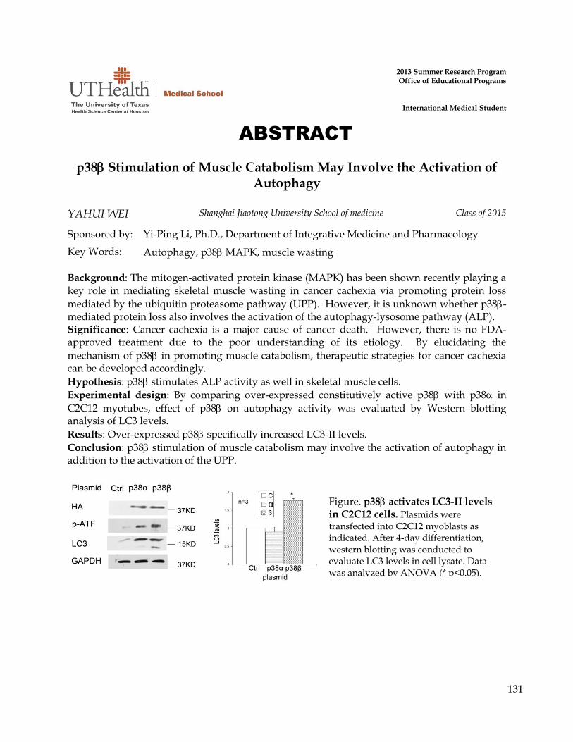
131
ABSTRACT
2013 Summer Research Program Office of Educational Programs
International Medical Student
p38 Stimulation of Muscle Catabolism May Involve the Activation of Autophagy
YAHUI WEI Shanghai Jiaotong University School of medicine Class of 2015
Sponsored by: Yi-Ping Li, Ph.D., Department of Integrative Medicine and Pharmacology
Key Words: Autophagy, p38 MAPK, muscle wasting Background: The mitogen-activated protein kinase (MAPK) has been shown recently playing a key role in mediating skeletal muscle wasting in cancer cachexia via promoting protein loss
mediated by the ubiquitin proteasome pathway (UPP). However, it is unknown whether p38-mediated protein loss also involves the activation of the autophagy-lysosome pathway (ALP). Significance: Cancer cachexia is a major cause of cancer death. However, there is no FDA-approved treatment due to the poor understanding of its etiology. By elucidating the
mechanism of p38 in promoting muscle catabolism, therapeutic strategies for cancer cachexia can be developed accordingly. Hypothesis: p38 stimulates ALP activity as well in skeletal muscle cells. Experimental design: By comparing over-expressed constitutively active p38 with p38 in
C2C12 myotubes, effect of p38 on autophagy activity was evaluated by Western blotting analysis of LC3 levels. Results: Over-expressed p38 specifically increased LC3-II levels. Conclusion: p38 stimulation of muscle catabolism may involve the activation of autophagy in addition to the activation of the UPP.
Figure. p38 activates LC3-II levels in C2C12 cells. Plasmids were
transfected into C2C12 myoblasts as indicated. After 4-day differentiation, western blotting was conducted to evaluate LC3 levels in cell lysate. Data was analyzed by ANOVA (* p<0.05).

132
ABSTRACT
2013 Summer Research Program Office of Educational Programs
International Medical Student
Association between Socioeconomic Status and Outcome for Descending Thoracic Aortic Aneurysm Repair
ZIPING WU Shanghai Jiao Tong University School of Medicine Class of 2018
Sponsored by: Anthony L. Estrera, MD and Charles C. Miller, PhD; Dept. of Cardiothoracic and Vascular Surgery
Key Words: Socioeconomic Status, Patients Outcome, Thoracic Aneurysm Repair Background: Previous studies report a difference in postoperative outcomes of descending thoracic aortic aneurysm (DTAA) repair based on race and ethnicity. Because race is often a surrogate for socioeconomic status, the goal of this study was to examine the effect of socioeconomic status on the surgical outcomes of our patients with DTAA. Methods: A prospectively maintained aortic database was queried for consecutive patients who underwent repair of DTAA between 1990 and 2012 (n = 567). Data was collected on patient demographics, comorbidities, hospital course, and early and late complications. Zip code level data were obtained from the US Census. A composite SES index was calculated based on a standardized algorithm utilizing principal SES census variables of occupational status, median household income, percentage below the poverty level, housing, and educational attainment. A Spearman ranked correlation analysis was performed for initial screening to determine association between patient’s SES and operative outcomes. Pre-planned analyses for mortality and major morbidity were performed using logistic regression and contingency table methods. Results: Of the total 567 patients, 412 were used for SES analysis due to the lack of information on the remainder. No meaningful associations were identified between SES score and any measure of mortality or major morbidity. Even in the screening analysis, only pump time was identified as statistically significant, and this seems likely to represent a type 1 statistical error given the large number of variables screened. Conclusion: Our results did not show any significant association between patient SES and postoperative outcomes after open DTAA repair. Zipcode areas are large and may be too economically heterogeneous to be useful for classifying person-level SES. We suggest using more incisive SES-measuring tools such as Hollingshead Index or Geocoding to estimate SES for future studies.

133
ABSTRACT
2013 Summer Research Program Office of Educational Programs
International Medical Student
Studies of Molecular Mechanisms Underlying CPS1 Induction by a Clock-enhancing Compound
TING-YU YEH Fu Jen Catholic University Class of 2018
Sponsored by: Dr. Zheng Chen PhD
Key Words: CPS1, High-fat diet, Promoter, cell line model Carbamoyl phosphate synthetase 1 (CPS1) is a liver-specific enzyme for the rate-limiting step of the urea cycle. Previous studies in Dr. Chen’s lab showed that a clock-enhancing compound, CEM5, can rescue the fatty liver phenotype of mice treated with high-fat diet (HFD). In accordance, CEM5 restored CPS1 protein level which was suppressed by HFD treatment. Using real-time qPCR analysis, I demonstrated a significant increase in CPS1 mRNA level. Together, these data indicates that CPS1 expression can be induced by CEM5 in HFD-fed mice. Given the fundamental role of CPS1 in liver metabolism, we are interested in the molecular mechanism whereby CEM5 restored CPS1 expression under HFD conditions. Literature study revealed several potential promoters and enhancers for CPS1. In particular, the TFSEARCH analysis identified a putative ROR response element (RORE) in the enhancer of CPS1. Since RORE is an essential responsive element in the core clock, future work will investigate the role of RORE in CPS1 induction by CEM5. To facilitate mechanistic studies, we also tried to establish a cell line model for CEM5 induction of Cps1. We have tried SV and Hepa1-6 cells. The qPCR and Western blotting results, however, showed low levels of Cps1 expression and thus rendered these cells not suitable for our studies. We are currently testing HeLa cells, which are known to express CPS1 to high levels. Together, these studies revealed an important transcriptional mechanism underlying CEM5 –mediated induction of Cps1, and identified a putative circadian transcription element which may be required for the transcriptional induction.

134
ABSTRACT
2013 Summer Research Program Office of Educational Programs
International Medical Student
Progression of Disease Despite Optimal Medical Management in Acute Type B Aortic Dissection
YW ZHU Shanghai JiaoTong University Class of 2015
Sponsored by: Kristofer M. Charlton-Ouw, MD, Cardiothoracic and Vascular Surgery
Key Words: aortic dissection, ischemia, surgical repair Background Treatment of acute type B aortic dissection (ABAD) is traditionally with medical management of hypertension. Emerging research suggests that thoracic endovascular aortic stent-graft repair (TEVAR) may play a role in preventing aortic-related mortality even in uncomplicated cases. We discuss the literature and present a patient with medically-managed uncomplicated aortic dissection that suffered progression of dissection with visceral malperfusion despite adequate blood pressure control. Case report A 50-year-old man with abdominal and chest pain was admitted and diagnosed with uncomplicated ABAD. After control of hypertension, his pain subsided. He had no evidence of branch vessel malperfusion but he did have incidental finding of a 3 cm right subclavian artery aneurysm. On office follow-up, his blood pressure was well controlled. One month after his original diagnosis, he suffered sudden onset of abdominal pain, nausea, vomiting and diarrhea and was readmitted. On CT, there was slight enlargement of the thoracoabdominal aorta with extension of his dissection into occluded celiac, superior mesenteric, and left renal arteries. The patient underwent emergent TEVAR. Completion angiogram showed persistent non-opacification of mesenteric arteries and an exploratory laparotomy was performed. He had no pulse in the mesenteric arteries and a right common iliac to superior mesenteric artery bypass was created. Postoperatively, the patient required hemodialysis and suffered severe liver dysfunction. Three weeks later, he became hemodynamically unstable with lactic acidosis and multi-organ dysfunction. Although the patient was intubated, he was lucid and indicated that he preferred no additional aggressive interventions. After a family meeting, he was made comfort care only and died. An autopsy showed stable thoracic aortic repair and patent visceral bypass but with hemorrhagic gastric necrosis. Conclusions TEVAR for uncomplicated ABAD is controversial. However, even patients with optimal blood pressure control can extend their dissection and suffer sequelae of malperfusion. Consideration should be given to revascularization of both the celiac and superior mesenteric artery branches in cases of acute malperfusion.