Embed Size (px)
Citation preview
NIA Clinical Guidelines
© 2016 Magellan Health, Inc. Proprietary Page 1 of 22
2016 NIA Clinical Guidelines for Medical Necessity Review
OB ULTRASOUND
_______________________________________________________________
NIA Clinical Guidelines
© 2016 Magellan Health, Inc. Proprietary Page 2 of 22
Guidelines for Clinical Review Determination
Preamble
NIA is committed to the philosophy of supporting safe and effective treatment for patients. The
medical necessity criteria that follow are guidelines for the provision of diagnostic imaging. These
criteria are designed to guide both providers and reviewers to the most appropriate diagnostic
tests based on a patient’s unique circumstances. In all cases, clinical judgment consistent with the
standards of good medical practice will be used when applying the guidelines. Guideline
determinations are made based on the information provided at the time of the request. It is
expected that medical necessity decisions may change as new information is provided or based on
unique aspects of the patient’s condition. The treating clinician has final authority and
responsibility for treatment decisions regarding the care of the patient.
Guideline Development Process
These medical necessity criteria were developed by NIA for the purpose of making clinical review
determinations for requests for diagnostic tests. The developers of the criteria sets included
representatives from the disciplines of radiology, internal medicine, nursing, and cardiology. They
were developed following a literature search pertaining to established clinical guidelines and
accepted diagnostic imaging practices.
All inquiries should be directed to:
National Imaging Associates, Inc.
6950 Columbia Gateway Drive
Columbia, MD 21046
Attn: NIA Associate Chief Medical Officer

NIA Clinical Guidelines
© 2016 Magellan Health, Inc. Proprietary Page 3 of 22
TABLE OF CONTENTS
TOC
OB ULTRASOUND GUIDELINES ___________________________________________________ 4
76805 – OB Ultrasound - Routine _____________________________________________________ 4
76811 – OB Ultrasound - Detailed ____________________________________________________ 6
76816 – OB Ultrasound - Monitoring __________________________________________________ 7
76818 – OB Biophysical Profile _____________________________________________________ 16
76820 – OB Ultrasound – Vessel Doppler ____________________________________________ 21
All guidelines reviewed between January – November 2015.

_______________________________________________________________
NIA Clinical Guidelines
© 2016 Magellan Health, Inc. Proprietary Page 4 of 22
TOC
OB ULTRASOUND GUIDELINES
76805 – OB Ultrasound - Routine
CPT Codes: 76801, +76802, 76805, +76810, 76813, +76814
INTRODUCTION:
A limited number of ultrasounds are considered standard of care in early pregnancy management.
These studies can be used to identify potential fetal abnormalities or other issues with the
pregnancy that are more amenable to resolution early in the pregnancy.
Ultrasounds required beyond the indications noted typically involve limited, follow-up or
transvaginal ultrasounds to monitor medical conditions and complexities and are covered in
Guideline for Obstetric Ultrasounds – Monitoring.
INDICATIONS FOR ROUTINE ULTRASOUND:
One ultrasound performed prior to fourteen (14) weeks gestation
One nuchal translucency measurement per pregnancy performed between eleven (11) and
fourteen (14) weeks gestation
One complete screening obstetric ultrasound, typically performed between 18 – 22 weeks
gestation
In some circumstances, such as late pregnancy care, the complete ultrasound may be done after
22 weeks
A second complete ultrasound may be approvable when the need is justified, such as when
patient is referred to another provider or specialist
ADDITIONAL INFORMATION RELATED TO OB US - ROUTINE:
Three-dimensional (3D) and Four-dimensional (4D) Ultrasounds are considered experimental and
investigational and are not indicated.
REFERENCES:
American College of Obstetricians and Gynecologists. (2009). ACOG practice bulletin No. 101:
Ultrasonography in pregnancy. Obstet Gynecol, 113, 451-461. doi:
10.1097/AOG.0b013e31819930b0.
American College of Radiology. (2014). ACR Appropriateness Criteria® Retrieved from
https://acsearch.acr.org/list.
American Institute of Ultrasound in Medicine. (2010). AIUM practice guideline for the performance
of obstetric ultrasound examinations. J Ultrasound Med, 9(1), 157-166. Retrieved from
http://www.jultrasoundmed.org/content/29/1/157.full.pdf+html.
Chen, M., Lee, C.P., Lam, Y.H., Tang, R.Y., Chan, B.C., Wong, S.F., . . . Tang, M.H. (2008).
Comparison of nuchal and detailed morphology ultrasound examinations in early pregnancy for

_______________________________________________________________
NIA Clinical Guidelines
© 2016 Magellan Health, Inc. Proprietary Page 5 of 22
fetal structural abnormality screening: A randomized controlled trial. Ultrasound Obstet Gynecol, 31(2), 136-146. doi: 10.1002/uog.5232.
Morin, L., Van den Hof, M.C. & Society of Obstetricians and Gynecologists of Canada. (June 2005).
SOGC clinical practice guidelines. Ultrasound evaluation of first trimester pregnancy
complications. Number 161, Int J Gynaecol Obstet, 93(1), 77-81. Retrieved from
http://sogc.org/guidelines/ultrasound-evaluation-of-first-trimester-pregnancy-complications.
Yagel, S., Cohen, S.M., Messing, B., & Valsky, D.V. (2009). Three-dimensional and four-dimensional
ultrasound applications in fetal medicine. Curr Opin Obstet Gynecol, 21(2), 167-174. doi:
10.1097/GCO.0b013e328329243c.

_______________________________________________________________
NIA Clinical Guidelines
© 2016 Magellan Health, Inc. Proprietary Page 6 of 22
TOC
76811 – OB Ultrasound - Detailed
CPT Codes: 76811, +76812
INTRODUCTION:
A detailed obstetric ultrasound “is not intended to be the routine scan performed for all
pregnancies. Rather, it is intended for a known or suspected fetal anatomic, genetic abnormality
(i.e., previous anomalous fetus, abnormal scan this pregnancy, etc.) or increased risk for fetal
abnormality (e.g. AMA, diabetic, fetus at risk due to teratogen or genetics, abnormal prenatal
screen). Thus, the performance of CPT 76811 is expected to be rare outside of referral practices with
special expertise in the identification of, and counseling about, fetal anomalies.” SMFM
INDICATIONS FOR DETAILED ULTRASOUND:
One detailed obstetric ultrasound per pregnancy is considered medically necessary for approved
medical conditions as listed in the Appendix.
ADDITIONAL INFORMATION RELATED TO OB US-DETAILED:
Three-dimensional (3D) and Four-dimensional (4D) Ultrasounds are considered experimental
and investigational and are not covered services.
REFERENCES:
American College of Obstetricians and Gynecologists. (2009). ACOG practice bulletin No. 101:
Ultrasonography in pregnancy. Obstet Gynecol, 113, 451-461. doi:
10.1097/AOG.0b013e31819930b0.
American College of Radiology. (2014). ACR Appropriateness Criteria® Retrieved from
https://acsearch.acr.org/list.
American Institute of Ultrasound in Medicine. (2010). AIUM practice guideline for the performance
of obstetric ultrasound examinations. J Ultrasound Med, 9(1), 157-166. Retrieved from
http://www.jultrasoundmed.org/content/29/1/157.full.pdf+html.
Society for Maternal Fetal Medicine, Coding Committee. (Revised December 27, 2012). White Paper on Ultrasound Code 76811. Retrieved from
https://www.smfm.org/attachedfiles/UltrasoundCode76811Revised-Dec272012.pdf
Yagel, S., Cohen, S.M., Messing, B., & Valsky, D.V. (2009). Three-dimensional and four-dimensional
ultrasound applications in fetal medicine. Curr Opin Obstet Gynecol, 21(2), 167-174. doi:
10.1097/GCO.0b013e328329243c.

_______________________________________________________________
NIA Clinical Guidelines
© 2016 Magellan Health, Inc. Proprietary Page 7 of 22
TOC
76816 – OB Ultrasound - Monitoring
CPT Codes: 76815, 76816, 76817
INTRODUCTION:
Prenatal ultrasounds may assist in the diagnosis and monitoring of complicating medical conditions
and major fetal anomalies. Some high-risk, complicated pregnancies may require regular
monitoring over time.
INDICATIONS FOR ULTRASOUND EXAMINATIONS TO ASSESS AND MONITOR HIGH-RISK
PREGNANCY:
Limited, follow-up transabdominal and transvaginal obstetric ultrasounds will be approved for
fetal, obstetrical or maternal complications when consistent with the indications and criteria below.
Condition Defined as or Evidenced by Frequency*
1. Advanced Maternal
Age
Maternal age of 35 years or older
for a screening ultrasound from 12
through 27 weeks of gestation.
Maternal age of thirty-eight (38)
years or older for antepartum
monitoring from 34 weeks.
One ultrasound from 12 through
27 weeks of gestation.
Ultrasounds (to accompany Non-
Stress Tests when needed for
amniotic fluid value checks) for
antepartum testing weekly from
34 weeks.
2. Amniotic fluid volume
abnormalities:
oligohydramnios Decreased amniotic fluid volume
relative to gestational age,
characterized by an amniotic fluid
index (AFI) less than 5 cm or
single deepest pocket less than 2
cm.
Ultrasounds once per week (to
accompany Non-Stress Tests
when needed for amniotic fluid
value checks) at diagnosis or as
determined by clinical reviewer.
polyhydramnios Increased amniotic fluid volume
relative to gestational age
characterized by an AFI greater
than or equal to 24 cm.
One ultrasound or as
determined by clinical reviewer.
3. Antiphospholipid
syndrome (APS) or
other maternal
autoimmune disease
such as Systemic
Lupus Erythematosis
(SLE)
Documented previous diagnosis of
antiphospholipid syndrome (APS),
or other maternal autoimmune
disease, such as Systemic Lupus
Erythematosis (SLE).
Ultrasounds every 4 weeks from
24-32 weeks, weekly thereafter
(to accompany Non-Stress Tests
when needed for amniotic fluid
value checks).
4. Asthma Severe, documented asthma
requiring daily medication such as
long-acting beta-agonist and/or
Ultrasounds every 4 weeks from
24-32 weeks, weekly thereafter
(to accompany Non-Stress Tests

_______________________________________________________________
NIA Clinical Guidelines
© 2016 Magellan Health, Inc. Proprietary Page 8 of 22
inhaled or oral steroids. when needed for amniotic fluid
value checks).
5. Cardiac disease,
maternal
Severe, with documented history of
structural, valvular or ischemic
heart disease.
Ultrasounds every 4 weeks from
24-32 weeks, weekly thereafter
or as determined by clinical
reviewer.
6. Cholestasis of
pregnancy
Documented elevated serum bile
acid (upper limit of normal is
between 10 and 14 µmol/L). or
physician diagnosis based on
patient symptoms.
Ultrasounds (to accompany Non-
Stress Tests when needed for
amniotic fluid value checks) for
antepartum testing weekly
starting at diagnosis.
7. Decreased fetal
movement
Documented maternal perception
of decreased fetal activity.
One ultrasound upon
occurrence.
8. Diabetes mellitus-
gestational
Diabetes arising or first diagnosed
during pregnancy.
Medication (e.g. insulin,
glyburide) is required to
control.
Ultrasounds at initiation of
medications, every 4 weeks until
32 weeks, weekly thereafter (to
accompany Non-Stress Tests
when needed for amniotic fluid
value checks).
Controlled by diet, without
requiring medications.
One ultrasound during third
trimester to screen for
macrosomia.
9. Diabetes mellitus-
Type I or Type II, pre-
gestational
Diabetes diagnosed prior to
pregnancy requiring medication
(e.g. insulin, glyburide) to control.
Ultrasounds every 4 weeks from
24-32 weeks, weekly thereafter
(to accompany Non-Stress Tests
when needed for amniotic fluid
value checks).
10. Drug/ ETOH abuse, or
methadone use/abuse
Active, documented in chart. Ultrasounds every 4 weeks from
24-32 weeks, weekly thereafter
(to accompany Non-Stress Tests
when needed for amniotic fluid
value checks).
11. Fetal anomaly, major Suspected or known major
structural anomaly, including
documented history of previous
congenital anomaly.
One ultrasound for screening of
suspected anomaly.
Follow-up ultrasounds for
observation of identified fetal
anomaly as determined by
clinical reviewer.
12. Fetal size/due date
discrepancy
A significant discrepancy of 3 or
more between fundal height
(centimeters) to gestational age
(weeks).
One ultrasound or as
determined by clinical reviewer.
13. Hypertension, chronic Blood pressure ≥ 140 mm Hg
systolic and/or 90 mm Hg diastolic,
diagnosed before conception or
Ultrasounds every 4 weeks from
24-32 weeks, weekly thereafter
(to accompany Non-Stress Tests

_______________________________________________________________
NIA Clinical Guidelines
© 2016 Magellan Health, Inc. Proprietary Page 9 of 22
before twenty (20) weeks gestation. when needed for amniotic fluid
value checks).
14. Hyperthyroid disease,
maternal
Uncontrolled, defined by
suppressed TSH level with related
maternal symptoms.
Ultrasounds every 4 weeks from
24-32 weeks, weekly thereafter
(to accompany Non-Stress Tests
when needed for amniotic fluid
value checks) as long as
treatment is ongoing, even if
TSH has normalized.
15. Hypothyroid disease,
maternal
Uncontrolled, defined by elevated
thyroid stimulating hormone
(TSH) and related maternal
symptoms.
Ultrasounds every 4 weeks from
24-32 weeks, weekly thereafter
(to accompany Non-Stress Tests
when needed for amniotic fluid
value checks) as long as
symptoms persist, even if TSH
has normalized during
treatment.
16. Human
Immunodeficiency
Virus (HIV) infection,
maternal
Confirmed HIV, documented in
chart.
Ultrasounds every 4 weeks from
24-32 weeks.
17. Incompetent cervix
and no cerclage
Premature opening of the cervix. Ultrasounds every two weeks
during 16-24 weeks of gestation
to determine need for
intervention. Berghella,
18. Intrauterine Fetal
Death (IUFD), history
Documented history of IUFD. Weekly ultrasounds from 32
weeks or from 2 weeks prior to
the gestational age of prior
IUFD (to accompany non-stress
test when needed for AFV
checks) or as determined by
clinical reviewer.
19. Intrauterine Growth
Restriction (IUGR)
Estimated fetal weight less than
the 10th percentile for gestational
age Scifres or an estimated fetal
weight between the 10th and 15th
percentile for gestational age and
an abdominal circumference less
than the 5th percentile.
Weekly ultrasounds at diagnosis
or as determined by clinical
reviewer.
20. Malpresentation Presentation other than vertex
after 36 weeks.
One ultrasound at or beyond 36
weeks of gestation or as
determined by clinical reviewer.
21. MSAFP (Maternal
serum alpha-
fetoprotein) level,
elevated
Unexplained, elevated MSAFP, >
2.5 MoMs (quantitative unit of
measure for MSAFP reported as
multiples of the median).
Ultrasounds every 4 weeks from
24-32 weeks, weekly thereafter
(to accompany Non-Stress Tests
when needed for amniotic fluid
value checks).
22. Multiple gestations
Two or more fetuses.
Ultrasounds every 4 weeks from
24-32 weeks, weekly thereafter

_______________________________________________________________
NIA Clinical Guidelines
© 2016 Magellan Health, Inc. Proprietary Page 10 of 22
(to accompany Non-Stress Tests
when needed for amniotic fluid
value checks).
Monochorionic
twins
Twins that share a placenta and
an outer membrane.
Ultrasounds every 2 weeks
between 16 and 24 weeks to
assess for twin-twin transfusion.
23. Obesity in pregnancy Maternal body mass index (BMI) >
30 kg/m2 conception (usually
determined during first obstetrical
exam).
One ultrasound between 30 and
34 weeks of gestation.
24. PAPP-A (Pregnancy-
associated plasma
protein A), abnormal
value
Unexplained, <0.3 MoMs
(multiples of the median).
Ultrasounds every 4 weeks from
24-32 weeks, weekly thereafter
(to accompany Non-Stress Tests
when needed for amniotic fluid
value checks).
25. Placenta previa Asymptomatic (without bleeding)
with documented prior ultrasound
report of placenta located near or
over the internal cervical orifice.
One ultrasound between 30-34
weeks; possible follow-up at 36 –
38 weeks if condition continues.
26. Placental abruption Vaginal bleeding with suspected
placental abruption.
One ultrasound or as
determined by physician
reviewer.
27. Post term pregnancy Pregnancy that is at or beyond
forty (40) weeks of gestation
Ultrasounds two times per week
post term (to accompany Non-
Stress Tests when needed for
amniotic fluid value checks).
28. Pre-eclampsia New onset of blood pressure
elevation exceeding 140/90 mm Hg
after twenty (20) weeks gestation.
Upon occurrence, every 4 weeks
until 32 weeks, weekly
thereafter (to accompany Non-
Stress Tests when needed for
amniotic fluid value checks).
29. Premature rupture of
membranes
Confirmed and documented in
chart.
One ultrasound or as
determined by physician
reviewer.
30. Pre-term delivery
history
Patient has had a previous
pregnancy that delivered between
20 and 37 weeks of gestation.
Ultrasounds every two weeks
during 16-24 weeks of gestation
to determine need for
intervention.
31. Pre-term labor Active labor defined as regular
painful contractions (≥4 in 20
minutes or ≥8 in one hour) and
documented cervical change.
One ultrasound upon
occurrence.
32. Renal disease,
maternal
Documented history of
parenchymal renal disease prior to
pregnancy.
Ultrasounds every 4 weeks from
24-32 weeks, weekly thereafter
(to accompany Non-Stress Tests
when needed for amniotic fluid
value checks).
33. Sickle cell disease,
maternal
Documented maternal sickle cell
disease (not just trait), normal Hb
Ultrasounds every 4 weeks from
24-32 weeks, weekly thereafter

_______________________________________________________________
NIA Clinical Guidelines
© 2016 Magellan Health, Inc. Proprietary Page 11 of 22
A is present in the blood of patient
at a lower level than Hb S. Frenette
(to accompany Non-Stress Tests
when needed for amniotic fluid
value checks).
34. Vaginal bleeding Suspected placental abruption,
suspected placenta previa,
suspected spontaneous abortion,
etc.
One ultrasound or as
determined by physician
reviewer.
Situations beyond the medical conditions above:
1. Adjunct to procedures An ultrasound may be indicated
for amniocentesis, amnioinfusion,
cervical cerclage, fetoscopy, shunt
placement, etc.
Upon occurrence when discussed
with a clinical reviewer.
2. Other high-risk
medical conditions
Medical conditions that
contribute to high risk that have
not been listed above.
Upon occurrence when discussed
with a clinical reviewer.
Transvaginal Ultrasounds are generally used for the following scenarios:
1. Incompetent cervix and
no cerclage
Premature opening of the cervix. Ultrasounds every two weeks
during 16-24 weeks of gestation
to determine need for
intervention.
2. Pre-term delivery
history
Patient has had a previous
pregnancy that delivered between
20 and 37 weeks of gestation.
Ultrasounds every two weeks
during 16-24 weeks of gestation
to determine need for
intervention.
3. Placenta previa Asymptomatic (without bleeding)
with documented prior
ultrasound report of placenta
located near or over the internal
cervical orifice.
One ultrasound between 30-34
weeks; possible follow-up at 36 –
38 weeks if condition continues.
4. Pre-term labor Active, regular painful
contractions (≥4 in 20 minutes or
≥8 in one hour) and documented
cervical change.
One ultrasound upon
occurrence.
*Typical frequency is provided as a guide for authorizations, though many patients may not need
monitoring this frequently. More frequent monitoring will require physician review.
ADDITIONAL INFORMATION RELATED TO OB US:
Antepartum Fetal Testing is appropriate for monitoring patients at increased risk for adverse
perinatal outcomes: Nageotte et al , Liston, et al
o Testing may start after 24 weeks but usually starts at 32 weeks or beyond;
o A reasonable first line antepartum fetal surveillance strategy includes a Non-Stress Test
(NST) and, when indicated, Amniotic Fluid Volume (AFV) assessment, reserving the
Biophysical Profile (BPP) for abnormal NST results. Haws, et al
A single transvaginal ultrasound for screening of cervical length in singleton gestations without
previous preterm birth (low risk patients) between 18 and 24 weeks gestation is supported by
the Society for Maternal Fetal Medicine, Society for Maternal Fetal Medicine. Screening of cervical length

_______________________________________________________________
NIA Clinical Guidelines
© 2016 Magellan Health, Inc. Proprietary Page 12 of 22
should be performed by an appropriately trained physician to determine possible need for
intervention. If cervical length is normal, no further action is required. If screening indicates
a short length, treatment may be indicated. No follow-up or serial cervical length exams are
required.
A biophysical profile (BPP) consists of a NST plus 4 ultrasound components (fetal movement,
fetal muscle tone, amniotic fluid volume and fetal breathing movement):
o A BPP is an appropriate second line (back-up) testing strategy and is performed on the
same day when the first line NST test is non-reactive or non-interpretable (non-
reassuring).
o See separate clinical guideline for Biophysical Profile.
A positive quad screen for fetal Down Syndrome is not considered an indication for antepartum
testing.
Three-dimensional (3D) and Four-dimensional (4D) Ultrasounds are considered experimental
and investigational as there is no evidence that they alter management over a two-dimensional
(2D) ultrasound in a way that improves outcomes.
REFERENCES:
Akkerman, D., Cleland, L., Croft, G., Eskuchen, K., Heim, C., Levine, A., . . . Westby, E. (2012).
Institute for Clinical Systems Improvement. Routine Prenatal Care. Retrieved from
http://bit.ly.Prenatal0712.
American College of Obstetricians and Gynecologists. (2001 Reaffirmed 2010). ACOG Practice
Bulletin, Clinical Management for Obstetrician-Gynecologists. No. 30: Gestational Diabetes.
Retrieved from
http://www.acog.org/~/media/List%20of%20Titles/PBListOfTitles.pdf?dmc=1&ts=20120711T160
6386613
American College of Obstetricians and Gynecologists. (2000 Reaffirmed 2010). ACOG Practice
Bulletin, Clinical Management for Obstetrician-Gynecologists. No. 22: Fetal Macrosomia.
Retrieved from:
http://www.acog.org/~/media/List%20of%20Titles/PBListOfTitles.pdf?dmc=1&ts=20120711T160
6386613
American College of Obstetricians and Gynecologists (ACOG), Committee on Obstetric Practice,
Opinion: “Incidentally Detected Short Cervical Length”, Number 522, April, 2012. doi:
10.1097/AOG.0b013e3182538e64.
American College of Obstetricians and Gynecologists. (1999). ACOG practice bulletin No. 9:
Antepartum fetal surveillance. Int J Gynaecol Obstet. 68, 175-185. Retrieved from
http://www.ncbi.nlm.nih.gov/pubmed/?term=American+College+of+Obstetricians+and+Gynecolo
gists.+(1999).+ACOG+practice+bulletin+No.+9+Antepartum+fetal+surveillance.+Int+J+Gynaec
ol+Obstet.+68%2C+175-185

_______________________________________________________________
NIA Clinical Guidelines
© 2016 Magellan Health, Inc. Proprietary Page 13 of 22
American College of Obstetricians and Gynecologists. (2004). ACOG practice bulletin No. 55:
Management of post term pregnancy. Obstet Gynecol. 104(3), 639-646. Retrieved from
http://www.ncbi.nlm.nih.gov/pubmed/15339790.
American College of Obstetricians and Gynecologists. (2009). ACOG practice bulletin No. 101:
Ultrasonography in pregnancy. Obstet Gynecol, 113, 451-461. doi:
10.1097/AOG.0b013e31819930b0.
American College of Radiology. (2014). ACR Appropriateness Criteria® Retrieved from
https://acsearch.acr.org/list.
American Institute of Ultrasound in Medicine. (2010). AIUM practice guideline for the performance
of obstetric ultrasound examinations. J Ultrasound Med, 9(1), 157-166. Retrieved from
http://www.jultrasoundmed.org/content/29/1/157.full.pdf+html.
Bellamy, L., Casas, J.P., Hingorani, A.D., & Williams, D.J. (2007). Pre-eclampsia and risk of
cardiovascular disease and cancer in later life: Systematic review and meta-analysis. British Medical Journal, 335(7627), 974. Retrieved from
http://www.ncbi.nlm.nih.gov/pubmed/17975258.
Berghella, V. (2012). The Society for Maternal-Fetal Medicine: Publications Committee,
Progesterone and Preterm Birth Prevention: Translating Clinical Trials Data into Clinical
Practice, American Journal of Obstetrics and Gynecology. doi: 10.1016/j.ajog.2012.03.010.
Bjorklund, N.K., Evans, J.A., Greenberg, C.R., Seargeant, C.R., Schneider, C.E., & Chodirker, B.N.
(2004). The C677T methylenetetrahydrofolate reductase variant and third trimester obstetrical
complications in women with unexplained elevations of maternal serum alpha-fetoprotein.
Reprod Biol Endocrinol., 2, 65. doi: 10.1186/1477-7827-2-65.
Caughey, A.B., Stotland, N.E., Washington, A.E., & Escobar, G.J. (2007). Maternal complications of
pregnancy increase beyond 40 weeks’ gestation. Am J Obstet Gynecol, 196(2), 155 el – 155e6.
doi: 10.1016/j.ajog.2006.08.040.
Centers for Medicare & Medicaid Services (CMS). Retrieved from
http://www.cms.gov/MCD/viewncd.asp?ncd_id=220.5&ncd_version=3&basket=ncd%3A220.5%3A
3%3AUltrasound+Diagnostic+Procedures.
Cejtin, H.E. (2008). Gynecologic issues in the HIV-infected woman. Infect Dis Clin North Am, 22(4),
709-vii. doi: 10.1016/j.idc.2008.05.006
Chen, M., Lee, C.P., Lam, Y.H., Tang, R.Y., Chan, B.C., Wong, S.F., . . . Tang, M.H. (2008).
Comparison of nuchal and detailed morphology ultrasound examinations in early pregnancy for
fetal structural abnormality screening: A randomized controlled trial. Ultrasound Obstet Gynecol, 31(2), 136-146. doi: 10.1002/uog.5232.
Clinical Practice Obstetrics Committee, Maternal Fetal Medicine Committee, Delaney,
M., Roggensack, A., Leduc, D.C., Ballermann, C., . . . Wison, K. (2008). Guidelines for the
management of pregnancy at 41+0 to 42+0 weeks. J Obstet Gynaecol Can, 30(9), 800-823.
Retrieved from http://www.ncbi.nlm.nih.gov/pubmed/18845050?dopt=Abstract.

_______________________________________________________________
NIA Clinical Guidelines
© 2016 Magellan Health, Inc. Proprietary Page 14 of 22
Davies, G.A.L., Maxwell, C., McLeod, L., Gagnon, R., Basso, M., Bos, H., . . . Society of Obstetricians
and Gynaecologists of Canada. (2010). SOGC clinical practice guideline: Obesity in pregnancy. J Obstet Gynaecol Can, 32, 165. Retrieved from http://www.ncbi.nlm.nih.gov.
Dobbenga-Rhodes, Y.A. & Prive, A.M. (2006). Assessment and evaluation of the woman with
cardiac disease during pregnancy. J Perinat Neonatal Nurs, 20(4), 295-302. Retreived from
http://www.ncbi.nlm.nih.gov.
Freeman, R.K. (2008). Antepartum testing in patients with hypertensive disorders in pregnancy.
Semin Perinatol, 32(4), 271-273. doi: 10.1053/j.semperi.2008.04.009.
Frenette, P.S., & Atweh, G.F. (2007). Sickle cell disease: old discoveries, new concepts, and future
promise. J Clin Invest, 117(4), 850-858. doi: 10.1172/JCI30920
Froen, J.F., Tveit, J.V.H., Saastad, E., Bordahl, P.E., Stray-Pedersen, B., Heazell, A.E., . . . Fretts,
R.C. (2008). Management of decreased fetal movement. Semin Perinatol, 32(4), 307-311.
Retrieved from http://www.ncbi.nlm.nih.gov/pubmed/18652933
Geenes, V., & Williamson, C. (2009). Intrahepatic cholestasis of pregnancy. World J Gastroenterol, 15(17), 2049-2066. doi: 10.3748/wjg.15.2049
Haws, R.A., Yakoob, M.Y., Soomro, T., Menezes, E.V., Darmstadt, G.L., . . . Bhutta, Z.A. (2009).
Reducing stillbirths: screening and monitoring during pregnancy and labour. BMC Pregnancy Childbirth, 9 Suppl 1, S5. Retrieved from http://www.ncbi.nlm.nih.gov/pubmed/19426468
Kelly, L., Evans, L., & Messenger, D. (2005). Controversies around gestational diabetes. Can Fam Physician, 51(5), 688-695. Retrieved from
http://www.ncbi.nlm.nih.gov/pmc/articles/PMC1472928/pdf/jCFP_v051_pg688.pdf.
Kennelly, M.M., & Sturgiss, S.N. (2007). Management of small-for-gestational-age twins with
absent/reversed end diastolic flow in the umbilical artery: Outcome of a policy of daily
biophysical profile (BPP). Prenat Diagn, 27(1), 77-80. doi: 10.1002/pd.1630
Lalor, J.G., Fawole, B., Alfirevic, Z., & Devane, D. (2008). Biophysical profile for fetal assessment in
high risk pregnancies. Cochrane Database of Systematic Reviews, Issue 1. Art. No.: CD000038.
doi: 10.1002/14651858.CD000038.pub2
Liston, R., Sawchuck, D., Young, D., Society of Obstetrics and Gynaecologists of Canada & British
Columbia Perinatal Health Program. (2007). Fetal health surveillance: Antepartum and
intrapartum consensus guideline. J Obstet Gynaecol Can, 29 (9 Suppl 4), 53-56. Retrieved from
http://www.ncbi.nlm.nih.gov/pubmed/17845745.
Morin, L., Van den Hof, M.C. & Society of Obstetricians and Gynecologists of Canada. (June 2005).
SOGC clinical practice guidelines. Ultrasound evaluation of first trimester pregnancy
complications. Number 161, Int J Gynaecol Obstet, 93(1), 77-81. Retrieved from
http://sogc.org/guidelines/ultrasound-evaluation-of-first-trimester-pregnancy-complications.
Roberts, C.L., Bell, J.C., Ford, J.B., Hadfield, R.M., Algert, C.S. & Morris, J.M. (2008). The accuracy
of reporting of the hypertensive disorders of pregnancy in population health data. Hypertens Pregnancy, 27, 285-297. Retreived from doi: 10.1080/10641950701826695.

_______________________________________________________________
NIA Clinical Guidelines
© 2016 Magellan Health, Inc. Proprietary Page 15 of 22
Scifres, C.M., & Nelson, D.M. (2009). Intrauterine growth restriction, human placental development
and trophoblast cell death. J Physiol, 587(pt 14), 3453-3458. doi: 10.1113/jphysiol.2009.173252.
Sigmore, C., Freeman, R.K., & Spong, C.Y. (2009). Antenatal testing – a reevaluation: Executive
summary of a Eunice Kennedy Shriver National Institute of Child Health and Human
Development workshop. Obstet Gynecol, 113(3), 687-701. doi: 10.1097/AOG.0b013e318197bd8a.
Society for Maternal Fetal Medicine. (June, 2013). Coding and billing for transvaginal ultrasound to
assess second-trimester cervical length, Contemporary OB/GYN. http://contemporaryobgyn.modernmedicine.com/contemporary-obgyn/news/coding-and-billing-
transvaginal-ultrasound-assess-second-trimester-cervical-?destination=node%2F370372.
Society for Maternal Fetal Medicine, Coding Committee. (Revised December 27, 2012). White Paper on Ultrasound Code 76811. Retrieved from
https://www.smfm.org/attachedfiles/UltrasoundCode76811Revised-Dec272012.pdf
Van den, Hof M., & Crane, J. (2001). SOGC clinical practice guidelines: Ultrasound cervical
assessment in predicting preterm birth. J Soc Obstet Gynaecol Can, 23(5), 418-421. Retrieved
from http://sogc.org/wp-content/uploads/2013/01/102E-CPG-May2001.pdf
Yagel, S., Cohen, S.M., Messing, B., & Valsky, D.V. (2009). Three-dimensional and four-dimensional
ultrasound applications in fetal medicine. Curr Opin Obstet Gynecol, 21(2), 167-174. doi:
10.1097/GCO.0b013e328329243c.
Yinon, Y., Nevo, O., Xu, J., Many, A., Rolf, A., Todros, T., . . . Canniggia, I. (2008). Severe
intrauterine growth restriction pregnancies have increased placental endoglin levels. Am J Pathol, 172(1), 77-85. doi: 10.2353/ajpath.2008.070640.

_______________________________________________________________
NIA Clinical Guidelines
© 2016 Magellan Health, Inc. Proprietary Page 16 of 22
TOC
76818 – OB Biophysical Profile
CPT Codes: 76818, 76819
INTRODUCTION:
Antepartum fetal testing is commonly performed in pregnancies at increased risk for fetal
compromise. The Non-Stress Test (NST) is the preferable first line antepartum fetal testing
modality and may be supplemented with serial assessments of amniotic fluid volume for clinical
scenarios with the potential for decreased amniotic fluid volume. The fetal biophysical profile is
best reserved as a back-up testing methodology for those fetuses in which the NST is non-
reassuring (non-reactive, non-interpretable). There is insufficient evidence at this time to support
the use of the BPP as a first line antepartum fetal testing modality. See Appendix for details.
INDICATIONS FOR BIOPHYSICAL PROFILE:
A biophysical profile BPP consists of a NST plus four (4) ultrasound components: fetal
movement, fetal muscle tone, amniotic fluid volume and fetal breathing movement. A BPP is an
appropriate second line (back-up) testing strategy when the NST component of the BPP is non-
reactive or non-interpretable (non-reassuring).
Each BPP performed for follow-up of a high risk patient must include a NST performed the
same day that is non-reassuring, unless the fetus has evidence of suspected congenital fetal
heart block and the heart rate is uninterpretable or an in-office NST is unavailable. .
There is insufficient evidence at this time to support use of the biophysical profile (BPP) for the
assessment of fetal well-being in high-risk pregnancies compared to a NST or NST and AFV.
Compared with conventional fetal monitoring, which is based primarily on
cardiotocography/NST, BPP appears to offer no improvement in pregnancy outcomes (Grade C
evidence). When a patient meets the indications for antepartum fetal surveillance noted below, a
NST would be done (when available), and when non-reactive, the 4 ultrasound components of
the BPP would be completed.
Condition Defined as or Evidenced by
1. Advanced Maternal Age Maternal age of thirty-eight (38)
years or older.
2. Amniotic fluid volume abnormalities:
oligohydramnios Decreased amniotic fluid volume
relative to gestational age,
characterized by an amniotic fluid
index (AFI) less than 5 cm. or single
deepest pocket is less than 1 cm by 2
cm.
polyhydramnios Increased amniotic fluid volume
relative to gestational age
characterized by an AFI greater
than or equal to 24 cm.
3. Antiphospholipid syndrome (APS) or other maternal
autoimmune disease such as Systemic Lupus
Erythematosis (SLE)
Documented previous diagnosis of
antiphospholipid syndrome (APS), or
other maternal autoimmune disease,
_______________________________________________________________
NIA Clinical Guidelines
© 2016 Magellan Health, Inc. Proprietary Page 17 of 22
such as Systemic Lupus
Erythematosis (SLE).
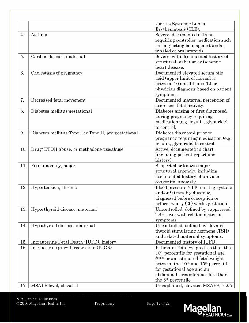
4. Asthma Severe, documented asthma
requiring controller medication such
as long-acting beta agonist and/or
inhaled or oral steroids.
5. Cardiac disease, maternal Severe, with documented history of
structural, valvular or ischemic
heart disease.
6. Cholestasis of pregnancy Documented elevated serum bile
acid (upper limit of normal is
between 10 and 14 µmol/L) or
physician diagnosis based on patient
symptoms.
7. Decreased fetal movement Documented maternal perception of
decreased fetal activity.
8. Diabetes mellitus-gestational Diabetes arising or first diagnosed
during pregnancy requiring
medication (e.g. insulin, glyburide)
to control.
9. Diabetes mellitus-Type I or Type II, pre-gestational Diabetes diagnosed prior to
pregnancy requiring medication (e.g.
insulin, glyburide) to control.
10. Drug/ ETOH abuse, or methadone use/abuse Active, documented in chart
(including patient report and
history).
11. Fetal anomaly, major Suspected or known major
structural anomaly, including
documented history of previous
congenital anomaly.
12. Hypertension, chronic Blood pressure ≥ 140 mm Hg systolic
and/or 90 mm Hg diastolic,
diagnosed before conception or
before twenty (20) weeks gestation.
13. Hyperthyroid disease, maternal Uncontrolled, defined by suppressed
TSH level with related maternal
symptoms.
14. Hypothyroid disease, maternal Uncontrolled, defined by elevated
thyroid stimulating hormone (TSH)
and related maternal symptoms.
15. Intrauterine Fetal Death (IUFD), history Documented history of IUFD.
16. Intrauterine growth restriction (IUGR) Estimated fetal weight less than the
10th percentile for gestational age, Scifres or an estimated fetal weight
between the 10th and 15th percentile
for gestational age and an
abdominal circumference less than
the 5th percentile.
17. MSAFP level, elevated Unexplained, elevated MSAFP, > 2.5

_______________________________________________________________
NIA Clinical Guidelines
© 2016 Magellan Health, Inc. Proprietary Page 18 of 22
MoMs (quantitative unit of measure
for MSAFP reported as multiples of
the median).
18. Multiple gestations Two or more fetuses.
Monochorionic twins
Twins that share a placenta and an
outer membrane.
19. PAPP-A, abnormal value Unexplained, <0.3 MoMs (multiples
of the median).
20. Placental abruption Vaginal bleeding with suspected
placental abruption.
21. Post term pregnancy Pregnancy that is at or beyond forty
(40) weeks of gestation.
22. Pre-eclampsia New onset of blood pressure
elevation exceeding 140/90 mm Hg
after twenty (20) weeks’ gestation.
23. Premature rupture of membranes Confirmed and documented in chart.
24. Renal disease, maternal Documented history of parenchymal
renal disease prior to pregnancy.
25. Sickle cell disease, maternal Documented maternal sickle cell
disease (not just trait) -, normal Hb
A is present in the blood of patient
at a lower level than Hb S.
26. Other high-risk medical conditions Medical conditions that contribute to
high risk that have not been listed
above.
REFERENCES:
American College of Obstetricians and Gynecologists. (2014). ACOG practice bulletin No. 145:
Antepartum fetal surveillance.
American College of Obstetricians and Gynecologists. (2004). ACOG practice bulletin No. 55:
Management of post term pregnancy. Obstet Gynecol. 104(3), 639-646. Retrieved from
http://www.ncbi.nlm.nih.gov/pubmed/15339790.
American College of Obstetricians and Gynecologists. (2009). ACOG practice bulletin No. 101:
Ultrasonography in pregnancy. Obstet Gynecol, 113, 451-461. doi:
10.1097/AOG.0b013e31819930b0.
American College of Radiology. (2014). ACR Appropriateness Criteria® Retrieved from
https://acsearch.acr.org/list.
American Institute of Ultrasound in Medicine. (2010). AIUM practice guideline for the performance
of obstetric ultrasound examinations. J Ultrasound Med, 9(1), 157-166. Retrieved from
http://www.jultrasoundmed.org/content/29/1/157.full.pdf+html.
Bellamy, L., Casas, J.P., Hingorani, A.D., & Williams, D.J. (2007). Pre-eclampsia and risk of
cardiovascular disease and cancer in later life: Systematic review and meta-analysis. British

_______________________________________________________________
NIA Clinical Guidelines
© 2016 Magellan Health, Inc. Proprietary Page 19 of 22
Medical Journal, 335(7627), 974. Retrieved from
http://www.ncbi.nlm.nih.gov/pubmed/17975258.
Bjorklund, N.K., Evans, J.A., Greenberg, C.R., Seargeant, C.R., Schneider, C.E., & Chodirker, B.N.
(2004). The C677T methylenetetrahydrofolate reductase variant and third trimester obstetrical
complications in women with unexplained elevations of maternal serum alpha-fetoprotein.
Reprod Biol Endocrinol., 2, 65. doi: 10.1186/1477-7827-2-65.
Caughey, A.B., Stotland, N.E., Washington, A.E., Escobar, G.J. (2007). Maternal complications of
pregnancy increase beyond 40 weeks’ gestation. Am J Obstet Gynecol, 196(2), 155 el – 155e6.
doi: 10.1016/j.ajog.2006.08.040.
Cejtin, H.E. (2008). Gynecologic issues in the HIV-infected woman. Infect Dis Clin North Am, 22(4),
709-vii. doi: 10.1016/j.idc.2008.05.006
Clinical Practice Obstetrics Committee, Maternal Fetal Medicine Committee, Delaney, M.,
Roggensack, A., Leduc, D.C., Ballermann, C., … Wison, K. (2008). Guidelines for the
management of pregnancy at 41+0 to 42+0 weeks. J Obstet Gynaecol Can, 30(9), 800-823.
Retrieved from http://www.ncbi.nlm.nih.gov/pubmed/18845050?dopt=Abstract.
Davies, G.A.L., Maxwell, C., McLeod, L., Gagnon, R., Basso, M., Bos, H., … Society of Obstetricians
and Gynaecologists of Canada. (2010). SOGC clinical practice guideline: Obesity in pregnancy. J Obstet Gynaecol Can, 32, 165. Retrieved from http://www.ncbi.nlm.nih.gov.
Dobbenga-Rhodes, Y.A. & Prive, A.M. (2006). Assessment and evaluation of the woman with
cardiac disease during pregnancy. J Perinat Neonatal Nurs, 20(4), 295-302. Retreived from
http://www.ncbi.nlm.nih.gov.
Freeman, R.K. (2008). Antepartum testing in patients with hypertensive disorders in pregnancy.
Semin Perinatol, 32(4), 271-273. doi: 10.1053/j.semperi.2008.04.009.
Frenette, P.S., & Atweh, G.F. (2007). Sickle cell disease: old discoveries, new concepts, and future
promise. J Clin Invest, 117(4), 850-858. doi: 10.1172/JCI30920
Froen, J.F., Tveit, J.V.H., Saastad, E., Bordahl, P.E., Stray-Pedersen, B., Heazell, A.E., …Fretts,
R.C. (2008). Management of decreased fetal movement. Semin Perinatol, 32(4), 307-311.
Retrieved from http://www.ncbi.nlm.nih.gov/pubmed/18652933
Gabbe Obstetrics, Fourth Edition (Eds Gabbe, Niebyl, Simpson) Chapter 12 Antepartum Fetal
evaluation (Auth Druzin, Gabbe, Reed)
Geenes, V., & Williamson, C. (2009). Intrahepatic cholestasis of pregnancy. World J Gastroenterol, 15(17), 2049-2066. doi: 10.3748/wjg.15.2049
Kelly, L., Evans, L., & Messenger, D. (2005). Controversies around gestational diabetes. Can Fam Physician, 51(5), 688-695. Retrieved from
http://www.ncbi.nlm.nih.gov/pmc/articles/PMC1472928/pdf/jCFP_v051_pg688.pdf.

_______________________________________________________________
NIA Clinical Guidelines
© 2016 Magellan Health, Inc. Proprietary Page 20 of 22
Kennelly, M.M., & Sturgiss, S.N. (2007). Management of small-for-gestational-age twins with
absent/reversed end diastolic flow in the umbilical artery: Outcome of a policy of daily
biophysical profile (BPP). Prenat Diagn, 27(1), 77-80. doi: 10.1002/pd.1630
Lalor, J.G., Fawole, B., Alfirevic, Z., & Devane, D. Biophysical profile for fetal assessment in high
risk pregnancies. Cochrane Database of Systematic Reviews 2008, Issue 1. Art. No.: CD000038.
doi: 10.1002/14651858.CD000038.pub2
Liston, R., Sawchuck, D., Young, D., Society of Obstetrics and Gynaecologists of Canada & British
Columbia Perinatal Health Program. (2007). Fetal health surveillance: Antepartum and
intrapartum consensus guideline. J Obstet Gynaecol Can, 29 (9 Suppl 4), 53-56. Retrieved from
http://www.ncbi.nlm.nih.gov/pubmed/17845745.
Management of High Risk Pregnancy, Eds Queenan, Spong, and Lockwood Fifth Edition
Antepartum fetal monitoring (Shaffer,Parer)
Manning, F.A. (1999) Fetal biophysical profile. Obstet Gynecol Clin North Am, 26(4), 557-577.
Retrieved from http://www.uptodate.com/contents/the-fetal-biophysical-profile.
Nageotte, M.P., Towers, C.V., Asrat, T., & Freeman, R.K. (1994). Perinatal outcome with the
modified biophysical profile. Am J Obstet Gynecol, 170(6), 1672-1676. Retreived from
http://www.ncbi.nlm.nih.gov/pubmed/8203424.
Roberts, C.L., Bell, J.C., Ford, J.B., Hadfield, R.M., Algert, C.S. & Morris, J.M. (2008). The accuracy
of reporting of the hypertensive disorders of pregnancy in population health data. Hypertens Pregnancy, 27, 285-297. Retreived from doi: 10.1080/10641950701826695.
Scifres, C.M., & Nelson, D.M. (2009). Intrauterine growth restriction, human placental development
and trophoblast cell death. J Physiol, 587(pt 14), 3453-3458. doi: 10.1113/jphysiol.2009.173252.
Sigmore, C., Freeman, R.K., & Spong, C.Y. (2009). Antenatal testing – a reevaluation: Executive
summary of a Eunice Kennedy Shriver National Institute of Child Health and Human
Development workshop. Obstet Gynecol, 113(3), 687-701. doi: 10.1097/AOG.0b013e318197bd8a.
Solt, I. & Divon, M.Y. (2005). Fetal Surveillance Tests. In S. Blazer MD, & E. Z. Zimmer MD (Eds.),
The Embryo: Scientific Discovery and Medical Ethics. 291-308. Retrieved from
http://content.karger.com/ProdukteDB/Katalogteile/isbn3_8055/_78/_02/embryo_3.pdf

_______________________________________________________________
NIA Clinical Guidelines
© 2016 Magellan Health, Inc. Proprietary Page 21 of 22
TOC
76820 – OB Ultrasound – Vessel Doppler
CPT Codes: 76820, 76821
INTRODUCTION:
Specialty vessel Doppler ultrasounds are indicated when an appropriate, approved medical
condition is present. Vessel Doppler exams are expected to be used infrequently for selected clinical
scenarios and performed by clinicians with specialized expertise in the performance and
interpretation of the study. See Appendix for diagnostic codes related to approved medical
conditions. (For ongoing monitoring of medical conditions causing complications to a pregnancy,
see clinical guideline for “OB Ultrasound-Monitoring”.)
INDICATIONS FOR VESSEL DOPPLER ULTRASOUNDS (UMBILICAL ARTERY DOPPLER
AND MIDDLE CEREBRAL ARTERY DOPPLER):
Umbilical artery Doppler exams for:
o poor fetal growth
o oligohydramnios
o twin to twin transfusion syndrome (TTTS)
Middle cerebral artery Doppler exams for:
o maternal viral diseases
o suspected viral disease-related damage to fetus
o fetal-maternal hemorrhage
o significant isoimmunization
o hydrops fetalis not due to isoimmunization or poor fetal growth
REFERENCES:
American College of Obstetricians and Gynecologists. (1999). ACOG practice bulletin No. 9:
Antepartum fetal surveillance. Int J Gynaecol Obstet. 68, 175-185. Retrieved from
http://www.ncbi.nlm.nih.gov/pubmed/?term=American+College+of+Obstetricians+and+Gynecolo
gists.+(1999).+ACOG+practice+bulletin+No.+9+Antepartum+fetal+surveillance.+Int+J+Gynaec
ol+Obstet.+68%2C+175-185.
American College of Radiology. (2014). ACR Appropriateness Criteria® Retrieved from
https://acsearch.acr.org/list.
Kennelly, M.M., & Sturgiss, S.N. (2007). Management of small-for-gestational-age twins with
absent/reversed end diastolic flow in the umbilical artery: Outcome of a policy of daily
biophysical profile (BPP). Prenat Diagn, 27(1), 77-80. doi: 10.1002/pd.1630
Liston, R., Sawchuck, D., Young, D., Society of Obstetrics and Gynaecologists of Canada & British
Columbia Perinatal Health Program. (2007). Fetal health surveillance: Antepartum and
intrapartum consensus guideline. J Obstet Gynaecol Can, 29 (9 Suppl 4), 53-56. Retrieved from
http://www.ncbi.nlm.nih.gov/pubmed/17845745.

_______________________________________________________________
NIA Clinical Guidelines
© 2016 Magellan Health, Inc. Proprietary Page 22 of 22
Oepkes, D., Seaward, P.G., Vandenbussche, F.P.H.A., Windrim, R., Kingdom, J., Beyene, J., …
DIAMOND Study Group. (2006). Doppler ultrasonography versus amniocentesis to predict fetal
anemia. The New England Journal of Medicine, 355, 156-164. doi: 10.1056/NEJMoa052855
Scifres, C.M., & Nelson, D.M. (2009). Intrauterine growth restriction, human placental development
and trophoblast cell death. J Physiol, 587(pt 14), 3453-3458. doi: 10.1113/jphysiol.2009.173252.
Sigmore, C., Freeman, R.K., & Spong, C.Y. (2009). Antenatal testing – a reevaluation: Executive
summary of a Eunice Kennedy Shriver National Institute of Child Health and Human
Development workshop. Obstet Gynecol, 113(3), 687-701. doi: 10.1097/AOG.0b013e318197bd8a.
Yinon, Y., Nevo, O., Xu, J., Many, A., Rolf, A., Todros, T., … Canniggia, I. (2008). Severe
intrauterine growth restriction pregnancies have increased placental endoglin levels. Am J Pathol, 172(1), 77-85.doi: 10.2353/ajpath.2008.070640.
APPENDIX
Diagnostic Codes for Approved Medical Conditions for Vessel Doppler Ultrasounds
Umbilical artery Doppler exams (76820) are allowed upon claim submittal with the appropriate
ICD9 code for poor fetal growth (656.53). oligohydramnios (658.03) or twin to twin transfusion
syndrome (TTTS) (678.03).
Middle cerebral artery Doppler exams (76821) are allowed upon claim submittal with the
appropriate ICD9 code for other viral diseases in mother (647.63), suspected damage to fetus
from viral disease (655.33), fetal-maternal hemorrhage (656.03), significant isoimmunization
(656.13 or 656.23), hydrops fetalis not due to isoimmunization (778.0) or poor fetal growth
(656.53).