Embed Size (px)
Citation preview
CANCERADVANCES
WINTER 2020
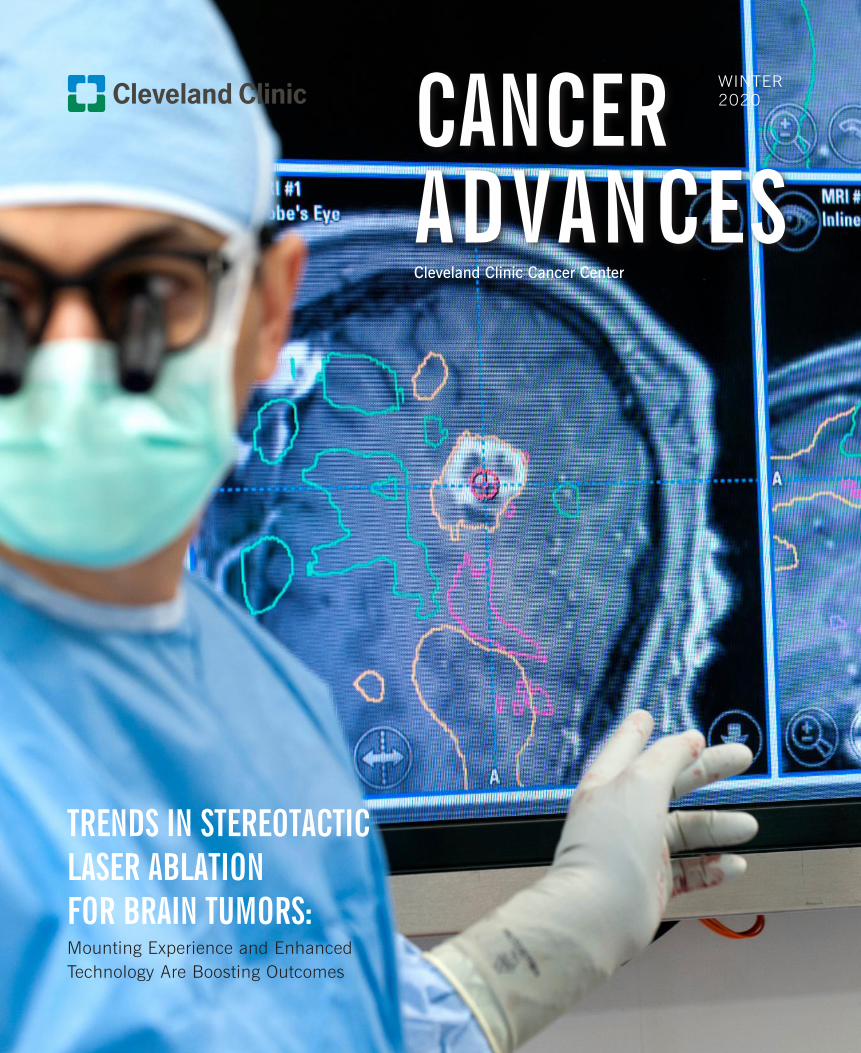
TRENDS IN STEREOTACTIC LASER ABLATION FOR BRAIN TUMORS: Mounting Experience and Enhanced Technology Are Boosting Outcomes
Cleveland Clinic Cancer Center

DEAR COLLEAGUES,
BRIAN J. BOLWELL, MD, FACP
Chairman, Taussig Cancer Institute
Cleveland Clinic Cancer Center
216.444.6922
On Twitter: @BrianBolwellMD
Improving cancer treatment for our patients depends on innovation and evaluation. We must be bold
enough to attempt difficult things, and disciplined enough to honestly assess those efforts and respond
accordingly. Informed risk-taking and rigorous appraisal go hand in hand. Both are essential if we are
to continue making progress against this relentless disease.
The accounts in Cancer Advances reflect Cleveland Clinic Cancer Center’s commitment to innovation
and evaluation.
We were one of the early adopters of stereotactic laser ablation to treat brain tumors. The insights
gained from nearly a decade’s experience with this leading-edge and still-evolving tool are the subject
of our cover story. Neurosurgeon Alireza M. Mohammadi, MD, reports (p. 24) that our outcomes and
operative times have improved dramatically, even as our case mix has become more challenging.
We also are among the initial practitioners of a novel two-stage surgical strategy to treat liver cancer
patients who do not qualify for traditional resection due to multicentric disease and an inadequate
future liver remnant. Federico Aucejo, MD, Director of the Liver Cancer Program, describes (p. 16) this
intriguing method.
Jame Abraham, MD, the newly appointed Chair of our Department of Hematology and Medical
Oncology, is part of a multicenter team that has discovered (p. 4) a radiogenomic MRI signature that
could identify HER2-positive breast cancer patients likely to benefit from targeted therapy.
Research (p. 8) conducted by Carol Burke, MD, Vice Chair of the Department of Gastroenterology,
Hepatology and Nutrition, suggests the traditional formula for staging duodenal polyposis to predict
cancer risk in familial adenomatous polyposis patients may need adjustment to account for previously
unappreciated factors.
Importantly, these and other results detailed in Cancer Advances are impacting or will soon impact
patient care. That tradition of translational research will be carried on in our new Center for Research
Excellence in Gynecologic Cancer (p. 10), which will investigate a wide range of subjects, from
targeted immunotherapy to the role of cancer stem cells.
None of this progress would be possible without the guidance of our many talented physician leaders.
In my Chairman’s Q&A (p. 28), I discuss our approach to physician leadership.
As always, I welcome opportunities to discuss the work we do and the possibility for collaboration.
Please let us know how we can help.
Sincerely,
Brian J. Bolwell, MD, FACP | Chairman, Taussig Cancer Institute, Cleveland Clinic Cancer Center
Cleveland Clinic Cancer Center provides complete cancer care enhanced by innovative basic, genetic and translational
research. It offers the most effective techniques to achieve long-term survival and improve patients’ quality of life. The
Cancer Center’s more than 700 physicians, researchers, nurses and technicians care for thousands of patients each year
and provide access to a wide range of clinical trials. Cleveland Clinic Cancer Center unites clinicians and researchers
based in Taussig Cancer Institute and in Cleveland Clinic’s 26 other clinical and special-expertise institutes, as well as
cancer specialists at our regional hospitals, health centers and Cleveland Clinic Florida. Cleveland Clinic is a nonprofit
academic medical center ranked as a top hospital in the country (U.S. News & World Report), where more than 3,900
staff physicians and researchers in 180 specialties collaborate to give every patient the best outcome and experience.
03 FROM THE INSTITUTE CHAIR
04 NEW APPROACH MAY HELP PREDICT HER2+ TUMOR RESPONSE TO TREATMENT
08 SHOULD WE RETHINK CANCER RISK STAGING IN FAMILIAL ADENOMATOUS POLYPOSIS?
10 CLEVELAND CLINIC FORMS CENTER FOR RESEARCH EXCELLENCE IN GYNECOLOGIC CANCER
12 REAFFIRMING THE OPTIMAL TOTAL DOSE OF CISPLATIN FOR HIGH-RISK ORAL CAVITY SQUAMOUS CELL CARCINOMA
14 INOTUZUMAB OZOGAMICIN PROVES SUPERIOR TO STANDARD CHEMOTHERAPY FOR RELAPSED/REFRACTORY ALL IN A LONG-TERM FOLLOW-UP STUDY
16 SURGERY OPENS NEW POSSIBILITIES FOR TREATING LIVER METASTASES FROM COLORECTAL CANCER
20 LOW CD30 EXPRESSION IN NON-HODGKIN’S LYMPHOMA PATIENTS DOES NOT PREDICT LOW RESPONSE TO BRENTUXIMAB VEDOTIN
24 TRENDS IN STEREOTACTIC LASER ABLATION FOR BRAIN TUMORS
26 POST-PROSTATECTOMY PROSTATE-SPECIFIC ANTIGEN KINETICS ARE ASSOCIATED WITH RECURRENCE AFTER SALVAGE RADIATION
28 CHAIRMAN’S Q&A: PHYSICIAN LEADERSHIP
29 JAME ABRAHAM, MD, FACP, NAMED CHAIR OF HEMATOLOGY AND MEDICAL ONCOLOGY
30 VELOSANO: A CATALYST FOR HIGH-IMPACT CANCER RESEARCH
FROM THE CHAIRMAN 3
ON THE COVER: Neurosurgeon Alireza Mohammadi, MD, of Cleveland Clinic’s Rose Ella Burkhardt Brain Tumor and Neuro-Oncology Center.
5
The emergence of HER2-targeted therapy,
including the monoclonal antibodies trastuzumab
and pertuzumab, has greatly improved survival
in HER2+ breast cancer. Yet a significant
percentage of patients will not achieve a
complete preoperative response to a combination
of anti-HER2 therapy and chemotherapy, and
no clinically validated biomarker is currently
available to indicate which patients are likely to
benefit from targeted therapies.
Breast radiogenomics — an investigational
diagnostic approach that integrates genomic data
and qualitative analysis of clinical radiology for
tumor characterization — has shown promise
in noninvasively identifying patients’ genetic
profile from imaging, but has not been applied in
the context of predicting clinical outcomes and
guiding targeted therapies.
Now, a multicenter team including researchers
from Cleveland Clinic has shown that a
combination of measurements within and outside
a tumor on clinical dynamic contrast-enhanced
magnetic resonance imaging is capable of
discriminating the response-associated HER2-
enriched (HER2-E) molecular subtype from other
subtypes among patients with HER2+ tumors.
(Current subtype identification requires costly
gene expression profiling using tissue obtained
by an invasive biopsy.)
When subsequently evaluated among recipients
of HER2-targeted therapy, the new intratumoral
and peritumoral imaging signature was found
to be associated with response to neoadjuvant
chemotherapy. The team’s findings were
published in the journal JAMA Network Open.
“Currently, if we see someone with a HER2-
positive tumor, we always just give them
chemotherapy and HER2-targeted medicine,”
says Jame Abraham, MD, Chair of Cleveland
Clinic Taussig Cancer Institute’s Department of
Hematology and Medical Oncology, Director of
the Breast Oncology Program and Co-Director
of the Comprehensive Breast Cancer Program.
“Until now, no one has looked at a predictive
model to see who will benefit. This is the first
use of radiology and radioanalysis to identify that
subset of patients.”
Potential treatment impacts
The approach uses computerized tissue
phenotyping on radiographic imaging
(radiomic) features extracted from breast MRI
to examine the appearance of the tumor and its
surroundings. A combination of local disorder,
especially within the peritumoral environment,
and larger-scale homogeneity near the tumor
were found to most effectively characterize the
treatment response-associated HER2-E molecular
subtype.
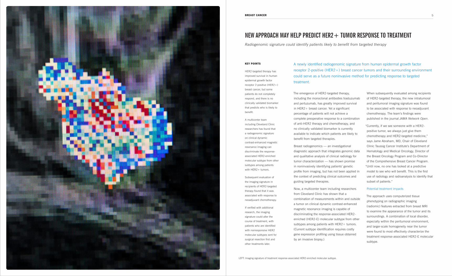
A newly identified radiogenomic signature from human epidermal growth factor
receptor 2-positive (HER2+) breast cancer tumors and their surrounding environment
could serve as a future noninvasive method for predicting response to targeted
treatment.
KEY POINTS
HER2-targeted therapy has
improved survival in human
epidermal growth factor
receptor 2-positive (HER2+)
breast cancer, but some
patients do not completely
respond, and there is no
clinically validated biomarker
that predicts who is likely to
benefit.
A multicenter team
including Cleveland Clinic
researchers has found that
a radiogenomic signature
on clinical dynamic
contrast-enhanced magnetic
resonance imaging can
discriminate the response-
associated HER2-enriched
molecular subtype from other
subtypes among patients
with HER2+ tumors.
Subsequent evaluation of
the imaging signature in
recipients of HER2-targeted
therapy found that it was
associated with response to
neoadjuvant chemotherapy.
If verified with additional
research, the imaging
signature could alter the
course of treatment, with
patients who are identified
with nonresponsive HER2
molecular subtypes sent for
surgical resection first and
other treatments later.
NEW APPROACH MAY HELP PREDICT HER2+ TUMOR RESPONSE TO TREATMENTRadiogenomic signature could identify patients likely to benefit from targeted therapy
BREAST CANCER
LEFT: Imaging signature of treatment response-associated HER2-enriched molecular subtype.

6 7CLEVELAND CLINIC CANCER CENTER BREAST CANCER
If confirmed in subsequent studies, the technique could alter the
course of treatment, since patients identified with nonresponsive
HER2 molecular subtypes could be sent for surgical resection
first and other treatments afterward, notes Abraham, who is
also Professor of Medicine at Cleveland Clinic Lerner College of
Medicine.
Alternatively, this approach could be used to identify patients who
might benefit from trastuzumab emtansine (T-DM1), an antibody-
drug conjugate of trastuzumab and the cytotoxic agent emtansine
(DM1), a maytansine derivative and microtubule inhibitor. In a
landmark study published in December 2018, T-DM1 reduced
the risk of recurrence of invasive breast cancer or death by 50%
in patients with HER2+ early breast cancer who had residual
invasive disease after completion of neoadjuvant therapy compared
with trastuzumab alone.
“We could potentially select patients upfront to treat with T-DM1.
This would represent a major step for personalized medicine,” Dr.
Abraham says.
Identifying responders radiographically
Initially, the investigators identified imaging features distinguishing
HER2+ tumors from other receptor subtypes among 117 patients
who received an MRI prior to neoadjuvant chemotherapy at a
single institution between 2012 and 2015.
Then, using imaging and genomic data from a previous multicenter
trial of 42 patients with HER2+ breast cancer and preoperative
FIGURE 2. Imaging signature of HER2-E is
associated with pathologic complete response to
anti-HER2 therapy, with rippled enhancement
patterns detected intratumorally by Laws feature
(left), and elevated local peritumoral heterogeneity
captured by CoLlAGe features 9 to 12 mm from the
tumor (right) characterizing both features. Radiomic
feature values are unitless, thus the scale depicts
relative expression values of radiomic features,
standardized between 0 and 1.0 based on the range
of their distribution. The blue color at 0 depicts the
minimum observed feature value; the red color at
1.0 depicts the maximum observed feature value.
Credit: Braman N, Prasanna P, Whitney J, et al. Association of Peritumoral Radiomics With Tumor Biology and Pathologic Response to Preoperative Targeted Therapy for HER2 (ERBB2)-Positive Breast Cancer. JAMA Netw Open. 2019 Apr 5;2(4):e192561.
FIGURE 1. Co-occurrence
of local anisotropy gradients
(CoLlAGe) feature expression
maps visualize the elevated
disorder of local intensity
gradient orientations within
the peritumoral region of
HER2-E relative to non–
HER2-E breast cancers.
Radiomic feature values
are unitless, thus the scale
depicts relative expression
values of radiomic features,
standardized between 0
and 1.0 based on the range
of their distribution. The
blue color at 0 depicts the
minimum observed feature
value; the red color at
1.0 depicts the maximum
observed feature value.
Credit: Braman N, Prasanna P, Whitney J, et al. Association of Peritumoral Radiomics With Tumor Biology and Pathologic Response to Preoperative Targeted Therapy for HER2 (ERBB2)-Positive Breast Cancer. JAMA Netw Open. 2019 Apr 5;2(4):e192561.
MRI and RNA sequencing data, they developed a signature to
identify the HER2-E subtype among clinically HER2+ tumors.
Previous radiomics studies have focused on analyzing the tumor
itself, but the team found that adding information about its
surroundings was critical to distinguishing HER2-E tumors.
To evaluate the utility of this signature in guiding treatment
decisions, the team explored whether it could be used to predict
targeted therapy outcome for HER2+ patients. When applied to
a set of 78 patients from two institutions who had received MRI
exams before HER2-targeted neoadjuvant chemotherapy, the
signature was found to significantly identify patients who would
achieve a complete response.
To better understand this signature, the researchers compared
radiomic features with biopsy samples from the same patients.
They observed that features from the 0-3 mm peritumoral region
on MRI were significantly associated with the density of tumor-
infiltrating lymphocytes on tissue samples — indicating a potential
link between the way the immune system responds to a tumor and
the appearance of its surroundings on imaging.
While the findings are compelling, Dr. Abraham cautions that “this
is a completely experimental, retrospective study — a proof of
concept. We need to validate this in larger datasets and confirm the
data. Then we can potentially apply it more widely.”
Dr. Abraham is Chair of Cleveland Clinic Taussig Cancer Institute’s
Department of Hematology and Medical Oncology, Director of
the Breast Oncology Program, Co-Director of the Comprehensive
Breast Cancer Program and Professor of Medicine at Cleveland
Clinic Lerner College of Medicine.
He can be reached at [email protected] or 216.445.0150.
On Twitter: @jamecancerdoc
“We could potentially select patients upfront to
treat with T-DM1. This would represent a major
step for personalized medicine.”
— JAME ABRAHAM, MD

8 CLEVELAND CLINIC CANCER CENTER 9DUODENAL CANCER
But recent Cleveland Clinic research published in
the journal Gastrointestinal Endoscopy highlights
the inconsistency of SS as a cancer prediction
risk indicator; more than half of FAP patients
diagnosed with duodenal cancer in the case-
control study lacked SS IV duodenal polyposis.
The results suggest that certain individual
characteristics of duodenal polyps in FAP
patients have heightened significance in
predicting cancer risk, irrespective of stage,
and that the formula for staging duodenal
polyposis may need to be adjusted to take that
into account, rather than focusing solely on the
presence or absence of SS IV disease.
“Traditionally, SS IV polyposis has been a trigger
for offering prophylactic duodenal surgery to
prevent duodenal cancer in FAP,” says study co-
author Carol Burke, MD, Vice Chair of Cleveland
Clinic Digestive Disease & Surgery Institute’s
Department of Gastroenterology, Hepatology and
Nutrition, Director of the Center for Colon Polyp
and Cancer Prevention, and Section Head of
Polyposis in the Sanford R. Weiss, MD, Center for
Hereditary Colorectal Neoplasia. “Our research
shows that earlier SS patients with large or
microscopically advanced polyps are also at
high risk of cancer, and aggressive endoscopic
intervention or duodenectomy should be
considered if polyp burden cannot be controlled.”
Dr. Burke and her colleagues believe their
research is the first to examine individual SS
components and papilla pathology in relation to
duodenal cancer risk in FAP.
The pluses and minuses of Spigelman staging
FAP is an inherited colorectal cancer syndrome
caused by a germline mutation in the
adenomatous polyposis coli gene. Without
colectomy, progression of colorectal polyposis to
colorectal cancer is inevitable, usually by ages
40 to 50. The second leading cause of cancer
in FAP is duodenal cancer, which arises from
duodenal adenomatous polyposis and has an
overall cumulative lifetime incidence of 4.5% by
age 57.
The five-stage (0 to IV) SS system was developed
30 years ago to predict duodenal cancer risk and
dictate the frequency of endoscopic surveillance
and the timing of prophylactic duodenectomy. In
calculating the SS score and stage, equal weight
is assigned to each of the four polyp criteria —
number, size, histology and degree of dysplasia.
Previous research showed FAP patients with SS
IV polyposis had 10-year cumulative risk levels
as high as 36% of developing duodenal cancer,
versus 2.5% risk in patients with SS 0-III. But
those data also showed that many FAP patients
who developed cancers did not have SS IV
polyposis.
That variability of SS predictive accuracy is what
prompted the Cleveland Clinic researchers to
assess the relationship of SS and other factors
with duodenal cancer in FAP.
Analyzing the data
The researchers queried the Cleveland Clinic
hereditary colon cancer database for FAP patients
The presence of Spigelman stage (SS) IV duodenal polyposis is considered the most
significant risk factor for duodenal cancer in patients with familial adenomatous
polyposis (FAP).
KEY POINTS
Spigelman stage (SS) IV
polyposis traditionally has
been a trigger for offering
prophylactic duodenal
surgery to prevent duodenal
cancer in patients with
familial adenomatous
polyposis (FAP).
More than half of FAP
patients diagnosed with
duodenal cancer in a
Cleveland Clinic case-control
study lacked SS IV duodenal
polyposis.
That inconsistency
suggests certain individual
characteristics of duodenal
polyps in FAP patients have
heightened significance
in predicting cancer risk,
irrespective of stage.
The formula for staging
duodenal polyposis may
need to be adjusted to give
greater consideration to large
polyp size and dysplasia,
rather than focusing solely
on the presence or absence
of SS IV disease.
SHOULD WE RETHINK CANCER RISK STAGING IN FAMILIAL ADENOMATOUS POLYPOSIS?Research suggests greater consideration for large polyp size, dysplasia
with duodenal polyposis seen between 1988 and 2013. They
identified 18 patients with duodenal cancer and, for comparison,
randomly selected 85 similarly aged patients with FAP but without
duodenal cancer. The researchers reviewed clinical data, including
results of esophagogastroduodenoscopy performed on the cases
and controls.
Statistical analysis found that SS IV polyposis was associated with
duodenal cancer, but that 53% of patients with duodenal cancer
had no prior SS IV polyposis — a considerably higher proportion
than in previous research.
Regarding individual SS characteristics, duodenal polyps larger
than 10 mm and polyps with high-grade dysplasia (HGD) were
positively associated with cancer. Large polyps were present in
76.5% of cancer patients versus 47.1% of FAP patients without
duodenal cancer (p = .027). Polyps with HGD were identified in
29.4% of cancer patients versus 5.9% of those without cancer
(p = .003). The presence of more than 20 duodenal polyps and
duodenal polyps with advanced histology (tubulovillous adenoma
or villous adenoma) were not associated with cancer risk.
“The demonstration that not all SS characteristics have comparable
predictive value for duodenal cancer begs the question of whether
it is prudent to rethink the equal weighting of those components in
risk stratification, instead giving greater consideration to large polyp
size and dysplasia,” Dr. Burke says.
The frequency of finding advanced pathology of the papilla with
any villous features or HGD was greater in the cancer patients than
in those without cancer (80% vs. 22% for villous features and
30% vs. 4% for HGD), regardless of whether the cancer was of the
papilla/ampulla or elsewhere in the duodenum.
Spigelman classification for duodenal polyps in FAP
Criteria Points
1 2 3
Polyp number 1-4 5-20 > 20
Polyp size (mm) 1-4 5-10 >10
Histology Tubular Tubulovillous Villous
Dysplasia Mild Moderate Severe
Stage 0 = 0 points; Stage I =1-4 points; Stage II = 5-6 points;
Stage III = 7-8 points; Stage IV = 9-12 points
“There is no consensus on including endoscopic or histologic
features of the duodenal papilla characterization in SS calculations,”
Dr. Burke says. “Our data support the importance of the histology of
the papilla in assessing duodenal cancer risk and bolster the case
for routine biopsy of the papilla and inclusion in SS.”
The study also identified a personal and family history of colon
cancer and the absence of desmoid tumors as characteristics in
FAP patients developing duodenal cancer, which may be a function
of the gene mutation causing the disease.
Clinical implications
Although larger studies are needed to validate the overall findings,
reassessing the Spigelman staging system with a larger population
should be considered, the authors conclude.
Meanwhile, Dr. Burke and her colleagues advocate regular
duodenal polyposis surveillance, biopsy of the duodenal papilla and
inclusion of histology findings of the papilla in the current SS. The
presence of HGD, whether papillary or in the duodenum, should
be a potential indicator of a high-risk patient and warrants close
follow-up, the researchers say.
Dr. Burke is Vice Chair of Cleveland Clinic Digestive Disease &
Surgery Institute’s Department of Gastroenterology, Hepatology
and Nutrition; Director of the Center for Colon Polyp and Cancer
Prevention; Section Head of Polyposis in the Sanford R. Weiss, MD,
Center for Hereditary Colorectal Neoplasia; and Clinical Assistant
Professor of Medicine at Cleveland Clinic Lerner College of Medicine.
She can be reached at [email protected] or 216.444.6864.
On Twitter: @burkegastrodoc

GYNECOLOGIC ONCOLOGYCLEVELAND CLINIC CANCER CENTER10 11
The center’s co-directors, Ofer Reizes, PhD,
Department of Cardiovascular and Metabolic
Sciences and the Cancer Impact Area, Lerner
Research Institute, and Peter Rose, MD,
Department of Gynecologic Oncology, Ob/Gyn &
Women’s Health Institute, believe the CREGC is
exceptionally positioned to change the landscape
of gynecologic cancer research and care.
The center will capitalize on the expertise and
patient volume of Cleveland Clinic’s Department
of Gynecologic Oncology, which is recognized
nationally by U.S. News & World Report for its
clinical prowess. Additionally, Lerner Research
Institute already has a portfolio of gynecologic
cancer research underway.
Breakthrough research opportunities
Through core resources and an infrastructure
designed to promote collaboration and accelerate
translational medicine, the CREGC supports
research projects that explore possible causes
of and treatments for a range of gynecologic
cancers, including:
› Characterizing genetic anomalies that confer
radiation resistance in endometrial cancer.
› Identifying candidates for targeted
immunotherapy to treat epithelial ovarian
cancers.
› Developing therapies to overcome drug
resistance in women with ovarian cancer who
have the BRCA mutation.
› Determining the role cancer stem cells and
related molecules play in chemotherapy
resistance.
“We are really driving research at the bench to
care at the bedside, ensuring that our research
informs clinical care and vice versa,” states Dr.
Reizes. “Our aim is to create tangible benefits for
patients by bringing together lab scientists with
front-line physicians to focus on the advances
most needed by patients, now.”
In addition to supporting specific research
projects, the CREGC will help drive research and
cures by developing a shared biorepository of
patient samples and establishing patient-derived
disease models for preclinical investigation of
new therapies. In 2018, the CREGC established
and successfully executed a process for
engrafting primary tumors for future testing. In
the coming months, the group will focus on
expanding its tissue and specimen collection and
explore the utilization of patient-derived xenograft
models in clinical practice.
Gynecologic cancers, including endometrial and ovarian cancers, are a leading cause
of cancer-related deaths in women. The ability of gynecologic tumors to adapt to
and evade treatment is a major factor contributing to the poor outcomes that many
patients face. The Center for Research Excellence in Gynecologic Cancer (CREGC) is
a collaborative network for the development of a comprehensive research program to
promote the translation of basic science investigation into the clinic.
KEY POINTS
Cleveland Clinic’s new
Center for Research
Excellence in Gynecologic
Cancer is developing a
comprehensive research
program to translate basic
science into clinical care.
The center will investigate
a wide range of subjects,
including genetic
anomalies that confer
radiation resistance in
endometrial cancer, targeted
immunotherapy for epithelial
ovarian cancers, and the
role of cancer stem cells in
chemotherapy resistance.
The center will create a
biorepository of patient
tissue samples and establish
patient-derived disease
models for preclinical
investigation of new
therapies.
CLEVELAND CLINIC FORMS CENTER FOR RESEARCH EXCELLENCE IN GYNECOLOGIC CANCERFocus is on delivering advances most needed by patients
“Opportunities for breakthroughs in gynecologic cancer are in
the team we’ve built,” Dr. Reizes says. “Bringing together a
multidisciplinary team to focus on this problem makes me hopeful
that we will move the needle on these diseases.”
Ensuring the best possible patient outcomes
In addition to the CREGC’s efforts, Cleveland Clinic’s Gynecologic
Oncology Cancer Program offers patients the latest in gynecologic
cancer management, including the newest drug therapies.
“As part of NRG Oncology, an international cooperative research
group funded by the National Cancer Institute and National
Institutes of Health, we can offer patients who qualify access to
investigational treatments through a wide range of clinical trials,”
states Dr. Rose. “Additional studies give eligible patients access to
other new treatments under investigation in the CREGC, such as
hyperthermic intraperitoneal chemotherapy, or HIPEC.”
The Gynecologic Oncology Cancer Program also offers minimally
invasive surgery, sophisticated radiation therapy techniques and
specialized imaging.
“Our team of highly trained specialists — including gynecologic
pathologists, radiation oncologists, nurse practitioners, physician
assistants, nurse navigators, chemotherapy coordinators,
genetic counselors and social workers — works with patients
to provide precise diagnosis, exacting surgical skill and leading-
edge therapies,” Dr. Rose notes. “Throughout the entire patient
experience, we emphasize comfort and empathy.”
Dr. Reizes holds the Laura J. Fogarty Endowed Chair for Uterine
Cancer Research and is a staff member of Cleveland Clinic Lerner
Research Institute’s Department of Cardiovascular and Metabolic
Sciences, the Department of Cellular and Molecular Medicine, and
the Cancer Impact Area. He is Assistant Professor of Molecular
Medicine at Cleveland Clinic’s Lerner College of Medicine.
He can be reached at [email protected] or 216.445.0880.
On Twitter: @oreizes
Dr. Rose is Section Head and Fellowship Director of Gynecologic
Oncology in the Ob/Gyn & Women’s Health Institute’s Department
of Obstetrics and Gynecology, and Professor of Surgery at
Cleveland Clinic Lerner College of Medicine.
He can be reached at [email protected] or 216.444.1712.
LEFT: Peter Rose, MD

HEAD AND NECK CANCERCLEVELAND CLINIC CANCER CENTER12 13
Of these patients:
› Median age was 56.
› 3% were men.
› 1% were Caucasian.
› 9% had significant tobacco history.
“Looking retrospectively at this cohort, we learned that patients who
received 200 mg/m2 or more of cisplatin had nearly double the median
disease-free survival of patients who received less,” says Dr. Geiger.
Median disease-free survival was:
› Five months in patients who received less than 200
mg/m2 of cisplatin.
› Eight months in patients who received 200 mg/m2 or
more of cisplatin.
There was no significant difference in disease-free
survival among patients who received cisplatin as a
bolus and those who received weekly dosing.
Univariate analysis also showed associations between
higher doses of cisplatin and improved locoregional
control (p = .131), metastatic disease (p = .084)
and overall survival (p = .187). However, none of
these associations was statistically significant, notes Dr.
Geiger.
Administration options
“This study reaffirms that patients with high-risk
resected OCSCC require systemic therapy with cisplatin
and need to receive as much of it as possible during
the course of radiation therapy,” says Dr. Geiger. “There
is a distinct benefit when patients get at least 200
mg/m2, whether in a bolus or weekly dosing.”
A prospective study is needed to evaluate different
cisplatin dosing schedules and determine the optimal
administration for high-risk OCSCC patients.
Dr. Geiger is a staff member of Cleveland Clinic Taussig
Cancer Institute’s Department of Hematology and
Medical Oncology and Assistant Professor of Medicine
at Cleveland Clinic Lerner College of Medicine.
She can be reached at [email protected] or
216.444.0888.
On Twitter: @JLGeigerMD
However, high-dose cisplatin is extremely
toxic and difficult for patients to tolerate. It is
highly emetogenic, nephrotoxic and ototoxic,
and patients often experience additional side
effects common with chemotherapy, including
myelosuppression and peripheral neuropathy.
These adverse effects are compounded by those
caused by radiation therapy to the head and
neck. For example, mucositis often causes
dysphagia and odynophagia, which can lead to
malnutrition, necessitating alternative means of
obtaining enteral nutrition.
“Regarding toxicities and side effects, I explain
to my patients that adding chemotherapy to
radiation can be a 1 + 1 = 10 situation,” says
Cleveland Clinic oncologist Jessica Geiger, MD.
High dose vs. weekly cisplatin dosing
Identifying therapies and administration
schedules with the best effectiveness and least
toxicity is always the goal, she notes.
To this end, Dr. Geiger and a multi-institutional
team established a large database of patients
treated for OCSCC. Patients were treated at one
of six academic institutions:
› Cleveland Clinic’s Taussig Cancer Institute.
› Lee Moffitt Cancer Center & Research Institute.
› Henry Ford Health System.
› Memorial Sloan Kettering Cancer Center.
› Princess Alexandra Hospital (Australia).
› University of Louisville Hospital.
With nearly 1,300 patients, the Institutional
Review Board-approved multi-institutional
database is one of the largest cohorts for OCSCC,
says Dr. Geiger. Many studies have mined the
extensive, long-term data for survival and toxicity
statistics.
Most recently, Dr. Geiger led a retrospective study
evaluating alternative cisplatin dosing schedules.
“We weren’t able to discern whether administering
cisplatin in a high-dose bolus or in weekly
cumulative doses affected survival end points,”
says Dr. Geiger. “But we did reaffirm an optimal
total dose that had been suggested previously in
the literature.”
Dr. Geiger presented results of the study at the
2019 American Society of Clinical Oncology
Annual Meeting.
Optimal total dose: at least 200 mg/m2
For this study, a subset of 196 patients met
inclusion criteria:
› Treated for OCSCC between 2005 and 2015.
› Had positive surgical margins (35.7%) and/
or extranodal extension (82.7%) following
resection.
› Treated concurrently with radiation therapy
and chemotherapy.
In oral cavity squamous cell carcinoma (OCSCC), the standard of care is resection.
In high-risk cases — those identified by positive surgical margins and extranodal
extension — resection is followed by radiation therapy and intravenous cisplatin.
KEY POINTS
Cisplatin and radiotherapy
following tumor resection
is the standard of care
in patients with high-risk
oral cavity squamous cell
carcinoma (OCSCC).
Since high-dose cisplatin is
extremely toxic and difficult
to tolerate, identifying
the dosage with the best
efficacy and least toxicity is
important.
A Cleveland Clinic-
led retrospective study
evaluating alternative
cisplatin dosing schedules
for high-risk OCSCC patients
reaffirmed a previously
suggested optimal total
cisplatin dose of 200 mg/m2
or more.
Patients who received this
dose had nearly double
the median disease-free
survival of patients who
received less. There was
no significant difference in
disease-free survival among
patients who received
cisplatin as a bolus and
those who received weekly
dosing.
REAFFIRMING THE OPTIMAL TOTAL DOSE OF CISPLATIN FOR HIGH-RISK ORAL CAVITY SQUAMOUS CELL CARCINOMAIt nearly doubles median disease-free survival
LEFT: Jessica Geiger, MD

14 CLEVELAND CLINIC CANCER CENTER 15HEMATOLOGY AND ONCOLOGY/LEUKEMIA
The final report of the INO-VATE (INotuzumab
Ozogamicin trial to inVestigAte Tolerability
and Efficacy) trial, published in the journal
Cancer, found that INO generated greater rates
of complete remission (CR) and longer median
overall survival (OS), but showed a greater
incidence of veno-occlusive disease (VOD),
compared with results in ALL patients treated
with standard-of-care chemotherapy.
“INO is a very encouraging drug in the setting
of relapsed/refractory ALL, and this long-term
follow-up study has validated its OS advantage,”
says study co-author Anjali Advani, MD,
Director of Cleveland Clinic Taussig Cancer
Institute’s Inpatient Leukemia Program. “The
main challenge we still have to deal with is
the risk of VOD, but INO definitely has an
advantage in patients with high tumor burden
or extramedullary disease. I also tend to favor
it in patients with central nervous system
disease, because it can be given with concurrent
intrathecal chemotherapy.”
The original INO-VATE trial assessed the safety
and efficacy of single-agent INO compared with
standard chemotherapy in relapsed/refractory
ALL. INO is a humanized monoclonal antibody
drug conjugate that binds to CD22+ ALL cells.
The antibody is conjugated to calicheamicin, a
cytotoxic compound that causes DNA damage
and apoptosis.
INO-VATE’s results led the Food and Drug
Administration to approve the drug’s use in adults
with relapsed or refractory B-cell precursor ALL.
Challenging VOD rates
The main purpose of the follow-up study was to
assess whether INO is superior to the standard-
of-care chemotherapy over a period of two years.
“We looked at the response/complete remission
rates, toxicity, OS, disease-free survival, minimal
residual disease (MRD) negativity and the
percentage of patients who were able to go on
to [hematopoietic stem cell] transplant,” says Dr.
Advani.
The two-year follow-up largely confirmed the
initial findings of the INO-VATE trial, with even
more impressive outcomes in terms of OS and
the percentage of patients who achieved MRD
negativity and proceeded to transplant.
“The difference in the OS in patients who received
INO (22.8%) compared with those who received
standard chemotherapy (10%) has become more
pronounced after the two-year follow-up,” she
says. “The outcome of patients who proceeded
to transplant is even more impressive in the
subgroup who received INO and went into
remission, achieved MRD negativity and went on
to transplant (39.6% INO vs. 10.5% standard of
care).”
The promising preliminary survival and remission outcomes that inotuzumab
ozogamicin (INO) produced in relapsed or refractory acute lymphoblastic leukemia
(ALL) patients in the antibody-drug conjugate’s phase 3 trial have been sustained in a
long-term follow-up study.
KEY POINTS
A long-term follow-up
study has verified the
superiority of inotuzumab
ozogamicin (INO) to
standard chemotherapy for
relapsed/refractory acute
lymphoblastic leukemia.
INO produced higher rates
of complete remission
and longer median overall
survival, but showed a
greater incidence of veno-
occlusive disease (VOD).
39.6% of patients who
received INO achieved
remission and minimal
residual disease
negativity and went on
to hematopoietic stem
cell transplant, versus
10.5% who were treated
with standard-of-care
chemotherapy.
Researchers are now
examining whether the
heightened VOD risk can be
reduced with prophylactic
medications prior to
transplant or by reducing the
dose of INO and combining
it with other agents.
INOTUZUMAB OZOGAMICIN PROVES SUPERIOR TO STANDARD CHEMOTHERAPY FOR RELAPSED/REFRACTORY ALL IN A LONG-TERM FOLLOW-UP STUDYTwo-year follow-up confirms initial findings of the INO-VATE trial
However, the risk of VOD in patients who are transplanted remains
a concern, she notes. VOD/sinusoidal obstruction syndrome was
significantly more frequent in the INO arm (14%) compared with
the standard-of-care arm (2.1%).
“We are now looking at how we can decrease that toxicity by either
giving these patients medications (i.e., defibrotide) prophylactically
prior to transplant or reducing the dose of INO and combining it
with other agents,” says Dr. Advani.
In both treatment arms, the most frequent all-grade and grade 3 or
higher adverse events were hematologic.
“Hematologic events are very common with both INO and the
standard-of-care chemotherapy,” she says. “But the neutropenia,
thrombocytopenia and anemia we saw were fairly easily managed.
For those patients who are not going on to transplant and are
receiving multiple cycles, the low platelet count can become an
issue.”
Next research steps
In terms of continuing research on INO, Dr. Advani says upfront
use of the drug is currently being investigated in several clinical
trials. In the ALLIANCE (A041501) trial, led by Daniel J. DeAngelo,
MD, PhD, of the Dana Farber Cancer Institute, INO is being
evaluated in combination with chemotherapy in young adults with
newly diagnosed CD22+ B-cell acute lymphoblastic leukemia.
“The question we are trying to answer in this trial is, if we use the
drug in the upfront setting, will we have better outcomes, fewer
relapses and maybe lower toxicity, because those patients hopefully
won’t be going on to transplant,” explains Dr. Advani, who is one of
the study’s principal investigators.
A second planned U.S. intergroup trial led by Elias Jabbour, MD, of
the University of Texas MD Anderson Cancer Center, will randomize
elderly patients with ALL to either mini-hyper-CVD (low-intensity
chemotherapy) or mini-hyper-CVD plus INO.
A third trial (S1312) that has completed accrual is reviewing INO
plus chemotherapy (cyclophosphamide, vincristine sulfate and
prednisone [CVP]) in patients with relapsed or refractory CD22+
acute leukemia. This is a Southwest Oncology Group trial in which
Dr. Advani serves as a principal investigator.
Dr. Advani is Director of Cleveland Clinic Taussig Cancer Institute’s
Inpatient Leukemia Program, a staff member of the Department
of Hematology and Medical Oncology and Department of
Translational Hematology and Oncology Research, and Professor of
Medicine at Cleveland Clinic Lerner College of Medicine.
She can be reached at [email protected] or 216.445.9354.
LEFT: Photomicrograph of ALL bone marrow showing
small, medium and large hemoblasts.
Med
ical
Im
ages
RM

17LIVER CANCER /COLORECTAL CANCER
The two-stage surgery, known as associating
liver partition and portal vein ligation for staged
hepatectomy (ALPPS), makes it possible to treat
patients who are not appropriate candidates
for traditional liver resection due to multicentric
disease and an inadequately small future liver
remnant.
When both lobes contain tumors, hepatobiliary
surgeons and radiologists in Cleveland Clinic
Digestive Disease & Surgery Institute’s Liver
Tumor Cancer Program work together to identify
the lobe with the lower tumor burden. After
excising the lesions in this lesser-affected lobe,
the true novelty of their approach begins. They
simultaneously enhance the growth of this lobe
while working to shrink the contralateral lobe —
the one with the greater tumor burden — so that
it can be more easily resected during a follow-up
procedure.
“The ALPPS procedure allows us to treat patients
who have a substantial amount of tumor, with
both lobes affected by tumor,” says Federico
Aucejo, MD, Director of the Liver Cancer
Program, Surgical Director of the Liver Tumor
Clinic and Co-Director of the Liver Tumor Center
of Excellence.
In the first stage of ALPPS surgery, at the same
time that surgeons resect the metastases from
one lobe, they ligate the portal vein supplying
blood to the other. They also partition the liver
with a transecting incision of the parenchyma to
interrupt intrahepatic vascular connections. This
approach serves a dual purpose — starving the
metastatic hemi-liver of its blood supply while
diverting as much as possible to the lobe that
will be preserved.
Partitioning the liver helps enhance growth of
the future liver remnant by stimulating increased
expression of growth factors and cytokines and
inducing hyperplasia, Dr. Aucejo explains. “This
not only makes the lobe of the liver grow larger,
but does it faster.”
“We can then perform the second and larger
operation, where we remove the contralateral
side — the lobe with the greater tumor burden,”
he says. The second stage typically is performed
one to two weeks after the first operation.
Embolization and venous deprivation approaches
Portal vein embolization is another method
to convert unresectable bilobar cancer into
resectable by inducing hypertrophy of liver
segments. Following excision of the small
metastatic lesions in the future liver remnant,
rather than performing surgical ligation of the
portal vein, interventional radiologists perform
perioperative embolization (using a percutaneous
catheter to deliver embolic agents to occlude the
vein), redirecting portal blood to the future liver
remnant.
An innovative surgical treatment option that leverages the liver’s regenerative capacity
is showing promise for some patients with previously unresectable liver metastases
from colorectal cancer.
KEY POINTS
A novel two-stage surgery
offered at Cleveland Clinic,
known as associating
liver partition and portal
vein ligation for staged
hepatectomy (ALPPS),
makes it possible to
treat patients with liver
metastases from colorectal
cancer who don’t qualify for
traditional liver resection due
to multicentric disease and
an inadequately small future
liver remnant.
In the first stage,
hepatobiliary surgeons excise
tumors in the lesser-affected
liver lobe, ligate the portal
vein supplying blood to the
other lobe and partition the
liver to interrupt intrahepatic
vascular connections.
This simultaneously
enhances growth of the
now tumor-free lobe while
shrinking the contralateral
lobe with the greater tumor
burden, which will be
resected during a follow-up
procedure.
With careful application,
ALPPS can improve survival
in patients who have no
alternative treatments.
SURGERY OPENS NEW POSSIBILITIES FOR TREATING LIVER METASTASES FROM COLORECTAL CANCERTwo-stage procedure capitalizes on liver’s ability to regenerate
LEFT: Federico Aucejo, MD

18 19LIVER CANCER /COLORECTAL CANCERCLEVELAND CLINIC CANCER CENTER
A distinct advantage of occlusion is less manipulation of the
local anatomy. “When we come back for the second stage of the
procedure, we find virgin tissue there,” Dr. Aucejo says. “Generally,
there is less inflammation, and dissection of the tissue is easier.”
Interventional radiologists also can perform a venous deprivation
technique. In addition to cutting off the blood flow to half the liver,
they embolize the hepatic vein coming out of the same lobe. “That
allows for substantial atrophy of that lobe and substantial growth
of the contralateral lobe, the one that we are going to leave in the
patient,” Dr. Aucejo says.
The importance of careful patient selection
Physicians in the Liver Tumor Cancer Program have performed
about 10 ALPPS and venous embolization/deprivation procedures
to date. Whichever approach is used, preserving at least 30%
of total liver volume is the mandatory safety margin. In terms of
patient selection, patients 60 years old or younger generally have
better outcomes with these two-stage interventions.
“This surgical technique is applied in very select patients,” Dr.
Aucejo says. “But it clearly opens an opportunity for improved
patient survival when there are no other alternatives.”
For example, recently an out-of-state colorectal cancer patient with
extensive bilobar metastases that were not amenable to traditional
resection sought a consult with Cleveland Clinic. “After evaluating
the liver volumetry and tumor burden affecting both lobes, I
decided to offer an ALPPS procedure, as it was the best possible
option to clear the liver of disease,” Dr. Aucejo says.
Months after surgery, the patient developed a small recurrence in
the remaining liver, which was treated with external beam radiation.
“As of today, the patient is back in his hometown, reconnecting with
his normal life and family,” Dr. Aucejo says.
An extensive toolkit
Cleveland Clinic is one of the few centers offering a comprehensive
range of advanced techniques to treat complex liver cancer cases.
They require “a high level of surgical expertise,” Dr. Aucejo says.
“ALPPS is a valuable part of a comprehensive toolkit we have to
treat these patients, which includes systemic treatment, surgical
therapies and interventional radiology strategies. Despite the
evolution of nonsurgical treatments when surgical options are not
possible, five-year survival ranges between 10% and 20%, as
opposed to 30% to 60% when surgery can be performed.”
Dr. Aucejo is Director of Cleveland Clinic Digestive Disease &
Surgery Institute’s Liver Cancer Program, Surgical Director of
the Liver Tumor Clinic, Co-Director of the Liver Tumor Center of
Excellence, and Associate Professor of Surgery at Cleveland Clinic
Lerner College of Medicine.
He can be reached at [email protected] or 216.445.7159.
On Twitter: @FAucejo
FIGURE 1. Preoperative computed tomography (CT) shows extensive bilobar liver
metastases from colorectal cancer.
FIGURE 3. First-stage hepatectomy showing left lateral segment after metastases
resection.
FIGURE 5. First-stage hepatectomy showing transection of the liver.
FIGURE 7. Resected extended right lobe.
FIGURE 2. First-stage hepatectomy to clear metastases from the left lateral segment. FIGURE 4. First-stage hepatectomy showing ligation of right anterior and right
posterior portal veins.
FIGURE 6. Second-stage (extended right) hepatectomy.
FIGURE 8. CT scan after second-stage hepatectomy, showing enlarged left lateral
segment without radiologic evidence of tumor.

LOW CD30 EXPRESSION IN NON-HODGKIN’S LYMPHOMA PATIENTS DOES NOT PREDICT LOW RESPONSE TO BRENTUXIMAB VEDOTIN
RIGHT: Deepa Jagadeesh, MD

23LYMPHOMA
The results of a new study, presented at the
American Society of Clinical Oncology’s 2019
annual meeting, invite questions about the
association between the efficacy of BV, an anti-
CD30 antibody drug conjugate, and the level of
CD30 expression.
Deepa Jagadeesh, MD, first author of the study,
is a Cleveland Clinic oncologist and assistant
professor at Cleveland Clinic Lerner College of
Medicine. She presented her team’s findings that
having CD30 levels that are < 10% or absent, as
detected through immunohistochemistry (IHC)
stain, did not predict patients’ objective response
to BV.
CD30 is a membrane protein of the tumor
necrosis factor receptor family, and it plays a role
in cell proliferation and apoptosis. While it can
be expressed in healthy cells, its prevalence in
multiple types of lymphoma cells has rendered it
a chemotherapy target.
BV works by becoming internalized in the cell
after binding to CD30, causing cell death. It is
approved in the relapsed setting for Hodgkin’s
lymphoma, CD30-positive cutaneous T-cell
lymphoma (CTCL) and systemic anaplastic
large cell lymphoma. In late 2018 it was
also approved for upfront treatment of CD30-
positive peripheral T-cell lymphoma (PTCL),
in combination with chemotherapy, based on
ECHELON-2 results.
No predictive correlation
Since patients with CD30 expression of < 10%
were not included in the ECHELON-2 trial, this
study sought to find out whether this particular
subgroup responded to BV. To do this, the
researchers analyzed data on 275 patients
from five different studies on T-cell and B-cell
lymphomas.
A total of 140 of these patients had tumors with
CD30 expression of < 10%, including 60 with
undetectable expression. Findings showed that
patients with PTCL, CTCL and B-cell lymphoma
with low/undetectable CD30 expression
responded to BV.
“It’s intriguing to see responses even in patients
with zero CD30 expression, and some of the
responses are durable,” says Dr. Jagadeesh.
Digging deeper for explanations
Dr. Jagadeesh points to several evidence-based
hypotheses that might explain the clinical
activity seen in these patients who have low or
no detected CD30 expression. One is that the
current method for detecting CD30 by IHC stain
may not be sensitive enough to detect all the
cells expressing this marker. Another possible
explanation is that there could be intrapatient
variability in CD30 expression within biopsy
samples.
The ECHELON-2 trial, published in 2018, helped establish the efficacy of brentuximab
vedotin (BV), in combination with chemotherapy, for treating CD30-positive peripheral
T-cell lymphoma subtypes.
KEY POINTS
The membrane protein
CD30’s prevalence in
multiple types of lymphoma
cells has made it a
chemotherapy target.
Previous research
established the efficacy of
the anti-CD30 antibody
drug conjugate brentuximab
vedotin (BV) in treating
CD30-positive peripheral
T-cell lymphoma subtypes,
but did not examine whether
patients with low (< 10%)
CD30 expression levels were
responsive to BV.
Cleveland Clinic researchers
analyzed data from T- and
B-cell lymphoma patients
with low to undetectable
CD30 expression levels and
identified BV responsiveness,
some of it durable.
Variability of CD30
expression within a patient’s
biopsy samples or lack
of sensitivity in CD30
expression assays may
explain BV’s clinical activity
in patients with low CD30
expression levels.
LOW CD30 EXPRESSION IN NON-HODGKIN’S LYMPHOMA PATIENTS DOES NOT PREDICT LOW RESPONSE TO BRENTUXIMAB VEDOTINStudy raises questions about association between expression level and efficacy
An ongoing phase II clinical trial at Cleveland Clinic is seeking to
determine the best method to measure CD30 expression and to
identify the patient population that may benefit from this treatment.
T-cell lymphoma is a rare heterogeneous disease compared
with B-cell lymphoma, comprising only 15% of non-Hodgkin’s
lymphoma cases. Prognosis is poor in this entity as the overall
survival rates are around 30%-35% for the most common subtypes
and 5%-10% in some of the rarer subtypes.
“Because it is rarer, there is much less research related to T-cell
lymphoma than to B-cell, so our knowledge about the disease
biology is sparse,” says Dr. Jagadeesh.
CLEVELAND CLINIC CANCER CENTER22
She and her study team will be further reviewing the data to
better understand the relationship between CD30 expression
and clinical response to BV. Ideally, ongoing research efforts will
continue to identify novel agents that are active in T-cell lymphoma,
and studies evaluating combination therapies will help improve
outcomes for patients with this disease.
Dr. Jagadeesh is a staff member of Cleveland Clinic Taussig Cancer
Institute’s Department of Hematology and Medical Oncology and
Clinical Assistant Professor of Medicine at Cleveland Clinic Lerner
College of Medicine.
She can be reached at [email protected] or 216.444.0857.
On Twitter: @DeepJagMD
LEFT: CD30 staining in non-
Hodgkin’s lymphoma.
“It’s intriguing to see responses even in
patients with zero CD30 expression, and
some of the responses are durable.”
— DEEPA JAGADEESH, MD

NEURO-ONCOLOGYCLEVELAND CLINIC CANCER CENTER24 25
These findings — from a retrospective review
of 240 Cleveland Clinic patients since 2011 —
were detailed in a platform presentation at the
2019 annual scientific meeting of the American
Association of Neurological Surgeons.
“Cleveland Clinic was one of the early adopters
of stereotactic laser ablation treatment for brain
tumors, so we have good data starting in 2011,
when the technology became commercially
available after FDA approval,” says the study’s
principal investigator, Alireza M. Mohammadi,
MD, a neurosurgeon with Cleveland Clinic’s Rose
Ella Burkhardt Brain Tumor and Neuro-Oncology
Center. “Assessing our experience allows us to
detect important trends and develop evidence-
based best practices for other centers with more
limited experience to follow.”
Study design and findings
The study categorized patients into two time
periods for comparison: the early period, from
2011 to 2014 (102 patients), and the more
recent period, from 2015 to 2018 (138 patients).
Extensive data on patient demographics, surgical
and tumor characteristics, and temporary and
permanent complications (the latter defined as
unresolved after six months) were assessed.
The following differences were detected between
the early and recent periods:
› Tumor types changed. In the early years,
stereotactic laser ablation was predominantly
used for upfront and recurrent gliomas
(76.6%). Over time, utilization markedly
increased for treating metastases and radiation
necrosis following radiosurgery failure,
changing from 25 combined cases (23.4%) in
the early years to 58 cases (42.6%) in recent
years.
› Operative time shortened, from 6.25 hours in
the early years to 3.6 hours in recent years.
› Complication rates improved. Rates of
permanent postoperative deficits declined
from 15% to 4%, a significant change. Dr.
Mohammadi says this decline was likely due
in part to modification of the team’s surgical
techniques, following review of the initial
series of cases, to protect eloquent brain area
close to the tumor and laser field (Neurosurg
Focus. 2016;41:E11). “Additionally,
there were no cases of infection or large
hemorrhage needing surgery in the second
cohort,” he notes.
› Postoperative mortality and severe morbidity
decreased. Mortality improved from 4.2% in
the early group to 1.5% in the recent period.
Outcomes and operative times associated with stereotactic laser ablation for treating
brain tumors dramatically improved over the past eight years at a single institution
even as the procedure was increasingly used to treat metastases and radiation
necrosis from radiosurgery failure.
KEY POINTS
Cleveland Clinic’s growing
experience with stereotactic
laser ablation for brain
tumors and the deployment
of next-generation devices
helped drive dramatic
improvements in operative
times and outcomes between
2011 and 2018, according
to a retrospective case
review.
The predominant tumor
types treated with
stereotactic laser ablation
shifted from upfront and
recurrent gliomas to
metastases and radiation
necrosis following
radiosurgery failure.
Operative time was reduced
by almost half during the
study period. The rate of
permanent postoperative
complications fell to 4%,
and postoperative mortality
declined to 1.5%.
TRENDS IN STEREOTACTIC LASER ABLATION FOR BRAIN TUMORS: MOUNTING EXPERIENCE AND ENHANCED TECHNOLOGY ARE BOOSTING OUTCOMESInsights from 240 cases over eight years at Cleveland Clinic
Dr. Mohammadi credits these substantial changes over the years
to growing experience on the part of the multidisciplinary team
as well as evolving technology. In 2013, a new generation of the
stereotactic laser ablation device (NeuroBlate® System) became
available, allowing more efficient delivery of energy from the laser
probe as well as improved planning and placement of the laser
probe into the tumor.
Most common applications
Stereotactic laser ablation is a powerful tool that plays an
increasing role in treating challenging brain tumors and their
complications, Dr. Mohammadi notes. Some of the most important
evolving applications include:
› Radiation necrosis. Radiation necrosis is a frequent
complication of radiosurgery for brain metastases, occurring
in about 10% to 15% of cases, often causing neurological
deterioration. A multicenter study led by Cleveland Clinic
(J Neurosurg. 2018;130:804-811) found that stereotactic
laser ablation offers good control for radiation necrosis, resulting
in stabilized performance and preserved quality of life and
cognition.
› High-grade gliomas. Complete resection of difficult-to-access
high-grade gliomas is rarely achievable surgically, according
to Dr. Mohammadi, who co-authored studies assessing the
role of stereotactic laser ablation for these tumors (Cancer
Med. 2014;3:971-979; and Neurosurgery. 2018 Sep
1;83(3):556-565. Laser ablation was shown to be safe and
effective in this setting.
“We now have enough evidence to confidently say that stereotactic
laser ablation can fill important roles for treating primary
and metastatic brain tumors and radiation necrosis,” says
Dr. Mohammadi. “We expect its uses to continue to evolve as
technology advances, further enhancing our capabilities.”
Dr. Mohammadi is a staff member of Cleveland Clinic Neurological
Institute’s Rose Ella Burkhardt Brain Tumor and Neuro-Oncology
Center and Assistant Professor of Neurological Surgery at
Cleveland Clinic Lerner College of Medicine.
He can be reached at [email protected] or 216.445.4290.
LEFT: MRI-guided stereotactic
laser ablation using the
NeuroBlate system.

26 CLEVELAND CLINIC CANCER CENTER 27RADIATION ONCOLOGY
In 2007, a multi-institutional cohort led by
Cleveland Clinic’s Glickman Urological & Kidney
Institute developed a nomogram to predict
prostate cancer-specific and all-cause mortality at
six years after salvage radiotherapy (SRT).
Almost a decade later, a study led by Cleveland
Clinic Cancer Center radiation oncologist Rahul
D. Tendulkar, MD, updated the nomogram with
evidence that early initiation of SRT following
radical prostatectomy reduced biochemical failure
(BF) and distant metastases (DM).
The researchers’ most recent update to the
nomogram was presented at the 2019 annual
meeting of the American Society for Radiation
Oncology (ASTRO).
“One critique of our previous update was the
lack of PSA kinetics among the predictive
factors,” says Shauna Campbell, DO, Chief
Resident, radiation oncology, Cleveland Clinic.
“This version of the nomogram adds initial
postoperative PSA level and PSA doubling time
[PSADT] to the picture.”
Refining the data
Early versions of the nomogram included patients
from 10 consortium institutions, in many of
whom researchers could not accurately estimate
PSADT. For this study, the team narrowed the
data to 1,005 patients from five institutions
with available PSA kinetic data, and were able
to calculate PSADT from 662 of them. Study
subjects had node-negative prostate cancer with
median pre-SRT PSA levels of 0.4 ng/mL, with a
median follow-up of five years.
The team performed multivariable analyses
(MVA) by Cox proportional hazards regression to
pinpoint risk factors for DM and BF (defined as
post-SRT PSA > 0.2 ng/mL). Most subjects had
a Gleason score (GS) of 7 (n = 632, 63%), with
20% having GS 6 (n = 197) and 9% each with
GS 8 (n = 86) and GS 9-10 (n = 90). Fifty-four
percent of subjects had extraprostatic extension,
and 19% had seminal vesicle invasion (SVI).
Margins were positive in 59%. Thirteen percent
received concurrent androgen deprivation therapy
(ADT), 46% were treated with ≥ 66 Gy and
20% received pelvic RT.
Impact of PSA kinetics
MVA showed that pre-SRT PSA and PSADT were
significantly associated with BF, along with GS,
surgical margins, use of ADT, RT dose ≥ 66 Gy,
pelvic nodal RT and SVI.
PSADT and pre-SRT PSA were also significantly
associated with DM, in addition to GS, surgical
margins, SVI and pelvic nodal RT. Specifically,
PSADT of less than six months was significantly
The integration of prostate-specific antigen (PSA) kinetics into a nomogram widely
used to predict outcomes of salvage therapy offers physicians and patients a more
nuanced, longer-term look at prostate cancer-specific outcomes.
KEY POINTS
The latest update to a
nomogram widely used
to predict prostate cancer
mortality after salvage
radiotherapy (SRT) adds
prostate-specific antigen
(PSA) kinetics to the
predictive factors.
The addition of initial
postoperative PSA level and
PSA doubling time is the
result of a Cleveland Clinic-
led analysis of PSA kinetic
data from 1,005 post-
prostatectomy patients.
That review found that an
initial postoperative PSA ≥
0.5 ng/mL is significantly
associated with risk of
biochemical failure, and PSA
doubling time < 6 months
is significantly associated
with increased rates of
biochemical failure and
distant metastases.
The incorporation of PSA
kinetics in the nomogram
provides a more precise
estimation of potential
outcomes following SRT.
POST-PROSTATECTOMY PROSTATE-SPECIFIC ANTIGEN KINETICS ASSOCIATED WITH RECURRENCE AFTER SALVAGE RADIATIONRelapse more likely with faster PSA doubling time
associated with increased DM (hazard ratio [HR] 2.6, p = 0.015)
and BF (HR 1.5, p = 0.018). “We did not find a significant
association in the PSADT six- to 12-month group,” notes Dr.
Campbell. “The faster a patient’s PSA doubling time, the more
likely they are to experience recurrence after SRT.”
The team also noted a significant association between an initial
postoperative PSA of ≥ 0.5 ng/mL and risk of BF, when compared
with a postoperative PSA of < 0.2 ng/mL (HR 1.4, p = 0.046).
“Our results confirm our previous finding that early SRT at lower
pre-SRT levels improves rates of recurrence,” says Dr. Campbell.
“Incorporating PSA kinetics allows us a more precise look at
potential outcomes.”
Calculating patients’ results
Patients with prostate cancer are a heterogeneous group, making
mortality and outcomes prediction complex for physicians to
calculate and share.
“Our nomogram update adds an important tool in the physician-
patient discussion, because it takes 12 individual characteristics
into consideration, so it’s quite personalized,” says Dr. Campbell.
“Patients with this disease are very interested in long-term
outcome estimates, and we want to offer them the best answers
possible with these 10- and 15-year predictions.”
A preview of the nomogram was presented at ASTRO, and the
team is developing an online calculator built from the nomogram
that will be available to providers and patients within the next
year.
BELOW: Axial and sagittal computed tomography images of the post-prostatectomy clinical target volume (delineated in aqua) for salvage radiation planning.
“Incorporating PSA kinetics allows us a more
precise look at potential outcomes.”
— SHAUNA CAMPBELL, DO

CHAIRMAN’S Q&ACLEVELAND CLINIC CANCER CENTER28 29
CHAIRMAN’S Q&A: Brian J. Bolwell, MD, FACP, Talks About Physician Leadership
Q: As someone who regularly reads leadership books, you’ve noted that few, if any, address leadership in a healthcare setting. Why is that?
Dr. Bolwell: Anyone who has tried to figure out medicine realizes how complex it is, organizationally. It is practiced in different environments. Big academic medical centers are usually the focus, but most medicine is practiced in the community. Then there is the complexity of the healthcare ecosystem, in which the pharmaceutical industry, the insurance industry and government regulation all play huge roles. It is very complicated. Having said that, I think leadership, especially physician leadership, has long been ignored in medicine. And I think there is a new appetite to try to learn about it, because both community and academic organizations realize if they want to get things done, they need physician leaders.
Q: Does medicine produce well-qualified leaders?
Dr. Bolwell: In academic medicine, we teach individuals to excel at skills that do not make a good leader. You are rewarded for self-promotion. You are rewarded for individual achievement — for getting grants, for being the first author of publications in high-impact journals. You are rewarded for being invited to speak in front of thousands of people at national meetings. It is a very egocentric system. Leadership has nothing to do with this. Leadership is about serving the team. I believe your job as a leader is to recruit good people, to support them, to create a psychologically safe atmosphere for people to participate and to remove obstacles, which are frequently political. And when success is achieved, to let the team celebrate it. So to think that an individual who has a well-funded lab and is on all sorts of podiums automatically has the skill
set to lead teams and be a serving leader is illogical. And yet, that is how leaders are chosen.
Q: How do you change that?
Dr. Bolwell: By changing the recruitment process for physician leaders. I recruit for emotional intelligence. I recruit for grit, which is a combination of passion and perseverance. I do not care all that much about whether you went to an Ivy League school. Having a huge academic pedigree is nice, but it certainly is not the No. 1 prerequisite for getting a job here.
Q: How do you identify those qualities in a candidate?
Dr. Bolwell: I ask questions. I try to measure how people react in real-world situations. You put candidates in a social situation. Things like recruiting dinners are time-consuming, but if you want to adequately evaluate someone, they are a very good idea. The other thing is, in my role as Director of Physician Leadership, I am revamping the search committee process to make it more uniform, by identifying the traits we are looking for and developing structured interview questions.
Q: Are search committees necessary?
Dr. Bolwell: Yes. They know the salient issues in fairly short order, which is useful. The key is to educate people within the search process. So we have brought in the Office of Physician Recruitment to sit on search committees, to bring a certain level of expertise.
Q: Does one have to have been a leader to be a good leader? Do you always look for leadership experience?
Dr. Bolwell: Not necessarily. I think you want someone who has the
skill set and is willing to learn, willing to be humble, willing to continually study, willing to receive constructive criticism, willing to want to get better. In academic medicine, many leadership jobs are simply about securing big grants and that is how you are judged, as opposed to managing people. Physicians are very difficult to manage. They are an independent lot. They tend to question everything — we’re trained to do that.
Q: In addition to administrative responsibilities, many of Cleveland Clinic’s physician leaders also care for patients. And you supervise other physicians who treat patients. How do you manage that dynamic, especially in the cancer setting, where illnesses often are life-threatening?
Dr. Bolwell: It can be emotionally intense. Some literature suggests that oncologists have the highest suicide rate of any subspecialty. You can probably understand why. We have to be aware of that — to be able to acknowledge the fact that this is a stressful thing to do, and that sometimes that stress carries over into what is going on in an individual’s home and family life. Part of my job is to make sure we have enough resources and support to help everyone get through their day to the best of their ability.
Q: Once you’ve found a good physician leader, how do you keep them?
Dr. Bolwell: If I am adhering to the principles I believe in, like being honest and transparent and creating a psychologically safe environment and being willing to accept feedback, hopefully that is an environment that people like to work in. One my favorite lines from one of the leadership books I have read is that great teams are a magnet for great talent. People are reluctant to leave a great team. Sometimes it happens, but it does not happen very frequently.
Q: What do you like about being a leader?
Dr. Bolwell: Leadership is about forming relationships and developing teams. I really like the people here. We have accomplished
a lot of great things together in the past decade, and that has been very rewarding. Being able to do some things that are starting to take hold on a national level, like significantly reducing cancer patients’ waiting times from diagnosis to treatment, is also rewarding. The positives are real, but the job is not for everyone.
Q: What have you learned from being a leader?
Dr. Bolwell: I have a lot of resources at my disposal. I have used executive coaching a couple of times, which has been really, really good. I am actually learning how to be an executive coach myself. Deeply exploring psychology and motivation — my own as well as others’ — has been a useful tool to try to manage the challenges and the solitary nature of the job. The hardest thing is to have the courage to speak up when the environment is not psychologically safe to do so. It is the right thing to do, but it is hard.
Q: How do you become a better leader?
Dr. Bolwell: You cannot just read about it. You cannot just talk about it. You actually have to change what you do. Your behaviors have to change. That is very hard for most people, but you can get better. You can transform, you can connect, you can elevate. It is very difficult, but if you do it well, you get a lot of nice rewards.
Dr. Bolwell is Chairman of Taussig Cancer
Institute, Cleveland Clinic Cancer Center,
Professor of Medicine at Cleveland Clinic
Lerner College of Medicine, and Director of
Physician Leadership and Development for
Cleveland Clinic. He speaks and writes often
about healthcare leadership topics, including
in his blog, “Straight Talk,” for Oncology
Times.
He can be reached at [email protected] or
216.444.6922.
On Twitter: @BrianBolwellMD
LEFT: John Suh, MD,
Brian Bolwell, MD
and Jame Abraham, MD,
of Cleveland Clinic
Cancer Center.
JAME ABRAHAM, MD, FACP, Named Chair of Hematology and Medical Oncology
Jame Abraham, MD, FACP, is Cleveland Clinic Taussig Cancer Institute’s new Chair of the Department of Hematology and Medical Oncology.
Dr. Abraham will recruit and develop staff and guide the department’s focus on patient access and multidisciplinary care.
Most recently, Dr. Abraham was Director of the Breast Oncology Program and Co-Director of the Cleveland Clinic Comprehensive Breast Cancer Program.
He served as Chief of Hematology/Oncology, Professor of Medicine and Bonnie Wells Wilson Distinguished Professor of Breast Cancer Research at West Virginia University before joining Cleveland Clinic in 2013.
Dr. Abraham is national principal investigator for multiple breast cancer clinical trials and has published and presented more than 200 papers. He is Founding Editor of The Bethesda Handbook of Clinical Oncology and is involved in national breast cancer committee and leadership activities.

CLEVELAND CLINIC CANCER CENTER30
STAY CONNECTED WITH CLEVELAND CLINIC CANCER CENTER
Consult QD – Cancer News, research and perspectives from Cleveland Clinic experts. consultqd.clevelandclinic.org/cancer
Facebook.com/CMEClevelandClinic
@CleClinicMD
clevelandclinic.org/MDlinkedin
clevelandclinic.org/cancer
Cancer Advances eNews Sign up to receive our bimonthly e-newsletter. clevelandclinic.org/canceradvances
› Referring Physician Hotline 24/7 access to information on our specialists and services 855.REFER.123 (855.733.3712) clevelandclinic.org/refer123
› Track Your Patients’ Care Online Establish a secure online DrConnect account at clevelandclinic.org /drconnect for real-time information about your patients’ treatment.
› Critical Care Transport Worldwide To arrange for a critical care transfer, call 216.448.7000 or 866.547.1467. clevelandclinic.org/criticalcaretransport
› Outcomes Data View Outcomes at clevelandclinic.org/outcomes.
› CME Opportunities Visit ccfcme.org for convenient learning opportunities from Cleveland Clinic’s Center for Continuing Education.
ABOUT CLEVELAND CLINIC
Cleveland Clinic is a nonprofit, multispecialty academic medical center integrating clinical and hospital care with research and education for better patient outcomes and experience. More than 3,900 staff physicians and researchers in 180 medical specialties provide services through 26 clinical and special expertise institutes. Cleveland Clinic comprises a main campus, 11 regional hospitals and more than 150 outpatient locations, with 19 family health centers and three health and wellness centers in northern Ohio, as well as medical facilities in Florida, Nevada, Toronto and Abu Dhabi. Cleveland Clinic is currently ranked as one of the nation’s top hospitals by U.S. News & World Report.
clevelandclinic.org
©2019 The Cleveland Clinic Foundation
CANCER ADVANCES STAFF
Medical Editor — Dale R. Shepard, MD, PhD, FACP
Marketing Manager — Nicole Eberle
Project Manager — Mary DaRif
Managing Editor — John Mangels
Graphic Designer — Chip Valleriano
Photography — Marty Carrick, Don Gerda, Russell Lee, YuKwan Lee,
Reen Nemeth, Steve Travarca
www
e
VELOSANO: A CATALYST FOR HIGH-IMPACT CANCER RESEARCHTreatment advances have produced significant declines in cancer mortality among children and adolescents in the
past two decades, but subsets of pediatric patients with solid tumors fail to fully respond and instead relapse.
A novel research project that aims to improve understanding of
solid tumor development, progression and therapeutic resistance
is underway at Cleveland Clinic Cancer Center. It would not have
been possible without funds raised by VeloSano, the annual cycling
event that benefits Cleveland Clinic cancer research.
The $213,000 project establishes a biorepository for sequential
blood and tumor samples collected from pediatric and young
adult patients with solid tumors and supports advanced testing
to analyze the tumor microenvironment and its interaction with
patients’ immune systems. It was one of 12 recipients of VeloSano
Impact Awards in 2018. Impact Awards address strategic cancer
research priorities at Cleveland Clinic.
By collecting multiple blood samples from diagnosis through
treatment, researchers can look for changes in immunobiology
that may be associated with therapeutic response or resistance,
says Principal Investigator Rabi Hanna, MD, Chair of Cleveland
Clinic Children’s Department of Pediatric Hematology and Blood
and Marrow Transplantation. A particular focus is myeloid-derived
suppressor cells, which can suppress T-cell function and prevent
effective anti-tumor response.
“We know that immunotherapy, including checkpoint inhibitors, has
not been successful in pediatric solid tumors, and part of that may
be due to the tumor microenvironment,” Dr. Hanna says. “This
project could help us understand the possible reasons for relapse,
and how we might use immunotherapy in a different way that
could make it more effective, with fewer side effects.”
The more than $21 million raised by VeloSano in its six-year history
— including $4.7 million from the 2019 event — has supported
139 research projects to date. One hundred percent of the dollars
raised are applied directly to cancer research. In addition to the
Impact Awards category, VeloSano Pilot Awards provide initial
funding for projects with a high likelihood of eventually qualifying
for external grants.
“As a researcher, VeloSano gives you the freedom to think outside
the box and explore concepts that could be high risk, but high
reward, too,” says Dr. Hanna, who participates on a Cleveland
Clinic Children’s cycling team of caregivers and former patients that
has raised $40,000. “The data we gather from our project could
be the seed for a bigger grant in the future. It’s research that will
hopefully change clinical practice. VeloSano makes those dreams
possible.”

The Cleveland Clinic Foundation
9500 Euclid Ave. / AC311
Cleveland, OH 44195
19-CNR-4126