Embed Size (px)
Citation preview

2021 SPONSORS
Canon Medical Systems
Carl Zeiss Meditec, Inc.
CenterVue
Genentech USA, Inc.
*Haag-Streit, USA
Merit CRO
OcuScience
Ophthalmic Labs
Optos, Inc.
*Phoenix Technology Group
Quantel Medical
Regeneron
Topcon America Corporation * Sustaining Member
The Ophthalmic Photographers’ Society wants to express gratitude to these companies and their representatives who have furnished financial contributions, equipment, supplies and technical support for the Annual Educational Program, the Midyear Education Program and other endeavors throughout the past year. Without their support and enthusiasm the workshops and other portions of our programs would not be possible.
1

Online Registration Instructions for the Annual Meeting The OPS will be granting continuing education credits (CECs) electronically through our website. It is necessary for EVERY program attendee to have a profile established through the OPS website as either a member or a non-member program attendee. This will be the only way to receive the credits for this program. If you are a current member of the OPS, please be sure to sign-in to the website before you complete the program registration. If you do not know your username, please contact Barbara in the OPS Central Office before registering. If you are NOT an OPS member and will ONLY be attending this program to receive the CEC’s, you will need to establish a profile on the OPS website before proceeding to the educational program registration. Go to the OPS home page (www.opsweb.org) and click on “Join OPS” in the upper right corner above the gray-shaded box. You will select the member type “Non-OPS Member – Meeting Attendee ONLY” when you register on the site. You will need to remember the username and password you create so you will be able to receive your CECs following the educational program. If you wish to become an active member of the OPS, then you need to join the Society as an Active member prior to completing your program registration. You may join the Society by clicking on “Join OPS” in the upper right corner of the home page. After reviewing the course material and deciding which courses you wish to attend, go to the website, sign-in and select “2021 Virtual Annual Program” from the Calendar on the right side of the home page, or click on the registration link provided on the home page. Please read the Online Registration Instructions before selecting your courses. Select the course package registration option based on the number of courses which you wish to view/receive continuing education credits. After selecting the courses you wish to attend, please proceed to the checkout process. Once payment has been made, you will receive a confirmation of the payment along with the link for accessing the videos, sponsor information and meeting rooms for networking with other attendees. The email you use for your registration is the email you will need to use to login to access the annual program materials on VFairs (online host). The credit card payment is a real-time payment meaning your card will be charged immediately upon checkout. The link provided for the lectures will not be active until October 23, 2021, even if you register prior to that date. No lecture changes can be made once the program starts on October 23rd. You will have a period of four weeks (October 23, 2021 - November 20, 2021), to complete all the lectures. The course surveys will be available until December 1, 2021. If you have any questions about online registration for the 2021 Virtual Annual Program, please contact Barbara in the OPS Central Office at 800-403-1677 or 417-725-0181.
# # # # # # # # # # # # # # # # # # # # # # # # # #
Continuing Education Credit Approved OPS continuing education credits are listed at the end of each course description. Due to the significant increase in application fees for IJCAHPO distant learning CECs, it is cost prohibitive to offer IJCAHPO credits for the OPS 2021 Virtual Annual Program. The OPS website will also list the approved credits for each course (www.opsweb.org). Continuing Education Credit will be awarded to all registrants who purchase and view the course, and properly complete the online course evaluation survey at the conclusion of the course. CEC documentation will only be available to each registrant through their OPS website registration or member profile. It will take approximately four to six weeks to verify attendance for the surveys completed.
2
October 23 ‐ November 20, 2021 Virtual On‐Demand Presenta ons
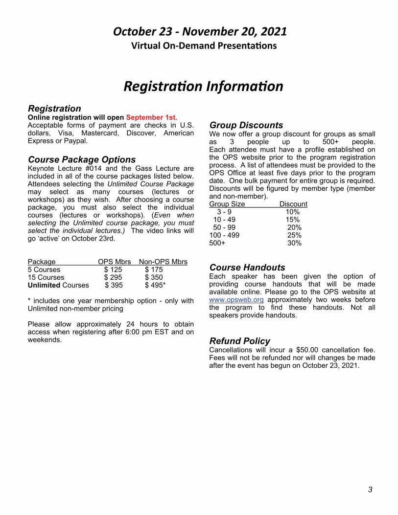
Registra on Informa on Registration Online registration will open September 1st. Acceptable forms of payment are checks in U.S. dollars, Visa, Mastercard, Discover, American Express or Paypal. Course Package Options Keynote Lecture #014 and the Gass Lecture are included in all of the course packages listed below. Attendees selecting the Unlimited Course Package may select as many courses (lectures or workshops) as they wish. After choosing a course package, you must also select the individual courses (lectures or workshops). (Even when selecting the Unlimited course package, you must select the individual lectures.) The video links will go ‘active’ on October 23rd. Package OPS Mbrs Non-OPS Mbrs 5 Courses $ 125 $ 175 15 Courses $ 295 $ 350 Unlimited Courses $ 395 $ 495* * includes one year membership option - only with Unlimited non-member pricing Please allow approximately 24 hours to obtain access when registering after 6:00 pm EST and on weekends.
Group Discounts We now offer a group discount for groups as small as 3 people up to 500+ people. Each attendee must have a profile established on the OPS website prior to the program registration process. A list of attendees must be provided to the OPS Office at least five days prior to the program date. One bulk payment for entire group is required. Discounts will be figured by member type (member and non-member). Group Size Discount 3 - 9 10% 10 - 49 15% 50 - 99 20% 100 - 499 25% 500+ 30% Course Handouts Each speaker has been given the option of providing course handouts that will be made available online. Please go to the OPS website at www.opsweb.org approximately two weeks before the program to find these handouts. Not all speakers provide handouts. Refund Policy Cancellations will incur a $50.00 cancellation fee. Fees will not be refunded nor will changes be made after the event has begun on October 23, 2021.
3
2021 Educational Program Team
General Chairman Robert Cavicchi, CRA, FOPS Beetham Eye Institute Joslin Diabetes Center One Joslin Place Boston, MA 02215 (617) 309-2778 email: [email protected] Registration, Sponsorship Barbara McCalley, Executive Director OPS Membership Office 1621 E Jody Circle Republic, MO 65738 1-800-403-1677 or (417) 725-0181 email: [email protected] Scientific Session Coordinator, Sponsorship Robert Cavicchi, CRA, FOPS Beetham Eye Institute Joslin Diabetes Center One Joslin Place Boston, MA 02215 (617) 309-2778 email: [email protected] Workshop Coordinators James Soque, COA, CRA, OCT-C, FOPS Island Retina 1500 William Floyd Parkway Shirley, NY 11967 (631) 924-4300 email: [email protected] Tim Steffens, CRA, OCT-C, FOPS Univ of Michigan Kellogg Eye Center 1000 Wall Street Ann Arbor, MI 48105 (734) 936-2283 email: [email protected]
Education Chairman, Graphic Design Anthony Medina, CRA Kellogg Eye Center, Univ of Michigan 3181 E. Grand Blanc Road Grand Blanc, MI 48439 (248) 408-7821 email: [email protected] Graphic Design, CECs Gary Rickert, CRA Retina Research Institute of Texas 5441 Health Center Drive Abilene, TX 79606 (325) 690-4408 email: [email protected] Don Wong Award Coordinator Elizabeth Affel, OCT-C, CDOS, ROUB, FOPS Phoenix Technology Group 8051 Stenton Avenue Wyndmoor, PA 19038 (215) 928-3114 email: [email protected] Scientific Exhibit Print Division John Grybas, CRA Henry Ford Health System Ophthalmology 2799 W. Grand Blvd, K-10 Detroit, MI 48202 (313) 916-4456 email: [email protected] Scientific Exhibit Stereo Division Leslie MacKeen, CRA email: [email protected]
4

Dear Colleagues, Welcome to the Ophthalmic Photographers’ Society 52nd Annual Education Program! This year’s event takes place virtually October 23rd - November 20th, 2021. Be sure to attend our Welcome Reception on October 23rd and reunite with your society. The 16th Annual J. Donald M. Gass Memorial Lecture will be given by Dr. Harry W. Flynn Jr., the J. Donald M. Gass Distinguished Chair in Ophthalmology at the University of Miami, Miller School of Medicine and Professor of Ophthalmology at the Bascom Palmer Eye Institute. We are offering over 40 on demand courses with topics including: Pediatric Ultra-Wide Field Imaging, The Future of Retinal Imaging, Cannabis 101, Organizing the Research Photographer and many other diverse topics targeted for the beginner through advanced imagers as well as ophthalmic technicians. This year’s program showcases the return of the Jamie Nicholl Symposium and 4 live broadcast workshops. In addition, we are welcoming back our supportive vendors, offering virtual exhibit halls, chat rooms and many exciting ways to experience our extra-curricular content. It is my pleasure to invite you to our second virtual Annual Program with a quote from an analog classic: “Step Inside! Hello! We’ve a most amazing show You’ll enjoy it all we know Step inside! Step inside!” Anthony P. Medina, CRA, FOPS Education Chair, OPS 52nd Annual Education Program University of Michigan, Kellogg Eye Center
5

SPECIAL EVENTS &
ORGANIZATIONAL MEETINGS
Saturday, October 23rd All courses scheduled for October 23rd and November 20th will be live including speaker
Q&A. Keynote Lecture* - The Future of Retinal Imaging by Yannis Paulus, MD, FACS - #014 - 11:00 am - 12:15 pm EDT - Dr. Yannis Paulus of the University of Michigan’s Kellogg Eye Center will be presenting ‘The Future of Retinal Imaging’, featuring a one-time ‘Live Question and Answer’ session. This interactive, complimentary lecture will open our program on Saturday, October 23rd and is included as part of your registration package. This lecture will also be available on-demand to all registered participants throughout the length of the Virtual Annual Program, but will not have a live Q & A when available on-demand. *This lecture will replace the Scientific Paper Session which has been postponed until the 2022 annual program due to lack of qualified abstract submissions. Welcome Reception - 6:00 pm - 7:00 pm EDT - Join us for our annual welcome reception virtually. It is definitely more fun to meet in-person to catch up with old friends and also meet new friends, but we have a room set aside to enjoy some socialization through our virtual platform. We hope you will join us following the live courses on the 23rd.
Saturday, November 20th The Sixteenth Annual J. Donald M. Gass Memorial Lecture - “Update on Imaging” – 4:00 pm - 5:00 pm EST - The J. Donald M. Gass Memorial lecture honors the memory of the father of macular diseases, a man celebrated for his depth of understanding of fluorescein angiography and is undisputedly regarded as one of the most influential ophthalmologists of the 20th century. This year’s lecture will be presented by Harry Flynn, Jr., MD. Harry W. Flynn Jr., M.D is the J. Donald M. Gass Distinguished Chair in Ophthalmology at the Bascom Palmer Eye Institute, University of Miami, Miller School of Medicine. Dr. Flynn has been author or co-author of more than 637 peer-reviewed publications as well as 122 book chapters. He has edited or co-edited 7 books. Dr. Flynn serves as Editor in 6 key peer-reviewed journals. He's received numerous awards nationally and internationally Immediately Following the Gass Lecture OPS Membership Meeting - 5:15 pm - 6:45 pm EST This is the annual meeting of the OPS membership, where the projects and progress of the Society are reported and discussed. This meeting will not only cover OPS business, but emphasize member participation in the Society. Help decide how the OPS can best serve our profession by supporting the Society with your presence and your participation. Contact President Elizabeth Affel, OCT-C, FOPS, c/o OPS Membership Office, (800) 403-1677 or by email: [email protected], for information about adding items to the meeting agenda.
6

52nd Virtual Annual Educational Program
1:00 pm - 2:00 pm
Course #002* Advanced Use of OCT Olivia Rainey, OCT-C, COA; James Soque, COA, CRA, OCT-C, FOPS Optical coherence tomography (OCT) has revolutionized ophthalmic imaging and continues to do so as we transition into the next decade. OCT primarily has been used for imaging the posterior pole and macular changes. This lecture will instruct how to utilize your system maximally, especially for documenting layers of the retinal pathology requiring surgery. We will emphasize the importance of understanding mechanical involvement of the vitreous cortex in both the macular region and the peripheral region and how these can play a role in retinal diseases. Fundamental and advanced techniques of image placement will be discussed and how these will aid diagnosis, monitor disease progression or morphology, and reveal response to treatments. Emphasis on surgical examples will be presented. Upon completion of this course the attendee should be able to further understand how their OCT device can aid in the decision of advanced treatment modalities made by their physician at the time of diagnosis. Level: Intermediate CEC OPS 1
2:15 pm - 3:15 pm Course #003* Color Management in Ophthalmology: Research Update Christye P. Sisson, MS, CRA Color consistency and management in fundus photography has long been an area of concern for ophthalmic photographers and reading centers, but has not been fully addressed, either through imaging standards or by the manufacturing community. This course will provide background on the principles and practices of color management in photography, and information on the specific issues surrounding color management in ophthalmic imaging. The course will provide information on research to potentially mitigate some of these issues. At the completion of the course, students will be able to describe the principles of color management, as well as how they relate and can potentially be applied to ophthalmic imaging. CEC OPS 1
3:30 pm - 4:30 pm
Course #004 Stereo Fundus Imaging Workshop James Soque, COA, CRA, OCT-C, FOPS Prerequisite: This workshop is NOT for beginners. Students must have a minimum of one (1) year
All of the lecture presentations are one hour in length. Please choose all of the courses you wish to view to obtain continuing education
credits. No course changes may be made after the start of the program. All times for live
presentations is in Eastern Standard Time.
Viewing all lectures and completing the course evaluation surveys must be done by
November 20, 2021.
Saturday, October 23, 2021 Live Presentations with Question and
Answer
11:00 am - 12:15 pm Scientific Paper Session - postponed until
2022 Annual Program.
11:00 am - 12:15 pm Course #014 - Keynote Lecture* The Future of Retinal Imaging Yannis M. Paulus, MD, FACS Newly-developed imaging techniques show extensive promise and potential to improve early disease detection, more accurate diagnosis, and improved patient management. This course will explore new optical imaging modalities used in retina and speculate on future technologies in the field including Swept Source Optical Coherence Tomography (SS-OCT), OCT angiography (OCTA), Photoacoustic Microscopy (PAM), Scanning Laser Ophthalmoscopy (SLO), adaptive optics (AO), fundus autofluorescence (FAF), and molecular imaging (MI). At the end of this course the students will be able to describe the difference between swept source and spectral-doman OCT, describe the photoacoustic principle, and understand how adaptive optics can be used to improve retinal image quality. CEC OPS 1
*This lecture is included in all of the course registration packages and does not count as one of
the courses being purchased. It will also be available on-demand throughout the virtual program.
12:30 pm
OPS Awards Presentations
7

available following the live lectures.
ON-DEMAND LECTURES Available October 23, 2021 through
November 20, 2021
Course #002 - Advanced Use of OCT - see page 7 for course description. Course #003 - Color Management in Ophthalmology: Research Update - see page 7 for course description Course #005 - Basics of Digital Imaging - see course description on page 8
Course #006 Understanding Swept Source OCT/OCTA and its Clinical Application Christopher Mody Since the introduction of Time Domain Optical Coherence Tomography (TD-OCT) in 1996, OCT has revolutionized retinal imaging, particularly since Spectral Domain technology became available in 2007, offering a significant jump in image quality. As the technology continues to progress, OCT imaging is benefiting patients in all sectors of eyecare. This lecture will consider the third generation of OCTs - Swept Source OCT, and how the technological differences between SD and SS-OCT impact capture and image quality. Signal-to-noise ratio, along with scan speed and hence data density will be considered. Swept source devices tend to use a longer wavelength which increases penetration of media opacities such as cataract and vitreous hemorrhage, with obvious advantages for tricky patients. In addition, the combination of wavelength with swept source technology increases the signal from deeper structures such as the chorioscleral interface. OCTA angiography is a relatively new concept that provides non-invasive imaging of retinal microvascular structure, the presentation will explore the implementation of swept source technology in OCTA and its clinical application. A series of clinical examples will be discussed which will include ocular conditions where choroidal, vitreous and OCTA imaging are increasing our understanding of the disease processes and providing more sensitive monitoring of ocular pathology. After this lecture, the attendee should be able to understand the technological differences between Swept Source and Spectral Domain OCT, relate the technological differences to differences in OCT capture and what structures can be imaged with one capture, understand which conditions particularly benefit from the use of SS-OCT and OCTA. Level: Intermediate CEC OPS 1 Course #007 OCT-A Practical Techniques Douglas B. Critser, CRA, OCT-C, FOPS This course will demonstrate how OCT angiography can be utilized as a diagnostic tool to classify and monitor retinal vasculature without the use of fluorescein dye. Multiple device types will be covered along with tips and tricks to optimize imaging techniques. After attending this
experience shooting seven modified fields and stereo. This course will provide live broadcast instruction for fundus imaging using the (7M) seven modified fields or the (FP-4W) four wide-angle fields technique as established by the Fundus Photography Reading Center, FPRC-Wisc [aka Eyekor/Merit]. This workshop will also provide assistance in troubleshooting. The participants will have the opportunity to send questions to the instructor during the live broadcast. Patient preparation and field definition will also be addressed. The workshop will be broadcast live from a remote clinic and attendees will be instructed using first person view, and review of some of the most difficult off axis fields will be emphasized using a live dilated volunteer. The camera of choice will be a Topcon 50 DX fundus camera with the O-Vision Software. Every effort will be made to ensure that the participant will have the best instruction possible in this ‘remote learning’ workshop. Upon completion of this course, participants will be able to identify and better work with difficult imaging scenarios encountered in clinical research imaging, including field definition, focus, and stereo pairs. When registering for this workshop, it is strongly encouraged you also register for the corresponding lecture: Strategies for Clinical and Research Imaging: Review of 7M and 4W Fields, #029. Level: Advanced (CRA recommended) CEC OPS 1
4:45 pm - 5:45 pm
Course #005* Basics of Digital Imaging Christye P. Sisson, MS, CRA Understanding the basics of photographic image formation in digital imaging provides the ophthalmic imager with troubleshooting skills when image quality is an issue. Knowing what your device is capable of is essential in knowing if a better image can be made. This course will provide the background to digital imaging, including the basics of light and color, digital image formation, and quality metrics of sensors, including resolution and bit depth. Storage and image file types will also be reviewed. At the completion of the course, students will be able to chronicle how a digital image is formed, describe the considerations for image quality in a sensor, and identify pros and cons in image file types CEC OPS 1 *Courses 002, 003 and 005 will also be available on-demand throughout the virtual program but will not have live Q & A when on-demand.
6:00 pm - 7:00 pm
Welcome Reception Join us for a virtual social event hosted by
President Libba Affel. A special room will be
8

course the student will have an increased understanding of the technical foundations of this emerging technology, be able to recognize retinal anatomy and vasculature, and also recognize basic pathologic findings such as AMD, PDR, and others. This lecture is strongly encouraged if you wish to take the corresponding OCTA workshop, Course #043. Level: Basic CEC OPS 1 Course #008 The Future of OCT Kevin Langton; Gary Michalec, CRA Optical Coherence Tomography (OCT) has revolutionized the eye care field with its ability to visualize ocular structures at high resolutions. New developments now position OCT to continue to transform imaging in ophthalmology and additional fields of medicine. This course will briefly review the history of OCT and focus on what the future holds for this transformational technology. Upon completion of this course attendees will understand the basic types of OCT technology, identify advantages/disadvantages of different OCT technologies, and identify additional fields of medicine that OCT technology is poised to impact. CEC OPS 1 Course #009 Intraoperative OCT (New) Houston Sharpe, III, OCT-C Today's operating environment is ushering in more technology & data than ever before for the surgeon. Learn how intraoperative OCT delivers sight saving images to surgeons around the world & what they glean from it. Learn how to use intraoperative OCT to improve outcomes & better visualize ocular anatomy during complex ophthalmic procedures. At the end of this lecture the student will be able to explain what intraoperative OCT is used for, understand why surgeons are using intraoperative OCT; and explain how to make the most of intraoperative OCT. CEC OPS 1 Course #010 Optical Coherence Tomography Angiography (OCTA) in Ocular Oncology Sandor Ferenczy, CRA, OCT-C The basic principles of OCTA will be covered briefly, however, the majority of the course will deal specifically with the application of OCTA imaging in a full-time ocular oncology service. There will be updates on the newest research in regards to OCTA in ocular oncology, as well as comparisons between both multiple manufacturers of OCTA systems and multiple OCT imaging implementations being used to create OCT- datasets - namely Spectral Domain OCT (SD-OCT) and Swept Source OCT (SS-OCT). At the end of this lecture the student will be able to define what OCT-A is and how it can be used in the oncology setting. He/she will also be able to discuss considerations pertaining to OCTA in studies and in creating data sets. CEC OPS 1
Course #011 OCT-A In The Age of Multi-Modal Imaging Ethan R. Priel, FOPS Following an introduction to OCT-A and an appreciation of select images, a comparison with the gold-standards of Fluorescein Angiography (FA) and Indocyanine Green Angiography (ICGA) will highlight the advantages and shortcomings of each of these modalities. Factors such as safety, time-consumption, patient comfort, identification of non-perfusion vs. new vessels, ease of interpretation, and the ideal of cross-platform uniformity of results will be discussed. In this course we will explore findings seen in Fluorescein and ICG Angiographies, OCT scans and color fundus photography and study their correlation to OCT-A images. At the end of this course the students will be able to describe the fundamentals of the OCT-A technology as well as compare and contrast the images produced by OCT-A to more traditional imaging modalities, such as Fluorescein Angiography and ICGA. CEC OPS 1 Course #012 OCT WORKSHOP LECTURE Darrin Landry, CRA, OCT-C This introductory course will briefly discuss the differences between Time-Domain OCT (TD-OCT), Spectral Domain OCT (SD-OCT), and Swept Source OCT (SS-OCT) and elaborate on fundamentals in the operation of OCT's. Examples of common scan patterns and analysis will be shown. Retinal anatomy as seen with OCT will be reviewed and examples of common abnormalities will be shown. Challenges in obtaining certain scans as well as how to overcome them will also be discussed. Upon completion of this course, participants will be able to describe the basic capabilities and limitations of TD-OCT, SD-OCT, and SS-OCT, discuss various aspects of retinal and optic nerve scan analysis as well as factors such as reliability and resolution. This lecture is strongly encouraged if you wish to take the corresponding OCT workshop, Course #042. Level: Basic CEC OPS 1 Course #014 - The Future of Retinal Imaging - see course description on page 7 Course #015 Fundus Autofluorescence Imaging Timothy Bennett, CRA, OCT-C, FOPS Fundus Autofluorescence Imaging (FAF) is a specialized form of fundus photography that documents naturally occurring fluorophores in the human eye. The crystalline lens, optic disc drusen, hammartomas and lipofuscin pigments in the RPE all possess fluorescent properties that can be captured with a fundus camera or scanning laser ophthalmoscope equipped for autofluorescent imaging. This course will present an overview of the uses, equipment and techniques for fundus autofluorescence. Upon completion of this course, the participant should be able to: list fluorescent
9

structures in the eye, describe equipment, wavelengths, and filters used in FAF, recognize common FAF clinical findings, and discuss challenges & solutions encountered in FAF imaging. Level: Intermediate CEC OPS 1 Course #016 Maximizing Clinical Flow (New) Tim Steffens, CRA, OCT-C, FOPS Do you have too many patients waiting long times for imaging or other services? This course will describe the LEAN continuous improvement cycle developed by Toyota and how to implement it. We will discuss at length how the process works, how to collect data and analyze it for implementation. Two clinical examples will be presented. After this lecture, attendees should be able to explain the LEAN continuous improvement cycle, identify opportunities in the workflow, plan how the current process can be improved and execute those plans and review the changes for more improvement. Level: Basic CEC OPS 1 Course #017 Clinical Applications of Slit Lamp Imaging Sarah Armstrong, CRA, OCT-C, FOPS In this case based course, we will walk through the different lighting techniques available on the slit lamp. How to use a slit lamp and make adjustments for ideal imaging settings will be discussed. The clinical benefit to acquiring images anterior segment disease will be reviewed. By the end of this course attendees should be able to identify several tips for adjusting slit lamp lighting for ideal image settings and will have a basic knowledge of which pathology should be imaged with which imaging techniques. Level: Intermediate CEC OPS 1 Course #018 Introduction to Slit Lamp Photography Michael Bono, COT, CRA, BFA In this course, we will take an in-depth look at the complexities and potential of an instrument first devised in 1911 and shaped by many hands to become an essential instrument in ophthalmology. We will demonstrate how to view, light and capture the structures in the anterior segment using all the full capabilities of the instrument. This course will cover preparation for an imaging session, patient management, how to use the slit lamp with an emphasis on basic lighting techniques and how they can be used to reveal specific structures in the anterior segment. The importance of instrument components, software, types of lighting schemes and what works best based on the pathology being documented will be discussed. Many examples of great slit lamp imaging from OPS photographers will be utilized. Upon completion of this
course students will be able to describe the essential parts of the instrument, the types of different lighting techniques, when they are applied and how images are captured and delivered to our physicians. Level: Basic CEC OPS 1 Course #019 Technician Safety and Efficiency During a Pandemic Jane T. Shuman, COT, COE, OCS As the effects of the 2020 pandemic have lasted longer than anticipated, it is easy for technicians to become complacent about patient safety as well as their own. This presentation will discuss how to remain efficient without compromising exposure as the volume of patients continues to increase. One such efficiency is the lasting effect of telemedicine in ophthalmology. At the conclusion of this program the attendee will be able to discuss the current risks of contagion in an outpatient healthcare setting, identify three areas of risk in the work-up area, discuss the history of telemedicine in ophthalmology and optometry, and list what a technician can do during a virtual visit. CEC OPS 1 Course #020 Pediatric and Neonatal Ultra-Wide Field Imaging (New) Candice Frazier, CRA, OCT-C, COA In this course, we will discuss basic anatomy of the eye along with a basic understanding of embryology and development of the eye. We will discuss how to prepare the RetCam and the infant prior to a photographic session. We will then cover how to combine the RetCam imaging technique with ocular anatomy to understand how to properly image pediatric patients. At the end of this course, participants will be able to Identify and define general ocular anatomy as well as understand basic embryology and development of the eye, prepare the RetCam System and position the infant for imaging the retina, and review proper imaging techniques and how to localize the posterior, nasal, temporal, superior and inferior retinal fields. CEC OPS 1 Course #021 Widefield Imaging for Tumors Sandor Ferenczy, CRA, OCT-C A brief overview of the lengthy history of widefield ophthalmic imaging will be given with specific discussion of its applications in ocular oncology. Case series of imaging from multiple cameras will be presented, providing comparisons of current technologies, as well as providing a baseline of what older cameras allowed photographers to capture. Technical differences of current cameras and the ensuing clinical impact will be discussed as well as the overall benefits of widefield retinal imaging for ophthalmology. At the end of the course, participants will be able to recognize images from various cameras based on their appearance. Students will be able to demonstrate knowledge of the differences between widefield instruments and the potential advantages and disadvantages of these differences in clinical use. CEC OPS 1 10

Course #022 Introduction to Ultra Widefield Imaging (WSL) Olivia Rainey, OCT-C, COA This lecture will introduce what ultra widefield (UWF) imaging is, how it is useful, and what instruments can be used to image the far periphery of the eye. It will elaborate on principle features of ultra widefield imaging and help identify how to improve imaging quality. It will discuss various imaging modalities such as fundus color photography, fundus autofluorescence, and fluorescein/indocyanine green angiography. It will help students understand how to recognize and image peripheral pathology with each modality. Upon completion of this course the student should understand the advantages of ultra widefield imaging systems, identify ultra widefield imaging modalities and artifacts, and identify pathology with ultra-widefield systems. This lecture is strongly encouraged if you wish to take the corresponding UWF workshop, Course #046. Level: Beginner CEC OPS 1 Course #023 Imaging Approaches to Challenging Cases (New) James Gilman, CRA, FOPS In the course of performing imaging on patients, in all cases we are faced with attempting to make an image on a patient that represents what they have. Some cases prove to be more challenging than others and will require a different approach than the usual procedure you perform on most patients. I will demonstrate a series of different techniques and how I approached some challenging patient situations to make images that were not your typical ophthalmic images. After this lecture, the attendee should be able to describe three techniques on ophthalmic imaging that they have never attempted before and be able to incorporate in their armentarium of procedures, describe a method for making measurements at the slit lamp and describe a way to transilluminate the sclera. Level: intermediate CEC OPS 1 Course #024 Corneal Endothelial Imaging, Analysis and Clinical Trials (New) Beth Ann Benetz, CRA, FOPS Specular microscopy, corneal endothelial imaging and analyses has long been used as a measure of corneal health, a measure of the endothelial cell reserve and monitoring corneal dystrophies. In clinical trials, endothelial cell density and morphology assessments over time are used as both safety and efficacy measures. In this presentation, clinical applications as well as clinical trials applications for endothelial imaging, various techniques and instruments for imaging, the most common analyses tools and common imaging and analyses challenges will be discussed. After this lecture the attendee will appreciate corneal endothelium imaging instruments, imaging techniques, analyses method selection and common imaging and analyses challenges. Level: Basic CEC OPS 1
Course #025 Issues of the Visually Impaired Paula F. Morris, CRA, FOPS As ophthalmic imagers we daily encounter and document patients whose vision may potentially be or is already impaired. But how well do we understand the realities of what it is like to have impaired vision? What is vision like for a patient with advanced glaucoma, macular degeneration, or retinitis pigmentosa? What difference do light and contrast make to impaired vision, and what does the photographic process do to our patients’ vision? This course will discuss and display the different ways that poor vision affects how our patients manage both in the clinic and away. Suggestions on how to make the clinical encounter less traumatic will be discussed. At the end of this course, students should be able to discuss the role of contrast in vision and demonstrate how to be a “sighted guide” escort. Level: Basic CEC OPS 1 Course #026 Imaging the Central Retinal Microvascular Pathology in the Diabetic Eye with AOSLO and OCTA (New) Konstantina Sampani, MD Advanced retinal imaging techniques are becoming indispensable for the retinal evaluation of patients with diabetes. Both AOSLO and OCTA can produce information-rich images in matter of seconds that must be quantified to further answer clinical questions. In this course learners will be introduced to the key concepts of AOSLO and OCTA images’ interpretation for effective DR management. Furthermore, attendees will learn how the use of high-resolution retinal images can contribute to the potential development of imaging biomarkers and the quantification of structural and hemodynamic changes on the retinal vasculature. At the end of this course, learners will be able to acknowledge the importance of AOSLO and OCTA in prevention of DR progression and will be equipped with the knowledge of how to evaluate the minimal micro-vessel alterations detected on the above imaging techniques. CEC OPS 1 Course #027 Evolving Treatment Paradigms in Diabetic Retinopathy (New) Dilsher Dhoot, MD This lecture will review the epidemiology and pathophysiology of diabetic retinopathy. Diagnosis and screening parameters in 2020 will be explained. Imaging and diabetic retinopathy screening and management will be discussed as well as the evolution of treatment for diabetic retinopathy and the evolving treatment paradigm. At the end of the course the student will be able to understand the importance of screening for diabetic retinopathy, understand the role of imaging including OCT-A, widefield imaging, and fluorescein
11
angiography in Diabetic Retinopathy, and characterize the treatment options for DR based on ETDRS staging. Level: Basic-Intermediate CEC OPS 1 Course #028 Advanced Fundus Imaging: Strategies for Clinical and Research Imaging Capturing 7M and 4W Fields Pamela Vargo, CRA This advanced course is a detailed overview of the imaging of the (7M) seven modified and (4W) four wide-angle stereoscopic fields used in diabetic studies. Emphasis will be focused on fine tuning techniques of field definition; stereoscopic imaging and imaging techniques will be discussed. Insightful and helpful tips will be offered. At the end of the course, the student will be able to identify the (7M) seven modified fields and the (4W) four wide-angle fields and understand the capture orientation to the optic nerve. This course is not for beginners, but intended for technicians with imaging experience of a minimum of one (1) year imaging the seven modified field. Level: Beginner with at least 1 year experience to Advanced. CEC OPS 1 Course #029 Introduction to Dye Angiography Darrin Landry, CRA, OCT-C Fluorescein angiography has been a staple of ophthalmic diagnostic imaging for 60 years, and Indocyanine Green angiography for 25 years. The history of each dye, understanding how these dyes are used to illustrate retinal and choroidal vasculature, when either dye is used for a specific intent, and how this skill set is a foundation for any ophthalmic imager will be discussed. Upon completion of this course the attendee will be able to explain the difference between fluorescein and ICG angiography, describe common findings in FA and ICG angiography and understand which disease pathologies are best imaged with either dye. Level: Beginner CEC OPS 1 Course #030 Jamie Nicholl Symposium Paula Morris, CRA, FOPS - Moderator As the practice of ophthalmic photography evolves, so do the challenges confronting imagers in 2021. Besides the merits of new technologies and changes in treatment modalities, new approaches to professional development need to be considered as well as classic patient management topics. This year imagers are confronted with health and safety problems that have never had to be addressed before. This course provides an opportunity to discuss these topics, share expertise, and consider successful work and training strategies appropriate for these challenging times. At the end of this course, the student will be able to discuss the merit of new imaging technologies, list strategies for patient
and imager safety, and discuss the value of strategic professional development. Level: Basic CEC OPS 1 Course #031 Advanced Imaging in Animal Based Vision Research Monica Motta This course will cover a range of vision research testing performed in mice, rats, rabbits, dogs, and primates. Testing modalities covered will include; OCT, fundus photography, slit lamp digital capture, electroretinogram and confocal biomicroscopy. Imaging pros and challenges will be discussed for both research and clinical trial settings. Upon completion of this course students should have a clear understanding of the imaging modalities used in animal vision research, understand the challenges which come with imaging animals and the importance of animal based vision research and its contribution to one health. Level: Intermediate CEC OPS 1 Course #032 Introduction to Bscan Screening Techniques Adeline Stone, COT, CRA, CDOS This course introduces the principles and practices of posterior ophthalmic ultrasound while covering standard screening techniques. Examples of static ultrasound images and positioning are reviewed, following a standard screening process and labeling nomenclature. Objectives of this class include an introduction of the theory of ultrasound, probe positioning, labeling and how to visualize the clock hours of a posterior ultrasound. Level: Beginner CEC OPS 1 Course #033 Diagnostic Ultrasound in Vitreoretinal Pathologies M. Bernadette Ayres, MD; Tanya McClendon, CDOS, ROUB This course will provide a comprehensive review of ophthalmic A- and B-scan ultrasound of the posterior segment with emphasis on vitreous, retina and choroid. The sonographic features of the vitreoretinal disorders will be presented. Topics will include retinal detachment, ciliochoroidal effusion, pediatric retinal disorders, endophthalmitis, retinochoroidal tumors and ocular trauma. This will be an interactive course with audio-visual demonstration of examination techniques and a wide variety of pathologies related to the vitreoretinal interface. Attendees will be able to describe or demonstrate echographic examination techniques and they will be able to assess features seen when doing Ultrasound of the most common vitreoretinal diseases. CEC OPS 1
12

Course #034 Ophthalmic Imaging: Technical Basics for Non-Technical Professionals N. A. (Bert) Massie, PhD If you are a retinal imaging professional or use retinal images, this presentation is for you. An assessable review of imaging technology will be delivered that covers the entire process. This starts with defining imaging objectives and criteria and concludes with image processing. After this lecture the participant will have a basic understanding of the technology and optical systems that enable retinal imaging. Level: Intermediate CEC OPS 1 Course #035 Cannabis 101 (New) Denise Satterfield, MD, FAAP Cannabis use, both medical and adult recreational, are on the rise. Fewer than 10% of medical schools have any education about cannabis. Most health care workers have no working knowledge about cannabis. This course will enlighten the participant about the historical, political and racial landscape of cannabis in the United States, the current research on cannabis in the US and abroad, understand the physiology of cannabis and understand the risks and benefits of cannabis use. After this lecture the participant will have a better understanding of cannabis use and abuse as well as the current and historical background of cannabis in the US and physiology of cannabis with human consumption. Level: Basic CEC OPS 1 Course #037 Standard Techniques of Gonioscopy & Goniography Michael Bono, COT, CRA, BFA Successfully viewing and documenting the iridocorneal angle is a challenging and rewarding task for the ophthalmic photographer. Viewing and documenting angle structures requires skillful use of the slit lamp microscope to light and properly expose the subject matter all while maintaining patient compliance. This course will include a brief overview of the history of direct and indirect gonioscopy techniques to view the angle. Students will then learn the skill set needed to visualize and image the angle using a three or four mirror gonio lens. In this course lighting techniques, camera positioning, magnification and patient management will be discussed using images provided by our workshop instructors. Students will see excellent examples of angle pathology using the video series by Wallace Alward, MD. At the completion of this course, students will be able to discuss the origins of gonioscopy, define the concept of total internal reflection, identify the six key landmarks of the angle, and describe the correct positioning of the gonio lens on the eye to reveal the angle structures. Students will be able to identify common abnormalities as seen in the iridocorneal angle. Level: Intermediate CEC OPS 1
Course #038 Imaging in Uveitis: Past, Present and Future (New) Lisa Faia, MD Imaging in uveitis aids in the diagnosis and management of the disease. Newer modalities are less invasive but lacking in a consensus of their utility and interpretation. More classic modalities, such as fundus photography, fluorescein angiography, and indocyanine green angiography, have been jetted forward with wide-field capabilities. Optical coherence tomography has also evolved from time-domain to spectral domain, giving us a “photographic” in vivo pathology-like imaging of the retina and anterior segment. Fundus autofluorescence, using the properties of the retinal pigment epithelium and lipofuscin, has allowed us to detect activity in the absence of fluorescein angiograms. Going deeper into the eye and forward in imaging, we are using enhanced depth imaging for choroidal and scleral imaging and optical coherence tomography – angiography to give us 3D angiograms of the retinal and choroidal vasculature without dye exposure. In this presentation, applications of the different modalities and the challenges in obtaining and interpreting these images will be discussed. After this presentation the attendee will better understand how the classic modalities aid in the diagnosis and management of uveitis and for what diseases each modality may better define, how the newer, less invasive modalities have given us more information on the collection of diseases known as uveitis, and the controversies that exist with the new modalities and what is needed in the future for more consistent interpretations. Level: Intermediate CEC OPS 1 Course #039 Pediatric Optic Nerve Drusen: Improving Detection with Multi-Modal Imaging and EDI OCT. Cynthia VandenHoven, CRA, FOPS In a pediatric population, optic nerve drusen (OND) is often buried, non-calcified, and difficult to detect on clinical examination compared to the adult presentation of optic nerve drusen. Many imaging modalities can be applied to help tease out the clinical signs such as fundus photos, Bscan ultrasonography, fundus autofluorescence (FAF), IVFA, OCT and more recently, Enhanced Depth Imaging (EDI) OCT. Although many research papers have explored how to improve detection of OND, no single method can be relied upon in all instances. After careful review of the current research, EDI-OCT is showing promise within our pediatric population. This presentation will detail how EDI-OCT using a modified approach in scanning technique, can complement other imaging modalities to improve detection of buried, non-calcified drusen bodies. At the end of this course the attendee will be able to review different imaging modalities for pediatric OND ranging from IVFA, FP, FAF and Ultrasound used to detect OND, understand the effect different OCT scan patterns make in the ability to detect clinical signs of OND, and understand that different resolution, image quality, and scan quality can make on the OCT image to improve detection of observable signs. Level: Advanced CEC OPS 1
13

is designed as an introduction to the use of Spectral Domain OCT (SD-OCT). The participants will have the opportunity to send questions to the instructor during the broadcast. The participants will have the opportunity to familiarize themselves with the various hardware and software of these imaging systems. Scan modes, their applications and some of the techniques needed to capture images with these instruments will be demonstrated. THIS WORKSHOP IS INTENDED FOR STUDENTS WITH LITTLE-TO-NO EXPERIENCE WITH THE SD-OCT. Upon completion of this course, participants will be able to perform basic techniques for SD-OCT procedures, and identify some of the common controls for SD-OCT operation. When registering for this workshop, it is strongly encouraged you also register for the corresponding OCT lecture, Course #012. Level: Basic CEC OPS 1
1:30 pm - 2:30 pm EST Course #043 OCTA WORKSHOP Tim Steffens, CRA, OCT-C, FOPS This workshop will provide live broadcast instruction and is designed to introduce the attendee to capturing OCT-A images with the current FDA approved devices on the market today for studying retinal vasculature. The participants will have the opportunity to send questions to the instructor during the broadcast. Entering patient data, capturing techniques, editing segmentation errors, exportation of reports, and scan assessment will be reviewed. Upon completion of this workshop, students will be able to capture an OCT-A scan, be able to generate OCT-A reports, know how to edit segmentation errors, and know the differences and similarities between OCT-A devices. When registering for this workshop, it is strongly encouraged you also register for the corresponding OCTA lecture, Course #007. CEC OPS 1
2:45 pm - 3:45 pm EST
Course #044 Ultra Widefield Imaging Workshop Tim Steffens, CRA, OCT-C, FOPS Prerequisites: A basic understanding of fundus photography is required. This workshop will provide live broadcast instruction and is designed as an introduction to the use of Wide Angle Fundus Imaging. The participants will have the opportunity to familiarize themselves with the various hardware and software of these imaging systems and have the opportunity to send questions to the instructor during the broadcast. It is intended for students with little-to-no experience with wide angle imaging. The Heidelberg Spectralis, the Optos California and the Zeiss Clarus 700 will be demonstrated. Imaging techniques for wide field imaging will be demonstrated for each device. Upon completion of this course, participants will be able to
14
Saturday, November 20th
Live Presentations with Question and Answer
9:45 am - 10:45 am EST Course #040 Organizing the Research Photographer (New) Holly Cheshier, CRA, OCT-C, COT Study imaging in clinic setting is overwhelming with learning various imaging protocols and reading center certification processes. We will discuss how to lessen the daily anxiety of the study imager by staying prepared through organizational forms and guides to increase productivity and efficiency. We will discuss tips to maintain high quality images with the various media and patient challenges we face in study imaging. At the completion of this course, the participants should be able to understand how to develop organizational forms for study protocols and how to image around media opacities. CEC OPS 1
11:00 am - 12:00 pm EST
Course #041 Difficult Imaging Made Easy Holly Cheshier, CRA, OCT-C, COT Prerequisite: Ability to perform fundus photography and understands basic ocular anatomy. This course will present a series of short tips and strategies for maximizing diagnostic information in Optical Coherence Tomography (OCT), fundus photography & fluorescein angiography. Troubleshooting techniques will be covered in getting around the pitfalls of difficult media opacities, patients with special needs and challenging pathologies. We will discuss achieving the optimal image for various pathologies of both central and peripheral retina. We will look at different imaging options to help the technician get the best image possible for the physician. Case examples will be given. At the conclusion of this course the student will be able to apply tips and strategies to maximize diagnostic information when performing imaging test, and understand how to achieve the best possible images when working with various challenges of both the central and peripheral retina. Level: Intermediate CEC OPS 1
12:15 pm - 1:15 pm EST
Course #042 OCT WORKSHOP Tim Steffens, CRA, OCT-C, FOPS This workshop will provide live broadcast instruction and

identify some of the common controls for wide angle imaging and acquire an acceptable wide angle image on each of the different instruments available. When registering for this workshop, it is strongly encouraged you also register for the corresponding UWF lecture, Course #022. Level: Basic CEC OPS 1
4:00 pm - 5:00 pm EST
This lecture is included in all of the course registration packages. It does not count toward
one of the lectures you purchase.
The Sixteenth Annual J. Donald M. Gass Memorial Lecture
Course #045 Update on Imaging Harry Flynn, MD Ophthalmic photography continues to evolve as new technology becomes available. Key components include optical coherence tomography (OCT), color photography, fluorescein angiography, OCT angiography and other methods of imaging documentation. The lecture will review current options in ophthalmic imaging as well as to present interesting clinical examples in each major area. At the end of the course attendees should be able to report treatment outcomes in multicenter clinical trials using OCT, demonstrate differences between spectral domain OCT and swept source OCT angiography, show the use of fluorescein angiography including widefield techniques, and document various modes of color photography ranging from standard posterior pole photography with montage to
widefield photography. The participant will gain knowledge regarding the clinical use of these modalities and their value to clinicians. Level: Intermediate CEC OPS 1 *This event is included as part of each of the lecture package options.
5:15 pm - 6:45 pm EST
OPS Membership Business Meeting This meeting will take place immediately following the
Gass lecture.
This is the annual business meeting for the Society. New Officers and Directors will be sworn in, Committee Chairs will update the membership of the committee’s accomplishments throughout the year, and the Treasurer will inform the membership of the financial health of the Society.
15

Sarah Armstrong, CRA, OCT-C, FOPS University of North Carolina Durham, NC M. Bernadete Ayres, MD Univ of Michigan, Kellogg Eye Center Ann Arbor, MI Beth Ann Benetz, CRA, FOPS Case Western Reserve University Cleveland, OH Timothy J. Bennett, CRA, OCT-C, FOPS Milton S. Hershey Medical Center Hershey, PA Michael Bono, COT, CRA, BFA, FOPS University of Colorado Eye Center Aurora, CO Holly N. Cheshier, CRA, OCT-C VitreoRetinal Surgery, PA Bloomington, MN Douglas B. Critser, CRA, OCT-C Merit CRO Iowa City, IA Dilsher Dhoot, MD California Retina Consultants Valencia, CA Lisa Faia, MD Associated Retinal Consultants, PC Royal Oak, MI Sandor Ferenczy, CRA, OCT-C Wills Eye Hospital, Ocular Oncology Service Philadelphia, PA Harry Flynn, Jr., MD Bascom Palmer Eye Institute Miami, FL Candice Frazier, CRA, OCT-C, COA Natus Ozark, MO James Gilman, CRA, FOPS Univ of Utah, John Moran Eye Center Salt Lake City, UT Darrin Landry, CRA, OCT-C Bryson Taylor Inc Saco, ME
Christye Sisson, MS, CRA Rochester Institute of Technology Rochester, NY James Soque, CRA, OCT-C, COA, FOPS Island Retina Shirley, NY Tim Steffens, CRA, OCT-C, FOPS Univ of Michigan/Kellogg Eye Center Ann Arbor, MI Adeline Stone, COT, CRA, CDOS Casey Eye Institute Vancouver, WA Cynthia VandenHoven, CRA, FOPS Hospital for Sick Children Toronto, ON, Canada Pamela Vargo, CRA Fundus Photograph Reading Center Madison, WI
16
Lecture Faculty Kevin Langton Carl Zeiss Meditec, Inc. South Orange, NJ N. A. (Bert) Massie, PhD San Ramon, CA Tanya McClendon, CDOS, ROUB Univ of Michigan, Kellogg Eye Center Ann Arbor, MI Gary Michalec, CRA Carl Zeiss Meditec, Inc. Minnetrista, MN Christopher Mody, FdSC Topcon (GB) Medical Ltd Newbury, Berkshire, UK Paula Morris, CRA, FOPS Ainsley Eye Brain Center, Jefferson University Philadelphia, PA Monica Motta UC Davis School of Veterinary Medicine Davis, CA Yannis M. Paulus, MD, FACS Univ of Michigan, Kellogg Eye Center Ann Arbor, MI Ethan R. Priel, FOPS MOR Medical Center Bnei-Brak, Israel Olivia Rainey, OCT-C, COA Retina Specialists of Michigan Grand Rapids, MI Konstantina Sampani, MD Joslin Diabetes Center Boston, MA Denise Satterfield, MD, FAAP Sutter Medical Group Sacramento, CA Houston Sharpe, III, COA, OCT-C Leica Microsystems Mebane, NC Jane Shuman, COT. COE, OCS Eyetechs, Inc. Ashland, MA

17
CONTINUING EDUCATION CREDIT
Approved OPS continuing education credits are listed at the end of each course description. Due to the significant increase in application fees for IJCAHPO distant learning CECs, it is cost prohibitive to offer IJCAHPO credits for the OPS 2021 Virtual Annual Program. Continuing Education Credit will be granted to all registrants who attend the course and complete the online course evaluation surveys at the conclusion of each course. CEC credits will also be granted for The Scientific Paper Session and Gass Lecture. CEC documentation will be available through the registrants’ profile on the OPS website following verification approximately five to six weeks after the Educational Program. Each attendee must be registered on the OPS website prior to registering for the Annual Program in order to complete the online course evaluation surveys. The course number and title will be provided at the beginning of each lecture. Students must complete their own online evaluation surveys when the course has ended. The evaluation surveys may be done via a smart phone, tablet, or PC at the conclusion of each course. The surveys will be available until December 1, 2021. If you require a hard-copy for your records, print your list of earned CECs from your profile on the OPS website. Letters will no longer be mailed to program attendees. To obtain credit, you must 1. Register for the course 2. View the recorded lecture. 3. Correctly complete the online course evaluation survey at the end of the course. 4. Print your list of CECs earned from your profile on the OPS website (Optional) Locating your online CEC Journal: Manage Profile > Content & Features > Professional Development
UPCOMING OPS EVENTS
EDUCATIONAL OPPORTUNITIES
Webinars on Demand available on the OPS website 24/7.
International Conference on Ophthalmic Photography (ICOP) Rotterdam, Netherlands
April 2022
2022 Regional Education Program Joslin Diabetes Center
Boston, MA TBD
53nd Annual Education Program
Chicago, IL September 30 - October 2, 2022
CERTIFICATION OPPORTUNITIES
The CRA and OCT-C Program Guides are available for download on the OPS web site - www.opsweb.org/page/CRAOCTrequirements

THE DON WONG AWARD In 1990 the Ophthalmic Photographers’ Society established a new award for the best scientific paper presented at each Annual Educational Program. The award is named for Don Wong (1931-1999), a founding member of our Society whose entire career exemplified literary and professional achievement. Don was the creator and first editor of our Journal of Ophthalmic Photography, one of the earliest proponents of the certification program, and the father of the international meeting series. He worked tirelessly to encourage professionalism in our technical work and high ethical standards in our lives. He was a mentor and friend to many. The Don Wong Award recognizes outstanding scientific achievement in our profession. Presentations will be judged by a panel of accomplished colleagues on the basis of content, originality, organization, presentation, delivery and importance to the field.
DON WONG AWARD RECIPIENTS 1990 Randall E. Verdick 2006 Ethan Priel, FOPS 1991 George Weir, CRA 2007 Dennis Orlock, CRA, FOPS 1992 Jeff Jacobs, CRA 2008 Robert G. Shutt, CRA, OCT-C 1993 Jim Gilman, CRA 2009 Ditte J. Hess, CRA, FOPS 1994 Randall E. Verdick 2010 Leslie D. MacKeen, CRA 1995 Lawrence M. Merin, FOPS 2011 Alexis Smith, CRA, OCT-C 1996 Linda M. Kelley, CRA 2012 Carl Glittenberg, MD 1997 Bobbie A. Turner, AA, CRA, COT 2013 Michael P. Kelly, FOPS 1998 Patrick J. Saine, CRA, FOPS 2014 Leslie D. MacKeen, CRA 1999 Csaba L. Martonyi, CRA, FOPS 2015 Mark Croswell, CRA, OCT-C, FOPS 2000 Ethan Priel 2016 Darrin Landry, CRA, OCT-C 2001 Dennis Orlock, CRA 2017 Darrin Landry, CRA, OCT-C 2002 Kevin Langton, CRA 2018 Douglas B. Critser, CRA, OCT-C 2003 Lawrence Merin, RBP, FOPS 2019 James D. Strong, CRA, OCT-C, FOPS 2004 Dennis Orlock, CRA 2020 Johnathan Hawkins, CRA, OCT-C 2005 Timothy J. Bennett, CRA, FOPS
18
Board of Certification Disclaimer
The OPS Annual Educational Program, nor any course or workshop offered during this Program, is reviewed, endorsed, or approved by The OPS Board of Certification for its specific content or efficacy in test preparation. These educational offerings are for information purposes and are only suggested as a study tool. Informational summaries, methods or techniques, and practical tips and tricks are only recommendations by the author, and in no way guarantee passing the CRA or OCT-C exams. The author has made reasonable efforts to provide current and accurate information to the students. The author, nor OPS, will be held liable for any unintentional errors or omissions that may be found. These educational materials, even in their entirety, should not be considered a complete and all-encompassing representation of test content for the CRA or OCT-C exam.
Participation by OPS Board of Certification members, in the OPS Annual Educational Program, is for the sole purpose of creating a more robust educational experience. This participation does not make any inference of validation or set any precedent that may be referenced during any Certification activities. The Certified Retinal Angiographer (CRA) and Optical Coherence Tomographer-Certified (OCT-C) credential programs are administered and maintained by the OPS Board of Certification in accordance with the BOC Standing Rules.

The Chris Barry Award
(Journal of Ophthalmic Photography Article of the Year) Since 2003, the Ophthalmic Photographers’ Society has awarded the “Journal of Ophthalmic Photography Article of the Year Award”. This award was inaugurated to highlight the skills and expertise of those OPS members and ophthalmic colleagues who spend their time and energy submitting their work for publication. They present us with new information, share with us interesting cases or the “nuts and bolts” of ophthalmic imaging. A committee judges the articles for the relevancy of the topic, degree of innovation, style of writing, quality of illustrations and value to an ophthalmic imager. This culminates in our celebration of the Best Journal Article of the Year and comes with a free one year OPS membership. We encourage all our readership to submit their work and share with us their expertise. Mentors are available to take you through the process, if necessary. Your continued involvement and support makes the Journal contemporary, vibrant and relevant. This award recognizes the excellence of published work in the Journal of Ophthalmic Photography, the “flagship” of the Ophthalmic Photographers’ Society.
Award Recipients
2019 Tim Steffens - External Photography 2018 Olivia Rainey - Ultra Wide Field Fundus Autofluorescence Imaging 2017 No Award Given 2016 Darrin Landry—Optical Coherence Tomography Angiography 2015 Melanie Fortin - Management of New or Recurrent Choroidal Neovascularisation in Telescope Implanted Eyes 2014 Peter Van Etten, MD - Zero Dilation Ophthalmoscopy 2013 Angela Chappell - Tales of the Unexpected: Incidental Findings in Ophthalmic Imaging 2012 Ditte Hess - ROP—A Visual Experience 2011 Alexis Smith - Correlation of Ocular Ultrasound and EDI of Ocular Lesions 2010 Kirsten Locke - Optical Coherence Tomography in Patients Diagnosed with North Carolina Macular Dystrophy 2009 Sarah Moyer - Anterior Segment OCT: A Comparison of Time Domain and Spectral Domain Technologies 2008 Alexander Walsh, MD - Spectral Domain OCT: An A to Z Guide 2007 Ethan Priel - Fundus Autofluorescence With a Confocal SLO 2006 Patrick Saine - Tutorial; External Eye Photography 2005 Richard Hackel and Patrick Saine - Creating Retinal Fundus Maps 2004 Lawrence Merin - Digital Detection of Diabetic Retinopathy 2003 Gregory Hoffmeyer - Mac Pac: A Systemic Protocol for OCT Scanning for Macular Pathology 2002 James Scott - An Affordable Alternative to the High Cost of Digital Fundus Photography (inaugural award)
19

THE CSABA L. MARTONYI AWARD In 2008 the OPS Board of Directors established the Csaba L. Martonyi Award, given annually to the best image from the OPS Scientific Exhibit. Csaba L. Martonyi, CRA, FOPS is Emeritus Associate Professor and Former Director of Ophthalmic Photography at the Kellogg Eye Center, University of Michigan Medical School. A longtime active member of the Ophthalmic Photographers’ Society, he has served on the OPS Board of Directors, was first Chair of the OPS Board of Certification, is a Past President, and recently retired from the post of OPS Parliamentarian. Csaba is well known for his teaching and writing, most notably for the classic text Slit Lamp Examination and Photography, now in its third edition. This award celebrates the high standards of excellence in imaging that Csaba has exemplified throughout his career. He has always stressed that it is not sufficient for us as professional imagers to simply take the picture that will "get by", but to put our effort and skill into producing images that both serve a medical purpose and demonstrate technical and artistic perfection. He has demonstrated his ability to accomplish this through the countless awards that his photographs have won, and he encouraged others in his profession to strive for these same goals through his teachings. This goal, which Csaba has championed throughout his career, is the heartbeat of our Scientific Exhibit. Please join the OPS Board of Directors in congratulating Csaba L. Martonyi, CRA, FOPS on the establishment of this award in his honor and consider entering your best work for consideration in the 2019 Scientific Exhibit competition and the opportunity to be the twelfth recipient of the Csaba L. Martonyi Award.
Award Recipients
2008 Robert Myles, CRA 2015 Ryan Terribilini, OCT-C 2009 David Miller, CRA 2016 John Golding 2010 Ditte Hess, CRA, FOPS 2017 Leslie D. MacKeen, CRA 2011 Zlatan Sadikovic, CRA 2018 Kit Morehead, CRA 2012 Allan Connor 2019 Katelyn Olney, CRA, OCT-C 2013 Mark Clark, CRA 2020 Timothy Costello, CRA, COA 2014 Gary Miller, CRA, OCT-C
20
Johnny Justice Jr. Scholarship
The first Johnny Justice Jr. Scholarship was awarded in 1996 at the 27th Annual Education Program of the Ophthalmic Photographers’ Society. Named in honor of Johnny Justice Jr., a principal founding member of the Ophthalmic Photographers’ Society, the JJJ Scholarship Award is available to assist in the education of persons actively pursuing careers in ophthalmic photography. The 2021 JJJ Scholarship Award will provide the chosen applicant with an ALL Lecture Package registration, if the award is used at the OPS Virtual Annual Education Program, or the general registration fee for the OPS Regional Education Program. The Johnny Justice Jr. Scholarship Award was created by the Board of Directors of the Ophthalmic Photographers’ Society to not only honor its founder, but also to assist its members in their efforts to gain knowledge and expertise in the field of ophthalmic photography. The Johnny Justice Jr. Scholarship Award and other special projects are supported by the OPS Endowment Fund. The fund is supported by contributions and fund raising activities such as the raffle held during the Annual Educational Program.
OUTSTANDING CONTRIBUTIONS TO OPHTHALMIC PHOTOGRAPHY AWARD
The Outstanding Contributions to Ophthalmic Photography Award, the OPS' highest honor, is awarded to select individuals who have promoted or advanced ophthalmic photography and imaging through their craft, writing, or innovations.
Recipients of the Outstanding Contributions to Ophthalmic Photography Award
1974 David Donaldson, MD* 2012 Mark Maio 1976 Matthew D. Davis, MD 2013 Timothy J. Bennett 1978 John L. Johnson 2013 Ditte J. Hess 1979 Don Wong* 2014 Chris Barry 1979 J. Donald Gass, MD 2019 Denice Barsness 1980 E. Lee Allen* 2019 Paula Morris 1980 Johnny Justice, Jr. 1982 Earl A. Choromokos* 1983 Terrance L. Tomer 1984 Csaba L. Martonyi 1988 Paul R. Montague* 1991 Marlene Fishman 1996 Joseph Warnicki 1997 Paul Rehkopf 2003 Patrick J. Saine 2011 J. Lawton Smith, MD
21

APPLICATION FOR MEMBERSHIP Name_______________________________________________________________________________ (Last, First, Middle Initial) (Certification or Licensure) Mailing Address for OPS Correspondence Credit Card Information _______________________________________ ____________________________________ Account Number
_______________________________________ ____________________________________ Expiration Date Verification Code _______________________________________ ____________________________________ Name on Card (please print) _______________________________________ ____________________________________ Office Telephone (include country and city codes if outside the USA) Signature _______________________________________ ____________________________________ Fax Billing Address (if different than the mailing address) _______________________________________ ____________________________________ Home Telephone (Optional) _______________________________________ Credit Cards Accepted: Visa, Mastercard, Discover, Amex Email
Annual Membership Fee - $99.00
A Member’s expiration date is 365 days from the date the initial payment is posted. Renewal notices are sent electronically 60 days prior to a member’s expiration date. Memberships not renewed within 60 days following their expiration date will stop receiving mailings from the Society.
Membership Office 1887 W. Ranch Rd. Nixa, MO 65714 USA 417-725-0181 (outside USA) 1-800-403-1677 (USA only) 417-724-8450 (Fax)
22