Embed Size (px)
Citation preview

Protein synthesis I - Nucleic A
cids
Bio
F actsheetA
pril 1998N
umber 22
1
Proteins are large, organic molecules w
hich play a fundamental role in
metabolic activities including nutrition, respiration, transport, sensitivity,
co-ordination and reproduction.
The characteristics of cells and organisms are determ
ined by the particularproteins w
hich are present. The synthesis of these proteins involves two
types of nucleic acid; DN
A and RN
A. D
NA
is contained within the nucleus
of a cell and carries the code to determine w
hich particular proteins arem
ade. Various forms of R
NA
then carry this information to the cytoplasm
of the cell and assemble the protein. To understand protein synthesis, you
must first have an understanding of D
NA
and RNA
.
Nucleic acids
DN
A and RN
A are both nucleic acids. N
ucleic acids are macrom
olecules(large m
olecules) made up of chains of individual units called nucleotides.
Each nucleotide is made up of 3 parts (Fig 1):
Fig 1. Diagram
matic representation of a nucleotide
PhosphatePentoseSugar
Base
1.A
phosphate group (H3 PO
4 ), which is the sam
e in all nucleotides.
2.A
pentose (5 carbon atoms) sugar. This sugar can either be ribose
sugar (C5 H
10 O5 ) or deoxyribose sugar (C
5 H10 O
4 )
3.O
ne of five nitrogenous bases. These bases are divided into two
types, depending on their structure (Fig 2):(a)
Purines - Bases made up of one six-sided ring and one five-sided
ring.(b)
Pyrimidines - Bases m
ade up of a single six-sided ring. The detailsof these rings is given in Table 1.
The three components of nucleotides are joined together by condensation
reactions (through the removal of w
ater). Individual nucleotides are thenjoined together by sim
ilar condensation reactions between the phosphate
group of one nucleotide and the pentose sugar of another (Fig 3). Thislinkage of nucleotides form
s long chains, called polynucleotides, which
make up nucleic acids.
A purine
eg. adenineA
pyrimidine
eg. thymine
Ring structure
Purine (double)
Pyrimidine
(single)
Base
Adenine
Guanine
Cytosine
Thymine
Uracil
Nucleic acid
DN
A/R
NA
DN
A/R
NA
DN
A/R
NA
DN
AR
NA
Symbol
AGCTU
Table 1. Nitrogenous bases in nucleic acids
From Fig 3, it can be seen that polynucleotides have a ‘backbone’ of
phosphate and sugar, with the nitrogenous bases projecting inw
ards.
Fig 3. Formation of a polynucleotide
Nucleotides are linked as follow
s
................................
................................
................................................
................................................
................................
................................
PS
A
PS
T
PS
G
PS
T
PS
G
PS
T
PS
A
PS
C
PS
A
PS
C
PS
A
PS
T
Fig 2. The ring structure of pyrimidines and purines
Exam hint - N
ot all Examination Boards require candidates to be
able to recognise purines and pyrimidines but all expect candidates
to know that purines are larger molecules than pyrim
idines and that Aand G
are purines etc.
NH
2
C
NCC
H-C
N
NNH
C-H
OCC
-CH
3
C-H
NH
H-N
O-C
hydrogen bond
ww
w.Xtrem
ePapers.net
Protein synthesis I - Nucleic A
cidsB
io Factsheet
2
Com
paring DN
A &
RN
AD
NA
and RNA
are both vital in protein synthesis. Table 2 summ
arises thesim
ilarities and differences between these tw
o macrom
olecules:
GA
C
TAG
CT
G
ATC
CG
GACTAG
CTGA
T
C
GACT
A
G
CTGATC
4.D
NA
polymerase continues to m
ove along the DN
A, exposing the
bases for free nucleotides to come into and bond. O
nce these newnucleotides are in place they bond together (phosphate to deoxyribosesugar) form
ing a new strand of D
NA
.
DN
A
Formed in nucleus
Predominantly found in nucleus
Double strand of nucleotides -
coiled into a double helix. Thetw
o strands are linked byhydrogen bonding betw
een thebases (Fig 3): C
ytosine with
Guanine, A
denine with Thym
ine
Pentose sugar
present -
Deoxyribose
Bases
present: C
ytosine,G
uanine, Adenine, Thym
ine
Larger molecule
One basic form
Ratio of 1:1 for adenine:thymine,
and cytosine:guanine
Table 2. Com
parison of DN
A and R
NARN
A
Formed in nucleus
Found throughout the cell
Single strand of nucleotidesw
hich can be folded intodifferent shapes
Pentose sugar present - Ribose
Bases
present: C
ytosine,G
uanine, Adenine, U
racil
Smaller m
olecule
Three main form
s: messenger
RNA
, transfer RNA
, ribosomal
RN
A
Ratio of adenine:thymine, and
cytosine:guanine variable
1.A
portion of the DN
A double helix about to be replicated
GACTAG
CTGATC
GA
C
TAG
CT
G
ATC
Movem
entof D
NA
polymerase
2.Replication has started. The enzym
e DN
A polym
erase moves along
the DN
A double helix unw
inding it and ‘unzipping it’ by breaking thehydrogen bonds betw
een the nitrogenous bases.
3.Free nucleotides in the nucleoplasm
of the nucleus are attracted tothe exposed com
plementary bases and form
new hydrogen bonds w
iththem
.
Fig 4. Replication of D
NA
To summ
arise, DN
A and RN
A are both m
ade up of nucleotides. In DN
A,
there are two nucleotide strands w
hich are wound around each other at
approximately every ten bases. Thus D
NA
forms a helix. The strands are
anti-parallel - i.e. they run in opposite directions to each other. The two
strands of nucleotides which m
ake up the DN
A double helix are held
together by the hydrogen bonding between nitrogenous bases. This paring
is always as follow
s:
•• •••A
denine with Thym
ine (A-T)
•• •••C
ytosine with G
uanine (C-G
)
The different structures of the bases result in two hydrogen bonds being
formed A
to T (A=T), and three hydrogen bonds betw
een C to G (C
≡G).
The bonding of the nitrogenous bases ensures that purines always bond
with pyrim
idines, and more specifically, A
to T and C to G. The precise
nature of this bonding is biologically important for tw
o reasons:
1.The structure of D
NA
remains exact and regular. This is vital since
DN
A carries the heredity m
aterial for an individual.2.
DN
A can exist as a very long sequence of bases, w
ith an enormous
variety in order, to carry the large amount of genetic inform
ation for anindividual.
DN
A R
eplicationThe replication of D
NA
takes place shortly before cell division, during aphase of the cell cycle called interphase. D
NA
replication is said to besem
i-conservative. This means that w
hen two new
double helixes ofD
NA
are produced, one of the strands of each helix is from the original
(parental) DN
A strand and the other is new. The sequence of diagram
s inFig 4 illustrate the replication of D
NA
.
Exam hint - Do not confuse thym
ine with thiamine.
5.Replication is now
complete, form
ing two identical strands of D
NA
which are exact copies of the original strand. This m
ethod is said to besem
i-conservative, since each strand retains half of the original DN
Am
aterial.
ww
w.Xtrem
ePapers.net
Nam
e: ______________________
Protein synthesis I - Nucleic A
cidsB
io Factsheet
3
Acknow
ledgements;
This Factsheet was researched and w
ritten by Jim Sharpe
Curriculum Press, U
nit 305B, The Big Peg,120 Vyse Street, B
irmingham
. B18 6N
FB
io Factsheets may be copied free of charge by teaching staff or students,
provided that their school is a registered subscriber.N
o part of these Factsheets may be reproduced, stored in a retrieval
system, or transm
itted, in any other form or by any other m
eans, without
the prior permission of the publisher.
ISSN 1351-5136
Evidence for semi-conservative D
NA
replicationThe evidence for sem
i-conservative DN
A replication cam
e fromexperim
ents by Matthew
Meselsohn and Franklin Stahl, tw
oscientists at the California Institute of Technology, using the bacteriumEscherichia coli. M
atthew and Franklin experim
ents can be explainedin the follow
ing series of steps:
1.E. coli w
ere cultured in a growth m
edium containing nitrogen in
the form of the isotope 15N
(known as ‘heavy nitrogen’).
2.By leaving the E. coli in the culture for a long enough period oftim
e, all DN
A in the E. coli becam
e made up of ‘heavy nitrogen’.
This meant that the m
olecular weight of the D
NA
in these E. coliw
as measurably greater.
3.The E. coli containing the ‘heavy nitrogen’ w
ere then placed intoa m
edium containing norm
al nitrogen ( 14N), so that any new
DN
Am
anufactured would be from
this normal nitrogen.
4.The E. coli w
as allowed to divide once and the first generation
cells were then collected.
5.W
hen the DN
A w
as extracted from these cells and the relative
weight determ
ined using a centrifugation technique, the molecular
weight of the D
NA
was found to be interm
ediate between heavy
and light types. This confirmed that the D
NA
was m
ade up of oneoriginal (heavy) strand of D
NA
and one new (light) strand of
DN
A - Sem
i-conservative replication.
Practice Questions
1.D
efine the following term
s:(a)
DN
A double helix
(3 marks)
(b)com
plementary base pairing
(3 marks)
(c)sem
i-conservative replication of DN
A (2 m
arks)
2.(a)
Read through the following account of D
NA
replication, thenfind the m
ost appropriate word or w
ords to complete the
account.
During D
NA
replication, the enzyme …
……
……
……
……
……
bindsto the D
NA
double ……
……
……
……
……
….This causes the D
NA
to…
……
……
……
……
……
and breaks the ……
……
……
……
……
…bonds betw
een the nucleotides. These nucleotides are bound together at…
……
……
……
……
……
bases. The base adenine binds with
……
……
……
……
……
… and …
……
……
……
……
……
binds with
guanine. Free nucleotides found in the ……
……
……
……
……
… bind
with the exposed bases producing tw
o strands of DN
A. The process is
said to be ……
……
……
……
……
… because in both of the tw
o DN
Astrands produced, one sequence of nucleotides is new
and the other isfrom
the ……
……
……
……
……
…D
NA
. (10 m
arks)
(b)W
hen a sample of D
NA
is extracted from the nucleus of a cell,
chemical analysis show
ed that 38% of the bases w
ere adenine. What
percentage of the bases are guanine (3 m
arks)
3.D
NA
and RNA
are major m
olecules involved in the transfer ofhereditary m
aterial and protein synthesis.(a)
To which group of m
olecules do DN
A and RN
A belong?
(1 mark)
(b)D
NA
and RNA
are both composed of nucleotide sub-units.
Describe the structure of a nucleotide.
(3 marks)
(c)State four sim
ilarities and four differences between a D
NA
molecule and an RN
A m
olecule (8 m
arks)
Answ
ers
Marking points are show
n by semicolons
1.(a)
Two strands of nucleotide;
held together by hydrogen bonding;coiled or tw
isted around each other (approximately every 10
bases).
(b)hydrogen bonding betw
een pairs of organic bases;(projecting from
the sugar-phosphate backbone of nucleicacids);pairing occurs betw
een adenine-thymine, guanine-cytosine in
DN
A;
pairing between adenine-uracil, guanine-cytosine in RN
A.
(Any 3)
(c)H
alf of the original parent molecule is retained/conserved;
half is composed of new
nucleotide molecules.
2.(a)
DN
A polym
erase;helix;unw
ind;hydrogen;nitrogenous/exposed;thym
ine;cytosine;nucleoplasm
/nucleus;sem
i-conservative;parental/original.
(b)38%
adenine, ∴ 38%
thymine;
remaining 24%
is cytosine and guanine (50% each);
∴ 12%
guanine.
3.(a)
nucleic acids.
(b)phosphate;ribose/5C sugar;nitrogenous base;com
ponents joined by condensations reactions
(c)(see Table 2)
ww
w.Xtrem
ePapers.net

Protein Synthesis II - Mechanism
s
Bio
F actsheetSeptem
ber 1999N
umber 49
1
Before studying this Factsheet the student should have fully mastered the inform
ation in Factsheet Num
ber 22 (Protein synthesis I,A
pril 1998).
This Factsheet summ
arises the key aspects of the mechanism
s of protein synthesis.1.
The nature of the genetic code.2.
The relationships of transfer RN
A (tR
NA
) to amino acids and their role in polypeptide synthesis.
3.The roles of m
essenger RN
A (m
RN
A), rough endoplasm
ic reticulum(R
ER) and ribosom
es in polypeptide synthesis (transcriptionand translation).
4.The m
odification of polypeptides into proteins in the RER
and Golgi body.
Questions on this topic usually test know
ledge and understanding, by using flow diagram
, tick box, ‘fill in the missing w
ord’ or continualprose questions.
The nature of the genetic codeThe genetic code can be found on D
NA
and on mRN
A.
Exam Hint - A frequent exam
error is to say that ‘protein synthesisoccurs at the ribosom
es’. Remem
ber, protein synthesis is a two stepprocess, polypeptide synthesis occurs at the ribosom
es, but theassem
bly of proteins occurs in the spaces of the rough endoplasmic
reticulum and G
olgi body.
This genetic code is universal to all life forms. Fig 1 illustrates the genetic
code in its mRN
A form
.
Fig 1. The genetic code on mR
NA
UC
AG
U
UU
UU
UC
UU
AU
UG
C
CU
UC
UC
CU
A
CU
G
A
AU
UA
UC
AU
A
UU
G
G
GU
UG
UC
GU
A
GU
G
UC
UU
CC
UC
A
UC
G
CC
UCCC
CC
A
CC
G
AC
UA
CC
AC
A
AC
G
GCU
GCC
GCA
GCG
UA
UU
AC
UA
A
UA
G
CA
UC
AC
CA
AC
AG
AA
UA
CC
AA
AA
AG
GA
UG
AC
GA
AC
AG
UG
UU
GC
UG
A
UG
G
CG
U
CG
CC
GA
CG
G
AG
UA
GC
AG
AA
GG
GG
UG
GC
GG
A
GG
G
PheTyr
Cys
Leu
Leulle
Met
Val
Ser
Pro
Thr
Ala
Asp
Glu
Asn
Lys
Gln
His
Stop
Stop
Stop
Trp
Arg
Ser
Arg
Gly
UCAGUCAGUCAGUCAG
Second base
First base
Third base
Remem
ber - DN
A contains the base thymine but m
RNA contains
uracil so the letters T or U m
ust be used accordingly.
U = uracil
C = cytosine
A = adenine
G = guanine
The triplets of bases shown in Fig 1 are codons. A
codon is the unit of thegenetic code and each codon w
ill always relate to the sam
e amino acid.
There are 64 possible codons but only 20 amino acids found in proteins,
thus some am
ino acids have several codons. Because of this, the code issaid to be degenerate and redundant. The code is also non- overlapping,m
eaning that adjacent codons do not share bases.
It is not necessary to learn this byheart, or to rem
ember the am
inoacids
ww
w.Xtrem
ePapers.net
Nam
e: ________________________________A
S Biology- G
enetic Control
Protein Synthesis II - Mechanism
sB
io Factsheet 49
2
A gene is a length of D
NA
or mRN
A w
hich codes for the assembly of a
specific polypeptide, and so the sequence of codons which m
ake up thegene w
ill determine the sequence in w
hich amino acids are assem
bled intothat polypeptide. This sequence of am
ino acids is the primary structure
of the polypeptide. This will govern how
the polypeptide folds and crossbonds into its secondary structure (alpha-helix or beta-pleated sheet) andtertiary structure (globular form
) at the ribosomes, and how
it will assem
bleinto its quaternary structure (the arrangem
ent and joining of polypeptidestogether) in the rough endoplasm
ic reticulum and G
olgi body.
Three codons mark the end of genes and are responsible for the release of
the polypeptides into the spaces of the rough endoplasmic reticulum
.They are referred to as chain term
ination codons or stop codons. Theym
ay also mark the start of the next gene along the D
NA
or mRN
A m
olecule.
Typical Exam Q
uestionA
n interesting task is to imagine that life in another solar system
has thesam
e code but that it is overlapping. Compare the polypeptides m
adefrom
identical base sequences with a non-overlapping code and an
overlapping code. One exam
board has asked a question on this theme.
tRN
A and its roles in polypeptide synthesis
Transfer RNA
is found in the cytoplasm. It is about 80 nucleotides long
and is clover leaf in shape (Fig 2). There are 20 types of tRNA
molecule,
one for each amino acid. O
ne end contains a triplet of exposed nucleotidescalled the anticodon, w
hich is complem
entary to one of the codons foundon the m
RNA
(Fig 1). The other end of the tRNA
molecule has a site for
the attachment of a specific am
ino acid. The amino acid w
hich becomes
attached must correspond to the anticodon at the other end, and thus also
to the codon on the mRN
A.
Fig 2. The structure of tRN
A
Each molecule of tRN
A thus picks up its ow
n amino acid, and by m
atchingits anticodon to the com
plementary codon on the m
RNA
the amino acids
can be assembled into the correct sequence.
Before amino acids can join w
ith tRNA
they have to be activated usingATP as an energy source. The activation and com
bination with tRN
Aoccurs in the cytoplasm
. Thus protein synthesis is an anabolic or energyrequiring process.
The roles of mR
NA
and ribosomes in polypeptide synthesis
The genetic code on the DN
A is passed onto m
RNA
by a process oftranscription. In this process the D
NA
helix unwinds for the part of its
length which contains the genes to be copied, and one of its strands (called
the coding strand) acts as a template for the synthesis of a com
plementary
single strand or mRN
A. The enzym
e RN
A polym
erase catalyses theprocess.
The process of transcription is shown in Fig 3. The m
RNA
is synthesisedfrom
free complem
entary nucleotides in the surrounding nuclear sap.
Fig 3. Transcription of mR
NA
from D
NA
RNA
polymerase
DN
A
mRN
A
DN
A
mR
NA
AU
CG
UU
AG
CA
CU
TAG
CA
ATCG
TGA
After transcription the D
NA
returns to its double stranded form and the
new m
RNA
passes through the pores in the nuclear mem
brane into thecytoplasm
to become associated w
ith the ribosomes that are fixed on the
rough endoplasmic reticulum
. Fig 4 shows the association betw
een mRN
Aand ribosom
es.
Fig 4. Ribosom
es and mR
NA
Ribosomes (fixed)
mRN
A (m
oves through ribosomes)
The process of translation can now take place. This is the synthesis of a
specific polypeptide by the ribosomes using the genetic code on the m
RNA
to assemble the am
ino acids in the correct sequence.
Remem
ber - Transcription is the copying of genentic code from D
NA
onto mRN
A. Translation is the assembly of a polypeptide from
thegenetic code on the m
RNA.
Remem
ber - complem
entary bases will join by hydrogen bonding, A to
U or A to T and C
to G. This is essential know
ledge to work out som
eexam
answers.
Ribosome binding sites
Anticodon
Site of attachment of
specific amino acid
Hydrogen bonding
between com
plementary
bases
ww
w.Xtrem
ePapers.net
3
In the first step of translation codon 1 of the first gene is covered by theribosom
e. This enables the complem
entary tRNA
to attach to the codonw
ith its anticodon, by hydrogen bonding and so the first specific amino
acid is brought into place (Fig 5).
Fig 5. Translation Step 1
Protein Synthesis II - Mechanism
sB
io Factsheet 49
In the second step of translation the mRN
A m
oves so that codon 2 of thegene is covered by the ribosom
e. This enables the second tRNA
molecule
to attach to the second codon by an anticodon-codon link and so thesecond specific am
ino acid is carried into place. The enzyme peptide
synthetase in the ribosome catalyses the condensation reaction to form
apeptide bond to join the first and second am
ino acids into a dipeptide. Thefirst tRN
A m
olecule is then released back to the cytoplasm for reuse (Fig
6).
Fig 6. Translation Step 2
second tRNA
Similar steps are repeated as each successive codon of the gene is covered
by the ribosome, and so a polypeptide is assem
bled, the amino acid sequence
of which is related to the codon sequence of the gene. A
t the end of the geneis a chain term
ination (stop) codon. When this is covered by the ribosom
ethere is no com
plementary tRN
A to join the codon and so the synthesised
polypeptide is released into the spaces of the rough endoplasmic reticulum
.The process of translation then proceeds along gene 2 of the m
RNA
.
The process of polypeptide synthesis is amplified by having the length
of mRN
A attached to several or m
any ribosomes at a tim
e so that they canall carry out translation at the sam
e time. Such an assem
bly of mRN
A and
ribosomes attached to the rough endoplasm
ic reticulum is called a
polyribosome. The sam
e length of mRN
A can pass through the sam
eassem
bly of ribosomes tim
e and time again. The polyribosom
es in anactivated plasm
a cell enable the production of around 2000 antibodym
olecules per cell per second for 4 to 5 days.
(The mRN
A and associated ribosom
es illustrated in Fig 4. is a polyribosome
system).
Modification of polypeptides into protein
The synthesised polypeptides are transferred to the Golgi body in vesicles
which bud off from
the rough endoplasmic reticulum
, migrate through the
cytoplasm and fuse w
ith the cisternae (cavities) of the Golgi body. H
ere(and also in the rough endoplasm
ic reticulum and its vesicles) the
polypeptides couple by hydrogen bonding and sulphur bonding, between
amino acid side chain groups, to form
proteins. Examples of proteins
formed in this w
ay are lysozyme and catalase.
The Golgi body also allow
s the assembly of other protein derivatives. For
instance, carbohydrates may be joined to proteins to m
ake glycoproteinssuch as m
ucus, lipids may be joined to proteins to m
ake lipoproteins, ironcontaining haem
groups may be joined to proteins to m
ake molecules such
as haemoglobin, m
yoglobin and cytochromes.
The products of the Golgi body are budded off as G
olgi vesicles.Theyeither rem
ain in the cytoplasm as, for exam
ple, lysosomes (containing
lysozyme) and peroxisom
es (containing catalase), or fuse together intosecretory granules. These can then fuse w
ith the plasma m
embrane to
secrete their contents out of the cell, for example, antibodies, plasm
aproteins, digestive system
enzymes. This process is called exocytosis.
The functions of the Golgi body are show
n in Fig 7.
Fig 7. The functions of the Golgi body
Specific amino acid
tRNA
attachedto ribosom
e atbinding sites
Ribosome at codon 1
Anticodon attached to codon of m
RNA
mRN
A
tRN
Ato cytoplasmRibosom
e (codon 2)
Peptide bond joining two am
ino acids
mR
NA
Remem
ber - It is now know
n that the ribosome covers tw
o codons of them
RNA at a tim
e.Thus two tRN
A molecules w
ith their amino acids can be
held in place while a peptide bond form
s.
Golgi BodyG
olgi vesicles
Secretorygranule form
edby fusion ofvesicles
Rough endoplasmic reticulum
RER vesicles Plasma m
embrane
Secretion
ww
w.Xtrem
ePapers.net4
Protein Synthesis II - Mechanism
sB
io Factsheet 49
123456
DN
A m
olecue
Nuclear envelope
X
Rough endoplasmic reticulum
P
YQZ
Enzyme secreted
DN
A
Acknowledgements;
This Factsheet was researched and w
ritten by Martin G
riffin.Curriculum
Press, Unit 305B, The Big Peg, 120 Vyse Street, Birm
ingham. B18 6N
FBio Factsheets m
ay be copied free of charge by teaching staff or students, provided that theirschool is a registered subscriber. N
o part of these Factsheets may be reproduced, stored in
a retrieval system, or transm
itted, in any other form or by any other m
eans, without the prior
permission of the publisher.
ISSN 1351-5136
4.The follow
ing sequence of codons is from the gene on D
NA
which
codes for part of the haemoglobin m
olecule.
CAT GTA
AAT TG
A G
GA
CTT CTC
(a)U
sing the genetic code shown on page I w
ork out the haemoglobin gene
codons on the mRN
A and the sequence of am
ino acids found in thehaem
oglobin molecule.
(3 marks)
(b)If the D
NA
base T, marked w
ith an arrow w
as substituted with A
,how
would the haem
oglobin chain differ?(1 m
ark)
Answ
ersSem
icolons indicate marking points.
1.transcription; nuclear m
embrane; ribosom
es; rough endoplasmic
reticulum; specific; tR
NA
; codons; anticodons; peptide bonds/condensation/peptide links; polypeptide; rough endoplasm
ic reticulum;
Golgi body;
2.
Featurem
RN
AtR
NA
Contains anticodons
✗✗ ✗✗✗✓✓ ✓✓✓
May contain several genes or alleles
✓✓ ✓✓✓✗✗ ✗✗✗
Has a clover leaf shape
✗✗ ✗✗✗✓✓ ✓✓✓
Can associate w
ith any amino acid
✗✗ ✗✗✗✗✗ ✗✗✗
Contains uracil instead of thym
ine✓✓ ✓✓✓
✓✓ ✓✓✓
A short m
olecule 70 –90 nucleotides long✗
✓
3.(a)
X = ribosom
e; Y = vesicle of RER;
Z = Golgi vesicle;
(b)1 = transcription; 2 = translation;4 = protein assem
bly/modification;
6 = exocytosis;
(c)P is a vesicle from
the rough endoplasmic reticulum
;Q
is a vesicle from the G
olgi body;
P contains polypeptides/proteins assembled in RER:
Q contains proteins assem
bled in Golgi body/m
odified proteins/glycoproteins/any correct exam
ple;
4.(a)
GU
A CA
U U
UA
ACU
CCU G
AA
GA
G;;
(deduct 1 mark per error)
Val His Leu Thr Pro G
lu Glu ;
(b)last but one am
ino acid/penultimate am
ino acid would be valine/
Val instead of glutamic acid/G
lu;
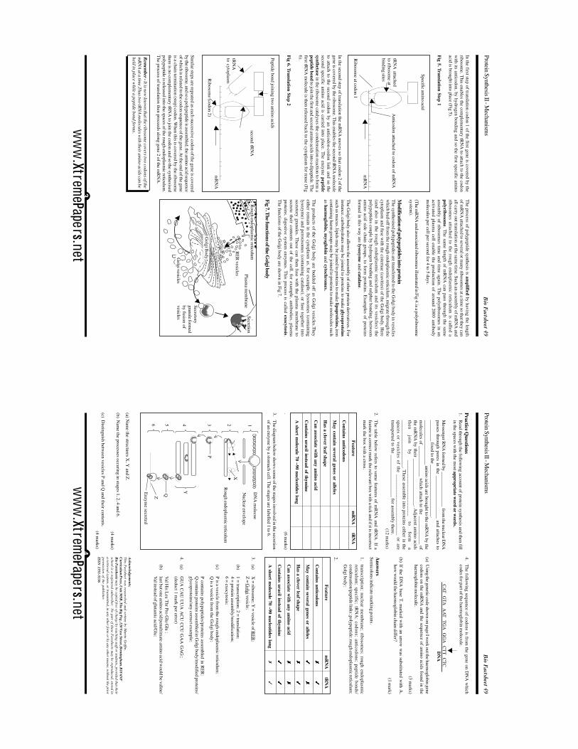
(a) Nam
e the structures X, Y
and Z.
(b)N
ame the processes occuring in stages 1, 2, 4 and 6.
(4 marks)
(c)D
istinguish between vesicles P and Q
and their contents.(4 m
arks)
Practice Questions
1.Read through the follow
ing account of protein synthesis and then fillin the spaces w
ith the most appropriate w
ord or words.
Messenger RN
A form
ed by _______________ from the nuclear D
NA
passes through pores in the __________________ and attaches to_________ fixed to the ______________________.___________________ am
ino acids are brought to the mRN
A by the
molecules of _____________ w
hich attach to the ____________ ofthe m
RNA
by their ____________________. Adjacent am
ino acidsthen
join by
_____________________ to
form
a____________________. These assem
ble into proteins either in thespaces or vesicles of the ________________________ or aretransported to the __________________ for assem
bly there.(12 m
arks)
2.The table below
refers to some features of m
RNA
and tRNA
. If afeature is correct m
ark the relevant box with a tick and if it is incorrect
mark the box w
ith a cross.
Featurem
RN
AtR
NA
Contains anticodons
May contain several genes or alleles
Has a clover leaf shape
Can associate w
ith any amino acid
Contains uracil instead of thym
ine
A short m
olecule 70 –90 nucleotides long
.(6 m
arks)
3.The diagram
below show
s some of the stages involved in the secretion
of an enzyme by a stom
ach cell. The stages are labelled 1 to 6.
ww
w.Xtrem
ePapers.net