Embed Size (px)
DESCRIPTION
a
Citation preview

Patil et al. International Nano Letters 2012, 2:17http://www.inl-journal.com/content/2/1/17
ORIGINAL ARTICLE Open Access
Preparation and characterization of SnO2nanoparticles by hydrothermal routeGanesh E Patil1, Dnyaneshwar D Kajale2, Vishwas B Gaikwad1 and Gotan H Jain1*
Abstract
This paper demonstrates the synthesis of SnO2 nanoparticles using a simple hydrothermal route in the presence ofthe surfactant hydrazine at 100 °C for 12 h. X-ray diffraction (XRD), field emission scanning electron microscopy, andtransmission electron microscopy (TEM) were employed to characterize the as-prepared product, and opticalproperty was studied by UV-visible diffuse reflectance spectroscopy (DRS). The XRD pattern of the as-preparedsample is indexed to the tetragonal structure of SnO2, and the calculated particle size is 22.4 nm, which is furtherconfirmed by TEM. The selected area electron diffraction patterns showed continuous ring patterns without anyadditional diffraction spots and rings of secondary phases, revealing their crystalline structure. Analysis of the DRSspectrum showed the bandgap of the synthesized SnO2 to be 3.6 eV. The anionic surfactant hydrazine plays a keyrole in the formation of the SnO2 nanostructures. A probable reaction for the formation of SnO2 nanoparticles isproposed.
Keywords: SnO2 nanoparticles, Hydrothermal route, FESEM, TEM
BackgroundNanomaterials have attracted great interest due to theirintriguing properties, which are different from those oftheir corresponding bulk state. In the past few years,SnO2 is an important n-type wide-energy-gap semicon-ductor (Eg = 3.64 eV, 330 K) which has a wide range ofapplications such as in solid-state gas sensors [1], trans-parent conducting electrodes [2], rechargeable Li batter-ies [3], and optical electronic devices [4]. During thepast decade, SnO2 nanostructures have been one of themost important oxide nanostructures due to their prop-erties and potential applications [5,6].Many processes have been developed to the synthesis
of SnO2 nanostructures, e.g., spray pyrolysis [5], hydro-thermal methods [6-8], evaporating tin grains in air [9],chemical vapor deposition [10], thermal evaporation ofoxide powders [11], rapid oxidation of elemental tin[12], the sol–gel method [13], etc. Davar et al. [14]reported the synthesis of SnO2 nanoparticles by thermaldecomposition using [bis(2-hydroxyacetophenato)tin(II)],[Sn(HAP)2], as precursor. Salavati-Niasari et al. [15]synthesized zinc blend ZnS nanoparticles by a thioglycolic
* Correspondence: [email protected] Research Laboratory, K.T.H.M. College, Nashik 422 002, IndiaFull list of author information is available at the end of the article
© 2012 Patil et al.; licensee Springer. This is anAttribution License (http://creativecommons.orin any medium, provided the original work is p
acid (HSCH2COOH)-assisted hydrothermal techniquevia the reaction between a new inorganic precursor[bis(2-hydroxyacetophenato)zinc(II)], [Zn(HAP)2], and thio-acetamide (CH3CSNH2). Gnanam and Rajendran [16]synthesized nanocrystalline tin oxide powders of about8 to 13 nm in size using different surfactants such ascetyltrimethyl ammonium bromide, sodium dodecylsulphate, and polyethylene glycol via hydrothermal reac-tion at 160°C for 12 h and studied their structural andphotoluminescence properties.A simple hydrazine-assisted hydrothermal route was
employed to synthesize nanocrystalline SnO2 powders inthis study, and structural, morphological, microstruc-tural, and optical properties were discussed.
MethodsAll reagents used were of analytical grade without fur-ther purification. First, 3.505 g of SnCl4�5H2O (0.1 M)was dissolved in 100 ml of distilled water, and then1.2800 g of hydrazine hydrate (0.01 M) was added withstirring. N2H4�H2O immediately reacted with SnCl4 inthe solution to form a slurry-like white precipitate of thehybrid complex between N2H4 and SnCl4. After 10 minof stirring, the solution was transferred into a Teflon-lined stainless steel autoclave with a capacity of 200 ml
Open Access article distributed under the terms of the Creative Commonsg/licenses/by/2.0), which permits unrestricted use, distribution, and reproductionroperly cited.
0
200
400
600
800
1000
1200
1400
20 30 40 50 60 70 80
(110
)
(101
)
Inte
nsit
y (a
.u.)
2 Theta
(200
)
(211
)
(220
)
(310
)
(301
)
(321
)
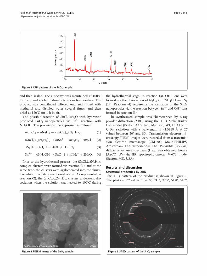
Figure 1 XRD pattern of the SnO2 sample.
Patil et al. International Nano Letters 2012, 2:17 Page 2 of 5http://www.inl-journal.com/content/2/1/17
and then sealed. The autoclave was maintained at 100°Cfor 12 h and cooled naturally to room temperature. Theproduct was centrifuged, filtered out, and rinsed withmethanol and distilled water several times, and thendried at 120°C for 1 h in air.The possible reaction of SnCl4�5H2O with hydrazine
produced SnO2 nanoparticles via Sn4+ reaction withNH4OH. The process can be expressed as follows:
mSnCl4 þ nN2H4 ! ðSnCl4ÞmðN2H4Þn ð1Þ
ðSnCl4ÞmðN2H4Þn ! mSn4þ þ nN2H4 þ 4mCl� ð2Þ3N2H4 þ 4H2O ! 4NH4OHþN2 ð3Þ
Sn4þ þ 4NH4OH ! SnO2 # þ4NH4þ þ 2H2O: ð4Þ
Prior to the hydrothermal process, the (SnCl4)m(N2H4)ncomplex clusters were formed via reaction (1), and at thesame time, the clusters were agglomerated into the slurry-like white precipitate mentioned above. As represented inreaction (2), the (SnCl4)m(N2H4)n clusters underwent dis-sociation when the solution was heated to 100°C during
Figure 2 FESEM image of the SnO2 sample.
the hydrothermal stage. In reaction (3), OH− ions wereformed via the dissociation of N2H4 into NH4OH and N2
[17]. Reaction (4) represents the formation of the SnO2
nanoparticles via the reaction between Sn4+ and OH− ionsformed in reaction (3).The synthesized sample was characterized by X-ray
powder diffraction (XRD) using the XRD Make-BrukerD-8 model (Bruker AXS, Inc., Madison, WI, USA) withCuKα radiation with a wavelength λ =1.5418 Å at 2θvalues between 20° and 80°. Transmission electron mi-croscopy (TEM) images were recorded from a transmis-sion electron microscope (CM-200, Make-PHILIPS,Amsterdam, The Netherlands). The UV-visible (UV–vis)diffuse reflectance spectrum (DRS) was obtained from aJASCO UV–vis/NIR spectrophotometer V-670 model(Easton, MD, USA).
Results and discussionStructural properties by XRDThe XRD pattern of the product is shown in Figure 1.The peaks at 2θ values of 26.6°, 33.8°, 37.9°, 51.8°, 54.7°,
Figure 3 SAED pattern of the SnO2 sample.

Figure 4 TEM image of the SnO2 sample.
Patil et al. International Nano Letters 2012, 2:17 Page 3 of 5http://www.inl-journal.com/content/2/1/17
61.9°, and 65.9° can be associated with (1 1 0), (1 0 1),(2 0 0), (2 1 1), (2 2 0), (3 1 0), and (3 0 1), respectively. Amatching of the observed and standard (hkl) planes con-firmed that the product is of SnO2 having a tetragonalstructure, which are in good agreement with the literaturevalues (JCPDS card no. 41–1445). The average particlesize (D) was estimated using the Scherrer equation [18]:
D ¼ 0:9λβ cosθ
; ð5Þ
where D is the crystallite size, λ is the X-ray wavelength,β is the full width at half maximum of the diffractionpeak, and θ is the Bragg diffraction angle of the diffrac-tion peaks. The average particle size was found to be22.4 nm.
Morphological properties by FESEMFigure 2 shows the field emission scanning electron mi-croscopy (FESEM) micrograph of the synthesized SnO2
sample. Clustering of particles seems to have occurredon the surface. In this image, cubic structures can beeasily seen.
Table 1 d values obtained from XRD and TEM
Reported d values (Å) XRD d values (Å)
Reciprocal
3.35 3.342 3.01
2.64 2.604 4.23
2.37 2.372 4.41
1.76 1.770 5.61
1.67 1.653 6.52
1.50 1.501 7.10
1.41 1.418 7.71
Microstructural properties by TEM and SAED patternFigure 3 shows the electron diffraction patterns of thesample. It is clear from the figure that the SnO2 particlesare crystalline in nature. The electron diffraction pat-terns show continuous ring patterns without any add-itional diffraction spots and rings of secondary phases,revealing their crystalline structure. Seven fringe pat-terns corresponding to planes (1 1 0), (1 0 1), (2 0 0),(2 1 1), (2 2 0), (3 1 0), and (3 0 1) are consistent withthe peaks observed in the XRD patterns. XRD and TEMstudies confirmed pure tetragonal structure of SnO2 asevidenced from Figures 1 and 4, respectively. The ring-to-the-center distance of each ring is measured as 3.01,4.23, 4.41, 5.61, 6.52, 7.10, and 7.71 and expressed interms of nm−1. The reciprocal of these values gives theinterplanar distance d. Details are given in Table 1.
Optical properties by UV–vis DRSTo determine the optical bandgap of synthesized SnO2,the reflectance spectra of the SnO2 thick film preparedby screen printing technique [19] on a glass substratewas measured. The reflectance (R) spectra of the SnO2
thin film were shown in Figure 5.As seen in Figure 5, the reflectance spectra show a
strong decrease after 360 nm. This decrease is related tooptical transitions occurring in the optical bandgap. Inorder to determine the precise value of the optical band-gap of the SnO2, the reflectance values were convertedto absorbance by application of the Kubelka-Munk func-tion [20,21].The Kubelka-Munk theory is generally used for the
analysis of diffuse reflectance spectra obtained fromweakly absorbing samples. The Kubelka-Munk formulais expressed by the following relation:
FðRÞ ¼ ð1� RÞ22R
¼ KS; ð6Þ
where F(R) is the Kubelka-Munk function which corre-sponds to the absorbance, R is the reflectance, K is theabsorption coefficient, and S is the scattering coefficient.
Electron diffraction (TEM) Planes(hkl)of d values δhkl (nm
−1) d values dhkl (Å)
3.320 (1 1 0)
2.364 (1 0 1)
2.267 (2 0 0)
1.782 (2 1 1)
1.533 (2 2 0)
1.408 (3 1 0)
1.297 (3 0 1)

Figure 5 Reflectance spectra of the SnO2 thin film.
Figure 6 Plot of (αhν)2 vs. hν for the SnO2 thin film.
Patil et al. International Nano Letters 2012, 2:17 Page 4 of 5http://www.inl-journal.com/content/2/1/17
It is well known that the optical transitions in semicon-ductor materials are taken place by direct and indirecttransitions. The absorption coefficient α for direct tran-sitions is expressed by the following relation [22]:
αhν ¼ Aðhν� EgÞn; ð7Þ
where α is the linear absorption coefficient of the mater-ial, A is an energy-independent constant, Eg is the op-tical bandgap, and n is a constant which determines thetype of optical transitions: for indirect allowed transition,n= 2; for indirect forbidden transition, m= 3; for directallowed transition, n= 1/2; and for direct forbidden tran-sition, m= 3/2. The F(R) values of the SnO2 film were
obtained using the ð1�RÞ22R relation in Equation 6 [23,24]
and the Kubelka-Munk function F(R) is directly propor-tional to the absorbance. Therefore, F(R) values wereconverted to the linear absorption coefficient by means
of the α ¼ FðRÞt ¼ Absorbance
t relation [25], where t is the
thickness of the SnO2 film. The curve of FðRÞhνt
� �2vs. hν
for the SnO2 film was plotted, as shown in Figure 6. Theoptical bandgap (Eg) of the SnO2 film was determined
from the curve of FðRÞhνt
� �2vs. hν and was found to be
3.6 eV. The optical bandgap of the SnO2 studied is simi-lar to that of undoped SnO2 materials obtained by vari-ous methods [26,27]. This suggests that the opticalbandgap of SnO2 semiconductors changes with respectto the synthesis method used.
ConclusionsSnO2 nanoparticles have been successfully synthesizedby a simple hydrothermal method at low temperatureusing hydrazine hydrate as a mediator. The structural,morphological, microstructural, and optical properties ofa SnO2 sample were investigated. XRD spectra indicatedthat the as-prepared product is polycrystalline in nature.
It was also shown from these spectra that the crystallitestructure was observed to be tetragonal. The surfacemorphology was investigated by FESEM. The crystallitesize (22.4 nm) of the SnO2 nanoparticles, estimated byXRD, is confirmed by TEM. The optical bandgap of theSnO2 film was found to be 3.6 eV.
Competing interestsThe authors declare that they have no competing interests.
Authors’ contributionsGEP synthesized the nanocrystalline SnO2 materials, carried out thecharacterization, and drafted the manuscript. DDK participated in thediscussions and interpretation of all characterization results. VBG and GHJgave the final approval of the version to be published. All the authors readand approved the final manuscript.
Authors’ informationGEP is an INSPIRE fellow at Materials Research Laboratory, KTHM College,Nashik, India. He received his B.Sc. and M.Sc. (Physics) degrees from NorthMaharashtra University, Jalgaon, in 2005 and 2007, respectively. He iscurrently pursuing a Ph.D. degree under the supervision of Dr. GHJ at theUniversity of Pune, Pune. He is a life member of the Indian ScienceCongress Association. His research interests are in the areas of preparationof binary oxide thin film by spray pyrolysis and its gas sensingapplications.DDK is an assistant professor at MVP’s Arts, Commerce and ScienceCollege, Nandgaon, India. He received his B.Sc. and M.Sc. (Chemistry)degrees from the University of Pune. He has 25 peer-reviewed researchpublications to his credit. He is a life member of the Indian ScienceCongress Association. His research interest is in the areas of perovskitematerials for gas sensors, thin films, and nanosized material preparation.Dr. VBG received his M.Sc., M.Phil., and Ph.D. degrees from the Universityof Pune, Pune, in 1981, 1990, and 2001, respectively. He is currently aprofessor and the Head of the KTHM College, Nashik, and a member ofthe Management Council, University of Pune. His research interest is in theareas of environmental science, material science, and nanomaterials. He isa member of the Indian Association of Nuclear Chemists and AlliedScientists, BARC, Mumbai.Dr. GHJ is an associate professor and the Head of the Department ofPhysics at MVP’s KTHM College, Nashik, India. He received his M.Sc.(Physics) degree from the University of Pune, Pune, in 1989 and Ph.D.(Materials Science) degree from Pratap College, Amalner, NorthMaharashtra University, Jalgaon, in 2007. He has published 44 researcharticles in the Journal of International Repute. His areas of interest areperovskite for gas sensors, nanomaterials, and thick and thin films. He has

Patil et al. International Nano Letters 2012, 2:17 Page 5 of 5http://www.inl-journal.com/content/2/1/17
delivered invited talks at MS&T 2008, USA; EUROMAT 2009, UK; ICST 2010,Italy; ICST 2011, New Zealand; and ICPAC-2012, Mauritius. He is a BOSmember in Physics at the University of Pune, Pune.
AcknowledgementsThe financial support for this work through the INSPIRE Fellowship fordoctoral degree from DST, New Delhi, is gratefully acknowledged. Theauthors thank the Sophisticated Analytical Instrument Facility, Indian Instituteof Technology (IIT), Bombay, for carrying out TEM characterization and C-MET,Pune, for providing the FESEM facility.
Author details1Materials Research Laboratory, K.T.H.M. College, Nashik 422 002, India.2Materials Research Laboratory, Arts, Commerce and Science College,Nandgaon 423 106, India.
Received: 1 April 2011 Accepted: 28 February 2012Published: 27 July 2012
References1. Ying, Z., Wan, Q., Song, Z.T., Feng, S.L.: SnO2 nanowhiskers and their ethanol
sensing characteristics. Nanotechnology 15, 1682 (2004)2. Chopra, K.L., Major, S., Pandya, D.K.: Transparent conductors–a status review.
Thin Solid Films 102, 1 (1983)3. Peng, Z., Shi, Z., Liu, M.: Mesoporous Sn–TiO2 composite electrodes for
lithium batteries. Chem. Commun. 21, 25 (2000)4. Aoki, A., Sasakura, H.: Tin oxide thin film transistors. Japan J. Appl. Phys. 9,
582 (1970)5. Paraguay-Delgado, F., Antúnez-Flores, W., Miki-Yoshida, M., Aguilar-
Elguezabal, A., Santiago, P., Diaz, R., Ascencio, J.A.: Structural analysis andgrowing mechanisms for long SnO2 nanorods synthesized by spraypyrolysis. Nanotechnology 16, 688 (2005)
6. Cheng, B., Russell, J.M., Shi, W., Zhang, L., Samulski, E.T.: Large-scale, solution-phase growth of single-crystalline SnO2 nanorods. J. Am. Chem. Soc. 126,5972 (2004)
7. Du, F., Guo, Z., Li, G.: Hydrothermal synthesis of SnO2 hollow microspheres.Mater. Lett. 59, 2563 (2005)
8. Fujihara, S., Maeda, T., Ohgi, H., Hosono, E., Imai, H., Kim, S.: Hydrothermalroutes to prepare nanocrystalline mesoporous SnO2 having high thermalstability. Langmuir 20, 6476 (2004)
9. Duan, J., Yang, S., Liu, H., Gong, J., Huang, H., Zhao, X., Zhang, R., Du, Y.:Single crystal SnO2 zigzag nanobelts. J. Am. Chem. Soc. 127, 6180 (2005)
10. Liu, Y., Koep, E., Liu, M.: A highly sensitive and fast-responding SnO2 sensorfabricated by combustion chemical vapor deposition. Chem Mater 17, 3997(2005)
11. Dai, Z.R., Gole, J.L., Stout, J.D., Wang, Z.L.: Tin oxide nanowires, nanoribbons,and nanotubes. J. Phys. Chem. B 106, 1274 (2002)
12. Hu, J.Q., Ma, X.L., Shang, N.G., Xie, Z.Y., Wong, N.B., Lee, C.S., Lee, S.T.: Largescale rapid oxidation synthesis of SnO2 nanoribbons. J. Phys. Chem. B 106,3823 (2002)
13. Pourfayaz, F., Khodadadi, A., Mortazavi, Y., Mohajerzadeh, S.S.: SnO2 sensorselective to ethanol in presence of CO, LPG and CH4. Sensors Actuators B108, 172 (2005)
14. Davar, F., Salavati-Niasaria, M., Fereshteh, Z.: Synthesis and characterizationof SnO2 nanoparticles by thermal decomposition of new inorganicprecursor. J. Alloys Compd. 496(1–2), 638–643 (2010)
15. Salavati-Niasari, M., Davar, F., Seyghalkar, H., Esmaeili, E., Mir, N.: Synthesisand characterization of SnO2 nanoparticles by thermal decomposition ofnew inorganic precursor. Cryst. Eng. Comm. 13, 2948 (2011)
16. Gnanam, S., Rajendran, V.: Anionic, cationic and nonionic surfactants-assistedhydrothermal synthesis of tin oxide nanoparticles and theirphotoluminescence properties. Digest Journal of Nanomaterials andBiostructures 5(2), 623 (2010)
17. Zhu, H., Yang, D., Yu, G., Zhang, H., Yao, K.: Hydrothermal synthesis ofZn2SnO4 nanorods in the diameter regime of sub-5 nm and theirproperties. Nanotechnology 17, 2386 (2006)
18. Cullity, B.D.: Elements of X-ray Diffraction. Boston, Addison-WesleyPublishing Co (1956)
19. Jain, G.H., Patil, L.A., Wagh, M.S., Patil, D.R., Patil, S.A., Amalnerkar, D.P.: Surfacemodified BaTiO3 thick film resistors as H2S gas sensors. Sensors Actuators BChemical 117, 159 (2006)
20. Escobedo Morales, A., Sanchez Mora, E., Pal, U.: Use of diffuse reflectancespectroscopy for optical characterization of un-supported nanostructures.Rev. Mexic. De Fisica S 53, 18 (2007)
21. Senthilkumar, V., Vickraman, P., Ravikumar, R.: Synthesis fluorine doped tinoxide nanoparticles by sol–gel technique and characterization. Journal ofSol–gel Science and Technology 53(2), 316 (2010)
22. Caglar, M., Ilican, S., Caglar, Y., Yakuphanoglu, F.: The effects of Al doping onthe optical constants of ZnO thin films prepared by spray pyrolysis method.Int. J. Mater. Sci. Elect. Res. 1, 21 (2010)
23. Caglar, M., Ilican, S., Caglar, Y., Yakuphanoglu, F.: Electrical conductivity andoptical properties of ZnO nanostructured thin film. Appl. Surf. Sci.255, 4491 (2009)
24. Liu, X.-C., Shi, E.-W., Chen, Z.-Z., Zhang, H.-W., Song, L.-X., Wang, H., Yao, S.-D.:Structural, optical and magnetic properties of co-doped ZnO films. J. Cryst.Growth 296, 135 (2006)
25. Silva, R.F., Darbello Zaniquelli, M.E.: Aluminium doped zinc oxide films:formation process and optical properties. J. Non-Cryst. Solids1247, 248 (1999)
26. Patil, G.E., Kajale, D.D., Ahire, P.T., Chavan, D.N., Pawar, N.K., Shinde, S.D.,Gaikwad, V.B., Jain, G.H.: Synthesis, characterization and gas sensingperformance of SnO2 thin films prepared by spray pyrolysis. Bull. Mater. Sci.120, 1 (2011)
27. Patil, G.E., Kajale, D.D., Gaikwad, V.B., Jain, G.H.: Nanocrystalline tin oxide thinfilm as a low level H2S gas sensor. Int. J. Nanosci. 10, 1 (2011)
doi:10.1186/2228-5326-2-17Cite this article as: Patil et al.: Preparation and characterization of SnO2
nanoparticles by hydrothermal route. International Nano Letters 2012 2:17.
Submit your manuscript to a journal and benefi t from:
7 Convenient online submission
7 Rigorous peer review
7 Immediate publication on acceptance
7 Open access: articles freely available online
7 High visibility within the fi eld
7 Retaining the copyright to your article
Submit your next manuscript at 7 springeropen.com