Embed Size (px)
Citation preview
Would you like to know more?
Carestream Health© Carestream Health, Inc., 2008.The Kodak trademark and trade dress are used under license from Kodak. RVG is a trademark of Carestream Health, Inc.
To schedule a demonstration or to receive further information, please contact your authorized dealer
or visit our website: www.my90003d.com
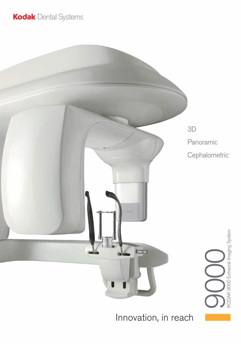
3D
Panoramic
Cephalometric
9000
K
OD
AK
9000
Ext
raor
al Im
agin
g Sy
stem
Innovation, in reach
Dealer stamp
24p_90003D_V4:Mise en page 1 9/09/08 12:09 Page 1

3
Innovation made simple
We believe in innovation. We always have. In fact, our productshave consistently distinguished themselves as groundbreakingsolutions to real challenges.
Nevertheless, innovation alone won't do. Products must also beeasy to understand and operate. Consequently, our designphilosophy has always emphasized a commitment to practicalingenuity. In other words, we make sure innovation remains simple, while staying focused on the evolving needs of modern dentistry.
Today's practitioner requires diagnostic tools that are completeand incomparably effective. This was our inspiration in creatingthe Kodak 9000 system, the single yet modular unit answer to the diagnostic needs of dentists, orthodontists, and maxillo-facialsurgeons alike.
2
Innovation made simple
Cephalometric
24p_90003D_V4:Mise en page 1 9/09/08 12:09 Page 2
5
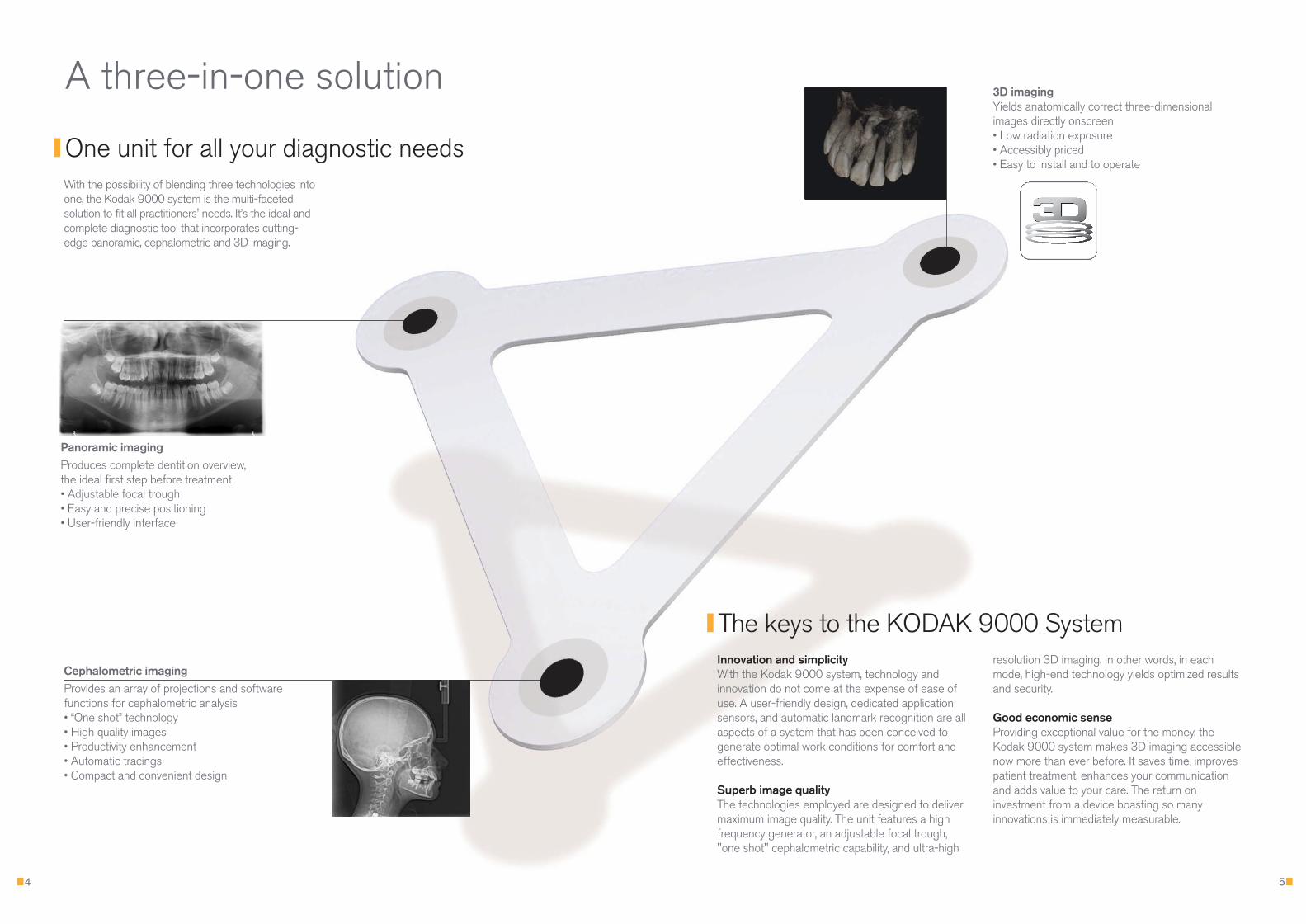
With the possibility of blending three technologies intoone, the Kodak 9000 system is the multi-facetedsolution to fit all practitioners' needs. It’s the ideal andcomplete diagnostic tool that incorporates cutting-edge panoramic, cephalometric and 3D imaging.
Innovation and simplicityWith the Kodak 9000 system, technology andinnovation do not come at the expense of ease ofuse. A user-friendly design, dedicated applicationsensors, and automatic landmark recognition are allaspects of a system that has been conceived togenerate optimal work conditions for comfort andeffectiveness.
Superb image qualityThe technologies employed are designed to delivermaximum image quality. The unit features a highfrequency generator, an adjustable focal trough,"one shot" cephalometric capability, and ultra-high
4
The keys to the KODAK 9000 System
Panoramic imaging Produces complete dentition overview, the ideal first step before treatment• Adjustable focal trough• Easy and precise positioning• User-friendly interface
3D imagingYields anatomically correct three-dimensionalimages directly onscreen• Low radiation exposure• Accessibly priced• Easy to install and to operate
Cephalometric imagingProvides an array of projections and softwarefunctions for cephalometric analysis• “One shot” technology• High quality images• Productivity enhancement• Automatic tracings • Compact and convenient design
resolution 3D imaging. In other words, in eachmode, high-end technology yields optimized resultsand security.
Good economic senseProviding exceptional value for the money, theKodak 9000 system makes 3D imaging accessiblenow more than ever before. It saves time, improvespatient treatment, enhances your communicationand adds value to your care. The return oninvestment from a device boasting so manyinnovations is immediately measurable.
A three-in-one solution
One unit for all your diagnostic needs
24p_90003D_V4:Mise en page 1 9/09/08 12:09 Page 4
7
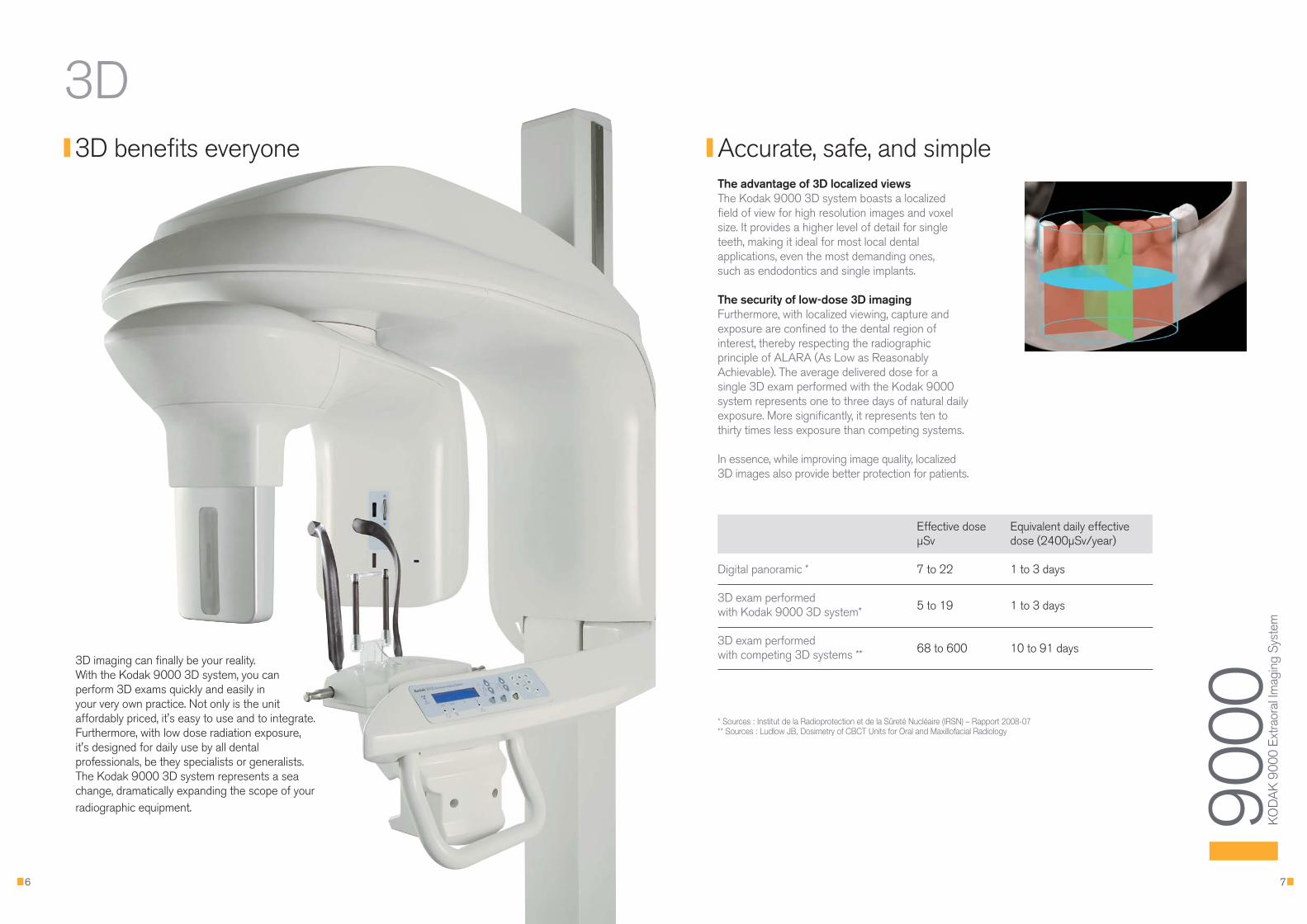
The advantage of 3D localized viewsThe Kodak 9000 3D system boasts a localizedfield of view for high resolution images and voxelsize. It provides a higher level of detail for singleteeth, making it ideal for most local dentalapplications, even the most demanding ones, such as endodontics and single implants.
The security of low-dose 3D imagingFurthermore, with localized viewing, capture andexposure are confined to the dental region ofinterest, thereby respecting the radiographicprinciple of ALARA (As Low as ReasonablyAchievable). The average delivered dose for asingle 3D exam performed with the Kodak 9000system represents one to three days of natural dailyexposure. More significantly, it represents ten tothirty times less exposure than competing systems.
In essence, while improving image quality, localized3D images also provide better protection for patients.
6
Accurate, safe, and simple
* Sources : Institut de la Radioprotection et de la Sûreté Nucléaire (IRSN) – Rapport 2008-07** Sources : Ludlow JB, Dosimetry of CBCT Units for Oral and Maxillofacial Radiology
3D imaging can finally be your reality. With the Kodak 9000 3D system, you canperform 3D exams quickly and easily inyour very own practice. Not only is the unitaffordably priced, it's easy to use and to integrate.Furthermore, with low dose radiation exposure,it's designed for daily use by all dentalprofessionals, be they specialists or generalists.The Kodak 9000 3D system represents a seachange, dramatically expanding the scope of yourradiographic equipment. 90
00
KO
DA
K90
00 E
xtra
oral
Imag
ing
Syst
em
3D
Effective dose Equivalent daily effective µSv dose (2400µSv/year)
Digital panoramic * 7 to 22 1 to 3 days
3D exam performed with Kodak 9000 3D system* 5 to 19 1 to 3 days
3D exam performed with competing 3D systems ** 68 to 600 10 to 91 days
3D benefits everyone
24p_90003D_V4:Mise en page 1 9/09/08 12:09 Page 6
9
A new perspectiveThe Kodak 9000 3D system gives you a new wayof looking at dental structures and pathologies. You get all your information more clearly and all the angles and slices you need within thevolume acquired.
A new exactitudeWith 3D imaging, you obtain precise visualization of dental structures in their actualspatial representation. Images are displayedin axial, coronal, sagittal, and custom slices.Meanwhile, the three-dimensional reconstructionprovides a reassuring and exact 1:1 scale. This “true to life” view of dental structuresunquestionably facilitates effective communication.
3D, the perfect complement3D imaging takes nothing away from theusefulness of traditional 2D imaging. On thecontrary, they are perfectly complementary. The panoramic and cephalometric image provide a global view/image while 3D goes into precise tooth detail, delivering complementaryinformation to refine your diagnosis.
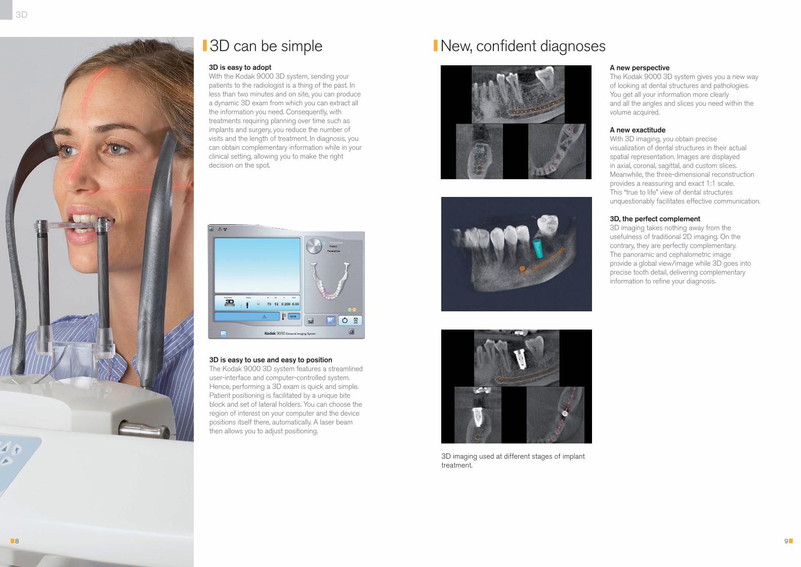
3D is easy to adoptWith the Kodak 9000 3D system, sending yourpatients to the radiologist is a thing of the past. Inless than two minutes and on site, you can producea dynamic 3D exam from which you can extract allthe information you need. Consequently, withtreatments requiring planning over time such asimplants and surgery, you reduce the number ofvisits and the length of treatment. In diagnosis, youcan obtain complementary information while in yourclinical setting, allowing you to make the rightdecision on the spot.
8
3D can be simple New, confident diagnoses
3D
3D is easy to use and easy to positionThe Kodak 9000 3D system features a streamlineduser-interface and computer-controlled system.Hence, performing a 3D exam is quick and simple.Patient positioning is facilitated by a unique biteblock and set of lateral holders. You can choose theregion of interest on your computer and the devicepositions itself there, automatically. A laser beamthen allows you to adjust positioning.
3D imaging used at different stages of implanttreatment.
24p_90003D_V4:Mise en page 1 9/09/08 12:09 Page 8
1110
3D
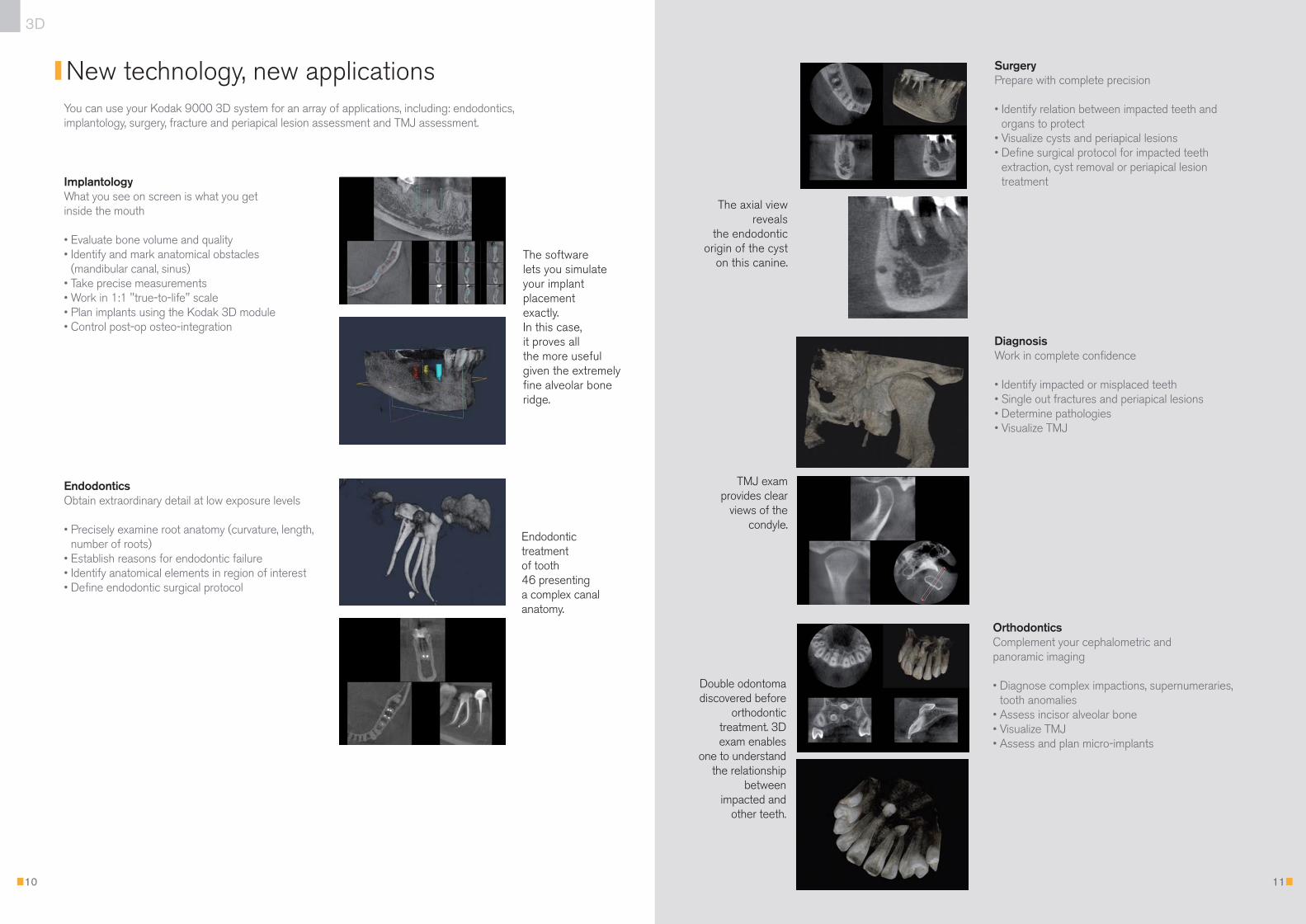
Orthodontics Complement your cephalometric and panoramic imaging
• Diagnose complex impactions, supernumeraries,tooth anomalies
• Assess incisor alveolar bone • Visualize TMJ • Assess and plan micro-implants
DiagnosisWork in complete confidence
• Identify impacted or misplaced teeth • Single out fractures and periapical lesions• Determine pathologies• Visualize TMJ
The software lets you simulate your implant placement exactly. In this case, it proves all the more useful given the extremelyfine alveolar boneridge.
Endodontic treatment of tooth 46 presenting a complex canalanatomy.
TMJ exam provides clear
views of thecondyle.
Double odontomadiscovered before
orthodontic treatment. 3Dexam enables
one to understandthe relationship
between impacted and
other teeth.
You can use your Kodak 9000 3D system for an array of applications, including: endodontics,implantology, surgery, fracture and periapical lesion assessment and TMJ assessment.
New technology, new applications
ImplantologyWhat you see on screen is what you get inside the mouth
• Evaluate bone volume and quality• Identify and mark anatomical obstacles
(mandibular canal, sinus)• Take precise measurements• Work in 1:1 "true-to-life" scale• Plan implants using the Kodak 3D module• Control post-op osteo-integration
EndodonticsObtain extraordinary detail at low exposure levels
• Precisely examine root anatomy (curvature, length,number of roots)
• Establish reasons for endodontic failure• Identify anatomical elements in region of interest • Define endodontic surgical protocol
SurgeryPrepare with complete precision
• Identify relation between impacted teeth andorgans to protect
• Visualize cysts and periapical lesions• Define surgical protocol for impacted teeth
extraction, cyst removal or periapical lesiontreatment
The axial view reveals
the endodonticorigin of the cyst
on this canine.
24p_90003D_V4:Mise en page 1 9/09/08 12:09 Page 10
13
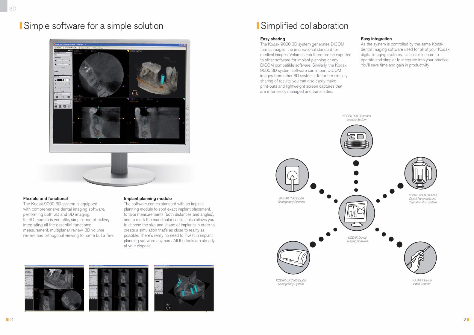
Easy sharingThe Kodak 9000 3D system generates DICOMformat images, the international standard formedical images. Volumes can therefore be exportedto other software for implant planning or anyDICOM compatible software. Similarly, the Kodak9000 3D system software can import DICOMimages from other 3D systems. To further simplifysharing of results, you can also easily make print-outs and lightweight screen captures that are effortlessly managed and transmitted.
Flexible and functional The Kodak 9000 3D system is equipped with comprehensive dental imaging software,performing both 2D and 3D imaging. Its 3D module is versatile, simple, and effective,integrating all the essential functions:measurement, multiplanar review, 3D volumereview, and orthogonal viewing to name but a few.
12
Simple software for a simple solution Simplified collaboration
3D
Implant planning moduleThe software comes standard with an implantplanning module to spot exact implant placement,to take measurements (both distances and angles),and to mark the mandibular canal. It also allows youto choose the size and shape of implants in order tocreate a simulation that's as close to reality aspossible. There's really no need to invest in implantplanning software anymore. All the tools are alreadyat your disposal.
Easy integrationAs the system is controlled by the same Kodakdental imaging software used for all of your Kodakdigital imaging systems, it’s easier to learn tooperate and simpler to integrate into your practice.You’ll save time and gain in productivity.
KODAK 9000 ExtraoralImaging System
KODAK RVG DigitalRadiography Systems
KODAK CR 7400 DigitalRadiography System
KODAK 8000 / 8000CDigital Panoramic andCephalometric System
KODAK Intraoral Video Camera
KODAK Dental Imaging Software
24p_90003D_V4:Mise en page 1 9/09/08 12:09 Page 12
15
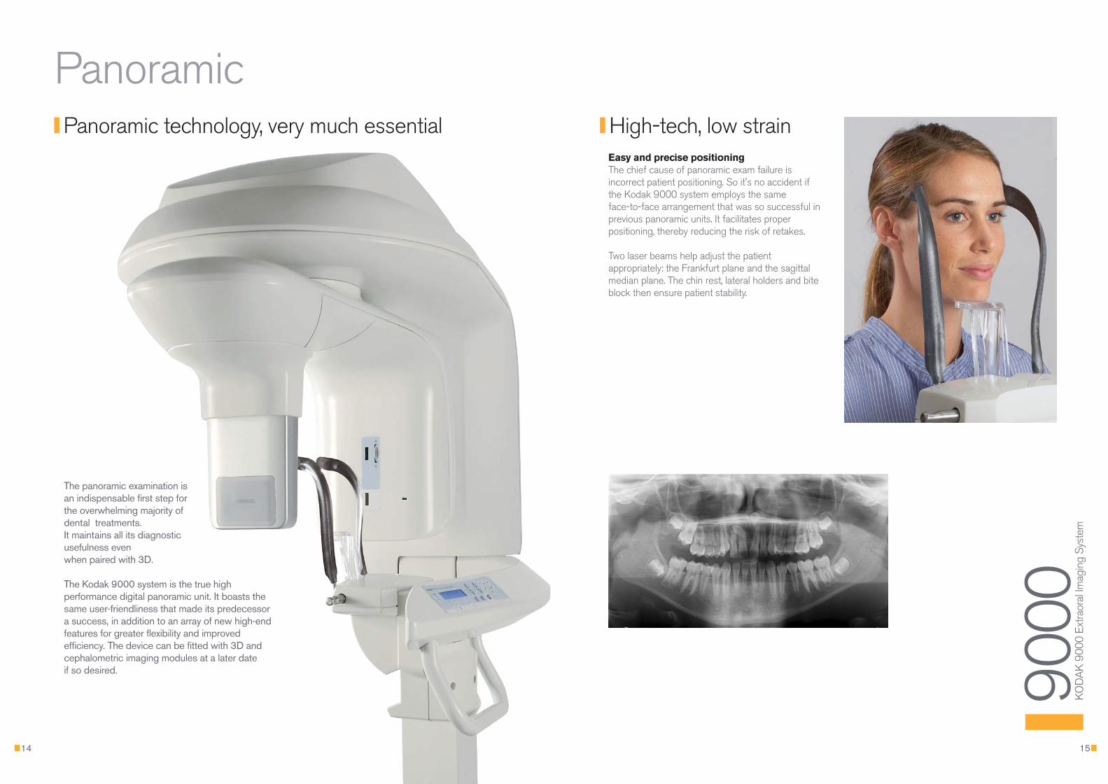
Easy and precise positioningThe chief cause of panoramic exam failure isincorrect patient positioning. So it's no accident ifthe Kodak 9000 system employs the same face-to-face arrangement that was so successful inprevious panoramic units. It facilitates properpositioning, thereby reducing the risk of retakes.
Two laser beams help adjust the patientappropriately: the Frankfurt plane and the sagittalmedian plane. The chin rest, lateral holders and biteblock then ensure patient stability.
14
High-tech, low strainPanoramic technology, very much essential
The panoramic examination is an indispensable first step for the overwhelming majority ofdental treatments. It maintains all its diagnosticusefulness even when paired with 3D.
The Kodak 9000 system is the true highperformance digital panoramic unit. It boasts thesame user-friendliness that made its predecessora success, in addition to an array of new high-endfeatures for greater flexibility and improvedefficiency. The device can be fitted with 3D andcephalometric imaging modules at a later date if so desired.
Panoramic
9000
K
OD
AK
9000
Ext
raor
al Im
agin
g Sy
stem
24p_90003D_V4:Mise en page 1 9/09/08 12:09 Page 14
17
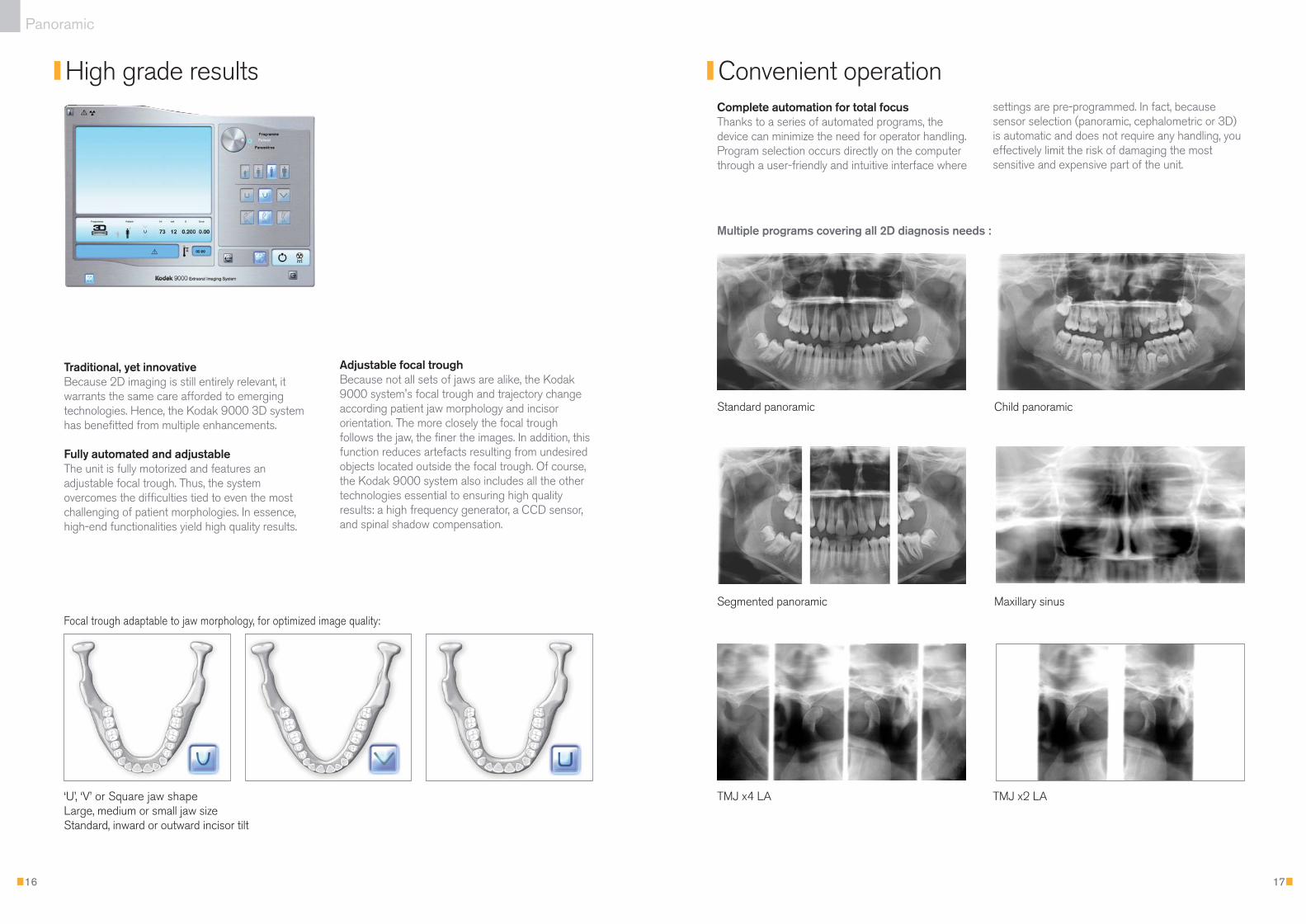
Complete automation for total focusThanks to a series of automated programs, thedevice can minimize the need for operator handling.Program selection occurs directly on the computerthrough a user-friendly and intuitive interface where
16
High grade results Convenient operation
Panoramic
Multiple programs covering all 2D diagnosis needs :
settings are pre-programmed. In fact, becausesensor selection (panoramic, cephalometric or 3D)is automatic and does not require any handling, youeffectively limit the risk of damaging the mostsensitive and expensive part of the unit.
‘U’, ‘V’ or Square jaw shape Large, medium or small jaw size Standard, inward or outward incisor tilt
Focal trough adaptable to jaw morphology, for optimized image quality:
Adjustable focal troughBecause not all sets of jaws are alike, the Kodak9000 system's focal trough and trajectory changeaccording patient jaw morphology and incisororientation. The more closely the focal troughfollows the jaw, the finer the images. In addition, thisfunction reduces artefacts resulting from undesiredobjects located outside the focal trough. Of course,the Kodak 9000 system also includes all the othertechnologies essential to ensuring high qualityresults: a high frequency generator, a CCD sensor,and spinal shadow compensation.
Traditional, yet innovativeBecause 2D imaging is still entirely relevant, itwarrants the same care afforded to emergingtechnologies. Hence, the Kodak 9000 3D systemhas benefitted from multiple enhancements.
Fully automated and adjustableThe unit is fully motorized and features anadjustable focal trough. Thus, the systemovercomes the difficulties tied to even the mostchallenging of patient morphologies. In essence,high-end functionalities yield high quality results.
Standard panoramic Child panoramic
Segmented panoramic Maxillary sinus
TMJ x4 LA TMJ x2 LA
24p_90003D_V4:Mise en page 1 9/09/08 12:09 Page 16
19
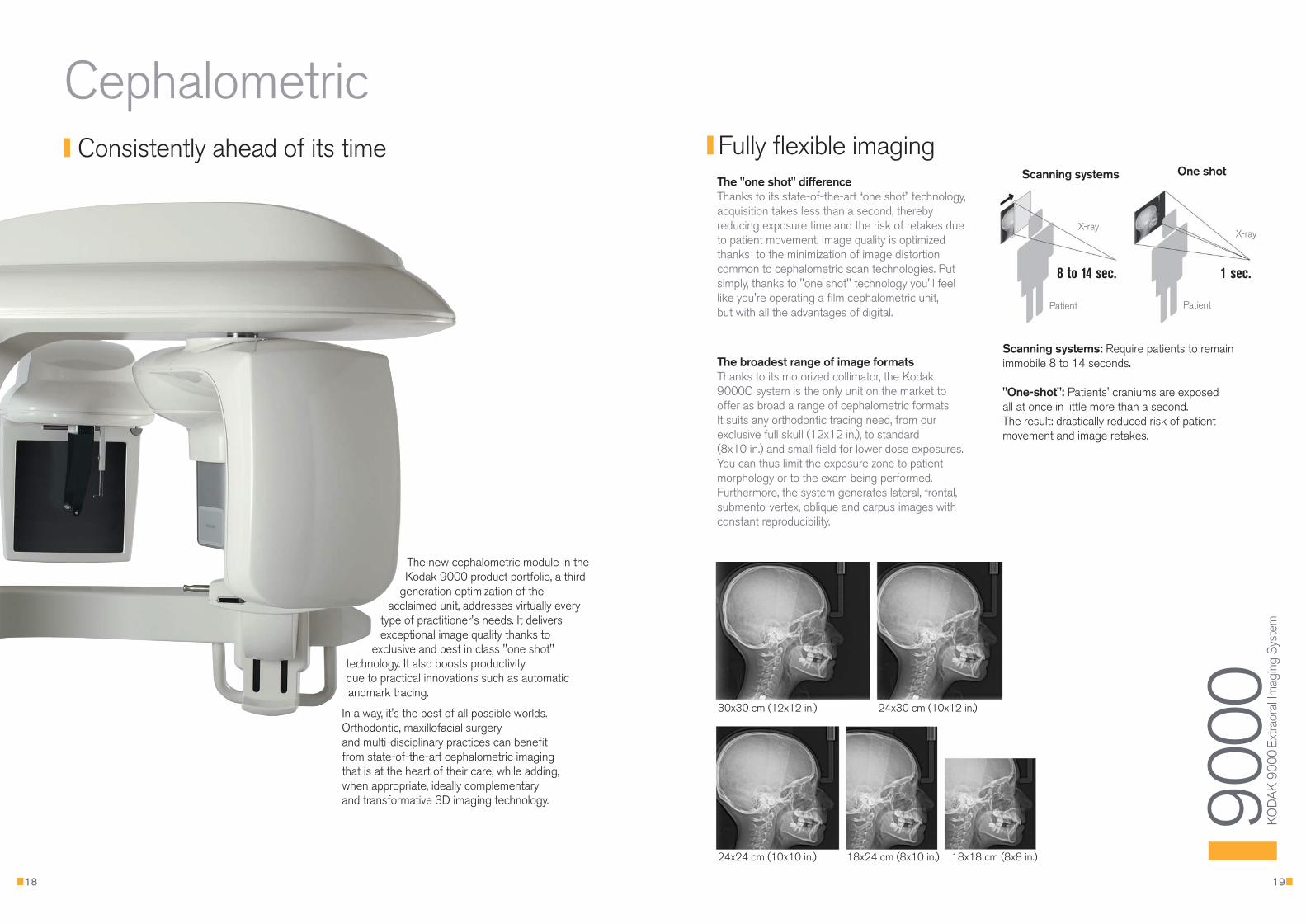
The broadest range of image formatsThanks to its motorized collimator, the Kodak9000C system is the only unit on the market tooffer as broad a range of cephalometric formats. It suits any orthodontic tracing need, from ourexclusive full skull (12x12 in.), to standard (8x10 in.) and small field for lower dose exposures.You can thus limit the exposure zone to patientmorphology or to the exam being performed.Furthermore, the system generates lateral, frontal,submento-vertex, oblique and carpus images withconstant reproducibility.
18
Fully flexible imagingThe "one shot" differenceThanks to its state-of-the-art “one shot” technology, acquisition takes less than a second, therebyreducing exposure time and the risk of retakes dueto patient movement. Image quality is optimizedthanks to the minimization of image distortioncommon to cephalometric scan technologies. Putsimply, thanks to "one shot" technology you'll feellike you're operating a film cephalometric unit, but with all the advantages of digital.
30x30 cm (12x12 in.) 24x30 cm (10x12 in.)
24x24 cm (10x10 in.) 18x24 cm (8x10 in.) 18x18 cm (8x8 in.)
Consistently ahead of its time
The new cephalometric module in theKodak 9000 product portfolio, a third
generation optimization of theacclaimed unit, addresses virtually every
type of practitioner's needs. It deliversexceptional image quality thanks to
exclusive and best in class "one shot"technology. It also boosts productivity due to practical innovations such as automaticlandmark tracing.
In a way, it's the best of all possible worlds.Orthodontic, maxillofacial surgery and multi-disciplinary practices can benefit from state-of-the-art cephalometric imaging that is at the heart of their care, while adding, when appropriate, ideally complementary and transformative 3D imaging technology.
Cephalometric
9000
K
OD
AK
9000
Ext
raor
al Im
agin
g Sy
stem
Scanning systems One shot
X-ray
Patient Patient
X-ray
1 sec.8 to 14 sec.
Scanning systems: Require patients to remain immobile 8 to 14 seconds.
"One-shot": Patients' craniums are exposed all at once in little more than a second. The result: drastically reduced risk of patient movement and image retakes.
24p_90003D_V4:Mise en page 1 9/09/08 12:09 Page 18
21
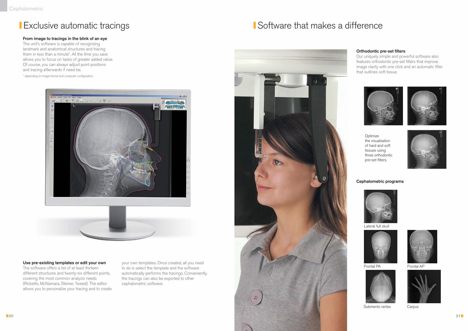
Orthodontic pre-set filtersOur uniquely simple and powerful software alsofeatures orthodontic pre-set filters that improveimage clarity with one click and an automatic filterthat outlines soft tissue.
Cephalometric programs
20
Exclusive automatic tracings Software that makes a difference
Cephalometric
From image to tracings in the blink of an eyeThe unit's software is capable of recognizinglandmark and anatomical structures and tracingthem in less than a minute*. All the time you saveallows you to focus on tasks of greater added value.Of course, you can always adjust point positionsand tracing afterwards if need be. * depending on image format and computer configuration.
Use pre-existing templates or edit your ownThe software offers a list of at least thirteendifferent structures and twenty-six different points,covering the most common analysis needs(Ricketts, McNamara, Steiner, Tweed). The editorallows you to personalize your tracing and to create
your own templates. Once created, all you need to do is select the template and the softwareautomatically performs the tracings. Conveniently,the tracings can also be exported to othercephalometric software.
Lateral full skull
Optimize the visualization of hard and soft tissues using three orthodonticpre-set filters.
Frontal PA Frontal AP
Submento vertex Carpus
24p_90003D_V4:Mise en page 1 9/09/08 12:09 Page 20
2322
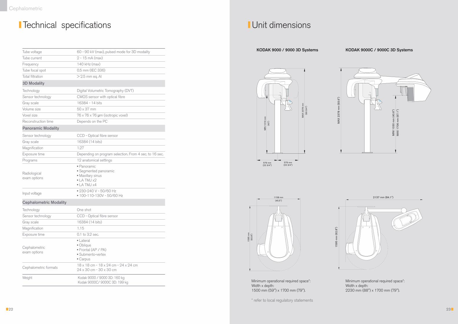
Technical specifications Unit dimensions
Cephalometric
Tube voltage 60 - 90 kV (max), pulsed mode for 3D modality
Tube current 2 - 15 mA (max)
Frequency 140 kHz (max)
Tube focal spot 0.5 mm (IEC 336)
Total filtration > 2.5 mm eq. Al
3D Modality
Technology Digital Volumetric Tomography (DVT)
Sensor technology CMOS sensor with optical fibre
Gray scale 16384 - 14 bits
Volume size 50 x 37 mm
Voxel size 76 x 76 x 76 µm (isotropic voxel)
Reconstruction time Depends on the PC
Panoramic Modality
Sensor technology CCD - Optical fibre sensor
Gray scale 16384 (14 bits)
Magnification 1.27
Exposure time Depending on program selection. From 4 sec. to 16 sec.
Programs 12 anatomical settings
• Panoramic
Radiological• Segmented panoramic
exam options• Maxillary sinus• LA TMJ x2• LA TMJ x4
Input voltage• 230-240 V - 50/60 Hz• 100-110-130V - 50/60 Hz
Cephalometric Modality
Technology One shot
Sensor technology CCD - Optical fibre sensor
Gray scale 16384 (14 bits)
Magnification 1.15
Exposure time 0.1 to 3.2 sec.
• Lateral
Cephalometric • Oblique
exam options• Frontal (AP / PA)• Submento-vertex• Carpus
Cephalometric formats18 x 18 cm - 18 x 24 cm - 24 x 24 cm24 x 30 cm - 30 x 30 cm
Weight Kodak 9000 / 9000 3D: 160 kgKodak 9000C/ 9000C 3D: 199 kg
KODAK 9000 / 9000 3D Systems KODAK 9000C / 9000C 3D Systems
Minimum operational required space*:Width x depth: 1500 mm (59") x 1700 mm (79").
* refer to local regulatory statements
Minimum operational required space*:Width x depth: 2230 mm (88") x 1700 mm (79").
24p_90003D_V4:Mise en page 1 9/09/08 12:09 Page 22

Would you like to know more?
Carestream Health© Carestream Health, Inc., 2008.The Kodak trademark and trade dress are used under license from Kodak. RVG is a trademark of Carestream Health, Inc.
To schedule a demonstration or to receive further information, please contact your authorized dealer
or visit our website: www.my90003d.com
3D
Panoramic
Cephalometric
9000
K
OD
AK
9000
Ext
raor
al Im
agin
g Sy
stem
Innovation, in reach
Dealer stamp
24p_90003D_V4:Mise en page 1 9/09/08 12:09 Page 1