Embed Size (px)
Citation preview

25th ANNUALPHARMACOLOGY RESEARCH DAYFriday & Saturday, October 25-26, 2019
Manoir Saint-Sauveur246 Chemin du Lac-Millette, Saint-Sauveur, QC J0R 1R3


Recherche & Innovation


Special Thanks To All Of Our Sponsors!
GE Healthcare
BioRad
BioBasic
Abbvie
Vertex
Innovative Medicines Canada
L’Oréal Research and Innovation
Nikon
Chemspace
Pfizer Canada Inc

Table of Contents
PRD Student Committee Welcome Keynote Speaker PRD Judges Program Departmental and PRD Prizes Abstracts - Oral Presentations Poster Session I - Odd Numbers Poster Session II - Even Numbers
2019 PHARMACOLOGYRESEARCH DAY COMMITTEE
Dr. Jean-François Trempe, Co-ChairDr. Maureen McKeague, Co-Chair
Tina TremblaySuleyman Can Akerman
Mariana AsslanMorgan Foret
Sally LeeSophie Lu
Lama IskandaraniAnne-Sophie Pepin
Han YanIssan Zhang
Special Thanks to
Anna CuccoviaChantal Grignon
Nadee Buddhiwickrama
Dear Colleagues,
Welcome to the 25th Annual Pharmacology Research Day!
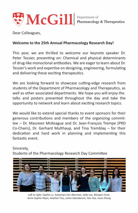
This year, we are thrilled to welcome our keynote speaker Dr. Peter Tessier, presenting on: Chemical and physical determinants of drug-like monoclonal antibodies. We are eager to learn about Dr. Tessier’s work and expertise on designing, engineering, formulating and delivering these exciting therapeutics.
We are looking forward to showcase cutting-edge research from students of the Department of Pharmacology and Therapeutics, aswell as other associated departments. We hope you will enjoy thetalks and posters presented throughout the day and take the opportunity to network and learn about exciting research topics.
We would like to extend special thanks to event sponsors for their generous contributions and members of the organizing commit-tee – Dr. Maureen McKeague and Dr. Jean-François Trempe (PRD Co-Chairs), Dr. Gerhard Multhaup, and Tina Tremblay – for their dedication and hard work in planning and implementing this fantastic event.
Sincerely,Students of the Pharmacology Research Day Committee
Department ofPharmacology & TherapeuticsMcGill
Left to right: Sophie Lu, Suleyman Can Akerman, Sally Lee, Morgan Foret, Anne-Sophie Pépin, Heather Fice, Lama Iskandarani, Han Yan, Issan Zhang
Keynote Speaker
Biography
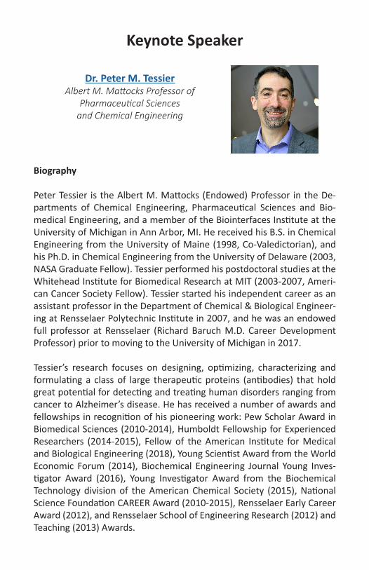
Peter Tessier is the Albert M. Mattocks (Endowed) Professor in the De-partments of Chemical Engineering, Pharmaceutical Sciences and Bio-medical Engineering, and a member of the Biointerfaces Institute at the University of Michigan in Ann Arbor, MI. He received his B.S. in Chemical Engineering from the University of Maine (1998, Co-Valedictorian), and his Ph.D. in Chemical Engineering from the University of Delaware (2003, NASA Graduate Fellow). Tessier performed his postdoctoral studies at the Whitehead Institute for Biomedical Research at MIT (2003-2007, Ameri-can Cancer Society Fellow). Tessier started his independent career as an assistant professor in the Department of Chemical & Biological Engineer-ing at Rensselaer Polytechnic Institute in 2007, and he was an endowed full professor at Rensselaer (Richard Baruch M.D. Career Development Professor) prior to moving to the University of Michigan in 2017.
Tessier’s research focuses on designing, optimizing, characterizing and formulating a class of large therapeutic proteins (antibodies) that hold great potential for detecting and treating human disorders ranging from cancer to Alzheimer’s disease. He has received a number of awards and fellowships in recognition of his pioneering work: Pew Scholar Award in Biomedical Sciences (2010-2014), Humboldt Fellowship for Experienced Researchers (2014-2015), Fellow of the American Institute for Medical and Biological Engineering (2018), Young Scientist Award from the World Economic Forum (2014), Biochemical Engineering Journal Young Inves-tigator Award (2016), Young Investigator Award from the Biochemical Technology division of the American Chemical Society (2015), National Science Foundation CAREER Award (2010-2015), Rensselaer Early Career Award (2012), and Rensselaer School of Engineering Research (2012) and Teaching (2013) Awards.
Dr. Peter M. TessierAlbert M. Mattocks Professor of
Pharmaceutical Sciences and Chemical Engineering
PHARMACOLOGY RESEARCH DAY PRIZES
Melville PrizesThis prize, established in 1994, is awarded annually to the Phar-macology students in the MSc and PhD category whose research poster presentation at the Pharmacology Research Day is judged to be the best. A Melville Prize is also awarded to a Pharmacolo-gy Postdoctoral Fellow whose research poster/oral presentation is judged to be the best.
Best Oral Presentation PrizeThis prize is awarded annually to a Pharmacology graduate student for the oral presentation that best exemplifies multidisciplinary ap-proach in Pharmacology.
PRD Life Sciences Prize(Anatomy and Cell Biology, Biochemistry, IPN)
This prize, established this year by Pharmacology & Therapeutics, is awarded to a graduate student presenting the best posters/orals at Pharmacology Research Day.
Pharmacology Research Day Judges
Dr. Agustin AlonsoDr. Bastien Castagner
Dr. Paul ClarkeDr. Juliana Dallagnol
Dr. John GillardDr. Barbara HalesDr. Mark HancockDr. Terry Hébert
Dr. Maureen McKeague
Dr. Nourhen MnasriDr. Gerhard Multhaup
Dr. Lisa MunterDr. Alfredo Ribeiro da Silva
Dr. Bernard RobaireDr. Adeolo ShoboDr. Jason Tanny
Dr. Jean-François Trempe

PROGRAM
Friday, October 25th, 2019
Bus Departure – by Thomson House(3650 McTavish)
Arrival at Manoir Saint-Sauveur
Registration/Room Assignments Hotel Lobby
Free TimeEnjoy the Manoir St. Saveur’s beautiful amenities and varied activities!
Welcome Cocktails – Matterhorn Room CA few words from Dr. Gerhard Multhaup, Chair
Johans Fakhoury – L’Oréal TalkTitle: Traversing Barriers: the Story of DNA (Im)migrationMatterhorn Room C
Dinner BanquetMatterhorn Room A/B
Entertainment - Matterhorn Room A/BAs part of the 25th anniversary of Pharmacology and Research Day, a featured event will be held both as a welcoming and team building exercise for all. Participation is strongly encouraged! You will have the chance to earn prizes from our spon-sors and from GAPTS
13h00-13h30
15h00
15h00-16h00
16h00-18h00
18h00
18h15-18h45
19h00
21h00
PROGRAM
Saturday, October 26th, 2019
Breakfast – Restaurant St. Moritz
Pharmacology Research Day IntroductionMatterhorn Room A/BDr. Jean Francois Trempe and Dr. Maureen McKeague, PRD Co-Chairs
KEYNOTE ADDRESS –Professor Peter TessierAlbert M. Mattocks Professor of Pharmaceutical Sciences and Chemical EngineeringTitle: Chemical and physical determinants of drug-like monoclonal antibodies
BREAK & POSTER SESSION 1 - Matterhorn COdd Numbered Posters
ORAL SESSION I - Matterhorn A/BSession Moderator: Dr. JF Trempe
Gauthier Schang, PhD - Bernard Lab
Lama Iskandarani, MSc - Hales/Robaire Lab
Anthony Duchesne, MSc - Trempe Lab
Jennifer Chen, PhD - Tanny Lab
Ryan Martin, PhD - Hébert Lab
Lunch - Restaurant St. Moritz
7h00-8h30
8h45
9h00-10h00
10h00-11h30
11h30-13h00
11h30
11h45
12h00
12h15
12h30
13h00-14h00

PROGRAM
Saturday, October 26th, 2019
POSTER SESSION 2 - Matterhorn CEven Numbered Posters
ORAL SESSION II - Matterhorn A/BSession Moderator: Dr. M. McKeague
Jace Jones-Tabah, PhD - Hébert Lab
Haley Deamond, MSc - Ribeiro-da-Silva Lab
Morgan Foret, PhD - Cuello Lab
Simon Veyron, Postdoctroal Fellow - Trempe Lab
Ariane Lismer, PhD - Kimmins Lab
Awards Presentation/Closing Remarks
Bus Departure – Hotel parking area
Arrival at McIntyre Med
14h00-15h30
15h30-17h00
15h30
15h45
16h00
16h15
16h30
17h00-17h30
17h30-18h00
19h30

Oral Presentations
O1 Gauthier SchangGonadotrope-specific Acvr2a and Acvr2b conditional knockout animals are hypogonadal and FSH-deficient
O2 Lama IskandaraniThe effects of bisphenol A on limb development in a murine limb bud culture system
O3 Anthony DuchesneMeasuring Mitochondrial Protein Turnover in a Human Midbrain Organoid Parkinson’s Model by Mass Spectrometry
O4 Jennifer ChenDissection of a Cdk9-dependent Transcription Elongation Path-way Uncovers Novel Functional Pathways of Transcription Fac-tors, Spt5 and Rtf1
O5 Ryan MartinA novel interaction between Gβγ and RNA polymerase II regu-lates angiotensin II type I receptor-mediated transcription
O6 Jace Jonas-TabahDevelopment of FRET Photometry to Track Dopamine Recep-tor-Dependent Protein Kinase Signalling in Parkinson’s Disease and L-DOPA Induced Dyskinesia
O7 Haley DeamondA “Joint” Effort Between Nociceptive and Neuropathic Pain in Arthritis
O8 Morgan ForetQuantification of Neuronal Lipid Peroxidation using a Novel Flu-orogenic Probe
O9 Simon VeyronStructure-based Design of Small-molecule Activators of Parkin
O10 Ariane LismerPaternal folate deficiency induces aberrant histone methylation in sperm which is transmitted to the pre-implantation embryo
Presenter: Gauthier Schang 11h30 / PhD Sr. / O1
Title: Gonadotrope-specific Acvr2a and Acvr2b conditional knockout ani-mals are hypogonadal and FSH-deficient
Authors: Gauthier Schang, Luisina Ongaro, Hailey Schultz, Ying Wang, Ul-rich Boehm, Se-Jin Lee, Daniel J. Bernard
Activins are selective regulators of follicle-stimulating hormone (FSH) production by pituitary gonadotrope cells. In a gonadotrope-like cell line, LβT2 cells, activins stimulate FSH via the type II receptors ACVR2A and/or BMPR2. In vivo, global Acvr2a knockout mice have ~50% lower serum FSH levels than controls. In contrast, gonadotrope-specific Bmpr2 knockouts exhibit normal FSH. Another type II receptor, ACVR2B, can bind activins but appears dispensable for activin-stimulated FSH production in vitro. Acvr2b global knockout mice die soon after birth, precluding their use to assess ACVR2B’s role in FSH production. In light of these previous results, we hypothesized that both ACVR2A and ACVR2B are required for normal FSH production in vivo. To investigate this idea, we crossed floxed Acvr2a or Acvr2b mice to GRIC mice, which express Cre recombinase specifically in gonadotropes. The resulting conditional knockout (cKO) animals were compared to littermate controls. Both Acvr2a and Acvr2b cKO females ex-hibited normal puberty onset (assessed by vaginal opening) and regular estrous cyclicity. However, when paired to wild-type males, Acvr2a and Acvr2b cKO females displayed ~70% and ~20% reductions in litter sizes, respectively. Similarly, Acvr2a and Acvr2b cKO males exhibited ~50% and ~20% reductions in testicular weights. Consistent with these phenotypes, serum FSH levels were lower in Acvr2a cKO males and females, as well as Acvr2b cKO males relative to controls. We failed to detect a decrease in FSH production in Acvr2b cKO females. Simultaneous deletion of both Acvr2a and Acvr2b in gonadotropes yielded females that were hypogo-nadal, acyclic, and sterile. Double knockout males were hypogonadal and had undetectable serum FSH levels. These data suggest that ACVR2A and, to a lesser extent, ACVR2B are the critical type II receptors through which activins (or related TGFβ ligands) signal to induce FSH production in mice in vivo.
Acknowledgements: CIHR (grant MOP-123447 to DJB; award 152308 to GS), NSERC (2015-05178 to DJB), FRQS (award 31338 to GS), Samuel Sol-omon (award to GS), and Ferring (award to LO). The authors would like to thank Xiang Zhou for her technical assistance.

Presenter: Lama Iskandarani 11h45 / MSc Jr. / O2
Title: The effects of bisphenol A on limb development in a murine limb bud culture system
Authors: Iskandarani, L.; Robaire, B.; Hales, B. F.
Environmental exposure to chemical toxicants and their impact on hu-man health are issues of growing concern in modern society. Bisphenols are a family of chemicals which are used to produce polycarbonate plas-tics and epoxy resins. Bisphenol A (BPA), the most prominent bisphenol, is found in food and drink packaging and reusable plastic water bottles. There is evidence suggesting that BPA is an endocrine disruptor that can act as a xenoestrogen. Since estrogens are involved in bone development, BPA may have detrimental effects on ossification during limb develop-ment. In fact, studies show that exposure to BPA is linked to alterations in skeletal development. However, the mechanisms by which BPA disrupts limb development are largely unknown. We hypothesize that BPA will ad-versely affect endochondral ossification in the limb due to its ability to mimic estrogens. Using an ex vivo limb bud culture system, as well as transgenic mice that express fluorescent markers of collagen, we tracked the development of cartilage and bone in forelimbs, and assigned scores based on their morphological development. Limbs were exposed to 1, 10, 50, or 100 µM BPA. Our results indicate that BPA delays endochondral ossification and has an adverse effect on the differentiation of the car-pals and the phalanges in the limb. These effects were observed as soon as 24 hours after initial culture and at concentrations as low as 10 μM. Furthermore, limbs treated with BPA had decreased limb differentiation scores at all concentrations. Further studies are underway to examine changes in the expression of genes involved in endochondral ossification (Sox9, Runx2, Sp7); this will allow us to elucidate the signalling pathways underlying the effects of BPA. By studying the morphological effects of BPA and its replacements on limb morphogenesis, and their effects on gene expression, we expect to identify responsible replacements for BPA in consumer goods.
Acknowledgements: These studies were funded by CIHR. IL is the recip-ient of a training award from CIHR. IL would also like to thank all of the members of the Robaire and Hales Labs for their support.

Presenter: Anthony Duchesne 12h00 / MSc Sr. /O3
Title: Measuring Mitochondrial Protein Turnover in a Human Midbrain Organoid Parkinson’s Model by Mass Spectrometry
Authors: Duchesne, Anthony; Mohamed, Nguyen-Vi; Yi, Wei; Mathur, Meghna; Boisvert, Francois-Michel; Trempe, Jean-Francois
Parkinson’s Disease (PD) is a currently incurable neurodegenerative dis-order that manifests in the elderly through motor symptoms of bradyki-nesia, rigidity and tremor. Those symptoms are caused by a dopamine (DA) deficit, which leads to ineffective neural motor function. Intriguingly, certain DA neuronal populations involved in the disease will die whilst others nearby that are very similar will survive. One of the prevalent the-ories explaining this selective death is the mitochondrial stress hypothe-sis, where affected neurons are more susceptible to mitochondrial dam-age. Therefore, understanding the mechanisms of mitochondrial quality control in these PD-associated neural populations is critical. PINK1, a mi-tochondrial-targeted kinase, and Parkin, a ubiquitin ligase, are two pro-teins implicated with early-onset PD. Previous studies have found that the turnover, or rate of degradation, of mitochondrial proteins in Dro-sophila is slowed down by mutations in Parkin and PINK1. Whether the loss of Parkin or PINK1 in mammals have similar effects on mitochondrial proteins has yet to be confirmed. To test this, we propose to measure protein turnover in the human induced pluripotent stem cell organoids (IPSC) model. We used mass spectrometry proteomics to examine the effect of a Parkin knock-out (KO) in the human IPSC organoid model. We used medium supplemented with deuterium-labeled leucine to measure protein turnover from time-course experiments. Our results show that a Parkin deletion mutation creates a selective slowing in mitochondrial protein turnover, notably in respiratory chain proteins, aligning with pre-vious Drosophila studies. Future experiments are planned to increase bi-ological replicates in the organoid model and within a mouse Parkin KO model while continuing to improve our mass spectrometry methods and proteomic software analysis.
Acknowledgements: Thank you to my supervisor, JF Trempe; my collab-orators, Nguyen-Vi Mohamed, Wei Yi, Mathur Meghna, Francois-Michel Boisvert; my graduate committee: Lisa Münter, Heidi McBride, Terry He-bert; and funding: CIHR, HBHL, Michael J. Fox Foundation, Parkinsons’ Canada.
Presenter: Jennifer Chen 12h15 / PhD Sr. /O4
Title: Dissection of a Cdk9-dependent Transcription Elongation Pathway Uncovers Novel Functional Pathways of Transcription Factors, Spt5 and Rtf1
Authors: Chen, J.; Mbogning, J.; Madhok, M.; Page, V.; Tanny, J.
Transcription elongation by RNA polymerase II is a critical stage in gene expression that is aberrantly regulated in a variety of diseases including cancer. Cyclin-dependent kinase 9 (Cdk9) is an essential positive regula-tor of elongation by phosphorylating multiple downstream targets and is the catalytic component of P-TEFb (positive transcription elongation factor b). P-TEFb activity drives elongation and co-transcriptional events such as mRNA processing and histone modifications. This study focus-es on Cdk9-dependent phosphorylation of the elongation factor Spt5, a unique Cdk9 target. The only established function of phosphorylated Spt5 (Spt5-P) is to act as a binding site for Rtf1 through its conserved Plus3 do-main. Rtf1 directly promotes co-transcriptional ubiquitination of histone H2B (H2Bub1), a modification that helps maintain chromatin structure during elongation. Deletion of the Plus3 domain or C-terminal repeat of Spt5 (CTD), or abolishing the CTD’s ability to be phosphorylated, all pre-vent Rtf1 from being recruited to transcribed chromatin, arguing Rtf1’s role as a direct effector of the Cdk9-Spt5 pathway.Using the model eukaryote S. pombe, I investigated the role of Cdk9 function in transcription by specifically dissecting the pathway that links Cdk9 phosphorylation of Spt5 to the recruitment of Rtf1 to chromatin and H2Bub1. Using structure-function analysis, I demonstrate functions of the Plus3 domain and Spt5-P that are not accounted for by the pro-posed linear pathway connecting Cdk9, Spt5, and Rtf1. I provide evidence that the Plus3 domain has a binding partner other than Spt5 and that Spt5 likely has a binding partner aside from Rtf1, both of which promote downstream Rtf1 function. Current studies are aimed at identifying these factors. My findings suggest a novel function of both the Plus3 domain and Spt5-P downstream of Cdk9 kinase activity and suggest a dynamic interplay of these functions which determine Cdk9’s effect on transcrip-tion.
Acknowledgements: This work was supported by funding from CIHR.

Presenter: Ryan Martin 12h30 / PhD Sr. /O5
Title: A novel interaction between Gβγ and RNA polymerase II regulates angiotensin II type I receptor-mediated transcription
Authors: Martin, Ryan; Khan, Shahriar; Bouazza, Celia; Jones-Tabah, Jace; Zhang, Andy; MacKinnon, Sarah; Trieu, Phan; Gora, Sarah; Clarke, Paul; Tanny, Jason; Hébert, Terence
Cardiac fibroblasts are critical for the formation of a fibrotic scar to dam-aged areas in the heart and during the development of heart failure. Ac-tivation of the angiotensin II (Ang II) type I receptor (AT1R) initiates sig-nalling pathways involving the Gβγ subunits of heterotrimeric G proteins, leading to induction of a pro-fibrotic gene expression program. While bulk inhibition of Gβγ attenuates the fibrotic response, the role of specific Gβ isoforms is unknown. Furthermore, the role of our identified interaction between Gβγ and RNA polymerase II (RNAPII) has not been determined.Following AT1R activation in rat cardiac fibroblasts, we identified an in-creased interaction between Gβγ and RNA polymerase II by co-immuno-precipitation. We determined that Gβ1γ was involved in the Ang II-in-duced interaction with RNAPII using isoform specific antibodies, whereas Gβ2γ regulated receptor proximal signalling. To determine the functional impact of this interaction on transcription, we assessed gene expression following Gβ1 knockdown using a fibrosis qPCR array. Gβ1 knockdown led to a potentiated response to Ang II indicating negative transcriptional reg-ulation by Gβ1γ under normal conditions. ChIP-qPCR demonstrated Ang II-mediated recruitment of Gβ1γ to fibrotic genes identified in the array, further supporting a direct role in transcriptional regulation. Preliminary analysis of a Gβ1γ ChIP-seq experiment to assess genome-wide recruit-ment aligns with our ChIP-qPCR data. We next sought to determine the mechanism underlying the negative regulation of transcription by Gβ1γ. Affinity purification mass spectrometry with Gβ1γ and RNA-seq of our HEK 293 Gβ1 KO cell lines indicates a potential mechanism involving the SWI/SNF chromatin remodelling complex which we are currently explor-ing. Taken together, our studies reveal Gβ1γ as a negative regulator of RNAPII, shedding light on the complex roles specific Gβγ dimers play in GPCR signalling.
Acknowledgements: The Heart and Stroke Foundation and CIHR

Presenter: Jace Jones-Tabah 15h30 / PhD Sr. /O6
Title: Development of FRET Photometry to Track Dopamine Receptor-De-pendent Protein Kinase Signalling in Parkinson’s Disease and L-DOPA In-duced Dyskinesia
Authors: Jones-Tabah, Jace; Mohammad, Hanan; Hadj-Youssef, Shadi; Kim, Lucy; Ryan, Martin; Clarke, Paul; Hébert, Terry
More than 50 years after the first Parkinson’s disease (PD) patients were treated with L-DOPA, dopamine replacement therapy targeting striatal dopamine receptors remains the only effective pharmacotherapy for PD. While initially effective at relieving motor symptoms, the long-term use of L-DOPA is associated with the development of a hyperkinetic syn-drome called L-DOPA-induced dyskinesia (LID) caused in part by mal-adaptive alterations in striatal D1 dopamine receptor (D1R) signalling. In the striatum, the D1R couples primarily to Gαolf and activates a cAMP/PKA/DARPP-32 signalling cascade that increases neuronal excitability, stimulates gene expression and facilitates synaptic plasticity. In order to detect D1R activation in vivo and study the progressive dysregulation of D1R signalling in PD and LID, we developed a ratiometric-photometry ap-proach to fluorescently detect protein kinase A (PKA) and extracellular regulated kinase (ERK1/2) signalling in live animals. We used viral vectors to express Förster resonance energy transfer (FRET) reporters in neurons of the dorsal striatum and used chronically implanted optic cannulae to perform FRET recording in conjunction with behavioral scoring over the course of L-DOPA treatment. Following a period of dopamine depletion, we showed that D1R signalling becomes sensitized, resulting in increased agonist-induced activation of PKA and ERK1/2. We further showed that L-DOPA induced signalling is potentiated by repeated administration and PKA is hyperactivated during the behavioral manifestation of LID. The on-going goals of this project are to continue to use in vivo-FRET photometry to gain further insights into the cell-specific changes in signalling, synaptic function and gene expression that mediate the therapeutic and adverse effects of dopamine replacement and to develop this technique as a new tool for the wider scientific community.
Acknowledgements: This project was supported by funding from CIHR and the Weston Brain Institute

Presenter: Haley Deamond 15h15 / MSc Sr. /O7
Title: A “Joint” Effort Between Nociceptive and Neuropathic Pain in Ar-thritis
Authors: Haley Deamond; Valerie Bourassa; Noosha Yousefpour; Alfredo Ribeiro-da-Silva
Osteoarthritis (OA), is a debilitating disease affecting 10% of Canadian adults, and unfortunately, OA pain is not effectively controlled in the majority of patients. Pain conditions are classified as either nociceptive (pain due to direct tissue injury) or neuropathic (NP) (pain due to lesion or disease to the somatosensory system). Thus one explanation as to why OA pain is not effectively managed, is that there is increasing evidence that OA pain is mixed and current treatments only target the nociceptive/inflammatory components. This project investigates if there are com-mon features between OA pain and NP pain that could be more effective therapeutic targets for OA patients. In NP pain models, microglial signal-ling downregulates the expression of the potassium-chloride co-trans-porter KCC2 in spinal cord neurons, disrupting inhibitory transmission in the spinal cord which contributes to hypersensitivity. However, this is only true in males; the signalling cascade that leads to KCC2 downregulation in females remains unknown. Our hypothesis, is that OA pain has a similar microglia-neuron signalling mechanism that could be a novel therapeutic target. To investigate this we used a rat-ankle MIA model of OA, immuno-histochemistry, and behavioural pharmacology.We observed microgliosis patterns similar to that in NP models in both sexes, and reversed pain behaviour in males by administering a glial inhib-itor. Moreover, membrane-bound KCC2 was downregulated in each lam-ina of the spinal dorsal horn, in both excitatory and inhibitory neurons in both sexes. We conclude that OA has common features with NP pathologies that war-rant further investigation. In particular, we are interested in investigating whether chloride extrusion enhancers would provide analgesia in males and females with OA.
Acknowledgements: This research was funded by CIHR grant MOP-136903. HD acknowledges studentships from the Faculty of Medicine. VB and NY acknowledge studentships from the Louise and Alan Edwards Foundation.
Presenter: Morgan Foret 15h30 / PhD Sr. /O8
Title: Quantification of Neuronal Lipid Peroxidation using a Novel Fluoro-genic Probe
Authors: Foret, MK; Do Carmo, S; Lincoln, R; Greene, LE; Zhang, W; Cosa, G; Cuello, AC
Lipid peroxidation is a self-propagating process initiated by free rad-icals and is implicated in neurodegenerative disease pathologies. Neu-ronal membranes are vulnerable to this form of oxidative damage and the by-products of lipid peroxidation are highly reactive species that fur-ther modify biomolecules, overall disrupting neuronal function. These by-products are elevated in the brains of individuals with mild cognitive impairment and Alzheimer’s disease (AD), as well as animal models of AD, suggesting an important role for this type of oxidative damage. However, the early imbalances in free radical levels that culminate in lipid peroxi-dation and related downstream by-products, remain elusive as they are challenging to quantify in vivo and in vitro. Detecting these subtle, patho-logically relevant changes in lipid peroxidation would elucidate early dis-ease mechanisms triggered by free radicals.Towards this goal, we opti-mized in vitro conditions for quantifying lipid peroxidation levels real-time in primary neurons using the fluorogenic probe H4BPMHC. Neurons were subjected to varying antioxidant loads over the culturing period then im-aged under stressed and non-stressed conditions in the presence of H4B-PMHC. Those neurons deprived of antioxidants for longer periods of time in culture resulted in sharper fluorescence intensity enhancements, indi-cating increased levels of lipid peroxidation and diminished antioxidant capacity. This experimental design maximized the sensitivity of H4BPMHC for distinguishing modest differences in lipid peroxidation. Our strategy provides a foundation for using H4BPMHC to investigate lipid peroxida-tion in neurodegenerative disease models. Both the sensitivity and spec-ificity of H4BPMHC allows for reliable visualization and quantification of free radicals that contribute to neuronal lipid peroxidation. Application of this method will ultimately help elucidate pathological mechanisms that exacerbate or alleviate neuronal oxidative damage.
Acknowledgements: This work was supported by the Canadian Institute of Health Research.

Presenter: Simon Veyron 15h45 / PDF /O9
Title: Structure-based Design of Small-molecule Activators of Parkin
Authors: S. Veyron, A. Bayne, M. Vargas, N. Croteau, D. Drewry, J-F. Trempe
Parkinson’s disease (PD) is a neurodegenerative disease characterized by motor symptoms that are largely caused by the loss of neurons in the substantia nigra. Parkin is a cytosolic E3 ubiquitin ligase basally autoin-hibited but that becomes activated by phosphorylation by PINK1, a Ser-Thr protein kinase, to induce autophagy of depolarized and defective mitochondria (mitophagy) [1,2]. Molecular mechanism of this activation has been recently described [3,4] and auto-inhibitory element have been identified. In particular, crystallographic studies revealed that tryptophan 403 (W403) is inhibiting Parkin activation binding a pocket and locking the protein [2] (Fig.1a). The importance of this residue in inhibition makes it a very good therapeutic target.Based on structural data, we synthetized indole-containing compounds that would bind in the W403 binding pocket. We used in silico, biophysic-al and biochemical technics such as Thermal Shift Assay and Satura-tion-Transfer Difference NMR (STD-NMR) and crystallography to screen the library and test the best compounds (Fig.1b) to develop the first phar-macological activator of Parkin.
Fig. 1: a: Close-up of W403 (yellow) binding pocket in RING1 domain (cyan) (PDB: 4K7D). b: CompoundM4ND_27 bind the protein using STD-NMR
[1] Bayne A, Trempe JF (2019) Cellular and Molecular Life Sciences 10.1007/s00018-019-03203-4.[2] Trempe JF et al. (2013) Science 340:1451–1455.[3] Sauvé V et al. (2018) Nat Struct Mol Biol 25:623- 630.[4] Gladkova C et al. (2018) Nature 559:410-414.
*0 500 1000 1500 2000 2500
0
5
10
15
20
M4ND_27 concentration [uM]
STD
-am
plifi
catio
n fa
ctor"* 8*
Presenter: Ariane Lismer 16h00 / PhD Sr. /O10
Title: Paternal folate deficiency induces aberrant histone methylation in sperm which is transmitted to the pre-implantation embryo
Authors: Lismer, A.; Lafleur, C.; Siklenka, K.; Lambrot, R.; Brind’Amour, J.; Lorincz, M.; Dumeaux, V.; Kimmins, S.
Birth defects are the leading cause of infant mortality and over 50% of them remain idiopathic. Paternal exposures to toxicants and poor nutri-tion lead to increased incidence of birth defects in the offspring. These inherited phenotypes have been attributed to altered epigenetic in-formation in sperm yet the mechanisms remain poorly defined. In this study, we elucidate how a paternal folate deficiency perturbs the sperm epigenome, alters the pre-implantation embryo chromatin landscape, and impacts offspring development. To determine the effects of a folate deficient diet on the sperm epigenome, we fed a folate sufficient (FS, 2.0 mg/kg) or folate deficient (FD, 0.3 mg/kg) diet to wildtype males for two full spermatogenic cycles and performed ChIP-Seq for histone 3 lysine 4 trimethylation (H3K4me3) on their sperm. We identified 1434 regions with altered H3K4me3 in the sperm of FD wildtype males. These regions intersected critical developmental promoters, enhancers and transpos-able elements. Ultra-Low-Input ChIP-Seq on 8-cell embryos from FS or FD wildtype males demonstrated that aberrant H3K4me3 patterns in FD wildtype sperm were retained in the pre-implantation embryo. We further demonstrated that feeding a FD diet to transgenic males overexpressing the histone demethylase KDM1A in their germ cells, further exacerbated sperm H3K4me3 enrichment defects. E18.5 skeletal analysis of fetuses from FD transgenic males revealed a significant increase in severe abnor-malities in the offspring. Finally, low-input RNA-Seq on 8-cell embryos from FS or FD males on a wildtype or transgenic background allowed us to assess how diet-induced H3K4me3 alterations in sperm impacted embryo gene expression. Our study demonstrates that sperm H3K4me3 serves as a key determinant in transmitting environmentally-induced phenotypes to the pre-implantation embryo, and contributes to the broader under-standing of how paternal lifestyles can influence embryonic development and offspring health.
Acknowledgements: Canadian Institute of Health Research (CIHR), Ré-seaux Québécois en Reproduction (RQR), Center for the Research on Re-production and Development (CRRD)
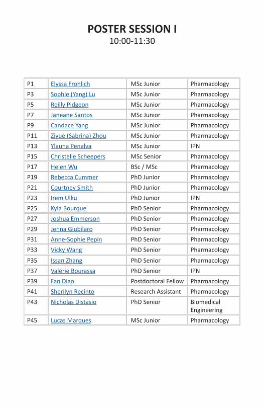
POSTER SESSION I10:00-11:30
P1 Elyssa Frohlich MSc Junior PharmacologyP3 Sophie (Yang) Lu MSc Junior PharmacologyP5 Reilly Pidgeon MSc Junior PharmacologyP7 Janeane Santos MSc Junior PharmacologyP9 Candace Yang MSc Junior PharmacologyP11 Ziyue (Sabrina) Zhou MSc Junior PharmacologyP13 Ylauna Penalva MSc Junior IPNP15 Christelle Scheepers MSc Senior PharmacologyP17 Helen Wu BSc / MSc PharmacologyP19 Rebecca Cummer PhD Junior PharmacologyP21 Courtney Smith PhD Junior PharmacologyP23 Irem Ulku PhD Junior IPNP25 Kyla Bourque PhD Senior PharmacologyP27 Joshua Emmerson PhD Senior PharmacologyP29 Jenna Giubilaro PhD Senior PharmacologyP31 Anne-Sophie Pepin PhD Senior PharmacologyP33 Vicky Wang PhD Senior Pharmacology
P35 Issan Zhang PhD Senior PharmacologyP37 Valérie Bourassa PhD Senior IPNP39 Fan Diao Postdoctoral Fellow PharmacologyP41 Sherilyn Recinto Research Assistant PharmacologyP43 Nicholas Distasio PhD Senior Biomedical
EngineeringP45 Lucas Marques MSc Junior Pharmacology
Presenter: Elyssa Frohlich 10h00 / MSc Jr. /P1
Title: Proteomic Screen for Novel Interactors of GPCR Heterodimers
Authors: Frohlich, Elyssa; Pétrin, Darlaine; Jiang, Sophie; Lau, Jenny; Dé-vost, Dominic; Bourque, Kyla; Zheng, Sindy; Hébert, Terry
G protein-coupled receptors (GPCRs) represent a large class of membrane receptors that mediate downstream signalling events in cells. Although GPCRs have been targeted by many therapeutics, their downstream sig-nalling remains incompletely understood, especially in the context of dimers. It is important to understand, as dimerization affects downstream signalling, trafficking of receptors to the membrane, and internalization kinetics of receptors. This project focuses on three GPCRs that are impli-cated in the regulation of blood pressure, the angiotensin II type 1 recep-tor (AT1R), the prostaglandin F receptor (FP), and the β2-adrenoreceptor (β2AR), and how their downstream pathways intertwine in the context of heterodimerization. To investigate their interactomes, we use the engin-eered ascorbate peroxidase 2 (APEX2), genetically fused to the C-tails. APEX2 is a biotin ligase that will allow the labelling and subsequent iden-tification of proximal proteins. We hypothesize that GPCR heterodimers have a different interactome than GPCR monomers or homodimers. First, we validated the expression, function and dimerization the APEX2-tagged constructs in HEK 293 F cells. Next, we will investigate the interactomes of heterodimers under different stimulation by co-expressing APEX2-tagged receptors with putative heterodimer partners. To confirm the function-ality of β2AR-APEX2, we used Bioluminescent Resonance Energy Trans-fer (BRET)-based signalling biosensors. When activated, β2AR leads to an increase in cAMP levels, which binds to an EPAC biosensor and causes a change in BRET. To confirm the functionality of AT1R-APEX2 and FP-APEX2, we used a Calflux biosensor. When activated, these receptors lead to an increase in intracellular Ca2+, which binds to the Calflux biosensor and causes a change in BRET. The BRET assays show that all three recep-tors couple functionally to Gα with APEX2 fused to their C-tail and west-ern blot analysis shows that APEX2 activity is functional.
Acknowledgements: McGill University, CIHR
Presenter: Sophie (Yang) Lu 10h00 / MSc Jr. /P3
Title: Elucidating the Mechanism underlying Parkin Substrate Selectivity
Authors: Lu, Yang; Vranas, Marta; Krett, Jonathan; Fon, Edward; Drucan, Thomas; Trempe, Jean-François.
Parkinson’s disease (PD) is the second most common neurodegenerative disease and it is characterized by a loss of nigrostriatal dopaminergic neu-rons. Although the majority of PD cases are late onset, some monogenic forms of PD display an early onset of motor symptoms as early as 18 years old. Parkin and PTEN-induced putative kinase 1 (PINK1) are two proteins involved in mitochondrial quality control pathways and their mutations cause early onset autosomal recessive PD. Upon mitochondrial damages, PINK1 accumulates on the outer mitochondrial membrane (OMM) where it phosphorylates ubiquitin (Ub) moieties to recruit Parkin. Parkin, an E3 ubiquitin ligase, catalyzes the addition of Ub onto target substrates. The buildup of Ub chain serves as a target signal for proteasomal degradation, formation of mitochondria-derived vesicles, or autophagy.Numerous Parkin substrates are located on the OMM, but at low phys-iological concentrations, Parkin primarily ubiquitinates its favorite sub-strate Mitofusin 2 (Mfn2). Mfn2 is a GTPase that catalyzes fusion of mi-tochondria and acts as a tether between the endoplasmic reticulum (ER) and mitochondria, thereby controlling homeostatic mechanisms such as calcium and lipids transfer. Although several groups have reported Mfn2 as a preferred substrate of Parkin, the molecular mechanisms underly-ing substrates’ recognition and specificity for Mfn2 are still unknown. Our overall objective is to determine the mechanism underlying Parkin substrate selectivity, more specifically how it preferentially ubiquitinates Mfn2 over other OMM substrates.Our mitochondrial ubiquitination assays confirmed Mfn2 as a preferred substrate of Parkin. Furthermore, cell-based assays showed that Mfn2 is indeed localized in proximity to PINK1, suggesting that it must be dec-orated with phospho-Ub (pUb) moieties at the onset of mitochondrial damage.
Acknowledgements: We acknowledge support from the FRQS groups GRASP and GEPROM, Parkinson Canada, NSERC and the Canadian Insti-tutes of Health Research (CIHR).

Presenter: Reilly Pidgeon 10h00 / MSc Jr. /P5
Title: Identifying Dietary Prebiotics to Improve PD-1 Immune Check-point Inhibition Response in Cancer: Preliminary Data
Authors: Pidgeon, R.; Messaoudene, M.; Routy, B.; Castagner, B.
Immune checkpoint inhibitors (ICIs) targeting the programmed cell death protein 1 (PD-1) and its ligand have revolutionized the way in which sev-eral cancers are treated, though overall response rates remain poor. Re-cent studies have shown that response failure is mediated, in part, by the gut microbiota and suggest that alterations in response status could be achieved through microbiota manipulation. We hypothesize that pre-biotics can stimulate the immune system through the gut microbiota and promote the response to PD-1 inhibition in cancer. Here, we pres-ent a methodology for the identification and evaluation of two classes of microbiome-modulating prebiotics: glycans and polyphenols. Two gly-cans (galactomannan and fructooligosaccharide) were identified for in vivo evaluation from the correlation between distinct bacteria present in ICI-responders and bacterial metabolic labelling experiments (met-FACSeq) performed in our laboratory previously. Simultaneously, crude extracts of the camu-camu berry, which improved ICI efficacy in past stud-ies, were prepared and iteratively fractionated to identify bioactive com-ponents using reverse-phase chromatographic techniques, then fed to a syngeneic mouse sarcoma model harboring an endogenous microbiota. Prebiotic glycan supplementation at repeated 200 mg/kg doses following tumor inoculation reduced the efficacy of PD-1 inhibition in mice, with the exception of galactomannan, which had no effect. Conversely, at the same dose, compounds present in the camu-camu extract polar fraction, but not others, significantly reduced tumor size in mice both alone and in combination with PD-1 inhibitors, pointing to a possible role in immune modulation through the microbiome. Future experiments will address the timing of prebiotic administration and expand our current metFACSeq data to identify candidate prebiotics for evaluation in anaerobic bioreac-tors and in humanized animal models.
Acknowledgements: The Canadian Glycomics Network
Presenter: Jeneane Santos 10h00 / MSc Jr. /P7
Title: Establishing a smoker-relevant rat model to study the behavioural pharmacology of nicotine withdrawal
Authors: Santos, Janeane; Clarke, Paul
Nicotine is a major contributor to tobacco addiction, a leading cause of preventable disease and death worldwide. During a quit attempt, smokers experience nicotine withdrawal syndrome that is characterized by symp-toms such as anxiety and irritability, which promote relapse and contin-ued tobacco use. Drug treatments designed to aid smoking cessation only partially reduce withdrawal symptoms, demonstrating the need to better understand nicotine withdrawal. In the standard rat model of nicotine withdrawal, nicotine is continuously administered before withdrawal is produced. Although withdrawal is reliably produced, nicotine plasma lev-els are three times higher in the model than in smokers. In turn, the rat model expresses opioid-like withdrawal that can be precipitated by the nicotinic antagonist mecamylamine; this does not occur in smokers. Due to these limitations, the standard model may not reveal smoker-relevant mechanisms of nicotine withdrawal. We aim to develop an animal model with improved relevance to smokers, and use the model to probe neuro-pharmacological mechanisms underlying nicotine withdrawal. The pres-ent study aimed to develop tests of affective withdrawal signs to be used in the nicotine withdrawal model. To this end, tests were established in rats in acute morphine withdrawal, which has similar affective signs to nicotine withdrawal but is produced rapidly. In a social interaction test of anxiety, withdrawal decreased the time that rats spent interacting so-cially. Analysis of ultrasonic calls, a measure of affect, revealed that 50-kHz call rate, but not call subtype profile, were altered by withdrawal. In a shock-induced aggression test of irritability, withdrawal decreased the duration of shock-induced 22-kHz calls whereas fighting behaviour was unaltered. These results suggest that social interaction, 50-kHz call rate, and shock-induced 22-kHz calls are appropriate measures of withdrawal signs for the nicotine withdrawal model.
Acknowledgements: The authors wish to thank Dr. Erik Cook (McGill Uni-versity) for technical assistance with the shock-induced aggression test. This work is supported by funding from CIHR.
Presenter: Candace Yang 10h00 / MSc Jr. /P9
Title: The role of ketone bodies in pathological angiogenesis
Authors: Yang, Candace; Cagnone, Gael; Kim, Jin Sung; Mitchell, Grant; Joyal, Jean-Sébastien
Pathological angiogenesis is a defining trait of many diseases, such as cancer and proliferative retinopathies. Vascular endothelial cells (EC) adapt metabolically to pathological conditions, using glucose and lipids as fuel or building blocks for proliferation. Ketone bodies are an alterna-tive source of fuel of the central nervous system, but their role in the eye and angiogenesis remain unexplored. Retinal single-cell RNA sequencing identified the expression of ketogenic enzymes, HMG-CoA synthase and lyase, specifically enriched in retinal EC, and more so in a murine mod-el of proliferative retinopathy. Hence, we hypothesized that endotheli-al-derived ketones might be an alternative source of fuel for proliferating pathological neovessels. Using a Cre/Lox system, we deleted HMG-CoA lyase (HmgclTie2-Cre) in vessels and studied pathological angiogene-sis in the murine oxygen-induced proliferative retinopathy (OIR) model. Retinas exposed to OIR were dissected, stained for vessels using lectin, flat-mounted, and imaged. Percentage of vaso-obliteration (VO) and neo-vascularization (NV) areas relative to the whole retina were measured using the SWIFT method. Conditional loss of Hmgcl in vessels significant-ly reduced pathological NV compared to wild type controls but did not affect VO. Our findings suggest ketone bodies are an alternative endog-enous metabolic substrate of vessels that play an essential role in patho-logical angiogenesis. Preventing ketogenesis could be a new therapeutic target in diseases associated with a neovascular phenotype.
Acknowledgements: This project is funded by the CIHR, the NSERC, and the Burroughs Wellcome Fund.

Presenter: Ziyue (Sabrina) Zhou 10h00 / MSc Jr. /P11
Title: Follicle-stimulating hormone (FSH) does not impact RANKL-in-duced osteoclastogenesis in RAW 264.7 cells
Authors: Zhou, Z.; Ongaro, L.; Bernard, D.
Postmenopausal osteoporosis has been attributed to decreased estradiol levels. In the hypothalamus-pituitary-gonadal (HPG) axis, estradiol syn-thesis is stimulated by follicle-stimulating hormone (FSH). FSH is secreted from the anterior pituitary gland and estradiol feeds back to the hypo-thalamus and pituitary to suppress FSH production. In postmenopausal women, the loss of estradiol negative feedback leads to elevated serum FSH levels. It was recently proposed that this increase in FSH also con-tributes to postmenopausal osteoporosis by stimulating differentiation and activation of bone-resorbing osteoclasts cells. Our objectives are to determine whether FSH has direct actions on osteoclast differentia-tion in vitro and, if so, its mechanism of action. First, a murine leukemic monocyte macrophage cell line, RAW 264.7, was differentiated into os-teoclasts by treatment with receptor activator of nuclear factor kappa-B ligand (RANKL, 50 ng/ml) for seven days. As expected, we observed the appearance of osteoclasts, characterized as tartrate-resistant acid phos-phatase (TRAP)-positive multinucleated cells. Also, RANKL treatment in-duced gene expression of established osteoclast differentiation markers, including Rank, Trap, cathepsin K (Ctsk), and matrix metalloproteinase-9 (Mmp-9). The mRNA expression of FSH receptor (Fshr), however, was very low and mostly undetectable before and after osteoclast differenti-ation. Second, RAW 264.7 cells were co-treated with FSH (35, 70 and 140 IU/L) during RANKL-induced osteoclastogenesis. FSH did not impact the expression of Rank, Trap, Ctsk, Mmp-9, and Fshr. In conclusion, FSH did not further induce osteoclast differentiation in the presence of RANKL; nonetheless, more experiments will be performed to determine whether and how FSH might directly regulate bone resorption.
Acknowledgements: The authors thank Dr. Bastien Castagner, Depart-ment of Pharmacology and Therapeutics, McGill University, for providing the RAW 264.7 cell line.
This research was funded by CIHR PJT-156249 to DJB

Presenter: Ylauna Penalva 10h00 / MSc Jr. /P13
Title: TCN-1, a novel mechanosensory channel involved in osmosensa-tion and motility in C. elegans
Authors: Penalva, Ylauna; Tirera, Fouley; Posner, Rachel; Hendricks, Mi-chael
Vital senses such as nociception, proprioception and osmosensation in-volve control of sensory neuron activity by mechanical force. Mechanical stimuli are transduced by neurons into electrical impulses through mech-anosensitive (MS) channels within their plasma membranes. In humans, various pathologies like chronic pain or salt-dependent hypertension are believed to be linked to mutations in these proteins. However, the iden-tity and mechanisms of the molecules underlying mechanotransduction are still not fully understood. A candidate MS ion channel named TACAN has recently been described in mice as a pore-forming subunit necessary for sensing mechanically induced pain by mediating mechanotransduc-tion in nociceptors. We wanted to further investigate this novel channel in a genetic model system. The Caenorhabditis elegans homolog of TA-CAN, which we called tcn-1, has not been characterized. We acquired tcn-1 mutant strains in C. elegans from a high-throughput mutagenesis li-brary. tcn-1 is found in an operon (CEOP3062) which also contains snf-6, a gene coding for an acetylcholine transporter in neuromuscular junctions. Consequently, we conducted behavioral sensory assays and motility stud-ies on the mutant worms. Our results did not corroborate the observa-tions made in mice, as they showed no significant difference between TACAN and wild-type worms in response to mechanical stimuli like body touch or mechanical pain. On the other hand, TACAN mutants displayed increased sensitivity to hyperosmotic barriers, implying that osmoregula-tion processes are impaired in these worms. Furthermore, we observed that TACAN worms also exhibited slight defects in swimming patterns. This suggested that TCN-1, in tandem with SNF-6, may also be implicated in movement coordination. Altogether, we have uncovered new func-tions for the TACAN channel in C. elegans, and these findings could, for example, give us insights on how osmosensitive neurons monitor body hydration and salt in humans.
Acknowledgements: CIHR, NSERC

Presenter: Christelle Scheepers 10h00 / MSc Sr. /P15
Title: Structural and functional characterization of amyloid beta (Aβ) variants
Authors: Scheepers, C.; Shobo, A.; Multhaup, G.
Alzheimer disease (AD) is a chronic neurodegenerative disease and is the most common cause of dementia. AD neuropathology includes the de-velopment of plaques in the brain, which are composed primarily of mis-folded amyloid beta (Aβ) protein. N-terminally truncated Aβ5-x species such as Aβ5-40 and Aβ5-42 have been identified in AD brains, yet their biophysical properties are currently unknown. Recently, a novel missense mutation in the amyloid precursor protein (APP) was discovered, in which the asparagine at position 687 is substituted for a lysine. In this study, it was noted that Aβ5-40 K16N was an additional product of APP K16N pro-cessing in cell-based experiments. The detection of these Aβ5-x variants supports further work to determine their potential contributions in AD pathology. Our aims include investigating whether the peptides Aβ5-40 and Aβ5-42 (wild-type [wt] and K16N) exert toxic effects in vitro and in vivo, their aggregation kinetics and their propensity to form fibrils. To as-sess the in vitro neurotoxicity of Aβ5-x peptides, the MTT assay was used to assess cell viability after treating SH-SY5Y neuroblastoma cells with varying concentrations of synthetic Aβ5-x peptides. Each of these Aβ5-x variants will also be transgenically expressed in Drosophila melanogaster to investigate in vivo toxicity based on whether eye-specific expression of these peptides disrupts the structural integrity of the fly eye. Thioflavin T assay results indicate that all the Aβ5-x peptides tested display the pro-pensity to aggregate. Preliminary electron microscopy data shows that all these Aβ variants form fibrils. Based on the MTT assay, treating cells with Aβ5-40 K16N, Aβ5-42 K16N, and Aβ5-42 wt resulted in a notable reduction in cell viability compared to untreated cells. As a follow up, we will generate four fly strains that will express these human N-terminally truncated peptides.
Acknowledgements: The authors would like to thank CIHR for financial contributions, and key collaborators in this project (Dai, D.; Bui, H.; Rao, Y.)

Presenter: Helen Wu 10h00 / BSc/MSc /P17
Title: Examining the regulation of GLUT1 trafficking by rhomboid prote-ase-related protein 4 and its implications in Alzheimer’s disease
Authors: Wu, Helen; Recinto, Sherilyn; Paschkowsky, Sandra; Münter, Lisa Marie
Initially identified by Alois Alzheimer, Alzheimer’s disease (AD) is a neu-rodegenerative disorder characterized by cognitive decline as well as memory loss. Although the pathology of AD remains elusive, a major hallmark of the disease is the deposition of amyloid beta (Abeta) protein into plaques in the brain. Abeta is generated following the enzymatic pro-cessing of the amyloid precursor protein (APP) by beta- and γ-secretases. Our lab has discovered that rhomboid-related protein 4 (RHBDL4) cleaves APP, bypassing the classical amyloidogenic processing and thus resulting in reduced production of Abeta peptides in cell culture experiments. Fur-ther, it is known that lowered glucose uptake into the brain is a character-istic of AD. We also observed that mouse embryonic fibroblasts deficient in RHBDL4 take up less glucose, leading us to question if this protease has a role in the regulation of glucose transporter 1 (GLUT1). Our goal is to uncover the potential interplay between the RHBDL4-mediated APP pro-cessing and the role of RHBDL4 in GLUT1 trafficking. In this study, we ex-amined the effect of different RHBDL4 mutants on the presence of GLUT1 at the cell surface. Different RHBDL4 mutants, a C terminus deletion and a catalytically inactive RHBDL4, was expressed in human embryonic kidney cells (HEK 293T) and mouse embryonic fibroblasts (MEF). We observed that GLUT1 levels at the cell surface was downregulated compared to cells transfected with wildtype RHBDL4. Through coimmunoprecipitation experiments, it was also evident that RHBDL4 interacts with GLUT1. The main goal of these studies is to investigate which domain of RHBDL4 is responsible for regulating the trafficking of GLUT1 to the cell surface and to gain insight into the physiological role of RHBDL4 and APP as metabolic sensors.
Acknowledgements: I would like to thank Dr. Lisa Münter for supervising me and giving me the opportunity to explore an area of research I am interested in. Thank you to Sherilyn Recinton, Sasen Efrem and Jaqueline Hsiao for being my mentors in the lab. Finally, thank you to students in Dr. Multhaup’s lab for helping me a long the way and answering my ques-tions.

Presenter: Rebecca Cummer 10h00 / PhD Jr. /P19
Title: Synthesis and evaluation of inositol phosphate analogs as thera-peutics against Clostridioides difficile toxin B
Authors: Cummer, Rebecca; Hadouch, Sarah; Castagner, Bastien
Virulent strains of Clostridioides difficile (C. difficile) infect 37 900 Canadi-ans annually. Symptoms associated with C. difficile infection are mediated by the secretion of toxins, and inactivating the toxins has shown to reduce recurrent C. difficile infection. The virulence of the toxins is associated with an auto-proteolysis event, which liberates an enzymatic domain of the toxin, causing cytoskeletal damage. Inactivation of auto-proteolysis has been shown to reduce the overall function of TcdB. Previously, our lab synthesized an IP6 analog that triggered toxin auto-proteolysis prior to its cellular uptake, thus preventing the internalization of the noxious en-zymatic domain. The group focused on optimizing the IP6 analog for the luminal environment. At present we are continuing the IP6 analog optimi-zation, focusing on improving the potency of the analog. We hypothesize that growing the IP6 analog further into an adjacent binding pocket could improve the potency, and therefore efficacy in vivo.We aim to synthesize analogs of IP6 with a side arm extension that inter-acts with a secondary binding pocket that extends from the IP6 binding site. We will first explore different attachment sites on the myo- and scyl-lo-inositol scaffold of IP6, before exploring different fragments that could bind in the secondary binding pocket and be attached to the IP6 analogs via click chemistry. Second, we plan to run an auto-processing functional assay using TcdB and the IP6 analogs. The activity will be quantified via SDS-PAGE with a silver stain.Synthesis of the final products: 1,3,4,5,6-phosphate-2-O-(prop-2-ynyl)-myo-inositol and 1,3,4,5,6-phosphate-2-O-benzyl-myo-inositol was suc-cessfully performed. Synthesis of the scyllo-inositol analogs are under-way. Confirmation of product synthesis was validated by 1H NMR, 31P NMR, and COSY NMR. Upon completion of the scyllo-inositol phosphate analogs, the IP6 analogs will be tested for their ability to trigger auto-pro-teolysis.
Acknowledgements: GlycoNet, CIHR IRSC, Canada Research Chairs, Can-ada Foundation for Innovation, and NSERC CRSNG

Presenter: Courtney Smith 10h00 / PhD Jr. /P21
Title: Discovering the Function of IGSF1 and Its Role in the Hypothalam-ic-Pituitary-Thyroid Axis
Authors: Courtney L. Smith, Daniel J. Bernard
The hypothalamic-pituitary-thyroid (HPT) axis controls the synthesis and secretion of thyroid hormones (THs). THs have many actions throughout the body, but are best known for their regulation of growth and metab-olism. Central hypothyroidism, a rare endocrine disorder, occurs when defects in the hypothalamus and/or the pituitary cause TH-deficiency. Mutations in the immunoglobulin superfamily, member 1 (IGSF1) gene are the most common cause of congenital central hypothyroidism. IGSF1 is a type 1 transmembrane protein of unknown function that is highly expressed in the pituitary gland. To define potential mechanism of IGSF1 action, we will identify its interacting partners using a new proximity in-teraction labelling method, BioID. The BioID method attaches a BirA* bi-otinylase onto the protein of interest (bait). In the presence of biotin, BirA* biotinylates interacting and proximal proteins. The biotinylated proteins (prey) are pulled down using streptavidin beads, which have a high affinity for biotin, and are identified using mass spectrometry. As we are most interested in intracellular signaling, we fused BirA* to the intracellular tail of IGSF1. 11 specific interacting proteins were identified in a preliminary BioID screen performed in a heterologous cell system. Several candidates identified in the BioID screen are expressed in thyro-trope cells, or have family members expressed in thyrotropes. Next, we will perform BioID in homologous cell lines. In the long-term, our work will define the function of IGSF1 in the pituitary, and may facilitate the discovery of novel causes of congenital central hypothyroidism.
Acknowledgements: Funding Sources: Supported by CIHR, RQR, and Mc-Gill Faculty of Medicine.
Presenter: Irem Ulku 10h00 / PhD Jr. /P23
Title: Endothelin-converting enzyme-1 as a Potential Amyloid-β34 De-grading Enzyme
Authors: Ulku, Irem; Multhaup, Gerhard
The canonical amyloidogenic pathway involves subsequent cleavage of the amyloid precursor protein first by β-secretase (BACE1) followed by γ-secretase to generate amyloid-β (Aβ) peptides of varying lengths [Chow 2010]. Recently, we have found a novel BACE1-catalyzed cleavage of Aβ40 and Aβ42 that results in the production of a non-amyloidogenic metasta-ble intermediate, i.e. Aβ34 [Liebsch 2019]. Unlike longer species, Aβ34 is soluble, non-toxic and non-aggregating [Hernandez 2015].Aβ production involves β- and γ-secretase, while Aβ clearance is mediat-ed by numerous proteases with distinct characteristics, including multiple Aβ cleavage site specificities, range of functional pH and varying subcellu-lar localization. Pharmacological experiments suggest that longer Aβ pep-tides can be directly degraded by a certain family of proteases, so-called Aβ-degrading enzymes (ADEs) [Saido 2012]. However, the protease(s) re-sponsible for Aβ34 degradation have not yet been identified. Therefore, we aim to analyze processes that are implicated in Aβ clearance in gen-eral and to identify proteases with a potential role in Aβ34 degradation.With regard to Aβ degradation, the most important difference among ADEs is their subcellular localization. BACE1 is located in endosomes and lysosomes [Vassar 2009] where it is active to produce Aβ34. Thus, Aβ34 is concentrated in acidic cell compartments and possibly recognized as a substrate for further degradation by the several candidate proteases. En-dothelin Converting Enzymes (ECEs), Insulin Degrading Enzyme, Cathep-sin B and Cathepsin D are of interest since these have been shown to be active in endosomes/lysosomes. Our preliminary data show that in BACE1 overexpressing SH-SY5Y cells, Aβ34 levels gradually increased upon ECE1 knockdown. Furthermore, in every condition, where ECE1 was knocked down either alone or in combination with other proteases, Aβ34 levels were elevated. Therefore, we suggest ECE1 as a potential Aβ34 degrading enzyme.
Acknowledgements: This project is funded by NSERC and CIHR.

Presenter: Kyla Bourque 10h00 / PhD Sr. /P25
Title: iPSC models of the cardiovascular system to interrogate GPCR sig-nalling and function
Authors: Devost, Dominic; Pétrin, Darlaine; Hébert, Terence E.
Cell surface expression of G protein-coupled receptors enhances their pharmacological tractability, making them ideal candidates for drug dis-covery. The development of novel therapeutic strategies to mitigate hu-man disease requires a more in-depth knowledge of GPCR signalling and function in situ. To address this, we have developed a toolbox of BRET and FRET-based biosensors with the ability to capture signalling and con-formational signatures originating at the level of the receptor, cognate G proteins and downstream effectors. As the AT1R is expressed throughout the cardiovascular system and dysregulated signalling has been linked to various pathologies, we are interested in understanding how cell context effects AT1R function. To increase the translatability of our results, we are using iPSCs as a physiologically relevant cellular model to explore AT1R conformational dynamics, agonist efficacy and G protein vs β-arrestin cou-pling. In order to build a comprehensive iPSC-based model of the human cardiovascular system, we are currently differentiating iPSCs towards the mesodermal lineage. iPSC-derived cardiomyocytes have been success-fully generated and validation of iPSC-derived vascular smooth muscle cells is still underway. Delivery of our BRET-based biosensors into cells is currently being accomplished using AAVs, however we are engineering an iPS master cell line for insertion of our biosensors at the AAVS1 safe harbour locus to enable comparisons between different cells in the same isogenic background. Working with an iPSC model allows access to clin-ically relevant cells such as those derived from patients suffering from cardiomyopathies. GPCR signalling can thus be studied in healthy versus patient lines to gain insight on how signalling modalities change as a con-sequence of disease. We predict that a deeper understanding of cell-type specific receptor function and signalling will advance rational drug design for the treatment of cardiovascular diseases.
Acknowledgements: This project is supported by the Canadian Institutes of Health Research (CIHR).

Presenter: Joshua Emmerson 10h00 / PhD Sr. /P27
Title: Generation of a novel bigenic rat model for Alzheimer’s dis-ease-like pathology: potential cross-talk between amyloid and tau
Authors: Emmerson, Joshua; Do Carmo, S; Cuello, AC
Alzheimer’s disease (AD) is a multifaceted neurodegenerative disease and is the leading cause of dementia. AD is pathologically characterized by the accumulation of extracellular amyloid Beta plaques and intracellular hy-perphosphorylated tau tangles. AD patients experience cognitive impair-ments in learning and memory and suffer from immense brain shrinkage. Numerous experimental drugs being tested on AD patients have failed, emphasizing a need to 1) to develop models that better recapitulate hu-man AD and 2) to better understand earlier stages of the disease and. Animal models have significantly improved our understanding of how AD affects the brain however current models do not reflect a complete human-like pathology or present with an overaggressive phenotype that may mask subtle changes occurring at early stages. Evidence implicates that amyloid and tau play an important role in AD however the way(s) by which amyloid and tau unfold together in early stages are not well understood.To address these research gaps, we present a newly generated bigenic rat model that possesses a relatively mild endogenous expression of both human amyloid and tau. At key time points of aging (representative of dif-ferent pathological stages), characterization of bigenic animals includes examination of AD-like pathology, behavioural assessment of cognitive function and downstream AD-associated mechanisms of disease such as inflammation. Preliminary findings from 12 month-old animals are dis-cussed, comparing bigenics to single transgenics and wild-type littermate controls. We anticipate that older animals will develop a complete human AD-like pathology.
Acknowledgements: This work is supported by funding from the McGill Faculty of Medicine (JTE) and CIHR (ACC).

Presenter: Jenna Giubilaro 10h00 / PhD Sr. /P29
Title: High-throughput Screening For AT1R Trafficking Reveals a New In-hibitor of Small GTPases
Authors: Giubilaro, Jenna; Schuetz, Doris A.; Namkung, Yoon; Bouvier, Michel; Marinier, Anne; Laporte, Stéphane A.
G protein–coupled receptors (GPCRs) are important therapeutic targets that activate various downstream signaling events, like mitogen-acti-vated protein kinases (MAPK), via G proteins and β-arrestins. β-arrest-ins can mediate MAPK signaling both at the plasma membrane and from endosomes. However, deciphering the role of β-arrestin-mediated MAPK signaling from these compartments remains a challenge due to the dearth of selective tools. Therefore, we performed a high-through-put screen using BRET-based sensors to identify modulators of GPCR traf-ficking, using the angiotensin II type 1 receptor (AT1R) as a prototypical GPCR. We identified a small molecule, Traf 21, that not only blocked the endocytosis of AT1R and other GPCRs, like the Bradykinin B2 and β2-ad-renergic receptors, but also potently inhibited the activation of MAPK by these receptors, including EGFR. Using a panel of new BRET sensors, we found that Traf 21 targets the small G proteins Arf6 and Ras (but not Rac and Rho), which are respectively involved in clathrin-mediated endocyto-sis and receptor-mediated MAPK activation. Furthermore, computational analysis of Traf 21 interaction with Ras reveals that it binds at the inter-face between Ras and its guanine nucleotide exchange factor (GEF) SOS1. To improve its functional selectivity, structure-activity relationship studies were performed on Traf 21 and analogues that inhibit endocytosis and/or MAPK were identified. Here, we discovered a new inhibitor of both Ras and Arf6, which we named Rasarfin, that can be optimized to selectively target either small G protein in order to study the internalization and sig-naling of GPCRs.
Acknowledgements: This work was supported by the CIHR and the RQRM.
Presenter: Anne-Sophie Pépin 10h00 / PhD Sr. /P31
Title: KDM1A mediates transgenerational metabolic disturbances in a sex-specific manner and is linked to diet-induced altered sperm H3K4me3
Authors: Pepin A.-S.; Lafleur C.; Dumeaux V.; Sloboda D. M.; Kimmins S.
Increasing evidence suggests that paternal diet may affect offspring risks to develop obesity. However, the molecular mechanisms underlying the non-genetic inheritance of complex disease remain elusive. The Kimmins lab has demonstrated using a genetic model of epigenetic inheritance, that sperm histone methylation is implicated in transgenerational epi-genetic inheritance. Using this model, our objectives were to (1) investi-gate whether epigenome-environment interactions can lead to enhanced metabolic phenotypes transgenerationally, and whether (2) transmission of phenotypes can be linked to sperm H3K4me3.Transgenic males (F0) with a pre-existing eroded sperm epigenome and wildtype sires, were fed either a low- or high-fat diet for 12 weeks. Their sperm was examined for diet induced-changes to the epigenome by chromatin immunoprecipitation sequencing targeting histone H3K4me3. Their regular chow-fed offspring (F1 and F2) were examined for inherited changes in metabolism.F0 males fed a high-fat diet became obese and showed signs of metabol-ic syndrome irrespective of their genotype. Intergenerational effects of paternal diet were observed in male offspring only, while female descen-dants were not impacted by paternal diet. Interestingly, transgeneration-al metabolic disturbances were only observed in transgenic descendants, suggesting that paternal exposure to a multitude of environmental stress-ors may exacerbate descendant risks to develop obesity. Sperm H3K4me3 profiling revealed diet-induced alterations in enrichment. Differentially enriched regions occurred at genes with functions corresponding to ob-served offspring phenotypes, including genes involved in placenta devel-opment and lipid metabolism.This is the first report linking high-fat feeding and alterations in the sperm epigenome at the level of histone H3K4me3. These findings shed light on the potential contribution of histone methylation in sperm in paternal transmission of complex diseases.
Acknowledgements: Funding sources: Supported by CIHR and CRRD.
Presenter: Xiaotong (Vicky) Wang 10h00 / PhD Sr. /P33
Title: Effects of environmentally-relevant mixtures of organophosphate ester (OPE) flame retardants on KGN cells, a human granulosa cell line
Authors: Wang, Xiaotong; Hales, Barbara F.; Robaire, Bernard
OPE flame retardants are found ubiquitously in the environment. Previ-ous studies suggest that exposure to individual OPEs may be detrimental to female fertility. Since ovarian granulosa cells play key roles in female reproduction, we tested the hypothesis that an environmentally relevant OPE mixture will adversely affect their function. KGN immortalized hu-man granulosa cells were exposed for 48h to one of two OPE mixtures. The first (total mixture) was composed of 13 OPEs detected in Canadian house dust; the second triaryl-OPE mixture, was a subset of 7 OPEs with 3 phenyl moieties in their structure. Cells were exposed to vehicle or 1/1,000,000X – 1/3,000X dilutions of these mixtures, where 1X repre-sents the OPE concentrations in 6.32g of dust. Effects on cell survival, lysosomes, ROS production, and lipid droplets were determined using fluorescent dyes and high content imaging. At dilutions of 1/10,000X, the total and triaryl mixtures decreased cell survival by more than 80% and 50%, respectively. At non-toxic dilutions, only the triaryl-OPE mixture de-creased the number of lysosomes/cell. Only the total mixture increased ROS production in cells at dilutions that were not cytotoxic. Both mixtures significantly increased the total area of lipid droplets at exposures as low as 1/300,000X; the total mixture induced this increase to a greater ex-tent. Together, these data show that the total and triaryl OPE mixtures af-fect specific endpoints differentially. We propose that exposure to “house dust” OPE mixtures may have adverse effects on female reproductive health.
Acknowledgements: Supported by CIHR and McGill University. We thank Dr. Mike Wade (Health Canada) for preparing and providing these mix-tures.
Presenter: Issan Zhang 10h00 / PhD Sr. /P35
Title: HDAC6 inhibition to kill glioblastoma and modulate its microenvi-ronment
Authors: Zhang, I.; Maysinger, D.
Background: Glioblastoma multiforme (GBM) is the most common and deadly type of brain tumor. Persistent STAT3 activation (p-STAT3) in the GBM microenvironment causes infiltrating immune cells to produce tu-mor-supporting growth factors and cytokines. However, direct STAT3 inhibitors have several drawbacks that limit their progression in clinical trials. Our studies showed that histone deacetylase 6 (HDAC6)-selective inhibitors ACY-1215 and sahaquine were effective against GBM growth and invasiveness. Because HDAC6 can promote STAT3 activation, we hy-pothesize that HDAC6 inhibitors will downregulate p-STAT3 in GBM and tumor-associated macrophages (TAMs). To test this hypothesis, we used direct and indirect GBM/TAMs co-cultures to measure p-STAT3 levels in response to ACY-1215 and sahaquine. We also investigated dendritic polyglycerol sulfates (dPGS) as nanostructures with inherent anti-inflam-matory properties in tumor-associated and normal neural cells.Results: We showed that GBM cells induced inflammatory markers (p-STAT3, NFkB, lipocalin-2, nitric oxide) in microglia and macrophages. In turn, activated microglia increased p-STAT3 in GBM, thereby contributing to a tumor-supportive feedback loop between GBM and TAMs. HDAC6 inhibitors ACY-1215 and sahaquine prevented p-STAT3 activation in TAMs without affecting their viability, whereas dPGS inhibited GBM- and mi-croglia-mediated inflammatory pathways in human neural cells.Conclusion: HDAC6 inhibitors and dPGS downregulated p-STAT3 and key inflammatory markers in GBM-associated cells. We propose the use of HDAC6 inhibitors and anti-inflammatory dPGS to modulate the tumor mi-croenvironment and enhance anti-cancer treatment effectiveness.
Acknowledgements: The authors wish to thank NSERC and CIHR for their financial support.
Presenter: Valérie Bourassa 10h00 / PhD Sr. /P37
Title: Cyclic AMP-dependent transcription factor 3 (ATF3) expression in proprioceptors correlates with consistent and irreversible pain in a MIA model of osteoarthritis in the rat ankle joint
Authors: Valérie Bourassa, Noosha Yousefpour, Haley Deamond, Valérie Cabana, Alfredo Ribeiro-da-Silva
There are many clinical and pre-clinical lines of evidence suggesting a neuropathic component to osteoarthritis (OA) pain. Here we studied the possible contribution of primary afferent damage following joint degen-eration to this neuropathic component by investigating the expression of Activating Transcription Factor 3 (ATF3), a marker of neuronal stress, in various cell populations of dorsal root ganglia (DRG). Male Sprague Dawley rats received an intra-articular injection of 2.4 mg of sodium mono-iodoacetate (MIA) in 40 µl saline in the tibio-talar joint (ankle joint), and were perfused at 1, 2, 3, 4, 5, and 6-week time-points. DRGs (L4-L6) were extracted and immunostained with anti-ATF3, anti-calcitonin gene-related peptide (CGRP), anti-neurofilament 200 (NF200), and an-ti-parvalbumin (PV) antibodies, as well as stained with fluorescent isolec-tin GS-IB4 and Nissl. Our characterization of ATF3 expression in dorsal root ganglia shows a biphasic pattern, with a large expression of ATF3 following MIA injection at week 1, which subsides completely by week 3. Interestingly, significant ATF3 expression return at week 5 principally in large-diameter cell bodies, which is the onset of the development of pain hypersensitivity in this model. Indeed, at this time point, 50 % of ATF3-positive cells also colocalize with PV, a marker of proprioceptors, while proprioceptors make up approximately 25% of cells in the rat DRG. This suggests that Aβ mechanoreceptors innervating the articular joint in OA pain are mostly affected following joint degeneration, which drives interest in understanding the contribution of lamina III-V that receives input from these fibers, in the persistence of OA pain.
Acknowledgements: Valérie Bourassa and Noosha Yousefpour are recipi-ents of Louise and Alan Edwards Foundation Doctoral Studentships. Haley Deamond is a recipient of studentship from the McGill University Faculty of Medicine. This work was funded by Canadian Institutes of Health Re-search Operating Grant MOP-136903.
Presenter: Fan Diao 10h00 / PDF /P39
Title: Paternal age differentially affects the transcriptome of spermato-zoa from the caput and cauda epididymidis of Brown Norway rats
Authors: Fan Diao, Bernard Robaire
Increasing number of couples choose to start having children at a later age. Mounting evidence indicates that advanced paternal age is associ-ated with decreased fertility and untoward effects on progeny outcome. To determine effects of advanced paternal age on sperm during epididy-mal maturation, we used a rat model to assess whether gene expression and epigenetic modification occur in aging male germ cells. We obtained spermatozoa from the caput and cauda regions of the epididymis of young (4-5 months) and aged (18-20 months) Brown Norway rats, isolat-ed long and small RNA, and performed mRNA and small RNA sequencing. By screening for differently expressed genes between sperm from the ca-put and cauda epididymidis and between young and aged animals, we found a large number of differentially expressed RNAs. Some of the most striking changes were noted in the β-defensin family, a group of proteins with antimicrobial activity that are essential for innate immune system and expressed at high levels in the epididymis. While aging related germ cell expression pattern and regulatory mechanism of β-defensins has not been reported yet. We found 30 members of rat β-defensins and Sperm Associated Antigen 11a/b stably expressed in caput and cauda sper-matozoa. Among these members, expression levels of Defb12, Defb22, Defb23, Defb25, Defb33, Defb41, Defb44 and Spag11a significantly de-creased with age only in spermatozoa from the cauda epididymis. Further analysis of the RNAseq results should provide new insight into how aging modulates RNAs in spermatozoa as they transit therough the epididymis.
Acknowledgements: Supported by CIHR.

Presenter: Sherilyn Recinto 10h00 / RA /P41
Title: Human rhomboid RHBDL4 as a modulator of cellular glucose me-tabolism
Authors: Recinto, Sherilyn Junelle; Paschkowsky, Sandra; Munter, Lisa
Rhomboid-related protease 4 (RHBDL4) is a member of an ancient class of serine intramembrane proteases found in all kingdoms of life. Recent breakthroughs in the identification of its substrates have paved way in understanding its potential cellular roles. Nevertheless, many of the proposed functions have been demonstrated in an overexpression cell system and have not been confirmed in vivo. We report here an altered cellular glucose metabolism in mouse embryonic fibroblasts (MEFs) upon RHBDL4 abrogation. Not only are MEFs lacking Rhbdl4 manifest reduced glucose consumption and glycolytic flux as compared to wild type cells, we further observed diminished total and cell surface glucose transport-er 1 (Glut1) levels. Endothelial cells lining the cerebral microvessels of Rhbdl4-deficient mice also exhibited less Glut1 expression. The overall systemic metabolic profile of these mice is, meanwhile, not impacted. Collectively, we propose that RHBDL4 has a physiological role in the an-terograde trafficking of GLUT1, albeit through a still unidentified manner.
Acknowledgements: We thank Dr. Matthew Freeman for kindly provid-ing the Rhbdl4-deficient mouse embryonic fibroblasts and mice. This re-search was supported by NSERC Discovery grant no. RGPIN-2015-04645, Canada Foundation of Innovation Leaders Opportunity Fund (CFI-LOF, 32565), Alzheimer Society of Canada Young Investigator award PT-58872 and Research Grant 17-02, Fonds d’innovation Pfizer-FRQS sur la mala-die d’Alzheimer et les maladies apparentées no. 31288, McGill Faculty of Medicine Bridge funding, and an award from The Scottish Rite Charitable Foundation of Canada 16112.
Presenter: Nicholas Distasio 10h00 / PhD Sr. /P43
Title: VCAM-1 targeted gene delivery nanoparticles localize to inflamed endothelial cells and atherosclerotic plaques
Authors: Nicholas Distasio, Hugo Salmon, Stephanie Lehoux, Maryam Ta-brizian
Introduction: Evidence continues to implicate the immune system in ath-erosclerosis, which is a leading cause of morbidity and mortality world-wide. However, broadly-acting immunosuppressants and anti-inflamma-tory therapy may decrease the immune system’s function elsewhere. New and innovative therapeutics must be targeted to the site of athero-sclerotic plaque growth. Along these lines, we synthesized biodegradable polymers capable of condensing plasmid DNA into nanoparticles (NPs) that could be targeted to VCAM-1, a receptor overexpressed by inflamed endothelial cells overlying the plaque. The nanoparticles contain plasmid DNA encoding interleukin-10 (IL-10), a potent anti-inflammatory cyto-kine. We hypothesize that by targeting endothelial cells at the plaque to produce IL-10, inflammation within the plaque only can be reduced.
Methods/Results: We show that NPs specifically bind VCAM-1 protein via surface plasmon resonance (SPRi) biosensing and are taken up by TN-Fα-activated endothelial cells under both static and flow conditions. NPs were found to be non-toxic and able to induce significant production of IL-10 in primary endothelial cells in vitro (~900 pg/ml per 200,000 cells). Macrophages cultured in media from IL10-transfected endothelial cells had increased phosphorylation of STAT3 (p=0.054) and reduced secretion of TNFα following stimulation with LPS (p<0.05). Furthermore, when in-cubated ex vivo on plaque-containing tissues harvested from LDLR-/- or ApoE-/- mice on high-fat diet at least 9 weeks, NPs bound to the endothe-lial layer and colocalized with VCAM-1 expression compared to non-spe-cific control NPs. The biodistribution of targeted NPs injected in vivo was also tracked via live fluorescent imaging and NP accumulation in plaque tissues was assessed after harvesting organs.
Conclusion: This strategy represents an innovative, multi-disciplinary and targeted approach capable of reducing inflammation in atherosclerosis via non-viral gene delivery and could be amenable to other applications.

Presenter: Lucas Marques 10h00 / MSc Jr. /P45
Title: Brain nicotinic receptor function assessed in vivo during chronic nicotine exposure and withdrawal
Authors: Marques, L.; Jones-Tabah, J.; Clarke, P.
The classical view of nicotine dependence suggests that tobacco smok-ers use nicotine to activate nicotinic acetylcholine receptors (nAChRs) in the brain. In this view, nicotine withdrawal reflects nAChR underactivity. However, nicotine acts as both an agonist and antagonist, as nAChRs can be both activated and inactivated when bound to nicotine. Nicotine’s an-tagonistic effects occur during chronic exposure as a result of receptor desensitization. Furthermore, there is in vitro and ex vivo evidence for nAChR overstimulation during nicotine withdrawal. Therefore, nicotine’s ability to alleviate withdrawal may derive from its time-averaged antago-nistic effect of nAChR desensitization.Using in vivo fibre photometry, we will measure neuronal activity in do-paminergic neurons following acute administration of nicotine in freely behaving rats that have been chronically exposed to the drug and then subsequently withdrawn.Hypotheses: 1) During chronic nicotine administration, the dopaminergic neuronal response provoked by an acute nicotine challenge injection will be reduced. 2) During withdrawal from chronic nicotine, this neuronal population will produce an exaggerated response to an acute nicotine challenge, indicating supersensitivity.Methodology: A Cre-dependent virus encoding a calcium indicator will be injected into the VTA of TH::Cre transgenic rats, so as to specifically target dopaminergic neurons. An optic probe will then be chronically positioned in the VTA to record neuronal activity in real-time. Nicotine will be intra-venously injected daily to provide chronic nicotine exposure; the cessa-tion of these daily injections will trigger spontaneous withdrawal. During both phases we will measure intracellular calcium responses to an acute intravenous nicotine injection using in vivo fibre photometry.Significance: This project will potentially provide the first in vivo evidence of a supersensitive dopaminergic neuronal response in nicotine with-drawal.
Acknowledgements: This project is funded by the Canadian Institutes of Health Research (CIHR).
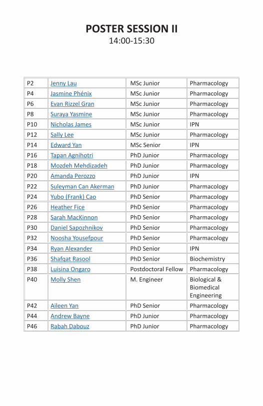
POSTER SESSION II14:00-15:30
P2 Jenny Lau MSc Junior PharmacologyP4 Jasmine Phénix MSc Junior PharmacologyP6 Evan Rizzel Gran MSc Junior PharmacologyP8 Suraya Yasmine MSc Junior PharmacologyP10 Nicholas James MSc Junior IPNP12 Sally Lee MSc Junior PharmacologyP14 Edward Yan MSc Senior IPNP16 Tapan Agnihotri PhD Junior PharmacologyP18 Mozdeh Mehdizadeh PhD Junior PharmacologyP20 Amanda Perozzo PhD Junior IPN
P22 Suleyman Can Akerman PhD Junior PharmacologyP24 Yubo (Frank) Cao PhD Senior PharmacologyP26 Heather Fice PhD Senior PharmacologyP28 Sarah MacKinnon PhD Senior PharmacologyP30 Daniel Sapozhnikov PhD Senior PharmacologyP32 Noosha Yousefpour PhD Senior Pharmacology
P34 Ryan Alexander PhD Senior IPNP36 Shafqat Rasool PhD Senior BiochemistryP38 Luisina Ongaro Postdoctoral Fellow PharmacologyP40 Molly Shen M. Engineer Biological &
Biomedical Engineering
P42 Aileen Yan PhD Senior PharmacologyP44 Andrew Bayne PhD Junior PharmacologyP46 Rabah Dabouz PhD Junior Pharmacology

Presenter: Jenny Lau 14h00/ MSc Jr. / P2
Title: Spatiotemporal characterization of G protein β and γ subunit in-teractors via APEX2 labeling
Authors: Pétrin, Darlaine; Bayne, Andrew; Trempe, Jean-Frnaçois; Hébert, Terence
GPCRs represent a large family of receptors involved in important physio-logical functions through regulation of myriad downstream signalling pathways. Many pathways work via heterotrimeric G proteins. In addi-tion to the important role played by Gα subunits, recent work has de-scribed non canonical roles for Gβγ subunits in downstream signalling of GPCRs in various organelles. Our lab has used TAP to identify cytosolic and nuclear Gβγ-interacting proteins. Nonetheless, methods such as TAP require protein interactions to be stable, missing transient interactions. The use of APEX2, a proximity-dependent labeling technique, provides both spatial and temporal information in endogenous protein expression level, allowing for in-depth characterization of protein networks. Brief-ly, cells expressing a construct comprised of a protein of interest fused genetically to APEX2 peroxidase are incubated with biotin-phenol. Then, hydrogen peroxide is added for a short 1-minute reaction to tag proteins within a 20nm radius with biotin, which can be purified using streptavi-din beads. Flag-tagged Gβ1 and Gγ5 constructs containing APEX2 have already been made and tested for proper expression and function in HEK 293F cells. These constructs were able to be pulled-down with Gαq, a known interactor of both Gβ1 and Gγ5 using co-immunoprecipitation with the Flag-tag. Labeling tests have also been done to validate the activ-ity of the APEX2 peroxidase. Through western blot, biotinylated protein can be observed only when both hydrogen peroxide and biotin-phenol are added. Currently, sample preparation for proteomic screens are being optimized. Further studies will use stimulated conditions as well as dif-ferent cell types as these factors highly influence the signalling pathways and interacting partners of G protein β and γ subunits. Using this tech-nique, we hope to undertake large proteomic screens to better identify Gβγ interactors and the role these interactions play in health and disease.
Acknowledgements: This project was supported by a grant from CIHR
Presenter: Jasmine Phénix 14h00 / MSc Jr. / P4
Title: Characterization of the C134W mutation in the FOXL2 transcrip-tion factor
Authors: Phénix J.; Schang G.; Krishnan S.; Guarné A.; Bernard D. J.
Follicle-stimulating hormone (FSH) is a hormone secreted by gonado-trope cells in the pituitary gland and is composed of an α and a β subunit. Endogenous expression of Fshb, which encodes the β subunit, relies on activin signaling, mainly through the stimulated interaction of transcrip-tion factors FOXL2, SMAD3, and SMAD4. The necessity of these transcrip-tion factors for Fshb expression has been shown by both in vitro and in vivo studies. While the elements of secondary structure mediating the SMAD3-SMAD4 interaction have been described, those required for the protein-binding C-terminal ‘Mad homology 2’ (MH2) domain of SMAD3 to interact with the DNA-binding forkhead domain (FHD) of FOXL2 re-main elusive. This is mainly because the crystal structure of FOXL2 alone or with SMAD3 remains unresolved. Such a crystal structure would al-low us to fully describe the nature of this interaction. A somatic mutation in FOXL2 (C134W) found in most ovarian granulosa cell tumours (GCT) has been suggested to stabilize the SMAD3-FOXL2 interaction. Thus, the FOXL2C134W mutation may enable us to co-crystallize the FOXL2-SMAD3 complex. In this study, we used a pituitary-like heterologous cell system to assess the functional interaction of FOXL2C134W with SMAD3. First, we showed that the C134W mutation enhanced FOXL2’s ability to acti-vate the Fshb promoter alone and in combination with SMAD3. Secondly, we successfully established a protocol to purify the FHD domain of FOX-L2C134W. Then, we worked on optimizing the purification of SMAD3’s MH2 domain. Based on these results, we will co-crystallize the FOX-L2C134W-SMAD3 complex to characterize the interaction with atomic resolution. Ultimately, this study may allow us to better understand: 1) how FOXL2 and SMAD3 physically and functionally interact, and 2) how the C134W substitution in the FHD domain of FOXL2 enhances the inter-action with SMAD3’s MH2 domain. This could inform novel therapeutic strategies to target FOXL2 in the context of GCT.
Acknowledgements: The authors thank Dr. Shunichi Shimasaki ,Depart-ment of Reproductive Medicine, School of Medicine, University of Califor-nia, San Diego, La Jolla, California, for providing the pFlag-FOXL2/pCS2+ and pFlag-FOXL2(C134W)/pCS2+ plasmids.

Presenter: Evan Rizzel Gran 14h00 / MSc Jr. / P6
Title: The role of ketone bodies in pathological angiogenesis
Authors: Gran, E. R.; Zhang, I.; Stochaj, U.; Maysinger, D.
Lysosomes are acidic organelles responsible for breaking down cellular components, as well as internalized drugs and nanostructures. Lysosomal function is impaired in various cancers, including glioblastoma. Given that most nanomaterials are internalized by the endo-lysosomal system, it is of great interest to explore the effects of nanostructures on lysosomes in human cells. Gold nanoclusters (AuNCs) are a unique type of nanostruc-ture whose physicochemical properties depend on gold surface accessi-bility and ligand. It is based on these properties that AuNCs can mimic redox-regulating enzymes. The objective of our study was to determine if AuNCs with 15-gold atom core with glutathione (Au15SG13) or PEG li-gands (Au15PEG) differentially modulate lysosomal biogenesis and activ-ity in human glioblastoma cells.
Non-cytotoxic concentrations of Au15SG13 or Au15PEG decreased lyso-somal abundance but induced a compensatory response in glioblasto-ma cells, where lysosome perinuclear positioning, lysosomal acidity and enzymatic activity were enhanced. The increased lysosomal activity was not sufficient to eliminate protein aggregates formed in the presence of Au15PEG. In contrast, Au15SG13 did not cause accumulation of protein aggregates. The dysregulation of lysosomes, at least in part, explains AuNC-induced cell loss in human glioblastoma cells, but no significant cell loss was found in human cerebral organoids or kidney cells after 72 hours. Results from these studies suggest 1) lysosomal adaptation to AuNCs is time and concentration-dependent, 2) ligands on AuNC surface play a critical role in determining their effects in human cells, and 3) AuNCs ex-ert cell type-dependent effects.
Acknowledgements: Special thanks to A. Rodolphe, F. Bertorelle, and H. Fakhouri for providing AuNCs for biological experiments. Funded by NSERC, CIHR, GE Fellowship.
Presenter: Suraya Yasmine 14h00 / MSc Jr. / P8
Title: Characterizing specific glycan metabolism of gut microbiota using “metFACseq”
Authors: Yasmine, Suraya; Dridi, Lharbi; Maurice, Corinne; Castagner, Bastien
The diverse microbial community residing in our gut, termed the gut mi-crobiota, is an ecosystem that has important functions in human physiol-ogy. Any alteration in this microbial composition (dysbiosis) can have po-tential influence on diseases such as inflammatory bowel disease, cancer and obesity. However, prebiotics - non-digestible dietary fibers - can be used to modulate the gut microbiota with beneficial outcome for the host health. Indeed, complex glycans contained in our diet are an important source of carbon for the gut microbiota, which collectively encodes the thousands of carbohydrate-active enzymes (CAZymes) necessary to me-tabolize them. We hypothesize that identifying which bacteria are respon-sible for the metabolism of specific glycans (“who eats what?”) will help us to design future prebiotic approaches. To identify bacteria that take up specific glycan structures in human microbiota samples, a functional and culture-independent method has been developed in the Castagner Lab called “metFACSeq”. This method combines metabolic labeling of bacteria using fluorescently labeled glycans with fluorescence-activated cell sorting (FACS) and 16S rDNA sequencing. I have started applying our metFACseq method on healthy stool samples with five probes: maltodex-trin, mannotetraose, arabinoxylotetraose, fructo-oligosaccharide, and xylotetraose). Around 200K cells were sorted via FACS from the labeled samples (1.5-3 % glycan+ cells). I am currently performing PCR amplifica-tion of the 16S rDNA (V4 region) for sequencing. We plan to run the met-FACSeq pipeline on 10 microbiota samples from healthy volunteers using 10-15 different fluorescent glycans. This will give us a broader idea about specific glycans and their consumers in the gut microbiota, and guide us for rational prebiotic approaches in the future.
Acknowledgements: Canadian Glycomics Network (GlycoNet)
Presenter: Nicholas James 14h00 / MSc Jr. / P10
Title: Characterization of the effects of anti-amyloid D-AIP on microglial response to amyloid-β42
Authors: James, N.; Shobo, A.; Multhaup, G.
Microglia perform essential roles in the healthy brain to support neurons and maintain homeostasis; however, in Alzheimer’s disease (AD), chronic activation of microglia due to the accumulation of amyloid-β (Aβ) results in a toxic neuroinflammatory state that can lead to neuronal death. The 42 amino acid long peptide Aβ42 is a rapidly aggregating Aβ isoform that is highly toxic to neurons when it forms soluble oligomers. Aβ42-oligomer interacting peptide (AIP) is a promising therapeutic compound to combat Aβ pathology in AD, though its effects on glial cell response to Aβ42 are unknown. AIP’s effects on neuronal and microglial cell response to Aβ42 in vitro were investigated using the SH-SY5Y neuroblastoma and BV-2 microglial cell lines. The aggregation kinetics of synthetic Aβ42 with or without D-amino acid AIP (D-AIP) were monitored using the Thioflavin T assay. Aβ42 toxicity to SH-SY5Y and BV-2 cells was examined by the MTT assay following treatment with preincubated synthetic Aβ42 with or with-out AIP. D-AIP was not cytotoxic, and was able to neutralize preincubated Aβ42 toxicity to SH-SY5Y and BV-2 cells. We are currently investigating the effects of D-AIP on fluorescently-labeled Aβ42 uptake and clearance by SH-SY5Y and BV-2 cells, Aβ42-induced cytokine release, and Aβ42-in-duced microglial toxicity to SH-SY5Y cells. We have recently found that D-AIP can cross the blood-brain barrier in wild type C57BL/6 mice, and will be investigating its effects on Aβ pathology and neuroinflammation in transgenic mouse models of AD.
Acknowledgements: This research was undertaken thanks in part to funding from CIHR and the Canada First Research Excellence Fund and Fonds de recherche du Québec, awarded to the Healthy Brains, Healthy Lives initiative at McGill University.
Presenter: Sally Lee 14h00 / MSc Jr. / P12
Title: Roles of periconceptional moderate and high dose folic acid sup-plementation in the prevention of epigenetic defects associated with the use of assisted reproduction
Authors: Lee, S; Martel, J; Rahimi, S; Trasler, J
Assisted reproductive technology (ART) currently accounts for 1-4% of all births in Western countries. Increases in growth and birth defects, ac-companied by DNA methylation abnormalities, have been reported in ART-conceived children. Proper DNA methylation is crucial for normal development of the embryo and placenta. Dietary folic acid is a major source of methyl groups used in DNA methylation. Here, our goal was to use a mouse model to determine how ART and different folic acid supple-mented diets (control diet, 4 times control diet, and 10 times control diet) could affect DNA methylation of selected non-imprinted and imprinted genes in placentas, shown to be more susceptible to the induction of epigenetic defects, from morphologically normal but growth retarded conceptuses. Bisulfite pyrosequencing was used to look more closely at DNA methylation of genes previously shown to be affected by ART in growth retarded embryos by genome-wide analysis. For Casz1, a non-im-printed gene, DNA methylation was not affected by ART or the different doses of folic acid. In contrast, although the mean DNA methylation of the germ line differentially methylated region (gDMR) of Nnat, an imprinted gene, was not affected by ART or folic acid supplementation, ART resulted in a significant increase in variance of Nnat methylation with evidence of correction of the ART effect with both doses of folic acid. Even more dramatic findings were evident for the imprinted gene Gnas, also one of the genes most highly affected in the growth retarded embryos. ART re-sulted in a decrease in mean methylation and an increase in variance for different regions of Gnas; partial correction of the ART effect occurred following the low dose of folic acid treatment. The results suggest that non-imprinted and imprinted genes are differentially affected in the pla-centas that folic acid supplements can partially correct ART induced DNA methylation defects in a dose-dependent manner.
Acknowledgements: Funding Sources: Department of Pharmacology & Therapeutics, CRRD Trainee Fellowship, RI-MUHC Desjardins Studentship, CIHR

Presenter: Yuhao Yan 14h00 / MSc Sr. / P14
Title: Alternative splicing of voltage-gated sodium channel Nav1.5 shift activation via two amino acids
Authors: Mancino, Adamo; Glass, William; Yan, Yuhao; Aurousseau, Mark; Biggins, Phil; Bowie, Derek
Voltage-gated sodium (Nav) channels are responsible for action potential upstroke across all excitable tissues. Given that there are nine distinct iso-forms, each of which having its own pattern of alternative splicing, a large diversity of Nav channels exists to fulfill their many roles. One recurring site of alternative splicing is the S3-S4 extracellular linker in domain I. In most isoforms, alternate splicing at this site modifies channel inactiva-tion, but, in Nav1.5, it shifts the voltage-dependence of channel activa-tion. Given that the specific structure-function relationships are still not fully explored, we sought to examine alternative splicing in the domain I S3-S4 linker of Nav1.5 in more detail. We identified the two amino acid residues, aspartate/lysine-211 and threonine/serine-207, responsible for altered channel gating. Molecular dynamics simulations suggest that these exchanges distort the network of gating charges and counter-char-ges within the voltage sensor domain. Furthermore, disrupting the volt-age sensor of each domain revealed a predominant role for domain I in setting the voltage-dependence of channel activation, explaining why this region may be subject to alternative splicing. Finally, we manipulated pos-itions 211 and 207 in a related Nav channel isoform, Nav1.4, which re-vealed that the shift in activation was transferable to other Nav channels. Our study sheds light on the mechanism by which alternative splicing of domain I modulates the functional properties of Nav channels, which may in turn contribute to fine-tuning neuronal excitability.
Acknowledgements: CIHR, NSERC, FRQS
Presenter: Tapan Agnihotri 14h00 / PhD Jr. / P16
Title: Heme metabolites contribute to pathological angiogenesis
Authors: Tapan Agnihotri, Nicholas Kim, Gael Cagnone, Jin Sung Kim, Emi-lie Heckel, Sheetal Pundir, Perrine Gaub, Florian Wunnemann, Walter Sza-rek, Hyman Schipper, Jean-Sebastien Joyal
Rationale: Retinopathy of prematurity (ROP) is a disease of the eye in premature newborns, characterized by pathological neovascularization (NV). The disease arises due to changes in oxygen availability in the retina but other factors such as red blood cell (RBC) transfusion, add to disease severity. RBCs are rich in heme molecule, which is metabolized by heme oxygenase (HO) into biliverdin, carbon monoxide and ferrous ions. Low iron levels and carbon monoxide are known to stabilize hypoxia-inducible factor 1α (HIF1 α), the main transcription factor of vascular endothelial growth factor (VEGF) that derives angiogenesis. We investigate the role of heme metabolites in pathological angiogenesis.Methods: ROP was studied using the oxygen-induced retinopathy (OIR) mouse model. Wild-type (WT) pups are exposed to high oxygen concen-trations for 5 days, from the post-natal day (P) 7 to 12, and then returned to room air until retinal collection at P12, P14, & P17. Retinas are then analyzed via single-cell RNAseq, RT-qPCR, western blot, and immunos-taining techniques to elucidate the effect of OIR on iron metabolism. We quantified vaso-obliteration (VO) and pathologic neovascular (NV) areas at P17 to assess the effects of (1) Hmox1 competitive inhibition, and (2) Hmox1 allosteric inhibition.Results: Iron trafficking genes in various cell-types were upregulated in OIR, including CP, FTH1, IREB2, TF, and Hmox1. In vitro, hemin and hypox-ia increased Hmox1 protein expression in microglial cells. Hmox1 mRNA and protein expression were increased 3.8 fold from P12 to P19 in the whole retina. Immunostaining confirmed Hmox1 in retinal microglial cells. Competitive and allosteric Hmox1 inhibitors decreased NV by 15% and 60% respectively.Conclusion: Iron and carbon monoxide have seldom been explored in the context of ROP. Perhaps heme metabolism by Hmox1 contributes to HIF1α stabilization and pathological NV.
Acknowledgements: Dr. Jean- Sebastien Joyal. Collaborator: Dr. Kostas Pantopoulos. Lab members: Dr. Gael Cagnone, Dr. Emilie Heckel, Jin-Sung Kim, Nicholas Kim, Dr. Sheetal Pundir,Dr. Perrine Gaub
Presenter: Mozhdeh Mehdizadeh 14h00 / PhD Jr. / P18
Title: The role of cellular senescence in Atrial fibrillation
Authors: Mehdizadeh, Mozhdeh. Hiram, Roddy. Naud, Patrice. Tardif, Jean-Claude. Nattel, Stanley.
Background: Atrial fibrillation (AF) is the most common sustained arrhyth-mias in clinical practice. The incidence of AF doubles with each decade of age after 65 years. Although age is the key determinant of AF risk, the underlying mechanism for this phenomenon is poorly understood. Aging and excessive stress could induce cellular senescence, a permanent state of cell cycle arrest, which affects different cardiac cell-types. Several lines of evidence have suggested that cellular senescence might be involved in AF-development. However, this possibility has not been proved yet. A closer look into the underlying mechanisms of age-dependence of AF, in-cluding analysis of the role of senescent cell (SNC) accumulation with age or cardiac pathology can provide new insights into the pathophysiology of AF.Approach: We employ two rat models, aging and myocardial infarction (MI) to explore the role of SNCs in AF susceptibility. We utilize detailed functional, structural and molecular approaches to evaluate the detailed role of senescence caused by aging or disease in AF.Results: Our preliminary data showed that AF susceptibility and duration are significantly higher in aged group compared to young one. Optical mapping data showed a trend to reduction of conduction velocity com-pared to control young rats. We have performed qPCR analysis of import-ant senescence biomarkers, and our data show an upregulation in mRNA expression level of p16 and p21, two important markers of senescence, in atrial cardiomyocytes and fibroblasts of aged rats. Echocardiographic analysis showed atrial dysfunction and dilation in aged rats compared to the young ones.Significance: Successful completion of this study can provide new infor-mation about the role of senescence in AF prevalence. From the clinical point of view, the obtained findings could open a promising horizon into the pathophysiology of AF which can help us better identify AF mecha-nisms and develop new targets for innovative therapies.
Acknowledgements: The authors thank Nathalie L’Heureux and Chantal St-Cyr for technical help.

Presenter: Amanda Perozzo 14h00 / PhD Jr. / P20
Title: Auxiliary subunits target different gating modalities of AMPA re-ceptors through discrete evolutionarily-conserved regulatory sites
Authors: Perozzo, Amanda; Arsenault, Marika; Brown, Patricia; Aurous-seau, Mark; Bowie, Derek
AMPA receptors (AMPARs), a family of ligand-gated ion channels, are fun-damental for synaptic transmission across all brain regions. Recent work has identified that the AMPAR pore-forming subunits co-assemble with a variety of auxiliary proteins. Proteomic analysis suggests that AMPAR signaling complexes are made up of at least 3 families of transmembrane proteins, namely the claudin family of proteins including TARPs and GS-G1L, the cornichon homologs (CNIHs) and the CKAMP family. These ac-cessory proteins are garnering much interest as they have been shown to not only regulate the trafficking of receptors into and out of the syn-apse, but also directly affect their functional behaviour. However, the underlying structural basis for auxiliary protein modulation of AMPARs remains poorly understood. We have identified a hotspot on the AMPAR ligand-binding domain (LBD) that governs auxiliary protein regulation of AMPARs. Our data show that the electropositive KGK motif, a conserved extracellular domain previously identified by our lab, exclusively medi-ates the function of Type I and Type II TARPs, as well as GSG1L. In contrast, an electronegative region on AMPARs is involved in CNIH-3 modulation of the receptor. CKAMP44 is unaffected by mutation of either of these regions, suggesting that this protein exerts its effects via other sites on the channel. Furthermore, we show that alternative splicing of AMPAR subunits selectively overrides TARP function, adding yet another level of complexity to auxiliary protein modulation. In summary, this work estab-lishes that auxiliary protein families modulate AMPARs by targeting dis-tinct structural sites, which confers unique gating properties and physio-logical identities to individual neuronal populations in the CNS.
Acknowledgements: NSERC and CIHR

Presenter: Suleyman Can Akerman 14h00 / PhD Jr. / P22
Title: Amyloid-β42 Disrupts Nucleocytoplasmic Transport while Tau Af-fects Nucleocytoskeleton Integrity
Authors: Akerman, Suleyman Can; Multhaup, Gerhard
Alzheimer disease (AD) is a debilitating proteinopathy lacking effective diagnostics, prevention, and treatment. Clinicians currently rely on the presentation of late-stage events such as cognitive decline, whereas the underlying pathophysiological events likely begin decades before the pre-sentation of clinical symptoms. The amyloid-β (Aβ) peptides are critical to AD pathogenesis, and intraneuronal accumulation of Aβ is seemingly the earliest detectable event (Selkoe 2016). There is growing evidence that neurodegeneration may have links to defects in the nucleocytoplasmic transport machinery (Zhang 2015), including AD. In fact, the Ras-relat-ed nuclear protein Ran, a key molecule that dictates the directionality of this transport, was found to be decreased in hippocampal neurons of AD patients (Mastroeni 2013). We have investigated whether Aβ was re-sponsible for the reduction of Ran protein levels in a cell culture model and have found that Aβ42 specifically caused a reduction in Ran levels. Furthermore, nuclear envelope irregularities have been observed in AD (Sheffield 2006), but the exact cause of this phenotype remains unknown. We hypothesized that Aβ42 could be causing these observations through genetic regulation of nucleocytoskeleton proteins. However, our experi-ments revealed that Aβ42 is not altering the permeability barrier of the nuclear pore complex. On the other hand, hyperphosphorylated tau seems to be responsible for these defects. We used a cell culture system where either wild-type or tau with P301L mutation (rendering tau more phosphorylation prone) is overexpressed and where phosphorylation is induced by hypothermic conditions. Our results suggest that when tau is overexpressed the tubulin structure changes, and the nuclear envelope is affected. As the nuclear envelop is linked to the cytoskeleton through the Linker of Nucleoskeleton to Cytoskeleton complex, we hypothesize that tau hyperphosphorylation results in the disruption of this complex.
Acknowledgements: This project is funded by CIHR and NHMRC/Edith Cowan University.
Presenter: Yubo (Frank) Cao 14h00 / PhD Sr. / P24
Title: Genetic code expansion and photocross-linking identify β-arrestin binding modes to the angiotensin II type 1 receptor
Authors: Laurence Gagnon, Yubo Cao, Aaron Cho, Dana Sedki, Thomas Huber, Thomas P. Sakmar, and Stéphane A. Laporte
The angiotensin II (AngII) type 1 receptor (AT1R) is a member of the G protein–coupled receptor (GPCR) family and binds β-arrestins (β-arrs), which regulate both AT1R signaling and trafficking. These processes can be biased by certain ligands or mutations in the AGTR1 gene. As for many GPCRs, the exact details for AT1R ̶ β-arr interactions directed by AngII or β-arr–biased ligands for AT1R remain largely unknown. Here, we used the amber-suppression technology to site-specifically introduce the unnatur-al amino acid p-azido-L-phenylalanine (azF) into the intracellular loops (ICLs) and the C-tail of AT1R to generate functional photoreactive recep-tors that can be cross-linked to β-arrs in cells. We performed UV-medi-ated photolysis of 25 different azF-labeled AT1Rs to cross-link β-arr1 to AngII-bound receptors, enabling us to map important contact sites in the ICL2, ICL3, and C-tail of the receptor. The level of AT1R ̶ β-arr1 cross-link-ing among azF-labeled receptors differed, revealing variability in β-arr’s contact mode with the different AT1R domains. Moreover, the signature of cross-linked AT1R ̶ β-arr complexes from a subset of azF-labeled recep-tors also differed between AngII– and β-arr–biased ligand stimulation of receptors, and between azF-labeled AT1R bearing or lacking a bias-sig-naling mutation. These observations imply distinct interaction modalities of the AT1R ̶ β-arr1 complex in different biased signaling conditions. Our findings demonstrate that this photocross-linking approach is useful for understanding GPCR ̶ β-arr complexes in different activation states, and can be extended to study other protein ̶ protein interactions in their na-tive cellular environment.
Acknowledgements: FRQS, CIHR

Presenter: Heather Fice 14h00 / PhD Sr. / P26
Title: Telomere Dynamics Throughout Spermatogenesis
Authors: Fice, Heather. Robaire, Bernard.
As delayed parenthood is an increasingly pertinent issue within our so-ciety more information is required regarding the influence of advanced parental age on offspring health. The field thus far has shown that off-spring from aged fathers have increased incidence of many multigene disorders, potentially due to a disruption in chromatin integrity. This dis-ruption may come from a variety of sources including: oxidative stress, accumulating DNA damage, and conflicting evidence suggests that the length of telomeres may also be altered in sperm of aged fathers. The present study aims to assess telomere dynamics across spermatogenesis during the aging process in an inbred rat model. Brown Norway (BN) rats were aged to 4 (young) and 18 (aged) months. The absolute telomere length (aTL) has been measured for pachytene spermatocytes (PS), round spermatids (RS), and spermatozoa from the caput and cauda epididymi-dis using qPCR. A significant age dependent decrease in aTL was observed from 115.6 kb (±6.5) to 93.3kb (±6.3) in caput sperm (p=0.04) and from 142.4 kb (±14.6) to 105.3 (±2.5) in cauda sperm (p=0.01) from BN rats. Additionally, a trend toward increased telomere length during epididym-al maturation was observed in all groups, strikingly from 115.6 kb (±6.5) to 142 kb (±14.6) in young BN rats; this is the first instance of telomere length changing during transit through the epididymis. These results sug-gest telomere length decreases in an age dependent manner, consistent with other rodent models for advanced paternal age.
Acknowledgements: Funding provided by: the CIHR IGH Team Grant TE1-138298, CRRD, and McGill University .
Presenter: Sarah MacKinnon 14h00 / PhD Sr. / P28
Title: The C-terminal domain of Spt5 prevents de novo heterochromatin nucleation and heterochromatin spreading
Authors: Sarah MacKinnon, Emily Schwenger, Ryan D. Martin, Nevan J. Krogan, Jason C. Tanny
The essential elongation factor Spt5 promotes processivity of RNA Poly-merase II (RNAPII) and couples transcription to RNA processing, DNA re-pair and histone modifications. The C-terminal domain (CTD) of Spt5 is required for full Spt5 function. The CTD is phosphorylated on threonine 1 (T1) allowing release of paused RNAPII into productive elongation. The CTD also binds chromatin-modifiers and RNA processing factors in a phos-phorylation-dependent manner to coordinate these activities with tran-scription. The mechanisms through which the CTD couples transcription to other processes are not understood. To address this gap, we complet-ed a genetic screen to identify mutations that enhance phenotypes of Spt5 CTD mutants as a means to functionally characterize the CTD, us-ing fission yeast as a model. The screen uncovered genetic interactions between Spt5 mutants and proteins associated with heterochromatin boundaries.Chromatin immunoprecipitation (ChIP) assays detected increased levels of H3K9me in spt5ΔC cells compared to wild-type, within heterochroma-tin domains and outside of them, consistent with inappropriate spread-ing of heterochromatin. Heterochromatin-proximal reporter gene assays were used to assess boundary function, and indicated that the increase in H3K9me was indeed associated with decreased gene expression. Thus, the Spt5 CTD antagonizes the spread of heterochromatin. We also showed that the CTD blocks de novo heterochromatin formation nucle-ated by small RNAs (sRNAs). In the presence of certain activating muta-tions (AMs), sRNAs can act in trans to silence genes through a pathway that leads to H3K9me. To determine whether Spt5 CTD mutations would act as AMs, we used a reporter gene system consisting of ade6 at its en-dogenous locus and an ade6 sRNA source. Whereas ade6 is expressed in wild-type cells, Spt5-T1A and Spt5ΔC mutants initiate silencing >7% of the time. Experiments to determine the mechanism of heterochromatin regulation by the Spt5 CTD are ongoing.
Acknowledgements: Funded by NSERC

Presenter: Daniel Sapozhnikov 14h00 / PhD Sr. / P30
Title: A tool for site-specific DNA methylation editing
Authors: Daniel M. Sapozhnikov & Moshe Szyf
Thirty years after the discovery of the modified nucleotide 5-methylcy-tosine, it was demonstrated that its frequency in gene promoters exhib-ited a negative correlation with the expression of the associated genes. After another forty years, there remains little causational evidence for the ability of 5-methylcytosine to inhibit gene expression, yet this no-tion is widely accepted as dogma in the field of epigenetics. The main goal of this research is to generate a tool that would allow gene-specific manipulation of DNA methyl marks while minimizing the numerous con-founds entangled in existing techniques and to use this tool to properly assess the contribution of 5-methylcytosine to gene repression. To this end, we have developed a CRISPR-Cas9-based system that inhibits methyl deposition at specific promoters and specific nucleotides – both in vitro and in dividing cells – and optimized this system to produce nearly com-plete demethylation at targeted sites. Thus far, we have observed minor changes in gene expression consequent to targeted demethylation at two CpG-poor genes and more pronounced upregulation at a mutated CpG-rich FMR1 promoter in Fragile X Syndrome patient fibroblasts. We are currently investigating the properties that may facilitate a robust gene upregulation in response to demethylation, developing an assay for more precise methylation quantification at the FMR1 promoter, as well as in-vestigating therapeutic utility of this system in the context of reduced in-sulin expression in specific forms of diabetes.

Presenter: Noosha Yousefpour 14h00 / PhD Sr. / P32
Title: A complement-microglia pathway drives spinal inhibitory synapse loss in neuropathic pain
Authors: Yousefpour, Noosha; Tansley, Shannon; Locke, Samantha, Wang, Chengyang; Cabana, Valerie; Khoutorsky, Arkady; De Koninck, Yves; Ri-beiro-da-Silva, Alfredo
Inhibitory synapse loss in the dorsal horn of the spinal cord strongly cor-relates with pain hypersensitivity in animal models of neuropathic pain. This selective synapse loss potentially contributes to spinal cord disinhib-ition and maintenance of neuropathic pain. The present study aims to find the mechanisms underlying this inhibitory synapse loss. Specifically, we investigate the role of microglia and the complement system, a well characterized synaptic pruning pathway, in selective inhibitory synapse loss in neuropathic pain. For this, in a mouse model of neuropathic pain, we analysed the integrity of inhibitory and excitatory synapses and their colocalization with complement factors using antibodies targeting pre- and post-synaptic elements and complement proteins. We further inves-tigated microglial involvement in phagocytosing inhibitory presynaptic inputs by performing an engulfment assay. Lastly, we depleted spinal microglia and complement factors in neuropathic and control mice and assessed the effect of microglial depletion on dorsal horn synapse loss and pain-related behaviour. We found a reduction in the number of intact inhibitory synaptic structures in the dorsal horn of neuropathic animals. A great proportion of the remaining synapses colocalized with comple-ment factors C1q and C3. Microglial engulfment assay studies showed colocalization of inhibitory pre-synaptic markers with a microglia-specif-ic lysosomal protein. Furthermore, depletion of microglia and comple-ment factors prevented inhibitory synapse loss and pain hypersensitivity. Together, these findings suggest that microglia contribute to disinhibition in neuropathic pain through engulfing inhibitory synapses in the spinal dorsal horn. The selectivity of microglia mediated synapse pruning in neuropathic pain is likely dependent on complement factors.
Acknowledgements: This research was funded by Louise and Alan Ed-wards Foundation (LAEF) project grant and N.Y. is a recipient of a LAEF studentship in pain research.

Presenter: Ryan P. D. Alexander 14h00 / PhD Sr. / P34
Title: NMDA receptor-dependent intrinsic plasticity in stellate cells
Authors: Ryan P.D. Alexander & Derek Bowie
Intrinsic plasticity is one of the strategies used by neurons to encode stimulus information across time. Activity-dependent changes in volt-age-gated ion channel activity either locally or globally fine-tunes the likelihood of postsynaptic action potential (AP) firing in response to sub-sequent input. Although demonstrated in other types of cerebellar neur-ons, whether intrinsic plasticity mechanisms are present in molecular layer interneurons (MLIs) remains unknown. Here we show that NMDA receptor signaling, already involved in other kinds of synaptic plasticity in these cells, also mediates a long-lasting increase in AP firing in cerebellar stellate cells (SCs). As described in our previous study, whole-cell record-ing in SCs initiates a gradual hyperpolarization of AP threshold over time that depends on shifts in voltage-gated Na+ channel (Nav) gating prop-erties. Perforated patch and calcium chelation experiments reveal that AP threshold hyperpolarization is independent of whole-cell dialysis. We find that the SC threshold decrease depends on intracellular calcium as well as the activity of protein kinases. We then demonstrate that CaMKII activity ultimately results in a negative shift in voltage-gated Na+ channel (Nav) gating properties, but not A-type K+ channels. Cell-attached elec-trophysiological recordings demonstrate that NMDA receptor stimulation causes an upregulation of spontaneous firing in a Ca2+- and CaMKII-de-pendent manner. These findings uncover a previously uncharacterized mode of Nav-dependent intrinsic plasticity in cerebellar MLIs and pro-vides an unconventional mechanism for fine-tuning motor behavior.
Presenter: Shafqat Rasool 14h00 / PhD Sr. / P36
Title: Novel features of a novel kinase: A stuctural study of PINK1
Authors: Truong, Luc; Soya, Naoto; Croteau, Nathalie; Lukacs, Gergely; Trempe, Jean-Francois
Mutations in PINK1 cause autosomal recessive Parkinson’s disease (PD), a neurodegenerative movement disorder. PINK1 is a kinase that acts as a sensor of mitochondrial damage and initiates Parkin-mediated clear-ance of the damaged organelle. PINK1 phosphorylates ubiquitin and the ubiquitin-like (Ubl) domain of Parkin, which stimulates its E3 ligase ac-tivity allowing it remove damaged mitochondrial from cells. Autophos-phorylation of human PINK1 at a conserved serine residue in the N-lobe of kinase domain is necessary to prime PINK1 for binding its substrates but not for its kinase activity. This presentation describes our work to understand the autophosphorylation mechanism of PINK1. An extensive series of crystallization trials with multiple mutants of PINK1 have yield-ed diffracting crystals, that are currently being improved to reveal the structure of its autophosphorylation complex. Enzymatic assays have also provided some interesting insights into the molecular requirements for autophosphorylation. Recently, we have also begun to uncover the role of a novel helical region upstream of the PINK1 kinase domain referred to as the N-terminal linker (NT). Our results indicate that this helix binds to the C-terminal extension (CTE) of the kinase domain, another region unique to PINK1. Our results provide some important insights into the anchoring PINK1 onto the Translocase of the Outer Membrane (TOM complex).
Acknowledgements: members of the Trempe lab

Presenter: Luisina Ongaro Gambino 14h00 / PDF / P38
Title: Mice lacking ALK4 and ALK5 in gonadotropes are FSH deficient and hypogonadal
Authors: Ongaro Luisina; Zhou Xiang; Schang Gauthier; Boehm Ulrich; Su Gloria; Bernard, Daniel J.
Follicle-stimulating hormone (FSH) is an essential regulator of mammal-ian fertility. The hormone is synthesized by pituitary gonadotrope cells in response to gonadotropin-releasing hormone (GnRH) secreted by the hypothalamus and pituitary activins. According to current dogma, activin B is the primary intra-pituitary stimulus for FSH synthesis. FSH is a dimeric glycoprotein, composed of an α subunit it shares with other glycopro-tein hormones and a hormone-specific FSHβ subunit (product of the Fshb gene) that confers biological specificity. Activins bind and signal via type II and type I serine/threonine receptor kinases to stimulate Fshb tran-scription in vitro. However, mice lacking activin B (Inhbb knockouts) have elevated rather than reduced levels of FSH. Moreover, bioneutralizing ac-tivin A and B antibodies fail to affect FSH synthesis in murine pituitary cultures. The type I receptor inhibitor SB431542 fully suppresses FSH in the same context. Therefore, it is presently unclear whether activins or other members of the TGFβ family play necessary physiological roles in FSH production in mice. SB431542 inhibits three type I receptors, activin receptor-like kinases (ALK) 4, 5, and 7. Using the Cre-lox system, we gen-erated mice that lack ALK4 (Acvr1b) and ALK5 (Tgfbr1), alone and togeth-er, in pituitary gonadotropes.ALK4 cKO exhibited normal fertility and serum FSH levels. In contrast, serum FSH levels were reduced in ALK5 conditional knockout mice rela-tive to controls in both males and females. The ALK5 knockout females also had reduced ovarian follicle development and litter sizes. Male and female mice lacking both ALK4 and ALK5 in gonadotropes were profound-ly FSH-deficient. Double knockout males had testes that were reduced by 50% in size relative to controls and females were sterile. These data challenge current dogma by showing that a non-activin TFGβ ligand that signals via ALK5 and, to a lesser extent, ALK4 is a main driver of FSH syn-thesis in mice.
Acknowledgements: This research was funded by CIHR MOP-133394 to DJB and a Ferring PDF Scholarship to LO.
Presenter: Molly Shen 14h00 / M.Eng / P40 Title: Extravesicular Protein Profiling of Cancer Cell-Derived Exosome Subpopulations Using Antibody Microarrays
Authors: Molly Shen, Rosalie Martel, Andy Ng, and David Juncker
Exosomes, in particular those shed by cancer cells, play an important role in the intercellular signal communication between tumour and the host. Its accessibility and extensive involvement in cancer makes exosome an attractive biomarker for liquid biopsy. Tetraspanin CD63, CD81 and CD9 are commonly found in exosomes and considered ubiquitous exosome markers for antibody-based exosome capture. Conventional antibody mi-croarray allows analysis of protein expression in exosome subpopulations where the fluorescence signal is dependent on both the protein expres-sion level and the number of exosomes captured. We report here a novel normalization approach employing dye-based exosome capture quantifi-cation for the estimation of detection signal per vesicle independent of its population size, and protein-independent, lipid-based exosome capture TIM4 for estimating the degree of protein enrichment relative to the pop-ulation average. Exosomes from various solid- and liquid-tumour-derived cancer cell lines were investigated using our normalization approach. We observed that CD63+, CD81+, and CD9+ exosome subpopulations exhibit different extravesicular protein profiles within and among the cancer cell lines, challenging the idea of tetraspanins being ubiquitous markers for all exosomes. Additionally, we identified Tetraspaninlow subpopulations such as CEA+ and CD44+ subpopulations that are enriched with cancer-as-sociated protein markers, suggesting the importance of non-convention-al Tetraspaninlow exosome subpopulations. Furthermore, exosomes from breast cancer cell line MDA-MB-231 and its organotrophic metastatic sub-lines (bone-, lung- and brain-tropic) were investigated. Preliminary result demonstrated that the organotrophic sub-lines exhibited signifi-cant difference in terms of not only their exosomal protein enrichment profile, but also the population size distribution of various exosome sub-populations.
Presenter: Han (Aileen) Yan 14h00 / PhD Sr. / P42
Title: An environmentally relevant mixture of organophosphate ester flame retardants negatively impacts endochondral ossification
Authors: Yan, Han; Hales, Barbara F.
Flame retardants are applied to many consumer goods to slow their burn-ing. With the global phase out of polybrominated diphenyl ether (PBDE) flame retardants, organophosphate esters (OPEs) have taken over the market. OPE levels in the environment have quickly surpassed those of PBDEs, but little is known about the safety of these new flame retard-ants. We previously showed that several individual OPEs, such as triph-enyl phosphate (TPHP) and tert-butylphenyl diphenyl phosphate (BPDP), adversely impact bone formation in the ex vivo mouse limb bud culture model. However, real life exposure often involves mixtures. As such, in this study, we investigated whether exposure to an environmentally relevant mixture of OPEs, whose composition is based on dust, a major route of FR exposure, could affect bone formation. We tested this using the 6-day limb bud culture system and a strain of transgenic mice expressing fluor-escently tagged collagen markers for the different stages of endochondral ossification: COL2A1-eCFP (chondrogenesis), COL10A1-mCherry (chon-drocyte maturation), and COL1A1-eYFP (osteogenesis). Limbs from ges-tation day 13 embryos were cultured in the presence of vehicle (DMSO), 1/1,000,000, 1/600,000, or 1/300,000 dilutions of the OPE mixture. Limb morphology scoring indicated that exposure to even the 1/1,000,000 di-lution significantly decreased the extent of cartilage template develop-ment, compared to controls. The fluorescent markers revealed that ex-posure to as low as the 1/600,000 dilution inhibited the progression of bone formation. This is the first evidence that an environmentally rel-evant mixture of OPEs may be detrimental to endochondral ossification.Acknowledgements
Acknowledgements: Supported by funding from CIHR, FRQS, RQR (FRQNT), CRRD, and McGill University. We thank Dr. Mike Wade (Health Canada) for providing the chemical mixture tested.

Presenter: Andrew Bayne 14h00 / PhD Sr. / P44
Title: Characterization of the human mitochondrial processing pepti-dase and its role in neurodegenerative disease
Authors: A.N. Bayne, J.F. Trempe
The import of mitochondrial proteins is innately coupled to their pro-teolysis. Although mitochondria contain their own genome, most mito-chondrial proteins are nuclear encoded and imported via an N-terminal mitochondrial targeting signal (MTS). To ensure proper sorting, folding, and function of these proteins, their MTS must be removed by the mito-chondrial processing peptidase (MPP) in the matrix. Recently, mutations in both MPP and its substrates MTS’s have been implicated in neurode-generative diseases, including Parkinson’s disease (PD). However, beyond siRNA knockdowns and genetic studies, the structural and mechanistic implications of MPP in these diseases remain unexplored. To this end, our work aims to characterize human MPP, solve its structure, and investigate its substrate processing with a specific focus on PINK1 – one cryptic MPP substrate whose N-terminal mutations cause early-onset PD, yet whose MPP cleavage site and precise MTS remain unknown. To shed light on these questions, we have devised a structural biology and biochemical approach: first, we developed a platform to express and purify recombinant human MPP from E. coli. We successfully reconstitut-ed human MPP activity in vitro against a panel of synthetic MTS peptides, using mass spectrometry to monitor reaction progress and kinetics. With this system, we identified the precise MPP cleavage site within the PINK1 N-terminus, and have begun to characterize the effects of PD-linked mutations. Our in vitro studies highlight the PINK1 MTS as a non-canon-ical MPP substrate that binds tightly within the MPP active site, yet is cleaved on a distinctly slow timescale and inhibits the processing of other substrates. We are investigating PINK1 processing in cells, using designer mutations against MPP cleavage motifs within the PINK1 MTS. Our data suggests that MPP may exert subtle roles in cells beyond proteolysis by acting as a tether for the PINK1 N-terminus during its import and mem-brane sorting.
Acknowledgements: Healthy Brains for Healthy Lives, CIHR CGS-D
Presenter: Rabah Dabouz 14h00 / PhD Jr. / P46
Title: A novel IL-1 receptor modulator prevents photoreceptor loss in a model of age-relatedmacular degeneration
Authors: Dabouz R, Cheng CWH, Omri S, Rivera JC, Chemtob S
Background: Age-related macular degeneration (AMD) is a leading cause of blindness in the elderly, of which two types are clinically recognized: dry and wet. Specifically, dry AMD is characterized by the formation of extracellular drusen deposits, degeneration of the retinal pigment epi-thelium and photoreceptor death. Pro-inflammatory cytokines secreted by immune cells contribute to the pathogenesis of AMD. Interleukin-1β (IL-1β), a pro-inflammatory cytokine implicated in pathological process-es including neurodegeneration, has been highly detected in the eyes of AMD patients. However, IL-1β’s role in AMD pathogenesis remains un-clear. Purpose: To evaluate the implications of IL-1β in photoreceptor degener-ation using a light model of retinal degeneration.Methods: CD-1 mice were exposed to blue LED light (BLE) for 1 h and then sacrificed day 3 post-illumination. Mice were intraperitoneally treat-ed or not with a, 101.10, a peptide IL-1β receptor antagonist, twice per day until sacrifice. Several markers of inflammation such as F4/80, NLRP3, caspase-1, IL-1β and Glial fibrillary acidic protein (GFAP) were evaluat-ed by immunohistochemistry. Photoreceptor cell death was assessed by TUNEL assay and caspase-3 immunofluorescence.Results: There was an infiltration of F4/80+ cells into the subretinal space in BLE mice which was significantly abrogated with 101.10 treatment. Co-localization of NLRP3, caspase-1, and IL-1β with F4/80 positive cells was clearly detected in the subretinal space, suggesting that these inflam-matory cells are the main source of IL-1β. Interestingly, GFAP immunore-activity, a marker of stress in Müller cells, was augmented in retinas ex-posed to blue light and reduced with 101.10 administration. The TUNEL assay and caspase-3 staining showed that 101.10 prevents photoreceptor apoptosis in the retina of BLE mice.Conclusion: These results show that 101.10 attenuated the inflammatory response and prevented the death of photoreceptors in a model of dry AMD.