Embed Size (px)
Citation preview
7/29/2019 2nd lecture on action potential by Dr. Roomi.
http://slidepdf.com/reader/full/2nd-lecture-on-action-potential-by-dr-roomi 1/14
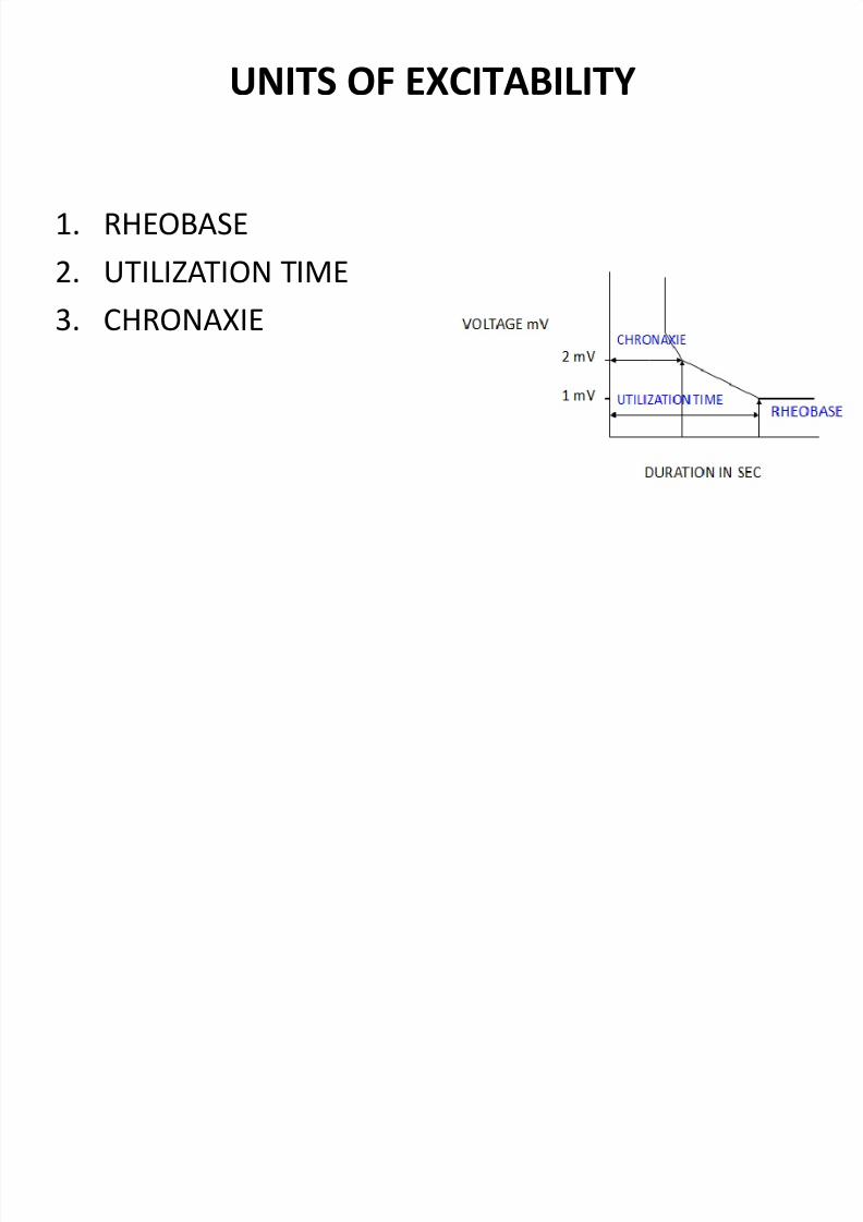
UNITS OF EXCITABILITY
1. RHEOBASE
2. UTILIZATION TIME
3. CHRONAXIE

7/29/2019 2nd lecture on action potential by Dr. Roomi.
http://slidepdf.com/reader/full/2nd-lecture-on-action-potential-by-dr-roomi 2/14
RHEOBASE, UTILIZATION TIME, CHRONAXIE
• RHEOBASE: Voltage /strength of stimulusrequired just to excite thetissue, e.g., 1 mV.
• UTILIZATION TIME: The timefor which Rheobase mustbe applied to excite thetissue.
• CHRONAXIE: A time forwhich a stimulus, double
the rheobase when applied, just excites the tissue, e.g.,2 mV. (Chronological is fromtime).

7/29/2019 2nd lecture on action potential by Dr. Roomi.
http://slidepdf.com/reader/full/2nd-lecture-on-action-potential-by-dr-roomi 3/14
CLINICAL APPLICATION / SIGNIFICANCE OF
CHRONAXIE
1. A particular value of it for a particular tissue. – Type A-alpha nerve fiber has minimum value of chronaxie,
i.e. they are more excitable as compared to cardiac muscle.(less chronaxiemore excitability)
2. In nerve injury repair procedureWe assessthe recovery by finding chronaxie of nerve affected& muscle affected. –
Damage to nerve fiber is determined by measuringchronaxie.
– It improves with recovery.

7/29/2019 2nd lecture on action potential by Dr. Roomi.
http://slidepdf.com/reader/full/2nd-lecture-on-action-potential-by-dr-roomi 4/14
Action potential
By
Dr. Mudassar Ali Roomi (MBBS, M. Phil)

7/29/2019 2nd lecture on action potential by Dr. Roomi.
http://slidepdf.com/reader/full/2nd-lecture-on-action-potential-by-dr-roomi 5/14
ACTION POTENTIAL OF NERVE FIBER /
SKELETAL MUSCLE
• Defintion: it is an abrupt pulse like change inmembrane potential, lasting for fraction of asecond.
•During action potential there is reversal of potential. (inside +, outside -).
• Nerve impulse is being conducted along a nervefiber = action potential is being conducted.
• Depolarization = loss of negativity inside.
• Repolarization = return of negativity inside.

7/29/2019 2nd lecture on action potential by Dr. Roomi.
http://slidepdf.com/reader/full/2nd-lecture-on-action-potential-by-dr-roomi 6/14
RMP -90 mV
Threshold -65 mV
0 mV
+35 to 40 mV
After Hyper-polarization
(Undershoot) / Sub-normal period ( -95 mV )
[K+ efflux continues, K+ channels remain
open for some time after RMP is reached].
Here tissue is difficult to be excited.
Depolarization Repolarization
[K+ efflux]
Time (msec)
(Overshoot)
Peak
Membrane Voltage(mV)
RMP -90 mV
[Rapid
Na+influx]
Complete
opening of
fast Na+ channels
Slow Repolarization/ K+accumulate
Excitable/ Super-normal period
After Depolarization
(70% of repolarization / start of
After-Depolarization)Spike potential
(First 1/3 of repolarization)
Relative Refractory period
Absolute Refractory period
[Na+ inactivation gates are still closed]
-65mV

7/29/2019 2nd lecture on action potential by Dr. Roomi.
http://slidepdf.com/reader/full/2nd-lecture-on-action-potential-by-dr-roomi 7/14
Properties of action potential
1. Sudden / abrupt in onset.
2. Of limited magnitude / amplitude.
3. It goes to +35 to 40 mV & comes back. (biphasic)
4. Short duration (may be few millisec).
5. It obeys all or none law. (if a stimulus is threshold orsuprathreshold action potential is produced with itsmaximum amplitude, if subthreshold stimulus notproduced at all).
6. Self propagating. (automatically propagated in bothdirections).
7. Has a refractory period. (when there won’t be response to2nd stimulus if the fiber is already stimulated).

7/29/2019 2nd lecture on action potential by Dr. Roomi.
http://slidepdf.com/reader/full/2nd-lecture-on-action-potential-by-dr-roomi 8/14
Ionic basis of action
potential
7/29/2019 2nd lecture on action potential by Dr. Roomi.
http://slidepdf.com/reader/full/2nd-lecture-on-action-potential-by-dr-roomi 9/14
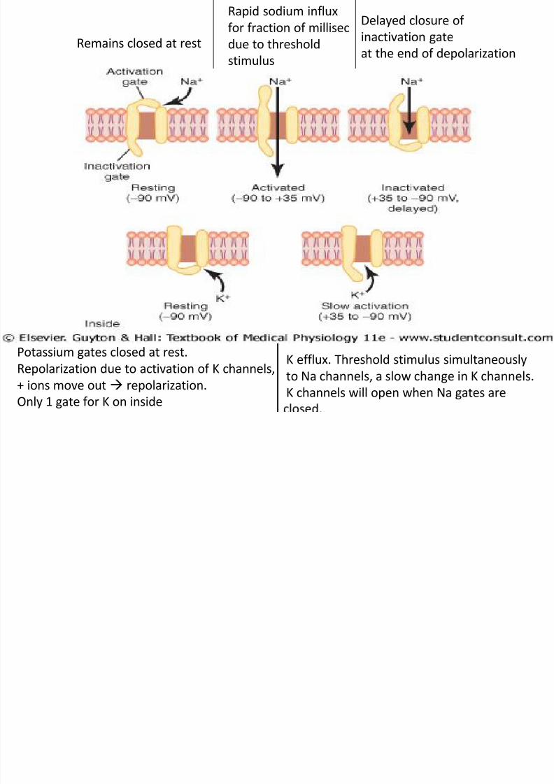
Remains closed at rest
Rapid sodium influx
for fraction of millisec
due to threshold
stimulus
Delayed closure of
inactivation gate
at the end of depolarization
Potassium gates closed at rest.
Repolarization due to activation of K channels,
+ ions move out repolarization.Only 1 gate for K on inside
K efflux. Threshold stimulus simultaneously
to Na channels, a slow change in K channels.
K channels will open when Na gates are

7/29/2019 2nd lecture on action potential by Dr. Roomi.
http://slidepdf.com/reader/full/2nd-lecture-on-action-potential-by-dr-roomi 10/14
Ionic basis of action potential
•
Voltage gated Na+ channels: – At rest (-90mV) sodium activation gates on outside of membrane remainclosed
– For fraction of m sec in presence of threshold stimulus, rapid sodiuminflux takes place depolarization (-90mV to +35mV)
– There is delayed closure of inactivation gate on the inside.
• Voltage gated K+ channels: – Repolarization due to activation of potassium channels, +ions move out regain of negativity inside (repolarization)
– At rest potassium gates situated on inside are closed (-90mV)
– There is slow activation of potassium gates between +35mV to -90mV.
Rapid potassium efflux occurs. – Threshold stimulus causes a slow change in potassium channels. It will
open when sodium gates are closed.

7/29/2019 2nd lecture on action potential by Dr. Roomi.
http://slidepdf.com/reader/full/2nd-lecture-on-action-potential-by-dr-roomi 11/14
threshold / firing / critical value: - 65 mV for sodium channels. It
causes change in activation gate of sodium channels at -65 mV,
complete opening of fast sodium channels.

7/29/2019 2nd lecture on action potential by Dr. Roomi.
http://slidepdf.com/reader/full/2nd-lecture-on-action-potential-by-dr-roomi 12/14
• K+ becomes accumulated on outer side of membrane during later part of
repolarization, which slows down further K+ efflux after 70% of repolarization
slow repolarization is called after depolarization.
• Super normal period: During After depolarization, there is super normal period.
Tissue is most excitable. Here potential is – 65 mV, so small change is required tostimulate.

7/29/2019 2nd lecture on action potential by Dr. Roomi.
http://slidepdf.com/reader/full/2nd-lecture-on-action-potential-by-dr-roomi 13/14
• When potential has reached the resting value, it does not stay there & becomesmore negative & called After hyperpolarization.
• Cause: It is because when potential has reached resting value, some K channels arestill open & K efflux continuesmembrane becomes more negative.
• Sub-normal period: During After hyper-polarization it occurs, because tissue isdifficult to be excited because potential becomes – 95 mV.
• Part of action potential between threshold value & beginning of afterdepolarization is called SPIKE POTENTIAL.

7/29/2019 2nd lecture on action potential by Dr. Roomi.
http://slidepdf.com/reader/full/2nd-lecture-on-action-potential-by-dr-roomi 14/14
Electrotonic potential or
graded potential or localized
potential
Action potential
1. Proportional to stimulus strength(graded) Independent of stimulus strength(all or none)
2. Not propagated but decremental
with distance
Propagated, unchanged in magnitude
3. Exhibits summation Summation not possible
4. magnitude: low Magnitude: high
5. Refractory period: absent Refractory period: present
6. duration: upto 20 msec duration: upto 2 msec
7. examples: subthreshold potential, EPSP,
IPSP
Action potential in nerve fibers